link. · Web viewElectronic Supplementary Material α-Hemolysin nanopore studies reveal...
24
1 Electronic Supplementary Material α-Hemolysin nanopore studies reveal strong interactions between biogenic polyamines and DNA hairpins Yun Ding, Aaron M. Fleming, and Cynthia J. Burrows* Department of Chemistry, University of Utah, 315 South 1400 East, Salt Lake City, UT 84112-0850, USA *To whom correspondence should be addressed: (801) 585-7290. E-mail: [email protected] Contents: Fig.S1 Characterization of the blunt ended hairpin. 2 Fig. S2 Voltage-dependent study of type 1 (A) and 2 (B) events for the hairpin in the nanopore. 3 Fig. S3 Duration of type 3 events with the presence of 1 mM Spm at different voltages. 4 Fig. S4 Voltage-dependent studies of 5’hp6 binding with Spm for the type 3 events. 5 Fig. S5 Sample i-t traces of the hairpin DNA with the presence of 1 mM Spm. 6 Fig. S6 Sample i-t traces of the hairpin DNA with the presence of 2 mM Spm. 7 Fig. S7 Sample i-t traces of the hairpin DNA with the presence of 3 mM Spm. 8 Fig. S8 Sample i-t traces of the hairpin DNA with the presence of 4 mM Spm. 9 Fig. S9 Sample i-t traces of the hairpin DNA with the presence of 1 mM Spd. 10
Transcript of link. · Web viewElectronic Supplementary Material α-Hemolysin nanopore studies reveal...

1
Electronic Supplementary Material
α-Hemolysin nanopore studies reveal strong interactions between biogenic
polyamines and DNA hairpins
Yun Ding, Aaron M. Fleming, and Cynthia J. Burrows*
Department of Chemistry, University of Utah, 315 South 1400 East, Salt Lake City, UT 84112-0850, USA
*To whom correspondence should be addressed: (801) 585-7290. E-mail: [email protected]
Contents:
Fig.S1 Characterization of the blunt ended hairpin. 2
Fig. S2 Voltage-dependent study of type 1 (A) and 2 (B) events for the hairpin in the nanopore. 3
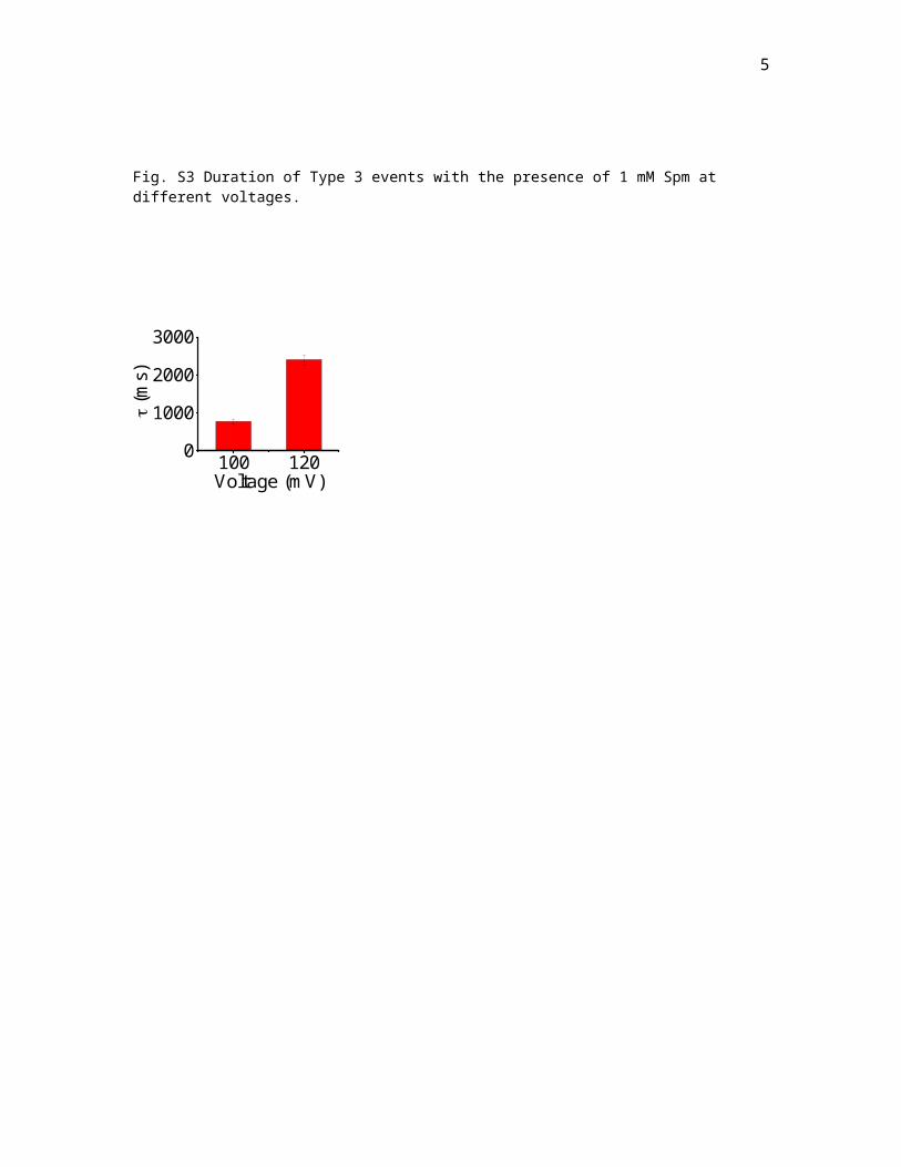
Fig. S3 Duration of type 3 events with the presence of 1 mM Spm at different voltages. 4
Fig. S4 Voltage-dependent studies of 5’hp6 binding with Spm for the type 3 events. 5
Fig. S5 Sample i-t traces of the hairpin DNA with the presence of 1 mM Spm. 6
Fig. S6 Sample i-t traces of the hairpin DNA with the presence of 2 mM Spm. 7
Fig. S7 Sample i-t traces of the hairpin DNA with the presence of 3 mM Spm. 8
Fig. S8 Sample i-t traces of the hairpin DNA with the presence of 4 mM Spm. 9
Fig. S9 Sample i-t traces of the hairpin DNA with the presence of 1 mM Spd. 10
Fig. S10 Sample i-t traces of the hairpin DNA with the presence of 2 mM Spd. 11
Fig. S11 Sample i-t traces of the hairpin DNA with the presence of 3 mM Spd. 12
Fig. S12 Sample i-t traces of the hairpin DNA with the presence of 4 mM Spd. 13
Fig. S13 Sample i-t traces of the hairpin DNA with the presence of 4 mM Put. 14
Fig. S14 Sample i-t traces of the hairpin DNA without the presence of polyamines. 15
Fig. S15 Sample i-t traces of the control DNA (poly-dC87) without the presence of polyamines. 16
Fig. S16 Sample i-t traces of the control DNA (poly-dC87) with the presence of 4 mM Spm. 17
Fig. S17 Example heat plots to show the variability in current blocking levels between proteins. 18
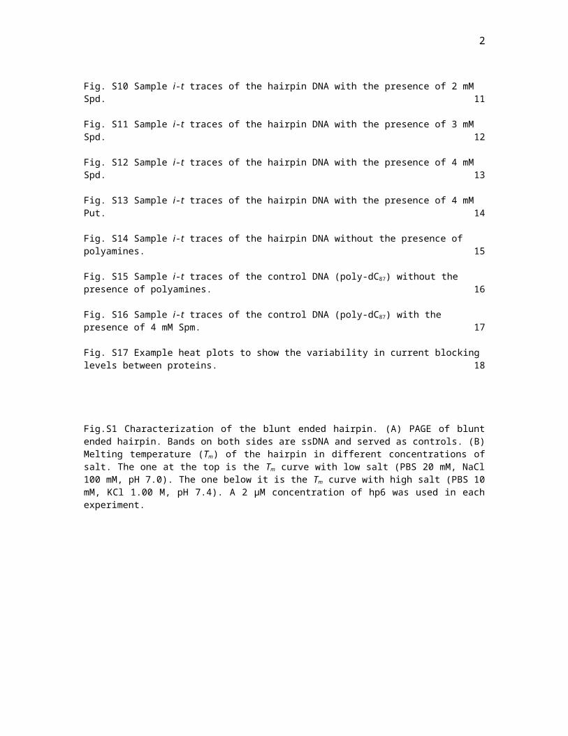
2
Fig.S1 Characterization of the blunt ended hairpin. (A) PAGE of blunt ended hairpin. Bands on both sides are ssDNA and served as controls. (B) Melting temperature (Tm) of the hairpin in different concentrations of salt. The one at the top is the Tm curve with low salt (PBS 20 mM, NaCl 100 mM, pH 7.0). The one below it is the Tm curve with high salt (PBS 10 mM, KCl 1.00 M, pH 7.4). A 2 µM concentration of hp6 was used in each experiment.
A
B Low Salt = 120 mM NaCl and High Salt = 1.00 M KCl

3
Fig. S2 Voltage-dependent study of Type 1 (A) and 2 (B) events for the hairpin in the nanopore.
A
B

4
Fig. S3 Duration of Type 3 events with the presence of 1 mM Spm at different voltages.
100 1200
1000
2000
3000
(m
s)
Voltage (mV)
5
Fig. S4 Voltage-dependent studies of the hairpin binding with Spm for the Type 3 events. (A) i-t traces of
the hairpin binding with Spm at different voltages. (B) Blocking current of the hairpin binding with Spm at
different voltages.

6
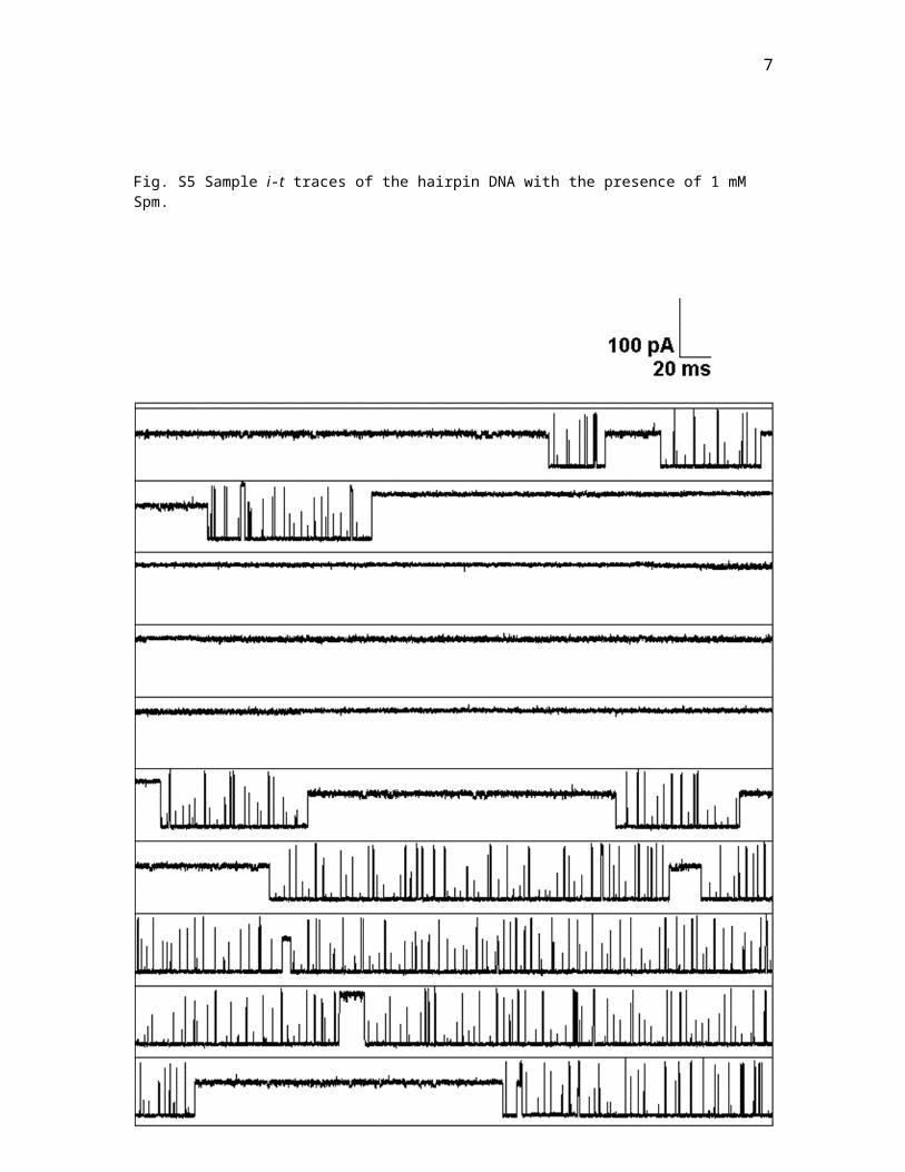
Fig. S5 Sample i-t traces of the hairpin DNA with the presence of 1 mM Spm.
7
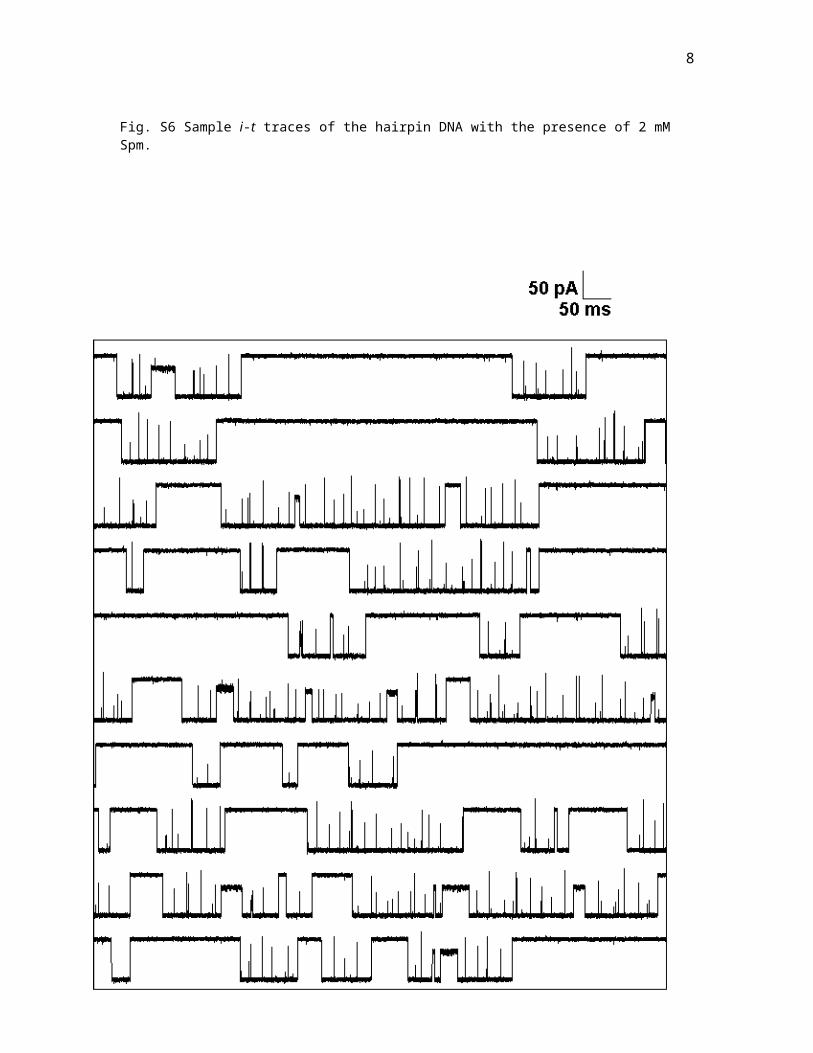
Fig. S6 Sample i-t traces of the hairpin DNA with the presence of 2 mM Spm.
8
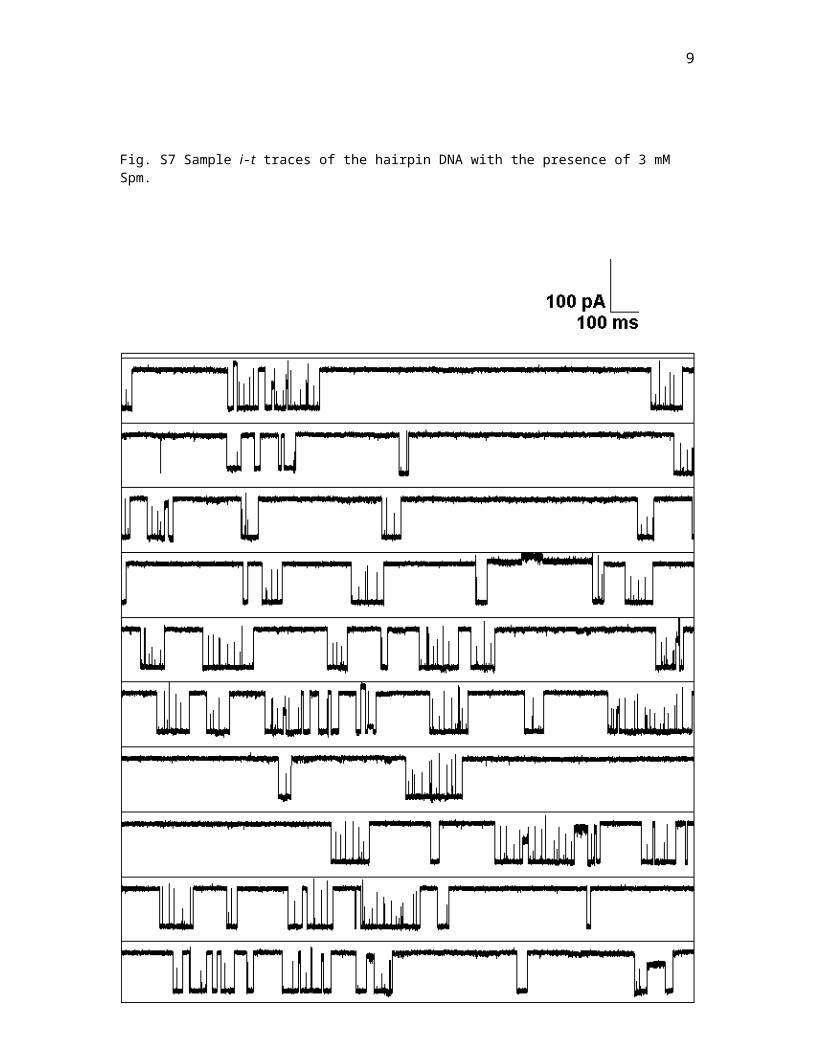
Fig. S7 Sample i-t traces of the hairpin DNA with the presence of 3 mM Spm.
9
Fig. S8 Sample i-t traces of the hairpin DNA with the presence of 4 mM Spm.

10
Fig. S9 Sample i-t traces of the hairpin DNA with the presence of 1 mM Spd.

11
Fig. S10 Sample i-t traces of the hairpin DNA with the presence of 2 mM Spd.

12
Fig. S11 Sample i-t traces of the hairpin DNA with the presence of 3 mM Spd.

13
Fig. S12 Sample i-t traces of the hairpin DNA with the presence of 4 mM Spd.
14
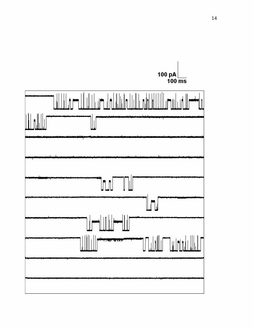
Fig. S13 Sample i-t traces of the hairpin DNA with the presence of 4 mM Put.

15
Fig. S14 Sample i-t traces of the hairpin DNA without the presence of polyamines.

16
Fig. S15 Sample i-t traces of the control DNA (poly-dC87) without the presence of polyamines.

17
Fig. S16 Sample i-t traces of the control DNA (poly-dC87) with the presence of 4 mM Spm.

18
Fig. S17 Example heat plots to show the variability in current blocking levels between proteins.
0.6
0.3
0
I/Io
0.05 500 5000t (ms)
No Spm
1
2
1
2
3
0.6
0.3
0
I/Io
1 mM Spm
0.05 500 5000t (ms)
0.6
0.3
0
I/Io
0.05 500 5000t (ms)
No Spm
1
2