3RD SEMINAR INNATE IMMUNITY: ANTIVIRAL STATE, KILLER CELLS, THE COMPLEMENT SYSTEM.
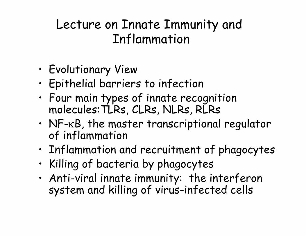
Lecture on Innate Immunity and Inflammation
• Evolutionary View • Epithelial barriers to infection • Four main types of innate recognition
molecules:TLRs, CLRs, NLRs, RLRs • NF-κB, the master transcriptional regulator
of inflammation • Inflammation and recruitment of phagocytes • Killing of bacteria by phagocytes • Anti-viral innate immunity: the interferon
system and killing of virus-infected cells
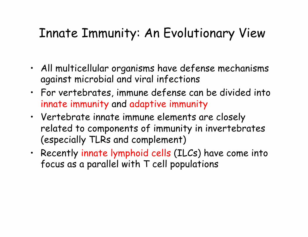
Innate Immunity: An Evolutionary View
• All multicellular organisms have defense mechanisms against microbial and viral infections
• For vertebrates, immune defense can be divided into innate immunity and adaptive immunity
• Vertebrate innate immune elements are closely related to components of immunity in invertebrates (especially TLRs and complement)
• Recently innate lymphoid cells (ILCs) have come into focus as a parallel with T cell populations
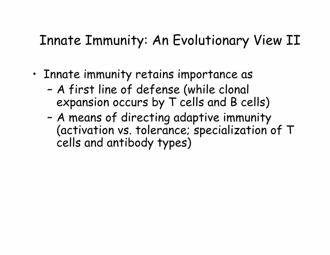
Innate Immunity: An Evolutionary View II
• Innate immunity retains importance as – A first line of defense (while clonal
expansion occurs by T cells and B cells) – A means of directing adaptive immunity
(activation vs. tolerance; specialization of T cells and antibody types)
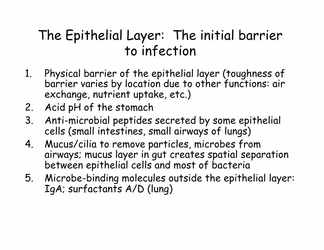
The Epithelial Layer: The initial barrier to infection
1. Physical barrier of the epithelial layer (toughness of barrier varies by location due to other functions: air exchange, nutrient uptake, etc.)
2. Acid pH of the stomach 3. Anti-microbial peptides secreted by some epithelial
cells (small intestines, small airways of lungs) 4. Mucus/cilia to remove particles, microbes from
airways; mucus layer in gut creates spatial separation between epithelial cells and most of bacteria
5. Microbe-binding molecules outside the epithelial layer: IgA; surfactants A/D (lung)
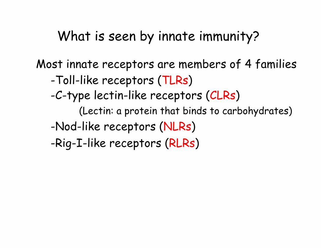
What is seen by innate immunity?
Most innate receptors are members of 4 families -Toll-like receptors (TLRs)
-C-type lectin-like receptors (CLRs) (Lectin: a protein that binds to carbohydrates)
-Nod-like receptors (NLRs) -Rig-I-like receptors (RLRs)
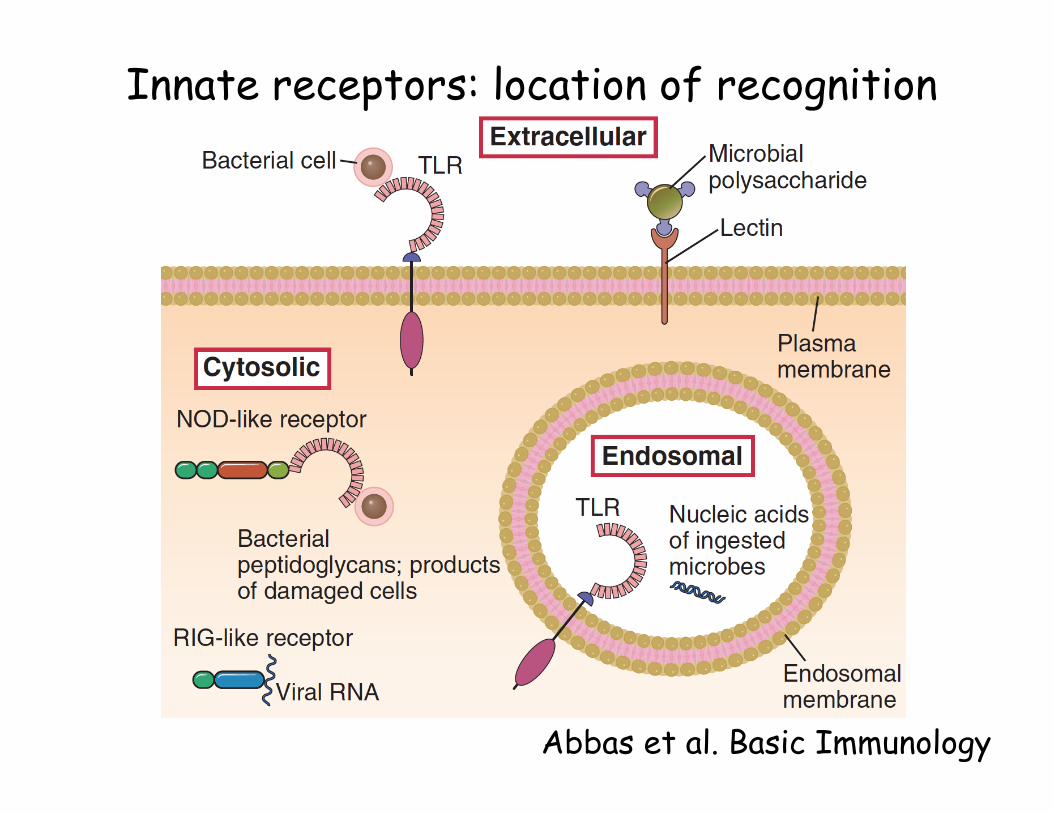
Innate receptors: location of recognition
Abbas et al. Basic Immunology
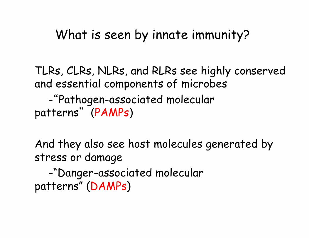
What is seen by innate immunity? TLRs, CLRs, NLRs, and RLRs see highly conserved and essential components of microbes
-“Pathogen-associated molecular patterns” (PAMPs) And they also see host molecules generated by stress or damage
-“Danger-associated molecular patterns” (DAMPs)
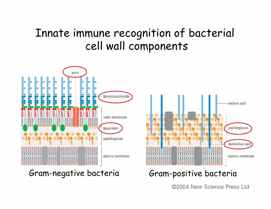
Innate immune recognition of bacterial cell wall components
Gram-negative bacteria Gram-positive bacteria
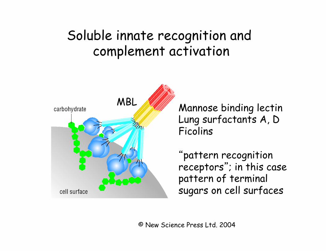
Soluble innate recognition and complement activation
© New Science Press Ltd. 2004
MBL Mannose binding lectin Lung surfactants A, D Ficolins “pattern recognition receptors”; in this case pattern of terminal sugars on cell surfaces
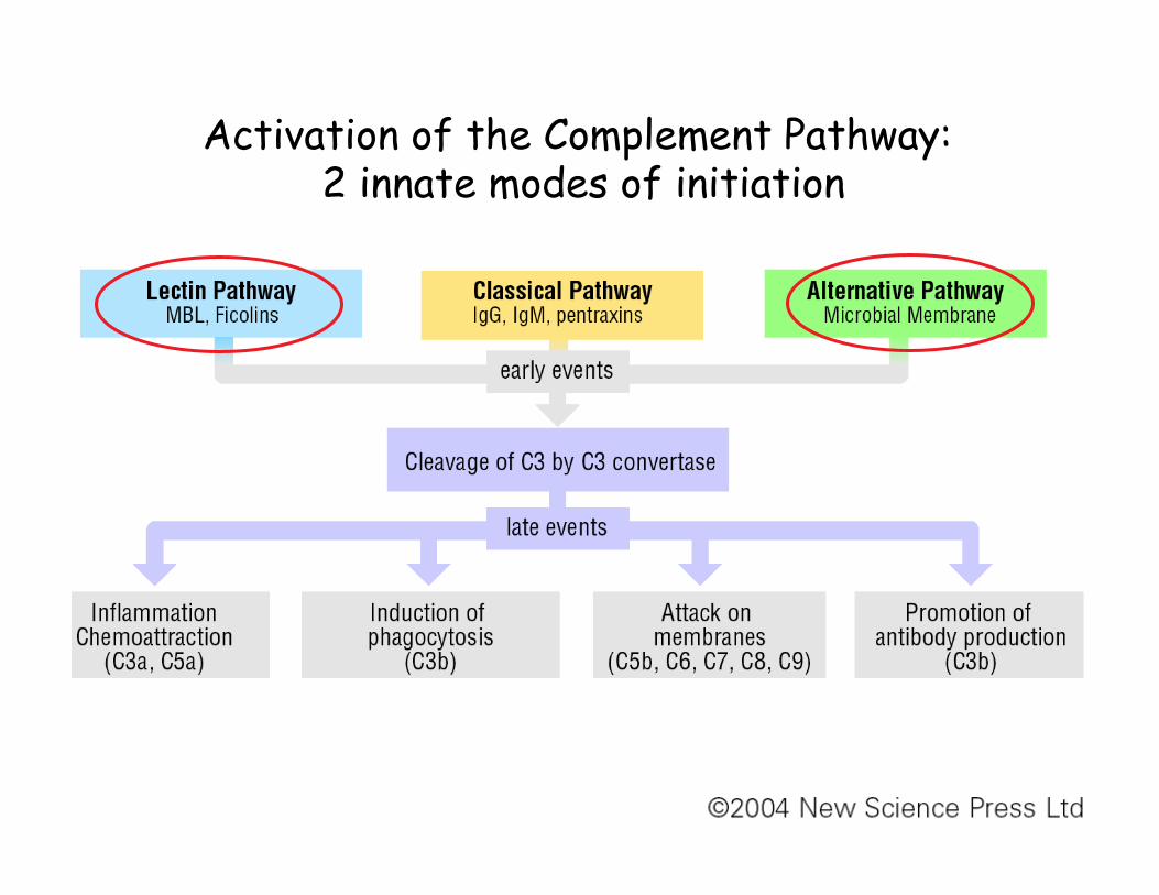
Activation of the Complement Pathway: 2 innate modes of initiation
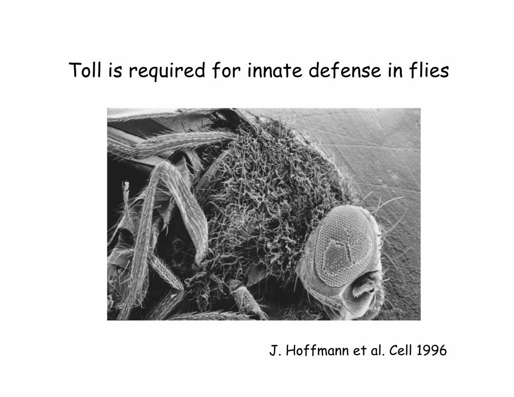
Toll is required for innate defense in flies
J. Hoffmann et al. Cell 1996
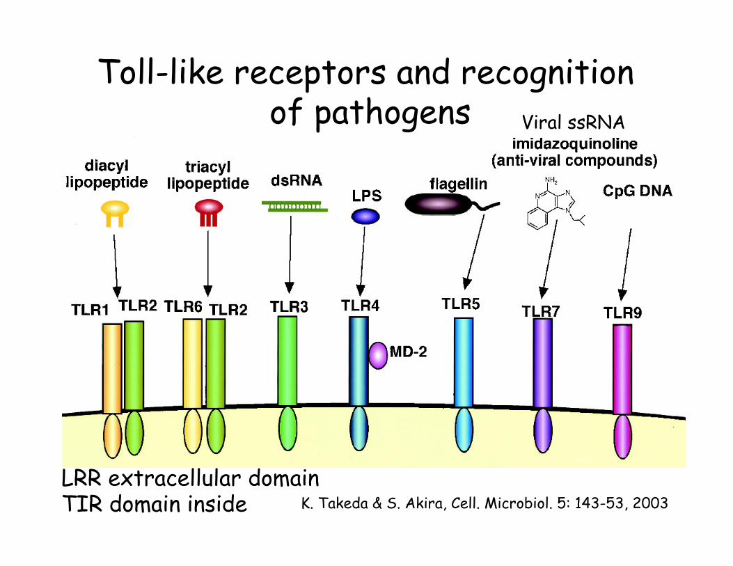
Toll-like receptors and recognition of pathogens
K. Takeda & S. Akira, Cell. Microbiol. 5: 143-53, 2003 LRR extracellular domain TIR domain inside
Viral ssRNA
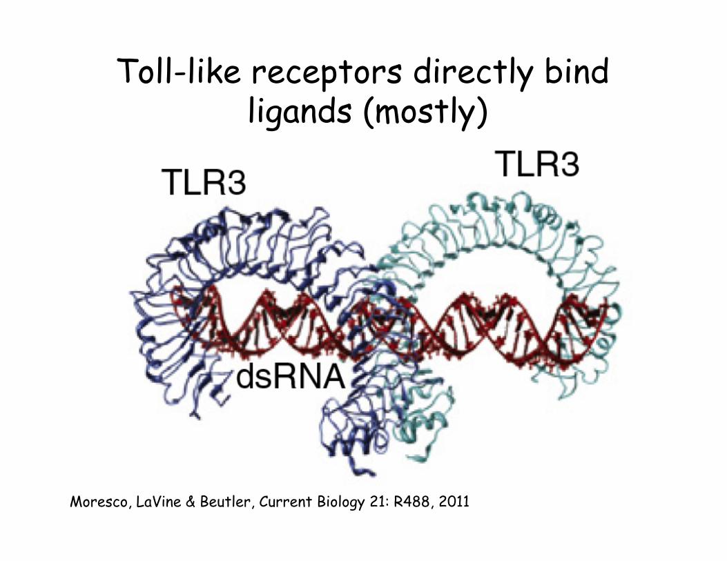
Toll-like receptors directly bind ligands (mostly)
Moresco, LaVine & Beutler, Current Biology 21: R488, 2011
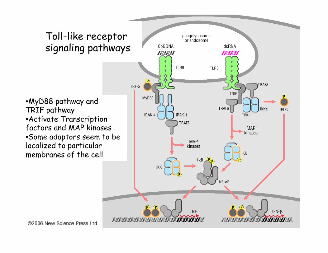
Toll-like receptor signaling pathways
• MyD88 pathway and TRIF pathway • Activate Transcription factors and MAP kinases • Some adaptors seem to be localized to particular membranes of the cell
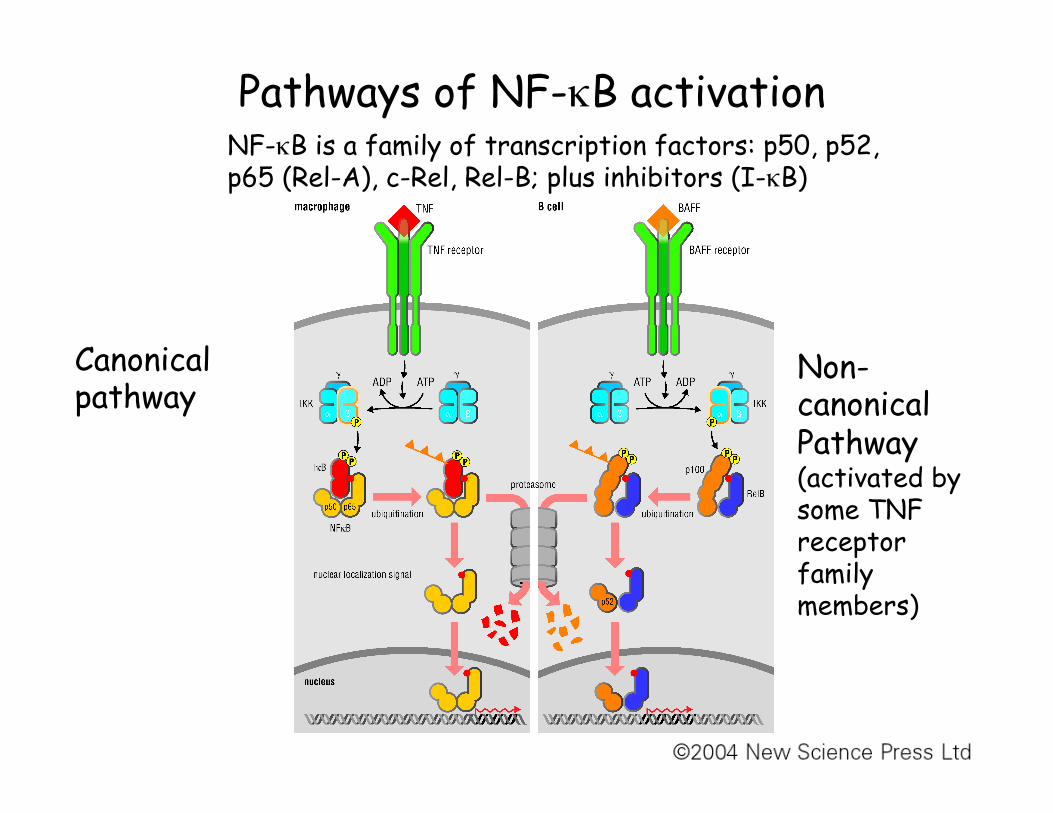
Pathways of NF-κB activation
Canonical pathway
Non-canonical Pathway (activated by some TNF receptor family members)
NF-κB is a family of transcription factors: p50, p52, p65 (Rel-A), c-Rel, Rel-B; plus inhibitors (I-κB)
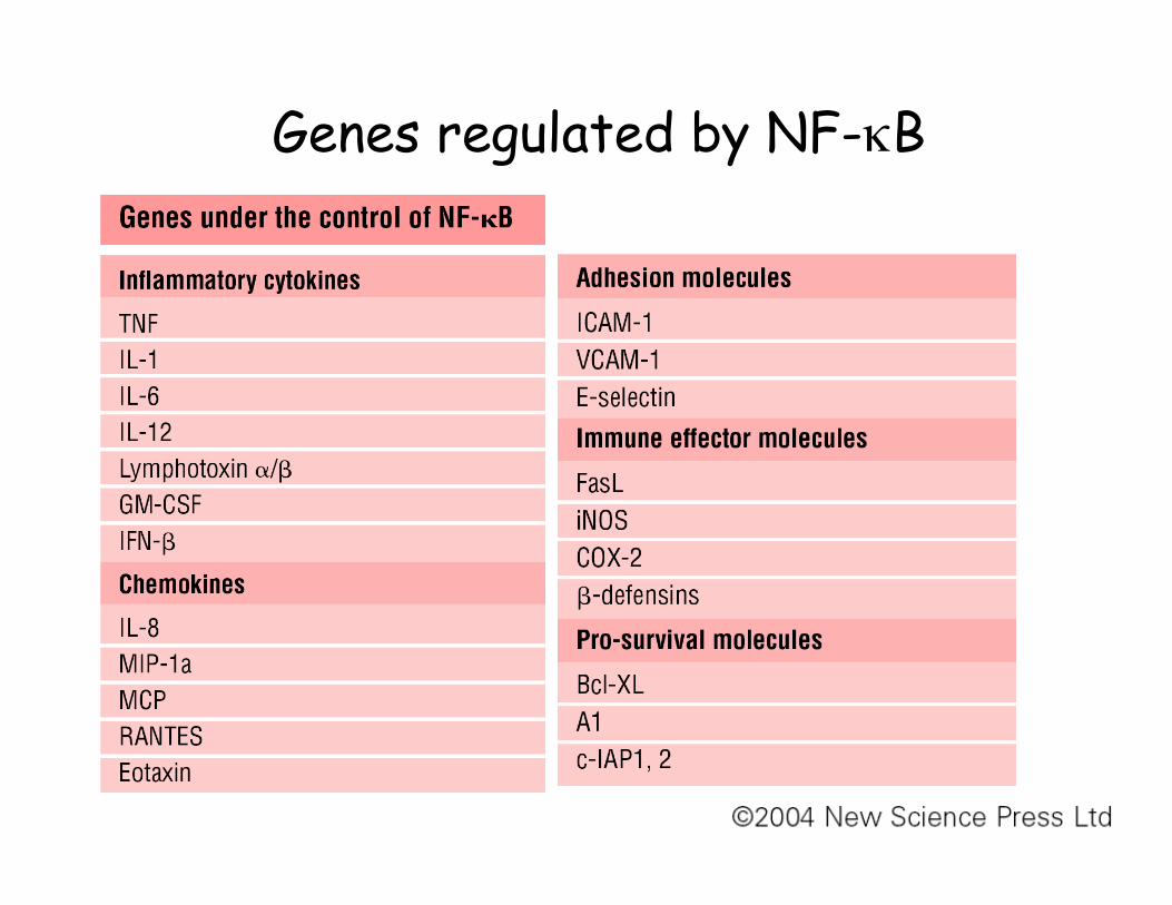
Genes regulated by NF-κB
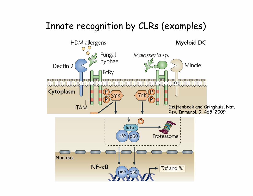
Innate recognition by CLRs (examples)
Geijtenbeek and Gringhuis, Nat. Rev. Immunol. 9: 465, 2009
NF-κB
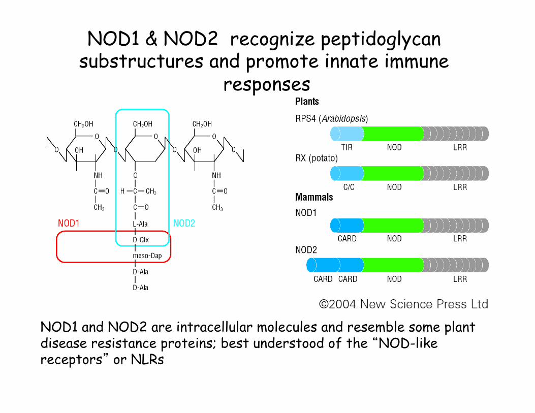
NOD1 & NOD2 recognize peptidoglycan substructures and promote innate immune
responses
NOD1 and NOD2 are intracellular molecules and resemble some plant disease resistance proteins; best understood of the “NOD-like receptors” or NLRs
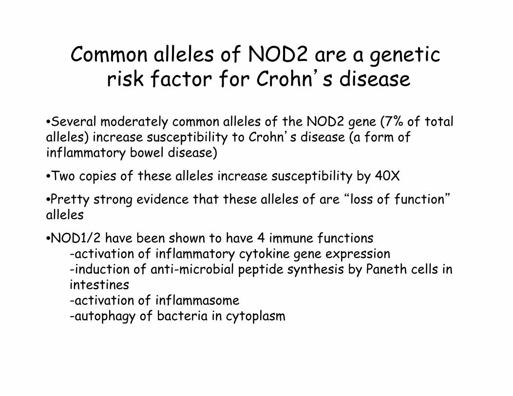
Common alleles of NOD2 are a genetic risk factor for Crohn’s disease
• Several moderately common alleles of the NOD2 gene (7% of total alleles) increase susceptibility to Crohn’s disease (a form of inflammatory bowel disease)
• Two copies of these alleles increase susceptibility by 40X
• Pretty strong evidence that these alleles of are “loss of function” alleles
• NOD1/2 have been shown to have 4 immune functions -activation of inflammatory cytokine gene expression -induction of anti-microbial peptide synthesis by Paneth cells in intestines -activation of inflammasome -autophagy of bacteria in cytoplasm

The NLRP3-inflammasome activates caspase 1 in response to cellular insults
Bacterial pore-forming toxins Efflux of K+ Bacterial flagellin, needle proteins Endocytosed crystals Other insults/stresses
• TLRs or NOD1/NOD2 induce synthesis of pro-IL-1 • Inflammasome processes it to generate active IL-1
(NLRP3)!
Pyroptosis!
Caspase 11!
?
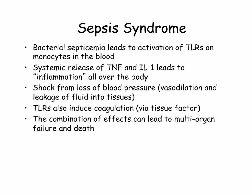
Sepsis Syndrome • Bacterial septicemia leads to activation of TLRs on
monocytes in the blood • Systemic release of TNF and IL-1 leads to
“inflammation” all over the body • Shock from loss of blood pressure (vasodilation and
leakage of fluid into tissues) • TLRs also induce coagulation (via tissue factor) • The combination of effects can lead to multi-organ
failure and death
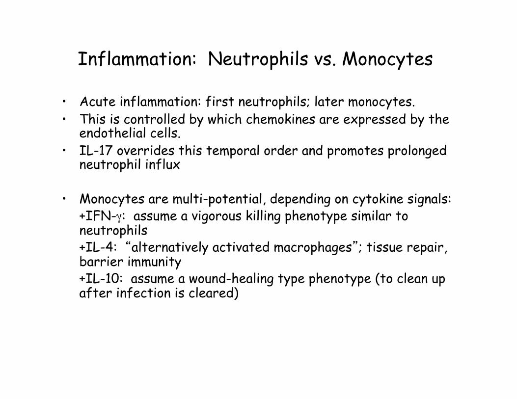
Inflammation: Neutrophils vs. Monocytes • Acute inflammation: first neutrophils; later monocytes. • This is controlled by which chemokines are expressed by the
endothelial cells. • IL-17 overrides this temporal order and promotes prolonged
neutrophil influx • Monocytes are multi-potential, depending on cytokine signals:
+IFN-γ: assume a vigorous killing phenotype similar to neutrophils +IL-4: “alternatively activated macrophages”; tissue repair, barrier immunity +IL-10: assume a wound-healing type phenotype (to clean up after infection is cleared)
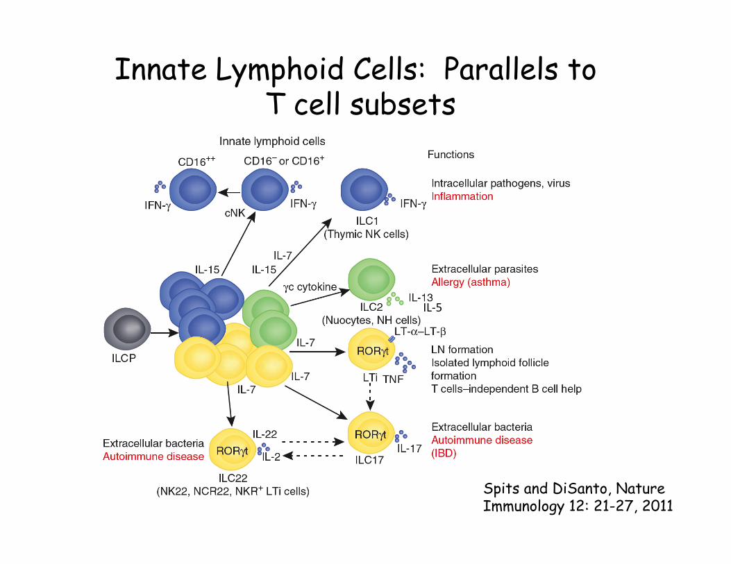
Innate Lymphoid Cells: Parallels to T cell subsets
Spits and DiSanto, Nature Immunology 12: 21-27, 2011
IL-‐5
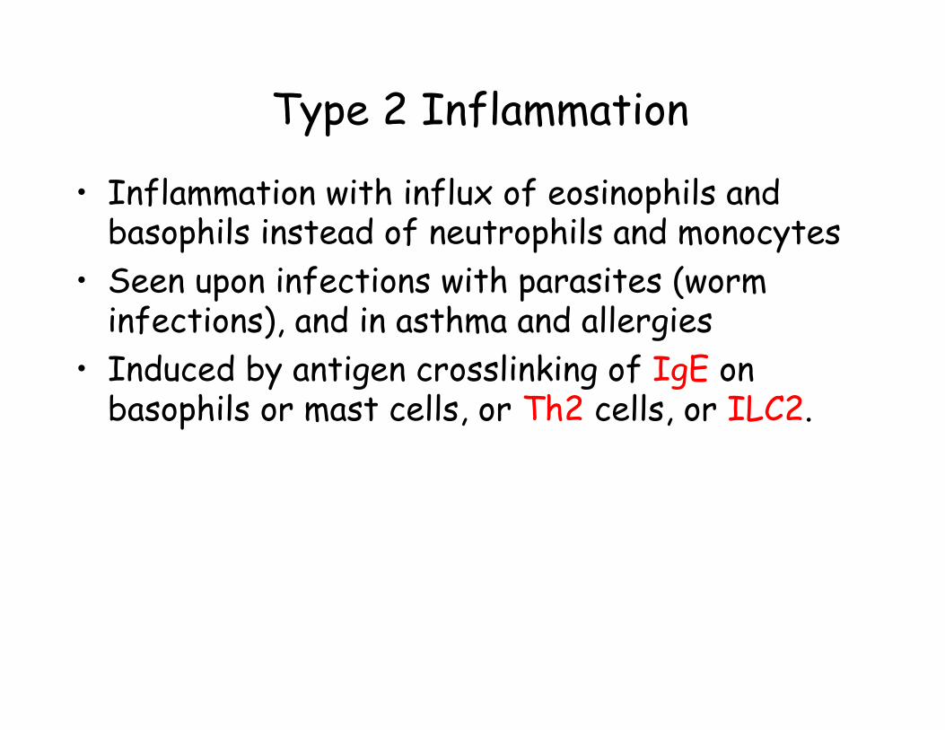
Type 2 Inflammation • Inflammation with influx of eosinophils and
basophils instead of neutrophils and monocytes • Seen upon infections with parasites (worm
infections), and in asthma and allergies • Induced by antigen crosslinking of IgE on
basophils or mast cells, or Th2 cells, or ILC2.
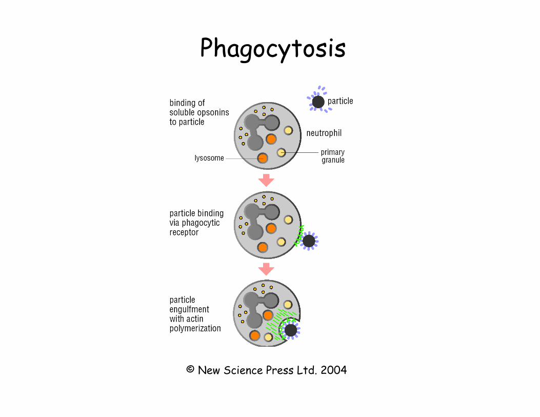
Phagocytosis
© New Science Press Ltd. 2004
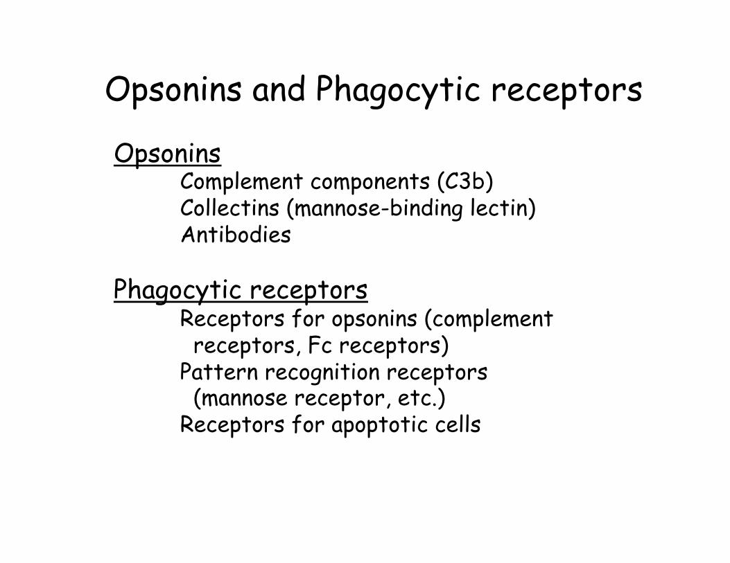
Opsonins and Phagocytic receptors Opsonins
Complement components (C3b) Collectins (mannose-binding lectin) Antibodies
Phagocytic receptors
Receptors for opsonins (complement receptors, Fc receptors) Pattern recognition receptors (mannose receptor, etc.) Receptors for apoptotic cells
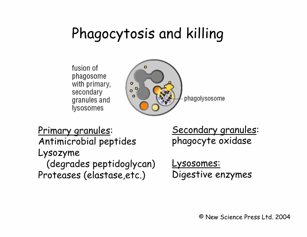
Phagocytosis and killing
Primary granules: Antimicrobial peptides Lysozyme (degrades peptidoglycan) Proteases (elastase,etc.)
Secondary granules: phagocyte oxidase Lysosomes: Digestive enzymes
© New Science Press Ltd. 2004
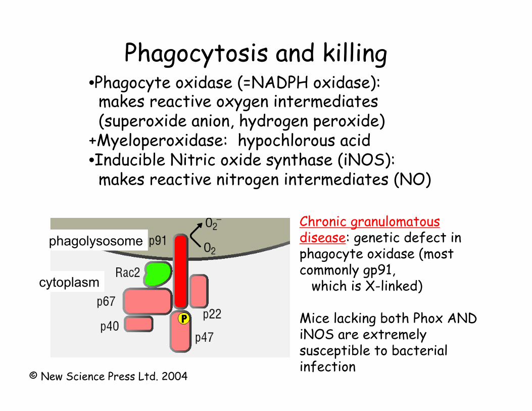
Phagocytosis and killing • Phagocyte oxidase (=NADPH oxidase): makes reactive oxygen intermediates (superoxide anion, hydrogen peroxide) +Myeloperoxidase: hypochlorous acid • Inducible Nitric oxide synthase (iNOS): makes reactive nitrogen intermediates (NO)
Chronic granulomatous disease: genetic defect in phagocyte oxidase (most commonly gp91, which is X-linked) Mice lacking both Phox AND iNOS are extremely susceptible to bacterial infection
phagolysosome
cytoplasm
© New Science Press Ltd. 2004
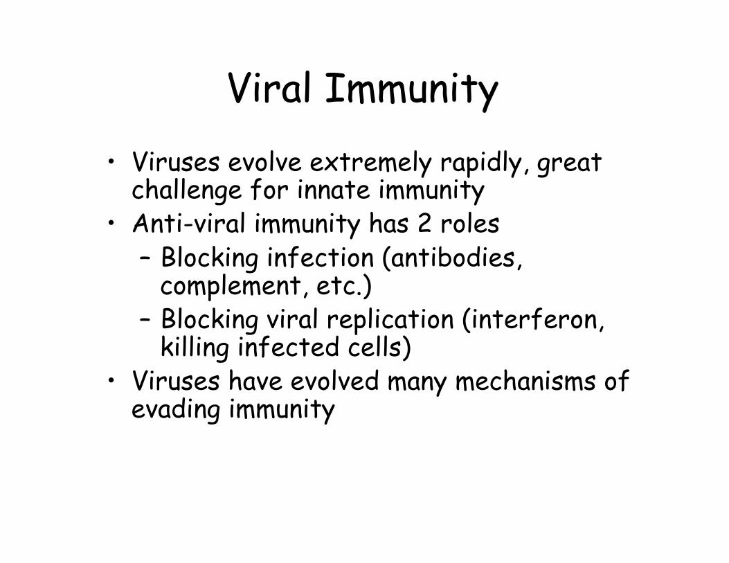
Viral Immunity • Viruses evolve extremely rapidly, great
challenge for innate immunity • Anti-viral immunity has 2 roles
– Blocking infection (antibodies, complement, etc.)
– Blocking viral replication (interferon, killing infected cells)
• Viruses have evolved many mechanisms of evading immunity
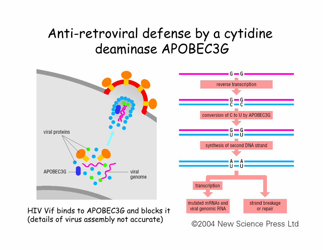
Anti-retroviral defense by a cytidine deaminase APOBEC3G
HIV Vif binds to APOBEC3G and blocks it (details of virus assembly not accurate)
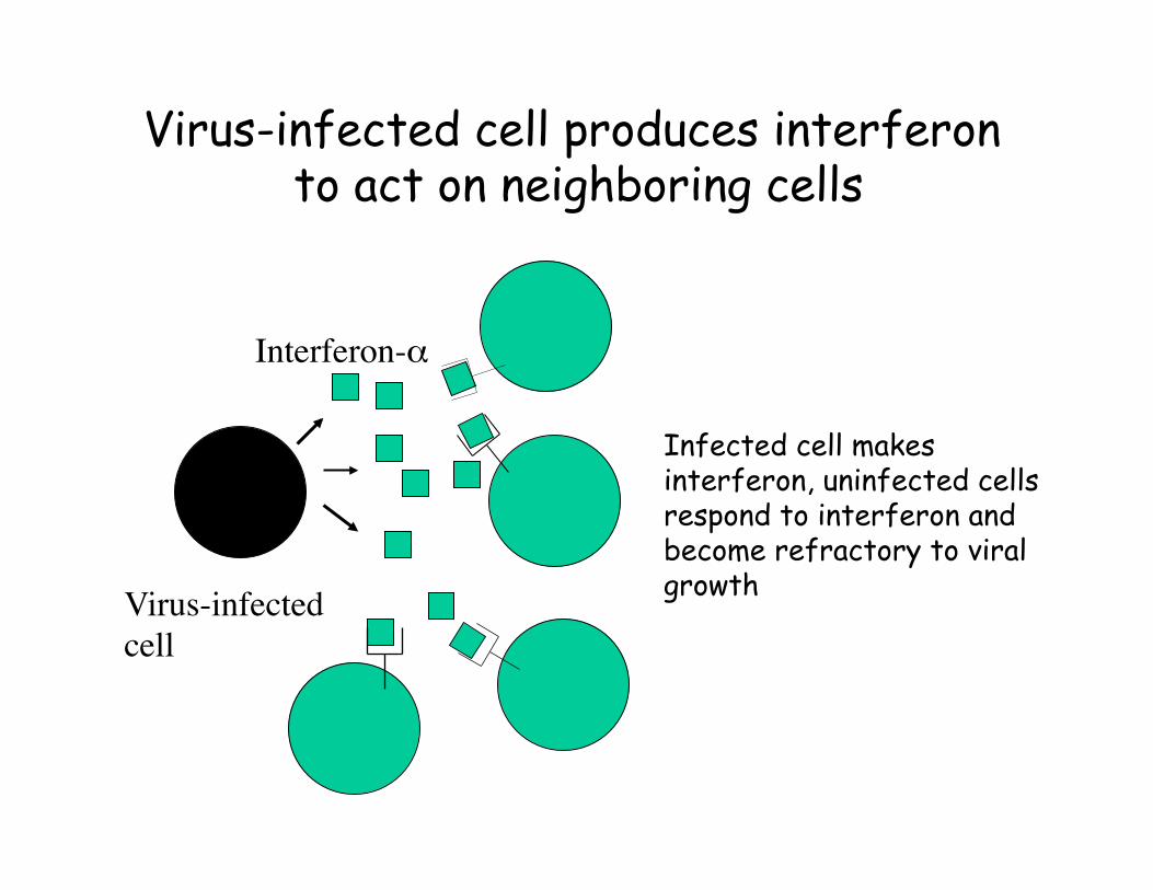
Virus-infected cell produces interferon to act on neighboring cells
Virus-infected cell
Interferon-α
Infected cell makes interferon, uninfected cells respond to interferon and become refractory to viral growth
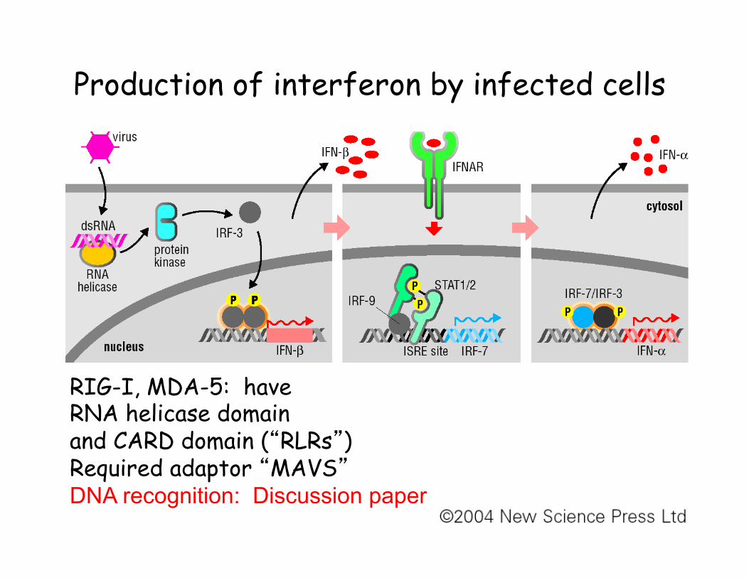
Production of interferon by infected cells
RIG-I, MDA-5: have RNA helicase domain and CARD domain (“RLRs”) Required adaptor “MAVS” DNA recognition: Discussion paper
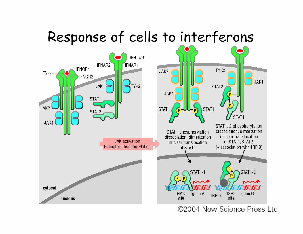
Response of cells to interferons
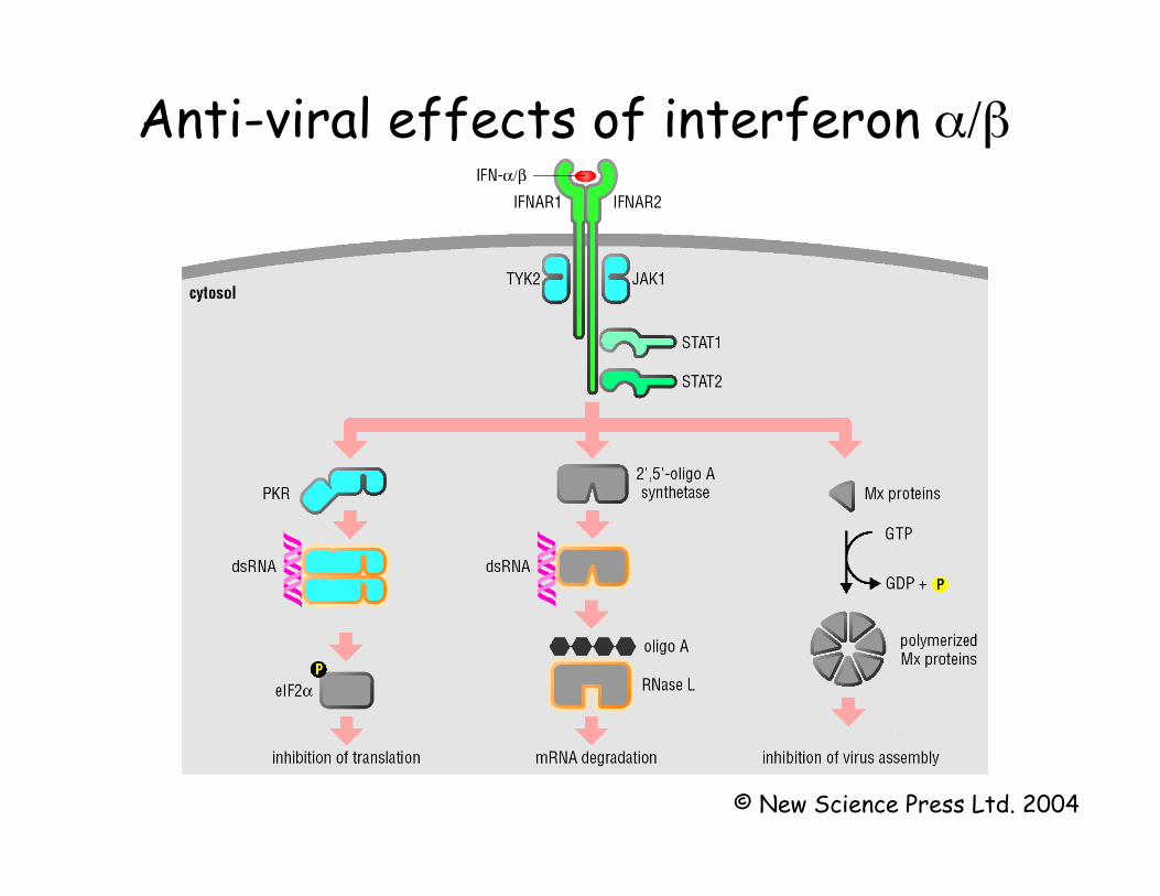
Anti-viral effects of interferon α/β
© New Science Press Ltd. 2004
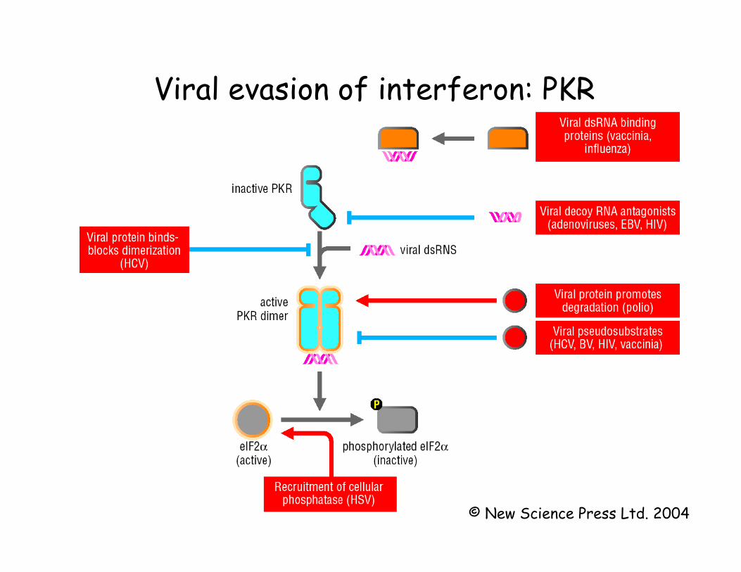
Viral evasion of interferon: PKR
© New Science Press Ltd. 2004
Plasmacytoid dendritic cells • Many cell types produce small amounts of
type 1 interferons upon infection • There is a dendritic cell subtype
(“plasmacytoid dendritic cell”; “natural interferon-producing cell”) that produces 100-1000x more interferon upon contact with viruses, does not need a productive infection.
• Also produces a large amount of TNF • Recognition mechanism: TLR7, TLR9 after
endocytosis of virus particles
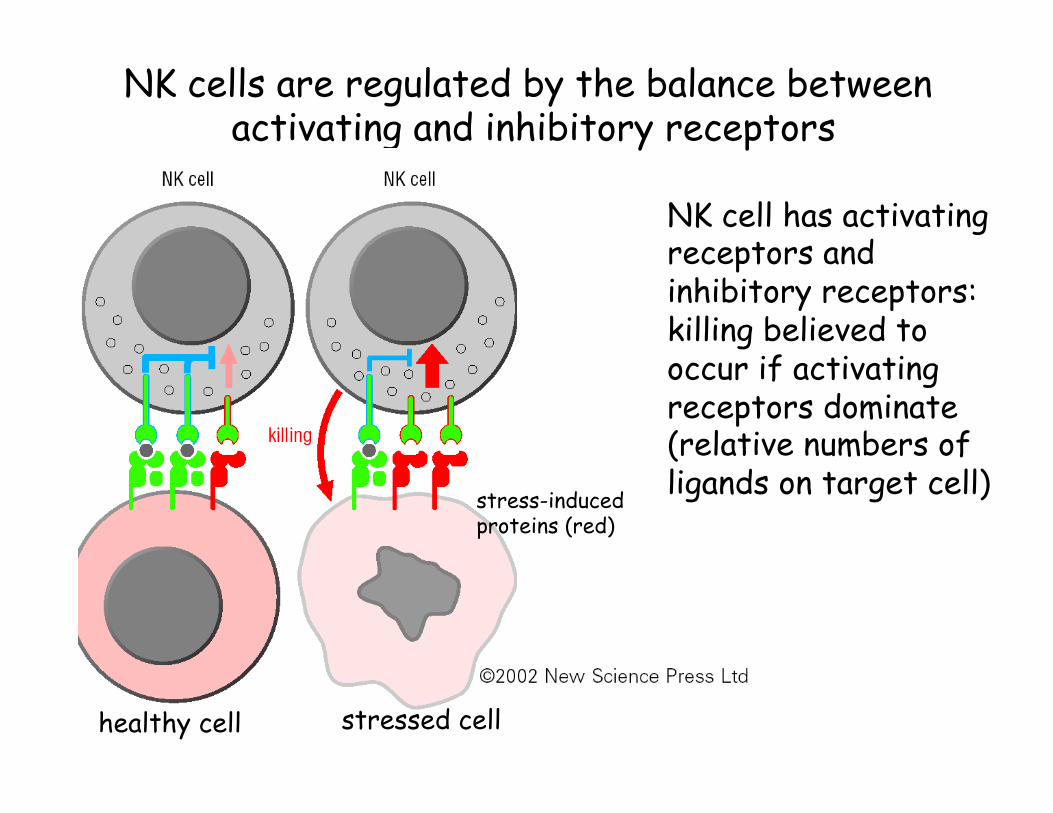
NK cells are regulated by the balance between activating and inhibitory receptors
stressed cell healthy cell
NK cell has activating receptors and inhibitory receptors: killing believed to occur if activating receptors dominate (relative numbers of ligands on target cell) stress-induced
proteins (red)
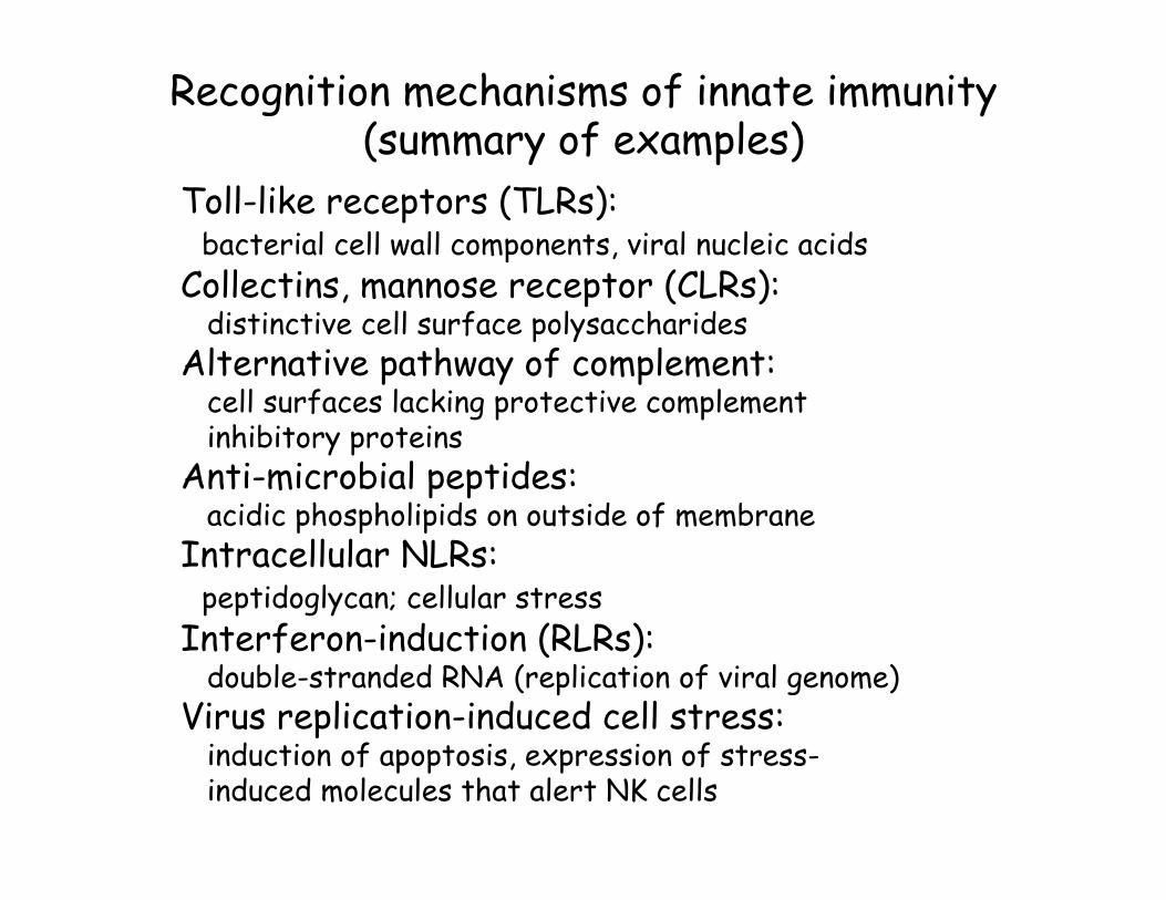
Recognition mechanisms of innate immunity (summary of examples)
Toll-like receptors (TLRs): bacterial cell wall components, viral nucleic acids Collectins, mannose receptor (CLRs): distinctive cell surface polysaccharides Alternative pathway of complement: cell surfaces lacking protective complement inhibitory proteins Anti-microbial peptides: acidic phospholipids on outside of membrane Intracellular NLRs: peptidoglycan; cellular stress Interferon-induction (RLRs): double-stranded RNA (replication of viral genome) Virus replication-induced cell stress: induction of apoptosis, expression of stress- induced molecules that alert NK cells