KJE-3900 Master thesis in organic chemistry
76
1 KJE-3900 Master thesis in organic chemistry Synthesis of Potential Metallo-β -Lactamase Inhibitors Britt Paulsen November, 2011 Faculty of Science and Technology Department of Chemistry University of Tromsø
Transcript of KJE-3900 Master thesis in organic chemistry
1
KJE-3900 Master thesis in organic chemistry
Synthesis of Potential Metallo-β-Lactamase Inhibitors
Britt Paulsen
November, 2011
Faculty of Science and Technology Department of Chemistry
University of Tromsø

3
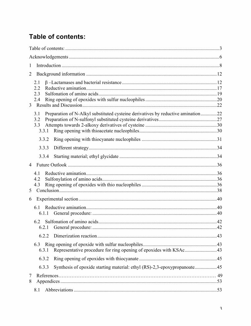
Table of contents: Table of contents: ............................................................................................................................. 3
Acknowledgements .......................................................................................................................... 6
1 Introduction ................................................................................................................................ 8
2 Background information .......................................................................................................... 12
2.1 β –Lactamases and bacterial resistance ............................................................................. 12 2.2 Reductive amination .......................................................................................................... 17 2.3 Sulfonation of amino acids ................................................................................................ 19 2.4 Ring opening of epoxides with sulfur nucleophiles .......................................................... 20
3 Results and Discussion ............................................................................................................. 22
3.1 Preparation of N-Alkyl substituted cysteine derivatives by reductive amination ............. 22 3.2 Preparation of N-sulfonyl substituted cysteine derivatives ............................................... 27 3.3 Attempts towards 2-alkoxy derivatives of cysteine .......................................................... 30
3.3.1 Ring opening with thioacetate nucleophiles ............................................................... 30
3.3.2 Ring opening with thiocyanate nucleophiles ............................................................. 31
3.3.3 Different strategy ........................................................................................................ 34
3.3.4 Starting material; ethyl glycidate ............................................................................... 34
4 Future Outlook ......................................................................................................................... 36
4.1 Reductive amination .......................................................................................................... 36 4.2 Sulfonylation of amino acids ............................................................................................. 36 4.3 Ring opening of epoxides with thio nucleophiles ............................................................. 36
5 Conclusion ................................................................................................................................ 38
6 Experimental section ................................................................................................................ 40
6.1 Reductive amination .......................................................................................................... 40 6.1.1 General procedure: ..................................................................................................... 40
6.2 Sulfonation of amino acids ................................................................................................ 42 6.2.1 General procedure: ..................................................................................................... 42
6.2.2 Dimerization reaction ................................................................................................. 43
6.3 Ring opening of epoxide with sulfur nucleophiles ............................................................ 43 6.3.1 Representative procedure for ring opening of epoxides with KSAc .......................... 43
6.3.2 Ring opening of epoxides with thiocyanate ............................................................... 45
6.3.3 Synthesis of epoxide starting material: ethyl (RS)-2,3-epoxypropanoate .................. 45
7 References…………………………………………………………………………………… 49 8 Appendices ............................................................................................................................... 53
8.1 Abbreviations .................................................................................................................... 53

4
8.2 NMR spectra of 1a-1c ....................................................................................................... 54 8.3 NMR spectra of protected 2a, 2b and cysteine .................................................................. 58 8.4 NMR spectra for 3a, 3b and ethyl glycidate ...................................................................... 61
9 Mass spectra ............................................................................................................................. 64
9.1 Mass spectra ...................................................................................................................... 64 9.2 IR spectra ........................................................................................................................... 72

5

6
Acknowledgements
The work presented in this thesis has been preformed at the Department of Chemistry, University
of Tromsø, in the period from August 2009 to November 2011. During this time, many have
contributed to this work.
First I like to thank my supervisor, Annette Bayer, for presenting me to a motivating and very
interesting project, helpful insights and proofreading my thesis.
Gratitude goes to my co-supervisor, Magnus Engquist, for helpful discussions, always patiently
answering my questions and for proof-reading my thesis.
To Jostein Johansen, Truls Ingebrigtsen and Arnfinn Kvarsnes that kindly helped me with MS
analysis and the operation of the analytical instruments.
To my lab and office partners; thank you for the good atmosphere and spirit.
My gratitudes also go to my family and friends who have supported me and listened to my
triumphs and complaints as the chemistry work evolved. To dad, having excellent knowledge
within the field of organic chemistry. To mom, who always inspire me to do my best. A special
thanks to Marianne H. Paulsen for helpful discussion (the legendary base trick) and support, and
to my sister Hanne-Kristin, for proof-reading my thesis. I am very grateful for your help.
Tromsø, November 20011 Britt Paulsen

7
8
1 Introduction
Bacterial resistance against antibiotics has in the last decades become an increasing problem
worldwide. One cause of antibiotic resistance is the bacterial production of enzymes that
hydrolyze the β-lactam ring found in many antibiotics. These enzymes, called β-lactamases, are
classified into four main groups, one of them being metallo-β-lactamases (MBL). The MBLs
subgroup currently has no clinically developed inhibitors, and due to the more recent
development of resistance against antibiotics due to MBLs the search for inhibitors is a highly
important strategy. The structural complexity and heterogeneity of MBLs have posed challenges
to find a common inhibitor to treat infections caused by MBL-producing bacteria.
The purpose of this project is to create different kinds of MBL inhibitors, and test for their
activities on two specifically selected Verona integron-borne metallo-β-lactamase (VIM)
enzymes, VIM-2 and VIM-7. VIM-2 is one of the most clinically relevant MBLs. One of the
characteristics of the VIM enzymes, is the presence of two Zn(II) ions in the catalytic site, that
are important for both enzymatic and inhibitory activity.
Only a few MBL inhibitors have been reported so far, and even fewer have been described for the
VIM class. No VIM-7 inhibitors have been found. For VIM-2, some inhibitors have been
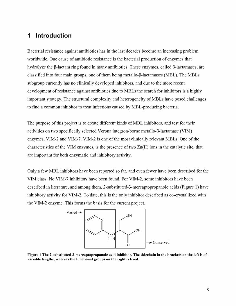
described in literature, and among them, 2-substituted-3-mercaptopropanoic acids (Figure 1) have
inhibitory activity for VIM-2. To date, this is the only inhibitor described as co-crystallized with
the VIM-2 enzyme. This forms the basis for the current project.
Figure 1 The 2-substituted-3-mercaptopropanoic acid inhibitor. The sidechain in the brackets on the left is of variable lengths, whereas the functional groups on the right is fixed.
9
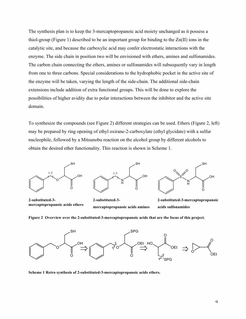
The synthesis plan is to keep the 3-mercaptopropanoic acid moiety unchanged as it possess a
thiol-group (Figure 1) described to be an important group for binding to the Zn(II) ions in the
catalytic site, and because the carboxylic acid may confer electrostatic interactions with the
enzyme. The side chain in position two will be envisioned with ethers, amines and sulfonamides.
The carbon chain connecting the ethers, amines or sulfonamides will subsequently vary in length
from one to three carbons. Special considerations to the hydrophobic pocket in the active site of
the enzyme will be taken, varying the length of the side-chain. The additional side-chain
extensions include addition of extra functional groups. This will be done to explore the
possibilities of higher avidity due to polar interactions between the inhibitor and the active site
domain.
To synthesize the compounds (see Figure 2) different strategies can be used. Ethers (Figure 2, left)
may be prepared by ring opening of ethyl oxirane-2-carboxylate (ethyl glycidate) with a sulfur
nucleophile, followed by a Mitsunobu reaction on the alcohol group by different alcohols to
obtain the desired ether functionality. This reaction is shown in Scheme 1.
2-substituted-3-mercaptopropanoic acids ethers
2-substituted-3-
mercaptopropanoic acids amines
2-substituted-3-mercaptopropanoic
acids sulfonamides
Figure 2 Overview over the 2-substituted-3-mercaptopropanoic acids that are the focus of this project.
Scheme 1 Retro synthesis of 2-substituted-3-mercaptopropanoic acids ethers.
10
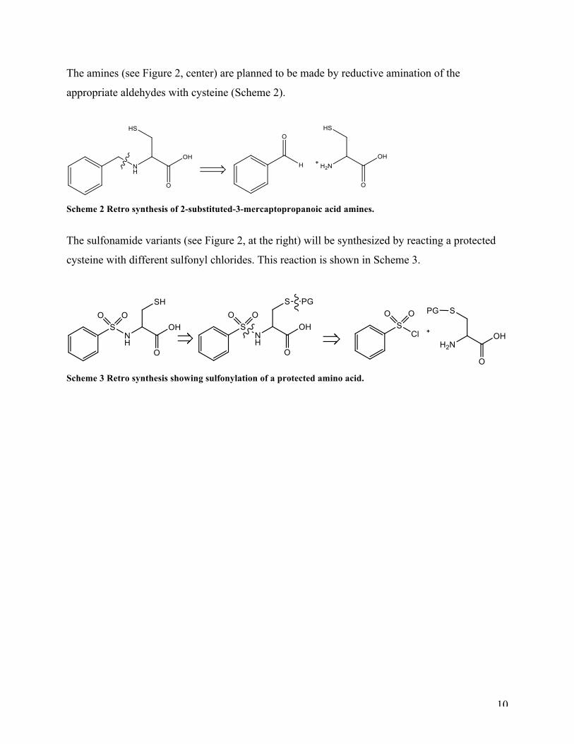
The amines (see Figure 2, center) are planned to be made by reductive amination of the
appropriate aldehydes with cysteine (Scheme 2).
Scheme 2 Retro synthesis of 2-substituted-3-mercaptopropanoic acid amines.
The sulfonamide variants (see Figure 2, at the right) will be synthesized by reacting a protected
cysteine with different sulfonyl chlorides. This reaction is shown in Scheme 3.
Scheme 3 Retro synthesis showing sulfonylation of a protected amino acid.

11
12
2 Background information
2.1 β –Lactamases and bacterial resistance The bacterial resistance against β-lactam antibiotics poses a continuous challenge, thus it is
highly relevant and important to develop general and more specific inhibitors conferring to the
antibiotic resistance. The β-lactam antibiotics hold a beta-lactam ring as its key feature (see
Figure 3). The β-lactam antibiotics works by inhibition of bacterial peptidyltransferase by
forming a stable acyl-enzyme intermediate after a nucleophilic attack governed by a serine
residue in the active site of peptidyltransferase, where the attack results in a cleavage of the β-
lactam ring1. Peptidyltransferase is critical for the peptidoglycan biosynthesis of bacterial cell
wall. Similar to peptidyltransferase, most beta-lactamases (not MBLs) contain a serine residue
within its active site, which performs a nucleophilic attack on the amide bond and cleaves the β-
lactam ring. These types of enzymes are called serine β-lactamases (SBLs), and on the basis of
sequence and structural homology, they have been suggested to be grouped into classes A, C and
D by Ambler5. β-lactamases that employ one or two active-site Zn(II) ions to catalyze the
cleavage of β-lactams are called metallo-β-lactamases and belong to class B2. Based on amino
acid sequence identity and structural features, MBLs can be further classified into three
subclasses: B1, B2 and B33. Similar for them all is that they have an αβ/βα-fold4, where the two
β-sheets are in between the two α-helices. This is called the metallo-β-lactamase fold5. The active
site with one or two Zn(II) ions is located at the edge of the two β-sheets, and the positions of the
Zn(II) ions are essential for substrate binding and hydrolysis6.
All three subclasses of MBLs have a binuclear active site, which requires one or two zinc ions for
full activity. For some binuclear B1 and B3 MBLs, two Zn(II) ions are not necessary for activity.
The second Zn(II) ion in the Zn2 binding site serves to stabilize the built-up negative charge on
the anionic N atom during hydrolysis of the peptide bond, thereby increasing the activation free
energy for nucleophilic attack. As a result, the binuclear enzyme has an overall better catalytic
efficiency than the mononuclear variant2. Subclass B2 enzymes are catalytically active with one
Zn(II) ion binding to the Zn2 binding site. MBLs hydrolyze all β-lactam antibiotics, except for
the monobactam aztreonam2.
13
The VIM enzymes are categorized into subclass B13. As shown by Oelschlaeger et al2 (2010)
VIM enzymes bind two Zn(II) ions in a binuclear center formed by the conserved amino acids
His116, His118, and His196; the Zn2 binding site consists of Asp120, Cys221, and His263. The
two Zn(II) ions activate a water/hydroxide ion nucleophile by coordination. Mutations at amino
acid position 120 have shown that the Zn2 binding site and the Zn1-Zn2 distance are flexible7.
Wang et al8 (1999) proposed a mechanism for nitrocefin hydrolysis of CcrA, and it is reasonable
to assume that the reaction mechanism is closely related to that of VIM. According to this
mechanism, the Zn(II) ions are coordinated by a bridging water/hydroxide molecule that
performs a nucleophilic attack on the carbonyl carbon in the β-lactam ring of the substrate,
resulting in cleavage of the amide bond and deactivation of the antibiotic activity.
So far, these types of MBLs have been identified9: IMP, VIM, GIM, SPM, SIM, KHM, AIM,
DIM, TMB, and NDM. The genes encoding these enzymes are associated with mobile genetic
elements, and are often carried together with other important antibiotic resistance genes as gene
cassettes in integrons, resulting in multidrug resistance that further limits treatment options10.
These integrons are frequently isolated from patients suffering from antibiotic resistant infections.
To this date, twenty-seven VIM variants have been reported9. The VIM enzymes are the globally
most widespread MBLs, and the VIM-2 was first isolated from Pseudomonas aeruginosa11.
VIM-7 is the most divergent VIM type in terms of amino acid sequence, sharing only 74 %
identity with VIM-29. It was first isolated in the United States in 2001, and its gene was found on
an integron in a Pseudomonas aeruginosa isolate. Due to the low sequence identity with VIM-2,
it has lead to the belief that VIM-7 has evolved independently in the US where it first was
isolated, rather than having been transferred from Europe or Asia2.
The two VIM enzymes that are used for testing in the current project possess a number of
different features. VIM-7 is more positively charged than VIM-2, carrying an overall charge of -9,
compared to -14 for VIM-29. Some of this negative charge in VIM-2 is confined to the active site
suggesting a possible influence in interactions with substrates. Because of this charge difference,
VIM-7 has a lower affinity and a reduced catalytic efficiency against cephalosporins relative to
VIM-2, particularly those with a bulky positively charged C3 substituent (see Figure 3). On the
other hand, penicillinase and carbapenemase activities are comparable with that of VIM-2. The
14
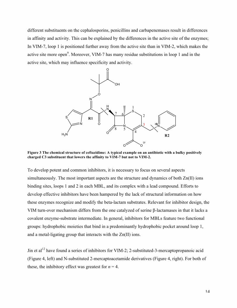
different substituents on the cephalosporins, penicillins and carbapenemases result in differences
in affinity and activity. This can be explained by the differences in the active site of the enzymes;
In VIM-7, loop 1 is positioned further away from the active site than in VIM-2, which makes the
active site more open9. Moreover, VIM-7 has many residue substitutions in loop 1 and in the
active site, which may influence specificity and activity.
Figure 3 The chemical structure of ceftazidime: A typical example on an antibiotic with a bulky positively charged C3 substituent that lowers the affinity to VIM-7 but not to VIM-2.
To develop potent and common inhibitors, it is necessary to focus on several aspects
simultaneously. The most important aspects are the structure and dynamics of both Zn(II) ions
binding sites, loops 1 and 2 in each MBL, and its complex with a lead compound. Efforts to
develop effective inhibitors have been hampered by the lack of structural information on how
these enzymes recognize and modify the beta-lactam substrates. Relevant for inhibitor design, the
VIM turn-over mechanism differs from the one catalyzed of serine β-lactamases in that it lacks a
covalent enzyme-substrate intermediate. In general, inhibitors for MBLs feature two functional
groups: hydrophobic moieties that bind in a predominantly hydrophobic pocket around loop 1,
and a metal-ligating group that interacts with the Zn(II) ions.
Jin et al12 have found a series of inhibitors for VIM-2; 2-substituted-3-mercaptopropanoic acid
(Figure 4, left) and N-substituted 2-mercaptoacetamide derivatives (Figure 4, right). For both of
these, the inhibitory effect was greatest for n = 4.
15
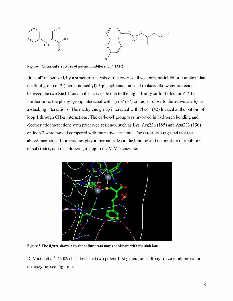
Figure 4 Chemical structure of potent inhibitors for VIM-2.
Jin et al6 recognized, by a structure analysis of the co-crystallized enzyme-inhibitor complex, that
the thiol group of 2-(mercaptomethyl)-5-phenylpentanoic acid replaced the water molecule
between the two Zn(II) ions in the active site due to the high affinity sulfur holds for Zn(II).
Furthermore, the phenyl group interacted with Tyr67 (47) on loop 1 close to the active site by π-
π-stacking interactions. The methylene group interacted with Phe61 (42) located at the bottom of
loop 1 through CH-π interactions. The carboxyl group was involved in hydrogen bonding and
electrostatic interactions with preserved residues, such as Lys. Arg228 (185) and Asn233 (190)
on loop 2 were moved compared with the native structure. These results suggested that the
above-mentioned four residues play important roles in the binding and recognition of inhibitors
or substrates, and in stabilizing a loop in the VIM-2 enzyme.
Figure 5 The figure shows how the sulfur atom may coordinate with the sink ions.
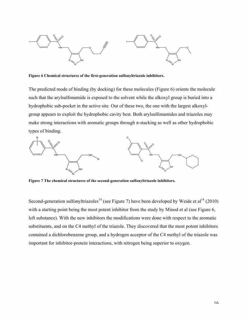
D. Minod et al13 (2009) has described two potent first generation sulfonyltriazole inhibitors for
the enzyme, see Figure 6.
16
Figure 6 Chemical structures of the first-generation sulfonyltriazole inhibitors.
The predicted mode of binding (by docking) for these molecules (Figure 6) orients the molecule
such that the arylsulfonamide is exposed to the solvent while the alkoxyl group is buried into a
hydrophobic sub-pocket in the active site. Out of these two, the one with the largest alkoxyl-
group appears to exploit the hydrophobic cavity best. Both arylsulfonamides and triazoles may
make strong interactions with aromatic groups through π-stacking as well as other hydrophobic
types of binding.
Figure 7 The chemical structures of the second-generation sulfonyltriazole inhibitors.
Second-generation sulfonyltriazoles14 (see Figure 7) have been developed by Weide et al14 (2010)
with a starting point being the most potent inhibitor from the study by Minod et al (see Figure 6,
left substance). With the new inhibitors the modifications were done with respect to the aromatic
substituents, and on the C4 methyl of the triazole. They discovered that the most potent inhibitors
contained a dichlorobenzene group, and a hydrogen acceptor of the C4 methyl of the triazole was
important for inhibitor-protein interactions, with nitrogen being superior to oxygen.
17
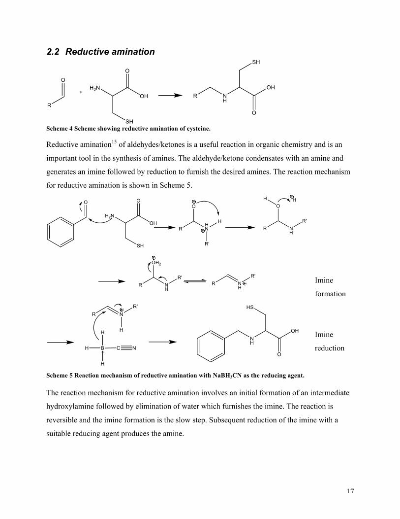
2.2 Reductive amination
Scheme 4 Scheme showing reductive amination of cysteine. Reductive amination15 of aldehydes/ketones is a useful reaction in organic chemistry and is an
important tool in the synthesis of amines. The aldehyde/ketone condensates with an amine and
generates an imine followed by reduction to furnish the desired amines. The reaction mechanism
for reductive amination is shown in Scheme 5.
Imine
formation
Imine
reduction
Scheme 5 Reaction mechanism of reductive amination with NaBH3CN as the reducing agent. The reaction mechanism for reductive amination involves an initial formation of an intermediate
hydroxylamine followed by elimination of water which furnishes the imine. The reaction is
reversible and the imine formation is the slow step. Subsequent reduction of the imine with a
suitable reducing agent produces the amine.
18
Among the various reduction methods at hand are catalytic hydrogenation, reduction with amine
borane complexes16 or by using metallo-hydride reducing agents such as sodium
cyanoborohydride17, sodium triacetoxyborohydride18, sodium19- or zinc borohydride20. Among
the latter, sodium cyanoborhydride and sodium triacetoxyborhydride are the most common
because of their versatility and will be described later. The success of the reaction depends on the
choice of the reducing agent, as it might have to reduce imines selectively over ketones/aldehydes
under the same reaction conditions. The rate of the reduction of iminium ions is in addition, much
faster than that for aldehydes or ketones, but requires catalytic amount of acid. As a result,
reductive amination can be carried out as a one-pot procedure by introducing the reducing agent
into a mixture of the amine and carbonyl compound at the start of the reaction.
NaBH3CN17 is a mild reducing agent that reduces a variety of organic functional groups21 with
good selectivity and will readily reduce imines to amines. The successful use of NaBH3CN is due
to its pH depending selectivity22. At pH 3-4 NaBH3CN reduces aldehydes and ketones efficiently,
and at pH 6-8, imines are preferentially protonated and reduced faster than aldehydes and ketones.
NaBH3CN is stabile in relatively strong acids (pH 3) and show good solubility in hydroxylic
solvents such as methanol. Limitations of this method are that the reaction may require up to a
fivefold excess of the amine in relation to the carbonyl compound22. For aromatic ketones and
with weakly basic amines, conversion can be slow. A drawback for the use of NaBH3CN in
reductive amination is that the highly toxic gas HCN may be liberated under strong acidic
conditions. Furthermore the products may be contaminated by HCN or a cyanide salt that might
follow the product through work up. Such contaminants have reportedly18 been hard to remove.
An other mild reducing agent, is Na(OAc)3BH18. The boron-hydride bond is stabilized by the
steric and electron-withdrawing influence of the acetoxy groups. Na(OAc)3BH works well in
reduction of aliphatic acyclic and cyclic ketones, aliphatic and aromatic aldehydes, and primary
and secondary amines including a variety of weakly basic and nonbasic amines. Some
challenging substrates for the reductive amination with Na(OAc)3BH include aromatic and
unsaturated ketones and some sterically hindered ketones and amines.

19
2.3 Sulfonation of amino acids
Scheme 6 Preparation of a sulfonamide with Cysteine using sulfonyl chloride
The most common way of preparing sulfonamide derivatives of unprotected amino acids is by
nucleophile acyl substitution at suitable sulfonyl chloride in aqueous solution in the presence of a
base (Scheme 6). Schröder et al25 have proposed a general procedure for preparing N-
sulfonylated amino acids involves addition of the sulfonyl chloride in a solution of the amino acid
in aqueous base before heating the mixture with vigorous stirring. This procedure works for most
of the natural amino acids but they tend to favor different bases for optimal yields. NaOH23,
DIEA24, K2CO325, and Na2CO3
26 are the most frequently used bases. The sulfonation requires a
reaction time between 10 min up to 2 hours. For some amino acids, heating the reaction mixture
up to 70° C is necessary for achieving an acceptable conversion. The differences in reaction
conditions required might be explained by solubility issues or steric hindrance among the amino
acids with charge carrying or bulkier side chains. There are no reported methods for sulfonation
of a cysteine unless its thiol group functionality is protected. For the protected cysteine27 the
sulfonyl chloride is dissolved in 1,4-dioxane prior to its addition to a solution of protected
cysteine in aqueous sodium hydroxide and the reaction is stirred at room temperature for 16 hours.
The reaction mechanism for sulfonation of amino acids is shown in Scheme 7.
Scheme 7 Reaction mechanism of sulfonation of amino acids.
The amine will react as a nucleophile and attack the electron poor sulfur and displace the leaving
group, in this case a chloride ion. The resulting amine cation will be deprotonated by the base
present in the system yielding the sulfonamide.
20
2.4 Ring opening of epoxides with sulfur nucleophiles
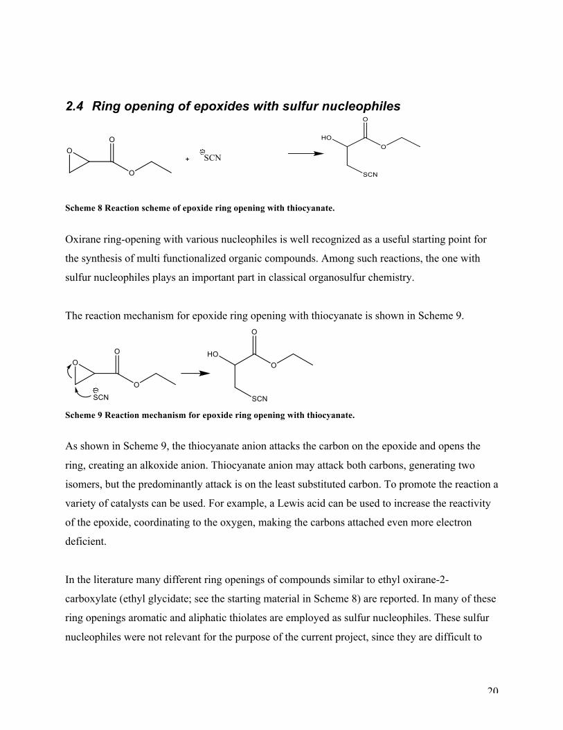
Scheme 8 Reaction scheme of epoxide ring opening with thiocyanate.
Oxirane ring-opening with various nucleophiles is well recognized as a useful starting point for
the synthesis of multi functionalized organic compounds. Among such reactions, the one with
sulfur nucleophiles plays an important part in classical organosulfur chemistry.
The reaction mechanism for epoxide ring opening with thiocyanate is shown in Scheme 9.
Scheme 9 Reaction mechanism for epoxide ring opening with thiocyanate.
As shown in Scheme 9, the thiocyanate anion attacks the carbon on the epoxide and opens the
ring, creating an alkoxide anion. Thiocyanate anion may attack both carbons, generating two
isomers, but the predominantly attack is on the least substituted carbon. To promote the reaction a
variety of catalysts can be used. For example, a Lewis acid can be used to increase the reactivity
of the epoxide, coordinating to the oxygen, making the carbons attached even more electron
deficient.
In the literature many different ring openings of compounds similar to ethyl oxirane-2-
carboxylate (ethyl glycidate; see the starting material in Scheme 8) are reported. In many of these
ring openings aromatic and aliphatic thiolates are employed as sulfur nucleophiles. These sulfur
nucleophiles were not relevant for the purpose of the current project, since they are difficult to
21
deprotect and the free thiol would not be available. Other nucleophiles at interest described are
KSAc28, NH4SCN/KSCN/HSCN29, and TMSNCS30.
One of the most described protection groups for sulfur is the cyano group. There are three
methods reported in literature for the synthesis of beta-hydroxythiocyanates, and the most
frequently used method is the epoxide ring opening with a thiocyanate anion. The thiocyanate
salts were used in both presence and absence31 of catalyst. Catalysts that has been used are;
crown ethers42, tetrabutylammonium fluoride (TBAF)32, Ti(OPri )433, various Lewis acids like
titanium-, zinc,- indium,- and gallium chlorides34, 2,3-Dichloro-5,6-dicyanobenzoquinone
(DDQ)35, triphenylphosphine-thiocyanogen (TPPT)36, triphenylphosphinepalladium (article not
found), polyacryloamides37, selectfluor38, 2-phenyl-2-(pyridyl)imidazolidine (PPI)39, 2,6-bis[2-
(o-aminophenoxy)-methyl]-4-bromo-1-methoxybenzene (BABMB)40, Schiff-base metal(II)
complexes (article not available), metalloporphyrins41, phenol-containing macrocyclic diamides42
and tetraarylporphyrins43. It has been reported that the presence of hydroquinone29b is required to
stabilize the beta-hydroxy thiocyanate to slow down the rapid conversion into thiiranes. This is
only valid if the beta-hydroxythiocyanate is contaminated in the reaction mixture. If it is in its
pure form, the addition of hydroxyquinone will catalyze the thiirane formation. Alternative
methods for making beta-hydroxythiocyanates include opening of a cyclic sulfate with NH4SCN
followed by hydrolysis of the corresponding beta-sulfate to form the thiocyanohydrins44, and
nucleophilic substitution of a bromine atom in bromohydrin by a thiocyanate anion45.
22
3 Results and Discussion
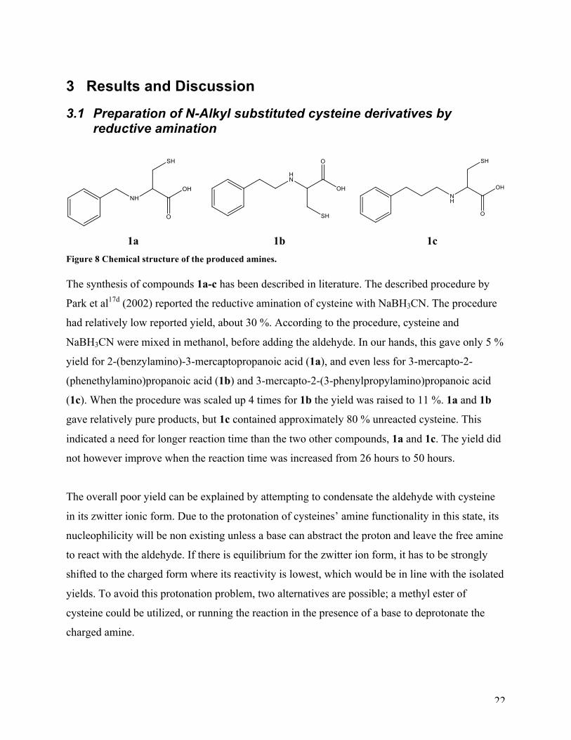
3.1 Preparation of N-Alkyl substituted cysteine derivatives by reductive amination
1a 1b 1c Figure 8 Chemical structure of the produced amines. The synthesis of compounds 1a-c has been described in literature. The described procedure by
Park et al17d (2002) reported the reductive amination of cysteine with NaBH3CN. The procedure
had relatively low reported yield, about 30 %. According to the procedure, cysteine and
NaBH3CN were mixed in methanol, before adding the aldehyde. In our hands, this gave only 5 %
yield for 2-(benzylamino)-3-mercaptopropanoic acid (1a), and even less for 3-mercapto-2-
(phenethylamino)propanoic acid (1b) and 3-mercapto-2-(3-phenylpropylamino)propanoic acid
(1c). When the procedure was scaled up 4 times for 1b the yield was raised to 11 %. 1a and 1b
gave relatively pure products, but 1c contained approximately 80 % unreacted cysteine. This
indicated a need for longer reaction time than the two other compounds, 1a and 1c. The yield did
not however improve when the reaction time was increased from 26 hours to 50 hours.
The overall poor yield can be explained by attempting to condensate the aldehyde with cysteine
in its zwitter ionic form. Due to the protonation of cysteines’ amine functionality in this state, its
nucleophilicity will be non existing unless a base can abstract the proton and leave the free amine
to react with the aldehyde. If there is equilibrium for the zwitter ion form, it has to be strongly
shifted to the charged form where its reactivity is lowest, which would be in line with the isolated
yields. To avoid this protonation problem, two alternatives are possible; a methyl ester of
cysteine could be utilized, or running the reaction in the presence of a base to deprotonate the
charged amine.
23
These compounds could not be purified by silica chromatography because of the low solubility in
any tested solvents, eg H2O, MeOH, CHCl3, dioxane, MeOH/H2O, THF, acetonitrile,
acetonitril+TFA, DMSO, DMF, Et2O, pyridine, and benzene. Several means of reducing reaction
time and increasing yield are reported in literature, such as the use of molecular sieves, azeotropic
removal of water and addition of acid22. We investigated the use of a dehydrating agent Na2SO4,
and catalytic addition of acid, HCl, in the synthesis of 1c. Due to the low solubility of 1c, the
dehydrating agent, Na2SO4, could not be separated from target compound.
The procedure reported in literature49 for compound 1a-1c has reported that the 1H NMR and 13C
NMR analysis were run in D2O. It was impossible to solve the target compounds 1a-1c in this
solvent. Therefore the use of NaOH was utilized, and the 1H NMR and 13C NMR analysis were
run with NaOH in D2O. The spectra from literature and the ones obtained can therefore not be
compared. 1H NMR and 13C NMR reference spectra with cysteine in NaOH/D2O were prepared
for comparison for the contamination in the spectra for 1b and 1c.
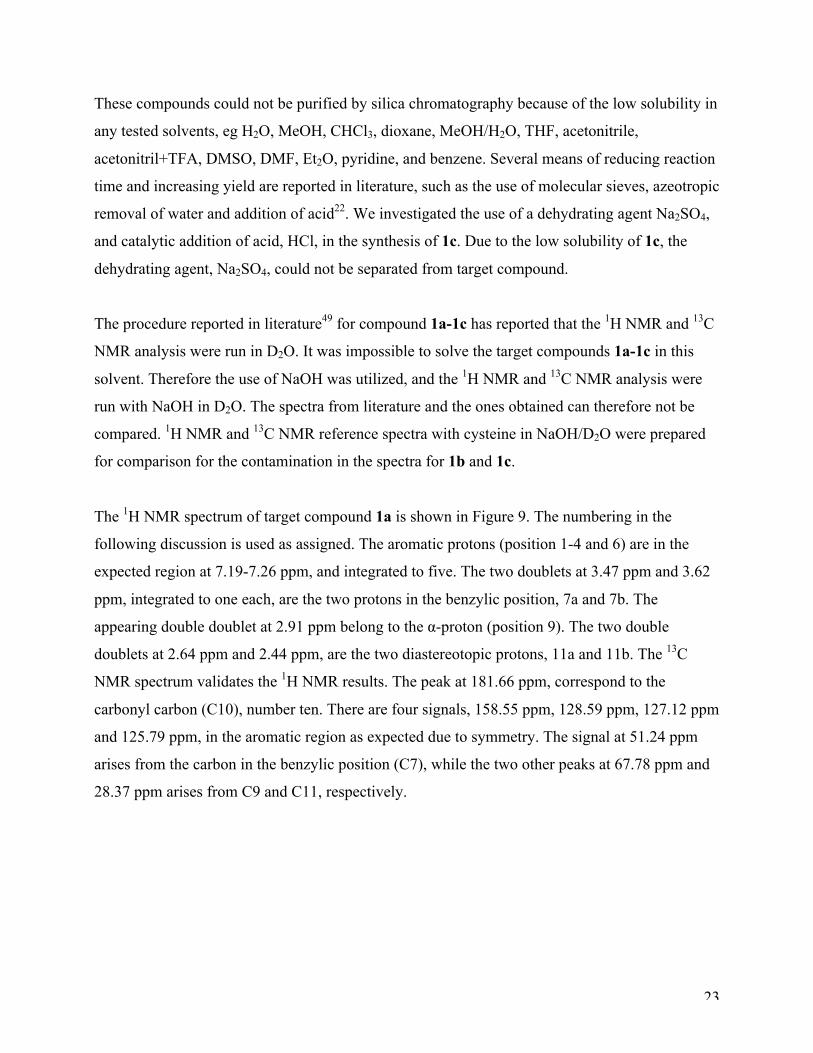
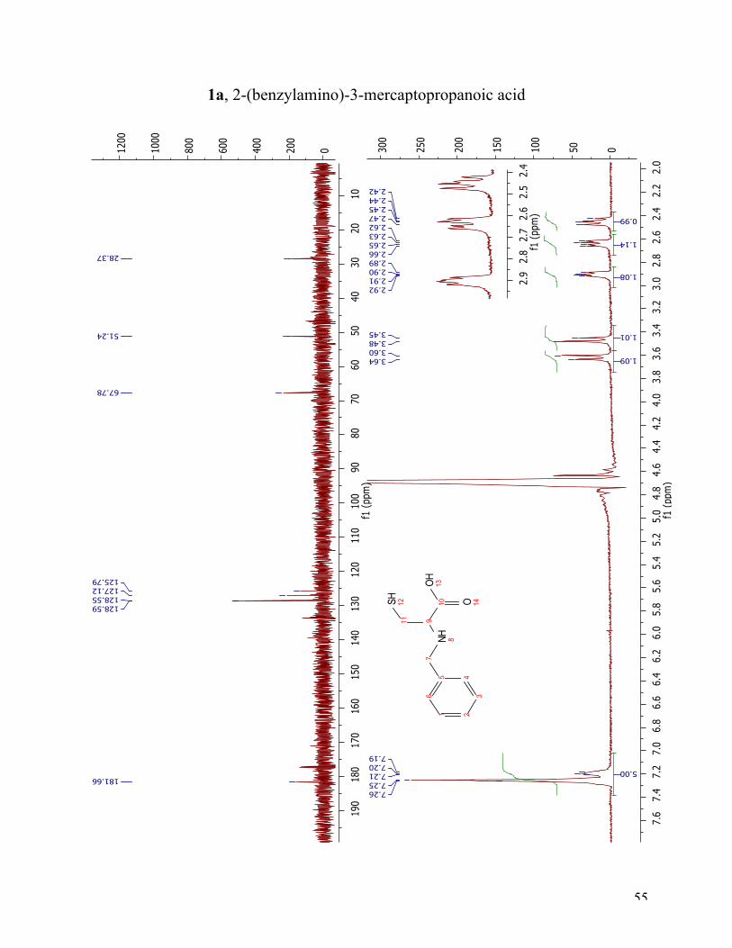
The 1H NMR spectrum of target compound 1a is shown in Figure 9. The numbering in the
following discussion is used as assigned. The aromatic protons (position 1-4 and 6) are in the
expected region at 7.19-7.26 ppm, and integrated to five. The two doublets at 3.47 ppm and 3.62
ppm, integrated to one each, are the two protons in the benzylic position, 7a and 7b. The
appearing double doublet at 2.91 ppm belong to the α-proton (position 9). The two double
doublets at 2.64 ppm and 2.44 ppm, are the two diastereotopic protons, 11a and 11b. The 13C
NMR spectrum validates the 1H NMR results. The peak at 181.66 ppm, correspond to the
carbonyl carbon (C10), number ten. There are four signals, 158.55 ppm, 128.59 ppm, 127.12 ppm
and 125.79 ppm, in the aromatic region as expected due to symmetry. The signal at 51.24 ppm
arises from the carbon in the benzylic position (C7), while the two other peaks at 67.78 ppm and
28.37 ppm arises from C9 and C11, respectively.
24
Figure 9 1H NMR and 13C NMR spectra of compound 1a.
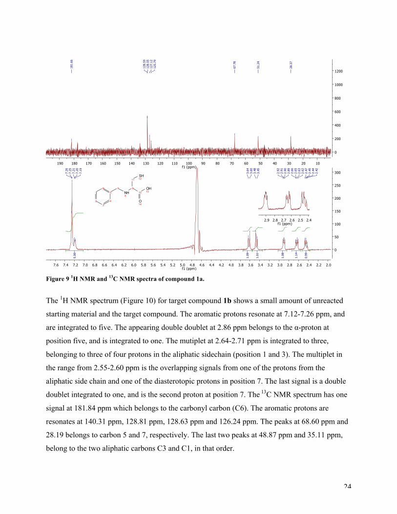
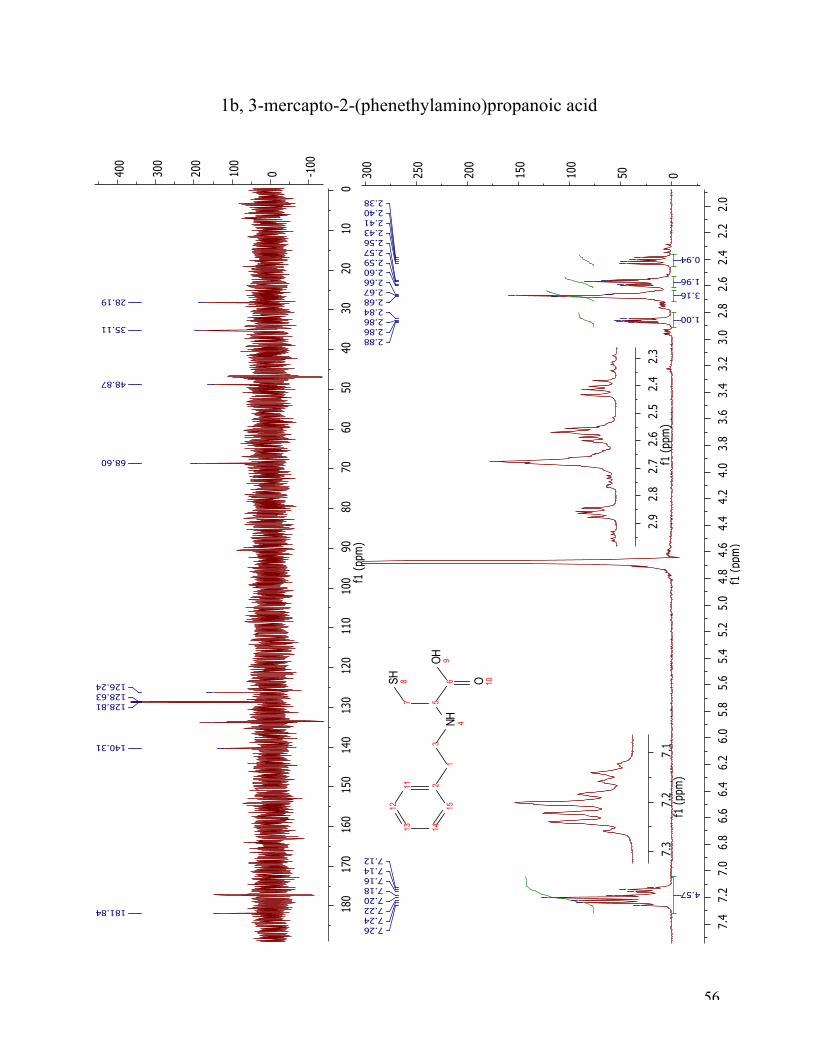
The 1H NMR spectrum (Figure 10) for target compound 1b shows a small amount of unreacted
starting material and the target compound. The aromatic protons resonate at 7.12-7.26 ppm, and
are integrated to five. The appearing double doublet at 2.86 ppm belongs to the α-proton at
position five, and is integrated to one. The mutiplet at 2.64-2.71 ppm is integrated to three,
belonging to three of four protons in the aliphatic sidechain (position 1 and 3). The multiplet in
the range from 2.55-2.60 ppm is the overlapping signals from one of the protons from the
aliphatic side chain and one of the diasterotopic protons in position 7. The last signal is a double
doublet integrated to one, and is the second proton at position 7. The 13C NMR spectrum has one
signal at 181.84 ppm which belongs to the carbonyl carbon (C6). The aromatic protons are
resonates at 140.31 ppm, 128.81 ppm, 128.63 ppm and 126.24 ppm. The peaks at 68.60 ppm and
28.19 belongs to carbon 5 and 7, respectively. The last two peaks at 48.87 ppm and 35.11 ppm,
belong to the two aliphatic carbons C3 and C1, in that order.
25
Figure 10 1H NMR and 13C NMR spectra of compound 1b.
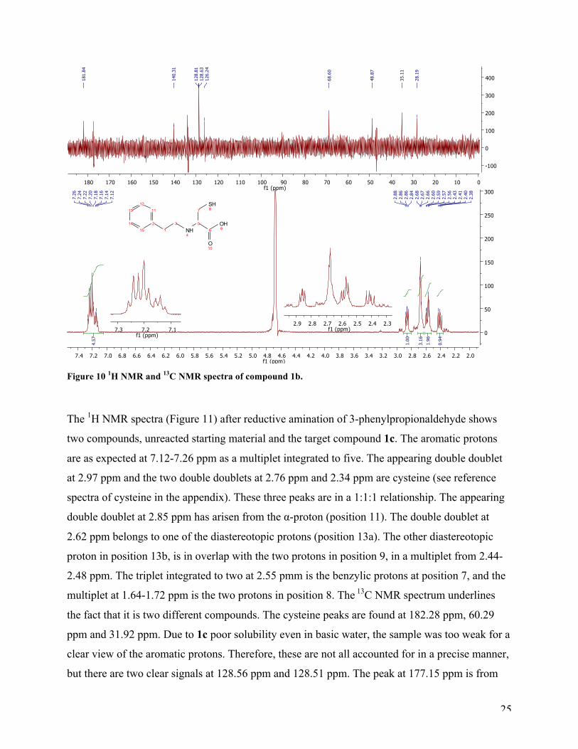
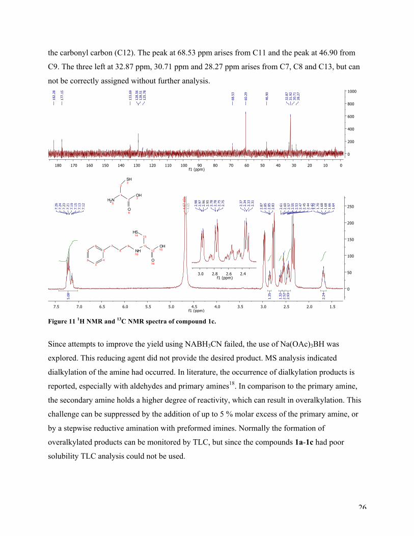
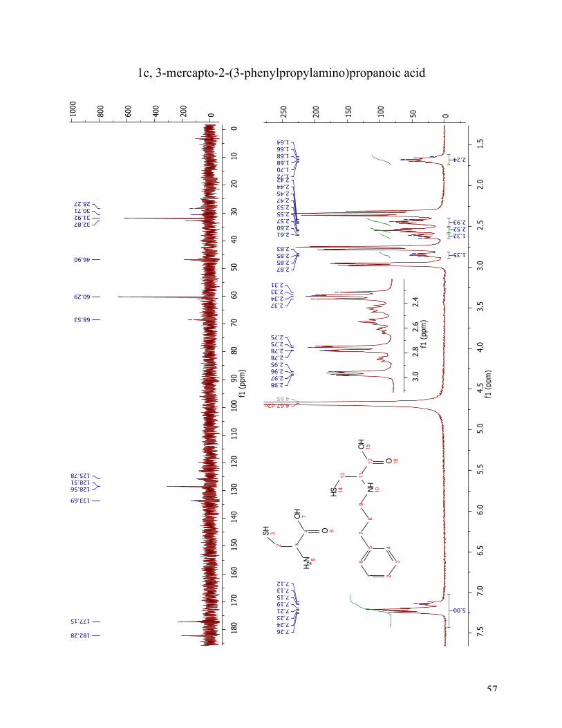
The 1H NMR spectra (Figure 11) after reductive amination of 3-phenylpropionaldehyde shows
two compounds, unreacted starting material and the target compound 1c. The aromatic protons
are as expected at 7.12-7.26 ppm as a multiplet integrated to five. The appearing double doublet
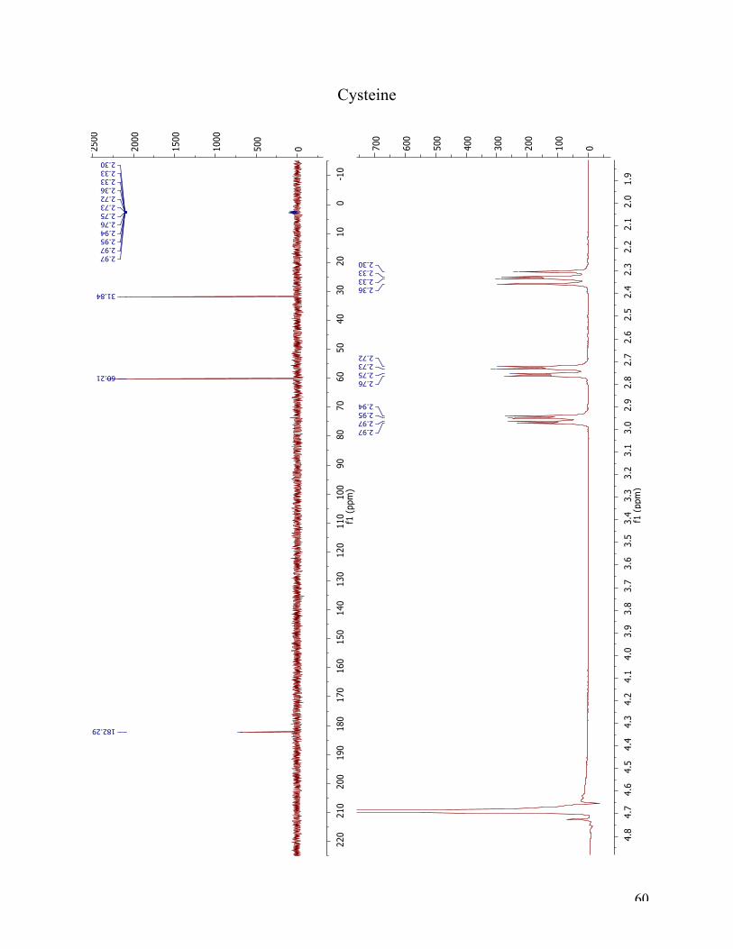
at 2.97 ppm and the two double doublets at 2.76 ppm and 2.34 ppm are cysteine (see reference
spectra of cysteine in the appendix). These three peaks are in a 1:1:1 relationship. The appearing
double doublet at 2.85 ppm has arisen from the α-proton (position 11). The double doublet at
2.62 ppm belongs to one of the diastereotopic protons (position 13a). The other diastereotopic
proton in position 13b, is in overlap with the two protons in position 9, in a multiplet from 2.44-
2.48 ppm. The triplet integrated to two at 2.55 pmm is the benzylic protons at position 7, and the
multiplet at 1.64-1.72 ppm is the two protons in position 8. The 13C NMR spectrum underlines
the fact that it is two different compounds. The cysteine peaks are found at 182.28 ppm, 60.29
ppm and 31.92 ppm. Due to 1c poor solubility even in basic water, the sample was too weak for a
clear view of the aromatic protons. Therefore, these are not all accounted for in a precise manner,
but there are two clear signals at 128.56 ppm and 128.51 ppm. The peak at 177.15 ppm is from
26
the carbonyl carbon (C12). The peak at 68.53 ppm arises from C11 and the peak at 46.90 from
C9. The three left at 32.87 ppm, 30.71 ppm and 28.27 ppm arises from C7, C8 and C13, but can
not be correctly assigned without further analysis.
Figure 11 1H NMR and 13C NMR spectra of compound 1c.
Since attempts to improve the yield using NABH3CN failed, the use of Na(OAc)3BH was
explored. This reducing agent did not provide the desired product. MS analysis indicated
dialkylation of the amine had occurred. In literature, the occurrence of dialkylation products is
reported, especially with aldehydes and primary amines18. In comparison to the primary amine,
the secondary amine holds a higher degree of reactivity, which can result in overalkylation. This
challenge can be suppressed by the addition of up to 5 % molar excess of the primary amine, or
by a stepwise reductive amination with preformed imines. Normally the formation of
overalkylated products can be monitored by TLC, but since the compounds 1a-1c had poor
solubility TLC analysis could not be used.
27
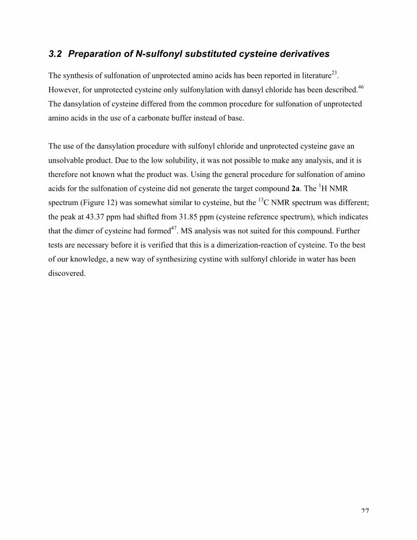
3.2 Preparation of N-sulfonyl substituted cysteine derivatives The synthesis of sulfonation of unprotected amino acids has been reported in literature23.
However, for unprotected cysteine only sulfonylation with dansyl chloride has been described.46
The dansylation of cysteine differed from the common procedure for sulfonation of unprotected
amino acids in the use of a carbonate buffer instead of base.
The use of the dansylation procedure with sulfonyl chloride and unprotected cysteine gave an
unsolvable product. Due to the low solubility, it was not possible to make any analysis, and it is
therefore not known what the product was. Using the general procedure for sulfonation of amino
acids for the sulfonation of cysteine did not generate the target compound 2a. The 1H NMR
spectrum (Figure 12) was somewhat similar to cysteine, but the 13C NMR spectrum was different;
the peak at 43.37 ppm had shifted from 31.85 ppm (cysteine reference spectrum), which indicates
that the dimer of cysteine had formed47. MS analysis was not suited for this compound. Further
tests are necessary before it is verified that this is a dimerization-reaction of cysteine. To the best
of our knowledge, a new way of synthesizing cystine with sulfonyl chloride in water has been
discovered.
28
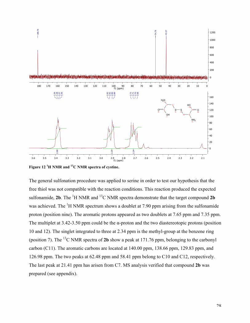
Figure 12 1H NMR and 13C NMR spectra of cystine.
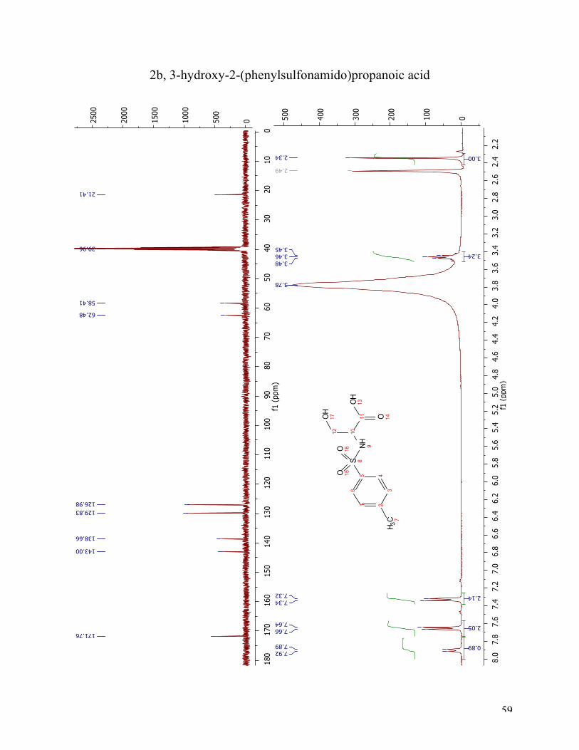
The general sulfonation procedure was applied to serine in order to test our hypothesis that the
free thiol was not compatible with the reaction conditions. This reaction produced the expected
sulfonamide, 2b. The 1H NMR and 13C NMR spectra demonstrate that the target compound 2b
was achieved. The 1H NMR spectrum shows a doublet at 7.90 ppm arising from the sulfonamide
proton (position nine). The aromatic protons appeared as two doublets at 7.65 ppm and 7.35 ppm.
The multiplet at 3.42-3.50 ppm could be the α-proton and the two diastereotopic protons (position
10 and 12). The singlet integrated to three at 2.34 ppm is the methyl-group at the benzene ring
(position 7). The 13C NMR spectra of 2b show a peak at 171.76 ppm, belonging to the carbonyl
carbon (C11). The aromatic carbons are located at 140.00 ppm, 138.66 ppm, 129.83 ppm, and
126.98 ppm. The two peaks at 62.48 ppm and 58.41 ppm belong to C10 and C12, respectively.
The last peak at 21.41 ppm has arisen from C7. MS analysis verified that compound 2b was
prepared (see appendix).
29
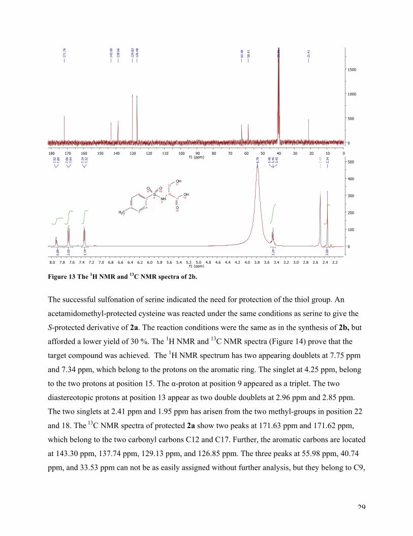
Figure 13 The 1H NMR and 13C NMR spectra of 2b.
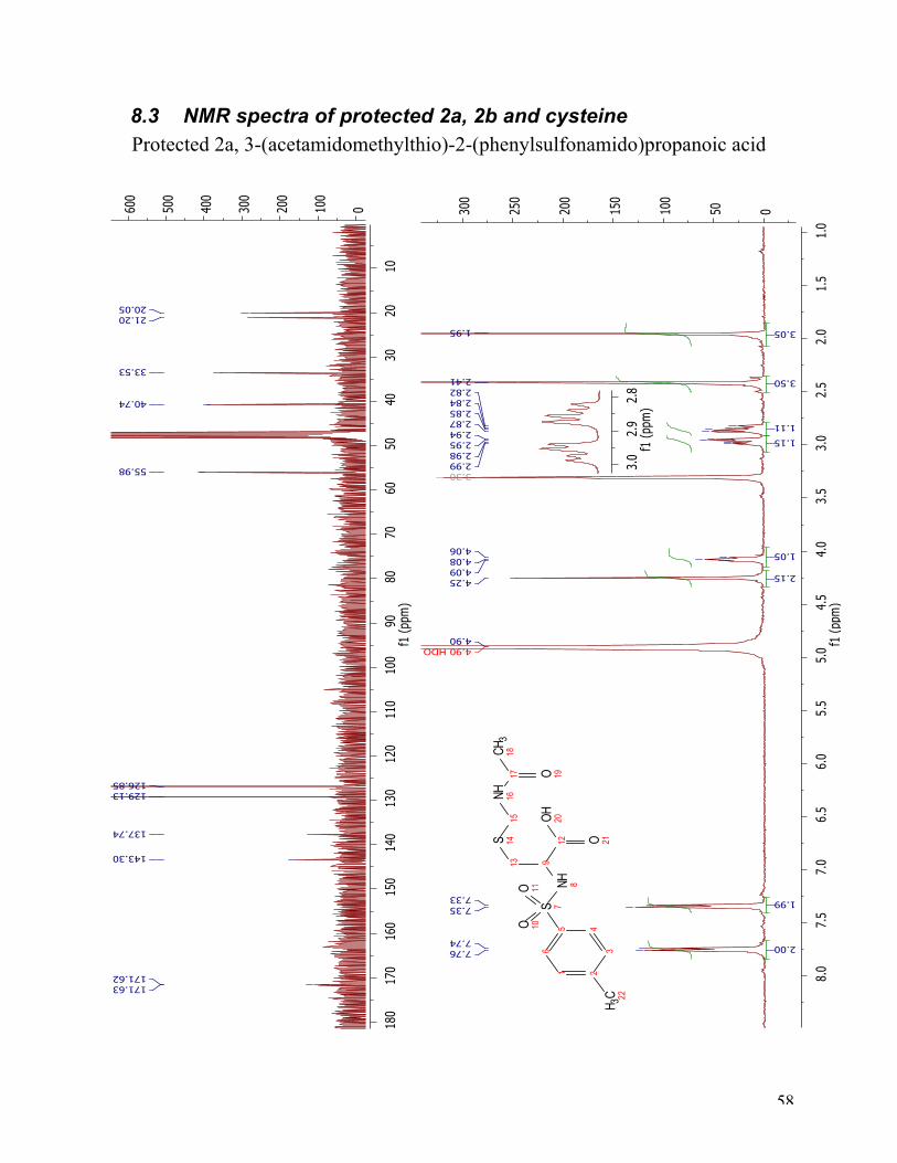
The successful sulfonation of serine indicated the need for protection of the thiol group. An
acetamidomethyl-protected cysteine was reacted under the same conditions as serine to give the
S-protected derivative of 2a. The reaction conditions were the same as in the synthesis of 2b, but
afforded a lower yield of 30 %. The 1H NMR and 13C NMR spectra (Figure 14) prove that the
target compound was achieved. The 1H NMR spectrum has two appearing doublets at 7.75 ppm
and 7.34 ppm, which belong to the protons on the aromatic ring. The singlet at 4.25 ppm, belong
to the two protons at position 15. The α-proton at position 9 appeared as a triplet. The two
diastereotopic protons at position 13 appear as two double doublets at 2.96 ppm and 2.85 ppm.
The two singlets at 2.41 ppm and 1.95 ppm has arisen from the two methyl-groups in position 22
and 18. The 13C NMR spectra of protected 2a show two peaks at 171.63 ppm and 171.62 ppm,
which belong to the two carbonyl carbons C12 and C17. Further, the aromatic carbons are located
at 143.30 ppm, 137.74 ppm, 129.13 ppm, and 126.85 ppm. The three peaks at 55.98 ppm, 40.74
ppm, and 33.53 ppm can not be as easily assigned without further analysis, but they belong to C9,
30
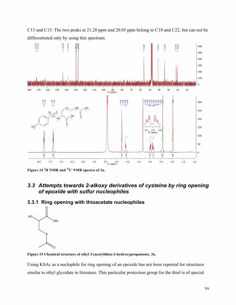
C13 and C15. The two peaks at 21.20 ppm and 20.05 ppm belong to C18 and C22, but can not be
differentiated only by using this spectrum.
Figure 14 1H NMR and 13C NMR spectra of 2a.
3.3 Attempts towards 2-alkoxy derivatives of cysteine by ring opening of epoxide with sulfur nucleophiles
3.3.1 Ring opening with thioacetate nucleophiles
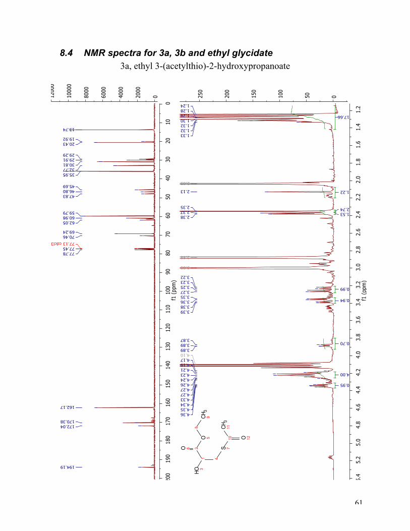
Figure 15 Chemical structure of ethyl 3-(acetylthio)-2-hydroxypropanoate, 3a. Using KSAc as a nuclophile for ring opening of an epoxide has not been reported for structures
similar to ethyl glycidate in literature. This particular protection group for the thiol is of special
31
interest because of its ability to be hydrolyzed under the same conditions as the ester. Many
different approaches were tried. The first reaction was conducted by mixing KSAc in DMF
before adding the solution slowly to a mixture of ethyl glycidate in DMF. The reaction mixture
was left in room temperature with stirring for two hours. This was not successful, and variations
in the reaction conditions with factors such as temperature, amount of Lewis acid, addition of
crown ether, and the addition speed of KSAc (in DMF) to the reaction mixture, was applied.
BF3*Et2O and 18-crown-6 was used to catalyze the reaction, and DMF was used as solvent. This
lead to a successful reaction with achievement of the target compound 3a. In this reaction the
ethyl glycidate, BF3*Et2O and 18-crown-6 was solved in DMF before the KSAc in DMF was
added slowly and left at room temperature with stirring for two hours. A fractional factorial
design was employed to get a feeling for the reaction. It underlined the successful reaction with
indications it was important to carry out the reaction at 0° C, with stociometric amount BF3*Et2O,
and slow addition of the KSAc solution. During work-up, the product, 3a, decomposed rapidly
and was impossible to isolate and store, even at low temperature, as a pure compound. Tanabe et
al32 have reported that acetylthiohydrin is unstable due to the weakness of the acetyl-sulfur bond
adjacent to the hydroxy group.
An attempt to trap the β-hydroxythioacetate with an acid chloride/ benzyl bromide before work-
up was unsuccessful. MS and NMR analysis did not indicate that the target compound had been
formed. In literature, trapping of an acetylthiohydrin has not been reported, only trapping of a β-
hydroxythiocyanate has been reported by Łukowska-Chojnacka et al48 (2011).
HSAc was also tested as nucleophile. The 1H NMR and 13C NMR spectra showed only unreacted
start material, which was not surprising considering its poor nucleophilic abilities.
3.3.2 Ring opening with thiocyanate nucleophiles
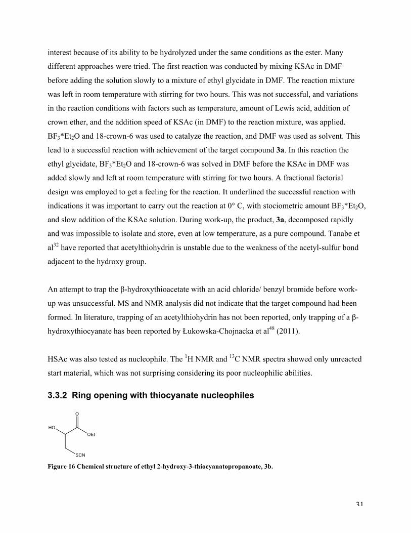
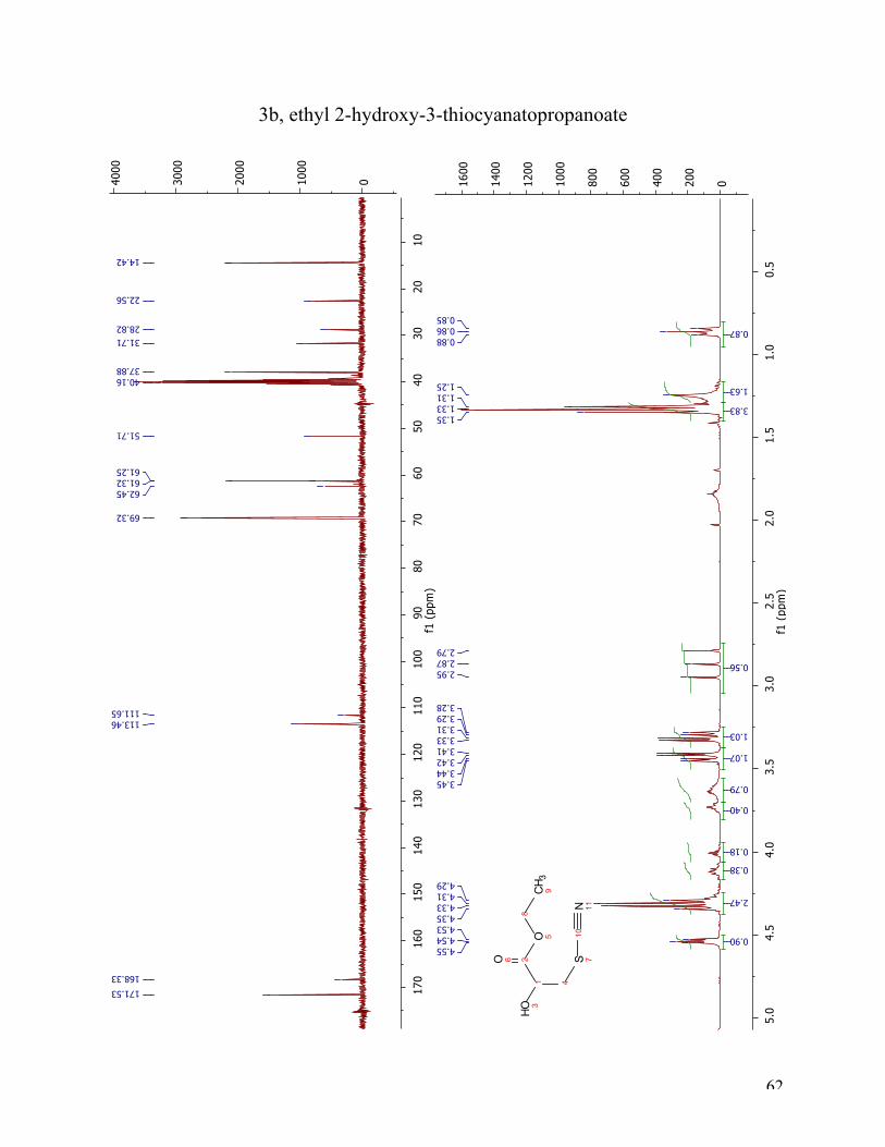
Figure 16 Chemical structure of ethyl 2-hydroxy-3-thiocyanatopropanoate, 3b.
32
Two thiocyanate nucleophiles were used; KSCN and TMSNCS. It was executed by dissolving
KSCN in DMF before adding in to a solution of ethyl glycidate and 18-crown-6 in DMF. The 1H
NMR and 13C NMR and MS analysis showed no conversion of the epoxide to the ethyl 2-
hydroxy-3-thiocyanatopropanoate.
Tanabe et al32 had reported a mild, effective and regioselective ring opening of oxiranes using
TBAF as catalyst. The article gave a general procedure for ring opening suggesting that the
TBAF and TMSNCS should be added successively to a stirred solution of the epoxide in DMF at
room temperature, before heating the reaction mixture to 50°C for three hours. Additionally, for
methyl 3-methyloxirane-2-carboxylate as substrate more specific reaction conditions were
reported asking for benzene as solvent and increased reaction time (24 hours). Since ethyl
glycidate is very similar to methyl 3-methyloxirane-2-carboxylate both approaches were tested.
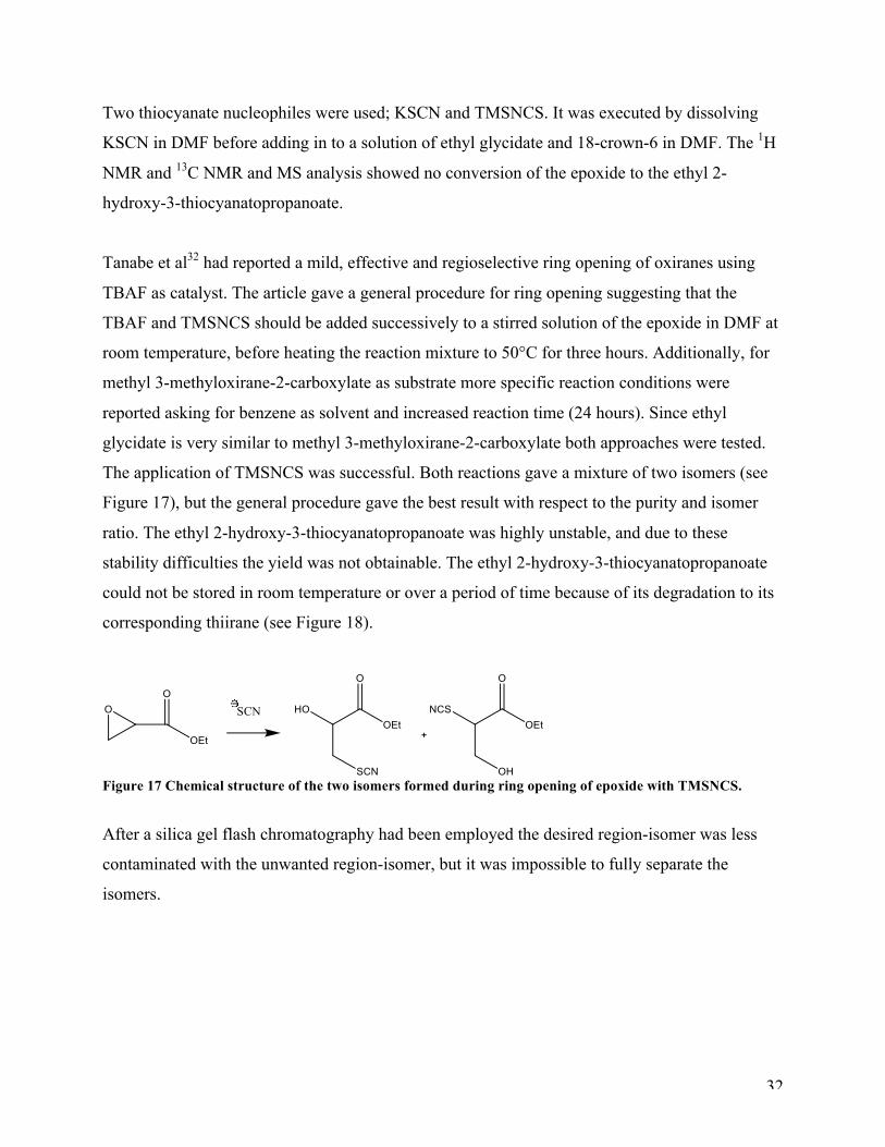
The application of TMSNCS was successful. Both reactions gave a mixture of two isomers (see
Figure 17), but the general procedure gave the best result with respect to the purity and isomer
ratio. The ethyl 2-hydroxy-3-thiocyanatopropanoate was highly unstable, and due to these
stability difficulties the yield was not obtainable. The ethyl 2-hydroxy-3-thiocyanatopropanoate
could not be stored in room temperature or over a period of time because of its degradation to its
corresponding thiirane (see Figure 18).
Figure 17 Chemical structure of the two isomers formed during ring opening of epoxide with TMSNCS.
After a silica gel flash chromatography had been employed the desired region-isomer was less
contaminated with the unwanted region-isomer, but it was impossible to fully separate the
isomers.
33
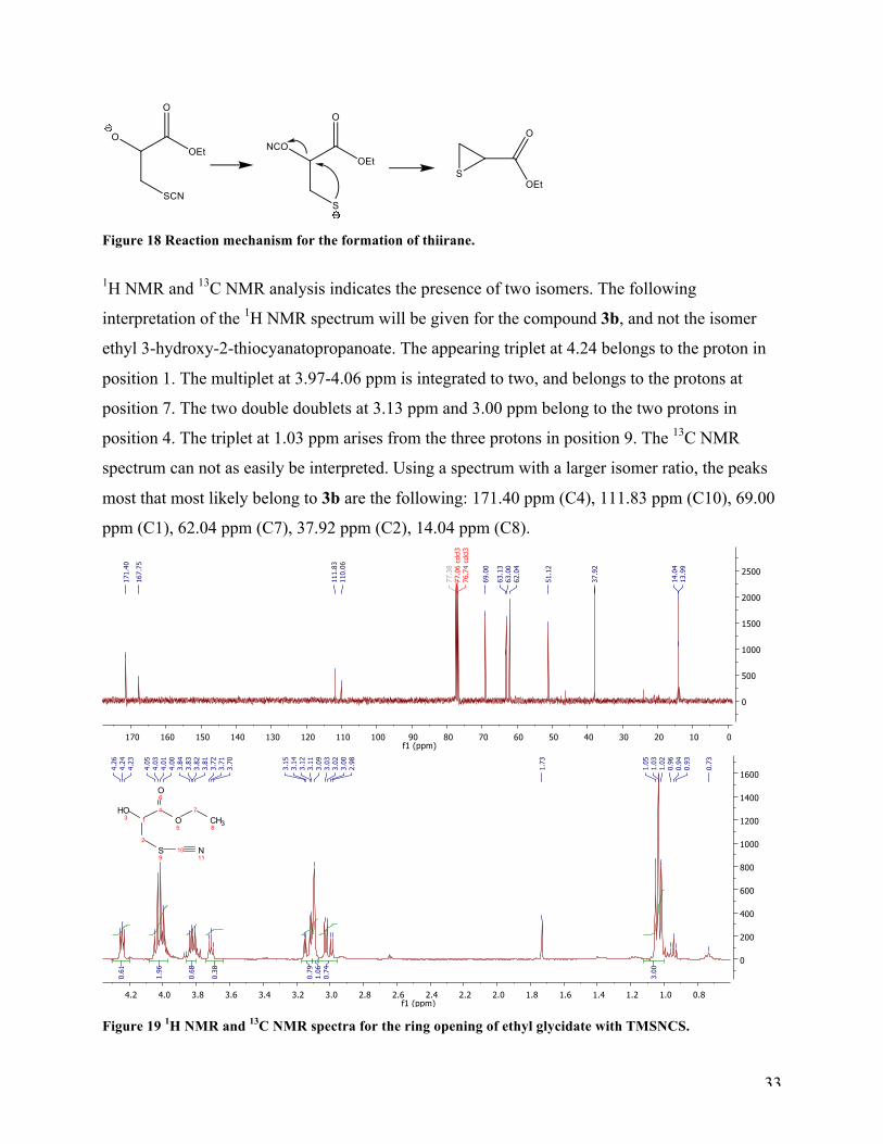
Figure 18 Reaction mechanism for the formation of thiirane.
1H NMR and 13C NMR analysis indicates the presence of two isomers. The following
interpretation of the 1H NMR spectrum will be given for the compound 3b, and not the isomer
ethyl 3-hydroxy-2-thiocyanatopropanoate. The appearing triplet at 4.24 belongs to the proton in
position 1. The multiplet at 3.97-4.06 ppm is integrated to two, and belongs to the protons at
position 7. The two double doublets at 3.13 ppm and 3.00 ppm belong to the two protons in
position 4. The triplet at 1.03 ppm arises from the three protons in position 9. The 13C NMR
spectrum can not as easily be interpreted. Using a spectrum with a larger isomer ratio, the peaks
most that most likely belong to 3b are the following: 171.40 ppm (C4), 111.83 ppm (C10), 69.00
ppm (C1), 62.04 ppm (C7), 37.92 ppm (C2), 14.04 ppm (C8).
Figure 19 1H NMR and 13C NMR spectra for the ring opening of ethyl glycidate with TMSNCS.
34
Two different approaches of the nuclophile were possible: -NCS or –SCN. To determine which of
these two nucleophiles had reacted, the 13C NMR spectrum was crucial. S-CN gives a signal at
approximately 112 ppm, while N=C=S gives signal a broad signal at 130 ppm49. The signals from
the 13C NMR spectrum for the ring opening of ethyl glycidate with TMSNCS are at 111.83 ppm,
110.06 ppm. This indicates clearly that the wanted S-CN product was obtained.
3.3.3 Different strategy
A different strategy that was utilized was to make the ethyl ester of glycolic acid and then make
the ether with benzyl bromide. However, the Williamson ether synthesis of the glycolic ester and
benzyl bromide yielded benzyl ethyl ether, and the strategy was therefore not pursued further.
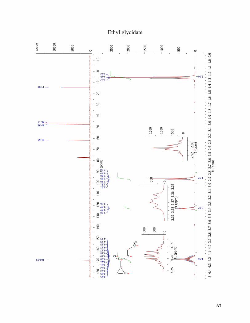
3.3.4 Starting material; ethyl glycidate
The starting material ethyl glycidate was made by reacting serine with KBr/HBr and NaNO2
using water as solvent. The crude oil from the first step was reacted with base in absolute ethanol
giving the epoxide salt. The epoxide salt was reacted with EtBr in DCM to give the ethyl
glycidate. First time the reaction was run the results were according to literature. Second time
around however, the end result was contaminated with a byproduct, in a 2:1 relationship, and
suspected formation of polymers since the yield was very low. The distillation gave no more than
approx. 4 grams, and it should have yielded 18 grams. The reason for the differences in yield and
purity has no obvious explanation.

35
36
4 Future Outlook
4.1 Reductive amination In an attempt to improve yields in this reaction, the use of not only catalytic amount of HCl but
stociometric amount of glacial acetic acid could be interesting approach as it has been
described.18 Molecular sieves (3 Å) 22 or azeotropic removal of water should also be considered
as a mean to drive the equilibrium in the imine formation in the desired direction.
To utilize the methyl ester of cysteine in the reductive amination with NaBH3CN would be an
interesting approach to investigate the suspicion of the formation of the zwitter ion causing the
low yields in this reaction.
Lowering of the pH with HCl to the isoelectric point upon the completion of the reduction has
been reported to cause the product to precipitate in methanol and should be considered as a way
to isolate the product18b.
4.2 Sulfonylation of amino acids The next step in the sulfonation of amino acids would be to remove the acetamidomethyl-
protection group on cysteine in product 2a with MeSiCl3, Ph2SO, TFA at 4°C for 30 minutes.
Other protection groups such as S-CN or S-Ac would be interesting to test, given that they would
be easier to detach. Another interesting suggestion is by reacting the appropriate sulfonyl chloride
with serine (as done) and then perform a Mitsunobu on the alcohol with a sulfur nucleophile with
an appropriate protection and then cleave the protection group to generate the target compound.
4.3 Ring opening of epoxides with thio nucleophiles The last step in the synthesis of the target ethers is performing a Mitsunobu with an alcohol, such
as 3-phenyl propan-1-ol, 2-phenylethanol and phenylmethanol, on the betahydroxythiocyanate
alcohol after the ring opening with a thio nuclophile. This must be preformed right after the ring

37
opening due to the short lifetime of the beta-hydroxythiocyanate. After the Mitsunobu reaction,
cleavage of the S-CN protection group is desirable.
38
5 Conclusion
In the reductive amination with NaBH3CN three target amines (1a-1c) was obtained. The yields
were very low; less than 5 % except for one reaction with 11 %. The solubility was poor which
can pose a challenge in the testing for inhibitor activity. 1a and 1b can be tested for inhibitory
activity without further purification; 1c needs to be purified before testing can be conducted.
Two sulfonamide compounds, protected 2a and 2b, were prepared. Sulfonation of serine with
para-toluene sulfonyl chloride gave 2b in good yields (85%). Acetamidomethyl-protected
cysteine gave the protected target compound 2a in acceptable yield (30%). Even though it has not
been deprotected, it is interesting to test the compound for inhibitor activity. 2b will also be
tested for inhibitor activity.
The ring opening of ethyl glycidate with KSAc and TMSNCS was successful, and gave the two
compounds 3a and 3b. Isolation of the products was possible, but due to their instability it was
not possible to obtain yields or store 3a and 3b over time. These two compounds were difficult to
work with, and in order to reach the ultimate goal of synthesizing 2-substituted-3-
mercaptopropanoic acids ethers there are one step left. The Mitsunobu reaction should be
performed in order to obtain the target ethers.
Three amines and two sulfonamides were prepared for VIM-2 and VIM-7 inhibitor testing.

39
40
6 Experimental section
The solvents and reagents were purchased from commercial suppliers and used without further
purification (except from para-toluene sulfonyl chloride). The reactions monitored by TLC was
on run on 60 F254 silica gel plates and visualized with UV light and staines. Column
chromatography was performed on silica gel (35-70 mesh) purchased from Merck. The IR
spectra were recorded on a Varian 7000e FT-IR spectrometer. Mass spectra were recorded on a
LTQ Orbitrap XL in a positive and negative Electron Spray Ionization mode. The samples were
run in methanol with some basic water in it. The 1H NMR and 13C NMR were recorded on a
Varian Mercury 400 MHz at room temperature. Significant 1H NMR data are reported in the
following order: multiplicity (s, singlet; d, dublet; dd, double dublet; t, triplet; m, multiplet) and
number of protons.
6.1 Reductive amination
6.1.1 General procedure: The aldehyde (1.5 eq) was added to a suspension of cysteine (1.0 eq) and NaBH3CN (1.0 eq) in
methanol (5 ml per 2 mmol) at room temperature and stirred for 22-26 hours. A white precipitate
formed and was filtered of and washed with methanol. During the reaction, the evolving gas was
bubbled through a mixture of 6 ml household chlorine bleach (a solution of approximately 3-6 %
sodium hypochlorite, NaClO) and 5 ml 10 % NaOH.
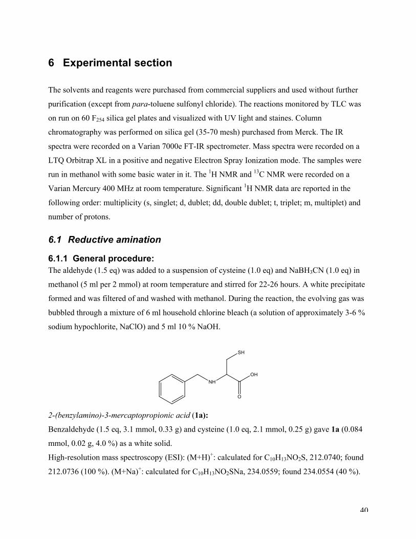
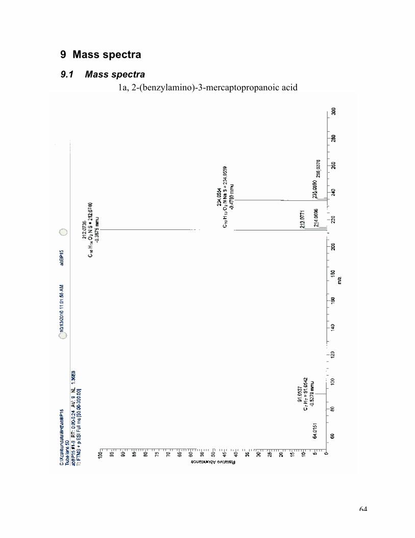
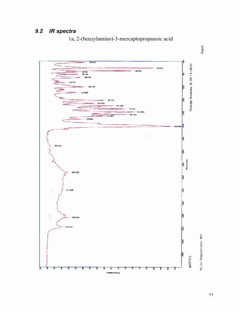
2-(benzylamino)-3-mercaptopropionic acid (1a):
Benzaldehyde (1.5 eq, 3.1 mmol, 0.33 g) and cysteine (1.0 eq, 2.1 mmol, 0.25 g) gave 1a (0.084
mmol, 0.02 g, 4.0 %) as a white solid.
High-resolution mass spectroscopy (ESI): (M+H)+: calculated for C10H13NO2S, 212.0740; found
212.0736 (100 %). (M+Na)+: calculated for C10H13NO2SNa, 234.0559; found 234.0554 (40 %).
41
1H NMR (D2O with NaOH, 400 MHz): δ 7.46 – 7.01 (m, 5H), 3.62 (d, J = 12.7 Hz, 1H), 3.47 (d,
J = 12.7 Hz, 1H), 2.90 (dd, J = 8.3, 5.0 Hz, 1H), 2.64 (dd, J = 12.6, 5.0 Hz, 1H), 2.45 (dd, J =
12.7, 8.4 Hz, 1H). 13C NMR (D2O with NaOH, 400 MHz): δ 181.7, 128.6, 128.5, 127.1, 125.8, 67.8, 51.2, 28.4.
IR: 3027 cm-1, 1606 cm-1
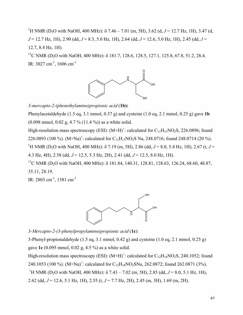
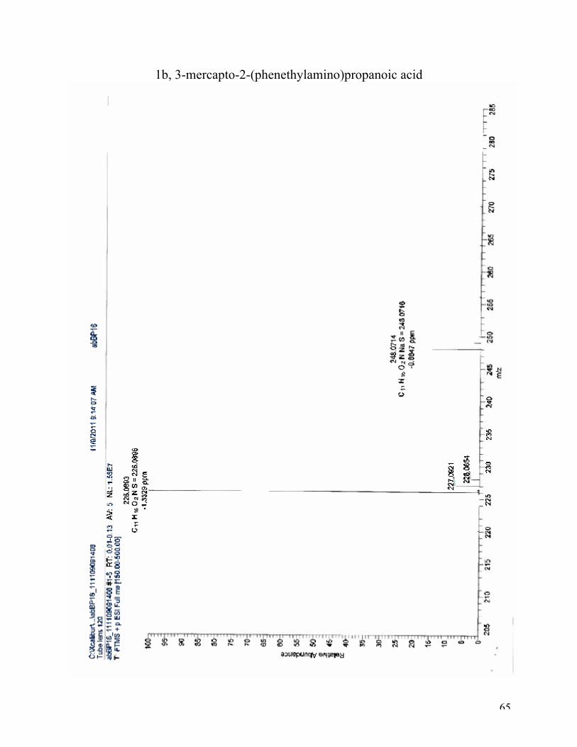
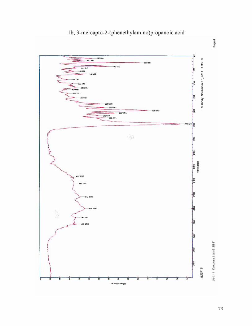
3-mercapto-2-(phenethylamino)propionic acid (1b):
Phenylacetaldehyde (1.5 eq, 3.1 mmol, 0.37 g) and cysteine (1.0 eq, 2.1 mmol, 0.25 g) gave 1b
(0.098 mmol, 0.02 g, 4.7 % (11.4 %)) as a white solid.
High-resolution mass spectroscopy (ESI): (M+H)+: calculated for C11H15NO2S, 226.0896; found
226.0893 (100 %). (M+Na)+: calculated for C11H15NO2S Na, 248.0716; found 248.0714 (20 %). 1H NMR (D2O with NaOH, 400 MHz): δ 7.19 (m, 5H), 2.86 (dd, J = 8.0, 5.4 Hz, 1H), 2.67 (t, J =
4.3 Hz, 4H), 2.58 (dd, J = 12.5, 5.3 Hz, 2H), 2.41 (dd, J = 12.5, 8.0 Hz, 1H). 13C NMR (D2O with NaOH, 400 MHz): δ 181.84, 140.31, 128.81, 128.63, 126.24, 68.60, 48.87,
35.11, 28.19.
IR: 2803 cm-1, 1581 cm-1
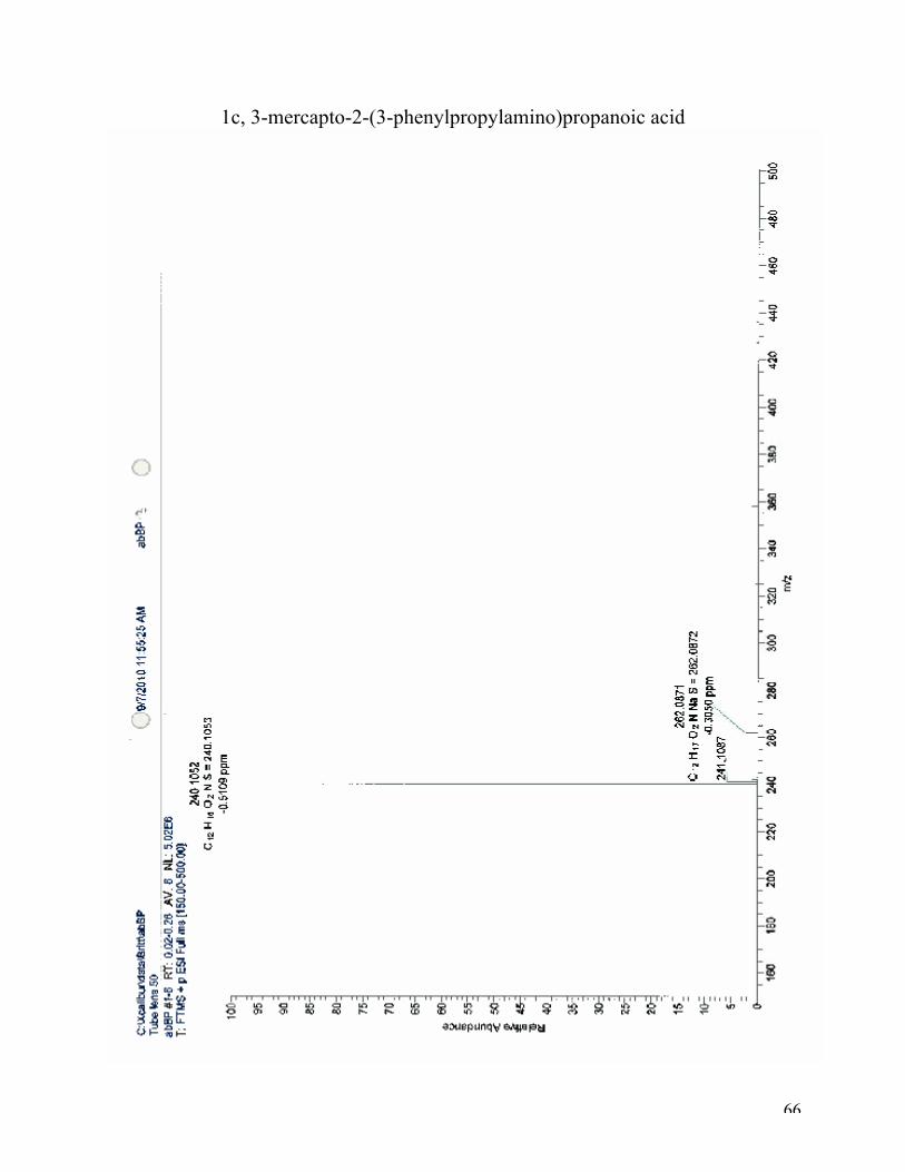
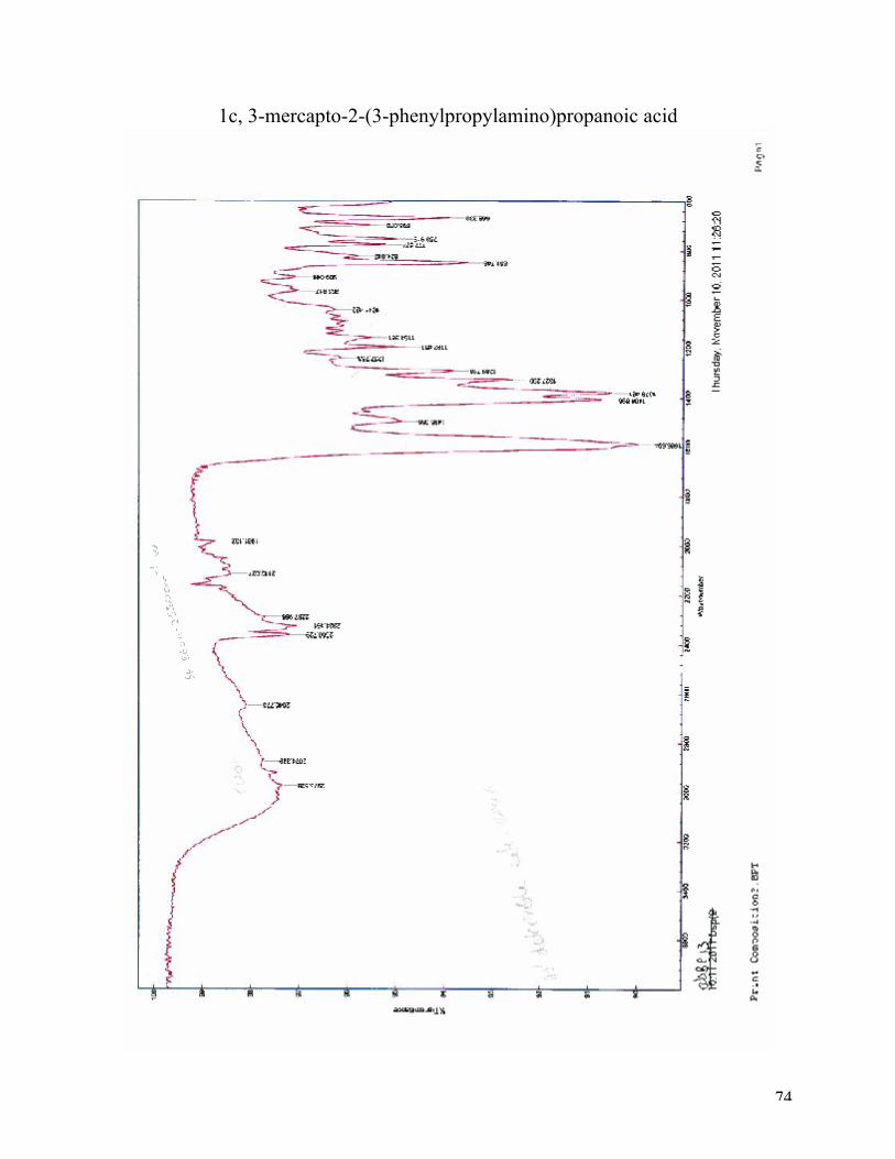
3-Mercapto-2-(3-phenylpropylamino)propionic acid (1c):
3-Phenyl-propionaldehyde (1.5 eq, 3.1 mmol, 0.42 g) and cysteine (1.0 eq, 2.1 mmol, 0.25 g)
gave 1c (0.095 mmol, 0.02 g, 4.5 %) as a white solid.
High-resolution mass spectroscopy (ESI): (M+H)+: calculated for C12H18NO2S, 240.1052; found
240.1053 (100 %). (M+Na)+: calculated for C12H18NO2SNa, 262.0872; found 262.0871 (3%).
1H NMR (D2O with NaOH, 400 MHz): δ 7.43 – 7.02 (m, 5H), 2.85 (dd, J = 8.0, 5.1 Hz, 1H),
2.62 (dd, J = 12.6, 5.1 Hz, 1H), 2.55 (t, J = 7.7 Hz, 2H), 2.45 (m, 3H), 1.69 (m, 2H).
42
13C NMR (D2O with NaOH, 400 MHz): δ 182.28, 177.15, 133.69, 128.56, 128.51, 125.78, 68.53,
60.29, 46.89, 46.87, 46.78, 32.87, 31.92, 28.27.
IR: 2973 cm-1, 1587 cm-1
6.2 Sulfonation of amino acids
6.2.1 General procedure:
The sulfonyl chloride (1 eq) was added to a solution of the amino acid (1.2 eq) in aqueous K2CO3
(2-3 eq). Under vigorous stirring, the mixture was heated to 70° C for 30 min, cooled in an
icebath, and acidified (HCl) to pH 2.5 by dropwise addition of concentrated HCl under stirring.
The precipitate was collected by filtration and washed with a minimum of cold water.
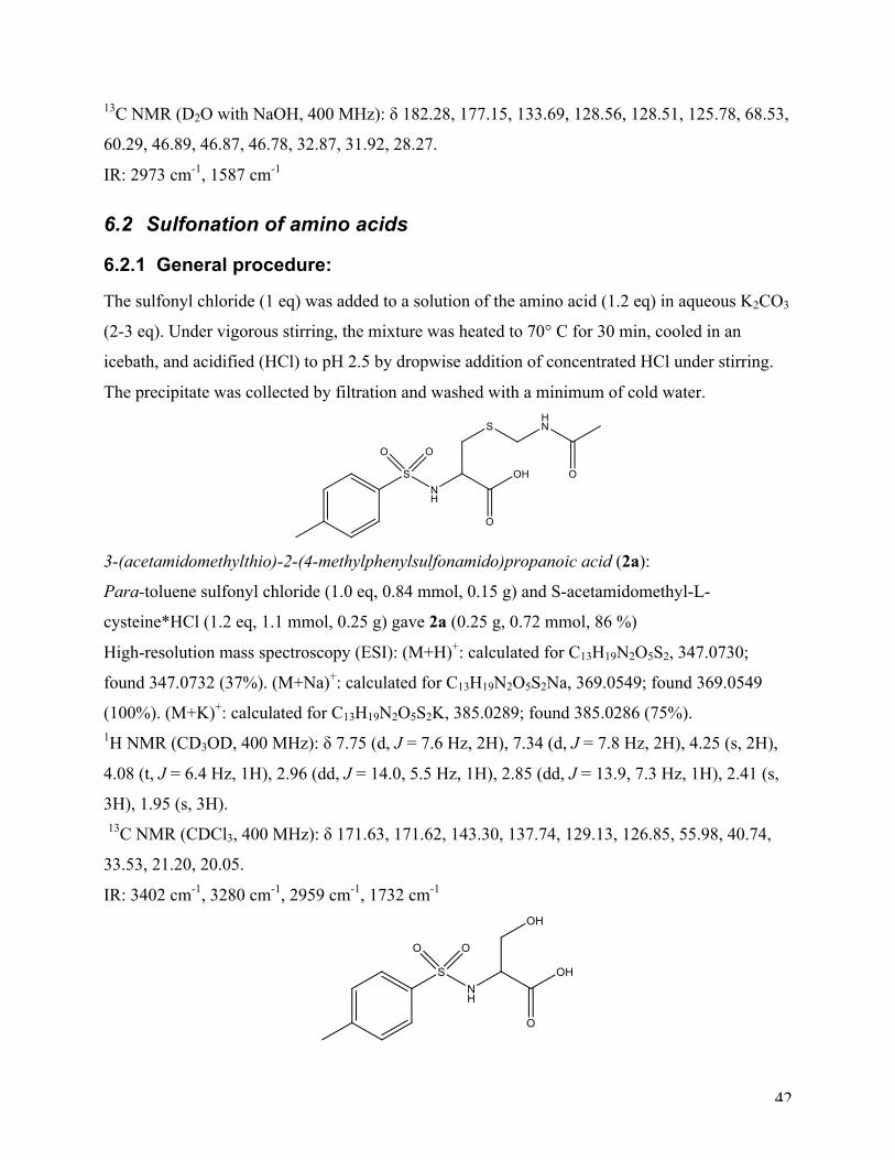
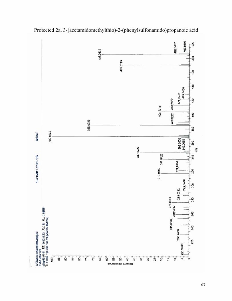
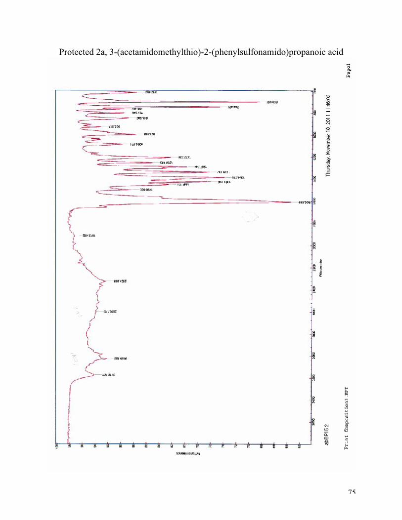
3-(acetamidomethylthio)-2-(4-methylphenylsulfonamido)propanoic acid (2a):
Para-toluene sulfonyl chloride (1.0 eq, 0.84 mmol, 0.15 g) and S-acetamidomethyl-L-
cysteine*HCl (1.2 eq, 1.1 mmol, 0.25 g) gave 2a (0.25 g, 0.72 mmol, 86 %)
High-resolution mass spectroscopy (ESI): (M+H)+: calculated for C13H19N2O5S2, 347.0730;
found 347.0732 (37%). (M+Na)+: calculated for C13H19N2O5S2Na, 369.0549; found 369.0549
(100%). (M+K)+: calculated for C13H19N2O5S2K, 385.0289; found 385.0286 (75%). 1H NMR (CD3OD, 400 MHz): δ 7.75 (d, J = 7.6 Hz, 2H), 7.34 (d, J = 7.8 Hz, 2H), 4.25 (s, 2H),
4.08 (t, J = 6.4 Hz, 1H), 2.96 (dd, J = 14.0, 5.5 Hz, 1H), 2.85 (dd, J = 13.9, 7.3 Hz, 1H), 2.41 (s,
3H), 1.95 (s, 3H). 13C NMR (CDCl3, 400 MHz): δ 171.63, 171.62, 143.30, 137.74, 129.13, 126.85, 55.98, 40.74,
33.53, 21.20, 20.05.
IR: 3402 cm-1, 3280 cm-1, 2959 cm-1, 1732 cm-1
43
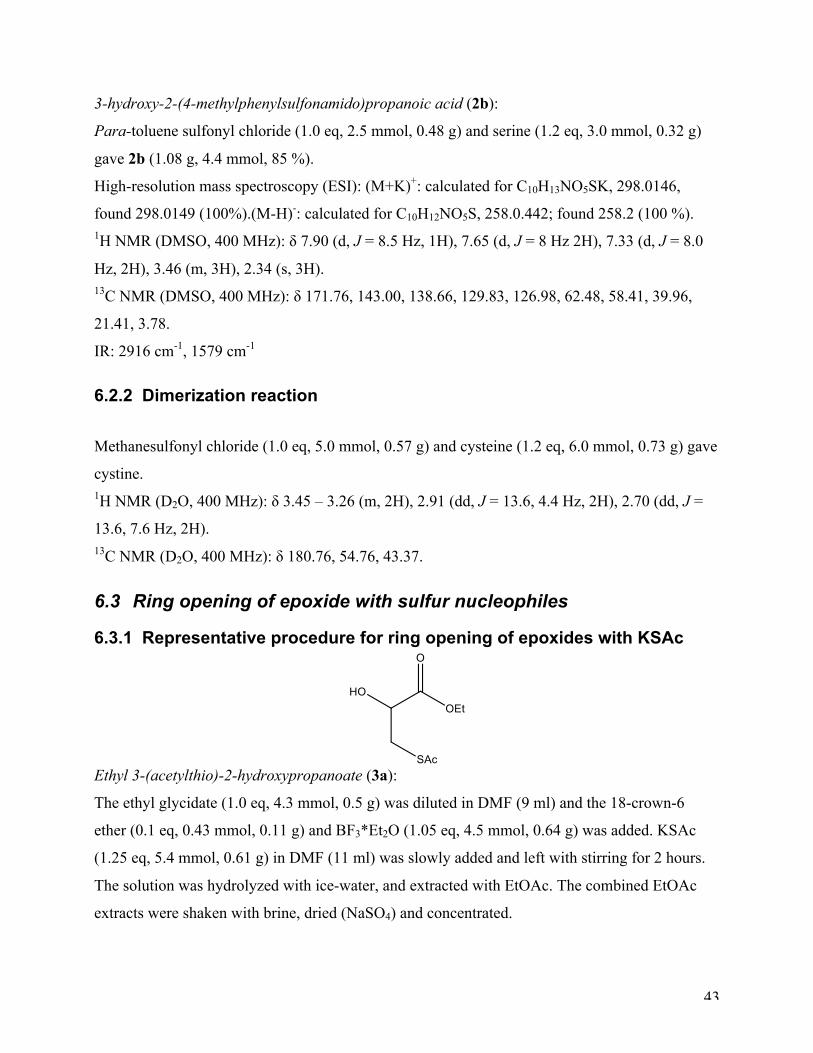
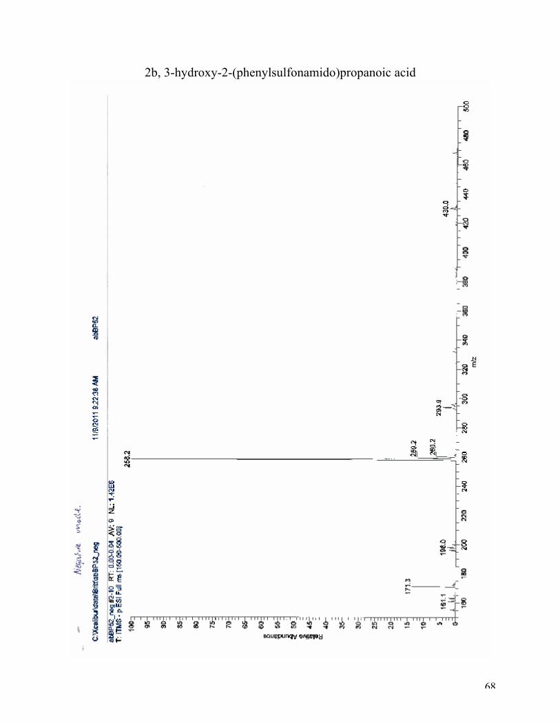
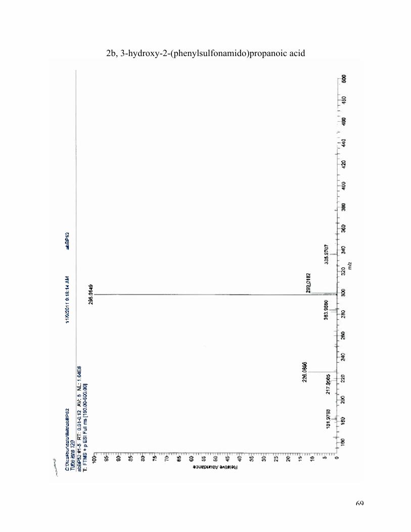
3-hydroxy-2-(4-methylphenylsulfonamido)propanoic acid (2b):
Para-toluene sulfonyl chloride (1.0 eq, 2.5 mmol, 0.48 g) and serine (1.2 eq, 3.0 mmol, 0.32 g)
gave 2b (1.08 g, 4.4 mmol, 85 %).
High-resolution mass spectroscopy (ESI): (M+K)+: calculated for C10H13NO5SK, 298.0146,
found 298.0149 (100%).(M-H)-: calculated for C10H12NO5S, 258.0.442; found 258.2 (100 %). 1H NMR (DMSO, 400 MHz): δ 7.90 (d, J = 8.5 Hz, 1H), 7.65 (d, J = 8 Hz 2H), 7.33 (d, J = 8.0
Hz, 2H), 3.46 (m, 3H), 2.34 (s, 3H). 13C NMR (DMSO, 400 MHz): δ 171.76, 143.00, 138.66, 129.83, 126.98, 62.48, 58.41, 39.96,
21.41, 3.78.
IR: 2916 cm-1, 1579 cm-1
6.2.2 Dimerization reaction
Methanesulfonyl chloride (1.0 eq, 5.0 mmol, 0.57 g) and cysteine (1.2 eq, 6.0 mmol, 0.73 g) gave
cystine. 1H NMR (D2O, 400 MHz): δ 3.45 – 3.26 (m, 2H), 2.91 (dd, J = 13.6, 4.4 Hz, 2H), 2.70 (dd, J =
13.6, 7.6 Hz, 2H). 13C NMR (D2O, 400 MHz): δ 180.76, 54.76, 43.37.
6.3 Ring opening of epoxide with sulfur nucleophiles
6.3.1 Representative procedure for ring opening of epoxides with KSAc
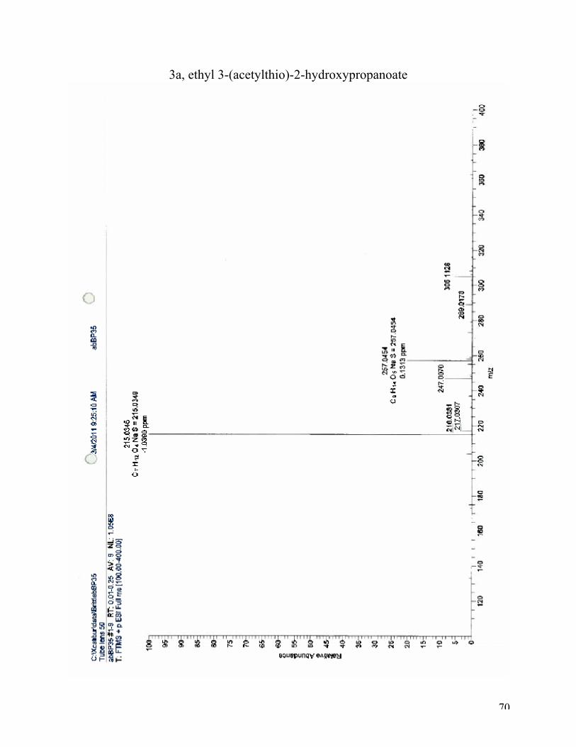
Ethyl 3-(acetylthio)-2-hydroxypropanoate (3a):
The ethyl glycidate (1.0 eq, 4.3 mmol, 0.5 g) was diluted in DMF (9 ml) and the 18-crown-6
ether (0.1 eq, 0.43 mmol, 0.11 g) and BF3*Et2O (1.05 eq, 4.5 mmol, 0.64 g) was added. KSAc
(1.25 eq, 5.4 mmol, 0.61 g) in DMF (11 ml) was slowly added and left with stirring for 2 hours.
The solution was hydrolyzed with ice-water, and extracted with EtOAc. The combined EtOAc
extracts were shaken with brine, dried (NaSO4) and concentrated.
44
High-resolution mass spectroscopy (ESI): (M+Na)+: calculated for C7H12O4SNa, 215.0349: found
215.0346. 1H NMR (CDCl3, 400 MHz): δ 4.34 (dd, J = 6.3, 4.9 Hz, 1H), 4.17-4.27 (m, 4H), 3.97 – 3.81 (m,
1H), 3.37 (dd, J = 13.7, 4.7 Hz, 1H), 3.24 (dd, J = 13.8, 6.5 Hz, 1H), 2.37 (d, J = 4.2 Hz, 2H),
2.35 (s, 3H), 2.13 (s, 1H), 1.39 – 1.16 (m, 14H). 13C NMR (CDCl3, 400 MHz): δ 194.19, 172.04, 170.38, 162.17, 70.46, 69.24, 62.05, 60.98,
59.79, 47.83, 46.80, 45.69, 35.95, 32.77, 30.81, 29.91, 29.29, 20.43, 19.92, 13.74.
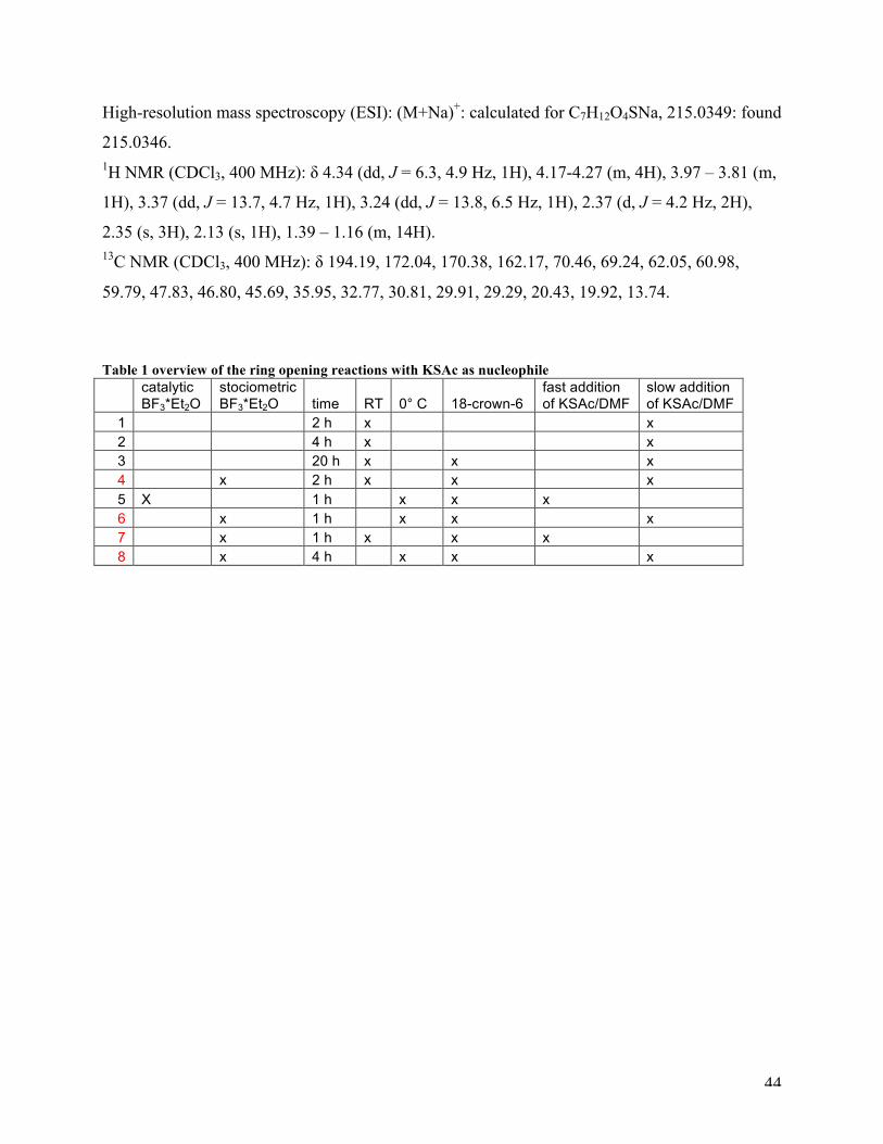
Table 1 overview of the ring opening reactions with KSAc as nucleophile
catalytic BF3*Et2O
stociometric BF3*Et2O time RT 0° C 18-crown-6
fast addition of KSAc/DMF
slow addition of KSAc/DMF
1 2 h x x 2 4 h x x 3 20 h x x x 4 x 2 h x x x 5 X 1 h x x x 6 x 1 h x x x 7 x 1 h x x x 8 x 4 h x x x
45
6.3.2 Ring opening of epoxides with thiocyanate
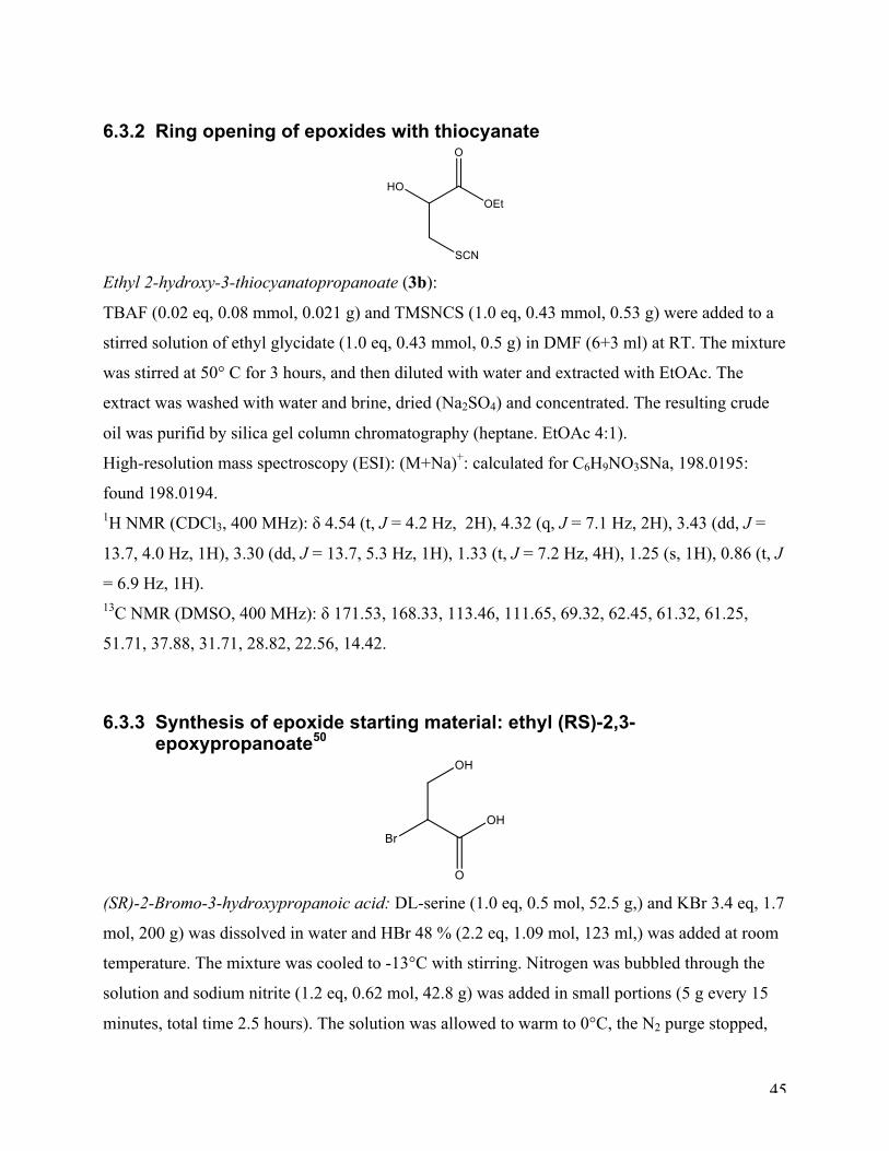
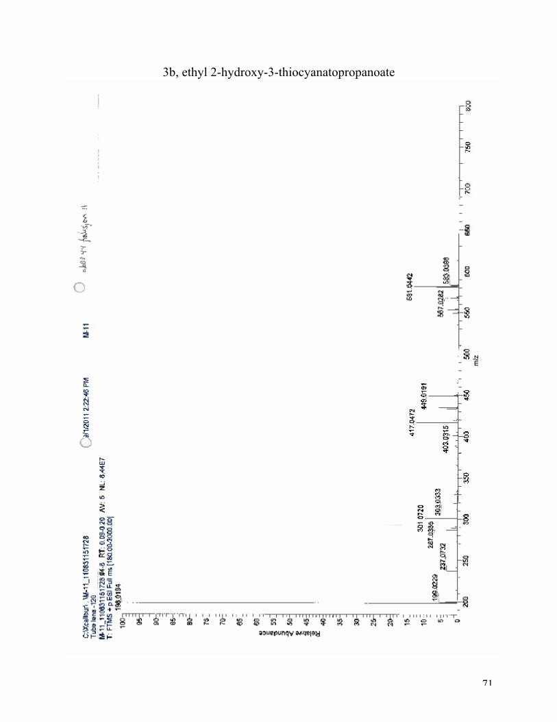
Ethyl 2-hydroxy-3-thiocyanatopropanoate (3b):
TBAF (0.02 eq, 0.08 mmol, 0.021 g) and TMSNCS (1.0 eq, 0.43 mmol, 0.53 g) were added to a
stirred solution of ethyl glycidate (1.0 eq, 0.43 mmol, 0.5 g) in DMF (6+3 ml) at RT. The mixture
was stirred at 50° C for 3 hours, and then diluted with water and extracted with EtOAc. The
extract was washed with water and brine, dried (Na2SO4) and concentrated. The resulting crude
oil was purifid by silica gel column chromatography (heptane. EtOAc 4:1).
High-resolution mass spectroscopy (ESI): (M+Na)+: calculated for C6H9NO3SNa, 198.0195:
found 198.0194. 1H NMR (CDCl3, 400 MHz): δ 4.54 (t, J = 4.2 Hz, 2H), 4.32 (q, J = 7.1 Hz, 2H), 3.43 (dd, J =
13.7, 4.0 Hz, 1H), 3.30 (dd, J = 13.7, 5.3 Hz, 1H), 1.33 (t, J = 7.2 Hz, 4H), 1.25 (s, 1H), 0.86 (t, J
= 6.9 Hz, 1H). 13C NMR (DMSO, 400 MHz): δ 171.53, 168.33, 113.46, 111.65, 69.32, 62.45, 61.32, 61.25,
51.71, 37.88, 31.71, 28.82, 22.56, 14.42.
6.3.3 Synthesis of epoxide starting material: ethyl (RS)-2,3-epoxypropanoate50
(SR)-2-Bromo-3-hydroxypropanoic acid: DL-serine (1.0 eq, 0.5 mol, 52.5 g,) and KBr 3.4 eq, 1.7
mol, 200 g) was dissolved in water and HBr 48 % (2.2 eq, 1.09 mol, 123 ml,) was added at room
temperature. The mixture was cooled to -13°C with stirring. Nitrogen was bubbled through the
solution and sodium nitrite (1.2 eq, 0.62 mol, 42.8 g) was added in small portions (5 g every 15
minutes, total time 2.5 hours). The solution was allowed to warm to 0°C, the N2 purge stopped,
46
and left with stirring for 6 hours. Excess nitrogen oxides were removed by bubbling N2 through
the solution for 1 hour. The pale green solution was extracted with 6*300 ml ether, dried over
MgSO4 and concentrated by rotary evaporation to give pale green oil (75.0 g, 89 %).
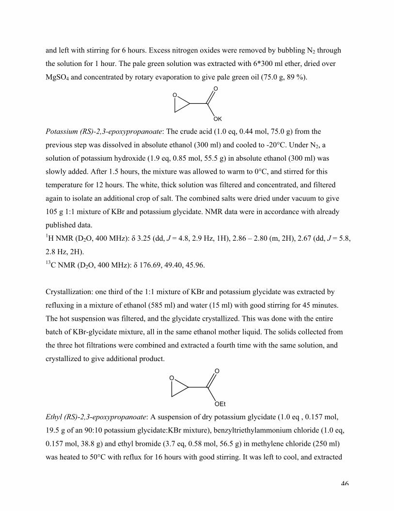
Potassium (RS)-2,3-epoxypropanoate: The crude acid (1.0 eq, 0.44 mol, 75.0 g) from the
previous step was dissolved in absolute ethanol (300 ml) and cooled to -20°C. Under N2, a
solution of potassium hydroxide (1.9 eq, 0.85 mol, 55.5 g) in absolute ethanol (300 ml) was
slowly added. After 1.5 hours, the mixture was allowed to warm to 0°C, and stirred for this
temperature for 12 hours. The white, thick solution was filtered and concentrated, and filtered
again to isolate an additional crop of salt. The combined salts were dried under vacuum to give
105 g 1:1 mixture of KBr and potassium glycidate. NMR data were in accordance with already
published data. 1H NMR (D2O, 400 MHz): δ 3.25 (dd, J = 4.8, 2.9 Hz, 1H), 2.86 – 2.80 (m, 2H), 2.67 (dd, J = 5.8,
2.8 Hz, 2H). 13C NMR (D2O, 400 MHz): δ 176.69, 49.40, 45.96.
Crystallization: one third of the 1:1 mixture of KBr and potassium glycidate was extracted by
refluxing in a mixture of ethanol (585 ml) and water (15 ml) with good stirring for 45 minutes.
The hot suspension was filtered, and the glycidate crystallized. This was done with the entire
batch of KBr-glycidate mixture, all in the same ethanol mother liquid. The solids collected from
the three hot filtrations were combined and extracted a fourth time with the same solution, and
crystallized to give additional product.
Ethyl (RS)-2,3-epoxypropanoate: A suspension of dry potassium glycidate (1.0 eq , 0.157 mol,
19.5 g of an 90:10 potassium glycidate:KBr mixture), benzyltriethylammonium chloride (1.0 eq,
0.157 mol, 38.8 g) and ethyl bromide (3.7 eq, 0.58 mol, 56.5 g) in methylene chloride (250 ml)
was heated to 50°C with reflux for 16 hours with good stirring. It was left to cool, and extracted
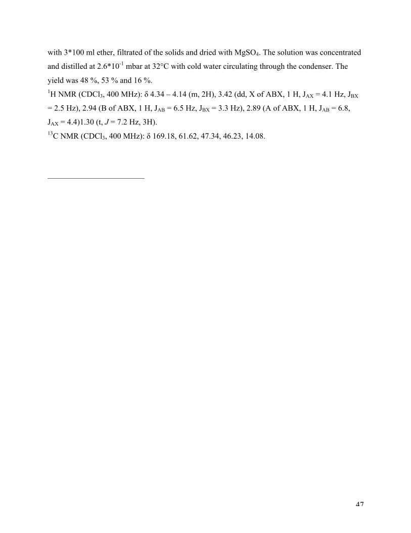
47
with 3*100 ml ether, filtrated of the solids and dried with MgSO4. The solution was concentrated
and distilled at 2.6*10-1 mbar at 32°C with cold water circulating through the condenser. The
yield was 48 %, 53 % and 16 %. 1H NMR (CDCl3, 400 MHz): δ 4.34 – 4.14 (m, 2H), 3.42 (dd, X of ABX, 1 H, JAX = 4.1 Hz, JBX
= 2.5 Hz), 2.94 (B of ABX, 1 H, JAB = 6.5 Hz, JBX = 3.3 Hz), 2.89 (A of ABX, 1 H, JAB = 6.8,
JAX = 4.4)1.30 (t, J = 7.2 Hz, 3H). 13C NMR (CDCl3, 400 MHz): δ 169.18, 61.62, 47.34, 46.23, 14.08.

48
49
7 References 1Sauvage, E.; Powell, A. J.; Heilemann, J.; Josephine, H. R.; Charlier, P.: Davies, C.;
Pratt, R. F. J. Mol. Biol. 2008, 381, 383-393 2 Oelschlaeger, P.; Ai, N, DuPrez, K. T.; Welsh, W. J.; Toney, J. H. J. Med. Chem., 2010, Vol. 53,
No.8, 3013-3027 3 Garau, G.; Garcia-Saez, I.; Bebrone, C.; Anne, C.; Mercuri, P.; Galleni, M.; Frere, J-M.;
Dideberg, O. Antimicrob. Agents. Chemother. 2004, 48, 2347-2349 4 Carfi, A.; Pares, S.; Duee, E.; Galleni, M.; Duez, C.; Frere, J. M.; Dideberg, O. E M B O. J.
1995, 14, 4914-4921 5 Ambler, R. P. Philos. Trans. R. Soc., B 1980, 289, 321-331 6 Yamaguchi, Y.; Jin, W.; Matsunaga, K.; Ikemizu, S.; Yamagata, Y.; Wachino, J.;
Shibata, N.; Arakawa, Y.; Kurosaki, H. J. Med. Chem. 2007, 50, 6647–6653 7 Jin, W.; Arakawa, Y.; Yasuzawa, H.; Taki, T.; Hashiguchi, R.; Mitsutani, K.; Shoga, A;
Yamaguchi, Y.; Kurosaki, H.; Shibata, N.; Ohta, M.; Goto, M. J. Biol. Chem. 2005, 280, 20824-
20832 8 Wang, Z.; Fast, W.; Benkovic, S. J. Biochemistry, 1999, 38, 10013-10023 9 Borra, P. S.; Leiros, H-K. S.; Ahmad, R.; Spencer, J.; Leiros, I.; Walsh, T.; Sundsfjord, A.;
Samuelsen, Ø. J. Mol. Biol. 2011, 411, 174–189 10 Miriagou, V.; Papagiannitsis, C. C.; Tzelepi, E.; Casals, J. Bou; Legakis, N. J.; Tzouvelekis, L.
S. J. Clin.Microbiol. 2010, 48, 667-668 11 Lauretti, L.; Riccio, M. L.; Mazzariol, Annarita; Cornaglia, G.; Amicosante, G.; Fontana, R.;
Rossolini, G. M. Antimicrob. Agents Chemother. 1999, 43, 1584-1590
12 Jin, W.; Arakawa, Y.; Yasuzawa, H.; Taki, T.; Hashiguchi, R.; Mitsutani, K.; Shoga, A;
Yamaguchi, Y.; Kurosaki, H.; Shibata, N.; Ohta, M.; Goto, M. Biol. Pharm. Bull. 2004, 27,
851—856 13 Minond, D.; Saldanha, S. A.; Subramaniam, P.; Spaargaren, M.; Spicer, Fotsing, T.; Joseph R.;
Weide, T.; Fokin, V. V.; Sharpless, K. Barry; Galleni, M.; Bebrone, C.; Lassaux, P.; Hodder, P.
Bioorg. Med. Chem. 2009, 17, 5027–5037
50
14 Weide, T.; Saldanha, S. A.; Minod, D.; Spicer, T. P.; Fotsing, J. R.; Spaargaren, M.; Frere, J-
M.; Bebrone, C. Sharpless, K. B.; Hodder, P. S.; Fokin, V. V. ACS Med Chem Lett. 2010, 1,
150-154 15 (a) Baxter, E.W.; Reitz, A.B.; Organic Reactions (Hoboken, NJ, United States), 2002. 59: p.
No pp given. (b) Nguyen, K.T.; Chemmedchem, 2008, 3, 1520-1524. (c) Liu, J.; Journal of
Organic Chemistry, 2008, 73, 4045-4052. (d) Dzygiel, P.; European Journal of Organic
Chemistry, 2008, 7, 1253-1264. (e) Mindt, T.L.; R. Schibli, Journal of Organic Chemistry, 2007,
72, 10247-10250 (f) Gibbs, T.J.K.;Boomhoff, M.; Tomkinson, N.C.O. Synlett, 2007, 10, 1573-
1576. (g) Hannachi, J.-C., Journal of Organic Chemistry, 2004, 69, 2367-2373. (h) Lelais,
G.;Seebach, D. Helvetica Chimica Acta, 2003, 86, 4152-4168. (i) Verardo, G.; Canadian Journal
of Chemistry, 2002, 80, 779-788. (j) Song, Y. Tetrahedron Letters, 2000, 41, 8225-8230. (k)
Ando, A.; Shioiri, T. Journal of the Chemical Society, Chemical Communications, 1987, 9, 656-
658. (l) Quitt, P.; Hellerbach, J.; Vogler, K. 1963, 46, 327-33. 16 (a) Parker, J. S.; Bowden, S. A.; Firkin, C. R.; Moseley, J. D.; Murray, P. M.;
Welham, M. J.; Wisedale, R.; Young, M. J.; Moss, W. O. Org. Process Res. Dev.
2003, 7, 67; (b) Moseley, J. D.; Moss, W. O.; Welham, M. J. Org. Process Res. Dev.
2001, 5, 491; (c) Bomann, M. D.; Guch, I. C.; DiMare, M. J. Org. Chem. 1995, 60,
5995. (d) Sato, S.; Sakamoto, T.; Miyazawa, E.; Kikugawa, Y. Tetrahedron 2004, 60,
7899; (e) Uchiyama, S.; Inaba, Y.; Matsumoto, M.; Suzuki, G. Anal. Chem. 2009,
81, 485. (f) Nose, A.; Kudo, T. Chem. Pharm. Bull. 1980, 34, 4817. (g) Heydari, A.; Tavakol, H.;
Aslanzadeh, S.; Azarnia, J.; Ahmadi, N. Synthesis 2005,
627. (h) Brown, H. C.; Kanth, J. V. B.; Dalvi, P. V.; Zaidlewicz, M. J. Org. Chem. 1999, 64,
6263. (i) Bruckhardt, E. R.; Coleridge, B. M. Tetrahedron Lett. 2008, 49, 5152. (j) Peterson, M.
A.; Bowman, A.; Morgan, S. Synth. Commun. 2002, 32, 443. (k) Cho, B. T.; Kang, S. K.
Tetrahedron 2005, 61, 5725. 17 (a) Markey, M. D.; Fu, Y.; Kelly, T. R. Org. Lett. 2007, 9, 3255; (b) Baxter, E. W.;
Reitz, A. B. Org. React. 2002, 59, 1; (c) Lewin, G.; Schaeffer, C. Heterocycles 1998,
48, 171. (d) Park, J. D.; Kim, D. H. J. Med. Chem. 2002, 45, 911-918 18 (a) Abdel-Magid, A. F.; Carson, G. C.; Harris, B. D.; Maryanoff, C. A.; Shah, R. D. J. Org.
Chem. 1996, 61, 3849-3862 (b) Abdel-Magid, A. F.; Mehrman, S. J. Organic process research
and development 2006, 10, 971-1031
51
19 (a) Alinezhad, H.; Ardestani, E. Lett. Org. Chem. 2007, 4, 473; (b) Saxena, I.;
Borah, R.; Sarma, J. C. J. Chem. Soc., Perkin Trans. 1 2000, 503; (c) Bhattacharyya,
S. J. Org. Chem. 1995, 60, 4928; (d) Verardo, G.; Giumanini, A. G.; Strazzolini, P.;
Poiana, M. Synthesis 1993, 121; (e) Itsuno, S.; Sakurai, Y.; Ito, K. Chem. Commun.
1988, 995. 20 (a) Heydari, A.; Khaksar, S.; Esfandyari, M.; Tajbakhsh, M. Tetrahedron 2007, 63,
3363; (b) Kotsuki, H.; Yoshimura, N.; Kadota, I.; Ushio, Y.; Ochi, M. Synthesis
1990, 401; (c) Bhattacharyya, S.; Chatterjee, A.; Williamson, J. S. Synth.
Commun. 1997, 27, 4265; (d) Ranu, B. C.; Majee, A.; Sarkar, A. J. Org. Chem.
1998, 63, 370. 21 Mackie, R. K.; Smith, D. M.; Aitken, R. A. Guidebook to Organic Synthesis, 2nd Edition, pp
171 22 Borch, R. F.; Bernstein, M. D.; Durst, H. D. Journal of the American Chemical Society 1, 1971,
93, 2897 23Luo, Z.; Zeng, C.; Yang, D.; Huang, Y.; Wang, F.; Du, H.; Hu, L. Chinese Journal of Chemistry,
2009, 27, 1333-1338 24Schweinitz, A.; Steinmetzer, T.; Banke, I. J.; Arlt, M. J. E.; Stürzebecher, A.; Schuster, O.;
Geissler, A.; Giersiefen, H.; Zeslawska E.; Jacob, U.; Krüger, A.; Stürzebecher, J. The journal of
biological chemistry, 2004, 279, 33613-33622 25 Schröder, J.; Henke, A.; Wenzel, H.; Brandstetter, H.; Stammler, H. G.; Stammler, A.; Pfeiffer,
W. D.; Tschesche, H. J. Med. Chem. 2001, 44, 3231-3243 26 Li1, S.; Li, G.; Huang, H.; Xiong, F.; Liu1, C.; Tu, G. Arch Pharm Res 2008, 31, 1231-1239. 27 Pickett, S. D.; Green, D. V. S.; Hunt, D. L.; Pardoe, D. A.; Hughes, I. ACS Med. Chem. Lett.
2011, 2, 28–33 28 Effenberger, F.; Gaupp, S. Tetrahedron: Asymmetry 10, 1999, 1765-1775 29 (a) Van Tamelen, E. E. J. Am. Chem. Soc. 1951, 73, 3444 (b) Wagner-Jauregg. Atylen-
rhodanhydrin. Liebigs Ann. Chem. 1949, 561, 87-98 30 Prusinowska, N.; Gawronski, J. Synthetic Communications 1, 2009, 39: 2795–2803 31 Aghapour, G. Hatefipour, R. Synth. Commun. 2009, 39, 1698-1707 32 Tanabe, Y; Mori, K.; Yoshida, Y. J. Chem. Soc., Perkin Trans. 1, 1997, 671-676 33 Caron, M.; Sharpless, K. B. J. Org. Chem. 1985, 50, 1557-1560
52
34 (a) Olszewski-Ortar, A.; Gros, P.; Fort, Y. Tetrahedron Lett. 1997, 38, 8699-8702 (b) Chen, X.;
Wu, H.; Xu, R.; Liu, M.; Ding, J.; Su, W. Synth. Commun. 2008, 38, 1855-1865 35 Iranpoor, N.; Kohmareh, G. A. Phosphorus, Sulfur and Silicon, 1999, 152, 135-139 36 Tamure, Y.; Kawasaki, T.; Yasuda, H.; Gohda, N.; Kita, Y. J. Chem. Soc. Perkins Trans. 1
1981, 1577-1581 37 Tamami, B.; Mahdavi, H. Tetrahedron Lett. 2002, 43, 6225-6228 38 Yadav, J. S.; Reddy, B. V. S.; Srinivas Reddy, C. Tetrahedron Lett. 2004, 45, 1291-1293 39 Shargi, H.; Nasseri, M. A. Phosphorus,Sulfur Silicon Relat. Elem. 2003, 178, 1353-1359 40 Niknam, K. Phosphorus,Sulfur Silicon Relat. Elem. 2004, 179, 499-506. 41 Shargi, H.; Nejad, A. H. New J. Chem. 2004, 28, 946-951. 42 Sharghi, H.; Nasseri, M. A.; Niknam, K. J. Org. Chem. 2001, 66, 7287-7293 43 Shargi, H.; Nejad, A. H. Phosphorus,Sulfur Silicon Relat. Elem. 2004, 179, 2297-2305 44 Gao, Y.; Sharpless, K. B. J. Am. Chem. SOC. 1988, 110, 7538-7539 45 Uchida, N.; Nakajima, Y.; Oda, J.; Inouye, Y. Agric. Biol. Chem. 1979, 43, 2169-2172 46 Mizrahi, S.; Gun, J.; Kipervaser, G.; Lev, O. Anal. Chem. 2004, 76, 5399-5404 47 http://riodb01.ibase.aist.go.jp/sdbs/cgi-bin/direct_frame_top.cgi 48 Łukowska-Chojnacka, E.; Plenkiewicz, J. Synthetic Communications1, 2011 41: 1999–2006 49 Pretsch, E.; Buhlmann, P.; Affolter, C. Structure and determination of organic compounds, 3rd
edition, p.127, p.149 50 Petit, Y.; Larchevêque, M. Organic synthesis, 2004, 10, 410
53
8 Appendices
8.1 Abbreviations DCM - dichloromethane
ddd - double double dublet
DMF - dimethylformamide
Ethyl glycidate - ethyl oxirane-2-carboxylate
IMP, GIM, SPM, SIM, KHM, AIM, DIM, TMB, NDM - different types of MBLs
IR - infrared spectroscopy
MBL - metallo-β-lactamase
MgSO4 - magnesium sulfate
MS - mass spectra
Na2SO4 - sodium sulfate
Nitrocefin (- 6R,7R)-3-[(2,4-dinitro)styryl]-8-oxo-7-[(2-thienylacetyl) amino]-5-thia-1-
azabicyclo[4.2.0]oct-2-ene-2-carboxylic acid
NMR - nuclear magnetic resonance
Quinoline - N-((7-chloroquinolin-4-ylamino)alkyl)-3-mercaptopropanamide
TLC - thin layer chromatography
VIM - verona integron-borne metallo-β-lactamase

54
8.2 NMR spectra of 1a-1c
55
1a, 2-(benzylamino)-3-mercaptopropanoic acid
56
1b, 3-mercapto-2-(phenethylamino)propanoic acid
57
1c, 3-mercapto-2-(3-phenylpropylamino)propanoic acid
58
8.3 NMR spectra of protected 2a, 2b and cysteine Protected 2a, 3-(acetamidomethylthio)-2-(phenylsulfonamido)propanoic acid
59
2b, 3-hydroxy-2-(phenylsulfonamido)propanoic acid
60
Cysteine
61
8.4 NMR spectra for 3a, 3b and ethyl glycidate 3a, ethyl 3-(acetylthio)-2-hydroxypropanoate
62
3b, ethyl 2-hydroxy-3-thiocyanatopropanoate
63
Ethyl glycidate
64
9 Mass spectra
9.1 Mass spectra 1a, 2-(benzylamino)-3-mercaptopropanoic acid
65
1b, 3-mercapto-2-(phenethylamino)propanoic acid
66
1c, 3-mercapto-2-(3-phenylpropylamino)propanoic acid
67
Protected 2a, 3-(acetamidomethylthio)-2-(phenylsulfonamido)propanoic acid
68
2b, 3-hydroxy-2-(phenylsulfonamido)propanoic acid
69
2b, 3-hydroxy-2-(phenylsulfonamido)propanoic acid
70
3a, ethyl 3-(acetylthio)-2-hydroxypropanoate
71
3b, ethyl 2-hydroxy-3-thiocyanatopropanoate
72
9.2 IR spectra 1a, 2-(benzylamino)-3-mercaptopropanoic acid
73
1b, 3-mercapto-2-(phenethylamino)propanoic acid
74
1c, 3-mercapto-2-(3-phenylpropylamino)propanoic acid
75
Protected 2a, 3-(acetamidomethylthio)-2-(phenylsulfonamido)propanoic acid
76
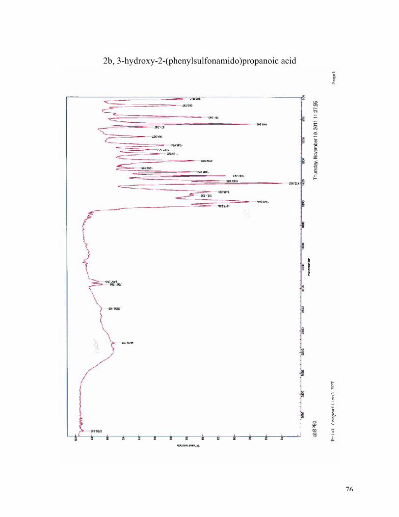
2b, 3-hydroxy-2-(phenylsulfonamido)propanoic acid