Kidney α–intercalated cells and lipocalin 2: defending...
3
Commentaries 2844 The Journal of Clinical Investigation http://www.jci.org Volume 124 Number 7 July 2014 Kidney α–intercalated cells and lipocalin 2: defending the urinary tract Yuxuan Miao 1 and Soman N. Abraham 1,2,3,4 1 Department of Molecular Genetics and Microbiology, 2 Department of Pathology, and 3 Department of Immunology, Duke University Medical Center, Durham, North Carolina, USA. 4 Program in Emerging Infectious Diseases, Duke–National University of Singapore, Singapore. A growing body of evidence indicates that the kidneys contribute substan- tially to immune defense against pathogens in the urinary tract. In this issue, Paragas et al. report that α–intercalated cells (A-ICs) within the nephron col- lecting duct sense infecting Gram-negative bacteria, resulting in simultane- ously secretion of the iron chelating protein lipocalin 2 (LCN2) and protons, which acidify the urine. A-IC–specific LCN2 and proton secretion markedly reduced the ability of infecting uropathogenic E. coli (UPEC) to grow and sustain infection. The capacity of A-ICs to sense and actively promote clear- ance of infecting bacteria in the lower urinary tract represents a novel func- tion for these specialized kidney cells, which are best known for their role in modulating acid-base homeostasis. The antimicrobial shield in the urinary tract Due to its close proximity to the gastroin- testinal tract, the urinary tract is subject to a constant barrage of bacteria, most of which are enteric in origin. To counter this microbial onslaught, the urinary tract has developed a highly effective antimi- crobial “shield” that can rapidly eliminate contaminating bacteria or prevent their growth. Both the flushing action of urine and urinary mucins are mechanical strat- egies that rapidly eliminate any contami- nating bacteria from the urinary tract (1). Additionally, various antimicrobial agents that directly kill pathogens, such as cat- helicidin (2) and RNAse7 (3), are consti- tutively secreted into urine. Periodically, pathogens overcome these antimicrobial defenses and begin to multiply in urine, a relatively rich growth medium, and induce a second wave of responses that involve the secretion of additional antimicrobial factors. Secondary responses to increased levels of bacteria in the urine appear to be triggered by immune sensory machinery in the urinary tract (4). Located at the sum- mit of the urinary tract, the kidneys are a major source of several potent antimi- crobial compounds that flow with urine and protect both upper and lower regions of the urinary tract. Kidney- secreted agents exhibit a wide range of antimicro- bial actions and include cathelicidin (2), human β-defensin-1 (HBD-1) (5), and RNAse7 (3), which all directly disrupt bacterial membranes, and Tamm-Horsfall protein (THP; also known as uromodulin) (6), which promotes bacterial aggregation and facilitates removal by the urine. In this issue, Paragas et al. have shown that α–intercalated cells (A-ICs) in the col- lecting ducts of the kidney serve as both sentinels and defenders of the urinary tract during infection (7). Specifically, they demonstrated that immune sensory TLR4 molecules on A-ICs detect the presence of uropathogenic E. coli (UPEC), and TLR4 signaling in A-ICs triggers secretion of the bacteriostatic protein lipocalin 2 (LCN2), as well as secretion of H + ions into the urine (Figure 1). Together, urine acidification and LCN2 drastically reduced the number of infecting bacteria in the urinary tract. Protective role of LCN2 against UPEC infections LCN2 is a member of the large lipocalin protein family, which has a wide range of biological functions (8). LCN2 binds to the secreted siderophore enterochelin (Ent), which UPEC and other pathogens release into the extracellular milieu to acquire essential iron (9), and then delivers Ent/Fe 3+ complexes to host cells for deg- radation, effectively abrogating bacterial iron acquisition. Pathogens are unable to grow in the absence of iron, allowing the immune system to eliminate the infection. Interestingly, Paragas et al. observed a sig- nificant elevation of LCN2 in the urine of mice with UTI compared with uninfected animals. The degree of LCN2 upregulation apparently associated with the number of infecting bacteria within the urine, and reduction of bacterial load with antibiotics resulted in decreased LCN2 production. Moreover, a similar correlation between bacterial numbers in the urine and LCN2 levels was also seen in patients with UTIs, providing direct clinical support for LCN2 production in immune defense of the uri- nary tract. Compared with WT animals, Lcn2-deficient mice were more susceptible to UTIs; however, both WT and Lcn2 –/– ani- mals were infected to similar extents with an ent mutant UPEC strain (7), which indi- cates that the suppressive actions of LCN2 on uropathogens involve iron acquisition. The observation by Paragas et al. that LCN2 is not constitutively present in the urinary tract, but rather produced in direct proportion to the bacterial numbers in urine (7), is similar to other antimicrobial agents, including pentraxin-3 and HBD-1, that are also secreted in direct proportion to the size of the bacterial threat (10). As urine is a very rich medium and can be retained for many hours in the bladder, the urinary tract can serve as a powerful incu- bator for bacterial growth. Indeed, bacterial numbers in urine can reach levels in excess of 10 8 bacteria/ml. The ability to produce LCN2 and other antibacterial agents in proportion to bacterial burden in the uri- nary tract may be adaptation by the kidney to maintain homeostasis in response to overwhelming bacterial infection. A-ICs: the cellular source of LCN2 Notably, Paragas and colleagues deter- mined that LCN2 is produced by highly specialized A-ICs located in the collecting duct of the kidney medulla. The renal col- lecting ducts are responsible for regulat- ing electrolyte and fluid balance through reabsorption and excretion processes. The collecting ducts are lined by both A-ICs Conflict of interest: The authors have declared that no conflict of interest exists. Citation for this article: J Clin Invest. 2014; 124(7):2844–2846. doi:10.1172/JCI76630.
Transcript of Kidney α–intercalated cells and lipocalin 2: defending...
Commentaries
2844 The Journal of Clinical Investigation http://www.jci.org Volume 124 Number 7 July 2014
Kidney α–intercalated cells and lipocalin 2: defending the urinary tract
Yuxuan Miao1 and Soman N. Abraham1,2,3,4
1Department of Molecular Genetics and Microbiology, 2Department of Pathology, and 3Department of Immunology, Duke University Medical Center, Durham, North Carolina, USA. 4Program in Emerging Infectious Diseases, Duke–National University of Singapore, Singapore.
A growing body of evidence indicates that the kidneys contribute substan-tially to immune defense against pathogens in the urinary tract. In this issue, Paragas et al. report that α–intercalated cells (A-ICs) within the nephron col-lecting duct sense infecting Gram-negative bacteria, resulting in simultane-ously secretion of the iron chelating protein lipocalin 2 (LCN2) and protons, which acidify the urine. A-IC–specific LCN2 and proton secretion markedly reduced the ability of infecting uropathogenic E. coli (UPEC) to grow and sustain infection. The capacity of A-ICs to sense and actively promote clear-ance of infecting bacteria in the lower urinary tract represents a novel func-tion for these specialized kidney cells, which are best known for their role in modulating acid-base homeostasis.
The antimicrobial shield in the urinary tractDue to its close proximity to the gastroin-testinal tract, the urinary tract is subject to a constant barrage of bacteria, most of which are enteric in origin. To counter this microbial onslaught, the urinary tract has developed a highly effective antimi-crobial “shield” that can rapidly eliminate contaminating bacteria or prevent their growth. Both the flushing action of urine and urinary mucins are mechanical strat-egies that rapidly eliminate any contami-nating bacteria from the urinary tract (1). Additionally, various antimicrobial agents that directly kill pathogens, such as cat-helicidin (2) and RNAse7 (3), are consti-tutively secreted into urine. Periodically, pathogens overcome these antimicrobial defenses and begin to multiply in urine, a relatively rich growth medium, and induce a second wave of responses that involve the secretion of additional antimicrobial factors. Secondary responses to increased levels of bacteria in the urine appear to be triggered by immune sensory machinery in the urinary tract (4). Located at the sum-mit of the urinary tract, the kidneys are a major source of several potent antimi-crobial compounds that flow with urine and protect both upper and lower regions of the urinary tract. Kidney- secreted
agents exhibit a wide range of antimicro-bial actions and include cathelicidin (2), human β-defensin-1 (HBD-1) (5), and RNAse7 (3), which all directly disrupt bacterial membranes, and Tamm-Horsfall protein (THP; also known as uromodulin) (6), which promotes bacterial aggregation and facilitates removal by the urine.
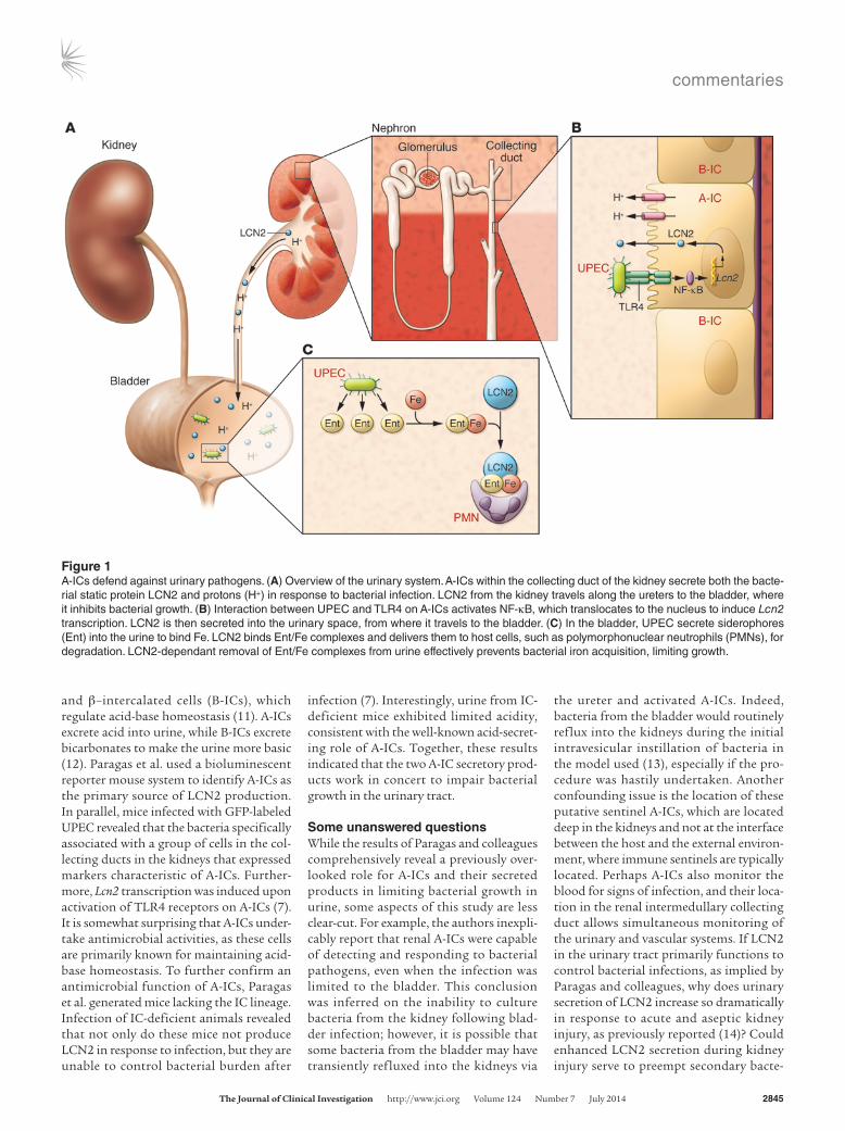
In this issue, Paragas et al. have shown that α–intercalated cells (A-ICs) in the col-lecting ducts of the kidney serve as both sentinels and defenders of the urinary tract during infection (7). Specifically, they demonstrated that immune sensory TLR4 molecules on A-ICs detect the presence of uropathogenic E. coli (UPEC), and TLR4 signaling in A-ICs triggers secretion of the bacteriostatic protein lipocalin 2 (LCN2), as well as secretion of H+ ions into the urine (Figure 1). Together, urine acidification and LCN2 drastically reduced the number of infecting bacteria in the urinary tract.
Protective role of LCN2 against UPEC infectionsLCN2 is a member of the large lipocalin protein family, which has a wide range of biological functions (8). LCN2 binds to the secreted siderophore enterochelin (Ent), which UPEC and other pathogens release into the extracellular milieu to acquire essential iron (9), and then delivers Ent/Fe3+ complexes to host cells for deg-radation, effectively abrogating bacterial iron acquisition. Pathogens are unable to grow in the absence of iron, allowing the immune system to eliminate the infection.
Interestingly, Paragas et al. observed a sig-nificant elevation of LCN2 in the urine of mice with UTI compared with uninfected animals. The degree of LCN2 upregulation apparently associated with the number of infecting bacteria within the urine, and reduction of bacterial load with antibiotics resulted in decreased LCN2 production. Moreover, a similar correlation between bacterial numbers in the urine and LCN2 levels was also seen in patients with UTIs, providing direct clinical support for LCN2 production in immune defense of the uri-nary tract. Compared with WT animals, Lcn2-deficient mice were more susceptible to UTIs; however, both WT and Lcn2–/– ani-mals were infected to similar extents with an ent mutant UPEC strain (7), which indi-cates that the suppressive actions of LCN2 on uropathogens involve iron acquisition.
The observation by Paragas et al. that LCN2 is not constitutively present in the urinary tract, but rather produced in direct proportion to the bacterial numbers in urine (7), is similar to other antimicrobial agents, including pentraxin-3 and HBD-1, that are also secreted in direct proportion to the size of the bacterial threat (10). As urine is a very rich medium and can be retained for many hours in the bladder, the urinary tract can serve as a powerful incu-bator for bacterial growth. Indeed, bacterial numbers in urine can reach levels in excess of 108 bacteria/ml. The ability to produce LCN2 and other antibacterial agents in proportion to bacterial burden in the uri-nary tract may be adaptation by the kidney to maintain homeostasis in response to overwhelming bacterial infection.
A-ICs: the cellular source of LCN2Notably, Paragas and colleagues deter-mined that LCN2 is produced by highly specialized A-ICs located in the collecting duct of the kidney medulla. The renal col-lecting ducts are responsible for regulat-ing electrolyte and fluid balance through reabsorption and excretion processes. The collecting ducts are lined by both A-ICs
Conflict of interest: The authors have declared that no conflict of interest exists.
Citation for this article: J Clin Invest. 2014; 124(7):2844–2846. doi:10.1172/JCI76630.
commentaries
The Journal of Clinical Investigation http://www.jci.org Volume 124 Number 7 July 2014 2845
and β–intercalated cells (B-ICs), which regulate acid-base homeostasis (11). A-ICs excrete acid into urine, while B-ICs excrete bicarbonates to make the urine more basic (12). Paragas et al. used a bioluminescent reporter mouse system to identify A-ICs as the primary source of LCN2 production. In parallel, mice infected with GFP-labeled UPEC revealed that the bacteria specifically associated with a group of cells in the col-lecting ducts in the kidneys that expressed markers characteristic of A-ICs. Further-more, Lcn2 transcription was induced upon activation of TLR4 receptors on A-ICs (7). It is somewhat surprising that A-ICs under-take antimicrobial activities, as these cells are primarily known for maintaining acid-base homeostasis. To further confirm an antimicrobial function of A-ICs, Paragas et al. generated mice lacking the IC lineage. Infection of IC-deficient animals revealed that not only do these mice not produce LCN2 in response to infection, but they are unable to control bacterial burden after
infection (7). Interestingly, urine from IC-deficient mice exhibited limited acidity, consistent with the well-known acid-secret-ing role of A-ICs. Together, these results indicated that the two A-IC secretory prod-ucts work in concert to impair bacterial growth in the urinary tract.
Some unanswered questionsWhile the results of Paragas and colleagues comprehensively reveal a previously over-looked role for A-ICs and their secreted products in limiting bacterial growth in urine, some aspects of this study are less clear-cut. For example, the authors inexpli-cably report that renal A-ICs were capable of detecting and responding to bacterial pathogens, even when the infection was limited to the bladder. This conclusion was inferred on the inability to culture bacteria from the kidney following blad-der infection; however, it is possible that some bacteria from the bladder may have transiently refluxed into the kidneys via
the ureter and activated A-ICs. Indeed, bacteria from the bladder would routinely reflux into the kidneys during the initial intravesicular instillation of bacteria in the model used (13), especially if the pro-cedure was hastily undertaken. Another confounding issue is the location of these putative sentinel A-ICs, which are located deep in the kidneys and not at the interface between the host and the external environ-ment, where immune sentinels are typically located. Perhaps A-ICs also monitor the blood for signs of infection, and their loca-tion in the renal intermedullary collecting duct allows simultaneous monitoring of the urinary and vascular systems. If LCN2 in the urinary tract primarily functions to control bacterial infections, as implied by Paragas and colleagues, why does urinary secretion of LCN2 increase so dramatically in response to acute and aseptic kidney injury, as previously reported (14)? Could enhanced LCN2 secretion during kidney injury serve to preempt secondary bacte-
Figure 1A-ICs defend against urinary pathogens. (A) Overview of the urinary system. A-ICs within the collecting duct of the kidney secrete both the bacte-rial static protein LCN2 and protons (H+) in response to bacterial infection. LCN2 from the kidney travels along the ureters to the bladder, where it inhibits bacterial growth. (B) Interaction between UPEC and TLR4 on A-ICs activates NF-κB, which translocates to the nucleus to induce Lcn2 transcription. LCN2 is then secreted into the urinary space, from where it travels to the bladder. (C) In the bladder, UPEC secrete siderophores (Ent) into the urine to bind Fe. LCN2 binds Ent/Fe complexes and delivers them to host cells, such as polymorphonuclear neutrophils (PMNs), for degradation. LCN2-dependant removal of Ent/Fe complexes from urine effectively prevents bacterial iron acquisition, limiting growth.
commentaries
2846 The Journal of Clinical Investigation http://www.jci.org Volume 124 Number 7 July 2014
links innate immune cell activation with adaptive immunity via a Toll-like receptor-4-dependent mechanism. J Clin Invest. 2005;115(2):468–475.
7. Paragas N, et al. α–Intercalated cells defend the uri-nary system from bacterial infection. J Clin Invest. 2014;124(7):2963–2976.
8. Goetz DH, Holmes MA, Borregaard N, Bluhm ME, Raymond KN, Strong RK. The neutrophil lipocalin NGAL is a bacteriostatic agent that interferes with siderophore-mediated iron acquisition. Mol Cell. 2002;10(5):1033–1043.
9. Flo TH, et al. Lipocalin 2 mediates an innate immune response to bacterial infection by seques-trating iron. Nature. 2004;432(7019):917–921.
10. Jaillon S, et al. The humoral pattern recognition molecule PTX3 is a key component of innate immunity against urinary tract infection. Immu-nity. 2014;40(4):621–632.
11. Al-Awqati Q. Cell biology of the intercalated cell in the kidney. FEBS Lett. 2013;587(13):1911–1914.
12. Wall SM. Recent advances in our understanding of intercalated cells. Curr Opin Nephrol Hypertens. 2005;14(5):480–484.
13. Chan CY, St John AL, Abraham SN. Mast cell inter-leukin-10 drives localized tolerance in chronic bladder infection. Immunity. 2013;38(2):349–359.
14. Mishra J, et al. Neutrophil gelatinase-associat-ed lipocalin (NGAL) as a biomarker for acute renal injury after cardiac surgery. Lancet. 2005; 365(9466):1231–1238.
15. DuBose TD, Alpern RJ. Renal tubular acidosis. In: Valle D, et al., eds. The Online Metabolic and Molecular Bases of Inherited Disease. New York, New York, USA: McGraw-Hill; 2007:Chapter 195. doi:10.1036/ommbid.228.
16. Raz R, Stamm WE. A controlled trial of intra-vaginal estriol in postmenopausal women with recurrent urinary tract infections. N Engl J Med. 1993;329(11):753–756.
rial infections? In view of the critical pro-tective role played by A-ICs, do their sister cells, B-ICs, play a complementary role in combating infection? Despite any linger-ing questions, the results of this study are important and reveal how a specialized kid-ney cell, previously implicated in acid-base homeostasis, combats bacterial infections of the urinary tract.
Highly specialized epithelial cells are dis-persed on various mucous membranes and are involved in maintaining the integrity of the mucosal barrier, mediating secretion, selective absorption, or transcellular trans-port. The revelation by Paragas and col-laborators that, in addition to maintaining acid-base homeostasis, kidney A-ICs play a key role in abrogating bacterial infection in the urinary tract (7), has implications for human renal diseases. For example, human diseases that involve A-IC dysfunction, such as chronic distal renal tubular acido-sis, are characterized by recurrent UTIs and pyelonephritis (15). As the aged population has dramatically grown in recent years, recurrent UTIs have become a substantial clinical problem in hospitals and nursing homes (16). Antibiotics are increasingly ineffective for combating UTIs; therefore, harnessing and boosting the innate anti-
microbial properties of cells in the urinary tract, such as A-ICs, may become viable therapeutic alternatives.
AcknowledgmentsThe authors were funded by NIH grants U01-AI082107, R01-AI096305, and R56-DK095198 and by a block grant from Duke-NUS, Singapore.
Address correspondence to: Soman N. Abraham, Department of Pathology, Duke University Medical Center, Box 3712, Dur-ham, North Carolina 27514, USA. Phone: 919.684.3630; Fax: 919.684.2021; E-mail: [email protected].
1. Chromek M, Brauner A. Antimicrobial mecha-nisms of the urinary tract. J Mol Med (Berl). 2008; 86(1):37–47.
2. Chromek M, et al. The antimicrobial peptide cat-helicidin protects the urinary tract against invasive bacterial infection. Nat Med. 2006;12(6):636–641.
3. Spencer JD, et al. Ribonuclease 7 is a potent anti-microbial peptide within the human urinary tract. Kidney Int. 2011;80(2):174–180.
4. Schilling JD, Martin SM, Hung CS, Lorenz RG, Hultgren SJ. Toll-like receptor 4 on stromal and hematopoietic cells mediates innate resistance to uropathogenic Escherichia coli. Proc Natl Acad Sci U S A. 2003;100(7):4203–4208.
5. Ganz T. Defensins in the urinary tract and other tissues. J Infect Dis. 2001;183(suppl 1):S41–S42.
6. Saemann MD, et al. Tamm-Horsfall glycoprotein
Loss of P2Y14 results in an arresting response to hematological stressBrian S. Garrison1,2,3 and Derrick J. Rossi1,2,3,4
1Department of Stem Cell and Regenerative Biology, Harvard University, Cambridge, Massachusetts, USA. 2Department of Pediatrics, Harvard Medical School, Boston, Massachusetts, USA. 3Program in Cellular and Molecular Medicine, Division of Hematology/Oncology, Boston Children’s Hospital,
Boston, Massachusetts, USA. 4Harvard Stem Cell Institute, Cambridge, Massachusetts, USA.
The regenerative capacity of tissues to recover from injury or stress is depen-dent on stem cell competence, yet the underlying mechanisms that govern how stem cells detect stress and initiate appropriate responses are poorly understood. In this issue of the JCI, Cho and Yusuf et al. demonstrate that the purinergic receptor P2Y14 may mediate the hematopoietic stem and pro-genitor cell regenerative response.
Senescence and stem cell declineCellular senescence, a state of permanent irreversible growth arrest, was initially described over half a century ago by Leon-
ard Hayflick and Paul Moorhead, who observed that normal human fibroblasts cease to replicate after 50 to 60 cellular divisions (1). This barrier to everlasting cellular proliferation later became termed the “Hayflick limit,” denoting the loss of proliferative potential even though the cell remains viable and metabolically active. While this phenomenon was originally connected to long-term in vitro cell propa-
gation, cellular senescence is now under-stood to be a complex mechanism that may limit cell growth as well as prevent cancer in vivo and that can be initiated in response to a variety of cellular stresses, including oxidative damage, telomere shortening, DNA damage, and gene deregulation (2–4).
As with the majority of tissues, the hematopoietic system exhibits signs of age-related decline, including immune dysfunction, decreased red blood cell pro-duction, increased incidence of malignan-cies, and impaired recovery from injury, much of which appears to arise through cell autonomous changes in the HSC com-partment (5–8). These age-related changes in the HSC compartment appear to be
Conflict of interest: Derrick Rossi has an ownership stake and provides consultation for Moderna Thera-peutics.
Citation for this article: J Clin Invest. 2014; 124(7):2846–2848. doi:10.1172/JCI76626.