Neuroprotection mediates through oestrogen receptor alpha in astrocytes
5385
Abstract. – OBJECTIVE: To investigate the effects of ketamine on autophagy and apopto-sis of astrocytes in the cerebral cortex of rats, and determine whether nuclear factor-κB (NF-κB) pathway is involved in the regulation of auto-phagy and apoptosis of astrocytes.
MATERIALS AND METHODS: A total of 36 male Sprague-Dawley (SD) rats were randomly divided into 3 groups: control group (Group C: intraperitoneal injection of equal amount of nor-mal saline), glutamic acid group (Group G: in-traperitoneal injection of 1 mg/kg glutamic ac-id) and glutamic acid + ketamine group (Group GK: intraperitoneal injection of 1 mg/kg glutam-ic acid and then injection of 5 mg/kg ketamine after 30 min). The cerebral cortex of rats in each group was taken after successive administration for 5 d. The number of glial fibrillary acidic pro-tein (GFAP)-positive cells in the cerebral cortex of rats in each group was detected via immuno-fluorescence. The number of terminal deoxynu-cleotidyl transferase-mediated dUTP nick end labeling (TUNEL)-positive cells (apoptotic cells) in the cerebral cortex was detected via TUNEL staining. The levels of inflammatory factors were detected using the enzyme-linked immunosor-bent assay (ELISA) kit. Moreover, the expres-sions of autophagy-related proteins and apopto-sis-related proteins in the cerebral cortex were detected via Western blotting, and the expres-sions of IκB-a and NF-κBp65 were also detected.
RESULTS: The results of immunofluores-cence showed that the number of GFAP-posi-tive cells in the cerebral cortex of rats in Group G was significantly increased compared with that in Group C (p<0.01), and it was significant-ly decreased in Group GK compared with that in Group G (p<0.01). The results of TUNEL stain-ing revealed that the number of TUNEL-posi-tive cells in the cerebral cortex in Group G was significantly larger than that in Group C, and it was significantly smaller in Group GK than that in Group G (p<0.01). Results of ELISA demon-strated that compared with those in Group C, the contents of interleukin-6 (IL-6) and tumor necro-sis factor-a (TNF-a) in Group G were significant-
ly increased (p<0.01), but the content of IL-10 was significantly decreased (p<0.01). Compared with those in Group G, the contents of IL-6 and TNF-a in Group GK were significantly decreased (p<0.01), but the level of IL-10 was statistically el-evated (p<0.01). Compared with those in Group C, the levels of LC3 II/I and cleaved caspase-3 in the cerebral cortex in Group G were significantly increased (p<0.01), but the p62 level and B-cell lymphoma-2/Bcl-2 associated X protein (Bcl-2/Bax) ratio were significantly decreased (p<0.01). In Group GK, the levels of LC3 II/I and cleaved caspase-3 were reduced, but the p62 level and Bcl-2/Bax ratio were increased. The expressions of IκB-α and NF-κBp65 in Group G were signifi-cantly decreased compared with those in Group C (p<0.01), and they were significantly higher in Group GK than those in Group G (p<0.01).
CONCLUSIONS: Ketamine can reduce the glu-tamic acid-induced activation of astrocytes in the cerebral cortex, inhibit the autophagy and alleviate the apoptosis of astrocytes, the pro-cess of which is mediated by the NF-κB pathway, which provides the new molecular basis of ket-amine in protecting astrocytes.
Key Words:Ketamine, Astrocytes, Autophagy, Apoptosis, NF-κB
pathway.
Introduction
Astrocytes are the most major macroglial cells in the central nervous system, which, together with neurons, regulate the development and other processes of the central nervous system1. Astrocytes can produce under the induction of neurotransmitter or spontaneously produce exci-tability, and they are also involved in signal transduction, neuroimmunity, and nerve tissue repair and regeneration, in addition to providing nutritional support for neurons2. Glutamic acid
European Review for Medical and Pharmacological Sciences 2018; 22: 5385-5393
J. YANG, X. LI, C. YANG, X.-X. BU, J. SHEN, T. HONG
Department of Anesthesiology, First People’s Hospital of Changzhou & Third Affiliated Hospital of Suzhou University, Changzhou, Jiangsu, China
Corresponding Author: Tao Hong, MD; e-mail: [email protected]
Research on ketamine in mediating autophagy and inhibiting apoptosis of astrocytes in cerebral cortex of rats through NF-κB pathway
J. Yang, X. Li, C. Yang, X.-X. Bu, J. Shen, T. Hong
5386
is an important excitatory neurotransmitter in the brain, and the accumulation of excessive glutamic acid will produce nervous excitability toxicity, leading to the death of nerve cells and astrocytes3. Hoshi et al4 found that glutamic acid promoted the production of a large number of calcium ions from hippocampal astrocytes and was involved in the regulation of intracellular calcium ions, sodium ions and oxidative stress response, leading to nerve injury. Ketamine, a kind of N-methyl-D-aspartate (NMDA) receptor antagonist, is often used as an anesthetic with both excitatory and inhibitory effects on the central system5,6. Liu et al7 found via the in vitro cell experiment that the single administration of ketamine can inhibit the glutamic acid-induced apoptosis of astrocytes, thus exerting a protecti-ve effect on astrocytes. Moreover, Steiner et al8 showed that the repeated administration of ke-tamine can mediate apoptosis through activating the nuclear factor-κB (NF-κB) signaling pathway. Recent evidence9 has shown that apoptosis and autophagy interact with each other and jointly participate in the regulation of cell fate. In this study, astrocytes in the cerebral cortex of rats were activated via intraperitoneal injection of glutamic acid to produce autophagy and apop-tosis, so as to study the effects of ketamine on autophagy and apoptosis of astrocytes and its possible mechanism.
Materials and Methods
Animal Feeding, Grouping and Drug Administration
A total of 36 male Sprague-Dawley (SD) rats weighing 240-260 g were purchased from the Laboratory Animal Center of Guangdong Pro-vince (Laboratory Animal Production License No. SCXK2012-0006) (Guangzhou, Guangdong, China), and they were fed in a specific patho-gen-free animal room to adapt to the environment for 1 week. Rats could take food and water freely under the temperature of 22-25°C, humidity of 45-50%, and light-dark time of 12/12 h. The abo-ve-mentioned rats were randomly divided into 3 groups: control group (Group C, n=12: intra-peritoneal injection of equal amount of normal saline), glutamic acid group (Group G, n=12: in-traperitoneal injection of 1 mg/kg glutamic acid) and glutamic acid + ketamine group (Group GK, n=12: intraperitoneal injection of 1 mg/kg gluta-mic acid and then injection of 5 mg/kg ketamine
after 30 min). The concentration of glutamic acid (Sigma-Aldrich, St. Louis, MO, USA) was adju-sted to 1 mg/ml with normal saline, and the con-centration of ketamine (Lingnan Pharmaceutical: NMPN H28308820) was 50 mg/mL and diluted into 5 mg/mL with normal saline. Both glutamic acid and ketamine were injected intraperitoneal-ly. After successive administration for 5 days, 6 rats in each group were subjected to cardiac per-fusion fixation, and the brain was taken for fixa-tion and dehydration. The remaining rats were executed, and the brain was taken to separate the cortex. Then, the cortex was quickly frozen with liquid nitrogen, and stored at -80°C for standby application. All the animal experiments involved in this study were approved by the Animal Ethics Review Committee of our hospital, and all ani-mal operations strictly followed the regulations of the National Institute about the Laboratory Animal Care and Health Guidance.
Immunofluorescence StainingAfter perfusion, the brain was removed from
rats, and fixed in 4% paraformaldehyde, followed by dehydration via 20% and 30% sucrose until the brain tissues completely sank to the bottom. Then, the brain tissues were taken, embedded into the embedding agent, fixed on the tray, and frozen in a freezer at -80°C. After the embedding agent was completely frozen, tissues were equi-librated in a freezing microtome for 30 min, the microtome was adjusted and tissues were cut into 40 μm-thick sections. The optimal brain sections were selected and collected into a 24-well plate. The sections were fixed with acetone at 4°C for 15 min and acetone was discarded and sections were washed with phosphate-buffered saline (PBS) for 3 times (3 min per time). After tran-sparency with 0.3% Triton X-100 for 15 min, the liquid was discarded, and sections were washed with PBS for 3 times (3 min per time). Then, sections were sealed with 10% goat serum sealing solution at room temperature for 1 h. The sealing solution was discarded and sections were incuba-ted with anti-glial fibrillary acidic protein (GFAP) antibody (Cell Signaling Technology, Danvers, MA, USA, 1:100) in a web box at 4°C overnight. After the antibody was recycled, sections were washed again with PBS for 3 times (3 min per time), incubated with Dylight 488 fluorescent secondary antibody (Abcam, Cambridge, MA, USA, 1:400) in a dark place at room temperature for 2 h and washed with PBS for 3 times (3 min per time). After nuclear staining with 4’,6-dia-
Ketamine in apoptosis of NF-κB pathway
5387
midino-2-phenylindole solution (2 μg/mL) for 10 min, sections were washed with PBS once for 3 min. After anti-fluorescence quenching sealing agent was added dropwise, sections were sealed with neutral resin, covered with the cover glass, observed and photographed under a confocal fluorescence microscope. Finally, the number of GFAP-positive cells in the cerebral cortex in each group was calculated.
Terminal Deoxynucleotidyl Transferase-Mediated dUTP Nick End Labeling (TUNEL) Staining
The optimal brain sections were selected, dropwise added with Proteinase K solution for reaction at 37°C for 10 min, and washed with PBS for 3 times (3 min per time). Then, brain sections were treated with a mixed solution of TdT and DIG-d-UTP, labeled and washed with PBS for 3 times (3 min per time) after 2 h. Brain sections were sealed with sealing solution at room tem-perature for 30 min, added with biotinylated an-ti-digoxin antibody (Cell Signaling Technology, Beverly, MA, USA, 1:100) for incubation at 37°C for 30 min, and washed with PBS for 3 times (3 min per time). After incubation with streptavidin biotin enzyme complex-fluorescein isothiocya-nate secondary antibody (Abcam, Cambridge, MA, USA, 1:100) at 37°C for 30 min, sections were washed with PBS for 3 times (3 min per time). After anti-fluorescence quenching sealing agent was dropwise added, sections were covered with the cover glass, observed and photographed under the confocal fluorescence microscope. Fi-nally, the number of TUNEL-positive cells in the brain section of each group was calculated, and cells with yellow-green fluorescence were positi-ve cells, namely apoptotic cells.
Detection of Content of Inflammatory Factors Via Enzyme-Linked Immunosorbent Assay (ELISA)
The cortical tissues in each group were ta-ken, and added with PBS buffer at a weight/volume (mg/μL) ratio of 1:9, followed by ultraso-nic dispersion using an ultrasonic homogenizer (placed on ice to avoid the protein degradation due to excess temperature). After there were no visible tissues to the naked eyes, centrifuga-tion was performed at 12,000 g and 4°C for 10 min. The supernatant was collected as the total protein. The standard solution was prepared to make the standard curve, and the standard curve was drawn using CurveExpert 1.4 software for
sample quantification. The 96-well plate coated with the corresponding antibodies was taken, added with the standard solution or sample solution, and affixed with the sealing membra-ne, followed by incubation at 37°C for 90 min. After the liquid in the plate was patted dry, the biotin-labeled antibody [anti-tumor necrosis fac-tor-α (TNF-α), interleukin-6 (IL-6) or IL-10 an-tibody] was added, and the plate was sealed with the sealing membrane, followed by incubation at 37°C for 60 min. After the plate was washed wi-th washing solution for 4 times (3 min per time), ABC working solution was added, and the plate was sealed, followed by incubation at 37°C for 30 min. After the plate was washed again with washing solution for 4 times, tetramethylbenzi-dine (TMB) developing solution was added, and the plate was sealed, followed by incubation in a dark place at 37°C for 20 min. Then, TMB stop buffer was added and mixed evenly. The absor-bance value at 450 nm was detected using a mi-croplate reader (GeneTex, Irvine, CA, USA), and substituted into the standard curve to calculate the concentrations of TNF-α, IL-6, and IL-10 in brain tissues in each group.
Western BlottingThe cortical tissues in each group were taken,
added with radioimmunoprecipitation assay (RI-PA) lysis solution at a weight/volume (mg/μL) ratio of 1:9, and added with protease inhibitor and phosphatase inhibitor at a volume ratio of 1%, followed by ultrasonic dispersion using the ul-trasonic homogenizer (placed on ice to avoid the protein degradation due to excess temperature). After there were no visible tissues to the naked eyes, centrifugation was performed at 12,000 g and 4°C for 10 min. The supernatant was col-lected as the total protein sample. The protein was quantified using the bicinchoninic acid (BCA) protein quantitative kit (Millipore, Temecula, CA, USA), and the total protein concentration in cortical tissues in each group was calculated. The sample loading system in an equal concen-tration was prepared, and heated at 95°C for 15 min to inactivate the protein. Then, the sample was added into the sodium dodecyl sulfate polya-crylamide gel electrophoresis (SDS-PAGE) gel slot prepared for electrophoresis until the protein completely reached the bottom of the gel. The protein was transferred onto a polyvinylidene difluoride membrane (Millipore, Billerica, MA, USA) under a constant current of 260 mA. The membrane was sealed with the freshly prepared
J. Yang, X. Li, C. Yang, X.-X. Bu, J. Shen, T. Hong
5388
5% skim milk powder for 2 h, and the target band was cut according to the protein size. The band was incubated with LC3, p62, cleaved caspase-3, B-cell lymphoma-2 (Bcl-2), Bcl-2 associated X protein (Bax), IκB-α, NF-κBp65, and glyceral-dehyde-3-phosphate dehydrogenase antibodies (Cell Signaling Technology, Danvers, MA, USA, 1:1000) overnight, washed with Tris-buffered sa-line with Tween-20 (TBST) for 3 times (10 min per time), incubated with horseradish peroxi-dase-conjugated secondary antibody (Millipore, Billerica, MA, USA) at room temperature for 2 h, and washed again with TBST for 3 times (10 min per time). Finally, enhanced chemiluminescence solution (Millipore, Billerica, MA, USA) was added for color development using a developing machine to calculate the relative expression level of the corresponding protein.
Statistical AnalysisIn this study, data were presented as mean ±
standard deviation, and processed using Stati-stical Product and Service Solutions (SPSS) 19.0 software (SPSS Inc., Chicago, IL, USA). The t-test was used for the intergroup comparison, x2-test was used for the enumeration data, and analysis of variance with Tukey’s post-hoc test
was used for the comparison among groups. p<0.05 suggested that the difference was statisti-cally significant.
Results
Activation of Astrocytes in the Cerebral Cortex of Rats
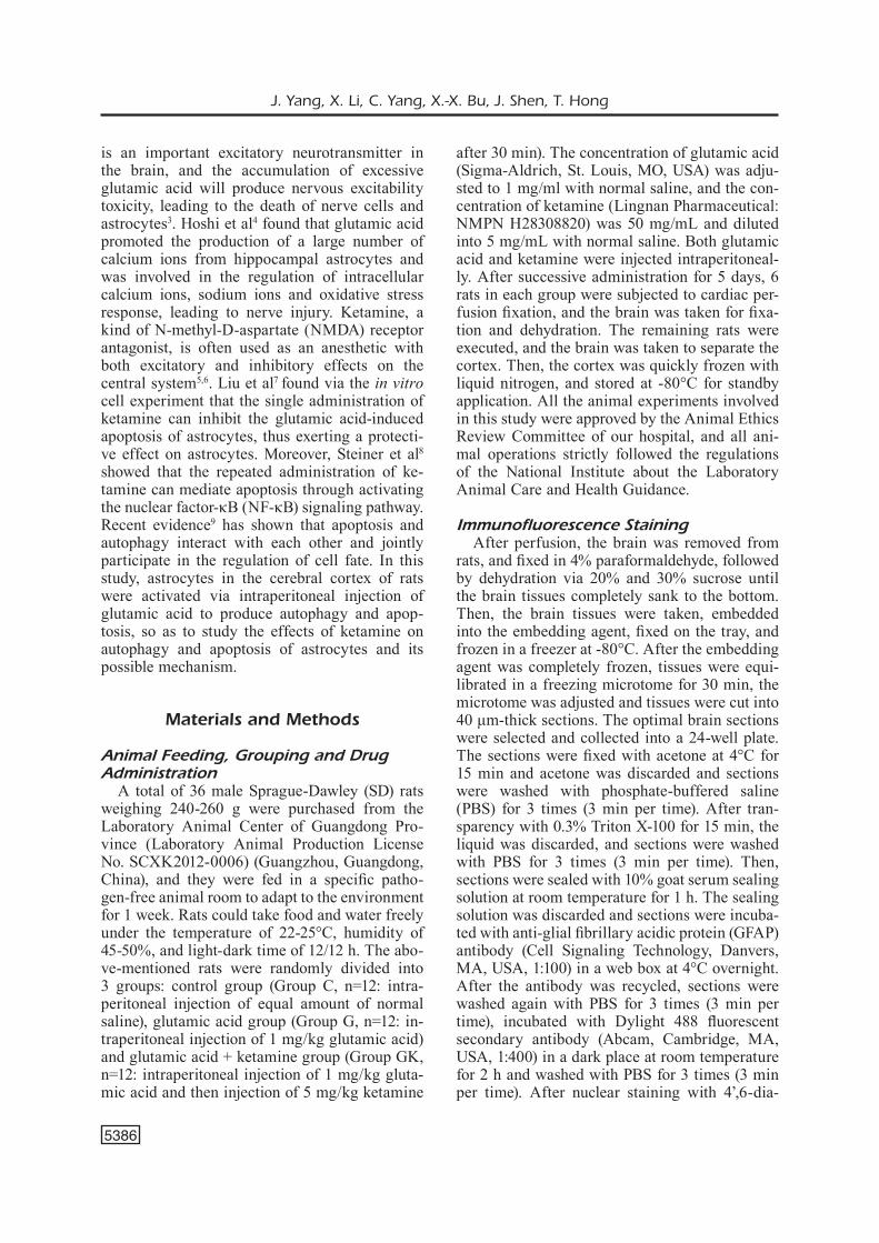
The expression of GFAP, an astrocyte activa-tion marker, in the cerebral cortex of rats was detected via immunofluorescence staining to in-vestigate the effect of glutamic acid on astrocyte activation. Results showed that compared with that in control group, the number of GFAP-posi-tive cells in the cerebral cortex of rats in Group G was increased significantly (p<0.01), and it was significantly decreased after administration of ketamine (p<0.01) (Figure 1).
Apoptosis of Astrocytes in the Cerebral Cortex of Rats in Each Group
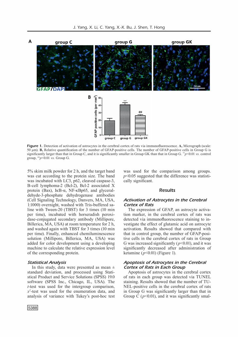
Apoptosis of astrocytes in the cerebral cortex of rats in each group was detected via TUNEL staining. Results showed that the number of TU-NEL-positive cells in the cerebral cortex of rats in Group G was significantly larger than that in Group C (p<0.01), and it was significantly smal-
Figure 1. Detection of activation of astrocytes in the cerebral cortex of rats via immunofluorescence. A, Micrograph (scale: 50 μm). B, Relative quantification of the number of GFAP-positive cells. The number of GFAP-positive cells in Group G is significantly larger than that in Group C, and it is significantly smaller in Group GK than that in Group G. **p<0.01 vs. control group, ##p<0.01 vs. Group G.
Ketamine in apoptosis of NF-κB pathway
5389
ler in Group GK than that in Group G after treat-ment with ketamine (p<0.01) (Figure 2).
Content of Inflammatory Factors in Rats in Each Group
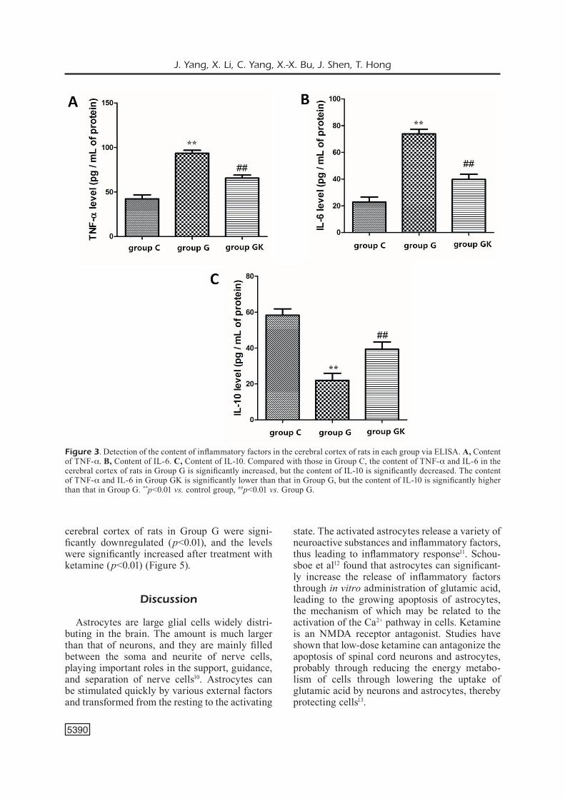
The content of inflammatory factors in the ce-rebral cortex of rats in each group was detected using the ELISA kit. We found that compared wi-th those in Group C, the levels of TNF-a and IL-6 in the cerebral cortex of rats in Group G were significantly increased (p<0.01), but the level of IL-10 was significantly decreased (p<0.01). After treatment with ketamine, the levels of TNF-a and IL-6 in the cerebral cortex of rats in Group GK were significantly lower than those in Group G (p<0.01), but the level of IL-10 was significantly higher than that in Group G (p<0.01) (Figure 3).
Expressions of Autophagy-Related Proteins and Apoptosis-Related Proteins
Expressions of autophagy-related proteins and apoptosis-related proteins in the cerebral cortex of rats in each group were detected via Western blotting. Results revealed that compared with
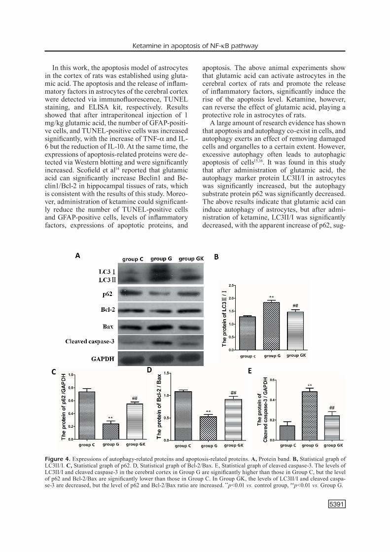
those in Group C, the level of LC3II/I in the ce-rebral cortex in Group G was statistically incre-ased (p<0.01) with a significant reduction of p62 (p<0.01). After administration of ketamine, the level of LC3II/I in Group GK was significantly decreased, but the level of p62 was significantly elevated (p<0.01). The level of cleaved caspase-3 in the cerebral cortex in Group G was signifi-cantly increased, but the Bcl-2/Bax ratio was si-gnificantly reduced (p<0.01). The level of cleaved caspase-3 in Group GK was significantly lower than that in Group G, but the Bcl-2/Bax ratio was significantly higher than that in Group G (p<0.01) (Figure 4).
Regulation of Autophagy and Apoptosis Via NF-κB Pathway
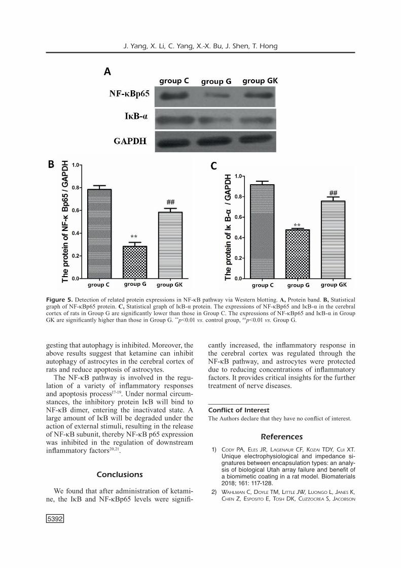
NF-κB pathway plays crucial roles in inflam-matory response, autophagy, and apoptosis. The roles of the NF-κB pathway in autophagy and apoptosis of astrocytes in the cerebral cortex of rats were detected via Western blotting. Our data indicated that compared with those in Group C, the expressions of NF-κBp65 and IκB-α in the
Figure 2. Detection of apoptosis of astrocytes in the cerebral cortex of rats in each group via TUNEL staining. A, Micro-graph (scale: 50 μm), B, Relative quantification of the number of TUNEL-positive cells. The number of TUNEL-positive cells in Group G is significantly larger than that in Group C, and it is significantly smaller in Group GK than that in Group. **p<0.01 vs. control group, ##p<0.01 vs. Group G.
J. Yang, X. Li, C. Yang, X.-X. Bu, J. Shen, T. Hong
5390
cerebral cortex of rats in Group G were signi-ficantly downregulated (p<0.01), and the levels were significantly increased after treatment with ketamine (p<0.01) (Figure 5).
Discussion
Astrocytes are large glial cells widely distri-buting in the brain. The amount is much larger than that of neurons, and they are mainly filled between the soma and neurite of nerve cells, playing important roles in the support, guidance, and separation of nerve cells10. Astrocytes can be stimulated quickly by various external factors and transformed from the resting to the activating
state. The activated astrocytes release a variety of neuroactive substances and inflammatory factors, thus leading to inflammatory response11. Schou-sboe et al12 found that astrocytes can significant-ly increase the release of inflammatory factors through in vitro administration of glutamic acid, leading to the growing apoptosis of astrocytes, the mechanism of which may be related to the activation of the Ca2+ pathway in cells. Ketamine is an NMDA receptor antagonist. Studies have shown that low-dose ketamine can antagonize the apoptosis of spinal cord neurons and astrocytes, probably through reducing the energy metabo-lism of cells through lowering the uptake of glutamic acid by neurons and astrocytes, thereby protecting cells13.
Figure 3. Detection of the content of inflammatory factors in the cerebral cortex of rats in each group via ELISA. A, Content of TNF-a. B, Content of IL-6. C, Content of IL-10. Compared with those in Group C, the content of TNF-a and IL-6 in the cerebral cortex of rats in Group G is significantly increased, but the content of IL-10 is significantly decreased. The content of TNF-a and IL-6 in Group GK is significantly lower than that in Group G, but the content of IL-10 is significantly higher than that in Group G. **p<0.01 vs. control group, ##p<0.01 vs. Group G.
Ketamine in apoptosis of NF-κB pathway
5391
In this work, the apoptosis model of astrocytes in the cortex of rats was established using gluta-mic acid. The apoptosis and the release of inflam-matory factors in astrocytes of the cerebral cortex were detected via immunofluorescence, TUNEL staining, and ELISA kit, respectively. Results showed that after intraperitoneal injection of 1 mg/kg glutamic acid, the number of GFAP-positi-ve cells, and TUNEL-positive cells was increased significantly, with the increase of TNF-a and IL-6 but the reduction of IL-10. At the same time, the expressions of apoptosis-related proteins were de-tected via Western blotting and were significantly increased. Scofield et al14 reported that glutamic acid can significantly increase Beclin1 and Be-clin1/Bcl-2 in hippocampal tissues of rats, which is consistent with the results of this study. Moreo-ver, administration of ketamine could significant-ly reduce the number of TUNEL-positive cells and GFAP-positive cells, levels of inflammatory factors, expressions of apoptotic proteins, and
apoptosis. The above animal experiments show that glutamic acid can activate astrocytes in the cerebral cortex of rats and promote the release of inflammatory factors, significantly induce the rise of the apoptosis level. Ketamine, however, can reverse the effect of glutamic acid, playing a protective role in astrocytes of rats.
A large amount of research evidence has shown that apoptosis and autophagy co-exist in cells, and autophagy exerts an effect of removing damaged cells and organelles to a certain extent. However, excessive autophagy often leads to autophagic apoptosis of cells15,16. It was found in this study that after administration of glutamic acid, the autophagy marker protein LC3II/I in astrocytes was significantly increased, but the autophagy substrate protein p62 was significantly decreased. The above results indicate that glutamic acid can induce autophagy of astrocytes, but after admi-nistration of ketamine, LC3II/I was significantly decreased, with the apparent increase of p62, sug-
Figure 4. Expressions of autophagy-related proteins and apoptosis-related proteins. A, Protein band. B, Statistical graph of LC3II/I. C, Statistical graph of p62. D, Statistical graph of Bcl-2/Bax. E, Statistical graph of cleaved caspase-3. The levels of LC3II/I and cleaved caspase-3 in the cerebral cortex in Group G are significantly higher than those in Group C, but the level of p62 and Bcl-2/Bax are significantly lower than those in Group C. In Group GK, the levels of LC3II/I and cleaved caspa-se-3 are decreased, but the level of p62 and Bcl-2/Bax ratio are increased. **p<0.01 vs. control group, ##p<0.01 vs. Group G.
J. Yang, X. Li, C. Yang, X.-X. Bu, J. Shen, T. Hong
5392
gesting that autophagy is inhibited. Moreover, the above results suggest that ketamine can inhibit autophagy of astrocytes in the cerebral cortex of rats and reduce apoptosis of astrocytes.
The NF-κB pathway is involved in the regu-lation of a variety of inflammatory responses and apoptosis process17-19. Under normal circum-stances, the inhibitory protein IκB will bind to NF-κB dimer, entering the inactivated state. A large amount of IκB will be degraded under the action of external stimuli, resulting in the release of NF-κB subunit, thereby NF-κB p65 expression was inhibited in the regulation of downstream inflammatory factors20,21.
Conclusions
We found that after administration of ketami-ne, the IκB and NF-κBp65 levels were signifi-
cantly increased, the inflammatory response in the cerebral cortex was regulated through the NF-κB pathway, and astrocytes were protected due to reducing concentrations of inflammatory factors. It provides critical insights for the further treatment of nerve diseases.
Conflict of InterestThe Authors declare that they have no conflict of interest.
References
1) Cody PA, ElEs JR, lAgEnAuR CF, KozAi Tdy, Cui XT. Unique electrophysiological and impedance si-gnatures between encapsulation types: an analy-sis of biological Utah array failure and benefit of a biomimetic coating in a rat model. Biomaterials 2018; 161: 117-128.
2) WAhlmAn C, doylE Tm, liTTlE JW, luongo l, JAnEs K, ChEn z, EsPosiTo E, Tosh dK, CuzzoCREA s, JACobson
Figure 5. Detection of related protein expressions in NF-κB pathway via Western blotting. A, Protein band. B, Statistical graph of NF-κBp65 protein. C, Statistical graph of IκB-α protein. The expressions of NF-κBp65 and IκB-α in the cerebral cortex of rats in Group G are significantly lower than those in Group C. The expressions of NF-κBp65 and IκB-α in Group GK are significantly higher than those in Group G. **p<0.01 vs. control group, ##p<0.01 vs. Group G.
Ketamine in apoptosis of NF-κB pathway
5393
KA, sAlvEmini d. Chemotherapy-induced pain is promoted by enhanced spinal adenosine kina-se levels via astrocyte-dependent mechanisms. Pain 2016; 159: 1025-1034.
3) du y, WAng W, luTTon Ad, Kiyoshi Cm, mA b, TAyloR AT, olEsiK JW, mCTiguE dm, AsKWiTh CC, zhou m. Dissipation of transmembrane potassium gra-dient is the main cause of cerebral ischemia-in-duced depolarization in astrocytes and neurons. Exp Neurol 2018; 303: 1-11.
4) hoshi A, TsunodA A, yAmAmoTo T, TAdA m, KAKiTA A, ugAWA y. Altered expression of glutamate tran-sporter-1 and water channel protein aquaporin-4 in human temporal cortex with Alzheimer’s disea-se. Neuropathol Appl Neurobiol 2018 Feb 6. doi: 10.1111/nan.12475. [Epub ahead of print].
5) zuo d, WAng C, li z, lin l, duAn z, Qi h, li l, sun F, Wu y. Existence of glia mitigated ketamine-in-duced neurotoxicity in neuron-glia mixed cultures of neonatal rat cortex and the glia-mediated pro-tective effect of 2-PMPA. Neurotoxicology 2014; 44: 218-230.
6) FEAThERsTonE RE, liAng y, sAundERs JA, TATARd-lEiTmAn vm, EhRliChmAn Rs, siEgEl sJ. Subchronic ketamine treatment leads to permanent changes in EEG, cognition and the astrocytic glutamate transporter EAAT2 in mice. Neurobiol Dis 2012; 47: 338-346.
7) liu WX, WAng J, XiE zm, Xu n, zhAng gF, JiA m, zhou zQ, hAshimoTo K, yAng JJ. Regulation of glutamate transporter 1 via BDNF-TrkB signaling plays a role in the anti-apoptotic and antidepres-sant effects of ketamine in chronic unpredictable stress model of depression. Psychopharmacolo-gy (Berl) 2016; 233: 405-415.
8) sTEinER J, bogERTs b, sARnyAi z, WAlTER m, gos T, bERnsTEin hg, myinT Am. Bridging the gap between the immune and glutamate hypotheses of schi-zophrenia and major depression: potential role of glial NMDA receptor modulators and impaired blood-brain barrier integrity. World J Biol Psychia-try 2012; 13: 482-492.
9) EREKAT ns. Autophagy precedes apoptosis among at risk cerebellar Purkinje cells in the shaker mu-tant rat: an ultrastructural study. Ultrastruct Pathol 2018; 42: 162-169.
10) du y, WAng W, luTTon Ad, Kiyoshi Cm, mA b, TAyloR AT, olEsiK JW, mCTiguE dm, AsKWiTh CC, zhou m. Dissipation of transmembrane potassium gra-dient is the main cause of cerebral ischemia-in-duced depolarization in astrocytes and neurons. Exp Neurol 2018; 303: 1-11.
11) hAsEl P, dAndo o, JiWAJi z, bAXTER P, Todd AC, hERon s, máRKus nm, mCQuEEn J, hAmPTon dW, ToRvEll m, TiWARi ss, mCKAy s, ERAso-PiChoT A, zoRzAno A, mA-sgRAu R, gAlEA E, ChAndRAn s, WylliE dJA, simPson
Ti, hARdinghAm gE. Author Correction: Neurons and neuronal activity control gene expression in astrocytes to regulate their development and metabolism. Nat Commun 2018; 9: 16176.
12) sChousboE A. Metabolic signaling in the brain and the role of astrocytes in control of gluta-mate and GABA neurotransmission. Neurosci Lett 2018 Feb 2. pii: S0304-3940(18)30044-2. doi: 10.1016/j.neulet.2018.01.038. [Epub ahead of print].
13) miyAzAKi h, nAKAmuRA y, ARAi T, KATAoKA K. Increa-se of glutamate uptake in astrocytes: a possible mechanism of action of volatile anesthetics. Ane-sthesiology 1997; 86: 1359-1366.
14) sCoFiEld md. Exploring the role of astroglial glu-tamate release and association with synapses in neuronal function and behavior. Biol Psychiatry 2017 Nov 11. pii: S0006-3223(17)32159-5. doi: 10.1016/j.biopsych.2017.10.029. [Epub ahead of print].
15) sun W, lin y, ChEn l, mA R, CAo J, yAo J, ChEn K, WAn J. Legumain suppresses OxLDL-induced macrophage apoptosis through enhancement of the autophagy pathway. Gene 2018; 652: 16-24.
16) huAng K, ChEn y, zhAng R, Wu y, mA y, FAng X, shEn s. Honokiol induces apoptosis and autopha-gy via the ROS/ERK1/2 signaling pathway in hu-man osteosarcoma cells in vitro and in vivo. Cell Death Dis 2018; 9: 157.
17) gAo y, luo Cl, li ll, yE gh, gAo C, WAng hC, huAng WW, WAng T, WAng zF, ni h, ChEn XP, TAo ly. IL-33 provides neuroprotection through sup-pressing apoptotic, autophagic and NF-κB-med-iated inflammatory pathways in a rat model of recurrent neonatal seizure. Front Mol Neurosci 2017; 10: 423.
18) huAng WQ, WEn Jl, lin RQ, WEi P, huAng F. Effects of mTOR/NF-κB signaling pathway and high thoracic epidural anesthesia on myocardial ischemia-reperfusion injury via autophagy in rats. J Cell Physiol 2018; 233: 6669-6678.
19) Xu Xm, liu W, CAo zh, liu mX. Effects of ZEB1 on regulating osteosarcoma cells via NF-kappaB/iNOS. Eur Rev Med Pharmacol Sci 2017; 21: 1184-1190.
20) liAng X, hou X, yAng y, liu h, guo R, yAng z, yAng l. The feedback loop of “EMMPRIN/NF-κB” wor-sens atherosclerotic plaque via suppressing au-tophagyin macrophage. J Mol Cell Cardiol 2018; 114: 129-140.
21) bAi F, huAng Q, niE J, lu s, lu C, zhu X, WAng y, zhuo l, lu z, lin X. Trolline ameliorates liver fi-brosis by inhibiting the NF-κB pathway, promoting HSC apoptosis and suppressing autophagy. Cell Physiol Biochem 2017; 44: 436-446.


![Genistein induces apoptosis of colon cancer cells by ...€¦ · pathway [3]. In this study, we demonstrated that GEN can inhibite proliferation and induce apoptosis of colon cancer](https://static.fdocument.org/doc/165x107/6091035508039222da437990/genistein-induces-apoptosis-of-colon-cancer-cells-by-pathway-3-in-this-study.jpg)