Journal of NeuroimmunologyCompound Molecular formula Molecular weight Structure DMF C 6H 8O 4 144.13...
12
DMF, but not other fumarates, inhibits NF-κB activity in vitro in an Nrf2-independent manner Geoffrey O. Gillard, Brian Collette, John Anderson, Jianhua Chao, Robert H. Scannevin, David J. Huss, Jason D. Fontenot ⁎ Biogen, Inc., 115 Broadway, Cambridge, MA 02142, USA abstract article info Article history: Received 19 February 2015 Received in revised form 7 April 2015 Accepted 9 April 2015 Keywords: Dimethyl fumarate (DMF) Monomethyl fumarate (MMF) Monoethyl fumarate (MEF) Nuclear factor kappa-light-chain-enhancer of activated B cells (NF-κB) Nuclear factor (erythroid-derived 2)-like 2 (Nrf2) Cytokine Fumarate-containing pharmaceuticals are potent therapeutic agents that influence multiple cellular pathways. Despite proven clinical efficacy, there is a significant lack of data that directly defines the molecular mechanisms of action of related, yet distinct fumarate compounds. We systematically compared the impact of dimethyl fuma- rate (DMF), monomethyl fumarate (MMF) and a mixture of monoethyl fumarate salts (Ca ++ , Mg ++ , Zn ++ ; MEF) on defined cellular responses. We demonstrate that DMF inhibited NF-κB-driven cytokine production and nuclear translocation of p65 and p52 in an Nrf2-independent manner. Equivalent doses of MMF and MEF did not affect NF-κB signaling. These results highlight a key difference in the biological impact of related, yet dis- tinct fumarate compounds. © 2015 The Authors. Published by Elsevier B.V. This is an open access article under the CC BY-NC-ND license (http://creativecommons.org/licenses/by-nc-nd/4.0/). 1. Introduction Fumarate esters, and particularly dimethyl fumarate (DMF), are ap- proved therapeutics for the treatment of two major autoimmune pa- thologies — multiple sclerosis (MS) and psoriasis. Delayed-release DMF (also known as gastro-resistant DMF) is an approved oral thera- peutic for the treatment of MS (Fox, 2012; Gold et al., 2012b; Kappos et al., 2008, 2012; Linker et al., 2011; MacManus et al., 2011). A mixture of DMF and three salt conjugates (Ca ++ , Mg ++ , and Zn ++ ) of monoethyl fumarate (MEF) (Hoefnagel et al., 2003) is an approved oral therapy for the treatment of psoriasis. Despite the demonstrable safety and efficacy of these therapeutics, the molecular mechanisms that underlie their efficacy have not been fully defined. Mechanism of action studies of the various fumarate esters [Table 1] have character- ized anti-inflammatory, cytoprotective, and immunomodulatory prop- erties (Fox et al., 2014; Linker and Gold, 2013; Linker et al., 2008; Moharregh-Khiabani et al., 2009; Mrowietz and Asadullah, 2005). How- ever, the unique and differential effects of these individual fumarate es- ters, as well as the major metabolite of DMF, monomethyl fumarate (MMF), remain largely unstudied. The majority of orally delivered DMF is rapidly metabolized to MMF by esterases in the intestine (Dibbert et al., 2013; Litjens et al., 2004c; Werdenberg et al., 2003). Some DMF forms long-lived glutathione con- jugates (Dibbert et al., 2013; Ghashghaeinia et al., 2010; Lehmann et al., 2007; Nelson et al., 1999; Rostami Yazdi and Mrowietz, 2008; Rostami-Yazdi et al., 2009; Schmidt and Dringen, 2010; Schmidt et al., 2007; Spencer et al., 1990). It is not clear precisely which cell popula- tions and the degree to which different immunological compartments are directly exposed to significant levels of DMF or which cell popula- tions and compartments are exposed exclusively to MMF and/or DMF- glutathione conjugates. Many preclinical studies have treated DMF and MMF as interchangeable, and not as two structurally related but distinct compounds. Although there have been fewer studies on the pharmacokinetic and pharmacodynamic properties of the MEF mixture as compared to DMF and MMF, the exposure levels of MEF and half-life in plasma are far more similar to those of MMF than DMF (Rostami- Yazdi et al., 2010). There remains a significant lack of data that directly compares and defines the molecular mechanisms of action for these related yet dis- tinct fumarate compounds and their contribution to the therapeutic ef- fects of the pharmaceuticals that deliver them. The majority of in vitro and in vivo pre-clinical research has been conducted with DMF. In vivo, numerous studies have reported that DMF reduces the severity of experimental autoimmune encephalomyelitis (EAE), a mouse model of MS (Chen et al., 2014; Fox et al., 2014; Ghashghaeinia et al., 2010; Ghoreschi et al., 2011; Linker et al., 2011; Reick et al., 2014; Schilling et al., 2006). Four mechanisms of action have been described. Journal of Neuroimmunology 283 (2015) 74–85 ⁎ Corresponding author. http://dx.doi.org/10.1016/j.jneuroim.2015.04.006 0165-5728/© 2015 The Authors. Published by Elsevier B.V. This is an open access article under the CC BY-NC-ND license (http://creativecommons.org/licenses/by-nc-nd/4.0/). Contents lists available at ScienceDirect Journal of Neuroimmunology journal homepage: www.elsevier.com/locate/jneuroim
Transcript of Journal of NeuroimmunologyCompound Molecular formula Molecular weight Structure DMF C 6H 8O 4 144.13...
Journal of Neuroimmunology 283 (2015) 74–85
Contents lists available at ScienceDirect
Journal of Neuroimmunology
j ourna l homepage: www.e lsev ie r .com/ locate / jneuro im
DMF, but not other fumarates, inhibits NF-κB activity in vitro in anNrf2-independent manner
Geoffrey O. Gillard, Brian Collette, John Anderson, Jianhua Chao, Robert H. Scannevin,David J. Huss, Jason D. Fontenot ⁎Biogen, Inc., 115 Broadway, Cambridge, MA 02142, USA
⁎ Corresponding author.
http://dx.doi.org/10.1016/j.jneuroim.2015.04.0060165-5728/© 2015 The Authors. Published by Elsevier B.V
a b s t r a c t
a r t i c l e i n f oArticle history:Received 19 February 2015Received in revised form 7 April 2015Accepted 9 April 2015
Keywords:Dimethyl fumarate (DMF)Monomethyl fumarate (MMF)Monoethyl fumarate (MEF)Nuclear factor kappa-light-chain-enhancer ofactivated B cells (NF-κB)Nuclear factor (erythroid-derived 2)-like 2(Nrf2)Cytokine
Fumarate-containing pharmaceuticals are potent therapeutic agents that influence multiple cellular pathways.Despite proven clinical efficacy, there is a significant lack of data that directly defines themolecular mechanismsof action of related, yet distinct fumarate compounds.We systematically compared the impact of dimethyl fuma-rate (DMF), monomethyl fumarate (MMF) and a mixture of monoethyl fumarate salts (Ca++, Mg++, Zn++;MEF) on defined cellular responses. We demonstrate that DMF inhibited NF-κB-driven cytokine productionand nuclear translocation of p65 and p52 in an Nrf2-independent manner. Equivalent doses of MMF and MEFdid not affect NF-κB signaling. These results highlight a key difference in the biological impact of related, yet dis-tinct fumarate compounds.
© 2015 The Authors. Published by Elsevier B.V. This is an open access article under the CC BY-NC-ND license(http://creativecommons.org/licenses/by-nc-nd/4.0/).
1. Introduction
Fumarate esters, and particularly dimethyl fumarate (DMF), are ap-proved therapeutics for the treatment of two major autoimmune pa-thologies — multiple sclerosis (MS) and psoriasis. Delayed-releaseDMF (also known as gastro-resistant DMF) is an approved oral thera-peutic for the treatment of MS (Fox, 2012; Gold et al., 2012b; Kapposet al., 2008, 2012; Linker et al., 2011; MacManus et al., 2011). A mixtureof DMF and three salt conjugates (Ca++, Mg++, and Zn++) ofmonoethyl fumarate (MEF) (Hoefnagel et al., 2003) is an approvedoral therapy for the treatment of psoriasis. Despite the demonstrablesafety and efficacy of these therapeutics, the molecular mechanismsthat underlie their efficacy have not been fully defined. Mechanism ofaction studies of the various fumarate esters [Table 1] have character-ized anti-inflammatory, cytoprotective, and immunomodulatory prop-erties (Fox et al., 2014; Linker and Gold, 2013; Linker et al., 2008;Moharregh-Khiabani et al., 2009;Mrowietz andAsadullah, 2005). How-ever, the unique and differential effects of these individual fumarate es-ters, as well as the major metabolite of DMF, monomethyl fumarate(MMF), remain largely unstudied.
The majority of orally delivered DMF is rapidly metabolized to MMFby esterases in the intestine (Dibbert et al., 2013; Litjens et al., 2004c;
. This is an open access article under
Werdenberg et al., 2003). Some DMF forms long-lived glutathione con-jugates (Dibbert et al., 2013; Ghashghaeinia et al., 2010; Lehmann et al.,2007; Nelson et al., 1999; Rostami Yazdi and Mrowietz, 2008;Rostami-Yazdi et al., 2009; Schmidt and Dringen, 2010; Schmidt et al.,2007; Spencer et al., 1990). It is not clear precisely which cell popula-tions and the degree to which different immunological compartmentsare directly exposed to significant levels of DMF or which cell popula-tions and compartments are exposed exclusively to MMF and/or DMF-glutathione conjugates. Many preclinical studies have treated DMFand MMF as interchangeable, and not as two structurally related butdistinct compounds. Although there have been fewer studies on thepharmacokinetic and pharmacodynamic properties of the MEF mixtureas compared to DMF and MMF, the exposure levels of MEF and half-lifein plasma are far more similar to those of MMF than DMF (Rostami-Yazdi et al., 2010).
There remains a significant lack of data that directly compares anddefines the molecular mechanisms of action for these related yet dis-tinct fumarate compounds and their contribution to the therapeutic ef-fects of the pharmaceuticals that deliver them. The majority of in vitroand in vivo pre-clinical research has been conducted with DMF. Invivo, numerous studies have reported that DMF reduces the severityof experimental autoimmune encephalomyelitis (EAE), a mousemodel of MS (Chen et al., 2014; Fox et al., 2014; Ghashghaeinia et al.,2010; Ghoreschi et al., 2011; Linker et al., 2011; Reick et al., 2014;Schilling et al., 2006). Four mechanisms of action have been described.
the CC BY-NC-ND license (http://creativecommons.org/licenses/by-nc-nd/4.0/).
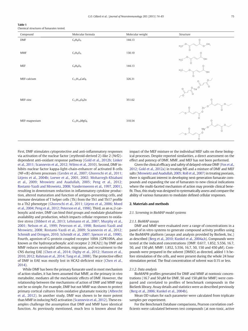
Table 1Chemical structures of fumarates tested.
Compound Molecular formula Molecular weight Structure
DMF C6H8O4 144.13
MMF C5H6O4 130.10
MEF C6H8O4 144.13
MEF-calcium C12H14CaO8 326.31
MEF-zinc C12H14O8Zn 351.62
MEF-magnesium C12H14MgO8 310.54
75G.O. Gillard et al. / Journal of Neuroimmunology 283 (2015) 74–85
First, DMF stimulates cytoprotective and anti-inflammatory responsesvia activation of the nuclear factor (erythroid-derived 2)-like 2 (Nrf2)-dependent anti-oxidant response pathway (Gold et al., 2012b; Linkeret al., 2011; Scannevin et al., 2012;Wilms et al., 2010). Second, DMF in-hibits nuclear factor kappa-light-chain-enhancer of activated B cells(NF-κB)-driven processes (Gerdes et al., 2007; Ghoreschi et al., 2011;Litjens et al., 2004b; Loewe et al., 2001, 2002; Moharregh-Khiabaniet al., 2009; Mrowietz and Asadullah, 2005; Peng et al., 2012;Rostami-Yazdi and Mrowietz, 2008; Vandermeeren et al., 1997, 2001),resulting in downstream reduction in inflammatory cytokine produc-tion, altered maturation and function of antigen-presenting cells, andimmune deviation of T helper cells (Th) from the Th1 and Th17 profileto a Th2 phenotype (Ghoreschi et al., 2011; Litjens et al., 2006; Moedet al., 2004; Peng et al., 2012; Peterson et al., 1998). Third, as anα, β car-boxylic acid ester, DMF can bind thiol groups andmodulate glutathioneavailability and production, which impacts cellular responses to oxida-tive stress (Dibbert et al., 2013; Lehmann et al., 2007; Murphy et al.,2001; Nelson et al., 1999; Peterson et al., 1998; Rostami-Yazdi andMrowietz, 2008; Rostami-Yazdi et al., 2009; Scannevin et al., 2012;Schmidt and Dringen, 2010; Schmidt et al., 2007; Spencer et al., 1990).Fourth, agonism of G-protein coupled receptor 109A (GPR109A, alsoknown as the hydroxycarboxylic acid receptor 2 (HCA2)) by DMF andMMF reduces neutrophil adhesion, migration, and recruitment to theCNS during EAE (Chen et al., 2014; Digby et al., 2012; Hanson et al.,2010, 2012; Rahman et al., 2014; Tang et al., 2008). The protective effectof DMF in EAE was mostly lost in HCA2-deficient mice (Chen et al.,2014).
While DMF has been the primary fumarate used inmostmechanismof action studies, it has been assumed that MMF, as the primary in vivometabolite, mediates all the mechanistic effects of DMF. However, therelationship between the mechanisms of action of DMF and MMF maynot be so simple. For example, DMF but not MMFwas shown to protectprimary cortical cultures from oxidative glutamate toxicity (Albrechtet al., 2012). In another study, DMF was shown to be more potentthanMMF in inducingNrf2 activation (Scannevin et al., 2012). These ex-amples challenge the assumption that DMF and MMF have identicalfunction. As previously mentioned, much less is known about the
impact of the MEF mixture or the individual MEF salts on these biolog-ical processes. Despite reported similarities, a direct assessment on theeffect and potency of DMF, MMF, and MEF has not been performed.
Given the clinical efficacy and safety of delayed-releaseDMF (Foxet al.,2012; Gold et al., 2012a) in treating MS and a mixture of DMF and MEFsalts (Mrowietz andAsadullah, 2005; Roll et al., 2007) in treatingpsoriasis,there is significant interest in developing next-generation fumarate com-pounds and expanding the use of fumarates to new clinical indicationswhere the multi-faceted mechanism of action may provide clinical bene-fit. Thus, this studywas designed to systematically assess and compare theability of various fumarates to modulate defined cellular responses.
2. Materials and methods
2.1. Screening in BioMAP model systems
2.1.1. BioMAP assaysDMF and MMF were evaluated over a range of concentrations in a
panel of in vitro systems to generate compound activity profiles usingthe BioMAP® platform (assays and analysis provided by BioSeek, Inc.)as described (Berg et al., 2010; Kunkel et al., 2004a,b). Compounds weretested at the indicated concentrations (DMF: 0.617, 1.852, 5.556, 16.7,50, and 150 μM; MMF: 1.852, 5.556, 16.7, 50, 150 and 450 μM). Com-pounds were prepared in the solvent (DMSO) as directed, added 1 h be-fore stimulation of the cells, and were present during the whole 24 hourstimulation period. The final concentration of solvent was 0.1% or less.
2.1.2. Data analysisBioMAP® profiles generated for DMF and MMF at nontoxic concen-
trations (16.7 and 50 μM for DMF, 50 and 150 μM for MMF) were com-pared and correlated to profiles of benchmark compounds in theBioSeek library. Assay details and statisticswere as described previously(Berg et al., 2006; Kunkel et al., 2004b).
Mean OD values for each parameter were calculated from triplicatesamples per experiment.
For the Benchmark Database comparisons, Pearson correlation coef-ficients were calculated between test compounds (at non-toxic, active
76 G.O. Gillard et al. / Journal of Neuroimmunology 283 (2015) 74–85
doses) and compounds from BioSeek's database (3C, 4H, LPS and SAgsystems). Statistically significant correlations were identified based oncomparisons of these true Pearson correlations to a set of Pearson corre-lations obtained from randomizing the experimentally observed values.
2.2. HCS screening of translocation of p65 and p52
2.2.1. Reference compound validation of assayThe reference compound for TNF-induced p65 nuclear translocation
is 2-Amino-6-[2-(cyclopropylmethoxy)-6-hydroxyphenyl]-4-(4-piperidinyl)-3-pyridinecarbonitrile (ACHP), a commercially availableIKKβ Inhibitor (Tocris Bioscience, Bristol, UK). The p52 translocationassay was validated using BIO-032202, an in-house reference compoundbased on a competitor's patented compound. All IC50 curves for ACHP andBIO-032202 met acceptance criteria.
2.2.2. Dilution of fumaratesCompoundswere added to the cells 30min before stimulation. Eight
doses of the fumaric acid esters (DMF, MMF, and MEF) were tested: 18,12, 6, 3, 1, 0.5, 0.25, and 0.125 μg/mL. In plates 3 and 4, dose–responsecurves were tested in the presence of 3 μg/mL DMF or MMF.
2.2.3. Stimulation and assessment of p65 and p52 translocationU-2 OS cells were stimulated with 10 ng/mL TNF-α for 30 min (p65
assay) or 10 ng/mL of the anti-lymphotoxin β receptor (LTβR) antibodyBS-1 for 4 h (p52 assay). The cells were then fixed and stained with anantibody that recognizes the appropriate protein. Images of each wellwere acquired on the PerkinElmer Operetta and the images were ana-lyzed using PerkinElmer Columbus Image Analysis software. All plateshad a Z′ score ≥0.5 and a signal-to-noise ratio of ≥5.
2.3. Ramos-Blue reporter assay
2.3.1. Culturing Ramos-Blue B lymphocyte cell lineRamos-Blue cells were thawed from the manufacturer (InvivoGen)
and cultured in Ramos-Blue cell culture medium without the selectiveantibiotic Zeocin. The cells were then passaged every 3–4 days in theRamos-blue cell culture mediumwith Zeocin at 100 μg/mL to a concen-tration of 1 × 10⁶ cells/mL, and never exceeding 6 × 10⁶ cells/mL.
2.3.2. Dilution of DMF, MMF, and MEFCompounds were prepared on a plate in DMSO at 15 μg/mL and se-
quentially diluted in order to generate the different fumarate stock con-centrations in a standard volume of DMSO. This dilution plate was usedto make the final dilution of 5× compound in Ramos-blue cell culturemedia. This was done by adding 3 μl from each well (concentration) ofthe dilution plate to a 1.7 mL microcentrifuge tube containing 997 μlof cell culture media.
2.3.3. Pre-incubation of Ramos-Blue cells with DMF, MMF, and MEFRamos-blue cells were re-suspended in Ramos-Blue culturemedium
without Zeocin at a concentration of 2 × 10⁶ cells/mL. 175 μl of cells wasadded to the appropriate number of wells in a 96-well round bottomplate. The wells were treated with 50 μl of the varying concentrationsof each compound from the dilution tubes. The wells were incubatedfor 30 min at 37 °C and 5% CO2.
2.3.4. Activation of Ramos-Blue cellsWells were treated with 25 μl of ODN 2006 at a final working con-
centration of 10 μg/mL. For CD40-stimulated cells, agonistic anti-human CD40 antibody clone G28.5 (Biolegend, Ultra-LEAF αCD40)was added to a final working concentration of 10 μg/mL. Isotype controlantibody (BioLegend; Ultra-LEAF mouse IgG1 κ, clone MOPC-21) wasused at equal concentration for unstimulated wells. The wells were in-cubated for 24 h at 37 °C and 5% CO2.
2.3.5. Detection of NF-κB activity through the production of secretedembryonic alkaline phosphatase (SEAP)
Theplatewas centrifuged at 1800 rpm for 10min to pellet cells. 40 μlof supernatant was taken from thewells and put into a new 96-well flatbottom plate. 200 μl of QUANTI-Blue reaction mixture, pre-warmed to37 °C, was added to the wells. The wells were incubated at 37 °C for75 min in the dark. The wells were read on a microplate reader usinga wavelength of 635 nm.
2.4. Cytokine responses in fumarate-treated primary splenocytes inresponse to lipopolysaccharide (LPS) or CpG-B stimulation
2.4.1. Preparation of splenocytesPrimary splenocyteswere isolated from age-matchedNRF2−/−mice
or age-matched control wild-type (WT) mice. Splenocytes were recov-ered via mechanical dissociation of spleens, followed by washing andlysis of RBS (Sigma). Cells were then counted, centrifuged, and resus-pended at 2 × 10⁶ cells/mL in X-VIVO 10 (Lonza) chemically definedmedia and stored on ice.
2.4.2. Preparation of fumarate dilutions and working stocksStocks of fumarates for dilution series were at an initial concentra-
tion of 30 mg/mL in DMSO. Final concentrations to be tested were9 μg/mL, 6 μg/mL, 3 μg/mL, and 1 μg/mL. Working stocks (4× final con-centration) of fumarateswere generated inX-VIVO 10media andmixedwell.
2.4.3. Preparations of working stocks of LPS, CpG-B ODN 2006, CpG-B ODN1826, and controls
Stocks of LPS (Sigma-Aldrich, ion-exchange chromatography puri-fied from Escherichia coli strain 0111:B4) and CpG-B oligonucleotides(1826 for murine cell stimulation and 2006 for human PBMC stimula-tion; InVivoGen, San Diego, CA) were prepared at 40 ng/mL and40 μg/mL, respectively, in X-VIVO 10media, to yieldfinal concentrationsof 10 ng/mL and 10 μg/mL, respectively; 2 replicates per condition withno stimulation (vehicle control for each fumarate condition). Wells re-ceiving vehicle (PBS) received 6 μg/mL (final concentration) offumarates.
2.4.4. Preparation of platesFor each plate, 50 μl of fumarate/DMSO control working stock were
added, followed by 50 μl of stimulation mix, followed by 100 μl of cellsat 2 × 10⁶ cells/mL. Combination ofworking stocks and cells in final vol-ume of 200 μl/well result in the desired final concentrations of fuma-rates (9, 6, 3, 1, 0 μg/mL) and stimuli (10 ng/mL for LPS and 10 μg/mLfor CpGs). Plates (2 plates per stimulus) were then transferred into37 °C incubator and supernatants were harvested 24 h later and trans-ferred to new 96 well plates that were immediately placed in −80 °C.
2.4.5. Quantification of cytokine/chemokine in cell culture supernatantsThe Milliplex Magnetic multiplex mouse cytokine/chemokine
magnetic premixed bead immunoassay (premixed 25-plex; Cat #MCYTOMAG-70 K-PMX) was used to quantify cytokine levels in tissueculture supernatants in direct accordance with the manufacturer's pro-tocol. Freshly thawed tissue culture supernatant (25 μl) was assayed induplicate. All plates were run with overnight incubation of sample andbeads at 4 °C with shaking. After washing and resuspension, beadswere read on either a Luminex 100 reader or a Luminex 200 reader.
3. Results
3.1. DMF and MMF show significant overlap in activity profiles in BioMAPassays but marked differences in potency
Given that numerous cellular pathways are affected by fumarates,we began our study with a systematic screen for similarities and
MMF 50 μM
DMF 50 μM
MMF
DMF
A
B
C
log
rat
iooitar
gol
oitarg
ol
150 μM50 μM16.667 μM5.556 μM1.852 μM0.617 μM
450 μM150 μM50 μM16.667 μM5.556 μM1.852 μM
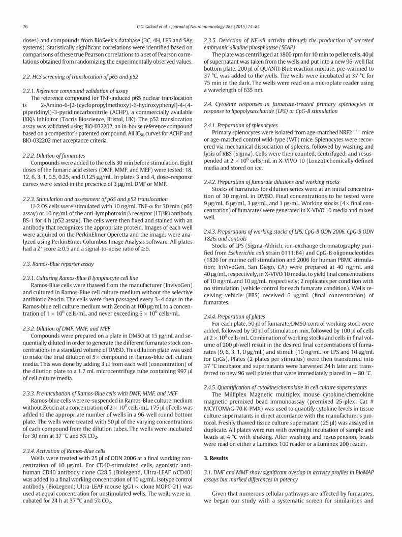
Fig. 1. BioMAP profiles for DMF and MMF. BioMAP profiles of DMF (A) and MMF (B), tested at multiple concentrations in 7 BioMAP systems. The biomarker readouts measured (see theMaterials andmethods section) are indicated along the x-axis. The y-axis shows the log10 expression ratios of the readout level measurements relative to solvent (DMSObuffer) controls.Each datapoint represents a single well. The gray area above and below the dashed line indicates the 95% significance envelope of DMSO negative controls. The BioMAP profiles for DMFand MMF at a standard concentration of 50 mM are plotted in C.
77G.O. Gillard et al. / Journal of Neuroimmunology 283 (2015) 74–85
differences between DMF and MMF using the BioMAP® system. TheBioMAP® analysis system consists of 7 individual assays that use com-plex co-cultures of primary human cells to model the effects of tested
Table 2Reproducibility between two repeat BioMAP experiments.
Compound Dose Pearson correlation across seven BioMAP systems
DMF 150 μM (toxic) 0.87DMF 50 μM 0.95DMF 16.7 μM 0.86MMF 450 μM (toxic) 0.93MMF 150 μM 0.83
compounds on cell intrinsic-responses and diverse cell–cell interactions(Kunkel et al., 2004a,b). The effects of tested compounds are read outusing multiple parameters for each of the seven assays. Additional de-tails on the seven individual assays and the readouts for each assaycan be found in Supplemental Table 1. The combined biological effectsof a compound on each analyte in each assay are used to generate an ac-tivity profile for the compound. The activity profile of a compound ob-served in BioMAP® systems can be used to identify molecules andpathways that are affected by a given compound, and can also becompared (“benchmarked”) against a database of activity profiles ofhundreds of experimental drugs and approved therapeutics. This“benchmarking” provides insight into the mechanism of action or
Table 3Benchmarking hits in BioSeek database identify DMF andMMF as NF-kB-pathwaymodulators. DMF andMMF profiles at non-toxic doseswere compared to profiles in the BioSeek Bench-mark Database as described in Appendix I. Compoundswith significantly related profiles (FDR b 0.05) are listed. The correlation coefficients (r) for the pairwise comparisons are shown forall four systems, and for individual systems.
r (individual systems)
Compound Database Match Mechanism r FDR LPS 3C SAG 4H
DMF (50 μM) Ro106-9920 (5.5 μM) IkBα ubiquitination inhibitor 0.820 0.012 0.960 0.926 0.362 0.936BAY 11-7085 (2.7 μM) IkBα phosphorylation inhibitor 0.782 0.012 0.954 0.900 0.569 0.852Parthenolide (3.7 μM) NF-κB inhibitor (p65 alkylation) 0.768 0.012 0.755 0.950 0.500 0.664GW8510 (8.3 μM) CDK inhibitor 0.805 0.014 0.936 0.772 0.884 0.988Roscovitine (25 μM) CDK inhibitor 0.666 0.063 0.669 0.766 0.312 0.915
DMF (16.7 μM) Ro106-9920 (1.8 μM) IkBα ubiquitination inhibitor 0.847 0.009 0.852 0.726 0.874 0.973Ro106-9920 (5.5 μM) IkBα ubiquitination inhibitor 0.773 0.012 0.546 0.958 0.893 0.968BAY 11-7085 (0.9 μM) IkBα phosphorylation inhibitor 0.842 0.009 0.860 0.801 0.973 0.713BAY 11-7085 (2.7 μM) IkBα phosphorylation inhibitor 0.833 0.009 0.694 0.983 0.964 0.902Parthenolide (3.7 μM) NF-κB inhibitor (p65 alkylation) 0.781 0.012 0.691 0.778 0.932 0.886
MMF (150 μM) Roscovitine (8.3 μM) CDK inhibitor 0.702 0.011 0.731 0.930 0.960 0.832Olomoucine (33 μM) CDK inhibitor 0.697 0.011 0.672 0.772 0.887 0.848BAY 11-7085 (0.9 μM) IkBα phosphorylation inhibitor 0.678 0.021 0.641 0.862 0.805 0.631BAY 11-7085 (2.8 μM) IkBα phosphorylation inhibitor 0.665 0.021 0.497 0.881 0.871 0.858Ro106-9920 (1.8 μM) IkBα ubiquitination inhibitor 0.675 0.021 0.695 0.735 0.611 0.899
78 G.O. Gillard et al. / Journal of Neuroimmunology 283 (2015) 74–85
secondary activities of test compounds (Berg et al., 2006, 2010; Kunkelet al., 2004a,b).
DMF and MMF were tested in 7 BioMAP® model systems at sevendoses starting at 150 μM (DMF) or 450 μM (MMF) with threefold dilu-tions. DMF and MMF were active in all 7 BioMAP® systems (Fig. 1).DMF was active between 5.5 and 50 μM (IC50 ~ 30 μM) and showedsigns of cellular toxicity at 150 μM (Fig. 1A). MMF was active between50 and 150 μM (IC50 ~ 190 μM) and was toxic at 450 μM (Fig. 1B).DMF and MMF exhibited similar BioMAP® profiles but at distinctly dif-ferent doses. Mechanistic clustering across multiple doses showed thebest correlation between the 16.7 μM DMF and 150 μM MMF doses(compare Fig. 1A and B) suggesting that the two compounds generatesimilar responses in these assays, albeit with different potencies. Thisis highlighted in Fig. 1C, where DMF and MMF are compared directlyat 50 μM. While direct dose and tissue exposure is difficult to deter-mine, the plasma concentration of MMF falls in a range between 10and 40 μM in vivo after an oral dose of 240 mg DMF (Litjens et al.,2004a,c). BioMAP profiles showed statistically significant inhibitionof VCAM-1 and E-selectin (but not ICAM-1) expression, in agree-ment with findings in psoriasis patients in vivo (Loewe et al.,2001). Additional markers affected by DMF andMMF, which fall out-side the 99.7% prediction envelope, included HLA-DR, Eotaxin-3,
BA
100
60
20
-20
80
40
10
5
-5
% in
hib
itio
n o
f S
EA
P p
rod
uct
ion
fumarate concentration (μμg/ml)
0
CpG
0 2 4 6 8 10
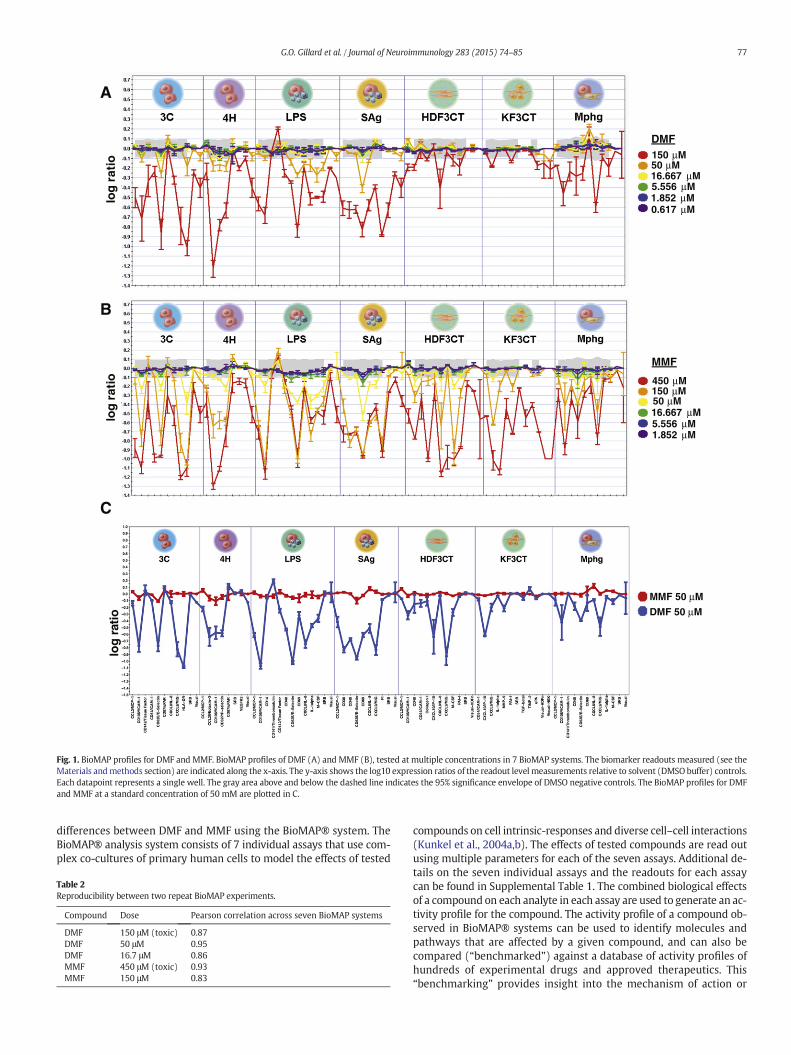
Fig. 2.DMF, but not other fumarates, inhibits canonical and non-canonical NF-κB reporter activNFκB/AP-1-inducible secreted alkaline phosphatase (SEAP) reporter gene following stimulationafter stimulation in vitro via TLR9 agonist CpG oligonucleotides ODN 2006 (A) or an agonisticshown on graph), DMF (blue diamonds), MMF (red circles), or MEF salt mixture (green invertassay. Relative alkaline phosphatase activity is presented as percent inhibition of alkaline phospiment of 3 independent experiments performed.
CD141/thrombomodulin, CD142/tissue factor, CD40, CD69, IL-1β,M-CSF, IL-8, IP-10, and MIG. The effect of DMF on IL-8, IP-10, andMIG production has been previously reported (Stoof et al., 2001).BioMAP activity profiles across the seven systems were highly repro-ducible, with a Pearson correlation between the two replicate exper-iments ranging from 0.83 to 0.95 (Table 2).
Statistical comparison of the DMF and MMF profiles (3C, 4H, LPS,SAg) with the profiles of compounds in the BioSeek database resultedin two types of hits: inhibitors of NF-κB signaling and cyclin-dependent kinase (CDK) inhibitors (Table 3). The NF-κB inhibitorgroup includes Ro106-9920, BAY 11-7085, and parthenolide. Thesehits suggest that DMF and MMF inhibit the NF-κB pathway at the levelof the NF-κB/IκB complex, in agreement with published studies(Loewe et al., 2001; Vandermeeren et al., 2001). However, the differ-ence in potency between DMF and MMF is profound.
The NF-κB pathway is a pivotal regulator of innate and adaptive im-munity, and its dysregulation is implicated in the chronic inflammationof autoimmune diseases. Given the published studies on fumaratemod-ulation of NF-κB signaling (Cross et al., 2011; Gerdes et al., 2007;Ghoreschi et al., 2011; Kim et al., 2010; Lee et al., 2009; Loewe et al.,2001, 2002; Lv et al., 2013; Peng et al., 2012; Vandermeeren et al.,2001) and the differences we saw between potency of DMF and MMF
0
0
0
0
fumarate concentration (μg/ml)
αCD40
0 2 4 6 8 10
DMF
MMF
MEF
ity in Ramos-Blue cells. Ramos-Blue B cells are a human B cell line that stably expresses anvia the NF-κB pathway. Supernatants from Ramos-blue cell cultures were harvested 24 hanti-CD40 antibody (B) in the presence of various concentrations of DMSO (vehicle; noted triangles) and assayed for alkaline phosphatase activity using a colorimetric enzymatichate activity, calculated vs. vehicle control (DMSO). Graphs depict a representative exper-
AIL-6 MIP-1ββ IP-10
0 5 10cyto
kin
e p
rod
uct
ion
(p
g/m
l) 1500
1000
500
0
1500
1000
500
0
60
20
40
0
B
LPS
* *
* ***
*
*
0 5 10 0 5 10
compound concentration (μg/mL)
CpG
IL-6 MIP-1β IP-10
0 2 4 6 8 10 0 2 4 6 8 10 0 2 4 6 8 10
cyto
kin
e p
rod
uct
ion
(p
g/m
l)
3000
2000
1000
0
8000
4000
2000
0
40
20
0***
**
** *
** * *
compound concentration (μg/mL)
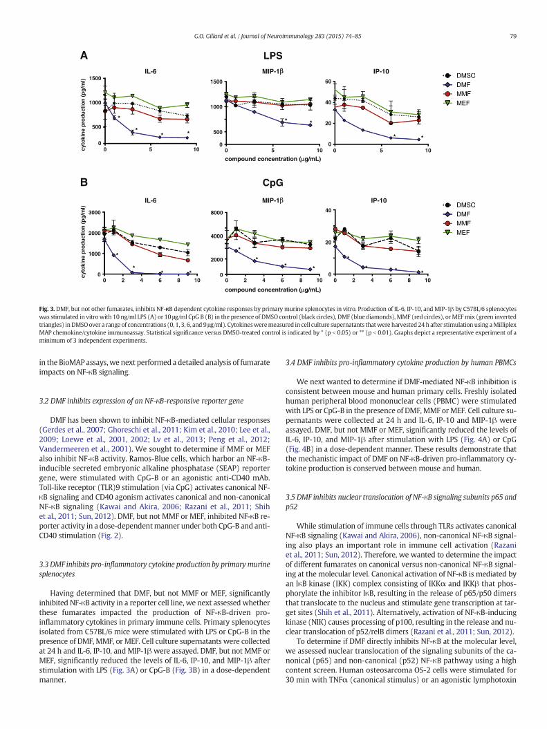
Fig. 3. DMF, but not other fumarates, inhibits NF-κB dependent cytokine responses by primary murine splenocytes in vitro. Production of IL-6, IP-10, and MIP-1β by C57BL/6 splenocyteswas stimulated in vitrowith 10 ng/ml LPS (A) or 10 μg/ml CpG B (B) in the presence of DMSO control (black circles), DMF (blue diamonds), MMF (red circles), orMEFmix (green invertedtriangles) inDMSOover a range of concentrations (0, 1, 3, 6, and 9 μg/ml). Cytokinesweremeasured in cell culture supernatants thatwere harvested 24 h after stimulation using aMilliplexMAP chemokine/cytokine immunoassay. Statistical significance versus DMSO-treated control is indicated by * (p b 0.05) or ** (p b 0.01). Graphs depict a representative experiment of aminimum of 3 independent experiments.
79G.O. Gillard et al. / Journal of Neuroimmunology 283 (2015) 74–85
in the BioMAP assays,we next performed a detailed analysis of fumarateimpacts on NF-κB signaling.
3.2 DMF inhibits expression of an NF-κB-responsive reporter gene
DMF has been shown to inhibit NF-κB-mediated cellular responses(Gerdes et al., 2007; Ghoreschi et al., 2011; Kim et al., 2010; Lee et al.,2009; Loewe et al., 2001, 2002; Lv et al., 2013; Peng et al., 2012;Vandermeeren et al., 2001). We sought to determine if MMF or MEFalso inhibit NF-κB activity. Ramos-Blue cells, which harbor an NF-κB-inducible secreted embryonic alkaline phosphatase (SEAP) reportergene, were stimulated with CpG-B or an agonistic anti-CD40 mAb.Toll-like receptor (TLR)9 stimulation (via CpG) activates canonical NF-κB signaling and CD40 agonism activates canonical and non-canonicalNF-κB signaling (Kawai and Akira, 2006; Razani et al., 2011; Shihet al., 2011; Sun, 2012). DMF, but not MMF or MEF, inhibited NF-κB re-porter activity in a dose-dependentmanner under both CpG-B and anti-CD40 stimulation (Fig. 2).
3.3 DMF inhibits pro-inflammatory cytokine production by primarymurinesplenocytes
Having determined that DMF, but not MMF or MEF, significantlyinhibited NF-κB activity in a reporter cell line, we next assessed whetherthese fumarates impacted the production of NF-κB-driven pro-inflammatory cytokines in primary immune cells. Primary splenocytesisolated from C57BL/6 mice were stimulated with LPS or CpG-B in thepresence of DMF, MMF, or MEF. Cell culture supernatants were collectedat 24 h and IL-6, IP-10, and MIP-1βwere assayed. DMF, but not MMF orMEF, significantly reduced the levels of IL-6, IP-10, and MIP-1β afterstimulation with LPS (Fig. 3A) or CpG-B (Fig. 3B) in a dose-dependentmanner.
3.4 DMF inhibits pro-inflammatory cytokine production by human PBMCs
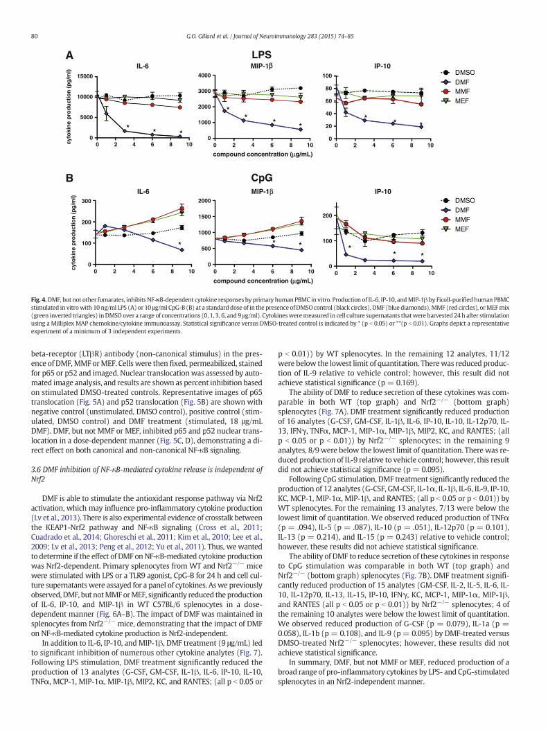
We next wanted to determine if DMF-mediated NF-κB inhibition isconsistent between mouse and human primary cells. Freshly isolatedhuman peripheral blood mononuclear cells (PBMC) were stimulatedwith LPS or CpG-B in the presence of DMF, MMF orMEF. Cell culture su-pernatants were collected at 24 h and IL-6, IP-10 and MIP-1β wereassayed. DMF, but not MMF or MEF, significantly reduced the levels ofIL-6, IP-10, and MIP-1β after stimulation with LPS (Fig. 4A) or CpG(Fig. 4B) in a dose-dependent manner. These results demonstrate thatthe mechanistic impact of DMF on NF-κB-driven pro-inflammatory cy-tokine production is conserved between mouse and human.
3.5 DMF inhibits nuclear translocation of NF-κB signaling subunits p65 andp52
While stimulation of immune cells through TLRs activates canonicalNF-κB signaling (Kawai and Akira, 2006), non-canonical NF-κB signal-ing also plays an important role in immune cell activation (Razaniet al., 2011; Sun, 2012). Therefore, we wanted to determine the impactof different fumarates on canonical versus non-canonical NF-κB signal-ing at the molecular level. Canonical activation of NF-κB is mediated byan IκB kinase (IKK) complex consisting of IKKα and IKKβ that phos-phorylate the inhibitor IκB, resulting in the release of p65/p50 dimersthat translocate to the nucleus and stimulate gene transcription at tar-get sites (Shih et al., 2011). Alternatively, activation of NF-κB-inducingkinase (NIK) causes processing of p100, resulting in the release and nu-clear translocation of p52/relB dimers (Razani et al., 2011; Sun, 2012).
To determine if DMF directly inhibits NF-κB at the molecular level,we assessed nuclear translocation of the signaling subunits of the ca-nonical (p65) and non-canonical (p52) NF-κB pathway using a highcontent screen. Human osteosarcoma OS-2 cells were stimulated for30 min with TNFα (canonical stimulus) or an agonistic lymphotoxin
A LPSIL-6 MIP-1ββ IP-10
cyto
kin
e p
rod
uct
ion
(p
g/m
l)
15000
10000
5000
0
* * *0 2 4 6 8 10 0 2 4 6 8 100 2 4 6 8 10
4000
3000
2000
0
1000
100
80
60
20
40
0
** * *
* * *
compound concentration (μg/mL)
B CpG1-PIM6-LI β IP-10
cyto
kin
e p
rod
uct
ion
(p
g/m
l)
0 2 4 6 8 10 0 2 4 6 8 10 0 2 4 6 8 10
300
200
100
0
2000
1500
1000
0
500
200
100
0
* * * ** *
compound concentration (μg/mL)
Fig. 4.DMF, but not other fumarates, inhibits NF-κB-dependent cytokine responses by primary human PBMC in vitro. Production of IL-6, IP-10, andMIP-1β by Ficoll-purified human PBMCstimulated in vitrowith 10 ng/ml LPS (A) or 10 μg/ml CpG-B (B) at a standard dose of in the presence of DMSO control (black circles), DMF (blue diamonds),MMF (red circles), orMEFmix(green inverted triangles) inDMSO over a range of concentrations (0, 1, 3, 6, and 9 μg/ml). Cytokinesweremeasured in cell culture supernatants thatwere harvested 24h after stimulationusing a Milliplex MAP chemokine/cytokine immunoassay. Statistical significance versus DMSO-treated control is indicated by * (p b 0.05) or **(p b 0.01). Graphs depict a representativeexperiment of a minimum of 3 independent experiments.
80 G.O. Gillard et al. / Journal of Neuroimmunology 283 (2015) 74–85
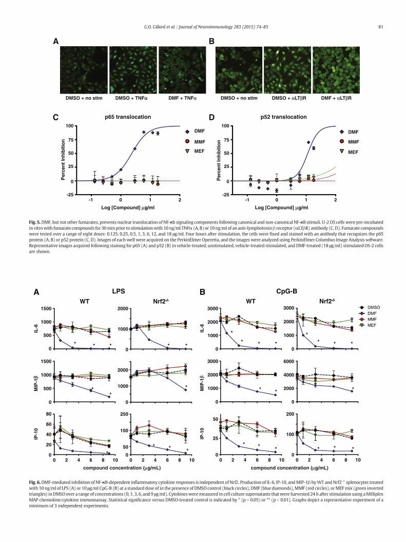
beta-receptor (LTβR) antibody (non-canonical stimulus) in the pres-ence of DMF,MMF orMEF. Cells were then fixed, permeabilized, stainedfor p65 or p52 and imaged. Nuclear translocation was assessed by auto-mated image analysis, and results are shown as percent inhibition basedon stimulated DMSO-treated controls. Representative images of p65translocation (Fig. 5A) and p52 translocation (Fig. 5B) are shown withnegative control (unstimulated, DMSO control), positive control (stim-ulated, DMSO control) and DMF treatment (stimulated, 18 μg/mLDMF). DMF, but not MMF or MEF, inhibited p65 and p52 nuclear trans-location in a dose-dependent manner (Fig. 5C, D), demonstrating a di-rect effect on both canonical and non-canonical NF-κB signaling.
3.6 DMF inhibition of NF-κB-mediated cytokine release is independent ofNrf2
DMF is able to stimulate the antioxidant response pathway via Nrf2activation, which may influence pro-inflammatory cytokine production(Lv et al., 2013). There is also experimental evidence of crosstalk betweenthe KEAP1-Nrf2 pathway and NF-κB signaling (Cross et al., 2011;Cuadrado et al., 2014; Ghoreschi et al., 2011; Kim et al., 2010; Lee et al.,2009; Lv et al., 2013; Peng et al., 2012; Yu et al., 2011). Thus, we wantedto determine if the effect of DMF onNF-κB-mediated cytokine productionwas Nrf2-dependent. Primary splenocytes from WT and Nrf2−/− micewere stimulated with LPS or a TLR9 agonist, CpG-B for 24 h and cell cul-ture supernatantswere assayed for a panel of cytokines. Aswe previouslyobserved,DMF, but notMMForMEF, significantly reduced theproductionof IL-6, IP-10, and MIP-1β in WT C57BL/6 splenocytes in a dose-dependent manner (Fig. 6A–B). The impact of DMF was maintained insplenocytes from Nrf2−/− mice, demonstrating that the impact of DMFon NF-κB-mediated cytokine production is Nrf2-independent.
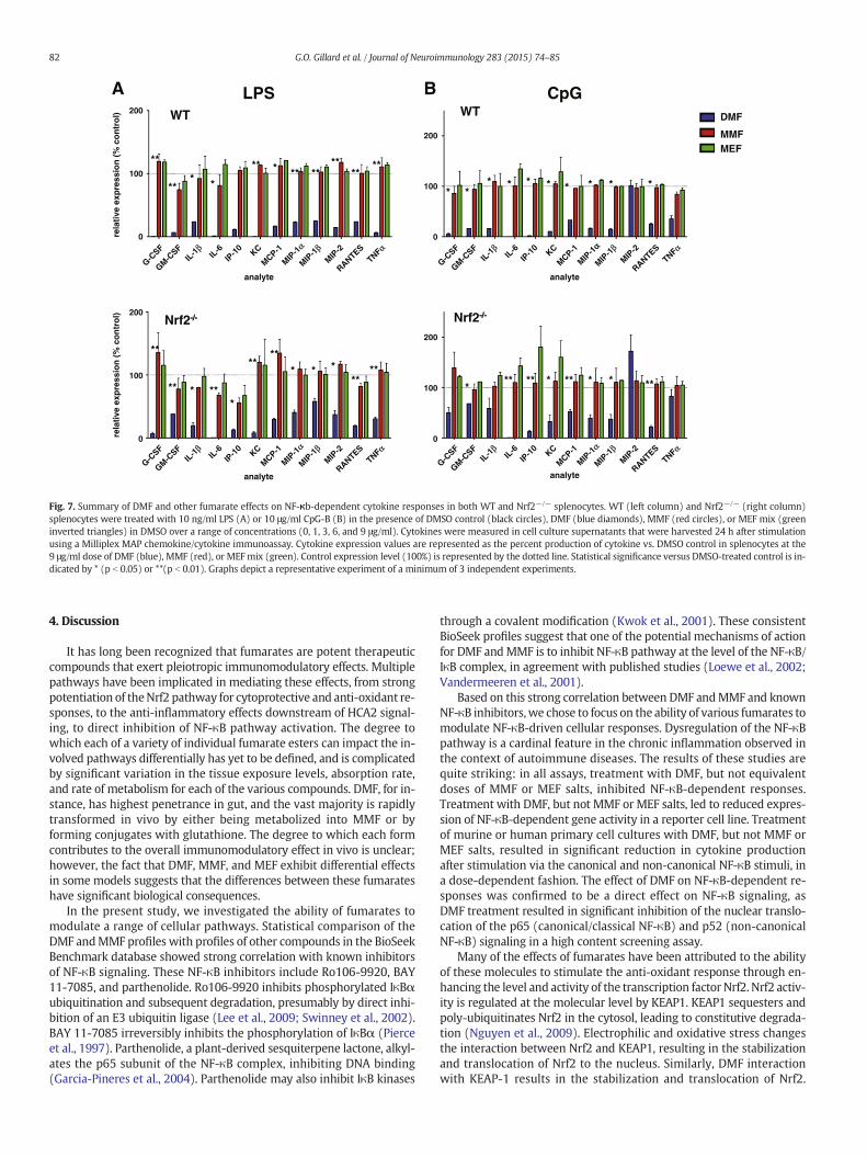
In addition to IL-6, IP-10, and MIP-1β, DMF treatment (9 μg/mL) ledto significant inhibition of numerous other cytokine analytes (Fig. 7).Following LPS stimulation, DMF treatment significantly reduced theproduction of 13 analytes (G-CSF, GM-CSF, IL-1β, IL-6, IP-10, IL-10,TNFα, MCP-1, MIP-1α, MIP-1β, MIP2, KC, and RANTES; (all p b 0.05 or
p b 0.01)) by WT splenocytes. In the remaining 12 analytes, 11/12were below the lowest limit of quantitation. Therewas reduced produc-tion of IL-9 relative to vehicle control; however, this result did notachieve statistical significance (p = 0.169).
The ability of DMF to reduce secretion of these cytokines was com-parable in both WT (top graph) and Nrf2−/− (bottom graph)splenocytes (Fig. 7A). DMF treatment significantly reduced productionof 16 analytes (G-CSF, GM-CSF, IL-1β, IL-6, IP-10, IL-10, IL-12p70, IL-13, IFNγ, TNFα, MCP-1, MIP-1α, MIP-1β, MIP2, KC, and RANTES; (allp b 0.05 or p b 0.01)) by Nrf2−/− splenocytes; in the remaining 9analytes, 8/9 were below the lowest limit of quantitation. There was re-duced production of IL-9 relative to vehicle control; however, this resultdid not achieve statistical significance (p = 0.095).
Following CpG stimulation, DMF treatment significantly reduced theproduction of 12 analytes (G-CSF, GM-CSF, IL-1α, IL-1β, IL-6, IL-9, IP-10,KC, MCP-1, MIP-1α, MIP-1β, and RANTES; (all p b 0.05 or p b 0.01)) byWT splenocytes. For the remaining 13 analytes, 7/13 were below thelowest limit of quantitation. We observed reduced production of TNFα(p = .094), IL-5 (p = .087), IL-10 (p = .051), IL-12p70 (p = 0.101),IL-13 (p = 0.214), and IL-15 (p = 0.243) relative to vehicle control;however, these results did not achieve statistical significance.
The ability of DMF to reduce secretion of these cytokines in responseto CpG stimulation was comparable in both WT (top graph) andNrf2−/− (bottom graph) splenocytes (Fig. 7B). DMF treatment signifi-cantly reduced production of 15 analytes (GM-CSF, IL-2, IL-5, IL-6, IL-10, IL-12p70, IL-13, IL-15, IP-10, IFNγ, KC, MCP-1, MIP-1α, MIP-1β,and RANTES (all p b 0.05 or p b 0.01)) by Nrf2−/− splenocytes; 4 ofthe remaining 10 analytes were below the lowest limit of quantitation.We observed reduced production of G-CSF (p = 0.079), IL-1a (p =0.058), IL-1b (p = 0.108), and IL-9 (p = 0.095) by DMF-treated versusDMSO-treated Nrf2−/− splenocytes; however, these results did notachieve statistical significance.
In summary, DMF, but not MMF or MEF, reduced production of abroad range of pro-inflammatory cytokines by LPS- and CpG-stimulatedsplenocytes in an Nrf2-independent manner.
DMSO + no stim DMSO + TNFαα DMF + TNFα DMSO + no stim DMSO + αLTβR DMF + αLTβR
BA
Log [Compound] μg/ml
Per
cen
t In
hib
itio
n
-1 0 1 2-25
0
25
50
75
100
Log [Compound] μg/ml
Per
cen
t In
hib
itio
n
-1 0 1 2-25
0
25
50
75
100DMF
MMF
MEF
DMF
MMF
MEF
DC noitacolsnart 25pnoitacolsnart 56p
Fig. 5.DMF, but not other fumarates, prevents nuclear translocation of NF-κb signaling components following canonical and non-canonical NF-κB stimuli. U-2 OS cells were pre-incubatedin vitrowith fumarate compounds for 30minprior to stimulationwith 10 ng/ml TNFα (A, B) or 10ng/ml of an anti-lymphotoxinβ receptor (αLTβR) antibody (C, D). Fumarate compoundswere tested over a range of eight doses: 0.125, 0.25, 0.5, 1, 3, 6, 12, and 18 μg/ml. Four hours after stimulation, the cells were fixed and stained with an antibody that recognizes the p65protein (A, B) or p52 protein (C, D). Images of eachwell were acquired on the PerkinElmer Operetta, and the images were analyzed using PerkinElmer Columbus Image Analysis software.Representative images acquired following staining for p65 (A) and p52 (B) in vehicle-treated, unstimulated, vehicle-treated stimulated, andDMF-treated (18 μg/ml) stimulatedOS-2 cellsare shown.
A
IL-6
IP-1
0M
IP-1
ββ
1000
0
2000
0
100
200
1000
0
150
0
50
250
IL-6
IP-1
0M
IP-1
β
1000
0
2000
3000
2000
0
4000
6000
0
25
50
1000
0
2000
3000
1000
0
2000
3000
2000
20
0
40
80
500
0
1000
1500
500
0
1000
1500
60
B
0 2 4 6 8 10 0 2 4 6 8 10 0 2 4 6 8 10 0 2 4 6 8 10
LPS CpG-B
WT 2frNTW -/--/-Nrf2-/--/-
* *
* * ** * ***
***
** **
**** * *
***
* *
*
compound concentration (μg/mL) compound concentration (μg/mL)
Fig. 6.DMF-mediated inhibition of NF-κB-dependent inflammatory cytokine responses is independent of Nrf2. Production of IL-6, IP-10, andMIP-1β byWT andNrf2-/- splenocytes treatedwith 10 ng/ml of LPS (A) or 10 μg/ml CpG-B (B) at a standard dose of in the presence of DMSO control (black circles), DMF (blue diamonds),MMF (red circles), orMEFmix (green invertedtriangles) inDMSOover a range of concentrations (0, 1, 3, 6, and 9 μg/ml). Cytokinesweremeasured in cell culture supernatants thatwere harvested 24 h after stimulation using aMilliplexMAP chemokine/cytokine immunoassay. Statistical significance versus DMSO-treated control is indicated by * (p b 0.05) or ** (p b 0.01). Graphs depict a representative experiment of aminimum of 3 independent experiments.
81G.O. Gillard et al. / Journal of Neuroimmunology 283 (2015) 74–85
Are
lati
ve e
xpre
ssio
n (
% c
on
tro
l)
analyte
0
100
200re
lati
ve e
xpre
ssio
n (
% c
on
tro
l)
analyte
0
100
200
LPS B CpG
analyte
200
100
0
0
100
200
analyte
G-CSF
GM-CSF
IL-6
IP-1
0KC
MIP-1
αα
MIP-1
βMIP
-2
RANTESTNFα
DMF
MMFMEF
Nrf2-/--/-
WT
Nrf2-/--/-
WT
MCP-1IL
-1β
G-CSF
GM-CSF
IL-6
IP-1
0KC
MIP-1
α
MIP-1
βMIP
-2
RANTESTNFα
MCP-1IL
-1β
G-CSF
GM-CSF
IL-6
IP-1
0KC
MIP-1
α
MIP-1
βMIP
-2
RANTESTNFα
MCP-1IL
-1β
G-CSF
GM-CSF
IL-6
IP-1
0KC
MIP-1
α
MIP-1
βMIP
-2
RANTESTNFα
MCP-1IL
-1β
***
*
* * *
**
** **
****
****
****
******
**
**
**
* *
*
* ** * * * * * * *
** * *** ** **
Fig. 7. Summary of DMF and other fumarate effects on NF-κb-dependent cytokine responses in both WT and Nrf2−/− splenocytes. WT (left column) and Nrf2−/− (right column)splenocytes were treated with 10 ng/ml LPS (A) or 10 μg/ml CpG-B (B) in the presence of DMSO control (black circles), DMF (blue diamonds), MMF (red circles), or MEF mix (greeninverted triangles) in DMSO over a range of concentrations (0, 1, 3, 6, and 9 μg/ml). Cytokines were measured in cell culture supernatants that were harvested 24 h after stimulationusing a Milliplex MAP chemokine/cytokine immunoassay. Cytokine expression values are represented as the percent production of cytokine vs. DMSO control in splenocytes at the9 μg/ml dose of DMF (blue), MMF (red), or MEF mix (green). Control expression level (100%) is represented by the dotted line. Statistical significance versus DMSO-treated control is in-dicated by * (p b 0.05) or **(p b 0.01). Graphs depict a representative experiment of a minimum of 3 independent experiments.
82 G.O. Gillard et al. / Journal of Neuroimmunology 283 (2015) 74–85
4. Discussion
It has long been recognized that fumarates are potent therapeuticcompounds that exert pleiotropic immunomodulatory effects. Multiplepathways have been implicated in mediating these effects, from strongpotentiation of theNrf2 pathway for cytoprotective and anti-oxidant re-sponses, to the anti-inflammatory effects downstream of HCA2 signal-ing, to direct inhibition of NF-κB pathway activation. The degree towhich each of a variety of individual fumarate esters can impact the in-volved pathways differentially has yet to be defined, and is complicatedby significant variation in the tissue exposure levels, absorption rate,and rate of metabolism for each of the various compounds. DMF, for in-stance, has highest penetrance in gut, and the vast majority is rapidlytransformed in vivo by either being metabolized into MMF or byforming conjugates with glutathione. The degree to which each formcontributes to the overall immunomodulatory effect in vivo is unclear;however, the fact that DMF, MMF, and MEF exhibit differential effectsin some models suggests that the differences between these fumarateshave significant biological consequences.
In the present study, we investigated the ability of fumarates tomodulate a range of cellular pathways. Statistical comparison of theDMF andMMF profiles with profiles of other compounds in the BioSeekBenchmark database showed strong correlation with known inhibitorsof NF-κB signaling. These NF-κB inhibitors include Ro106-9920, BAY11-7085, and parthenolide. Ro106-9920 inhibits phosphorylated IκBαubiquitination and subsequent degradation, presumably by direct inhi-bition of an E3 ubiquitin ligase (Lee et al., 2009; Swinney et al., 2002).BAY 11-7085 irreversibly inhibits the phosphorylation of IκBα (Pierceet al., 1997). Parthenolide, a plant-derived sesquiterpene lactone, alkyl-ates the p65 subunit of the NF-κB complex, inhibiting DNA binding(Garcia-Pineres et al., 2004). Parthenolide may also inhibit IκB kinases
through a covalent modification (Kwok et al., 2001). These consistentBioSeek profiles suggest that one of the potential mechanisms of actionfor DMF andMMF is to inhibit NF-κB pathway at the level of the NF-κB/IκB complex, in agreement with published studies (Loewe et al., 2002;Vandermeeren et al., 2001).
Based on this strong correlation between DMF andMMF and knownNF-κB inhibitors,we chose to focus on the ability of various fumarates tomodulate NF-κB-driven cellular responses. Dysregulation of the NF-κBpathway is a cardinal feature in the chronic inflammation observed inthe context of autoimmune diseases. The results of these studies arequite striking: in all assays, treatment with DMF, but not equivalentdoses of MMF or MEF salts, inhibited NF-κB-dependent responses.Treatment with DMF, but not MMF or MEF salts, led to reduced expres-sion of NF-κB-dependent gene activity in a reporter cell line. Treatmentof murine or human primary cell cultures with DMF, but not MMF orMEF salts, resulted in significant reduction in cytokine productionafter stimulation via the canonical and non-canonical NF-κB stimuli, ina dose-dependent fashion. The effect of DMF on NF-κB-dependent re-sponses was confirmed to be a direct effect on NF-κB signaling, asDMF treatment resulted in significant inhibition of the nuclear translo-cation of the p65 (canonical/classical NF-κB) and p52 (non-canonicalNF-κB) signaling in a high content screening assay.
Many of the effects of fumarates have been attributed to the abilityof these molecules to stimulate the anti-oxidant response through en-hancing the level and activity of the transcription factor Nrf2. Nrf2 activ-ity is regulated at the molecular level by KEAP1. KEAP1 sequesters andpoly-ubiquitinates Nrf2 in the cytosol, leading to constitutive degrada-tion (Nguyen et al., 2009). Electrophilic and oxidative stress changesthe interaction between Nrf2 and KEAP1, resulting in the stabilizationand translocation of Nrf2 to the nucleus. Similarly, DMF interactionwith KEAP-1 results in the stabilization and translocation of Nrf2.
83G.O. Gillard et al. / Journal of Neuroimmunology 283 (2015) 74–85
There is also a body of literature on the cross-talk between the KEAP1-Nrf2 pathway and NF-κB signaling. Lee et al. demonstrated that KEAP1binds IKKβ and mediates ubiquitination and subsequent degradation(Lee et al., 2009). Depletion of KEAP1 led to an increase in NF-κB signal-ing. A separate study demonstrated that genetic knockdown of KEAP1led to enhanced IL-6 production, an NF-κB target gene, in LPS-stimulated macrophages. Interestingly, we demonstrate in our studythat DMF treatment inhibited pro-inflammatory cytokine productionby culturedWT and Nrf2−/− splenocytes with equal efficiency, demon-strating that the ability of DMF to suppress NF-κB dependent responseswas independent of Nrf2 activity.
These results demonstrate that equivalent amounts of DMF, MMF,and MEF salts display distinct abilities to influence immune responsesvia the NF-κB pathway in these assays. As mentioned previously, DMFis rapidly metabolized to MMF (its primary metabolite) and also formsglutathione adjuncts. Based on the metabolism and pharmacokineticsof DMF it is impossible to know the in vivo targets and local tissue expo-sure of DMF. The assumption has been that significant direct exposureto unmodified/unmetabolized DMF is limited to a small population ofcells and that consequently, the therapeutic effects of DMF aremediatedby its primary metabolite MMF. As a consequence, preclinical studieshave traditionally used DMF and MMF interchangeably both in vitroand in vivo. However, this assumption isflawed, as the tissue concentra-tion and target cell exposure toMMF in vivo is not known, and thereforeit is difficult to draw meaningful parallels between in vitro and in vivoactive concentration for MMF. In the absence of knownMMF target tis-sue concentrations for modeling the effects of fumarates, we based therange of concentrations used in our NF-κB-driven assays on the report-ed concentration range for MMF in the plasma of patients, where MMFconcentration has been measured in a range from 1 to 5 μg/mL (≈10–40 μM) (Litjens et al., 2004a; Sheikh et al., 2013). The highest dose weused in our cytokine production assays, 9 μg/mL, corresponds to an ap-proximate concentration of 60 μM for DMF and 70 μM for MMF. Basedon the assumption that the range of concentrations used approximatesthe plasma concentration of MMF in patients, the differential fumarateeffects on NF-κB activity observed may well represent effects at amore “physiological” concentration. The BioSeek profiles for DMF andMMF indicate that these concentrations fall well below toxicity levelfor both compounds but reflect a significant difference in potency be-tween the two compounds for modifying NF-κB-driven responses, asDMF had an IC50 of approximately 30 μM versus 190 μM for MMF. Thepharmacokinetics and serum concentration of MEFs in vivo is similarto that of MMF, not DMF, so at similarly “physiological” concentrationsof MEFs, we did not observe any inhibition of NF-κB-driven responses.That we did not observe any significant effect of MMF or MEF on NF-kB-driven responses at these levels suggests that perhaps a significantcomponent of the therapeutic benefit of fumarates is mediated by ef-fects on cells and tissues that either have direct exposure to DMF or ex-perience very high local concentrations of MMF and MEF.
In this study, we have directly compared the activity of DMF andMMF.We observed significant differences between the biological activ-ities of these two closely related, yet distinct compounds. In addition,the inclusion of MEF salts without DMF also allowed us to separatethe activity of MEF salts on NF-κB-driven cytokine production fromthose mediated by DMF. These results establish that DMF is the mostpotent fumarate tested for modulating NF-κB signaling. The differentialeffects we observed on NF-κB-dependent responses were largely Nrf2-independent, despite the well-established role for Nrf2 in fumarate-mediated cellular responses. This suggests that the impact that fuma-rates exert on cellular responses is mediated by the integration oftheir effects on multiple and distinct pathways, each of which may bedifferentially affected depending on which fumarate is present. WhileDMF may be more effective than MMF or MEF salts at modulating NF-κB-driven responses, as these data indicate, the activation of Nrf2, themodulation of the glutathione system, or the activation of GPR109Amay all be mediated differentially depending on whether the cells
have been exposed to DMF, MMF, or MEF salts. In that vein, it is impor-tant to note that MMF is a significantly more potent activator of theHCA2 pathway (70 nM) than DMF (N10 μM) (Tang et al., 2008), andthat concentration of MMF falls well within the plasma exposure levelsfor MMF observed after patients receive a 240 mg dose of delayed-re-lease DMF (Cmax of 1–3 μg/mL MMF in plasma) (Sheikh et al., 2013).Given the common pharmacodynamic observation of flushing in de-layed-release DMF clinical trials (Fox, 2012; Fox et al., 2014; Gold,2011; Kappos et al., 2008, 2012), and the role of HCA2 in mediatingthese responses (Hanson et al., 2010, 2012), it seems likely that thereis activation of this receptor in patients taking delayed-release DMF. Al-thoughMMF andMEF were not active in modulating inflammatory NF-κB-related responses in the assays described here, given the emergingrole of HCA2 in regulating macrophage function it is likely that MMFand MEF exert other immunomodulatory activities that need to bemore fully explored (Chen et al., 2014; Hanson et al., 2010, 2012; Tanget al., 2008).
Additional studies will be necessary to further explore potential dif-ferential effects of these compounds and identify their relative contribu-tions to the various biological activities attributed to fumarates.Understanding how each fumarate impacts these various biologicalpathways relative to one another will be crucial to understanding thecontributions these compounds make to the therapeutic effects ob-served in patients.
Supplementary data to this article can be found online at http://dx.doi.org/10.1016/j.jneuroim.2015.04.006.
Disclosures
This researchwas supported by Biogen. All authors are employees ofand hold stock/stock options in Biogen.
References
Albrecht, P., Bouchachia, I., Goebels, N., Henke, N., Hofstetter, H.H., Issberner, A., Kovacs, Z.,Lewerenz, J., Lisak, D., Maher, P., et al., 2012. Effects of dimethyl fumarate on neuro-protection and immunomodulation. J. Neuroinflammation 9, 163.
Berg, E.L., Kunkel, E.J., Hytopoulos, E., Plavec, I., 2006. Characterization of compoundmechanisms and secondary activities by BioMAP analysis. J. Pharmacol. Toxicol.Methods 53, 67–74.
Berg, E.L., Yang, J., Melrose, J., Nguyen, D., Privat, S., Rosler, E., Kunkel, E.J., Ekins, S., 2010.Chemical target and pathway toxicity mechanisms defined in primary human cellsystems. J. Pharmacol. Toxicol. Methods 61, 3–15.
Chen, H., Assmann, J.C., Krenz, A., Rahman, M., Grimm, M., Karsten, C.M., Kohl, J.,Offermanns, S., Wettschureck, N., Schwaninger, M., 2014. Hydroxycarboxylic acid re-ceptor 2 mediates dimethyl fumarate's protective effect in EAE. J. Clin. Invest. 124,2188–2192.
Cross, S.A., Cook, D.R., Chi, A.W., Vance, P.J., Kolson, L.L., Wong, B.J., Jordan-Sciutto, K.L.,Kolson, D.L., 2011. Dimethyl fumarate, an immune modulator and inducer of the an-tioxidant response, suppresses HIV replication and macrophage-mediated neurotox-icity: a novel candidate for HIV neuroprotection. J. Immunol. 187, 5015–5025.
Cuadrado, A., Martin-Moldes, Z., Ye, J., Lastres-Becker, I., 2014. Transcription factors NRF2and NF-kappaB are coordinated effectors of the Rho family, GTP-binding proteinRAC1 during inflammation. J. Biol. Chem. 289, 15244–15258.
Dibbert, S., Clement, B., Skak-Nielsen, T., Mrowietz, U., Rostami-Yazdi, M., 2013. Detectionof fumarate–glutathione adducts in the portal vein blood of rats: evidence for rapiddimethylfumarate metabolism. Arch. Dermatol. Res. 305, 447–451.
Digby, J.E., Martinez, F., Jefferson, A., Ruparelia, N., Chai, J., Wamil, M., Greaves, D.R.,Choudhury, R.P., 2012. Anti-inflammatory effects of nicotinic acid in human mono-cytes are mediated by GPR109A dependent mechanisms. Arterioscler. Thromb.Vasc. Biol. 32, 669–676.
Fox, R., 2012. The New England Journal of Medicine publishes pivotal data demonstratingefficacy and safety of oral BG-12 (dimethyl fumarate) in multiple sclerosis. Can.J. Neurosci. Nurs. 34, 7–11.
Fox, R.J., Miller, D.H., Phillips, J.T., Hutchinson, M., Havrdova, E., Kita, M., Yang, M.,Raghupathi, K., Novas, M., Sweetser, M.T., Viglietta, V., Dawson, K.T., 2012. CONFIRMStudy Investigators, Placebo-controlled phase 3 study of oral BG-12 or glatiramer inmultiple sclerosis. N. Engl. J. Med. 367 (12), 1087–1097.
Fox, R.J., Kita, M., Cohan, S.L., Henson, L.J., Zambrano, J., Scannevin, R.H., O'Gorman, J.,Novas, M., Dawson, K.T., Phillips, J.T., 2014. BG-12 (dimethyl fumarate): a review ofmechanism of action, efficacy, and safety. Curr. Med. Res. Opin. 30, 251–262.
Garcia-Pineres, A.J., Lindenmeyer, M.T., Merfort, I., 2004. Role of cysteine residues of p65/NF-kappaB on the inhibition by the sesquiterpene lactone parthenolide and N-ethylmaleimide, and on its transactivating potential. Life Sci. 75, 841–856.
84 G.O. Gillard et al. / Journal of Neuroimmunology 283 (2015) 74–85
Gerdes, S., Shakery, K., Mrowietz, U., 2007. Dimethylfumarate inhibits nuclear binding ofnuclear factor kappaB but not of nuclear factor of activated T cells and CCAAT/en-hancer binding protein beta in activated human T cells. Br. J. Dermatol. 156, 838–842.
Ghashghaeinia, M., Bobbala, D., Wieder, T., Koka, S., Bruck, J., Fehrenbacher, B., Rocken, M.,Schaller, M., Lang, F., Ghoreschi, K., 2010. Targeting glutathione by dimethylfumarateprotects against experimental malaria by enhancing erythrocyte cell membranescrambling. Am. J. Physiol. Cell Physiol. 299, C791–C804.
Ghoreschi, K., Bruck, J., Kellerer, C., Deng, C., Peng, H., Rothfuss, O., Hussain, R.Z., Gocke,A.R., Respa, A., Glocova, I., et al., 2011. Fumarates improve psoriasis and multiple scle-rosis by inducing type II dendritic cells. J. Exp. Med. 208, 2291–2303.
Gold, R., 2011. Oral therapies for multiple sclerosis: a review of agents in phase III devel-opment or recently approved. CNS Drugs 25, 37–52.
Gold, R., Kappos, L., Arnold, D.L., Bar-Or, A., Giovannoni, G., Selmaj, K., Tornatore, C.,Sweetser, M.T., Yang, M., Sheikh, S.I., Dawson, K.T., 2012a. DEFINE Study Investigators,Placebo-controlled phase 3 study of oral BG-12 for relapsing multiple sclerosis. NEngl J Med. 367 (12), 1098–1107.
Gold, R., Linker, R.A., Stangel, M., 2012b. Fumaric acid and its esters: an emerging treat-ment for multiple sclerosis with antioxidative mechanism of action. Clin. Immunol.142, 44–48.
Hanson, J., Gille, A., Zwykiel, S., Lukasova, M., Clausen, B.E., Ahmed, K., Tunaru, S., Wirth, A.,Offermanns, S., 2010. Nicotinic acid- and monomethyl fumarate-induced flushing in-volves GPR109A expressed by keratinocytes and COX-2-dependent prostanoid for-mation in mice. J. Clin. Invest. 120, 2910–2919.
Hanson, J., Gille, A., Offermanns, S., 2012. Role of HCA(2) (GPR109A) in nicotinic acid andfumaric acid ester-induced effects on the skin. Pharmacol. Ther. 136, 1–7.
Hoefnagel, J.J., Thio, H.B., Willemze, R., Bouwes Bavinck, J.N., 2003. Long-term safety as-pects of systemic therapy with fumaric acid esters in severe psoriasis. Br.J. Dermatol. 149, 363–369.
Kappos, L., Gold, R., Miller, D.H., Macmanus, D.G., Havrdova, E., Limmroth, V., Polman, C.H.,Schmierer, K., Yousry, T.A., Yang, M., et al., 2008. Efficacy and safety of oral fumaratein patients with relapsing–remitting multiple sclerosis: a multicentre, randomised,double-blind, placebo-controlled phase IIb study. Lancet 372, 1463–1472.
Kappos, L., Gold, R., Miller, D.H., MacManus, D.G., Havrdova, E., Limmroth, V., Polman, C.H.,Schmierer, K., Yousry, T.A., Eraksoy, M., et al., 2012. Effect of BG-12 on contrast-enhanced lesions in patients with relapsing–remitting multiple sclerosis: subgroupanalyses from the phase 2b study. Mult. Scler. 18, 314–321.
Kawai, T., Akira, S., 2006. TLR signaling. Cell Death Differ. 13, 816–825.Kim, J.E., You, D.J., Lee, C., Ahn, C., Seong, J.Y., Hwang, J.I., 2010. Suppression of NF-kappaB
signaling by KEAP1 regulation of IKKbeta activity through autophagic degradationand inhibition of phosphorylation. Cell. Signal. 22, 1645–1654.
Kunkel, E.J., Dea, M., Ebens, A., Hytopoulos, E., Melrose, J., Nguyen, D., Ota, K.S., Plavec, I.,Wang, Y., Watson, S.R., et al., 2004a. An integrative biology approach for analysis ofdrug action in models of human vascular inflammation. FASEB J. 18, 1279–1281.
Kunkel, E.J., Plavec, I., Nguyen, D., Melrose, J., Rosler, E.S., Kao, L.T., Wang, Y., Hytopoulos,E., Bishop, A.C., Bateman, R., et al., 2004b. Rapid structure-activity and selectivity anal-ysis of kinase inhibitors by BioMAP analysis in complex human primary cell-basedmodels. Assay Drug Dev. Technol. 2, 431–441.
Kwok, B.H., Koh, B., Ndubuisi, M.I., Elofsson, M., Crews, C.M., 2001. The anti-inflammatorynatural product parthenolide from the medicinal herb Feverfew directly binds to andinhibits IkappaB kinase. Chem. Biol. 8, 759–766.
Lee, D.F., Kuo, H.P., Liu, M., Chou, C.K., Xia, W., Du, Y., Shen, J., Chen, C.T., Huo, L., Hsu, M.C.,et al., 2009. KEAP1 E3 ligase-mediated downregulation of NF-kappaB signaling bytargeting IKKbeta. Mol. Cell 36, 131–140.
Lehmann, J.C., Listopad, J.J., Rentzsch, C.U., Igney, F.H., von Bonin, A., Hennekes, H.H.,Asadullah, K., Docke, W.D., 2007. Dimethylfumarate induces immunosuppressionvia glutathione depletion and subsequent induction of heme oxygenase 1. J. Invest.Dermatol. 127, 835–845.
Linker, R.A., Gold, R., 2013. Dimethyl fumarate for treatment of multiple sclerosis: mech-anism of action, effectiveness, and side effects. Curr. Neurol. Neurosci. Rep. 13, 394.
Linker, R.A., Lee, D.H., Stangel, M., Gold, R., 2008. Fumarates for the treatment of multiplesclerosis: potential mechanisms of action and clinical studies. Expert. Rev. Neurother.8, 1683–1690.
Linker, R.A., Lee, D.H., Ryan, S., van Dam, A.M., Conrad, R., Bista, P., Zeng, W., Hronowsky,X., Buko, A., Chollate, S., et al., 2011. Fumaric acid esters exert neuroprotective effectsin neuroinflammation via activation of the Nrf2 antioxidant pathway. Brain 134,678–692.
Litjens, N.H., Burggraaf, J., van Strijen, E., van Gulpen, C., Mattie, H., Schoemaker, R.C., vanDissel, J.T., Thio, H.B., Nibbering, P.H., 2004a. Pharmacokinetics of oral fumarates inhealthy subjects. Br. J. Clin. Pharmacol. 58, 429–432.
Litjens, N.H., Rademaker, M., Ravensbergen, B., Rea, D., van der Plas, M.J., Thio, B., Walding,A., van Dissel, J.T., Nibbering, P.H., 2004b.Monomethylfumarate affects polarization ofmonocyte-derived dendritic cells resulting in down-regulated Th1 lymphocyte re-sponses. Eur. J. Immunol. 34, 565–575.
Litjens, N.H., van Strijen, E., van Gulpen, C., Mattie, H., van Dissel, J.T., Thio, H.B., Nibbering,P.H., 2004c. In vitro pharmacokinetics of anti-psoriatic fumaric acid esters. BMCPharmacol. 4, 22.
Litjens, N.H., Rademaker, M., Ravensbergen, B., Thio, H.B., van Dissel, J.T., Nibbering, P.H.,2006. Effects of monomethylfumarate on dendritic cell differentiation. Br.J. Dermatol. 154, 211–217.
Loewe, R., Pillinger, M., de Martin, R., Mrowietz, U., Groger, M., Holnthoner, W., Wolff, K.,Wiegrebe, W., Jirovsky, D., Petzelbauer, P., 2001. Dimethylfumarate inhibits tumor-necrosis-factor-induced CD62E expression in an NF-kappa B-dependent manner.J. Invest. Dermatol. 117, 1363–1368.
Loewe, R., Holnthoner,W., Groger, M., Pillinger, M., Gruber, F., Mechtcheriakova, D., Hofer,E., Wolff, K., Petzelbauer, P., 2002. Dimethylfumarate inhibits TNF-induced nuclearentry of NF-kappa B/p65 in human endothelial cells. J. Immunol. 168, 4781–4787.
Lv, P., Xue, P., Dong, J., Peng, H., Clewell, R., Wang, A., Wang, Y., Peng, S., Qu, W., Zhang, Q.,et al., 2013. Keap1 silencing boosts lipopolysaccharide-induced transcription of inter-leukin 6 via activation of nuclear factor kappaB in macrophages. Toxicol. Appl.Pharmacol. 272, 697–702.
MacManus, D.G., Miller, D.H., Kappos, L., Gold, R., Havrdova, E., Limmroth, V., Polman, C.H.,Schmierer, K., Yousry, T.A., Eraksoy, M., et al., 2011. BG-12 reduces evolution of newenhancing lesions to T1-hypointense lesions in patients with multiple sclerosis.J. Neurol. 258, 449–456.
Moed, H., Stoof, T.J., Boorsma, D.M., von Blomberg, B.M., Gibbs, S., Bruynzeel, D.P., Scheper,R.J., Rustemeyer, T., 2004. Identification of anti-inflammatory drugs according to theircapacity to suppress type-1 and type-2 T cell profiles. Clin. Exp. Allergy 34,1868–1875.
Moharregh-Khiabani, D., Linker, R.A., Gold, R., Stangel, M., 2009. Fumaric Acid and its es-ters: an emerging treatment for multiple sclerosis. Curr. Neuropharmacol. 7, 60–64.
Mrowietz, U., Asadullah, K., 2005. Dimethylfumarate for psoriasis: more than a dietary cu-riosity. Trends Mol. Med. 11, 43–48.
Murphy, T.H., Yu, J., Ng, R., Johnson, D.A., Shen, H., Honey, C.R., Johnson, J.A., 2001. Prefer-ential expression of antioxidant response element mediated gene expression in as-trocytes. J. Neurochem. 76, 1670–1678.
Nelson, K.C., Carlson, J.L., Newman, M.L., Sternberg Jr., P., Jones, D.P., Kavanagh, T.J., Diaz,D., Cai, J., Wu, M., 1999. Effect of dietary inducer dimethylfumarate on glutathionein cultured human retinal pigment epithelial cells. Invest. Ophthalmol. Vis. Sci. 40,1927–1935.
Nguyen, T., Nioi, P., Pickett, C.B., 2009. The Nrf2-antioxidant response element signalingpathway and its activation by oxidative stress. J. Biol. Chem. 284, 13291–13295.
Peng, H., Guerau-de-Arellano, M., Mehta, V.B., Yang, Y., Huss, D.J., Papenfuss, T.L., Lovett-Racke, A.E., Racke, M.K., 2012. Dimethyl fumarate inhibits dendritic cell maturationvia nuclear factor kappaB (NF-kappaB) and extracellular signal-regulated kinase 1and 2 (ERK1/2) and mitogen stress-activated kinase 1 (MSK1) signaling. J. Biol.Chem. 287, 28017–28026.
Peterson, J.D., Herzenberg, L.A., Vasquez, K., Waltenbaugh, C., 1998. Glutathione levels inantigen-presenting cells modulate Th1 versus Th2 response patterns. Proc. Natl.Acad. Sci. U. S. A. 95, 3071–3076.
Pierce, J.W., Schoenleber, R., Jesmok, G., Best, J., Moore, S.A., Collins, T., Gerritsen, M.E.,1997. Novel inhibitors of cytokine-induced IkappaBalpha phosphorylation and endo-thelial cell adhesion molecule expression show anti-inflammatory effects in vivo.J. Biol. Chem. 272, 21096–21103.
Rahman, M., Muhammad, S., Khan, M.A., Chen, H., Ridder, D.A., Muller-Fielitz, H., Pokorna, B.,Vollbrandt, T., Stolting, I., Nadrowitz, R., et al., 2014. The beta-hydroxybutyrate receptorHCA2 activates a neuroprotective subset of macrophages. Nat. Commun. 5, 3944.
Razani, B., Reichardt, A.D., Cheng, G., 2011. Non-canonical NF-kappaB signaling activationand regulation: principles and perspectives. Immunol. Rev. 244, 44–54.
Reick, C., Ellrichmann, G., Thone, J., Scannevin, R.H., Saft, C., Linker, R.A., Gold, R., 2014.Neuroprotective dimethyl fumarate synergizes with immunomodulatory interferonbeta to provide enhanced axon protection in autoimmune neuroinflammation. Exp.Neurol. 257, 50–56.
Roll, A., Reich, K., Boer, A., 2007. Use of fumaric acid esters in psoriasis. Indian J. Dermatol.Venereol. Leprol. 73, 133–137.
Rostami Yazdi, M., Mrowietz, U., 2008. Fumaric acid esters. Clin. Dermatol. 26, 522–526.Rostami-Yazdi, M., Clement, B., Schmidt, T.J., Schinor, D., Mrowietz, U., 2009. Detection of
metabolites of fumaric acid esters in human urine: implications for their mode of ac-tion. J. Invest. Dermatol. 129, 231–234.
Rostami-Yazdi, M., Clement, B., Mrowietz, U., 2010. Pharmacokinetics of anti-psoriaticfumaric acid esters in psoriasis patients. Arch. Dermatol. Res. 302, 531–538.
Scannevin, R.H., Chollate, S., Jung, M.Y., Shackett, M., Patel, H., Bista, P., Zeng, W., Ryan, S.,Yamamoto, M., Lukashev, M., Rhodes, K.J., 2012. Fumarates promote cytoprotectionof central nervous system cells against oxidative stress via the nuclear factor (ery-throid-derived 2)-like 2 pathway. J. Pharmacol. Exp. Ther. 341, 274–284.
Schilling, S., Goelz, S., Linker, R., Luehder, F., Gold, R., 2006. Fumaric acid esters are effec-tive in chronic experimental autoimmune encephalomyelitis and suppress macro-phage infiltration. Clin. Exp. Immunol. 145, 101–107.
Schmidt, M.M., Dringen, R., 2010. Fumaric acid diesters deprive cultured primary astro-cytes rapidly of glutathione. Neurochem. Int. 57, 460–467.
Schmidt, T.J., Ak, M., Mrowietz, U., 2007. Reactivity of dimethyl fumarate andmethylhydrogen fumarate towards glutathione and N-acetyl-L-cysteine-preparationof S-substituted thiosuccinic acid esters. Bioorg. Med. Chem. 15, 333–342.
Sheikh, S.I., Nestorov, I., Russell, H., O'Gorman, J., Huang, R., Milne, G.L., Scannevin, R.H.,Novas, M., Dawson, K.T., 2013. Tolerability and pharmacokinetics of delayed-releasedimethyl fumarate administered with and without aspirin in healthy volunteers.Clin. Ther. 35, 1582–1594 (e1589).
Shih, V.F., Tsui, R., Caldwell, A., Hoffmann, A., 2011. A single NFkappaB system for both ca-nonical and non-canonical signaling. Cell Res. 21, 86–102.
Spencer, S.R., Wilczak, C.A., Talalay, P., 1990. Induction of glutathione transferases andNAD(P)H:quinone reductase by fumaric acid derivatives in rodent cells and tissues.Cancer Res. 50, 7871–7875.
Stoof, T.J., Flier, J., Sampat, S., Nieboer, C., Tensen, C.P., Boorsma, D.M., 2001. The antipsoriaticdrug dimethylfumarate strongly suppresses chemokine production in humankeratinocytes and peripheral blood mononuclear cells. Br. J. Dermatol. 144, 1114–1120.
Sun, S.C., 2012. The noncanonical NF-kappaB pathway. Immunol. Rev. 246, 125–140.Swinney, D.C., Xu, Y.Z., Scarafia, L.E., Lee, I., Mak, A.Y., Gan, Q.F., Ramesha, C.S., Mulkins,
M.A., Dunn, J., So, O.Y., et al., 2002. A small molecule ubiquitination inhibitor blocksNF-kappa B-dependent cytokine expression in cells and rats. J. Biol. Chem. 277,23573–23581.
Tang, H., Lu, J.Y., Zheng, X., Yang, Y., Reagan, J.D., 2008. The psoriasis drugmonomethylfumarate is a potent nicotinic acid receptor agonist. Biochem.Biophys. Res. Commun. 375, 562–565.

85G.O. Gillard et al. / Journal of Neuroimmunology 283 (2015) 74–85
Vandermeeren, M., Janssens, S., Borgers, M., Geysen, J., 1997. Dimethylfumarate is an in-hibitor of cytokine-induced E-selectin, VCAM-1, and ICAM-1 expression in humanendothelial cells. Biochem. Biophys. Res. Commun. 234, 19–23.
Vandermeeren, M., Janssens, S., Wouters, H., Borghmans, I., Borgers, M., Beyaert, R.,Geysen, J., 2001. Dimethylfumarate is an inhibitor of cytokine-induced nuclear trans-location of NF-kappa B1, but not RelA in normal human dermal fibroblast cells.J. Invest. Dermatol. 116, 124–130.
Werdenberg, D., Joshi, R., Wolffram, S., Merkle, H.P., Langguth, P., 2003. Presystemic me-tabolism and intestinal absorption of antipsoriatic fumaric acid esters. Biopharm.Drug Dispos. 24, 259–273.
Wilms, H., Sievers, J., Rickert, U., Rostami-Yazdi, M., Mrowietz, U., Lucius, R., 2010.Dimethylfumarate inhibits microglial and astrocytic inflammation by suppressingthe synthesis of nitric oxide, IL-1beta, TNF-alpha and IL-6 in an in-vitro model ofbrain inflammation. J. Neuroinflammation 7, 30.
Yu, M., Li, H., Liu, Q., Liu, F., Tang, L., Li, C., Yuan, Y., Zhan, Y., Xu, W., Li, W., et al., 2011. Nu-clear factor p65 interacts with Keap1 to repress the Nrf2-ARE pathway. Cell. Signal.23, 883–892.


![Nernstsche Gleichung: [ ] - Biochemie · -Bromlösung ist unbeständig, deshalb wird Brom intermediär durch Komproportionierung von Bromat und Bromid erzeugt: BrO + 5 Br + 6H Br](https://static.fdocument.org/doc/165x107/605f27c2cef5f16d4a2e8178/nernstsche-gleichung-biochemie-bromlsung-ist-unbestndig-deshalb-wird.jpg)