Journal of 15 Rats NUCLEAR
18
Effect of Silver Nanoparticles on distribution of Certain Trace Elements in Various Organs of Normal and Gamma Irradiated Rats Amin, Y.M. 1 ; Hawas, A.M. 1 ; Elsayed, M.E. 2 ; Kenawy, S.A. 2 ; Hassan, S.H.M. 1 ; El-Batal A.I. 1 KEYWORDS AgNPs, γ-radiation, iron, zinc, calcium. 1. 1. National Center for Radiation Research and Technology, Atomic Energy Authority, 11787, Egypt 2. 2. Department of Pharmacology & Toxicology, Faculty of Pharmacy, Cairo University, 11562, Egypt ABSTRACT E.mail: [email protected] Received: 16/01/2019 Accepted: 01/04/2019 The present study was performed to evaluate the effect of 28 days subchronic oral administration of AgNPs in normal and irradiated (4 Gy) rats on the distribution of certain trace elements (silver (Ag), zinc (Zn), copper (Cu), iron (Fe), calcium (Ca), magnesium (Mg) and manganese (Mn) contents in various organs tissues including liver, kidney, lung and testes. AgNPs were orally administered daily for 28 days continuously and the vehicle as well in the vehicle group. The study was performed to investigate the effect of AgNPs on trace elements in different organs and as compared with irradiated treated AgNPs group. Results of the present study demonstrated that subchronic oral administration of Ag- NPs (26.878 mg/kg b. wt.) induced changed in the concentration of the estimated essential elements in the various tissues examined of normal rats. The recorded changes in the tested trace elements pointed to (Zn), (Fe) and (Ca) content in liver, kidney, lung and testes tissues. Radiation exposure (4Gy) of rats produced marked alterations in the distribution of tested trace elements in all tissues investigated of irradiated rats, con- cerned with (Zn), (Cu), (Fe), (Ca), (Mg) and (Mn) in liver, kidney, lung and testes tissues. Combined exposure to γ-radiation (4Gy) and AgNPs (42.599 mg/kg b. wt.) treatment showed marked variation in trace ele- ments content of (Zn), (Cu), (Fe), (Ca), (Mg) and (Mn) of the selected tissues examined. Such variations were more or less pronounced than the individual effects of either AgNPs treatments or γ-radiation expo- sure. The detected variation in the concentrations of the estimated trace Journal of NUCLEAR Technology in Applied Science ISSN 2314-8209 e-ISSN 2314-8217 J. Nucl. Tech. Appl. Sci., Vol. 7, PP. 133 : 150 (2019)
Transcript of Journal of 15 Rats NUCLEAR
( 133 )Effect of Silver Nanoparticles on distribution of Certain Trace Elements in Various Organs of Normal and Gamma Irradiated Rats
Effect of Silver Nanoparticles on distribution of Certain Trace Elements in Various Organs of Normal and Gamma Irradiated Rats
Amin, Y.M.1; Hawas, A.M.1; Elsayed, M.E.2; Kenawy, S.A.2; Hassan, S.H.M.1; El-Batal A.I.1
KEYWORDS
AgNPs, γ-radiation, iron, zinc, calcium.
1. 1. National Center for Radiation Research and Technology, Atomic Energy Authority, 11787, Egypt
2. 2. Department of Pharmacology & Toxicology, Faculty of Pharmacy, Cairo University, 11562, Egypt
ABSTRACTE.mail: [email protected]
Received: 16/01/2019
Accepted: 01/04/2019
The present study was performed to evaluate the effect of 28 days subchronic oral administration of AgNPs in normal and irradiated (4 Gy) rats on the distribution of certain trace elements (silver (Ag), zinc (Zn), copper (Cu), iron (Fe), calcium (Ca), magnesium (Mg) and manganese (Mn) contents in various organs tissues including liver, kidney, lung and testes. AgNPs were orally administered daily for 28 days continuously and the vehicle as well in the vehicle group. The study was performed to investigate the effect of AgNPs on trace elements in different organs and as compared with irradiated treated AgNPs group. Results of the present study demonstrated that subchronic oral administration of Ag-NPs (26.878 mg/kg b. wt.) induced changed in the concentration of the estimated essential elements in the various tissues examined of normal rats. The recorded changes in the tested trace elements pointed to (Zn), (Fe) and (Ca) content in liver, kidney, lung and testes tissues. Radiation exposure (4Gy) of rats produced marked alterations in the distribution of tested trace elements in all tissues investigated of irradiated rats, con-cerned with (Zn), (Cu), (Fe), (Ca), (Mg) and (Mn) in liver, kidney, lung and testes tissues. Combined exposure to γ-radiation (4Gy) and AgNPs (42.599 mg/kg b. wt.) treatment showed marked variation in trace ele-ments content of (Zn), (Cu), (Fe), (Ca), (Mg) and (Mn) of the selected tissues examined. Such variations were more or less pronounced than the individual effects of either AgNPs treatments or γ-radiation expo-sure. The detected variation in the concentrations of the estimated trace
Physico-Chemical and Organolyptical Characteristics Of Cake Fortified By Irradiated Broccoli (Brassica Oleracea L.Var Italica) Powder ( 15 )
ABSTRACT
KEYWORDS
Physico-Chemical and Organolyptical Characteristics Of Cake Fortified By Irradiated Broccoli (Brassica Oleracea L.Var Italica) Powder
Khalaf, H.H.A.1; El Saadani, R.M.A.1; Mervat, M. Anwar2 and Hamed Aly2
Received: 10/01/2018
Accepted: 13/02/2018
E.mail:[email protected]
Broccoli Heads Powder, Broccoli Leaves Powder, Low Caloric Cake, Replacement, Total Phenolic Compounds, Antioxidant, Physical Properties, Sensory Properties.
J. Nucl. Tech. Appl. Sci, Vol. 6, No. 1, PP. 15 : 32 (2018)
Journal of
NUCLEARTechnology in Applied ScienceISSN 2314-8209 e-ISSN 2314-8217
1. Food Tech. Dept., Fac. of Agric., Benha Univ., Egypt.2. Plant Research Department, Nuclear Research Center, Atomic Energy Authority, Inshas, 13759, Egypt.
The present study aims to improve the antioxidant and antimicrobial properties of cake and produce low calorie cake through substitution of wheat flour (WF) by irradiated broccoli (Brassica oleracea L.var italica) powder. In this study broccoli heads powder and broccoli leaves pow-der were gamma irradiated at dose levels of 0, 3, 5 and 7 kGy. Results showed that ethanolic (70%) extract of irradiated broccoli heads powder (IBHP) and irradiated broccoli leaves powder (IBLP) at a dose level of 5 kGy had higher total phenolic compounds (TPC) and antioxidant activ-ity (AOA) compared to control and other doses. Thus, IBHP and IBLP at dose level of 5 kGy were selected for fortification of cake. IBHP was used to substitute (0, 1.5, 3, and 4.5 %) of WF in making cake, as well, replacement of WF (0, 1, 2 and 3%) by IBLP. The results showed that the cake processed from IBHP and IBLP had pronounced improvement (%) in its chemical composition (protein, lipids, ash and fiber content) while, the energy value and carbohydrate content decreased with increasing the replacement level. Also, the results showed that the TPC content, AOA, volume and specific volume were increased by increasing substitution level of IBHP and IBLP compared to control samples. On the other hand, total intensity, L*and a* values of the crust and crumb were decreased, whereas Chroma and b* values were increased for crumb and decreased for crust for all cake treatments by the addition of IBHP and IBLP com-pared to control sample. For microbiological properties, the results
J. Nucl. Tech. Appl. Sci., Vol. 7, PP. 133 : 150 (2019)
( 134 ) Amin, Y.M. et al.J. Nucl. Tech. Appl. Sci., Vol.7, 2019
elements of various tested organs might be attrib-uted to the selective responsiveness of each tissue to the combined effect of both AgNPs treatment and γ-radiation. In conclusion, AgNPs administration af-ter γ-radiation has considerate effect on the levels of metals in liver and lung tissues which give attention that these organs more sensitive to AgNPs.
INTRODUCTION
The novel characters of nanoparticles (NPs) have been introduced in a wide range of applications in medicine, cos-metics, renewable energies, environ-mental remediation and biomedical
apparatus (De et al., 2008; Ghosh Chaudhuri and Paria, 2012). Silver nanoparticles (AgNPs) have unique physical, chemical and biological properties compared to their macro-scaled counterparts (Shar-ma et al., 2009). Thus, AgNPs were used as anti-microbial coatings in medical devices as catheters to reduce infections at hospitals (Roe et al., 2008). AgNPs were also used in therapeutics, especially for treating burn wounds which it can suppresses both local and systemic inflammation in wound healing model. Moreover, AgNPs have been demonstrated as an effective biocide against broad-spectrum bac-teria including many highly pathogenic bacterial strains as gram-negative and gram-positive bacteria (Marambio-Jones and Hoek 2010, Morones et al., 2005). Also AgNPs have been reported to have inhibitory effects on H1N1 influenza A virus, HIV-1 virus and on hepatitis B virus (Lu et al., 2008). Consequently, the increasing usage of AgNPs in a wide range of medical applications has need for as-sessment its toxic effects on health (Christensen et al., 2010). AgNPs ability to penetrate through bio-logical barriers (Van der Zande et al., 2012) lead to accumulation and release high concentrations of the univalent silver ions in the cells under the action of oxidizers (Lubick, 2008, Xiu et al., 2012), which is called “the Trojan horse effect” (Choi et al., 2008). On the other hand, Ag salts form can interact with
the protein thiol groups that are responsible on trans-port and metabolism of trace elements, which can cause their imbalance and lead to the development of toxic effects (Benetti et al., 2014). However, there is no sufficient information in the literature about the influence of AgNPs introduced through the gastroin-testinal tract on the homeostasis of the main essential and toxic trace elements (Gmoshinski et al., 2016).
Essential trace elements are minerals found in the organism nutriment which they cannot produce by itself. These elements play a vital role in the body to perform the functions properly. These elements should present in the body in specific amounts to be available for reacting with other elements as well as to participate in various chemical reactions in the cell. Deficiency of the essential elements can lead to problems. The action of all these mineral ions natu-rally restores optimum performance to the cell (Sha-zia et al., 2012). Essential trace elements comprise Fe, Cu, Zn, Mg, Ca and Mn. While the non-essential trace elements are not usually found in the human organism and have no natural physiological role but they do have pharmacological properties as alumini-um, silver, bismuth, lithium and gold.
Radiation is known to produce noticeable dam-age to living tissues. Free radicals are generated when biological systems are exposed to ionizing radiation. The generation of radiation-induced free radicals is considered to be the primary cause of the radiation induced damage to biological systems. The deleterious effects of ionizing radiation could be re-lated to free radicals propagation. Radiation injury to living biological tissues is usually characterizes by changes in membranes permeability which accom-panied with corresponding alteration in the essential trace elements contents of Fe, Cu, Zn and Mg in var-ious organs including liver, spleen, kidney, heart and lung (Hassan et al., 2005).
The effect of silver nanoparticles (AgNPs) on the vital role of trace elements in the body to perform
( 135 )Effect of Silver Nanoparticles on distribution of Certain Trace Elements in Various Organs of Normal and Gamma Irradiated Rats
its function properly remains a controversial point of research concerning the risk/benefit ratio of their use alone and in combination with a gamma irradiation. Given that the AgNPs and γ-radiation have medical applications on the organism, the present study con-ducted to evaluate the alone or combined effect of them on the distribution of essential trace elements, Zn, Cu, Fe, Ca, Mg, Mn as well as Ag in various organs including: liver, kidney, lung and testes in re-sponse to AgNPs treatment in normal and irradiated rats.
MATERIAL AND METHODS
Material
Animals: Wistar male albino rats weighing 150-180 g were used. Animals were obtained from National Research Center, Cairo, Egypt. Rats were housed in plastic cages and were maintained under conventional laboratory conditions throughout the study. They were fed standard pellet chow (El-Nasr chemical Co., Abu Zaabal, Cairo and Egypt) and water ad libitum. The animal treatment protocol has been approved by Research Ethics committee (REC) (S.N.: 8A/17) of the National Center for Radiation Research and Technology (NCRRT), Cairo, Egypt.
Drugs and chemicals:
AgNPs were synthesized by chemical method at the national center for radiation research and tech-nology (NCRRT). AgNPs were administered orally for 28 days. The selected doses of AgNPs were car-ried out according to the preliminary experimental trials. According to the previous work (Amin et al., 2015) LD50 of AgNPs in normal mice was 268.787 mg/kg b. wt. however LD50 of AgNPs was 429.55 mg/kg in irradiated rats. The chosen doses of 26.878 mg/kg and 42.599 mg/kg b. wt. corresponds to 1/10 LD50 in normal and irradiated animals respectively (Amin et al., 2015). Polyvinylepyrrolidone was pur-chased from Sigma-Aldrich chemical co. (U.S.A), to be orally administered as the vehicle control group
in equivalent volume to the AgNPs and irradiated treated AgNPs groups.
Preparation of silver nanoparticles:
AgNPs were synthesized at Drug Radiation Re-search Dep. labs, at the national center for radiation research and technology (NCRRT) according to a modified method of Mao et al., (2012); El-Batal et al. (2013a) and El-Batal et al. (2013b).
Irradiation
Rat’s whole body gamma irradiation was per-formed at the national center for radiation research and technology (NCRRT), Cairo, Egypt, using an AECL Gamma cell-40 biological indicator. Animals were irradiated at an acute single dose level of 4 Gy delivered at a dose rate of 0.758 rad/sec.
Experimental design:
Rats were classified into 5 groups, each consists of 8 rats. The examined groups were: normal con-trol received saline, vehicle control group received polyvinylpyrrolidone (PVP), silver nanoparticles (AgNPs) normal group in which rats were received 26.878 mg/kg body wt., irradiated group in which rats exposed to 4 Gy gamma radiations, irradiated treated group where rats were exposed to 4 Gy gamma irradiation followed by oral administration of 42.599 mg/kg body wt. AgNPs. Saline, PVP, Ag-NPs were administered orally for 28 consecutive days (Van der Zande et al., 2012 and Loeschner et al., 2011) and were fasted overnight. On the day 29, all groups of the rats were anaesthetized by diethyl ether. Rats from the previously mentioned groups were sacrificed, and the selected organs (liver, kid-ney, lung and testes) were isolated.
Sample preparation:
Tissue samples (liver, kidney, lung and testes) were washed thoroughly by deionized water. Ap-proximately 0.5 g of tissues were put in special ves-sels with 4 ml concentrated pure nitric acid (65%)
( 136 ) Amin, Y.M. et al.J. Nucl. Tech. Appl. Sci., Vol.7, 2019
and 1 ml hydrogen peroxide (IAEA, 1980 and Ha-was, 2013) for the digestion process using Milestone MLS 1200 Mega, High Performance Microwave Digestor Unit. The sample was converted to soluble matter then diluted with deionized water to appro-priate concentration level. Trace elements were de-tected quantitatively using standard curve method (Kingston and Jassie, 1988).
Trace elements assessment:
Assessments of trace element levels in the vari-ous tissues of rats were carried out using Thermo Scientific ICE 3000 Series Atomic Absorption Spec-trometry, equipped with deuterium background cor-rection. Estimation of the tested trace elements (Ag, Zn, Cu, Fe, Ca, Mg and Mn,) of the selected tissues under investigations was carried out using hollow cathode lambs for each element. Each element con-centration was calculated via applying the method of calibration curve using standard stock solutions 1000 microgram/ml for each studied element.
The element concentration in the original sample could be determined from the following equation:
C2 microgram/gram = C1 microgram X D/sample weight
Where:
C1 = metal concentration in solution C2 = metal concentration in sample D = dilution factor
The concentrations of the different studied ele-ments were calculated through solar data station.
Statistical analysis:
The values of the measured parameters were presented as mean ± SE. Comparisons between dif-ferent treatments were carried out using one way analysis of Variance (ANOVA) followed by Tukey-Kramer as post ANOVA multiple comparisons test. Differences were considered statistically significant at p<0.05.
RESULTS
Characteriztion of silver nanoparticles
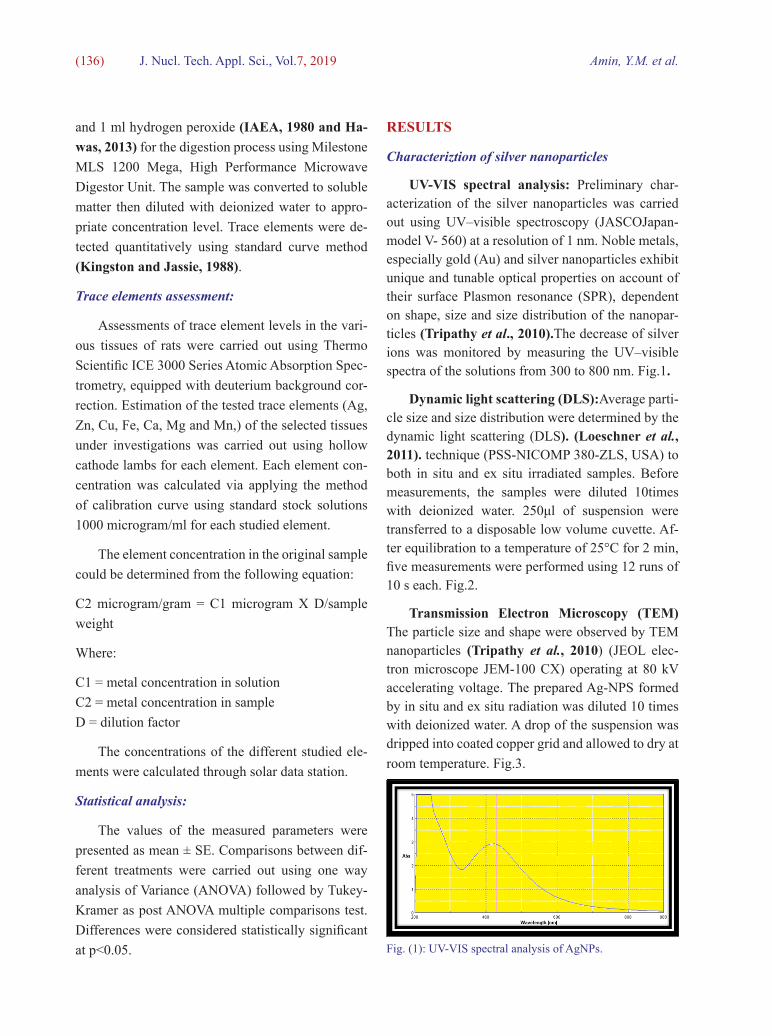
UV-VIS spectral analysis: Preliminary char-acterization of the silver nanoparticles was carried out using UV–visible spectroscopy (JASCOJapan-model V- 560) at a resolution of 1 nm. Noble metals, especially gold (Au) and silver nanoparticles exhibit unique and tunable optical properties on account of their surface Plasmon resonance (SPR), dependent on shape, size and size distribution of the nanopar-ticles (Tripathy et al., 2010).The decrease of silver ions was monitored by measuring the UV–visible spectra of the solutions from 300 to 800 nm. Fig.1.
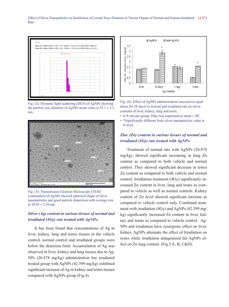
Dynamic light scattering (DLS):Average parti-cle size and size distribution were determined by the dynamic light scattering (DLS). (Loeschner et al., 2011). technique (PSS-NICOMP 380-ZLS, USA) to both in situ and ex situ irradiated samples. Before measurements, the samples were diluted 10times with deionized water. 250μl of suspension were transferred to a disposable low volume cuvette. Af-ter equilibration to a temperature of 25°C for 2 min, five measurements were performed using 12 runs of 10 s each. Fig.2.
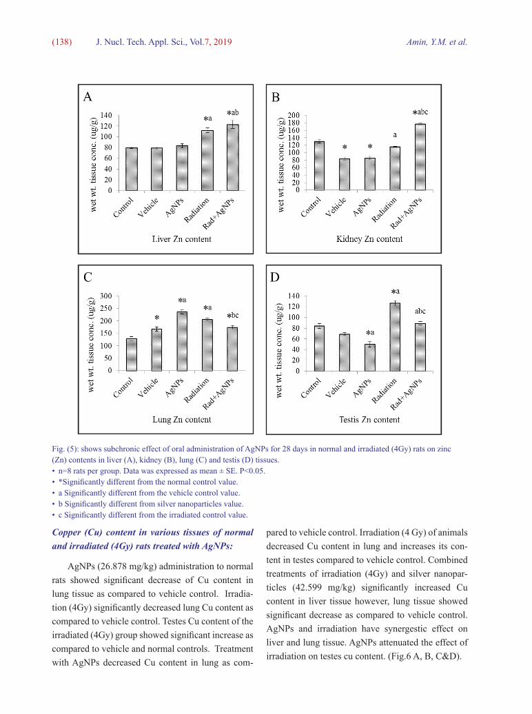
Transmission Electron Microscopy (TEM) The particle size and shape were observed by TEM nanoparticles (Tripathy et al., 2010) (JEOL elec-tron microscope JEM-100 CX) operating at 80 kV accelerating voltage. The prepared Ag-NPS formed by in situ and ex situ radiation was diluted 10 times with deionized water. A drop of the suspension was dripped into coated copper grid and allowed to dry at room temperature. Fig.3.
Fig. (1): UV-VIS spectral analysis of AgNPs.
( 137 )Effect of Silver Nanoparticles on distribution of Certain Trace Elements in Various Organs of Normal and Gamma Irradiated Rats
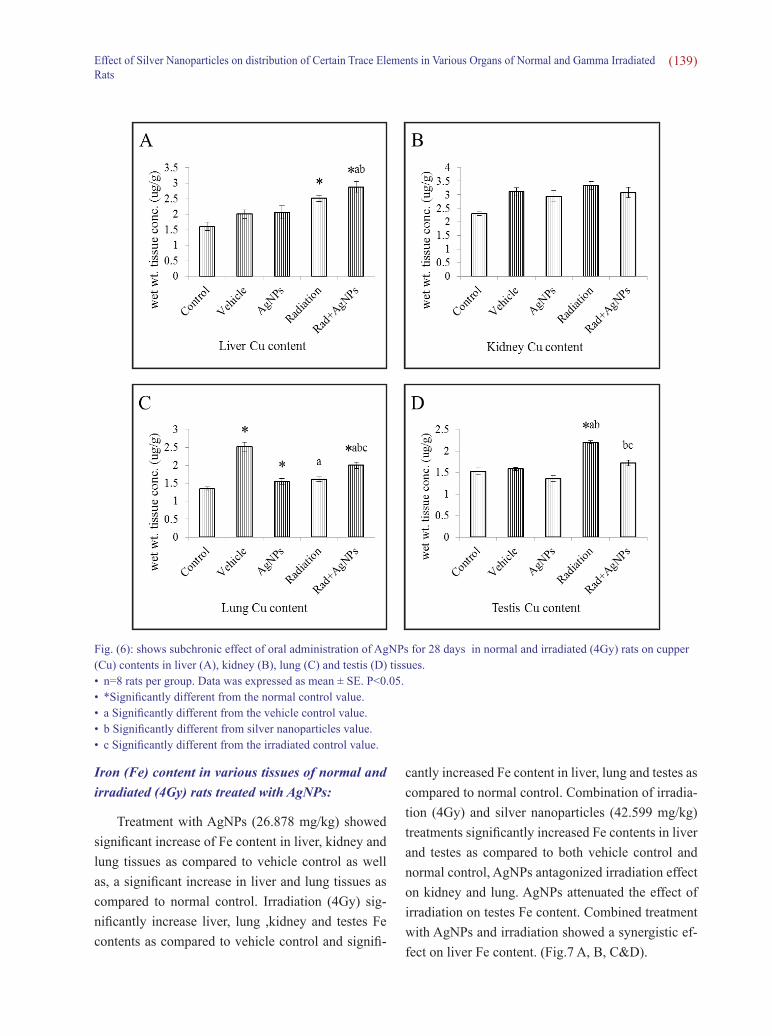
Silver (Ag) content in various tissues of normal and irradiated (4Gy) rats treated with AgNPs:
It has been found that concentrations of Ag in liver, kidney, lung and testes tissues in the vehicle control, normal control and irradiated groups were below the detection limit. Accumulation of Ag was observed in liver, kidney and lung tissues due to Ag-NPs (26.878 mg/kg) administration but irradiated treated group with AgNPs (42.599 mg/kg) exhibited significant increase of Ag in kidney and testes tissues compared with AgNPs group (Fig.4).
Zinc (Zn) content in various tissues of normal and irradiated (4Gy) rats treated with AgNPs:
Treatment of normal rats with AgNPs (26.878 mg/kg), showed significant increasing in lung Zn content as compared to both vehicle and normal control. They showed significant decrease in testes Zn content as compared to both vehicle and normal control. Irradiation treatment (4Gy) significantly in-creased Zn content in liver, lung and testes as com-pared to vehicle as well as normal controls. Kidney content of Zn level showed significant increase as compared to vehicle control only. Combined treat-ment with irradiation (4Gy) and AgNPs (42.599 mg/kg) significantly increased Zn content in liver, kid-ney and testes as compared to vehicle control. Ag-NPs and irradiation have synergistic effect on liver, kidney. AgNPs attenuate the effect of Irradiation on testes while irradiation antagonized the AgNPs ef-fect on Zn lung content. (Fig.5 A, B, C&D).
Fig. (2): Dynamic light scattering (DLS) of AgNPs showing the particle size diameter of AgNPs mean value is 23.1 ± 3.3 nm.
Fig. (3): Transmission Electron Microscopy (TEM) examination of AgNPs showed spherical shape of silver nanoparticles and good particle dispersion with average size at 30.92 ± 2.56 nm.
Fig. (4): Effect of AgNPs administration (successive equal doses for 28 days) to normal and irradiated rats on silver contents of liver, kidney, lung and testis.• n=8 rats per group. Data was expressed as mean ± SE.• *Significantly different from silver nanoparticles value at
P<0.05.
( 138 ) Amin, Y.M. et al.J. Nucl. Tech. Appl. Sci., Vol.7, 2019
Copper (Cu) content in various tissues of normal and irradiated (4Gy) rats treated with AgNPs:
AgNPs (26.878 mg/kg) administration to normal rats showed significant decrease of Cu content in lung tissue as compared to vehicle control. Irradia-tion (4Gy) significantly decreased lung Cu content as compared to vehicle control. Testes Cu content of the irradiated (4Gy) group showed significant increase as compared to vehicle and normal controls. Treatment with AgNPs decreased Cu content in lung as com-
pared to vehicle control. Irradiation (4 Gy) of animals decreased Cu content in lung and increases its con-tent in testes compared to vehicle control. Combined treatments of irradiation (4Gy) and silver nanopar-ticles (42.599 mg/kg) significantly increased Cu content in liver tissue however, lung tissue showed significant decrease as compared to vehicle control. AgNPs and irradiation have synergestic effect on liver and lung tissue. AgNPs attenuated the effect of irradiation on testes cu content. (Fig.6 A, B, C&D).
Fig. (5): shows subchronic effect of oral administration of AgNPs for 28 days in normal and irradiated (4Gy) rats on zinc (Zn) contents in liver (A), kidney (B), lung (C) and testis (D) tissues.• n=8 rats per group. Data was expressed as mean ± SE. P<0.05.• *Significantly different from the normal control value.• a Significantly different from the vehicle control value.• b Significantly different from silver nanoparticles value.• c Significantly different from the irradiated control value.
( 139 )Effect of Silver Nanoparticles on distribution of Certain Trace Elements in Various Organs of Normal and Gamma Irradiated Rats
Fig. (6): shows subchronic effect of oral administration of AgNPs for 28 days in normal and irradiated (4Gy) rats on cupper (Cu) contents in liver (A), kidney (B), lung (C) and testis (D) tissues.• n=8 rats per group. Data was expressed as mean ± SE. P<0.05.• *Significantly different from the normal control value.• a Significantly different from the vehicle control value.• b Significantly different from silver nanoparticles value.• c Significantly different from the irradiated control value.
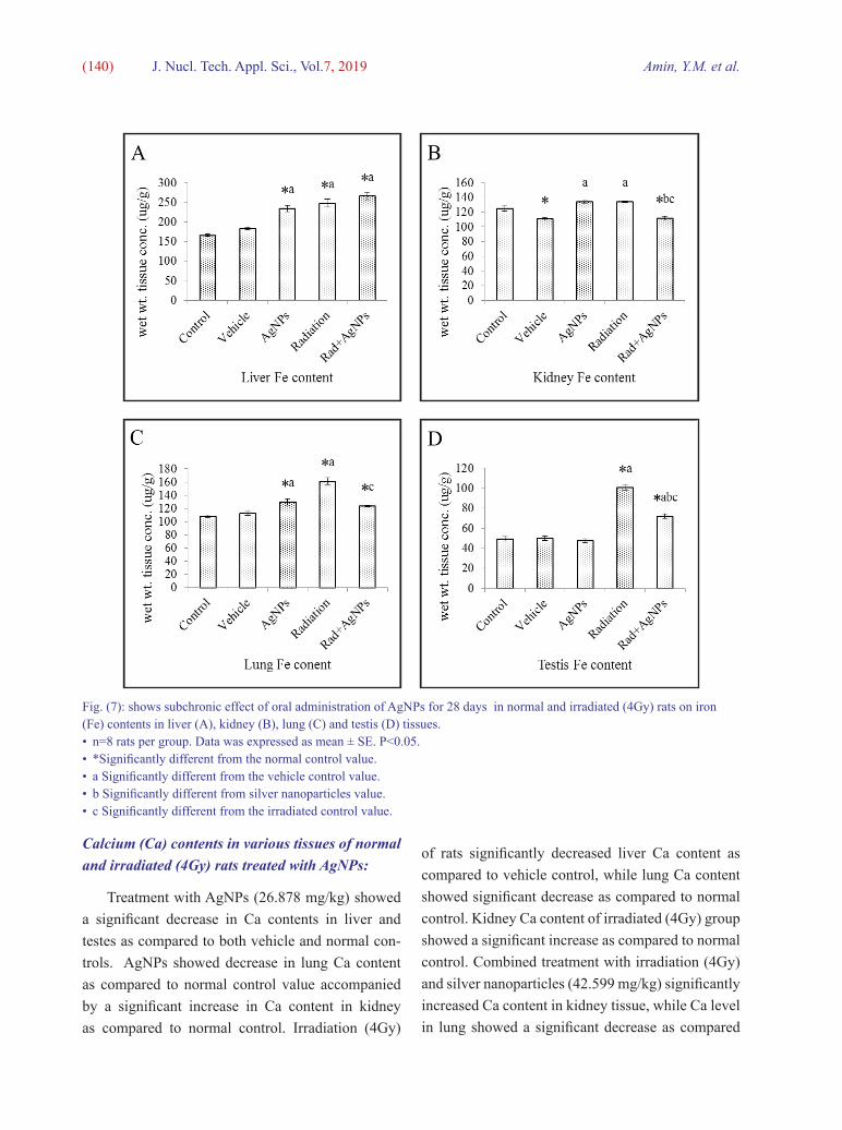
Iron (Fe) content in various tissues of normal and irradiated (4Gy) rats treated with AgNPs:
Treatment with AgNPs (26.878 mg/kg) showed significant increase of Fe content in liver, kidney and lung tissues as compared to vehicle control as well as, a significant increase in liver and lung tissues as compared to normal control. Irradiation (4Gy) sig-nificantly increase liver, lung ,kidney and testes Fe contents as compared to vehicle control and signifi-
cantly increased Fe content in liver, lung and testes as compared to normal control. Combination of irradia-tion (4Gy) and silver nanoparticles (42.599 mg/kg) treatments significantly increased Fe contents in liver and testes as compared to both vehicle control and normal control, AgNPs antagonized irradiation effect on kidney and lung. AgNPs attenuated the effect of irradiation on testes Fe content. Combined treatment with AgNPs and irradiation showed a synergistic ef-fect on liver Fe content. (Fig.7 A, B, C&D).
( 140 ) Amin, Y.M. et al.J. Nucl. Tech. Appl. Sci., Vol.7, 2019
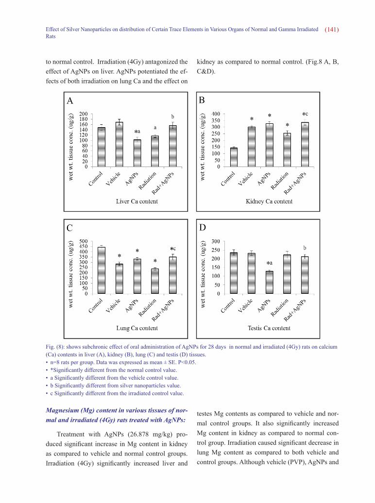
Calcium (Ca) contents in various tissues of normal and irradiated (4Gy) rats treated with AgNPs:
Treatment with AgNPs (26.878 mg/kg) showed a significant decrease in Ca contents in liver and testes as compared to both vehicle and normal con-trols. AgNPs showed decrease in lung Ca content as compared to normal control value accompanied by a significant increase in Ca content in kidney as compared to normal control. Irradiation (4Gy)
Fig. (7): shows subchronic effect of oral administration of AgNPs for 28 days in normal and irradiated (4Gy) rats on iron (Fe) contents in liver (A), kidney (B), lung (C) and testis (D) tissues.• n=8 rats per group. Data was expressed as mean ± SE. P<0.05.• *Significantly different from the normal control value.• a Significantly different from the vehicle control value.• b Significantly different from silver nanoparticles value.• c Significantly different from the irradiated control value.
of rats significantly decreased liver Ca content as compared to vehicle control, while lung Ca content showed significant decrease as compared to normal control. Kidney Ca content of irradiated (4Gy) group showed a significant increase as compared to normal control. Combined treatment with irradiation (4Gy) and silver nanoparticles (42.599 mg/kg) significantly increased Ca content in kidney tissue, while Ca level in lung showed a significant decrease as compared
( 141 )Effect of Silver Nanoparticles on distribution of Certain Trace Elements in Various Organs of Normal and Gamma Irradiated Rats
to normal control. Irradiation (4Gy) antagonized the effect of AgNPs on liver. AgNPs potentiated the ef-fects of both irradiation on lung Ca and the effect on
Fig. (8): shows subchronic effect of oral administration of AgNPs for 28 days in normal and irradiated (4Gy) rats on calcium (Ca) contents in liver (A), kidney (B), lung (C) and testis (D) tissues.• n=8 rats per group. Data was expressed as mean ± SE. P<0.05.• *Significantly different from the normal control value. • a Significantly different from the vehicle control value.• b Significantly different from silver nanoparticles value.• c Significantly different from the irradiated control value.
kidney as compared to normal control. (Fig.8 A, B, C&D).
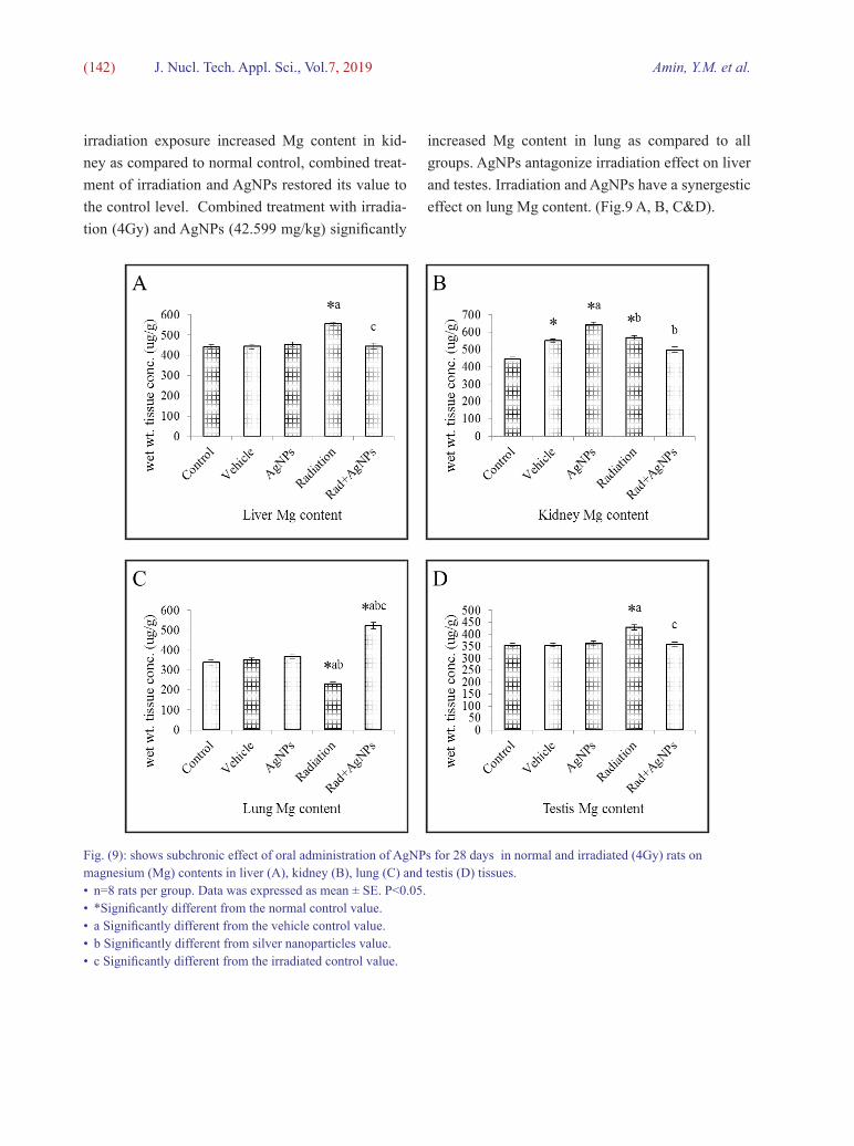
Magnesium (Mg) content in various tissues of nor-mal and irradiated (4Gy) rats treated with AgNPs:
Treatment with AgNPs (26.878 mg/kg) pro-duced significant increase in Mg content in kidney as compared to vehicle and normal control groups. Irradiation (4Gy) significantly increased liver and
testes Mg contents as compared to vehicle and nor-mal control groups. It also significantly increased Mg content in kidney as compared to normal con-trol group. Irradiation caused significant decrease in lung Mg content as compared to both vehicle and control groups. Although vehicle (PVP), AgNPs and
( 142 ) Amin, Y.M. et al.J. Nucl. Tech. Appl. Sci., Vol.7, 2019
irradiation exposure increased Mg content in kid-ney as compared to normal control, combined treat-ment of irradiation and AgNPs restored its value to the control level. Combined treatment with irradia-tion (4Gy) and AgNPs (42.599 mg/kg) significantly
increased Mg content in lung as compared to all groups. AgNPs antagonize irradiation effect on liver and testes. Irradiation and AgNPs have a synergestic effect on lung Mg content. (Fig.9 A, B, C&D).
Fig. (9): shows subchronic effect of oral administration of AgNPs for 28 days in normal and irradiated (4Gy) rats on magnesium (Mg) contents in liver (A), kidney (B), lung (C) and testis (D) tissues.• n=8 rats per group. Data was expressed as mean ± SE. P<0.05.• *Significantly different from the normal control value.• a Significantly different from the vehicle control value.• b Significantly different from silver nanoparticles value.• c Significantly different from the irradiated control value.
( 143 )Effect of Silver Nanoparticles on distribution of Certain Trace Elements in Various Organs of Normal and Gamma Irradiated Rats
Fig. (10): shows subchronic effect of oral administration of AgNPs for 28 days in normal and irradiated (4Gy) rats on manganese (Mn) contents in liver (A), kidney (B), lung (C) and testis (D) tissues.• n=8 rats per group. Data was expressed as mean ± SE. P<0.05.• *Significantly different from the normal control value. • a Significantly different from the vehicle control value.• b Significantly different from silver nanoparticles value.• c Significantly different from the irradiated control value.
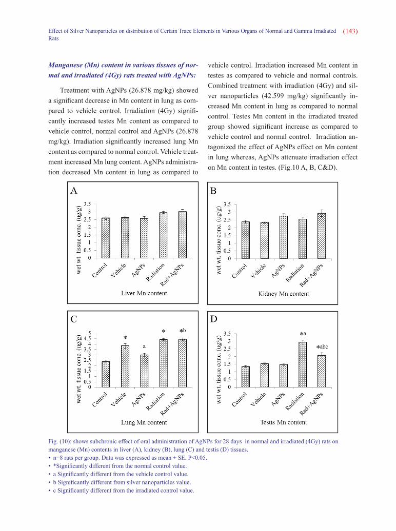
Manganese (Mn) content in various tissues of nor-mal and irradiated (4Gy) rats treated with AgNPs:
Treatment with AgNPs (26.878 mg/kg) showed a significant decrease in Mn content in lung as com-pared to vehicle control. Irradiation (4Gy) signifi-cantly increased testes Mn content as compared to vehicle control, normal control and AgNPs (26.878 mg/kg). Irradiation significantly increased lung Mn content as compared to normal control. Vehicle treat-ment increased Mn lung content. AgNPs administra-tion decreased Mn content in lung as compared to
vehicle control. Irradiation increased Mn content in testes as compared to vehicle and normal controls. Combined treatment with irradiation (4Gy) and sil-ver nanoparticles (42.599 mg/kg) significantly in-creased Mn content in lung as compared to normal control. Testes Mn content in the irradiated treated group showed significant increase as compared to vehicle control and normal control. Irradiation an-tagonized the effect of AgNPs effect on Mn content in lung whereas, AgNPs attenuate irradiation effect on Mn content in testes. (Fig.10 A, B, C&D).
( 144 ) Amin, Y.M. et al.J. Nucl. Tech. Appl. Sci., Vol.7, 2019
Discussion
In the present study, trace elements Fe, Cu, Zn, Mg, Ca and Mn as well as Ag were estimated in the examined four organs (liver, kidney, lung and testes) of rats after 28 days subchronic oral administration of AgNPs to normal and to irradiated rats (4 Gy) at a dose level of 26.878 mg/kg and 42.599 mg/kg re-spectively (Amin et al., 2015). Results of the present study indicated that, accumulation of Ag due to oral administration of AgNPs (26.878 mg/kg) in the stud-ied organs is in the ascending order of lung>liver> kidney>testes. Whereas, rats treated with AgNPs (42.599 mg/kg) after exposure to γ-irradiation (4Gy) led to different rank as kidney > testes > lung > liver. According to Gmoshinski et al. (2016), oral treat-ment of rats with AgNPs resulted in a dose-depen-dent Ag accumulation in liver tissue at a dose of 0.1–10 mg/kg whereas in kidney tissue at a dose of 0.1–1 mg/kg. Data of the present study revealed that, orally administration of AgNPs showed significant increase of Zn, Cu and Fe contents in lung tissue as compared to untreated control rats whereas AgNPs administration after γ-radiation led to significant in-crease in liver and lung Zn, Cu, Fe. The uptake of NPs occurs by the mononuclear phagocyte system (MPS) (Van Furth et al., 1972). The MPS comprises phagocytic cells, which are located in reticular con-nective tissue, as Kupffer cells in liver, alveolar and splenic macrophages. MPS has an important role in defense against bacteria, mycobacteria, viruses, pro-tozoa, and fungi, also macrophages remove senes-cent erythrocytes, leukocytes, and megakaryocytes by phagocytosis and digestion (Das et al., 2015), also MPS can store Fe in liver, which resulted from heme catabolism after the breakdown of red blood cells (Hull et al., 2014). AgNPs uptakes depend on NPs ability to cross the capillary wall of the organs and phagocytosed in MPS (Brandenberger et al., 2010). It was recorded that the uptake/accumula-tion of AgNPs by the MPS increases with its higher concentration in the blood (Kim et al., 2008). Our data further indicated that, accumulation of Ag in
kidney and testis tissues accompanied with elevation of Zn and Fe levels due to combined exposure with γ-radiation and AgNPs. It is not completely clear that the dissolved AgNPs interact with cell components, although it is suspected that AgNPs has physico-chemical properties lead to unknown adverse health effects (Chen and Schluesener, 2008), such as the “Trojan Horse” effect (Kreuter, 2004). In “Trojan-horse” mechanism, NPs are internalized within cells and then release many of toxic ions (Hsiao et al., 2015). On the other hand, AgNPs may cause adverse effects by the generation of reactive oxygen species (ROS) and through the interaction with the thiol groups of proteins (Chen and Schluesener, 2008). In a study on the metabolism of AgNPs, detect that accumulation of NPs in the organs of rats exposed to NPs consist of sulfur and selenium (Kim et al., 2008 and Loeschner et al., 2011).
Results of the present study revealed that irradia-tion (4Gy) significantly increased Zn and Fe content in liver, lung and testes tissues as compared to normal control. These results are in agreement with previous investigators Beregovskaia et al., (1988), Nada et al., (2008) and Ashry et al. (2010) who found that whole body γ-irradiation induced an elevation of Zn in different organs. The increase of Fe level may be due to oxidative stress inducing proteolytic modifi-cation of ferritin and transferrtin Garcia-Fernander et al. (2005). In addition Nada et al., (2008) reported an increase of Fe content in liver after whole body irradiation. In the study of Crowe and Morgan (1996), Fe is required in oxidative metabolism to DNA synthesis and cell division. For resisting cell damage and inflammation, liver cells deliver more Zn to synthesize nucleic acids, proteins and enzymes related to zinc (Shiraishi et al., 1986). Combina-tion treatment with irradiation (4Gy) and AgNPs (42.599 mg/kg) in the present study significantly in-creased Zn content in liver, kidney and testes tissues as compared to vehicle control, where significantly increased Fe content in liver and testes as compared to both vehicle control and normal control. Thus, ir-
( 145 )Effect of Silver Nanoparticles on distribution of Certain Trace Elements in Various Organs of Normal and Gamma Irradiated Rats
radiation and combined exposure of irradiation with AgNPs treatment led to accumulation of Zn and Fe in liver and lung organs. AgNPs potentiated the effect of irradiation on liver and kidney tissue content of trace elements. Zn is known as a free radical scaven-ger which affects the body’s immune system through its essential role in the synthesis of nucleic acid as well as protein. The antioxidant role of Zn could be related to its ability to induce metalothioneins (Wi-num et al., 2007). There is increasing evidence that metalothioneins can reduce several types of free radical including superoxides, hydroxyl and peroxyl radicals (Pierrel et al., 2007). On the other hand, the present data revealed that AgNPs attenuated the effect of irradiation on testes. The redistribution of Zn after irradiation may be a biological protection behavior against irradiation Sorenson, (2002); these may include DNA repair, protein synthesis and scav-enging the toxic free radicals. As essential metal, Zn is required for many cellular functions (Micheletti et al., 2001). Also Zn protects various membrane sys-tems from peroxidation damages induced by irradia-tion (Shiraishi et al., 1983; Matsubara et al., 1987) and stabilizes the membrane perturbation (Markant and Pallauf 1996 and Morcillo et al., 2000). Com-bined treatment with AgNPs and irradiation has syn-ergistic effect on liver and kidney tissues content of the tested trace elements. AgNPs attenuated the ef-fect of irradiation on testes tissues Zn content while irradiation antagonized AgNPs effect on lung tissue Zn content. Moreover, the high accumulation of Fe in liver due to radiation is closely correlated with the inhibition of ceruloplasmin which is essential for Fe metabolism and distribution (Osman et al., 2003 and Harris, 1995). The loss of feroxidase activity of ceruloplasmin resulted in systemic Fe deposition and tissue damage (Okamoto et al., 1996). Accord-ing to Underwood, (1977), there is inverse correla-tion between hepatic Fe and Cu concentration in rats. Fe is an essential trace element that is vital for the function of Fe dependent enzymes as peroxidases, ribonucleuotide reductase, catalase and cytochrome.
Fe is also necessary for the function of hemoglobin. It is also associated with Cu in several functions as it is involved in the constituent of other oxidative system for amino acid. According to Papanikolaou and Pantopoulos (2005) Fe homeostasis has to be tightly controlled. Free Fe has been catalyzing the generation of ROS which attack and damage cellular macromolecules and promote tissue injury and cell death. The present study indicated that Cu content of testes and liver tissue, in the irradiated group showed significant increase compared to normal control, where whole body X-ray irradiation increased Cu concentration Tamanoi et al. (1995).
In the study of AshaRani et al. (2009), cells treated with AgNPs for 4 hours showed a drop in Ca2+ concentration, as well as Moutin et al. (1989) provided evidence that Ag+ ions act on the same site as Ca2+ ions, regulating the release of Ca2+ from sar-coplasmic reticulum. On the other hand Orrenius et al., (1992) showed that higher concentration of Ag+ ion inhibited Ca2+ release from the intracellular stores. AgNPs could release Ag+ ions through sur-face oxidation which lead to fluctuations of Ca2+ in a similar way AshaRani et al. (2009); it may explain the increase of Ca content in kidney tissue of irradi-ated rats. Disruption of calcium homeostasis is an early sign of cell injury which plays a major role in pathological and toxicological conditions. Repeated calcium influx and efflux in mitochondria could re-sult in mitochondrial membrane damage, resulting in ROS production and inhibition of ATP synthesis Orrenius et al. (1992). Whole body γ-radiation ex-posure caused disturbance and alteration of Ca level in the blood Fauxcheux et al., (1976). El Nimr and Abdel-Rahim (1998) found that exposure of rats to γ-radiation (5Gy) increase levels of Ca in lung, liver and kidney tissues. The disturbances of calcium metabolism after irradiation may be attributed to the insufficient renal function Kotb et al. (1990). On the other hand, results of the present study revealed that AgNPs (26.878 mg/kg), irradiated and irradiat-ed rats treated with AgNPs (42.599 mg/kg) showed
( 146 ) Amin, Y.M. et al.J. Nucl. Tech. Appl. Sci., Vol.7, 2019
increased Ca content in kidney tissue as compared to normal control. The increase of Ca content may be attributed to hypoxia induced by oxidative stress this explanation is in accordance with the results of Berna et al. (2001).
Irradiation (4Gy) significantly increased Mg content in liver, kidney and testes tissue as compared to normal control. Irradiation caused significant de-crease in lung tissue Mg content as compared to both vehicle and control groups. The present results are in accordance with the results Yukawa et al. (1980) and Symthe et al. (1982). Combination of irradia-tion (4Gy) and AgNPs (42.599 mg/kg) significantly increased Mg content in lung tissue as compared to vehicle control and normal control. AgNPs antago-nized irradiation effect on liver, kidney and testes. Ir-radiation and AgNPs have synergestic effect on lung Mg content. Furthermore, Nada et al. (2008) report-ed that the redistribution of Ca and Mg in tissue or-gan may respond in recovery from radiation-induced pathology or in repairable damage in biomemebrane and to prevent irreversible cell damage.
Results of the present study demonstrated that Mn and Fe content in lung and testes tissues in-creased in irradiated and the irradiated group treated with AgNPs. Fe and Mn can share common absorp-tion and transport pathways Fitsanakis et al. (2010). Liver content of Mn in AgNPs, irradiated (4Gy) and irradiated treated group with AgNPs did not change. Mn is eliminated from the body mainly in the bile, thus the impaired liver function may lead to de-creased Mn secretion Keen et al. (1999). However irradiation induced decrease of Mn in liver and other organ tissues Nada and Azab (2005).
In conclusion, radiation damage to living biolog-ical tissues is associated with membrane permeabil-ity which accompanied with corresponding altera-tions in the essential trace element content of Zn, Cu, Fe, Ca, Mg, and Mn in the various examined organs tissue, liver, kidney, lung and testes. Subchronic oral administration of AgNPs induced marked changes in
the concentration of the estimated essential elements in the various examined organs tissue of normal rats. Combined treatment with irradiation and AgNPs showed alterations in the elements content of the various organs tissue investigated. The demonstrated variation of the essential elements in the various or-gans examined might be attributed to the selective responsiveness of each tissue to the AgNPs and/or γ-irradiation treatments. AgNPs administration to normal rats or γ-irradiated rats has considerate effect on the levels of trace elements especially Zn, Cu and Fe in liver and lung tissues which give attention that these organs more sensitive to AgNPs.
Additional studies however, several studies are needed to gain further insight into the underlying mechanisms of the observed disturbances of essen-tial trace element contents in the various examined organs (liver, kidney, lung, testes) in response to Ag-NPs treatment in normal and irradiated rats.
ACKNOWLEDGEMENT
This study was performed in the National Center for Radiation Research and Technology (NCRRT), Atomic Energy Authority, Cairo, Egypt. We are grateful to Professor Dr. Ahmed Shafik Nada, Prof. of physiology, Drug Radiation Research Depart-ment, NCRRT for offering all facilities to measure the trace elements in Atomic Absorption Spectrom-etry lab. Also the authors are appreciating to the staff members of gamma irradiation unit of NCRRT for carrying out the irradiation process of experiment.
REFERENCES
• Amin,Y.M.; Hawas, A.M.; El-Batal, A.I.; Hassan, S.H.M. and Elsayed, M.E. (2015): Evaluation of acute and subchronic toxicity of Silver nanoparticles in normal and irradiated animals. BJPT, 6(2): 22.
• AshaRani, P.V.; Hande, M.P. and Valiyaveettil, S. (2009): Anti-proliferative activity of silver nanopar-ticles. BMC Cell Biology, 10:65.
( 147 )Effect of Silver Nanoparticles on distribution of Certain Trace Elements in Various Organs of Normal and Gamma Irradiated Rats
• Ashry, O.M.; Kafafy, Y.A. and El-Tawil, G.A. (2010): Influence of hesperidin on lipid metabo-lism, oxidative stress and related trace elements in γ-irradiated rats. Egypt. J. Rad. Sci. Applic., 23(1): 15.
• Benetti, F.; Bregoli, L.; Olivato, I. and Sabbioni E. (2014): Effects of metal(loid)-based nanomaterials on essential element homeostasis: the central role of nanometallomics for nanotoxicology. Metallomics., 6(4): 729.
• Beregovskaia, N.N.; Sidorik, E.P.; Udovichenko, T.V. and Chebotarev, E.E. (1988): The state of the molecular electron carriers of the cell energy system at early periods following irradiation. Radiobiologi-ia., 28(5): 588.
• Berna, N.; Arnould, T.; Remacle, J. and Michiels, C. (2001): Hypoxia-induced increase in intracellular calcium concentration in endothelial cells: role of the Na(+)-glucose cotransporter. J. Cell. Biochem., 84(1): 115.
• Brandenberger, C.; Mühlfeld, C.; Ali, Z.; Lenz, A.G.; Schmid, O.; Parak, W.J.; Gehr, P. and Rothen-Rutishauser, B. (2010): Quantitative evalu-ation of cellular uptake and trafficking of plain and polyethylene glycol-coated gold nanoparticles. Small, 6(15): 1669.
• Chen, X. and Schluesener, H.J. (2008): Nanosilver: a nanoproduct in medical application. Toxicol. Lett., 176(1): 1.
• Choi, O.; Deng, K.K.; Kim, N.J.; Ross Jr., L.; Sur-ampalli, R.Y. and Hu, Z. (2008): The inhibitory ef-fects of silver nanoparticles, silver ions, and silver chloride colloids on microbial growth. Water Res.. 42(12): 3066.
• Christensen, F.M.; Johnston, H.J.; Stone, V.; Ait-ken, R.J.; Hankin, S.; Peters, S. and Aschberger, K. (2010): Nano-silver - feasibility and challenges for human health risk assessment based on open litera-ture. Nanotoxicology, 4(3): 284.
• Crowe, A. and Morgan, E.H. (1996): Iron and cop-per interact during their uptake and deposition in the brain and other organs of developing rats exposed to
dietary excess of the two metals. J. Nutr., 126: 183.
• Das, A.; Sinha, M.; Datta, S.; Abas, M.; Chaffee, S.; Sen, C.K. and Roy, S. (2015): Monocyte and macrophage plasticity in tissue repair and regenera-tion. Am. J. Pathol., 185(10): 2596.
• De, M.; Ghosh, P.S. and Rotello, V.M. (2008): Ap-plications of nanoparticles in biology. Adv. Mater., 20(22): 4225.
• El-Batal, A.I.; Amin, M.A.; Shehata, M.M.K. and Hallol, M.M.A. (2013a): Synthesis of silver nanopar-ticles by bacillus stearothermophilus using gamma ra-diation and their antimicrobial activity. World Appl. Sci. J., 22(1):1.
• El-Batal, A.I.; El-Baz, A.F.; Abo Mosalam, F.M. and Tayel, A.A. (2013b): Gamma irradiation induced silver nanoparticles synthesis by Monascus purpure-us. J. Chem. Pharm. Res., 5(8): 1.
• El-Nimr, T. and Abdel-Rahim, S.M. (1998): Effect of gamma radiation on some elements in certain or-gans of albino rats. Biol. Trace Elem. Res., 68(1−2): 25.
• Fauxcheux, B.; Kuchel, O.; Cuche, J.L.; Messerli, F.H.; Buu, N.T.; Barbeau, A. and Genest, J. (1976): Circadian variations of the urinary excretion of cat-echolamines and electrolytes. Endocr. Res. Commun., 3(5): 257.
• Fitsanakis, V.A.; Zhang, N.; Garcia, S. and As-chner, M. (2010): Manganese (Mn) and iron (Fe): in-terdependency of transport and regulation. Neurotox. Res., 18(2): 124.
• Garcia-Fernandez, M.; Castilla-Cortazar, I.; Diaz-Sanchez, M.; Navarro, I.; Puche, J.E.; Castilla, A.; Casares, A.D.; Clavijo, E. and González-Barón, S. (2005): Antioxidant effects of insulin-like growth factor-1(GF-1) in rats with advances liver cirrhosis. BMC Gastroenterol., 5(7): 1.
• Ghosh Chaudhuri, R. and Paria, S. (2012): Core/shell nanoparticles: Classes, properties, synthesis Mechanisms, characterization, and applications. Chem Rev., 112(4): 2373.
( 148 ) Amin, Y.M. et al.J. Nucl. Tech. Appl. Sci., Vol.7, 2019
• Gmoshinski, I.V.; Shumakova, A.A.; Shipelin, V.A.; Maltsev, G.Yu. and Khotimchenko, S.A. (2016): Influence of orally introduced silver nanopar-ticles on content of essential and toxic trace elements in organism. Nanotechnologies in Russia, 11(9−10): 646.
• Harris, E.D. (1995): The iron-copper connection: the link to ceruloplasmin grows stronger. Nutr Rev., 53(6): 170.
• Hassan, S.; El-Sayed, N.M.; Hussein, A.H. and Anis, L.M. (2005): Disturbance in certain essential elements concentrations in various tissues of mice by exposure to gamma radiation and/or magnetic field. Isotope & Rad. Res., 37(6): 1658.
• Hawas, A.M. (2013): Effect of low dose gamma rays on certain essential metals and oxidative stress in dif-ferent rat organs. J. Rad. Res. Appl. Sci., 6(2): 1
• Hsiao, I.L.; Hsieh, Y.K.; Wang, C.F.; Chen, I.C. and Huang, Y.J. (2015): Trojan-horse mechanism in the cellular uptake of silver nanoparticles verified by direct intra- and extracellular silver speciation analy-sis. Environ. Sci. Technol., 49(6): 3813.
• Hull, T.D.; Agarwal, A. and George, J.F. (2014): The mononuclear phagocyte system in homeostasis and disease: a role for heme oxygenase-1. Antioxid. Redox Signal., 20(11): 1770.
• IAEA. (1980): Elemental analysis of biological mate-rials; international Atomic Energy Agency. IAEA, No 197, 379. Veinna Technical report series.
• Keen, C.L.; Ensunsa, J.L.; Watson, M.H.; Baly, D.L.; Donovan, S.M.; Monaco, M.H. and Clegg, M.S. (1999): Nutritional aspects of manganese for experimental studies. Neurotoxicology, 20(2-3): 213.
• Kim, Y.S.; Kim, J.S.; Cho, H.S.; Rha, D.S.; Kim, J.M.; Park, J.D.; Choi, B.S.; Lim, R.; Chang, H.K.; Chung, Y.H.; Kwon, I.H.; Jeong, J.; Han, B.S. and Yu, I.J. (2008): Twenty-eight-day oral tox-icity, genotoxicity, and gender-related tissue distribu-tion of silver nanoparticles in Sprague-Dawley rats. Inhal. Toxicol., 20(6): 575.
• Kingston, H.M. and Jassei, L.B. (1988): Introduc-tion to microwave sample preparation. ACS, Wash-ington: DC, p 126.
• Kotb, M.A.; El-khatib, A.M.; Morsey, A.A.; Ra-madan, M.I.A. and El-Bassiouni, E.A. (1990): Changes in mineral elements in some tissues of mice following neutron irradiation. Isotop-enpaxis., 26(7): 297.
• Kreuter, J. (2004): Influence of the surface proper-ties on nanoparticle-mediated transport of drugs to the brain. J. Nanosci. Nanotechnol., 4(5): 484.
• Loeschner, K.; Hadrup, N.; Qvortrup, K.; Larsen, A.; Gao, X.; Vogel, U.; Mortensen, A.; Lam, H.R. and Larsen, E.H. (2011): Distribution of silver in rats following 28 days of repeated oral exposure to silver nanoparticles or silver acetate. Part. Fibre. Toxicol., 8: 18.
• Lu, L.; Sun, R.W.; Chen, R.; Hui, C.K.; Ho, C.M.; Luk, J.M.; Lau, G.K. and Che, C.M. (2008): Silver nanoparticles inhibit hepatitis B virus replication. An-tivir. Ther., 13(2): 253.
• Lubick, N. (2008): Nanosilver toxicity: ions, nanoparticles--or both?. Environ. Sci. Technol., 42(23): 8617.
• Mao, A.; Jin, X.; Gu, X.; Wei, X. and Yong, G. (2012): Rapid green synthesis and surface enhanced Raman scattering effect of single crystal silver nano-cupes. J. Mol. Struc., 1021: 158.
• Marambio-Jones, C. and Hoek, E.M. (2010): A re-view of the antibacterial effects of silver nanomateri-als and potential implications for human health and the environment. J. Nanopart. Res., 12: 1531.
• Markant, A. and Pallauf, J. (1996): Metallothionein and zinc as potential antioxidants in radical-induced lipid peroxidation in cultured hepatocytes. J. Trace Elem. Med. Biol., 10(2): 88.
• Matsubara, J.; Tajima, Y. and Karasawa, M. (1987): Metallothionein Induction as a Potent Means of Radiation Protection in Mice. Radiat. Res., 111(2): 267.
( 149 )Effect of Silver Nanoparticles on distribution of Certain Trace Elements in Various Organs of Normal and Gamma Irradiated Rats
• Micheletti,A.;Rossi,R.andRufini,S.(2001): Zinc status in athletes: relation to diet and exercise. Sports Med., 31(8): 577.
• Morcillo, M.A.; Rucandio, M.I. and Snatamaría, J. (2000): Effect of gamma radiation on liver metallo-thionein synthesis and lipid peroxidation in rats. Cell. Mol. Biol., 46(2): 435.
• Morones, J.R.; Elechiguerra, J.L.; Camacho, A.; Holt, K.; Kouri, J.B.; Ramírez, J.T. and Yaca-man; M.J. (2005): The bactericidal effect of silver nanoparticles. Nanotechnology, 16(10): 2346.
• Moutin, M.J.; Abramson, J.J.; Salama, G. and Du-pont, Y. (1989): Rapid Ag+-induced release of Ca2+ from sarcoplasmic reticulum vesicles of skeletal mus-cle: a rapid filtration study. Biochim. Biophys. Acta, 984: 289.
• Nada, A.S. and Azab, K. (2005): Induction of cel-lular metallothionein in irradiated rats supplemented with Egyptian proplis extract. Egypt. J. Rad. Sci. Ap-plic., 18(2): 351.
• Nada, A.H.; Gharib, O.A.; Noaman, E. and Amin, N.E. (2008): Early sings of trace element alterations induced by environmental pollutants and radiation exposure in rats. Egypt. J. Rad. Sci. Applic., 21(2): 515.
• Okamoto, N.; Wada, S.; Oga, T.; Kawabata, Y.; Baba, Y.; Habu, D.; Takada, Z. and Wada, Y. (1996): Hereditary ceruloplasmin deficiency with he-mosiderosis. Hum. Genet., 97(6): 755.
• Orrenius, S.; McCabe Jr., M.J. and Nicotera, P. (1992): Ca(2+)-dependent mechanisms of cyto-toxicity and programmed cell death. Toxicol. Lett., 64−65:357.
• Osman, S.A.; Abu Ghadeer, A.R.; Yousri, R.M.; Abdallah, N.M. and El-Fatih, N.M. (2003): Protec-tive role of 6, 8-thioctic acid against metalloproteins impairment induced by gamma irradiation in rats. Egypt. J. Rad. Sci. Applic., 16(1): 27.
• Papanikolaou, G. and Pantopoulos, K. (2005): Iron metabolism and toxicity. Toxicol. Appl. Pharmacol., 202(2): 199.
• Pierrel, F.; Cobine, P.A. and Winge, D.R. (2007): Metal ion availability in mitochondria. Biometals., 20(3−4): 657.
• Roe, D.; Karandikar, B.; Bonn-Savage, N.; Gib-bins, B. and Roullet, J.B. (2008): Antimicrobial surface functionalization of plastic catheters by silver nanoparticles. J. Antimicrob. Chemother., 61(4): 869.
• Sharma, V.K.; Yngard, R.A. and Lin, Y. (2009): Silver nanoparticles: green synthesis and their an-timicrobial activities. Adv. Colloid. Interface Sci., 145(1−2): 83.
• Shazia, Q.; Mohammad, Z.H.; Rahman, T. and Shekhar, H.U. (2012): Correlation of oxidative stress with serum trace element levels and antioxidant en-zyme status in beta thalassemia major patients: a re-view of the literature. Anemia., 2012: 1.
• Shiraishi, N.; Aono, K. and Utsumi, K. (1983): In-creased metallothionein content in rat liver induced by X irradiation and exposure to high oxygen tension. Radiat. Res., 95(2): 298.
• Shiraishi, N.; Yamamoto, H.; Takeda, Y.; Kon-doh, S.; Hayashi, H.; Hashimoto, K. and Aono, K. (1986): Increased metallothionein content in rat liver and kidney following X-irradiation. Toxicol. Appl. Pharmacol., 85(2): 128.
• Sorenson, J.R. (2002): Cu, Fe, Mn, Zn Chelates offer a medicinal chemistry approach to overcoming radia-tion injury. Curr. Med. Chem., 9(6): 639.
• Symthe, W.R.; Alfrey, A.C.; Crawell, P.W.; Crouch, C.A.; Ibels, L.S.; Kubo, H.; Nunnelley, L.L. and Rudolph, H. (1982): Trace element abnor-malities uremia. Ann. Intern. Med., 96(3): 302.
• Tamanoi, I.; Nakamura, A.; Hashikawa, K.; Ka-chi, M.; Goto, B.; Joshima, H. and Matsumoto, S. (1995): Changes of blood plasma element contents in X-rays irradiated mice by Pixe Analysis. Int. J. PIXE, 5: 85.
• Tripathy, A.; Raichur, A.M.; Chandrasekaran, N.; Prathna, T.C. and Mukherjee, A. (2010): Process variables in biomimetic synthesis of silver nanoparti-
( 150 ) Amin, Y.M. et al.J. Nucl. Tech. Appl. Sci., Vol.7, 2019
cles by aqueous extract of Azadirachta indica (Neem) leaves. J. Nanoparticle Res., 12(1):237-246.
• Underwood, E.J. (1977): Trace Elements in Human and Animal Nutrition; 4th Ed. New York, San Fran-cisco and London: Academic Press Inc.
• Van Der Zande, M.; Vandebriel, R.J.; Van Doren, E.; Kramer, E.; Herrera Rivera, Z.; Serrano-Ro-jero, C.S.; Gremmer, E.R.; Mast, J.; Peters, R.J.; Hollman, P.C.; Hendriksen, P.J.; Marvin, H.J.; Peijnenburg, A.A. and Bouwmeester, H. (2012): Distribution, elimination, and toxicity of silver nanoparticles and silver ions in rats after 28-day oral exposure. ACS Nano., 6(8): 7427.
• Van Furth, R.; Cohn, Z.A.; Hirsch, J.G.; Hum-phrey, J.H.; Spector, W.G. and Langevoort HL. (1972): The mononuclear phagocyte system: a new classification of macrophages; monocytes; and their precursor cells. Bull. World Health Organ., 46(6): 845.
• Winum, J.Y.; Scozzfava, A.; Montero, J.L. and Supuran, C.T. (2007): Metal binding functions in design of carbonic anhydrase inhibitors. Curr. Top. Chem., 7(9): 835.
• Xiu, Z.M.; Zhang, Q.B.; Puppala, H.L.; Colvin, V.L. and Alvarez, P.J. (2012): Negligible particle-specific antibacterial activity of silver nanoparticles. Nano Lett., 12(8): 4271.
• Yukawa, M.; Amano, K.; Suzuk-Yosumot, M. and Terai, M. (1980): Distribution of trace elements in the human body determined by neutron activation analysis. Arch. Environ. Health, 35(1): 36.


![Arab Journal of Nuclear Sciences and Applications · M. DIAA EL-DIN H. FARAG et.al and polysaccharides and high levels of bioactive compounds [6]. Pomegranate peel, being free from](https://static.fdocument.org/doc/165x107/604ffea56941bf0b4c2e87d8/arab-journal-of-nuclear-sciences-and-applications-m-diaa-el-din-h-farag-etal.jpg)