irradiated SCID mice - NIBSnibs.ac.cn/articles/20120210_01.pdf · 2012-02-10 · ORIGINAL ARTICLE...
12
ORIGINAL ARTICLE Genetic correction of β-thalassemia patient-specific iPS cells and its use in improving hemoglobin production in irradiated SCID mice Yixuan Wang 1, * , Chen-Guang Zheng 2, * , Yonghua Jiang 1 , Jiqin Zhang 1 , Jiayu Chen 1 , Chao Yao 1 , Qingguo Zhao 1 , Sheng Liu 1 , Ke Chen 2 , Juan Du 2 , Ze Yang 3 , Shaorong Gao 1 1 National Institute of Biological Sciences, Zhongguancun Life Science Park, #7 Science Park Road, Beijing 102206, China; 2 Guangxi Zhuang Autonomous Region Women and Children Care Hospital, Nanning, Guangxi 530003, China; 3 National Institute of Geriatrics, Beijing Hospital, Chinese Ministry of Health, Beijing 100730, China *These two authors contributed equally to this work. Correspondence: Shaorong Gao a , Ze Yang b a Tel: +86-10-80728967; Fax: +86-10-80727535 E-mail: [email protected] b Tel: +86-10-58115081 E-mail: [email protected] Received 5 September 2011; revised 6 December 2011; accepted 27 De- cember 2011 The generation of induced pluripotent stem cells (iPSCs) from differentiated somatic cells by over-expression of several transcription factors has the potential to cure many genetic and degenerative diseases currently recalcitrant to traditional clinical approaches. One such genetic disease is β-thalassemia major (Cooley’s anemia). This disease is caused by either a point mutation or the deletion of several nucleotides in the β-globin gene, and it threatens the lives of millions of people in China. In the present study, we successfully generated iPSCs from fibroblasts collected from a 2-year-old patient who was diagnosed with a homozygous 41/42 deletion in his β-globin gene. More importantly, we successfully corrected this genetic mutation in the β-thalassemia iPSCs by homologous recombination. Further- more, transplantation of the genetically corrected iPSCs-derived hematopoietic progenitors into sub-lethally irradi- ated immune deficient SCID mice showed improved hemoglobin production compared with the uncorrected iPSCs. Moreover, the generation of human β-globin could be detected in the mice transplanted with corrected iPSCs-derived hematopietic progenitors. Our study provides strong evidence that iPSCs generated from a patient with a genetic dis- ease can be corrected by homologous recombination and that the corrected iPSCs have potential clinical uses. Keywords: iPS; thalassemia Cell Research advance online publication 7 February 2012; doi:10.1038/cr.2012.23 npg Cell Research (2012) :1-12. © 2012 IBCB, SIBS, CAS All rights reserved 1001-0602/12 $ 32.00 www.nature.com/cr Introduction Induced pluripotent stem cells (iPSCs) can be derived from differentiated somatic cells by simultaneously ex- pressing several transcription factors that are normally expressed at high levels in embryonic stem cells (ESCs) [1-6]. This process allows for the generation of disease- specific iPSCs that can be used to model human diseases, and ultimately iPSCs may lead to the development of cures for many genetic and degenerative diseases [7-10]. β-thalassemia, one of the most common genetic dis- eases, is an inherited blood disorder that is characterised by a reduction in the synthesis of hemoglobin (HB) subunit β (HB β chain). The most common molecular defects are either point mutations or small deletions that affect the transcription, splicing or translation of the HBB gene mRNA. Individuals with β-thalassemia major (also called Cooley’s anemia) have severe anemia and hepa- tosplenomegaly. Without treatment, affected children fail to thrive and have a shortened life expectancy. More importantly, this genetically inherited disease, which is prevalent throughout the southern part of China, has threatened millions of people’s lives for decades, and no effective treatments are available. The generation of iPSCs from patients has raised hopes for curing blood diseases caused by genetic muta- tions, and a proof of principle study has shown that a
Transcript of irradiated SCID mice - NIBSnibs.ac.cn/articles/20120210_01.pdf · 2012-02-10 · ORIGINAL ARTICLE...
ORIGINAL ARTICLE
Genetic correction of β-thalassemia patient-specific iPS cells and its use in improving hemoglobin production in irradiated SCID miceYixuan Wang1, *, Chen-Guang Zheng2, *, Yonghua Jiang1, Jiqin Zhang1, Jiayu Chen1, Chao Yao1, Qingguo Zhao1, Sheng Liu1, Ke Chen2, Juan Du2, Ze Yang3, Shaorong Gao1
1National Institute of Biological Sciences, Zhongguancun Life Science Park, #7 Science Park Road, Beijing 102206, China; 2Guangxi Zhuang Autonomous Region Women and Children Care Hospital, Nanning, Guangxi 530003, China; 3National Institute of Geriatrics, Beijing Hospital, Chinese Ministry of Health, Beijing 100730, China
*These two authors contributed equally to this work.Correspondence: Shaorong Gaoa, Ze Yangb
aTel: +86-10-80728967; Fax: +86-10-80727535E-mail: [email protected]: +86-10-58115081E-mail: [email protected] 5 September 2011; revised 6 December 2011; accepted 27 De-cember 2011
The generation of induced pluripotent stem cells (iPSCs) from differentiated somatic cells by over-expression of several transcription factors has the potential to cure many genetic and degenerative diseases currently recalcitrant to traditional clinical approaches. One such genetic disease is β-thalassemia major (Cooley’s anemia). This disease is caused by either a point mutation or the deletion of several nucleotides in the β-globin gene, and it threatens the lives of millions of people in China. In the present study, we successfully generated iPSCs from fibroblasts collected from a 2-year-old patient who was diagnosed with a homozygous 41/42 deletion in his β-globin gene. More importantly, we successfully corrected this genetic mutation in the β-thalassemia iPSCs by homologous recombination. Further-more, transplantation of the genetically corrected iPSCs-derived hematopoietic progenitors into sub-lethally irradi-ated immune deficient SCID mice showed improved hemoglobin production compared with the uncorrected iPSCs. Moreover, the generation of human β-globin could be detected in the mice transplanted with corrected iPSCs-derived hematopietic progenitors. Our study provides strong evidence that iPSCs generated from a patient with a genetic dis-ease can be corrected by homologous recombination and that the corrected iPSCs have potential clinical uses.Keywords: iPS; thalassemiaCell Research advance online publication 7 February 2012; doi:10.1038/cr.2012.23
npgCell Research (2012) :1-12.© 2012 IBCB, SIBS, CAS All rights reserved 1001-0602/12 $ 32.00 www.nature.com/cr
Introduction
Induced pluripotent stem cells (iPSCs) can be derived from differentiated somatic cells by simultaneously ex-pressing several transcription factors that are normally expressed at high levels in embryonic stem cells (ESCs) [1-6]. This process allows for the generation of disease-specific iPSCs that can be used to model human diseases, and ultimately iPSCs may lead to the development of
cures for many genetic and degenerative diseases [7-10].β-thalassemia, one of the most common genetic dis-
eases, is an inherited blood disorder that is characterised by a reduction in the synthesis of hemoglobin (HB) subunit β (HB β chain). The most common molecular defects are either point mutations or small deletions that affect the transcription, splicing or translation of the HBB gene mRNA. Individuals with β-thalassemia major (also called Cooley’s anemia) have severe anemia and hepa-tosplenomegaly. Without treatment, affected children fail to thrive and have a shortened life expectancy. More importantly, this genetically inherited disease, which is prevalent throughout the southern part of China, has threatened millions of people’s lives for decades, and no effective treatments are available.
The generation of iPSCs from patients has raised hopes for curing blood diseases caused by genetic muta-tions, and a proof of principle study has shown that a
Genetically corrected β-thalassemia iPSCs function in vivo2
npg
Cell Research | www.cell-research.com
humanized sickle cell anemia mouse model could be res-cued by transplantation of genetically corrected iPSCs-derived hematopoietic stem cells [11]. Recent studies from others and ourselves have clearly shown that iPSCs can be produced from human fetal fibroblasts carrying a β-thalassemia mutation [12, 13]. However, β-thalassemia iPSCs have not yet been genetically corrected and the potential in vivo functionality of β-thalassemia disease-free iPSCs has not been evaluated.
In the present study, we aimed to further investigate if the genetic mutation in the β-thalassemia patient-specific iPSCs can be successfully corrected by homologous recombination. Furthermore, the functionality of the ge-netically corrected iPSCs was examined through in vitro differentiation and in vivo transplantation. Our results demonstrate that genetically corrected iPSCs-derived he-matopoietic progenitors (HPs) could differentiate in vivo and produce human β-globin in a mouse model. These results have important implications for personalized treatment of β-thalassemia in the future.
Results
Establishment of patient-specific iPS cell linesIn the present study, fibroblasts were obtained and
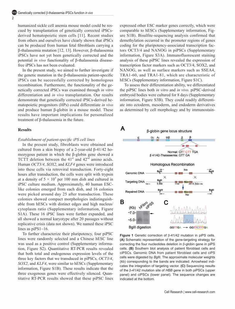
cultured from a skin biopsy of a 2-year-old β-41/42 ho-mozygous patient in which the β-globin gene showed a TCTT deletion between the 41st and 42nd amino acids. Human OCT3/4, SOX2, and KLF4 genes were introduced into these cells via retroviral transduction. Forty-eight hours after transduction, the cells were split with trypsin at a density of 5 × 10⁴ per 100 mm dish and cultured in iPSC culture medium. Approximately, 40 human ESC-like colonies emerged from each dish, and 16 colonies were picked around day 25 after transduction. These colonies showed compact morphologies indistinguish-able from hESCs with distinct edges and high nucleus/cytoplasm ratio (Supplementary information, Figure S1A). These 16 iPSC lines were further expanded, and all showed a normal karyotype after 20 passages without replicative crisis (data not shown). We named these iPSC lines as piPS1~16.
To further characterize their pluripotency, four piPSC lines were randomly selected and a Chinese hESC line was used as a positive control (Supplementary informa-tion, Figure S2). Quantitative RT-PCR results revealed that both total and endogenous expression levels of the three key factors that we transduced in piPSCs, OCT3/4, SOX2, and KLF4, were similar to hESCs (Supplementary information, Figure S1B). These results indicate that the three exogenous genes were effectively silenced. Quan-titative RT-PCR results showed that these piPSC lines
expressed other ESC marker genes correctly, which were comparable to hESCs (Supplementary information, Fig-ure S1B). Bisulfite-sequencing analysis confirmed that demethylation occurred in the promoter regions of genes coding for the pluripotency-associated transcription fac-tors OCT3/4 and NANOG in piPSCs (Supplementary information, Figure S3A). Immunofluorescent staining analysis of these piPSC lines revealed the expression of transcription factor markers such as OCT3/4, SOX2, and NANOG, as well as surface markers such as SSEA4, TRA1-60, and TRA1-81, which are characteristic of hESCs (Supplementary information, Figure S1C).
To assess their differentiation ability, we differentiated the piPSC lines both in vitro and in vivo. piPSC-derived embryoid bodies were cultured for 8 days (Supplementary information, Figure S3B). They could readily differenti-ate into ectoderm, mesoderm, and endoderm derivatives as determined by cell morphology and by immunostain-
Figure 1 Genetic correction of β-41/42 mutation in piPS cells. (A) Schematic representation of the gene-targeting strategy for correcting the four nucleotides deletion in β-globin gene in piPS cells. (B) Southern blot analysis of patient fibroblast cells and ciPSCs. Genomic DNA from patient fibroblast cells and ciPS cells were digested by BglII. The approximate molecular weights (kb) corresponding to the bands are indicated. Arrowhead indi-cates the integration of targeting vector. (C) Sequencing results of the β-41/42 mutation site of HBB gene in both piPSCs (upper panel) and ciPSCs (lower panel). The sequence changes are indicated at the bottom.
www.cell-research.com | Cell Research
Yixuan Wang et al.3
npg
ing with specific antibodies against SMA, Troponin I, Vi-mentin, and PDX1 (Supplementary information, Figure S3C). Directed in vitro differentiation results showed that these iPSCs could give rise to hematopoietic progenitor cells (see below). We also injected the four piPSC lines into SCID mice subcutaneously for teratoma formation, which is the most stringent test available for assessing the pluripotency of hESCs. In all cases, teratomas were formed 8 weeks after injection. They contained deriva-tives from all three embryonic germ layers, including columnar epithelium (endoderm), muscle, cartilage (mesoderm), cuticular epithelium, and neural rosette (ectoderm) (Supplementary information, Figure S1D). In summary, we successfully generated 16 β-thalassemia patient-specific iPSC lines. Furthermore, there were no conspicuous differences between these iPSCs and hESCs in their ability to either self-renew or differentiate.
Genetic correction of β-41/42 mutation in piPS cellsNext, we tried to correct the genetic mutation in the
piPSCs by specific gene targeting. We used the classic homologous recombination method following established protocols [14]. The targeting scheme was summarized in
Figure 1A. The targeting plasmid was constructed as pre-viously reported [15]. After linearization, the vector was introduced into piPS7 cells by electroporation, followed by G418 selection. Drug-resistant colonies emerged 2 weeks after electroporation, which were picked and ex-panded in iPSC culture medium. They were then further verified by PCR screen for Neo fragment integration into the genomic DNA (data not shown), Southern blot analy-sis (Figure 1B), and direct sequencing of the HBB gene (Figure 1C). The targeting efficiency was summarized in Supplementary information, Table S1. In the targeting process, we used 107 piPSCs in each of the three inde-pendent electroporation experiments, and finally obtained two successfully corrected iPSC lines with morpholo-gies similar to hESCs (Figure 2A). Both of the corrected iPSC lines showed a normal karyotype without replica-tive crisis after more than 30 passages (data not shown). We named these iPSCs as ciPSCs.
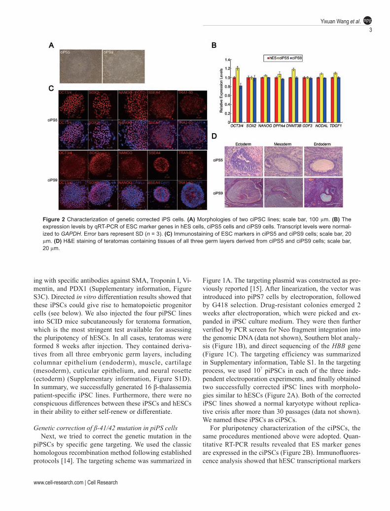
For pluripotency characterization of the ciPSCs, the same procedures mentioned above were adopted. Quan-titative RT-PCR results revealed that ES marker genes are expressed in the ciPSCs (Figure 2B). Immunofluores-cence analysis showed that hESC transcriptional markers
Figure 2 Characterization of genetic corrected iPS cells. (A) Morphologies of two ciPSC lines; scale bar, 100 µm. (B) The expression levels by qRT-PCR of ESC marker genes in hES cells, ciPS5 cells and ciPS9 cells. Transcript levels were normal-ized to GAPDH. Error bars represent SD (n = 3). (C) Immunostaining of ESC markers in ciPS5 and ciPS9 cells; scale bar, 20 µm. (D) H&E staining of teratomas containing tissues of all three germ layers derived from ciPS5 and ciPS9 cells; scale bar, 20 µm.
Genetically corrected β-thalassemia iPSCs function in vivo4
npg
Cell Research | www.cell-research.com
(OCT3/4, SOX2, NANOG) and surface markers (SSEA4, TRA1-60) were all expressed in the ciPSCs (Figure 2C). Teratomas consisting of derivatives from all three em-bryonic germ layers also formed after the subcutaneous injection of the two cell lines into SCID mice (Figure 2D). Overall, the process of generating disease-corrected iPSCs by targeted homologous recombination did not compromise their ability to either self-renew or differ-entiate, which is a prerequisite for further hematopoietic applications.
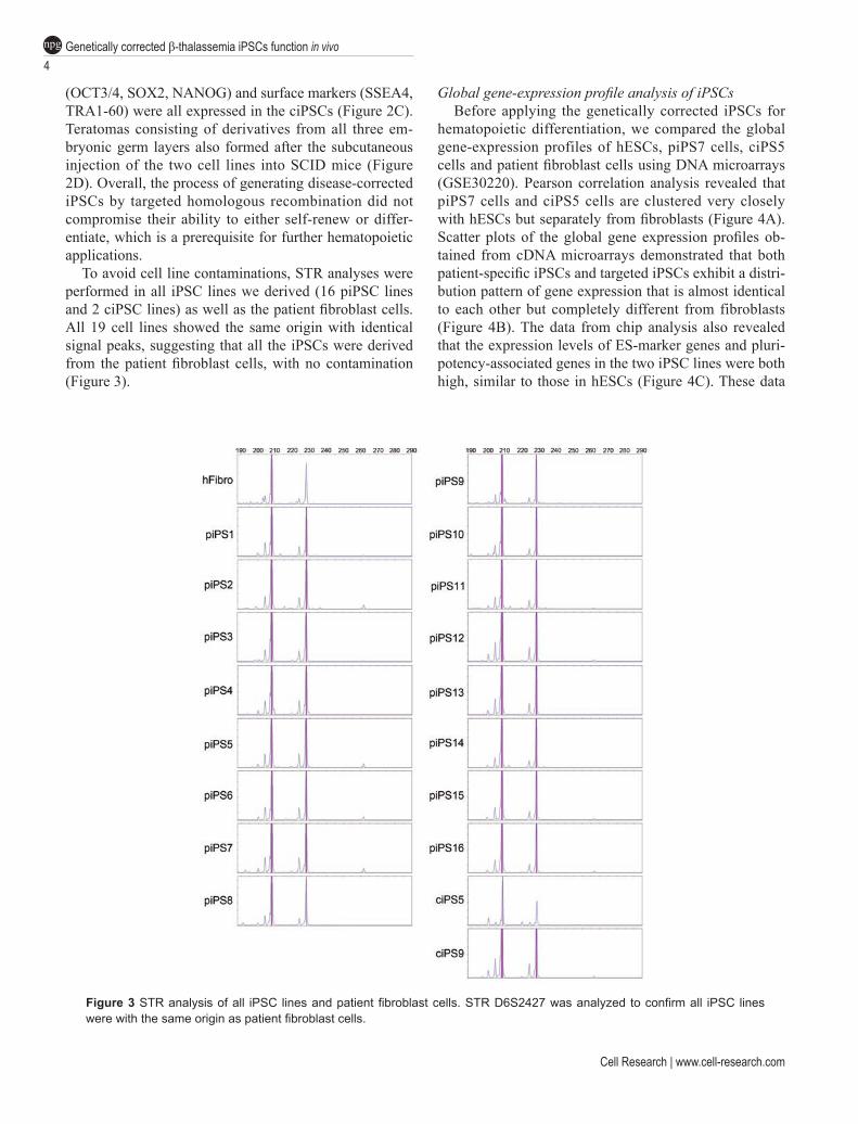
To avoid cell line contaminations, STR analyses were performed in all iPSC lines we derived (16 piPSC lines and 2 ciPSC lines) as well as the patient fibroblast cells. All 19 cell lines showed the same origin with identical signal peaks, suggesting that all the iPSCs were derived from the patient fibroblast cells, with no contamination (Figure 3).
Global gene-expression profile analysis of iPSCsBefore applying the genetically corrected iPSCs for
hematopoietic differentiation, we compared the global gene-expression profiles of hESCs, piPS7 cells, ciPS5 cells and patient fibroblast cells using DNA microarrays (GSE30220). Pearson correlation analysis revealed that piPS7 cells and ciPS5 cells are clustered very closely with hESCs but separately from fibroblasts (Figure 4A). Scatter plots of the global gene expression profiles ob-tained from cDNA microarrays demonstrated that both patient-specific iPSCs and targeted iPSCs exhibit a distri-bution pattern of gene expression that is almost identical to each other but completely different from fibroblasts (Figure 4B). The data from chip analysis also revealed that the expression levels of ES-marker genes and pluri-potency-associated genes in the two iPSC lines were both high, similar to those in hESCs (Figure 4C). These data
Figure 3 STR analysis of all iPSC lines and patient fibroblast cells. STR D6S2427 was analyzed to confirm all iPSC lines were with the same origin as patient fibroblast cells.
www.cell-research.com | Cell Research
Yixuan Wang et al.5
npg
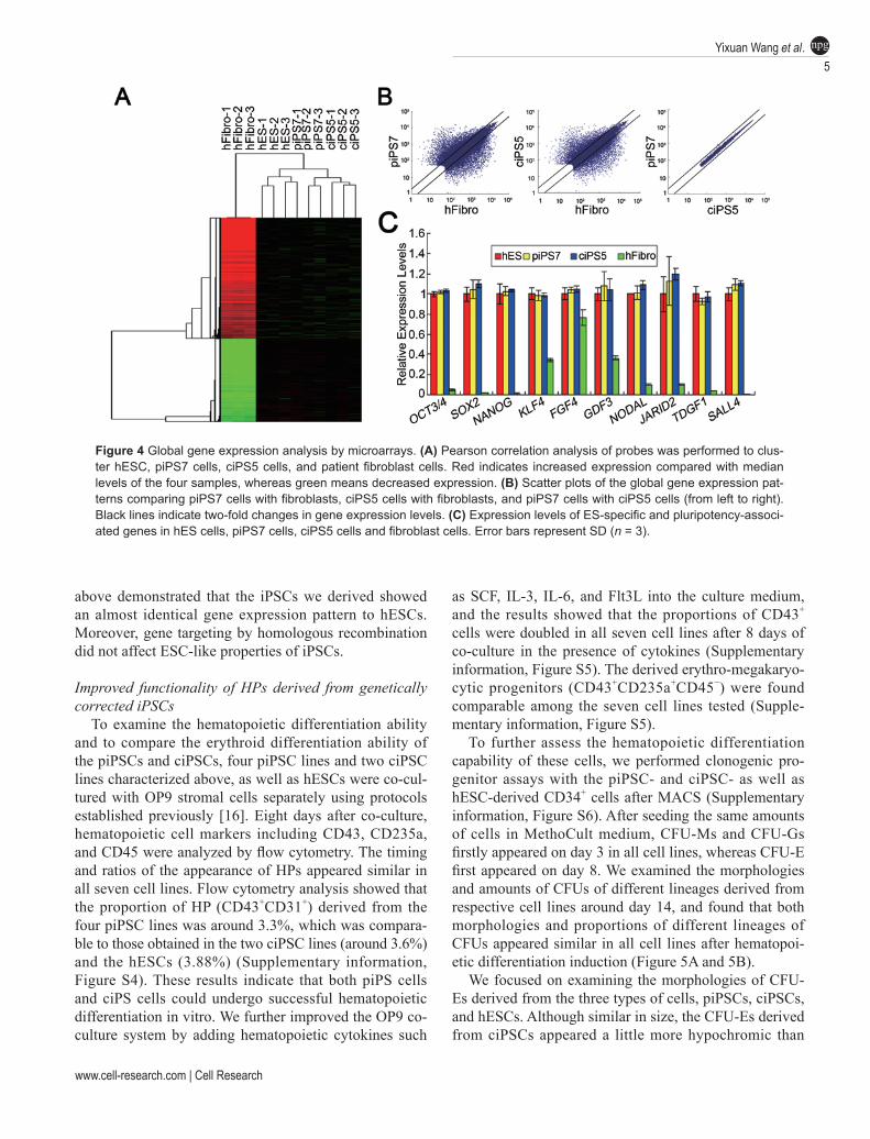
Figure 4 Global gene expression analysis by microarrays. (A) Pearson correlation analysis of probes was performed to clus-ter hESC, piPS7 cells, ciPS5 cells, and patient fibroblast cells. Red indicates increased expression compared with median levels of the four samples, whereas green means decreased expression. (B) Scatter plots of the global gene expression pat-terns comparing piPS7 cells with fibroblasts, ciPS5 cells with fibroblasts, and piPS7 cells with ciPS5 cells (from left to right). Black lines indicate two-fold changes in gene expression levels. (C) Expression levels of ES-specific and pluripotency-associ-ated genes in hES cells, piPS7 cells, ciPS5 cells and fibroblast cells. Error bars represent SD (n = 3).
above demonstrated that the iPSCs we derived showed an almost identical gene expression pattern to hESCs. Moreover, gene targeting by homologous recombination did not affect ESC-like properties of iPSCs.
Improved functionality of HPs derived from genetically corrected iPSCs
To examine the hematopoietic differentiation ability and to compare the erythroid differentiation ability of the piPSCs and ciPSCs, four piPSC lines and two ciPSC lines characterized above, as well as hESCs were co-cul-tured with OP9 stromal cells separately using protocols established previously [16]. Eight days after co-culture, hematopoietic cell markers including CD43, CD235a, and CD45 were analyzed by flow cytometry. The timing and ratios of the appearance of HPs appeared similar in all seven cell lines. Flow cytometry analysis showed that the proportion of HP (CD43+CD31+) derived from the four piPSC lines was around 3.3%, which was compara-ble to those obtained in the two ciPSC lines (around 3.6%) and the hESCs (3.88%) (Supplementary information, Figure S4). These results indicate that both piPS cells and ciPS cells could undergo successful hematopoietic differentiation in vitro. We further improved the OP9 co-culture system by adding hematopoietic cytokines such
as SCF, IL-3, IL-6, and Flt3L into the culture medium, and the results showed that the proportions of CD43+ cells were doubled in all seven cell lines after 8 days of co-culture in the presence of cytokines (Supplementary information, Figure S5). The derived erythro-megakaryo-cytic progenitors (CD43+CD235a+CD45−) were found comparable among the seven cell lines tested (Supple-mentary information, Figure S5).
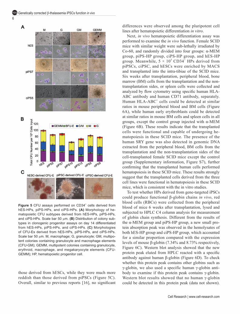
To further assess the hematopoietic differentiation capability of these cells, we performed clonogenic pro-genitor assays with the piPSC- and ciPSC- as well as hESC-derived CD34+ cells after MACS (Supplementary information, Figure S6). After seeding the same amounts of cells in MethoCult medium, CFU-Ms and CFU-Gs firstly appeared on day 3 in all cell lines, whereas CFU-E first appeared on day 8. We examined the morphologies and amounts of CFUs of different lineages derived from respective cell lines around day 14, and found that both morphologies and proportions of different lineages of CFUs appeared similar in all cell lines after hematopoi-etic differentiation induction (Figure 5A and 5B).
We focused on examining the morphologies of CFU-Es derived from the three types of cells, piPSCs, ciPSCs, and hESCs. Although similar in size, the CFU-Es derived from ciPSCs appeared a little more hypochromic than
Genetically corrected β-thalassemia iPSCs function in vivo6
npg
Cell Research | www.cell-research.com
those derived from hESCs, while they were much more reddish than those derived from piPSCs (Figure 5C). Overall, similar to previous reports [16], no significant
differences were observed among the pluripotent cell lines after hematopoietic differentiation in vitro.
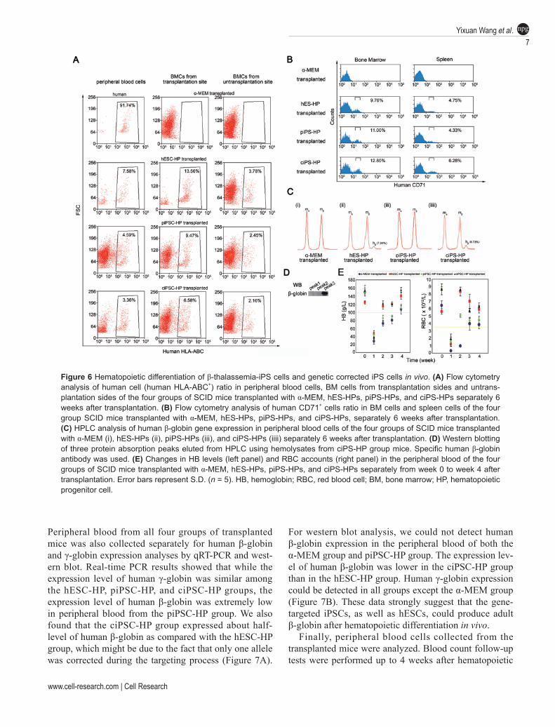
Next, in vivo hematopoietic differentiation assay was performed to examine the in vivo function. Female SCID mice with similar weight were sub-lethally irradiated by Co-60, and randomly divided into four groups: α-MEM group, piPS-HP group, ciPS-HP group, and hES-HP group. Meanwhile, 5 × 10⁵ CD34+ HPs derived from piPSCs, ciPSC, and hESCs were enriched by MACS and transplanted into the intra-tibiae of the SCID mice. Six weeks after transplantation, peripheral blood, bone marrow (BM) cells from the transplantation and the non-transplantation sides, or spleen cells were collected and analyzed by flow cytometry using specific human HLA-ABC antibody and human CD71 antibody, separately. Human HLA-ABC+ cells could be detected at similar ratios in mouse peripheral blood and BM cells (Figure 6A), while human early erythroblasts could be detected at similar ratios in mouse BM cells and spleen cells in all groups, except the control group injected with α-MEM (Figure 6B). These results indicate that the transplanted cells were functional and capable of undergoing he-matopoiesis in these SCID mice. The presence of the human SRY gene was also detected in genomic DNA extracted from the peripheral blood, BM cells from the transplantation and the non-transplantation sides of the cell-transplanted female SCID mice except the control group (Supplementary information, Figure S7), further confirming that the transplanted human cells performed hematopoiesis in these SCID mice. These results strongly suggest that the transplanted cells derived from the three cell lines were functional in hematopoiesis in these SCID mice, which is consistent with the in vitro studies.
To test whether HPs derived from gene-targeted iPSCs could produce functional β-globin chains in vivo, red blood cells (RBCs) were collected from the peripheral blood of mice 6 weeks after transplantation, lysed and subjected to HPLC C4 column analysis for measurement of globin chain synthesis. Different from the results of the α-MEM group and piPS-HP group, a new small pro-tein absorption peak was observed in the hemolysates of both hES-HP group and ciPS-HP group, which accounted for a similar proportion compared with the expression levels of mouse β-globin (7.34% and 8.73% respectively, Figure 6C). Western blot analysis showed that the new protein peak eluted from HPLC reacted with a specific antibody against human β-globin (Figure 6D). To check whether this protein peak contains other globins such as γ-globin, we also used a specific human γ-globin anti-body to examine if this protein peak contains γ-globin. Western blot results showed that no human γ-globin could be detected in this protein peak (data not shown).
Figure 5 CFU assays performed on CD34+ cells derived from hES-HPs, piPS-HPs, and ciPS-HPs. (A) Morphology of he-matopoietic CFU subtypes derived from hES-HPs, piPS-HPs, and ciPS-HPs. Scale bar 50 µm. (B) Distribution of colony sub-types in clonogenic progenitor assays on day 14 differentiated from hES-HPs, piPS-HPs, and ciPS-HPs. (C) Morphologies of CFU-Es derived from hES-HPs, piPS-HPs, and ciPS-HPs. Scale bar 50 µm. M, macrophage; G, granulocyte; GM, multipo-tent colonies containing granulocyte and macrophage elements (CFU-GM); GEMM, multipotent colonies containing granulocyte, erythroid, macrophage, and megakaryocyte elements (CFU-GEMM); HP, hematopoietic progenitor cell.
www.cell-research.com | Cell Research
Yixuan Wang et al.7
npg
Peripheral blood from all four groups of transplanted mice was also collected separately for human β-globin and γ-globin expression analyses by qRT-PCR and west-ern blot. Real-time PCR results showed that while the expression level of human γ-globin was similar among the hESC-HP, piPSC-HP, and ciPSC-HP groups, the expression level of human β-globin was extremely low in peripheral blood from the piPSC-HP group. We also found that the ciPSC-HP group expressed about half-level of human β-globin as compared with the hESC-HP group, which might be due to the fact that only one allele was corrected during the targeting process (Figure 7A).
For western blot analysis, we could not detect human β-globin expression in the peripheral blood of both the α-MEM group and piPSC-HP group. The expression lev-el of human β-globin was lower in the ciPSC-HP group than in the hESC-HP group. Human γ-globin expression could be detected in all groups except the α-MEM group (Figure 7B). These data strongly suggest that the gene-targeted iPSCs, as well as hESCs, could produce adult β-globin after hematopoietic differentiation in vivo.
Finally, peripheral blood cells collected from the transplanted mice were analyzed. Blood count follow-up tests were performed up to 4 weeks after hematopoietic
Figure 6 Hematopoietic differentiation of β-thalassemia-iPS cells and genetic corrected iPS cells in vivo. (A) Flow cytometry analysis of human cell (human HLA-ABC+) ratio in peripheral blood cells, BM cells from transplantation sides and untrans-plantation sides of the four groups of SCID mice transplanted with α-MEM, hES-HPs, piPS-HPs, and ciPS-HPs separately 6 weeks after transplantation. (B) Flow cytometry analysis of human CD71+ cells ratio in BM cells and spleen cells of the four group SCID mice transplanted with α-MEM, hES-HPs, piPS-HPs, and ciPS-HPs, separately 6 weeks after transplantation. (C) HPLC analysis of human β-globin gene expression in peripheral blood cells of the four groups of SCID mice transplanted with α-MEM (i), hES-HPs (ii), piPS-HPs (iii), and ciPS-HPs (iiii) separately 6 weeks after transplantation. (D) Western blotting of three protein absorption peaks eluted from HPLC using hemolysates from ciPS-HP group mice. Specific human β-globin antibody was used. (E) Changes in HB levels (left panel) and RBC accounts (right panel) in the peripheral blood of the four groups of SCID mice transplanted with α-MEM, hES-HPs, piPS-HPs, and ciPS-HPs separately from week 0 to week 4 after transplantation. Error bars represent S.D. (n = 5). HB, hemoglobin; RBC, red blood cell; BM, bone marrow; HP, hematopoietic progenitor cell.
Genetically corrected β-thalassemia iPSCs function in vivo8
npg
Cell Research | www.cell-research.com
transplantation (Table 1). Due to irradiation, both levels of HB and RBCs in the peripheral blood dropped greatly under the base line (which is 100 g/l and 3.6 × 10¹²/l, respectively) in all four groups of transplanted mice one week after transplantation. The HB levels in the hES-HP and ciPS-HP-transplanted mice elevated above 100 g/l from the second week after transplantation. In contrast, the HB levels in the rest two groups of mice increased slowly and remained below the normal base line until the 4th week after transplantation (Figure 6E, Table 1), from which time point, all four groups exhibited normal HB levels. These blood routine data suggest that genetically corrected iPSCs-derived HP can positively stimulate he-matopoiesis in these SCID mice after irradiation. Thus, our results collectively demonstrate that the genetically corrected iPSCs, like human ESCs, have the ability to give rise to hematopoietic progenitors that undergo nor-mal hematopoiesis in a sub-lethally SCID mouse model.
Discussion
In this study, we successfully derived 16 patient-specific iPS cell lines from a 2-year-old boy suffering from β-thalassemia major. Furthermore, we corrected the genetic mutation of the disease by homologous recombi-nation using a classic gene-targeting approach. By trans-plantation into an immune-deficient mouse model, we demonstrated that the production of HB could be signifi-cantly increased when disease-corrected iPSCs-derived HPs were transplanted and moreover, the production of human-specific β-globin could be detected. Our study advances the field by demonstrating a potential use of the iPSC technology in clinical treatment of β-thalassemia major.
Correction of the genetic mutation in iPS cells de-rived from the individual patient with genetic disease is the most critical step towards personalized regenerative medicine practice. Although two corrected colonies were finally obtained using the traditional homologous recom-bination approach, the targeting efficiency is very low and the targeting method needs to be further optimized. Recently, introduction of a transgene into the iPS cells or correction of the genetic mutation by zinc finger nu-clease- or bacteria artificial chromosome (BAC)-based methods offers alternative ways to obtain genetically cor-rected iPS cells [17-19]. More recently, helper-dependent adenoviral vectors were used to provide a more efficient method for correcting mutations in human pluripotent stem cells [20, 21]. It is desirable to further improve the efficiency of correcting the β-globin gene mutation in the iPS cells produced in the present study by comparing the different approaches listed above.
Before the clinical application of iPSCs can finally be realized, a number of hurdles must be overcome. The recent generation of protein, mRNA, and microRNA reprogrammed iPS cells provides a more ideal approach to obtaining safer patient-specific iPS cells [22-25]. Sub-sequently, selecting good quality iPS cells will be needed for further genetic manipulation. One major possibility causing incomplete erasing of the epigenetic memory in-herited from the somatic cells and the immunogenicity of iPS cells as observed recently might be due to incomplete reprogramming of the somatic genome [26-29]. Finally, the strategy of directed differentiation of corrected iPS cells into specific cell types has to be optimized in the future. Recent studies have shown that the direct conver-sion of one cell type into another is feasible both in vivo and in vitro [30-34]. It remains unknown, however, if these trans-differentiated cells are functional, because the length of the telomeres in these cells may not be prop-erly restored. Interestingly, telomere length can indeed
Figure 7 Analysis of human β-globin and γ-globin expression in the peripheral blood collected from all groups of transplanted mice. (A) qRT-PCR analysis of human β-globin and γ-globin mRNA expression in the peripheral blood collected from hESC-HP, piPSC-HP, and ciPSC-HP groups. (B) Western blot analysis of human β-globin and γ-globin expression using specific anti-bodies in the peripheral blood collected from all groups of trans-planted mice.
www.cell-research.com | Cell Research
Yixuan Wang et al.9
npg
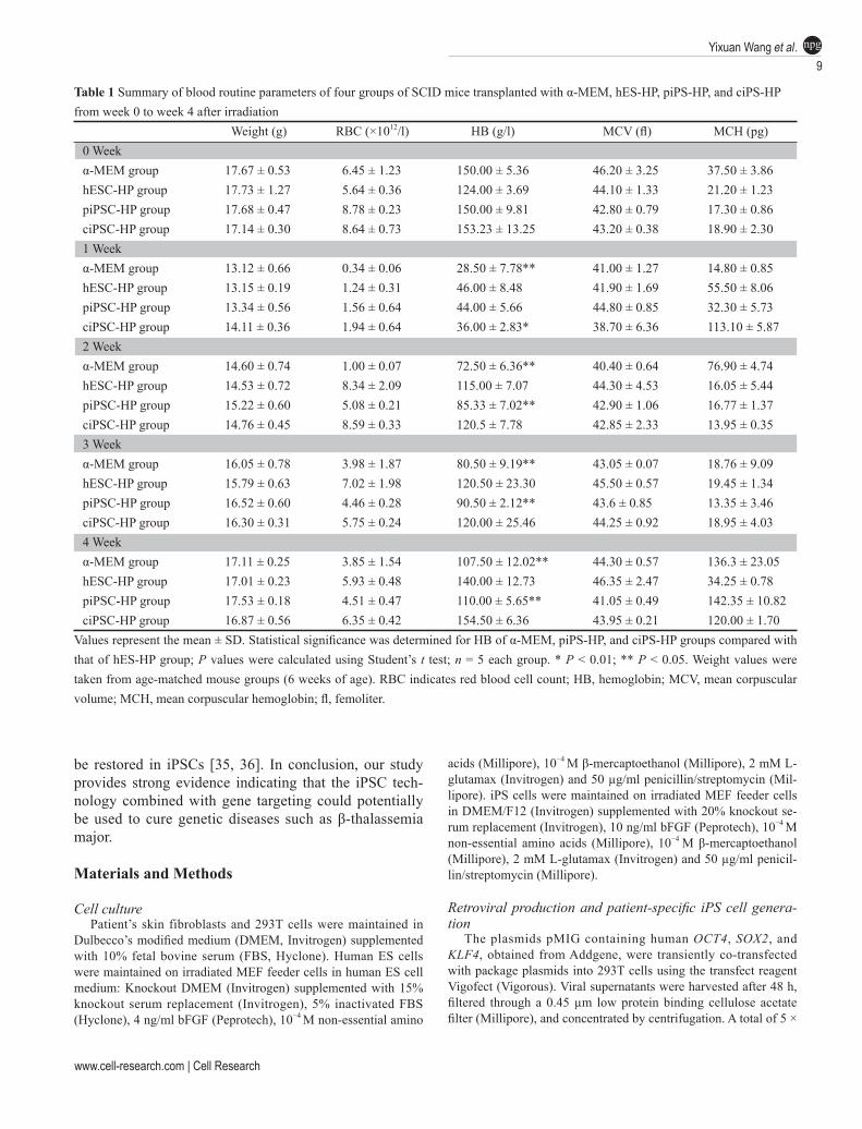
Table 1 Summary of blood routine parameters of four groups of SCID mice transplanted with α-MEM, hES-HP, piPS-HP, and ciPS-HP from week 0 to week 4 after irradiation
Weight (g) RBC (×1012/l) HB (g/l) MCV (fl) MCH (pg)0 Weekα-MEM group 17.67 ± 0.53 6.45 ± 1.23 150.00 ± 5.36 46.20 ± 3.25 37.50 ± 3.86hESC-HP group 17.73 ± 1.27 5.64 ± 0.36 124.00 ± 3.69 44.10 ± 1.33 21.20 ± 1.23piPSC-HP group 17.68 ± 0.47 8.78 ± 0.23 150.00 ± 9.81 42.80 ± 0.79 17.30 ± 0.86ciPSC-HP group 17.14 ± 0.30 8.64 ± 0.73 153.23 ± 13.25 43.20 ± 0.38 18.90 ± 2.301 Weekα-MEM group 13.12 ± 0.66 0.34 ± 0.06 28.50 ± 7.78** 41.00 ± 1.27 14.80 ± 0.85hESC-HP group 13.15 ± 0.19 1.24 ± 0.31 46.00 ± 8.48 41.90 ± 1.69 55.50 ± 8.06piPSC-HP group 13.34 ± 0.56 1.56 ± 0.64 44.00 ± 5.66 44.80 ± 0.85 32.30 ± 5.73ciPSC-HP group 14.11 ± 0.36 1.94 ± 0.64 36.00 ± 2.83* 38.70 ± 6.36 113.10 ± 5.872 Weekα-MEM group 14.60 ± 0.74 1.00 ± 0.07 72.50 ± 6.36** 40.40 ± 0.64 76.90 ± 4.74hESC-HP group 14.53 ± 0.72 8.34 ± 2.09 115.00 ± 7.07 44.30 ± 4.53 16.05 ± 5.44piPSC-HP group 15.22 ± 0.60 5.08 ± 0.21 85.33 ± 7.02** 42.90 ± 1.06 16.77 ± 1.37ciPSC-HP group 14.76 ± 0.45 8.59 ± 0.33 120.5 ± 7.78 42.85 ± 2.33 13.95 ± 0.353 Weekα-MEM group 16.05 ± 0.78 3.98 ± 1.87 80.50 ± 9.19** 43.05 ± 0.07 18.76 ± 9.09hESC-HP group 15.79 ± 0.63 7.02 ± 1.98 120.50 ± 23.30 45.50 ± 0.57 19.45 ± 1.34piPSC-HP group 16.52 ± 0.60 4.46 ± 0.28 90.50 ± 2.12** 43.6 ± 0.85 13.35 ± 3.46ciPSC-HP group 16.30 ± 0.31 5.75 ± 0.24 120.00 ± 25.46 44.25 ± 0.92 18.95 ± 4.034 Weekα-MEM group 17.11 ± 0.25 3.85 ± 1.54 107.50 ± 12.02** 44.30 ± 0.57 136.3 ± 23.05hESC-HP group 17.01 ± 0.23 5.93 ± 0.48 140.00 ± 12.73 46.35 ± 2.47 34.25 ± 0.78piPSC-HP group 17.53 ± 0.18 4.51 ± 0.47 110.00 ± 5.65** 41.05 ± 0.49 142.35 ± 10.82ciPSC-HP group 16.87 ± 0.56 6.35 ± 0.42 154.50 ± 6.36 43.95 ± 0.21 120.00 ± 1.70
Values represent the mean ± SD. Statistical significance was determined for HB of α-MEM, piPS-HP, and ciPS-HP groups compared with that of hES-HP group; P values were calculated using Student’s t test; n = 5 each group. * P < 0.01; ** P < 0.05. Weight values were taken from age-matched mouse groups (6 weeks of age). RBC indicates red blood cell count; HB, hemoglobin; MCV, mean corpuscular volume; MCH, mean corpuscular hemoglobin; fl, femoliter.
be restored in iPSCs [35, 36]. In conclusion, our study provides strong evidence indicating that the iPSC tech-nology combined with gene targeting could potentially be used to cure genetic diseases such as β-thalassemia major.
Materials and Methods
Cell culturePatient’s skin fibroblasts and 293T cells were maintained in
Dulbecco’s modified medium (DMEM, Invitrogen) supplemented with 10% fetal bovine serum (FBS, Hyclone). Human ES cells were maintained on irradiated MEF feeder cells in human ES cell medium: Knockout DMEM (Invitrogen) supplemented with 15% knockout serum replacement (Invitrogen), 5% inactivated FBS (Hyclone), 4 ng/ml bFGF (Peprotech), 10−4 M non-essential amino
acids (Millipore), 10−4 M β-mercaptoethanol (Millipore), 2 mM L-glutamax (Invitrogen) and 50 µg/ml penicillin/streptomycin (Mil-lipore). iPS cells were maintained on irradiated MEF feeder cells in DMEM/F12 (Invitrogen) supplemented with 20% knockout se-rum replacement (Invitrogen), 10 ng/ml bFGF (Peprotech), 10−4 M non-essential amino acids (Millipore), 10−4 M β-mercaptoethanol (Millipore), 2 mM L-glutamax (Invitrogen) and 50 µg/ml penicil-lin/streptomycin (Millipore).
Retroviral production and patient-specific iPS cell genera-tion
The plasmids pMIG containing human OCT4, SOX2, and KLF4, obtained from Addgene, were transiently co-transfected with package plasmids into 293T cells using the transfect reagent Vigofect (Vigorous). Viral supernatants were harvested after 48 h, filtered through a 0.45 µm low protein binding cellulose acetate filter (Millipore), and concentrated by centrifugation. A total of 5 ×
Genetically corrected β-thalassemia iPSCs function in vivo10
npg
Cell Research | www.cell-research.com
104 patient fibroblast cells were incubated with virus for 24 h and then seeded onto irradiated MEF feed cells after 5 days. iPS cell culture medium was substituted after 7 days. iPSC colonies were manually picked and mechanically dissociated for following pas-sage.
Karyotype analysisThe iPS cells were first incubated in culture medium contain-
ing 0.25 µg/ml colcemid (Invitrogen) for 4 h and then the cells were harvested and incubated in 0.4% sodium citrate, 0.4% chloratum Kaliumat (1:1, v/v) at 37 °C for 5 min and fixed in a methanol:acetic acid mixture (3:1, v/v) three times. After Giemsa staining, at least 20 chromosome karyoschisis images were exam-ined.
qRT-PCR analysisTotal RNA was extracted from cells using TRIzol reagent (In-
vitrogen) according to the manufacturer’s instructions. Comple-mentary DNA synthesis was performed with the M-MLV Reverse Transcriptase Kit (Promega) following the manufacturer’s instruc-tions. Quantitative RT-PCR was performed using a SYBR Green-based PCR Master Mix (Takara) and signals were detected with ABI7500 Real-Time PCR System (Applied BioSystems).
Alkaline phosphatase staining and immunocytochemistryAlkaline phosphatase staining was performed using the ALKA-
LINE PHOSPHATASE SUBSTRATE Kit (Sigma) according to the manufacturer’s instructions. For immunocytochemistry, cells were fixed with PBS containing 4% paraformaldehyde for 15 min at room temperature. After being washed with TBST, the cells were treated with PBS containing 0.1% Triton X-100 for 15 min at room temperature and then incubated with PBS containing 4% normal goat serum (Millipore) for 30 min at room temperature. The following primary antibodies were used in this study: SSEA4 (1:50, ES Cell Marker Sample Kit, Millipore), TRA1-60 (1:50, ES Cell Marker Sample Kit, Millipore), TRA-1-81 (1:50, ES Cell Marker Sample Kit, Millipore), SOX2 (1:500, Abcam), NANOG (1:500, Abcam), and OCT4 (1:50, ES Cell Marker Sample Kit, Millipore), PDX-1 (1:200, Millipore), Troponin (1:200, Millipore), SMA (1:200, Millipore), Vimentin ((1:200, Millipore), and Pan Cytokeratin ((1:200, Millipore). The following secondary antibod-ies were used in this study: Alexa 594-conjugated goat anti-rabbit IgG (1:500, Invitrogen), Alexa 594-conjugated goat anti-mouse IgM (1:500, Invitrogen) and Alexa 594-conjugated goat anti-mouse IgG (1:500, Invitrogen). Nuclei were stained with DAPI. Stained cells mounted on slides were observed on an LSM 510 META confocal microscope (Zeiss).
In vitro differentiationTo generate the embryoid bodies, iPS cells were harvested by
mechanical dissociation. The clumps of cells were transferred to a 6-well ultralow attachment plates (Costa, Corning) in IMDM (Invitrogen) containing 15% FBS (HyClone), 10−4 M non-essential amino acids (Millipore), 10−4 M β-mercaptoethanol (Millipore), 2 mM L-glutamax (Invitrogen) and 50 µg/ml penicillin/streptomycin (Millipore). The medium was changed every other day. After 8 days, EBs were transferred to gelatin-coated plates and cultured in the same medium for another 8 days.
Teratoma formationThe iPS cells were harvested by mechanical dissociation, col-
lected into tubes and centrifuged. The pellets were resuspended in DMEM/F12 (Invitrogen). Cells from a confluent 35-mm dish were injected subcutaneously into the groin of a SCID mouse. Eight weeks after injection, the tumors were dissected and fixed with PBS containing 10% paraformaldehyde (Sigma). Paraffin-embed-ded tissue was sliced and stained with hematoxylin and eosin.
Plasmid construction and gene targeting of patient-specific iPS cells
Targeting plasmid was constructed using two-round recombi-nation as previously reported. Freshly prepared BAC DNA con-taining the HBB locus was transformed into EL350 cells. pL253 plasmid containing the recombinant fragments was linearized by HindIII and electroporated into EL350 cells to generate the recom-binant colonies. The fragment containing the Neo resistant gene was used for the second-round recombination.
Before electroporation, iPS cells were first adapted to enzy-matically disaggregation into single cells using TrypLE select (Invitrogen) for several passages. On the day of electroporation, 1 × 10⁷ iPS cells were suspended in ice-cold PBS containing 50 µg linearized DNA and electroporated by electroporator (Gene Pulser II System, Bio-Rad) using the parameters 250 V and 500 µF. G418 selection started 5 days after electroporation with the concentration of 50 µg/ml and maintained for 5-7 days. Colonies were allowed to grow for an additional 7 days before picking.
Southern blot analysis and DNA sequencing analysisFor Southern blot analysis, genomic DNA (30 µg) from the iP-
SCs was digested with BglII overnight. Digested DNA fragments were separated on a 0.8% agarose gel and transferred to a nylon membrane (Amersham). The sequences of DNA probe primers are: F-GTAGAGGCTTGATTTGGAGGTT; R-TTATGGTGCT-TCTGGCTCTG. The membrane was incubated with 32P-labeled DNA probe supplied by Amersham Rediprime II Random Prime Labeling System (GE Health). For sequencing analysis, the muta-tion site of HBB gene were amplified by PCR from genomic DNA of piPSCs and ciPSCs and sequenced by BGI Life Tech company. The sequences of HBB sequencing primer are: F-CACTAGCAAC-CTCAAACAGACA; R-CTCAAGGCCCTTCATAATATCC.
STR analysisFor STR analysis, genomic DNA was extracted from 16 piPSC
lines, 2 ciPSC lines and the patient fibroblast cells, respectively, and used as templates for PCR. Specific primers with 5′ FAM were used in PCR system for STR D6S2427 cloning. STR signal analy-sis was performed by BGI Life Tech company.
Hematopoietic differentiationFor hES and iPS cell differentiation, OP9 cells were plated
onto gelatinized 35-mm or 100-mm dishes in α-MEM (Invitrogen) containing 20% FBS (HyClone), 2 mM L-glutamax (Invitrogen) and 50 µg/ml penicillin/streptomycin (Millipore). After forma-tion of confluent cultures on days 4 and 5, half of the medium was changed, and cells were cultured for an additional 3 to 4 days. Undifferentiated hESCs and iPSCs were harvested by mechanical dissociation into small clumps. Both hESCs and iPSCs were added to OP9 cultures at a density of 1.5 × 10⁶/20 ml per 100-mm dish
www.cell-research.com | Cell Research
Yixuan Wang et al.11
npg
or 0.3 × 10⁶/4 ml per 35-mm dish in α-MEM (Invitrogen) supple-mented with 10% FBS (HyClone) and 100 µM monothioglycerol (MTG; Sigma). The hESC/OP9 and iPSC/OP9 co-cultures were incubated for up to 10 days with a half-medium change every oth-er day. Cells were harvested on day 10, and single-cell suspension was prepared for clonogenic and flow-cytometric assays.
Flow cytometry analysisTo analyze the phenotype of HPs, hESC/OP9 and iPSC/OP9
co-culture were collected and washed with FACS buffer (phos-phate-buffered salt solution with 2% FBS). Cells were stained with anti-human CD43-PE monoclonal antibody (BD), anti-human CD31-APC monoclonal antibody (BD), anti-human CD45-PE/Cy7 monoclonal antibody (BD), and anti-human CD235a-PE/Cy5 monoclonal antibody (BD) with different combinations. Cells were then washed and resuspended in FACS buffer. Gating was done with matched isotype control monoclonal antibodies. 7-AAD (BD) was used to exclude dead cells. All analyses were performed on a MoFlo XDP cell sorter (Beckman Coulter Inc) running Sum-mit software.
For analysis of human cell ratio, the peripheral blood cells were collected and the BM cells were flushed from the intra tibia of transplanted SCID mice. After washing with FACS buffer, cells were stained with anti-human HLA-ABC-FITC monoclonal anti-body (BD), and incubated with Red Blood Cell Lysis Buffer (Tia-gen) to remove the RBC, and then resuspended in FACS buffer and analyzed using MoFlo XDP cell sorter (Beckman Coulter Inc).
For analysis of human β-globin, the peripheral blood cells were collected. After fixation and permeabilization, cells were incubated with anti-human β-globin monoclonal antibody (Santa Cruz), and then washed with FACS buffer and analyzed by using MoFlo XDP cell sorter (Beckman Coulter Inc).
Clonogenic progenitor cell assaysHematopoietic clonogenic assays were performed in 6-well
ultralow attachment plates (Costa, Corning) using a 2 ml/well of Methylcellulose-based media HSC003 (Stem Cell Technologies). Cells from hES cell/OP9 and iPS cell/OP9 co-cultures were col-lected, sorted by MACS and plated at 1 × 10⁴ / well after 8 days of differentiation. All clonogenic progenitor assays were performed in triplicate. CFCs were scored after 14 days of incubation. Cytospin preparations from single colonies were made using a cytospin centrifuge, then fixed with methanol and stained with Wright stain (Sigma) to confirm the cell content of appropriate colonies.
MACS analysisCells from hESC/OP9 and iPSC/OP9 co-cultures were col-
lected, washed with MACS buffer (phosphate-buffered salt solu-tion with 0.5% BSA, and 2 mM EDTA), and incubated with CD34 MicroBeads (Miltenyi Biotec) for magnetic labeling. Then cells were loaded onto the column. Magnetic separation was performed in the magnetic field following the manufacturer’s instructions.
BM transplantationImmune deficient NOD/SCID mice at the age of 8 to 10 weeks
were sublethally irradiated with 3.0 Gry 6 h before transplantation. For transplantation, 5 × 10⁵ MACS sorted CD34+ hematopoietic progenitors were collected as previously described and resus-pended in IMDM medium, and then transferred to a 28 G insulin
syringe. The cell suspension in the syringe was gently inserted in a twisting motion vertically and was transplanted by intra-tibiae injection.
HPLCRBC (20 µl) from mouse peripheral blood were lysed in 500 µl
of lysis buffer (0.1 M 2-mercaptoethanol, 0.1 M HCl), and then made up to 1 ml with 500 µl of 50% aqueous acetonitrile. The ly-sed red cells were centrifuged and the supernatant was analyzed by HPLC. Globin chain separation was performed by loading 30 µl of lysed RBC onto a Vydac C4 column (Vydac, Hesperia, USA). At the start of each run, the buffer ratio between buffer A (0.1% trifluoroacetic acid, Sigma) and buffer B (95% acetonitrile, Sigma, 4.0% water, 0.1% trifluoroacetic acid) was 65%:35%. Buffer B was gradually increased to 56% after 1 h with a constant flow rate of 1 ml/min. The proteins eluting from the column were measured at 220 nm, 280 nm, and 413 nm with Agilent 1100 Diode Array Detector (Waters). Peak protein fractions were collected, and ana-lyzed by tandem mass spectrometry or western blot using specific human β-globin (Santa Cruz) or γ-globin (Santa Cruz) antibody in order to confirm globin chain type.
Microarray analysisAffymetrix GeneChip Human Genome U133 plus 2.0 Array
(Affymetrix, Inc.) was used for microarray hybridizations to ex-amine the global gene expression of hES cells, piPS7 cells, ciPS5 cells, and patient fibroblast cells. Briefly, total RNA was isolated using Trizol reagent (Invitrogen). cDNA was synthesized and amplified with WT cDNA Synthesis and Amplification Kit (Af-fymetrix, Inc.), labeled with biotin using WT Terminal Labeling Kit (Affymetrix, Inc.), hybridized to Affymetrix GeneChip Human Genome U133 plus 2.0 Array and analyzed by GeneChip Scanner 3000 7G3000 (Affymetrix, Inc.). All samples were prepared in three biological repeats. Data were analyzed using GCOS 1.4 soft-ware provided by Affymetrix. Signal values of probes presented in two samples were plotted in a scatter graph. Pearson’s Correla-tion Coefficient (R) between samples was calculated by Excel. We have submitted the microarray data to GEO database (GSE30220).
Acknowledgments
We are grateful to colleagues in our laboratory for their as-sistance with experiments and in the preparation of the manu-script. This project was supported by the Chinese Ministry of Science and Technology (2011CB964800, 2011ZX102-010, and 2010CB944900 to SG, and 2009AZ4006-F to ZY), and the “stem cell therapy for β-thalassemia” of Guangxi Zhuang Autonomous Region Science and Technology Bureau (GKZ0897009 to ZY).
References
1 Takahashi K, Yamanaka S. Induction of pluripotent stem cells from mouse embryonic and adult fibroblast cultures by de-fined factors. Cell 2006; 126:663-676.
2 Okita K, Ichisaka T, Yamanaka S. Generation of germline-competent induced pluripotent stem cells. Nature 2007; 448:313-317.
3 Wernig M, Meissner A, Foreman R, et al. In vitro reprogram-ming of fibroblasts into a pluripotent ES-cell-like state. Na-
Genetically corrected β-thalassemia iPSCs function in vivo12
npg
Cell Research | www.cell-research.com
ture 2007; 448:318-324.4 Takahashi K, Tanabe K, Ohnuki M, et al. Induction of pluri-
potent stem cells from adult human fibroblasts by defined fac-tors. Cell 2007; 131:861-872.
5 Park IH, Zhao R, West JA, et al. Reprogramming of human somatic cells to pluripotency with defined factors. Nature 2008; 451:141-146.
6 Yu J, Vodyanik MA, Smuga-Otto K, et al. Induced pluripotent stem cell lines derived from human somatic cells. Science 2007; 318:1917-1920.
7 Dimos JT, Rodolfa KT, Niakan KK, et al. Induced pluripotent stem cells generated from patients with ALS can be differenti-ated into motor neurons. Science 2008; 321:1218-1221.
8 Ebert AD, Yu J, Rose FF Jr, et al. Induced pluripotent stem cells from a spinal muscular atrophy patient. Nature 2009; 457:277-280.
9 Raya A, Rodriguez-Piza I, Guenechea G, et al. Disease-corrected haematopoietic progenitors from Fanconi anaemia induced pluripotent stem cells. Nature 2009; 460:53-59.
10 Soldner F, Hockemeyer D, Beard C, et al. Parkinson’s disease patient-derived induced pluripotent stem cells free of viral reprogramming factors. Cell 2009; 136:964-977.
11 Hanna J, Wernig M, Markoulaki S, et al. Treatment of sickle cell anemia mouse model with iPS cells generated from au-tologous skin. Science 2007; 318:1920-1923.
12 Wang Y, Jiang Y, Liu S, Sun X, Gao S. Generation of induced pluripotent stem cells from human beta-thalassemia fibroblast cells. Cell Res 2009; 19:1120-1123.
13 Ye L, Chang JC, Lin C, Sun X, Yu J, Kan YW. Induced pluri-potent stem cells offer new approach to therapy in thalassemia and sickle cell anemia and option in prenatal diagnosis in ge-netic diseases. Proc Natl Acad Sci USA 2009; 106:9826-9830.
14 Costa M, Dottori M, Sourris K, et al. A method for genetic modification of human embryonic stem cells using electropo-ration. Nat Protoc 2007; 2:792-796.
15 Liu P, Jenkins NA, Copeland NG. A highly efficient recom-bineering-based method for generating conditional knockout mutations. Genome Res 2003; 13:476-484.
16 Vodyanik MA, Slukvin II. Hematoendothelial differentiation of human embryonic stem cells. Curr Protoc Cell Biol 2007; Chapter 23:Unit 23.6.
17 Hockemeyer D, Soldner F, Beard C, et al. Efficient targeting of expressed and silent genes in human ESCs and iPSCs using zinc-finger nucleases. Nat Biotechnol 2009; 27:851-857.
18 Cui X, Ji D, Fisher DA, Wu Y, Briner DM, Weinstein EJ. Tar-geted integration in rat and mouse embryos with zinc-finger nucleases. Nat Biotechnol 2011; 29:64-67.
19 Song H, Chung SK, Xu Y. Modeling disease in human ESCs using an efficient BAC-based homologous recombination sys-tem. Cell Stem Cell 2010; 6:80-89.
20 Liu GH, Suzuki K, Qu J, et al. Targeted gene correction of laminopathy-associated LMNA mutations in patient-specific
iPSCs. Cell Stem Cell 2011; 8:688-694.21 Li M, Suzuki K, Qu J, et al. Efficient correction of hemoglo-
binopathy-causing mutations by homologous recombination in integration-free patient iPSCs. Cell Res 2011; 21:1740-1744.
22 Warren L, Manos PD, Ahfeldt T, et al. Highly efficient re-programming to pluripotency and directed differentiation of human cells with synthetic modified mRNA. Cell Stem Cell 2010; 7:618-630.
23 Anokye-Danso F, Trivedi CM, Juhr D, et al. Highly efficient miRNA-mediated reprogramming of mouse and human so-matic cells to pluripotency. Cell Stem Cell 2010; 8:376-388.
24 Miyoshi N, Ishii H, Nagano H, et al. Reprogramming of mouse and human cells to pluripotency using mature microR-NAs. Cell Stem Cell 2011; 8:633-638.
25 Kim D, Kim CH, Moon JI, et al. Generation of human in-duced pluripotent stem cells by direct delivery of reprogram-ming proteins. Cell Stem Cell 2009; 4:472-476.
26 Lister R, Pelizzola M, Kida YS, et al. Hotspots of aberrant epigenomic reprogramming in human induced pluripotent stem cells. Nature 2011; 471:68-73.
27 Ohi Y, Qin H, Hong C, et al. Incomplete DNA methylation underlies a transcriptional memory of somatic cells in human iPS cells. Nat Cell Biol 2011; 13:541-549.
28 Zhao T, Zhang ZN, Rong Z, Xu Y. Immunogenicity of in-duced pluripotent stem cells. Nature 2011; 474:212-215.
29 Papapetrou EP, Lee G, Malani N, et al. Genomic safe harbors permit high beta-globin transgene expression in thalassemia induced pluripotent stem cells. Nat Biotechnol 2011; 29:73-78.
30 Zhou Q, Brown J, Kanarek A, Rajagopal J, Melton DA. In vivo reprogramming of adult pancreatic exocrine cells to beta-cells. Nature 2008; 455:627-632.
31 Vierbuchen T, Ostermeier A, Pang ZP, Kokubu Y, Sudhof TC, Wernig M. Direct conversion of fibroblasts to functional neu-rons by defined factors. Nature 2010; 463:1035-1041.
32 Huang P, He Z, Ji S, et al. Induction of functional hepatocyte-like cells from mouse fibroblasts by defined factors. Nature 2011; 475:386-389.
33 Ieda M, Fu JD, Delgado-Olguin P, et al. Direct reprogram-ming of fibroblasts into functional cardiomyocytes by defined factors. Cell 2010; 142:375-386.
34 Szabo E, Rampalli S, Risueno RM, et al. Direct conversion of human fibroblasts to multilineage blood progenitors. Nature 2010; 468:521-526.
35 Agarwal S, Loh YH, McLoughlin EM, et al. Telomere elon-gation in induced pluripotent stem cells from dyskeratosis congenita patients. Nature 2010; 464:292-296.
36 Marion RM, Strati K, Li H, et al. Telomeres acquire embry-onic stem cell characteristics in induced pluripotent stem cells. Cell Stem Cell 2009; 4:141-154.
(Supplementary information is linked to the online version of the paper on the Cell Research website.)


![Counterfactual Model for Learning Systems · Definition [IPS Utility Estimator]: Given 𝑆= 1, 1,𝛿1,…, 𝑛, 𝑛,𝛿𝑛 collected under 𝜋0, Unbiased estimate of utility](https://static.fdocument.org/doc/165x107/605dfe01ff887e0a3a7cc2b8/counterfactual-model-for-learning-definition-ips-utility-estimator-given-.jpg)