Interleukin-1β induces human proximal tubule cell injury ... · Kidney International, Vol. 62...
10
Kidney International, Vol. 62 (2002), pp. 31–40 Interleukin-1 induces human proximal tubule cell injury, -smooth muscle actin expression and fibronectin production 1 DAVID A. VESEY,CATHERINE W.Y. CHEUNG,LEILA CUTTLE,ZOLTAN A. ENDRE, GLENDA GOBE ´ , and DAVID W. JOHNSON Department of Renal Medicine, Princess Alexandra Hospital, Brisbane, and Departments of Pathology and Medicine, University of Queensland, Woolloongabba, Queensland, Australia muscle actin expression. These actions appear to be mediated Interleukin-1 induces human proximal tubule cell injury, by a TGF- 1 dependent mechanism and are independent of -smooth muscle actin expression and fibronectin production. nitric oxide release. Background. Tubulointerstitial lesions, characterized by tu- bular injury, interstitial fibrosis and the appearance of myofi- broblasts, are the strongest predictors of the degree and pro- gression of chronic renal failure. These lesions are typically In all forms of progressive kidney disease, the devel- preceded by macrophage infiltration of the tubulointerstitium, opment of tubulointerstitial lesions, characterized by raising the possibility that these inflammatory cells promote progressive renal disease through fibrogenic actions on resident tubular injury and interstitial fibrosis, most reliably pre- tubulointerstitial cells. The aim of the present study, therefore, dicts progression to end-stage renal failure [1]. However, was to investigate the potentially fibrogenic mechanisms of the mechanisms by which these lesions develop are not interleukin-1 (IL-1), a macrophage-derived pro-inflamma- clearly understood. Based on histological evidence that tory cytokine, on human proximal tubule cells (PTC). Methods. Confluent, quiescent, passage 2 PTC were estab- interstitial infiltration by macrophages predates inter- lished in primary culture from histologically normal segments stitial fibrosis [2, 3] and that regions of active intersti- of human renal cortex (N 11) and then incubated in serum- tial fibrosis predominantly exhibit a peritubular rather and hormone-free media supplemented with either IL-1 (0 to than perivascular distribution [4], it has been hypothe- 4 ng/mL) or vehicle (control). sized that infiltrating macrophages relay fibrogenic sig- Results. IL-1 significantly enhanced fibronectin secretion by up to fourfold in a time- and concentration-dependent fash- nals to resident tubulointerstitial cells, such as proximal ion. This was accompanied by significant (2.5- to 6-fold) in- tubule cells (PTC), in the settings of experimental and creases in -smooth muscle actin (-SMA) expression, trans- clinical renal disease. One of the putative cytokine me- forming growth factor beta (TGF- 1 ) secretion, nitric oxide (NO) diators of this paracrine fibrogenic “cross-talk” is inter- production, NO synthase 2 (NOS2) mRNA and lactate dehy- leukin-1 (IL-1). drogenase (LDH) release. Cell proliferation was dose-depen- dently suppressed by IL-1.N G -methyl-l-arginine (L-NMMA; Infiltrating macrophages are the major source of IL-1 1 mmol/L), a specific inhibitor of NOS, blocked NO production in renal disease and the degree of interstitial staining for but did not alter basal or IL-1–stimulated fibronectin secre- IL-1 is strongly associated with the severity of tubuloin- tion. In contrast, a pan-specific TGF- neutralizing antibody terstitial lesions, proteinuria and renal impairment [5, 6]. significantly blocked the effects of IL-1 on PTC fibronectin secretion (IL-1, 268.1 30.6 vs. IL-1TGF- 157.9 Moreover, some in vitro studies have demonstrated that 14.4%, of control values, P 0.001) and DNA synthesis (IL-1 IL-1 can exert potentially pro-fibrotic effects on a range 81.0 6.7% vs. IL-1TGF- 93.4 2.1%, of control of cell types, including stimulation of proliferation and values, P 0.01). extracellular matrix production [5], induction of leuko- Conclusion. IL-1 acts on human PTC to suppress cell prolif- cyte chemotactic molecule release [5, 6], augmentation of eration, enhance fibronectin production and promote -smooth adhesion molecule expression [6], and induction of the release of potentially noxious agents such as nitric oxide 1 See Editorial by Burns, p. 346. (NO), transforming growth factor- (TGF-) and reac- tive oxygen species (ROS) [7–9]. However, the relevance Key words: interstitial fibrosis, chronic kidney failure, myofibroblast, nitric oxide synthase, transforming growth factor-. of these findings to the pathogenesis of progressive renal scarring and failure is uncertain because widely varying Received for publication August 27, 2001 results have been obtained with cells isolated from differ- and in revised form January 9, 2002 Accepted for publication February 15, 2002 ent tissues [5] and because the direct effects of IL-1 on PTC have not been fully evaluated. 2002 by the International Society of Nephrology 31
Transcript of Interleukin-1β induces human proximal tubule cell injury ... · Kidney International, Vol. 62...
Kidney International, Vol. 62 (2002), pp. 31–40
Interleukin-1� induces human proximal tubule cell injury,�-smooth muscle actin expression and fibronectin production1
DAVID A. VESEY, CATHERINE W.Y. CHEUNG, LEILA CUTTLE, ZOLTAN A. ENDRE,GLENDA GOBE, and DAVID W. JOHNSON
Department of Renal Medicine, Princess Alexandra Hospital, Brisbane, and Departments of Pathology and Medicine,University of Queensland, Woolloongabba, Queensland, Australia
muscle actin expression. These actions appear to be mediatedInterleukin-1� induces human proximal tubule cell injury,by a TGF-�1 dependent mechanism and are independent of�-smooth muscle actin expression and fibronectin production.nitric oxide release.Background. Tubulointerstitial lesions, characterized by tu-
bular injury, interstitial fibrosis and the appearance of myofi-broblasts, are the strongest predictors of the degree and pro-gression of chronic renal failure. These lesions are typically
In all forms of progressive kidney disease, the devel-preceded by macrophage infiltration of the tubulointerstitium,opment of tubulointerstitial lesions, characterized byraising the possibility that these inflammatory cells promote
progressive renal disease through fibrogenic actions on resident tubular injury and interstitial fibrosis, most reliably pre-tubulointerstitial cells. The aim of the present study, therefore, dicts progression to end-stage renal failure [1]. However,was to investigate the potentially fibrogenic mechanisms of the mechanisms by which these lesions develop are notinterleukin-1� (IL-1�), a macrophage-derived pro-inflamma-
clearly understood. Based on histological evidence thattory cytokine, on human proximal tubule cells (PTC).Methods. Confluent, quiescent, passage 2 PTC were estab- interstitial infiltration by macrophages predates inter-
lished in primary culture from histologically normal segments stitial fibrosis [2, 3] and that regions of active intersti-of human renal cortex (N � 11) and then incubated in serum- tial fibrosis predominantly exhibit a peritubular ratherand hormone-free media supplemented with either IL-1� (0 to
than perivascular distribution [4], it has been hypothe-4 ng/mL) or vehicle (control).sized that infiltrating macrophages relay fibrogenic sig-Results. IL-1� significantly enhanced fibronectin secretion
by up to fourfold in a time- and concentration-dependent fash- nals to resident tubulointerstitial cells, such as proximalion. This was accompanied by significant (2.5- to 6-fold) in- tubule cells (PTC), in the settings of experimental andcreases in �-smooth muscle actin (�-SMA) expression, trans- clinical renal disease. One of the putative cytokine me-forming growth factor beta (TGF-�1) secretion, nitric oxide (NO)
diators of this paracrine fibrogenic “cross-talk” is inter-production, NO synthase 2 (NOS2) mRNA and lactate dehy-leukin-1� (IL-1�).drogenase (LDH) release. Cell proliferation was dose-depen-
dently suppressed by IL-1�. NG-methyl-l-arginine (L-NMMA; Infiltrating macrophages are the major source of IL-1�1 mmol/L), a specific inhibitor of NOS, blocked NO production in renal disease and the degree of interstitial staining forbut did not alter basal or IL-1�–stimulated fibronectin secre-
IL-1� is strongly associated with the severity of tubuloin-tion. In contrast, a pan-specific TGF-� neutralizing antibodyterstitial lesions, proteinuria and renal impairment [5, 6].significantly blocked the effects of IL-1� on PTC fibronectin
secretion (IL-1�, 268.1 � 30.6 vs. IL-1� � �TGF-� 157.9 � Moreover, some in vitro studies have demonstrated that14.4%, of control values, P � 0.001) and DNA synthesis (IL-1� IL-1� can exert potentially pro-fibrotic effects on a range81.0 � 6.7% vs. IL-1� � �TGF-� 93.4 � 2.1%, of control of cell types, including stimulation of proliferation andvalues, P � 0.01).
extracellular matrix production [5], induction of leuko-Conclusion. IL-1� acts on human PTC to suppress cell prolif-cyte chemotactic molecule release [5, 6], augmentation oferation, enhance fibronectin production and promote �-smoothadhesion molecule expression [6], and induction of therelease of potentially noxious agents such as nitric oxide
1 See Editorial by Burns, p. 346. (NO), transforming growth factor-� (TGF-�) and reac-tive oxygen species (ROS) [7–9]. However, the relevanceKey words: interstitial fibrosis, chronic kidney failure, myofibroblast,
nitric oxide synthase, transforming growth factor-�. of these findings to the pathogenesis of progressive renalscarring and failure is uncertain because widely varying
Received for publication August 27, 2001results have been obtained with cells isolated from differ-and in revised form January 9, 2002
Accepted for publication February 15, 2002 ent tissues [5] and because the direct effects of IL-1� onPTC have not been fully evaluated. 2002 by the International Society of Nephrology
31
Vesey et al: IL-1b and tubulointerstitial fibrosis32
The aims of the present study were to investigate the denced by the uniform negative staining of cultures forvimentin, desmin and factor VIII, respectively [10].effects of IL-1� on extracellular matrix production by
primary cultures of human PTC and to examine theExperimental protocolpossible contributions of NO and TGF-�.
All experiments were performed on confluent, pass-age 2 PTC cultured in 24- or 48-well tissue culture plates
METHODSor 56 cm2 culture dishes (cells for Western analysis). Cells
Patients were made quiescent by two washes followed by incuba-tion for 24 hours in basic media (DMEM/F12 contain-Segments of macroscopically and histologically nor-
mal renal cortex (5 to 10 g) were obtained aseptically ing 5 �g/mL transferrin). The time- and concentration-dependent effects of IL-1� on PTC morphology, growth,from adult human kidneys removed surgically for small
(�6 cm) renal adenocarcinomas (N � 7), benign onco- matrix production and cytokine expression were thenassessed by incubating PTC with basic media containingcytoma (N � 1) or benign renal cysts (N � 3). The
average patient age was 53.6 � 3.4 years and the male: various concentrations of IL-1� (0, and 0.065 to 4 ng/mL;Sigma) for incubation times ranging between 16 and 48female ratio was 8:3. Patients were otherwise healthy and
were on no medications. Informed consent was obtained hours. Further experiments were designed to determinewhether the pro-fibrotic effects of IL-1� on PTC wereprior to each operative procedure and the use of human
renal tissue for primary culture was reviewed and ap- mediated indirectly via reactive nitrogen species (RNS),or TGF-�. This was achieved by measuring fibronectin,proved by the Princess Alexandra Hospital Research
Ethics Committee. RNS and TGF-�1 concentrations in PTC-conditionedmedia and by incubating PTC with 1 ng/mL IL-1� for
Cell culture 24 hours in the presence or absence of either 1 mmol/LL-NMMA (NOS inhibitor; Sigma) or 20 �g/mL pan-The method for primary culture of human PTC is de-
scribed in detail elsewhere [10, 11]. Briefly, renal cortical specific rabbit anti-human TGF-� neutralizing antibody(R&D Systems, Minneapolis, MN, USA). Controls weretissue was dissected from the medulla, minced, digested
with collagenase (class 2, 383 U/mg; Worthington, Free- exposed to equivalent concentrations of vehicle or non-immune globulin (R&D Systems), as appropriate. Con-hold, NJ, USA) and passed through a 100 �m mesh. Fil-
tered cells were resuspended in 45% Percoll (Amersham- ditioned media harvested from PTC conditioned mediawas mixed with a cocktail of protease inhibitors (Com-Pharmacia-Biotech, Uppsala, Sweden) and separated
into four distinct bands by isopycnic ultracentrifugation. plete, mini, Roche) and stored at �80C until assayed.The lowermost band was removed for PTC culture. PTC
PTC morphologywere resuspended in serum-free, hormonally-defined Dul-becco’s modified Eagle’s medium (DMEM)/F-12 media For immunocytochemistry, cells were grown on 13 mm
Thermanox coverslips (Nalge Nunc International, Na-(Trace Scientific, Melbourne, Australia), supplementedwith penicillin (50 U/mL), streptomycin (50 �g/mL), hu- persville, IL, USA) in 24 well plates and treated as de-
scribed above. After a phosphate-buffered saline (PBS)man transferrin (5 �g/mL) bovine insulin (0.87 �mol/L),hydrocortisone (0.05 �mol/L), epidermal growth factor wash, cold 4% buffered paraformaldehyde was adminis-
tered to coverslip cultures for 10 minutes followed by a(10 ng/mL; Collaborative Research Inc, Bedford, MA,USA), prostaglandin E1 (50 �mol/L; Sigma, St. Louis, further wash with PBS. Coverslips were stored at 4C in
PBS until processed. Coverslips were washed thrice inMO, USA), selenium (50 nmol/L) and tri-iodothyronine(5 pmol/L; Sigma). The tubular fragments were plated Tris-buffered saline (TBS), blocked for non-specific per-
oxidase activity with 0.1% sodium azide and 2% H2O2at a density of 1.5 mg pellet/cm2 (�5000 fragments/cm2)in 75 cm2 flasks. Cells were incubated in humidified atmo- in TBS, then blocked for non-specific antibody binding
with 4% blotto in TBS followed by a 1:100 dilution ofsphere of 95% air/5% CO2 at 37C and media changedevery 48 hours. Confluence was reached at approxi- horse serum in 1% bovine serum albumin in TBS (BSA/
TBS). The �-smooth muscle actin (�-SMA) primary anti-mately ten days after plating. Cells were passaged usingdispase II (Roche Diagnostics, Indianapolis, IN, USA) body (Clone 1A4; Sigma) diluted 1:400 in 2% BSA/TBS
was applied at room temperature for one hour. This wasand frozen cultures stored in liquid nitrogen.Cytological examination of PTC preparations from all followed by 3 TBS washes, and then the secondary
antibody (Vecta Labs universal antibody horseradishdonors failed to reveal any evidence of cellular atypia.The morphological, biochemical and functional charac- peroxidase conjugated, 1:100 dilution) was applied in
1% BSA/TBS for 30 minutes at room temperature. Afterteristics of these cells have been previously studied inthis laboratory and found to reproducibly exhibit the a further 3 TBS washes, the samples were developed in
diaminobenzidine tetrahydrochloride (DAB), washed infeatures of PTC in vivo [10]. Fibroblast, mesangial andendothelial cell contamination was negligible, as evi- water, lightly counterstained with hematoxylin, dehy-
Vesey et al: IL-1b and tubulointerstitial fibrosis 33
drated in alcohols, cleared in xylene and mounted with BSA as the standard. Either equal amounts of proteinfrom each sample or aliquots of conditioned media wereDepex. Negative control coverslips had the primary anti-
body replaced with a non-immune serum. diluted in a reducing sodium dodecyl sulfate-polyacryl-amide gel electrophoresis (SDS-PAGE) sample buffer,A blinded observer counted at least 10 randomly se-
lected fields at 400 magnification per coverslip for each heated to 70C for 10 minutes, separated by a 4 to 12%SDS-PAGE gel and then electro-transferred to a poly-parameter (morphology, immunohistochemistry), using
a 10 10-eyepiece grid. Expression patterns for immu- vinylidene difluoride (PVDF) membrane. Efficient pro-tein transfer to the PVDF membrane was routinely con-nohistochemistry were scored as a proportion of positive
cells: 0 � no staining; 1 � positive labeling in �25% of firmed by the reversible staining of membranes withPonceau S. Membranes were blocked overnight with 5%the grid; 2 � labeling in �25% but �50%; and 3 �
labeling in �50% of the grid. skim milk powder in PBS containing 0.1% Tween-20.The primary antibodies used to probe the membranes
Fibronectin determination (�SMA or fibronectin) were diluted in PBS/T containing1% BSA. An anti-mouse peroxidase conjugate was usedThe fibronectin concentration in the culture superna-
tant was determined using a sandwich enzyme-linked at recommended dilutions for the secondary antibody.Detection was either with enhanced chemiluminescenceimmunosorbent assay (ELISA), as previously described
by Christiansen et al [12]. The wash buffer used was PBS (ECL; Amersham Pharmacia Biotech) or Opti-4CN (Bio-Rad, Hercules, CA, USA).containing 0.05% Tween 20, pH 7.4. The coating buffer
was carbonate/bicarbonate (0.05 mol/L carbonate) pHCell viability9.7. Each well of a flat-bottomed 96-well microtiter plate
(Nalge Nunc International) was coated with 100 �L of Proximal tubule cell viability was assessed using a cyto-toxicity detection kit (Roche) that measures lactate de-rabbit anti-human fibronectin (Dako) diluted to a con-
centration of 10 mg/mL in coating buffer for 24 hours hydrogenase (LDH) release into the culture media. Themanufacturer’s protocols were followed.at 4C. The plates were then washed four times with
wash buffer. Appropriately diluted test samples wereCell growththen added to the assay plate along with fibronectin
standards (1 ng/mL to 800 ng/mL). The plates were incu- Tritiated thymidine incorporation into cellular DNA,an index of DNA synthesis, was measured according tobated for one hour at room temperature and then washed
a further three times with wash buffer. One hundred a previously described method [13]. Briefly, cells wereincubated with control or test media for 24 hours. Formicroliters of the secondary peroxidase-conjugated anti-
human fibronectin antibody (Dako) diluted 1/3000 in the final four hours of this culture, [methyl-3H]-thymidine(Amersham-Pharmacia-Biotech, Uppsala, Sweden), 4 �CiPBS containing 0.05% Tween-20 were then added to
each well of the plate. After a further one-hour incuba- (0.15 MBq) per mL of media, was added. Cells werethen washed twice with ice-cold PBS, thrice with ice-tion at room temperature, plates were washed as before
and then incubated with OPD substrate (Dako) ac- cold 10% trichloroacetic acid for 20 minutes, and oncewith methanol. Monolayers were allowed to dry and thencording to the manufacturer’s instructions. The reaction
was stopped with 100 �L of H2SO4 (0.5 mol/L) and the dissolved in 300 �L of 0.3 mol/L NaOH containing 1%sodium dodecylsulfate. Aliquots of cell lysate were takenabsorbance determined at 492 nm. A standard curve was
constructed and unknowns determined from this curve. for liquid scintillation counting in a �-counter. Resultswere corrected for cellular protein content. The proteinThe intra-assay variation at 50 ng/mL was less than 10%.content of the cell lysate was measured by a commer-
Western blot analysis cially available protein assay using BSA as the standard.Following treatment, cell-associated fibronectin and
Determination of reactive nitrogen species in�-SMA expression were determined by Western blotconditioned mediaanalysis. PTC were washed twice with ice cold PBS
and incubated at 4C for 10 minutes with lysis buffer Reactive nitrogen species (RNS; nitrite and nitrate), thestable metabolic end-products of NO, were determined(50 mmol/L Tris-HCl pH 7.5, 150 mmol/L NaCl, 1%
Triton X-100, 0.1% SDS, 1 mmol/L sodium orthovani- by a modification of the method according to Grisham,Johnson and Lancaster [14]. The assay was performed indate, 25 mmol/L sodium fluoride and 0.5 mmol/L EDTA)
containing a cocktail of protease inhibitors (Complete, 96-well flat-bottomed plates. Stock solutions of sodiumnitrite (1 mol/L), sodium nitrate (1 mol/L), HEPES (500Mini; Roche). Cells were scraped free of the dish, passed
10 times through a 27-gauge needle and clarified by cen- mmol/L), N-(-1-naphthyl)ethylene-diamine dihydrochlo-ride (NED; 0.2%) and sulfanilamide (2% with 5% phos-trifugation at 13000 g for 15 minutes. The lysate was
stored at �80C until use. The protein content was deter- phoric acid) were prepared in water and stored at 4C.Stock solutions of nitrate reductase (4 U/mL), flavinmined by a commercially available protein assay kit using
Vesey et al: IL-1b and tubulointerstitial fibrosis34
adenosine dinucleotide (FAD; 100 �mol/L), pyruvate photographed under ultraviolet light. A HaeIII digestφx174 was used as a marker.(50 mmol/L) and nicotinamide adenine dinucleotide
phosphate (NADPH; 500 �M) also were prepared inMeasurement of TGF-� in conditioned mediawater and stored at �80C. Just before use, one part
Transforming growth factor-�1 levels in PTC-condi-nitrate reductase, one part FAD and two parts HEPEStioned media were measured using a commercially avail-were mixed and 10 �L added to 30 �L of sample orable ELISA kit (Promega TGF�1 ELISA; Promega,standard and 10 �L NADPH. The plate was incubatedMadison, WI, USA). Total (active plus latent) TGF-�1for 30 minutes at room temperature before addition ofwas determined following transient acidification of con-10 �L LDH (250 U/mL) and 10 �L pyruvate. The platesditioned media (CM). Briefly, 1 mL of CM was incubatedwere incubated a further 10 minutes before the additionwith 30 �L of 1 mol/L HCl for 15 minutes at roomof 100 �L of Griess reagent (1:1 mixture of NED andtemperature, followed by neutralization with an equiva-sulfanilamide). The absorbance at 550 nm was measuredlent volume of 1.2 mol/L NaOH/0.5 mol/L HEPES. Theafter 10 minutes at room temperature.minimum detectable concentration of TGF-�1 by theassay was 15.6 pg/mL.Semiquantitative RT-PCR analysis of NOS1, NOS2,
NOS3 and TGF-� mRNA levelsStatistical analysis
Total RNA was extracted from human PTC and mi-All studies were performed triplicate or more fromcrovascular endothelial cells (positive controls, gift of
PTC cultures obtained from at least three separate hu-Dr. John Prins, Princess Alexandra Hospital) using Tri-man donors. Each experiment contained internal con-reagent (Sigma) according to the manufacturer’s proto-trols originating from the same culture preparation. Forcols. Two micrograms of RNA was converted into cDNAthe purposes of analysis, each experimental result wasusing Expand Reverse Transcriptase (Roche) with stan-expressed as a change from the control value, which wasdard methodologies. PCR was performed in a total reac-regarded as 100%, and analyzed independently. Re-tion volume of 25 �L containing 1 PCR buffer, 1 �Lsults are expressed as mean � SEM. Statistical com-of cDNA, 12.5 pmol of each primer, 1.5 mmol/L MgCl2, parisons between groups were made by analysis of vari-and 0.625 U of Taq DNA polymerase. The NOS, TGF-�ance (ANOVA). Pair-wise multiple comparisons wereand HPRT primer sequences and thermal cycling tem-made by the Fisher protected least-significant differencesperatures have been previously published and are giventest. Analyses were performed using the software pack-below [15, 16].age, Statview version 5.0 (Abacus Concepts Inc., Berke-
NOS1 primers: 5�-GAATACCAGCCTGATCCCTGGley, CA, USA). P values less than 0.05 were considered
AA-3� and 5�-TCCAGGAGGGTGTCCACAGCGTG-3� significant.(599 bp product), 1 94C for 5 minutes; 30 94C for1 minute, 64C for 1 minute and 72C for 1 minute.
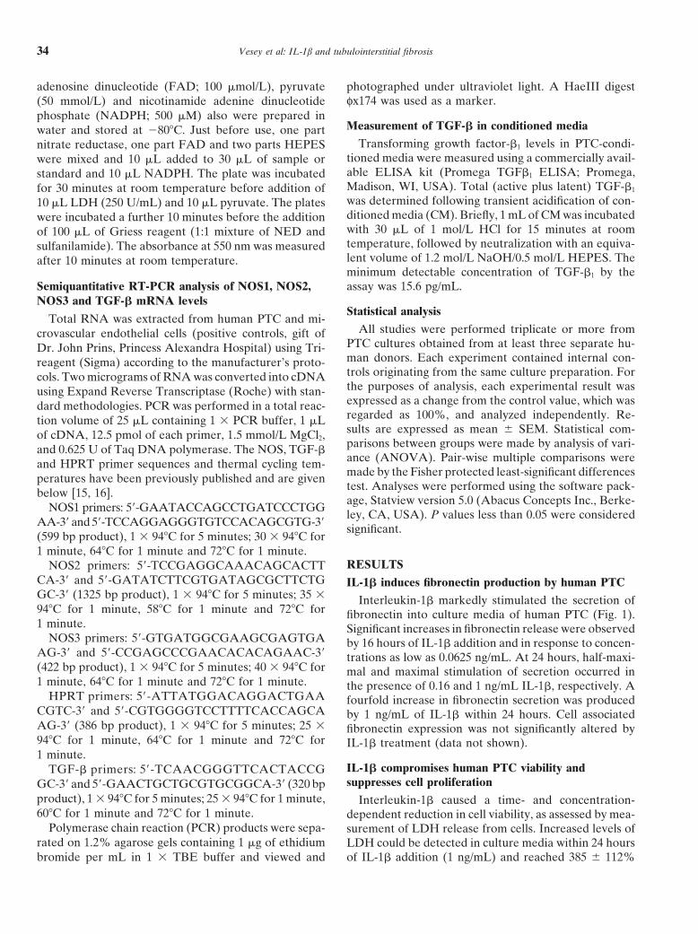
RESULTSNOS2 primers: 5�-TCCGAGGCAAACAGCACTTCA-3� and 5�-GATATCTTCGTGATAGCGCTTCTG IL-1� induces fibronectin production by human PTCGC-3� (1325 bp product), 1 94C for 5 minutes; 35 Interleukin-1� markedly stimulated the secretion of94C for 1 minute, 58C for 1 minute and 72C for fibronectin into culture media of human PTC (Fig. 1).1 minute. Significant increases in fibronectin release were observed
NOS3 primers: 5�-GTGATGGCGAAGCGAGTGA by 16 hours of IL-1� addition and in response to concen-AG-3� and 5�-CCGAGCCCGAACACACAGAAC-3� trations as low as 0.0625 ng/mL. At 24 hours, half-maxi-(422 bp product), 1 94C for 5 minutes; 40 94C for mal and maximal stimulation of secretion occurred in1 minute, 64C for 1 minute and 72C for 1 minute. the presence of 0.16 and 1 ng/mL IL-1�, respectively. A
HPRT primers: 5�-ATTATGGACAGGACTGAA fourfold increase in fibronectin secretion was producedCGTC-3� and 5�-CGTGGGGTCCTTTTCACCAGCA by 1 ng/mL of IL-1� within 24 hours. Cell associatedAG-3� (386 bp product), 1 94C for 5 minutes; 25 fibronectin expression was not significantly altered by94C for 1 minute, 64C for 1 minute and 72C for IL-1� treatment (data not shown).1 minute.
IL-1� compromises human PTC viability andTGF-� primers: 5�-TCAACGGGTTCACTACCGsuppresses cell proliferationGC-3� and 5�-GAACTGCTGCGTGCGGCA-3� (320 bp
product), 1 94C for 5 minutes; 25 94C for 1 minute, Interleukin-1� caused a time- and concentration-60C for 1 minute and 72C for 1 minute. dependent reduction in cell viability, as assessed by mea-
Polymerase chain reaction (PCR) products were sepa- surement of LDH release from cells. Increased levels ofrated on 1.2% agarose gels containing 1 �g of ethidium LDH could be detected in culture media within 24 hours
of IL-1� addition (1 ng/mL) and reached 385 � 112%bromide per mL in 1 TBE buffer and viewed and
Vesey et al: IL-1b and tubulointerstitial fibrosis 35
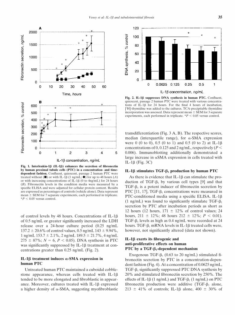
Fig. 2. IL-1� suppresses DNA synthesis in human PTC. Confluent,quiescent, passage 2 human PTC were treated with various concentra-tions of IL-1� for 24 hours. For the final 4 hours of incubation,[3H]-thymidine was added to the cultures. TCA-precipitable thymidineincorporation was assessed. Data represent mean � SEM for 3 separateexperiments, each performed in triplicate. *P � 0.05 versus control.
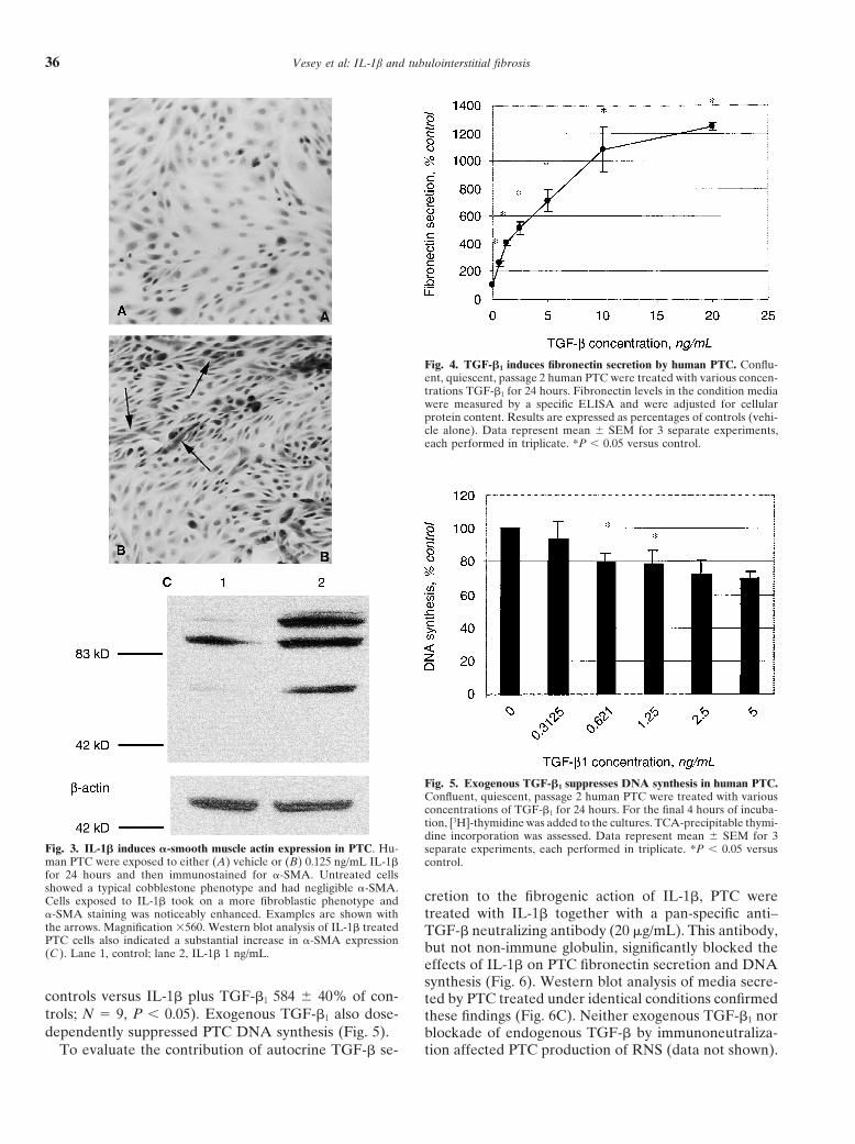
transdifferentiation (Fig. 3 A, B). The respective scores,median (interquartile range), for �-SMA expressionwere 0 (0 to 0), 0.5 (0 to 1) and 0.5 (0 to 2) at IL-1�concentrations of 0, 0.125 and 2 ng/mL, respectively (P �0.006). Immunoblotting additionally demonstrated alarge increase in �SMA expression in cells treated withIL-1� (Fig. 3C)
Fig. 1. Interleukin-1� (IL-1�) enhances the secretion of fibronectinby human proximal tubule cells (PTC) in a concentration- and time- IL-1� stimulates TGF-�1 production by human PTCdependent fashion. Confluent, quiescent, passage 2 human PTC weretreated without (�) or with IL-1� (1 ng/mL; �) for up to 48 hours (A) As there is evidence that IL-1� can stimulate the pro-or with increasing concentrations of IL-1� (0 to 4ng/mL) for 24 hours duction of TGF-�1 by various cell types [9] and that(B). Fibronectin levels in the condition media were measured by a
TGF-�1 is a potent inducer of fibronectin secretion byspecific ELISA and were adjusted for cellular protein content. Resultsare expressed as percentages of controls (vehicle alone). Data represent PTC [11, 17], TGF-�1 concentrations were measured inmean � SEM for 7 separate experiments, each performed in triplicate. PTC-conditioned media using a specific ELISA. IL-1�*P � 0.05 versus control.
(1 ng/mL) was found to significantly stimulate TGF-�1
secretion by PTC after incubation periods as short as12 hours (12 hours, 171 � 12% of control values; 24hours, 211 � 12%; 48 hours 212 � 12%; P � 0.01).of control levels by 48 hours. Concentrations of IL-1�TGF-�1 levels as high as 0.4 ng/mL were recorded at 24of 0.5 ng/mL or greater significantly increased the LDHhours. TGF-�1 mRNA levels in IL-1� treated cells were,release over a 24-hour culture period (0.25 ng/mLhowever, not significantly altered (data not shown).137.2 � 20.6% of control values, 0.5 ng/mL 143 � 9.94%,
1 ng/mL 153.7 � 2.1%, 2 ng/mL 189.5 � 21.7%, 4 ng/mLIL-1� exerts its fibrogenic and275 � 87%; N � 6, P � 0.05). DNA synthesis in PTCanti-proliferative effects on humanwas significantly suppressed by IL-1� treatment at con-PTC by a TGF-�1-dependent mechanismcentrations greater than 0.25 ng/mL (Fig. 2).
Exogenous TGF-�1 (0.63 to 20 ng/mL) stimulated fi-IL-1� treatment induces �-SMA expression in bronectin secretion by PTC in a concentration-depen-human PTC dent fashion (Fig. 4). At a concentration of 0.0625 ng/mL,
TGF-�1 significantly suppressed PTC DNA synthesis byUntreated human PTC maintained a cuboidal cobble-20% and stimulated fibronectin secretion by 250%. Thestone appearance, whereas cells treated with IL-1�effects of IL-1� (1 ng/mL) and TGF-�1 (1 ng/mL) on PTCtended to be more elongated and fibroblastic in appear-
ance. Moreover, cultures treated with IL-1� expressed fibronectin production were additive (TGF-�1 alone,213 � 41% of controls; IL-1� alone, 400 � 30% ofa higher density of �-SMA, suggesting myofibroblastic
Vesey et al: IL-1b and tubulointerstitial fibrosis36
Fig. 4. TGF-�1 induces fibronectin secretion by human PTC. Conflu-ent, quiescent, passage 2 human PTC were treated with various concen-trations TGF-�1 for 24 hours. Fibronectin levels in the condition mediawere measured by a specific ELISA and were adjusted for cellularprotein content. Results are expressed as percentages of controls (vehi-cle alone). Data represent mean � SEM for 3 separate experiments,each performed in triplicate. *P � 0.05 versus control.
Fig. 5. Exogenous TGF-�1 suppresses DNA synthesis in human PTC.Confluent, quiescent, passage 2 human PTC were treated with variousconcentrations of TGF-�1 for 24 hours. For the final 4 hours of incuba-tion, [3H]-thymidine was added to the cultures. TCA-precipitable thymi-dine incorporation was assessed. Data represent mean � SEM for 3
Fig. 3. IL-1� induces �-smooth muscle actin expression in PTC. Hu- separate experiments, each performed in triplicate. *P � 0.05 versusman PTC were exposed to either (A) vehicle or (B) 0.125 ng/mL IL-1� control.for 24 hours and then immunostained for �-SMA. Untreated cellsshowed a typical cobblestone phenotype and had negligible �-SMA.
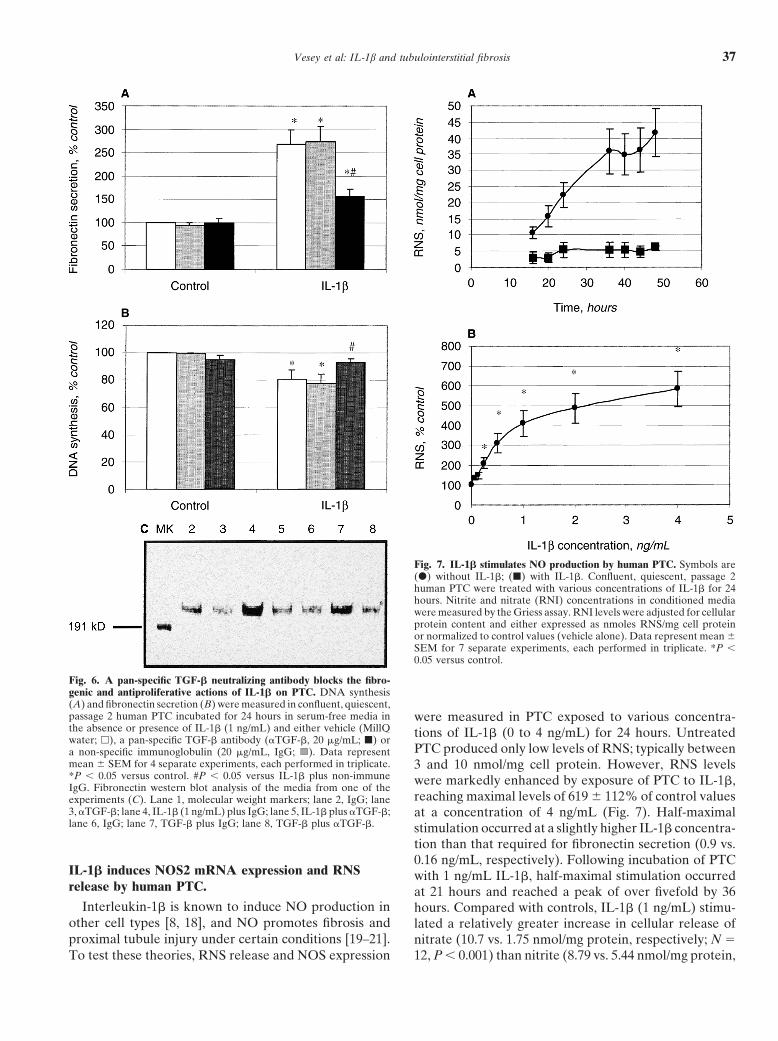
cretion to the fibrogenic action of IL-1�, PTC wereCells exposed to IL-1� took on a more fibroblastic phenotype and�-SMA staining was noticeably enhanced. Examples are shown with treated with IL-1� together with a pan-specific anti–the arrows. Magnification 560. Western blot analysis of IL-1� treated TGF-� neutralizing antibody (20 �g/mL). This antibody,PTC cells also indicated a substantial increase in �-SMA expression but not non-immune globulin, significantly blocked the(C ). Lane 1, control; lane 2, IL-1� 1 ng/mL.
effects of IL-1� on PTC fibronectin secretion and DNAsynthesis (Fig. 6). Western blot analysis of media secre-
controls versus IL-1� plus TGF-�1 584 � 40% of con- ted by PTC treated under identical conditions confirmedtrols; N � 9, P � 0.05). Exogenous TGF-�1 also dose- these findings (Fig. 6C). Neither exogenous TGF-�1 nordependently suppressed PTC DNA synthesis (Fig. 5). blockade of endogenous TGF-� by immunoneutraliza-
tion affected PTC production of RNS (data not shown).To evaluate the contribution of autocrine TGF-� se-
Vesey et al: IL-1b and tubulointerstitial fibrosis 37
Fig. 7. IL-1� stimulates NO production by human PTC. Symbols are(�) without IL-1�; (�) with IL-1�. Confluent, quiescent, passage 2human PTC were treated with various concentrations of IL-1� for 24hours. Nitrite and nitrate (RNI) concentrations in conditioned mediawere measured by the Griess assay. RNI levels were adjusted for cellularprotein content and either expressed as nmoles RNS/mg cell proteinor normalized to control values (vehicle alone). Data represent mean �SEM for 7 separate experiments, each performed in triplicate. *P �0.05 versus control.
Fig. 6. A pan-specific TGF-� neutralizing antibody blocks the fibro-genic and antiproliferative actions of IL-1� on PTC. DNA synthesis(A) and fibronectin secretion (B) were measured in confluent, quiescent,passage 2 human PTC incubated for 24 hours in serum-free media in were measured in PTC exposed to various concentra-the absence or presence of IL-1� (1 ng/mL) and either vehicle (MillQ tions of IL-1� (0 to 4 ng/mL) for 24 hours. Untreatedwater; �), a pan-specific TGF-� antibody (�TGF-�, 20 �g/mL; �) or
PTC produced only low levels of RNS; typically betweena non-specific immunoglobulin (20 �g/mL, IgG; ). Data representmean � SEM for 4 separate experiments, each performed in triplicate. 3 and 10 nmol/mg cell protein. However, RNS levels*P � 0.05 versus control. #P � 0.05 versus IL-1� plus non-immune were markedly enhanced by exposure of PTC to IL-1�,IgG. Fibronectin western blot analysis of the media from one of the
reaching maximal levels of 619 � 112% of control valuesexperiments (C). Lane 1, molecular weight markers; lane 2, IgG; lane3, �TGF-�; lane 4, IL-1� (1 ng/mL) plus IgG; lane 5, IL-1� plus �TGF-�; at a concentration of 4 ng/mL (Fig. 7). Half-maximallane 6, IgG; lane 7, TGF-� plus IgG; lane 8, TGF-� plus �TGF-�. stimulation occurred at a slightly higher IL-1� concentra-
tion than that required for fibronectin secretion (0.9 vs.0.16 ng/mL, respectively). Following incubation of PTC
IL-1� induces NOS2 mRNA expression and RNS with 1 ng/mL IL-1�, half-maximal stimulation occurredrelease by human PTC. at 21 hours and reached a peak of over fivefold by 36
Interleukin-1� is known to induce NO production in hours. Compared with controls, IL-1� (1 ng/mL) stimu-other cell types [8, 18], and NO promotes fibrosis and lated a relatively greater increase in cellular release ofproximal tubule injury under certain conditions [19–21]. nitrate (10.7 vs. 1.75 nmol/mg protein, respectively; N �
12, P � 0.001) than nitrite (8.79 vs. 5.44 nmol/mg protein,To test these theories, RNS release and NOS expression
Vesey et al: IL-1b and tubulointerstitial fibrosis38
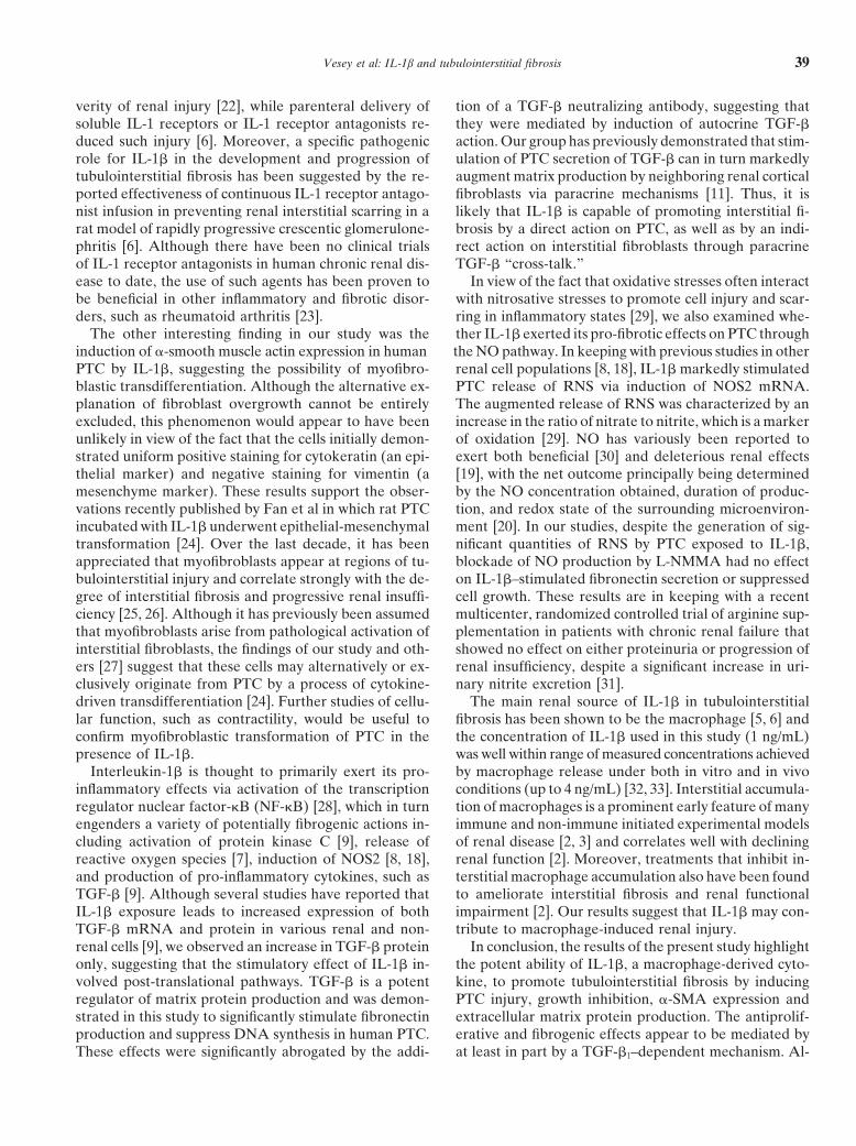
Fig. 9. IL-1�–induced fibronectin secretion by human PTC is unaf-fected by inhibition of NO production. Confluent, quiescent, passage 2human PTC were incubated for 24 hours in serum-free media containingvehicle (control), 1 mmol/L L-NMMA (a NOS inibitor), 1 ng/mLIL-1�, � 1ng/mL TGF-�1, IL-1� plus L-NMMA, or TGF-�1 plusL-NMMA. Fibronectin concentrations in conditioned media were thenmeasured by a specific ELISA. Data represent mean � SEM for 6separate experiments, each performed in triplicate. The results are ex-pressed as a percentage of control values. NS is non-significant; P � 0.1.
5% vs. IL-1� � L-NMMA 100 � 5% of control values,respectively; N � 12, P � 0.001). However, L-NMMAdid not significantly affect either basal or IL-1�–stim-ulated fibronectin secretion by PTC (Fig. 9). No changeswere observed in cell viability, protein content or DNA
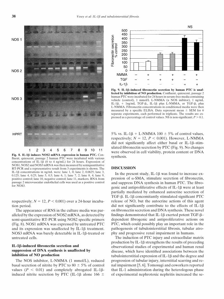
Fig. 8. IL-1� induces NOS2 mRNA expression in human PTC. Con- synthesis.fluent, quiescent, passage 2 human PTC were incubated with variousconcentrations of IL-1� (0 to 4 ng/mL) for 24 hours. Expression ofNOS1, NOS2 and NOS3 mRNA was then measured by semiquantitative
DISCUSSIONRT-PCR and a representative result from 3 experiments is shown. TheIL-1� concentrations in ng/mL were: lane 1, 0; lane 2, 0.0625; lane 3, In the present study, IL-1� was found to increase ex-0.125; lane 4, 0.25; lane 5, 0.5; lane 6, 1; lane 7, 2; lane 8, 4; lane 9,
pression of �-SMA, stimulate secretion of fibronectin,positive control; lane 10, negative control; lane 11, markers. RNA frompassage 2 microvascular endothelial cells was used as a positive control and suppress DNA synthesis in human PTC. The fibro-for NOS3. genic and antiproliferative effects of IL-1� were at least
partially mediated by enhanced autocrine secretion ofTGF-�. IL-1� concomitantly stimulated significant PTCrelease of NO, but the autocrine actions of this agentrespectively; N � 12, P � 0.001) over a 24-hour incuba-did not significantly contribute to the effects of IL-1�tion period.on fibronectin secretion and DNA synthesis. These novelThe appearance of RNS in the culture media was par-findings demonstrated that IL-1� exerted potent TGF-�–alleled by the expression of NOS2 mRNA, as detected bydependent fibrogenic and antiproliferative actions onsemi-quantitative RT-PCR using NOS2 specific primersPTC, which could possibly play an important role in the(Fig. 8). NOS1 mRNA was expressed by untreated PTCpathogenesis of tubulointerstitial fibrosis, tubular atro-and its expression was unaffected by IL-1� treatment.phy and progressive renal impairment in humans.NOS3 mRNA was barely detectable in IL-1�-treated or
The induction of PTC injury and extracellular matrixuntreated cells.production by IL-1� strengthens the results of preceding
IL-1�-induced fibronectin secretion and observational studies of experimental and human renalsuppression of DNA synthesis is unaffected by disease, which have identified associations between theinhibition of NO production tubulointerstitial expression of IL-1� and the degree and
progression of tubular injury, interstitial scarring and re-The NOS inhibitor, L-NMMA (1 mmol/L), reducednal impairment [5, 6]. Tomosugi and coworkers reportedbasal secretion of nitrite by PTC to 80 � 5% of controlthat IL-1 administration during the heterologous phasevalues (P � 0.01) and completely abrogated IL-1�-
induced nitrite secretion by PTC (IL-1� alone 166 � of experimental nephrotoxic nephritis increased the se-
Vesey et al: IL-1b and tubulointerstitial fibrosis 39
verity of renal injury [22], while parenteral delivery of tion of a TGF-� neutralizing antibody, suggesting thatthey were mediated by induction of autocrine TGF-�soluble IL-1 receptors or IL-1 receptor antagonists re-
duced such injury [6]. Moreover, a specific pathogenic action. Our group has previously demonstrated that stim-ulation of PTC secretion of TGF-� can in turn markedlyrole for IL-1� in the development and progression of
tubulointerstitial fibrosis has been suggested by the re- augment matrix production by neighboring renal corticalfibroblasts via paracrine mechanisms [11]. Thus, it isported effectiveness of continuous IL-1 receptor antago-
nist infusion in preventing renal interstitial scarring in a likely that IL-1� is capable of promoting interstitial fi-brosis by a direct action on PTC, as well as by an indi-rat model of rapidly progressive crescentic glomerulone-
phritis [6]. Although there have been no clinical trials rect action on interstitial fibroblasts through paracrineTGF-� “cross-talk.”of IL-1 receptor antagonists in human chronic renal dis-
ease to date, the use of such agents has been proven to In view of the fact that oxidative stresses often interactwith nitrosative stresses to promote cell injury and scar-be beneficial in other inflammatory and fibrotic disor-
ders, such as rheumatoid arthritis [23]. ring in inflammatory states [29], we also examined whe-ther IL-1� exerted its pro-fibrotic effects on PTC throughThe other interesting finding in our study was the
induction of �-smooth muscle actin expression in human the NO pathway. In keeping with previous studies in otherrenal cell populations [8, 18], IL-1� markedly stimulatedPTC by IL-1�, suggesting the possibility of myofibro-
blastic transdifferentiation. Although the alternative ex- PTC release of RNS via induction of NOS2 mRNA.The augmented release of RNS was characterized by anplanation of fibroblast overgrowth cannot be entirely
excluded, this phenomenon would appear to have been increase in the ratio of nitrate to nitrite, which is a markerof oxidation [29]. NO has variously been reported tounlikely in view of the fact that the cells initially demon-
strated uniform positive staining for cytokeratin (an epi- exert both beneficial [30] and deleterious renal effects[19], with the net outcome principally being determinedthelial marker) and negative staining for vimentin (a
mesenchyme marker). These results support the obser- by the NO concentration obtained, duration of produc-tion, and redox state of the surrounding microenviron-vations recently published by Fan et al in which rat PTC
incubated with IL-1� underwent epithelial-mesenchymal ment [20]. In our studies, despite the generation of sig-nificant quantities of RNS by PTC exposed to IL-1�,transformation [24]. Over the last decade, it has been
appreciated that myofibroblasts appear at regions of tu- blockade of NO production by L-NMMA had no effecton IL-1�–stimulated fibronectin secretion or suppressedbulointerstitial injury and correlate strongly with the de-
gree of interstitial fibrosis and progressive renal insuffi- cell growth. These results are in keeping with a recentmulticenter, randomized controlled trial of arginine sup-ciency [25, 26]. Although it has previously been assumed
that myofibroblasts arise from pathological activation of plementation in patients with chronic renal failure thatshowed no effect on either proteinuria or progression ofinterstitial fibroblasts, the findings of our study and oth-
ers [27] suggest that these cells may alternatively or ex- renal insufficiency, despite a significant increase in uri-nary nitrite excretion [31].clusively originate from PTC by a process of cytokine-
driven transdifferentiation [24]. Further studies of cellu- The main renal source of IL-1� in tubulointerstitialfibrosis has been shown to be the macrophage [5, 6] andlar function, such as contractility, would be useful to
confirm myofibroblastic transformation of PTC in the the concentration of IL-1� used in this study (1 ng/mL)was well within range of measured concentrations achievedpresence of IL-1�.
Interleukin-1� is thought to primarily exert its pro- by macrophage release under both in vitro and in vivoconditions (up to 4 ng/mL) [32, 33]. Interstitial accumula-inflammatory effects via activation of the transcription
regulator nuclear factor- B (NF- B) [28], which in turn tion of macrophages is a prominent early feature of manyimmune and non-immune initiated experimental modelsengenders a variety of potentially fibrogenic actions in-
cluding activation of protein kinase C [9], release of of renal disease [2, 3] and correlates well with decliningrenal function [2]. Moreover, treatments that inhibit in-reactive oxygen species [7], induction of NOS2 [8, 18],
and production of pro-inflammatory cytokines, such as terstitial macrophage accumulation also have been foundto ameliorate interstitial fibrosis and renal functionalTGF-� [9]. Although several studies have reported that
IL-1� exposure leads to increased expression of both impairment [2]. Our results suggest that IL-1� may con-tribute to macrophage-induced renal injury.TGF-� mRNA and protein in various renal and non-
renal cells [9], we observed an increase in TGF-� protein In conclusion, the results of the present study highlightthe potent ability of IL-1�, a macrophage-derived cyto-only, suggesting that the stimulatory effect of IL-1� in-
volved post-translational pathways. TGF-� is a potent kine, to promote tubulointerstitial fibrosis by inducingPTC injury, growth inhibition, �-SMA expression andregulator of matrix protein production and was demon-
strated in this study to significantly stimulate fibronectin extracellular matrix protein production. The antiprolif-erative and fibrogenic effects appear to be mediated byproduction and suppress DNA synthesis in human PTC.
These effects were significantly abrogated by the addi- at least in part by a TGF-�1–dependent mechanism. Al-
Vesey et al: IL-1b and tubulointerstitial fibrosis40
blasts regulate proximal tubule cell growth and transport via thethough IL-1� induced NO release, this effect did notIGF-I axis. Kidney Int 52:1486–1496, 1997
appear to mediate the actions of IL-1� on PTC fibronec- 14. Grisham MB, Johnson GG, Lancaster JRJ: Quantitation of ni-trate and nitrite in extracellular fluids. Methods Enzymol 268:237–tin secretion and DNA synthesis. Our studies suggest that246, 1996targeting renal IL-1� production or action would provide
15. Olive C, Cheung C, Nicol DL, Falk M: Expression of cytokinea useful strategy for preventing progressive renal scarring mRNA transcripts in renal cell carcinoma. Immunol Cell Biol 76:
357–362, 1998and dysfunction in patients with chronic renal disease.16. Rockett KA, Brookes R, Udalova I, et al: 1,25-Dihydroxyvitamin
D3 induces nitric oxide synthase and suppresses growth of Myco-ACKNOWLEDGMENTS bacterium tuberculosis in a human macrophage-like cell line. Infect
Immun 66:5314–5321, 1998This study was supported, in part, by funds from the National Health17. Johnson DW, Saunders HJ, Johnson FJ, et al: Cyclosporin exertsand Medical Research Council, the Australian Kidney Foundation,
a direct fibrogenic effect on human tubulointerstitial cells: Rolesand the Princess Alexandra Hospital Research and Developmentof IGF-I, TGF�1 and PDGF. J Pharm Exp Ther 289:535–542, 1998Foundation. The invaluable assistance provided by the urologists of
18. Kihara M, Yabana M, Toya Y, et al: Angiotensin II inhibitsthe Princess Alexandra Hospital in the procurement of human renalinterleukin-1�-induced nitric oxide production in cultured rat mes-tissue is gratefully acknowledged. Dr. Vesey’s salary is supported byangial cells. Kidney Int 55:1277–1283, 1999the Vincent Fairfax Family Foundation Fellowship awarded by the
19. Klahr S, Morrissey J: Renal disease: The two faces of nitric oxide.Royal Australasian College of Physicians.Lab Invest 72:1–3, 1995
20. Lane P, Gross SS: Cell signaling by nitric oxide. Semin NephrolReprint requests to Dr. David A. Vesey, Department of Renal Medi-19:215–229, 1999cine, Level 2, Ambulatory Renal and Transplant Services Building,
21. Kabore AF, Denis M, Bergeron MG: Association of nitric oxidePrincess Alexandra Hospital, Ipswich Road, Woolloongabba, Brisbane,production by kidney proximal tubular cells in response to lipopoly-Queensland 4102, Australia.saccharide and cytokines with cellular damage. Antimicrob AgentsE-mail: [email protected] 41:557–562, 1997
22. Tomosugi NI, Cashman SJ, Hay H, et al: Modulation of antibody-mediated glomerular injury in vivo by bacterial lipopolysaccharide,REFERENCEStumor necrosis factor, and IL-1. J Immunol 142:3083–3090, 1989
1. Bohle A, Muller GA, Wehrmann M, et al: Pathogenesis of 23. Bresnihan B, Alvaro Gracia JM, Cobby M, et al: Treatment ofchronic renal failure in the primary glomerulopathies, renal vascu- rheumatoid arthritis with recombinant human interleukin-1 recep-lopathies, and chronic interstitial nephritides. Kidney Int 49(Suppl tor antagonist. Arthritis Rheum 41:2196–2204, 199854):S2–S9, 1996 24. Fan JM, Huang XR, Ng YY, et al: Interleukin-1-induces tubular
2. Nikolic Paterson DJ, Lan HY, Hill PA, Atkins RC: Macro- epithelial myofibroblast transdifferentiation through a transform-phages in renal injury. Kidney Int 45(Suppl 45):S79–S82, 1994 ing growth factor-�1-dependent mechanism in vitro. Am J Kidney
3. Diamond JR: Macrophages and progressive renal disease in experi- Dis 37:820–831, 2000mental hydronephrosis. Am J Kidney Dis 26:133–140, 1995 25. Zhang G, Moorhead PJ, el Nahas AM: Myofibroblasts and the
4. Fine LG, Norman JT, Ong A: Cell-cell cross-talk in the patho- progression of experimental glomerulonephritis. Exp Nephrol 3:genesis of renal interstitial fibrosis. Kidney Int 47(Suppl 49):S48– 308–318, 1995S50, 1995 26. Hewitson TD, Becker GJ: Interstitial myofibroblasts in IgA glo-
5. Nikolic Paterson DJ, Main IW, Tesch GH, et al: Interleukin-1 merulonephritis. Am J Nephrol 15:111–117, 1995in renal fibrosis. Kidney Int 49(Suppl 54):S88–S90, 1996 27. Ng YY, Huang TP, Yang WC, et al: Tubular epithelial-myofi-
6. Nikolic Paterson DJ, Lan HY, Atkins RC: Interleukin-1 receptor broblast transdifferentiation in progressive tubulointerstitial fi-antagonism. Semin Nephrol 16:583–590, 1996 brosis in 5/6 nephrectomized rats. Kidney Int 54:864–876, 1998
7. Radeke HH, Meier B, Topley N, et al: Interleukin 1-alpha and 28. Stewart RJ, Marsden PA: Biologic control of the tumor necrosistumor necrosis factor-alpha induce oxygen radical production in factor and interleukin-1 signaling cascade. Am J Kidney Dis 25:954–mesangial cells. Kidney Int 37:767–775, 1990 966, 1995
8. Jiang B, Haverty M, Brecher P: N-acetyl-L-cysteine enhances 29. Eu JP, Liu L, Zeng M, Stamler JS: An apoptotic model forinterleukin-1beta-induced nitric oxide synthase expression. Hyper- nitrosative stress. Biochemistry 39:1040–1047, 2000tens 34:574–579, 1999 30. Morrissey JJ, Ishidoya S, McCracken R, Klahr S: Nitric oxide
9. Phillips AO, Topley N, Steadman R, et al: Induction of TGF- generation ameliorates the tubulointerstitial fibrosis of obstructivebeta 1 synthesis in D-glucose primed human proximal tubular cells nephropathy. J Am Soc Nephrol 7:2202–2212, 1996by IL-1 beta and TNF alpha. Kidney Int 50:1546–1554, 1996 31. De Nicola L, Bellizzi V, Minutolo R, et al: Randomized, double-
10. Johnson DW, Brew BK, Poronnik P, et al: Transport characteris- blind, placebo-controlled study of arginine supplementation intics of human proximal tubule cells in primary culture. Nephrol chronic renal failure. Kidney Int 56:674–684, 19993:183–194, 1997 32. Zucali JR, Broxmeyer HE, Levy D, Morse C: Lactoferrin de-
11. Johnson DW, Saunders HJ, Baxter RC, et al: Paracrine stimula- creases monocyte-induced fibroblast production of myeloid col-tion of human renal fibroblasts by proximal tubule cells. Kidney ony-stimulating activity by suppressing monocyte release of in-Int 54:747–757, 1998 terleukin-1. Blood 74:1531–1536, 1989
12. Christiansen BS, Ingerslev J, Heickendorff L, Petersen CM: 33. Brauner A, Hylander B, Wretlind B: Tumor necrosis factor-Human hepatocytes in culture synthesize and secrete fibronectin. alpha, interleukin-1 beta, and interleukin-1 receptor antagonistScand J Clin Lab Invest 48:685–690, 1988 in dialysate and serum from patients on continuous ambulatory
peritoneal dialysis. Am J Kidney Dis 27:402–408, 199613. Johnson DW, Saunders HJ, Brew BK, et al: Human renal fibro-

![Mechanische Beanspruchung und subchondrale · PDF fileDie 4-Fragment-Fraktur des proximalen Oberarms 422 The four-fragment fracture of the proximal humerus W. Knopp, Β. ... [14],](https://static.fdocument.org/doc/165x107/5a7b54f17f8b9a2e6e8bd25f/mechanische-beanspruchung-und-subchondrale-4-fragment-fraktur-des-proximalen.jpg)

![High Expression of XRCC6 Promotes Human Osteosarcoma Cell ... fileas distal femur and proximal radius [2,3]. With the aid of effective chemotherapeutic drugs the survival With the](https://static.fdocument.org/doc/165x107/5d57b6dd88c99399618ba79e/high-expression-of-xrcc6-promotes-human-osteosarcoma-cell-distal-femur-and-proximal.jpg)