Interaction of α-and β-Oligoarginine-Acids and Amides with Anionic Lipid Vesicles: A Mechanistic...
13
Interaction of R-and -Oligoarginine-Acids and Amides with Anionic Lipid Vesicles: A Mechanistic and Thermodynamic Study ² Thomas Hitz, ‡,| Rico Iten, ‡ James Gardiner, § Kenji Namoto, §,⊥ Peter Walde,* ,‡ and Dieter Seebach § Department of Materials, Institute of Polymers, and Department of Chemistry and Applied Biosciences, Laboratory of Organic Chemistry, ETH-Zu ¨rich, Wolfgang-Pauli-Strasse 10, CH-8093 Zu ¨rich, Switzerland ReceiVed February 10, 2006; ReVised Manuscript ReceiVed March 20, 2006 ABSTRACT: The interaction of R- and -oligoarginine amides and acids and of R-polyarginine with anionic lipid vesicles was studied. The -oligoarginines used were 3 -homologues of the R-oligoarginines. Lipid bilayers were composed of POPC (1-palmitoyl-2-oleoyl-sn-glycero-3-phosphocholine) and POPG (1- palmitoyl-2-oleoyl-sn-glycero-3-[phospho-rac-(1-glycerol)]) containing 5 mol % pyrene-PG (1-hexade- canoyl-2-(1-pyrenedecanoyl)-sn-glycero-3-[phospho-rac-1-glycerol]). Kinetic analysis of the binding process onto large unilamellar POPC/POPG (3:7, molar ratio) vesicles (100 nm diameter) shows biphasic time courses for all tested peptides. The first binding step is fast and takes place within ∼10 s with no disruption of the membrane as indicated by corresponding calcein release measurements. The second binding phase is slow and occurs within the next 30-300 s with substantial membrane disruption. In this context, -hexa- and octaarginine amides possess higher second half-times than the -hexa- and octaarginine acids of the same chain length. Furthermore -octaarginine amide induces a calcein release approximately twice as large as that of the -octaarginine acid. Thermodynamic analysis of the binding process, using the complex formation model that assumes that each peptide binds independently to n POPG lipids, reveals apparent binding constants (K app1 ) of ∼5 × 10 6 - 10 8 M -1 and n-values from 3.7 for -hexaarginine acid up to 24.8 for R-polyarginine. Although the K app1 -values are similar, the number of binding sites clearly depends on the chemical nature of the oligoarginine: -oligoarginine amides and R-oligoarginine acids interact with more lipids than -oligoarginine acids of the same length. Calculation of the electrostatic contribution to the total free energy of binding reveals that for all oligoarginines only 25-30% has electrostatic origin. The remaining ∼70-75% is nonelectrostatic, corresponding to hydrogen bonding and/or hydrophobic interactions. From the obtained data, a mechanism is suggested by which oligoarginines interact with anionic vesicles: (1) initial electrostatic interaction that is fast, nonspecific, and relatively weak; (2) nonelectrostatic interaction that is rate-limiting, stronger, and induces bilayer rigidification as well as release of aqueous contents from the vesicles. Developing technologies that allow the enhanced transport of hydrophilic and charged molecules across the hydrophobic plasma membrane into living cells are of substantial interest for biomedical applications such as the delivery of genes or proteins (1, 2). Cell-penetrating peptides (CPPs 1 , for a recent review see ref 3) belong to a class of oligopeptides that efficiently mediate the transport of extracellular cargo into cells. Such transport properties were demonstrated during the past decade for a number of hydrophilic molecules such as oligonucleotides, RNA, short interfering RNA, peptides, proteins, and even vesicles (4-7). Two of the best-characterized CPPs are the Tat 48-60 - transduction domain derived from the transcriptional activator of the HIV-1 virus and the Drosophila Antennapedia homeodomain peptide, penetratin (8, 9). Both peptides have a high proportion of arginine residues in their sequence. Recent reports show that the guanidine-moiety of the arginine-residue is critical to observe substantial peptide internalization: (i) replacing all nonarginine residues in Tat with arginine resulted in superior cellular uptake (factor of 20-100) (10); (ii) oligoarginines enter cells more efficiently than cationic homopolymers of lysine, ornithine, and histidine of the same length (11); (iii) inverting the absolute config- uration of the arginine residues in the peptide chain does not affect cellular uptake (12); (iv) arginine-rich -peptides and -oligoarginines efficiently permeate into mammalian and bacterial cells (13-16); and (v) even highly branched guanidinoglycosides and guanidinium-rich dendrimers are transported across the plasma membrane (17-18). It has also been shown that branched octaarginine is internalized to the same extent as linear octaarginine (19). In the case of fluorescein-labeled amide derivatives of -oligoarginines, it has been shown that a chain length of g7 leads to efficient ² J.G. acknowledges financial support from a New Zealand Founda- tion for Research Science and Technology postdoctoral fellowship (No. SWISS0401). K.N. acknowledges partial financial support from the Swiss National Science Foundation (SNF Project 20020-100 182). * To whom correspondence should be addressed: Telephone: +41- 6320473. Fax: +41-6321265. E-mail: [email protected]. ‡ Department of Materials, Institute of Polymers, ETH-Zu ¨rich. § Department of Chemistry and Applied Biosciences, Laboratory of Organic Chemistry, ETH-Zu ¨rich. | Present address: Mettler-Toledo GmbH, 8603 Schwerzenbach, Switzerland. ⊥ Present address: Novartis Pharma AG, NIBR, 4002 Basel, Switzerland. 5817 Biochemistry 2006, 45, 5817-5829 10.1021/bi060285d CCC: $33.50 © 2006 American Chemical Society Published on Web 04/12/2006
Transcript of Interaction of α-and β-Oligoarginine-Acids and Amides with Anionic Lipid Vesicles: A Mechanistic...
Interaction ofR-andâ-Oligoarginine-Acids and Amides with Anionic LipidVesicles: A Mechanistic and Thermodynamic Study†
Thomas Hitz,‡,| Rico Iten,‡ James Gardiner,§ Kenji Namoto,§,⊥ Peter Walde,*,‡ and Dieter Seebach§
Department of Materials, Institute of Polymers, and Department of Chemistry and Applied Biosciences,Laboratory of Organic Chemistry, ETH-Zu¨rich, Wolfgang-Pauli-Strasse 10, CH-8093 Zu¨rich, Switzerland
ReceiVed February 10, 2006; ReVised Manuscript ReceiVed March 20, 2006
ABSTRACT: The interaction ofR- andâ-oligoarginine amides and acids and ofR-polyarginine with anioniclipid vesicles was studied. Theâ-oligoarginines used wereâ3-homologues of theR-oligoarginines. Lipidbilayers were composed of POPC (1-palmitoyl-2-oleoyl-sn-glycero-3-phosphocholine) and POPG (1-palmitoyl-2-oleoyl-sn-glycero-3-[phospho-rac-(1-glycerol)]) containing 5 mol % pyrene-PG (1-hexade-canoyl-2-(1-pyrenedecanoyl)-sn-glycero-3-[phospho-rac-1-glycerol]). Kinetic analysis of the binding processonto large unilamellar POPC/POPG (3:7, molar ratio) vesicles (100 nm diameter) shows biphasic timecourses for all tested peptides. The first binding step is fast and takes place within∼10 s with no disruptionof the membrane as indicated by corresponding calcein release measurements. The second binding phaseis slow and occurs within the next 30-300 s with substantial membrane disruption. In this context,â-hexa-and octaarginine amides possess higher second half-times than theâ-hexa- and octaarginine acids of thesame chain length. Furthermoreâ-octaarginine amide induces a calcein release approximately twice aslarge as that of theâ-octaarginine acid. Thermodynamic analysis of the binding process, using the complexformation model that assumes that each peptide binds independently ton POPG lipids, reveals apparentbinding constants (Kapp1) of ∼5 × 10⁶ - 108 M-1 andn-values from 3.7 forâ-hexaarginine acid up to24.8 forR-polyarginine. Although theKapp1-values are similar, the number of binding sites clearly dependson the chemical nature of the oligoarginine:â-oligoarginine amides andR-oligoarginine acids interactwith more lipids thanâ-oligoarginine acids of the same length. Calculation of the electrostatic contributionto the total free energy of binding reveals that for all oligoarginines only 25-30% has electrostatic origin.The remaining∼70-75% is nonelectrostatic, corresponding to hydrogen bonding and/or hydrophobicinteractions. From the obtained data, a mechanism is suggested by which oligoarginines interact withanionic vesicles: (1) initial electrostatic interaction that is fast, nonspecific, and relatively weak; (2)nonelectrostatic interaction that is rate-limiting, stronger, and induces bilayer rigidification as well asrelease of aqueous contents from the vesicles.
Developing technologies that allow the enhanced transportof hydrophilic and charged molecules across the hydrophobicplasma membrane into living cells are of substantial interestfor biomedical applications such as the delivery of genes orproteins (1, 2). Cell-penetrating peptides (CPPs1, for a recentreview see ref3) belong to a class of oligopeptides thatefficiently mediate the transport of extracellular cargo intocells. Such transport properties were demonstrated duringthe past decade for a number of hydrophilic molecules suchas oligonucleotides, RNA, short interfering RNA, peptides,proteins, and even vesicles (4-7).
Two of the best-characterized CPPs are the Tat48-60-transduction domain derived from the transcriptional activatorof the HIV-1 virus and theDrosophila Antennapediahomeodomain peptide, penetratin (8, 9). Both peptides havea high proportion of arginine residues in their sequence.Recent reports show that the guanidine-moiety of thearginine-residue is critical to observe substantial peptideinternalization: (i) replacing all nonarginine residues in Tatwith arginine resulted in superior cellular uptake (factor of20-100) (10); (ii) oligoarginines enter cells more efficientlythan cationic homopolymers of lysine, ornithine, and histidineof the same length (11); (iii) inverting the absolute config-uration of the arginine residues in the peptide chain doesnot affect cellular uptake (12); (iv) arginine-richâ-peptidesand â-oligoarginines efficiently permeate into mammalianand bacterial cells (13-16); and (v) even highly branchedguanidinoglycosides and guanidinium-rich dendrimers aretransported across the plasma membrane (17-18). It has alsobeen shown that branched octaarginine is internalized to thesame extent as linear octaarginine (19). In the case offluorescein-labeled amide derivatives ofâ-oligoarginines, ithas been shown that a chain length ofg7 leads to efficient
† J.G. acknowledges financial support from a New Zealand Founda-tion for Research Science and Technology postdoctoral fellowship (No.SWISS0401). K.N. acknowledges partial financial support from theSwiss National Science Foundation (SNF Project 20020-100 182).
* To whom correspondence should be addressed: Telephone:+41-6320473. Fax:+41-6321265. E-mail: [email protected].
‡ Department of Materials, Institute of Polymers, ETH-Zu¨rich.§ Department of Chemistry and Applied Biosciences, Laboratory of
Organic Chemistry, ETH-Zu¨rich.| Present address: Mettler-Toledo GmbH, 8603 Schwerzenbach,
Switzerland.⊥ Present address: Novartis Pharma AG, NIBR, 4002 Basel,
Switzerland.
5817Biochemistry2006,45, 5817-5829
10.1021/bi060285d CCC: $33.50 © 2006 American Chemical SocietyPublished on Web 04/12/2006
cell uptake (14). Corresponding cell uptake data for free acidsare not available.
Despite the importance of CPPs as efficient deliveryvectors and the important role of the guanidinium-moietyfor peptide transduction, the mechanisms of cellular uptakeremain elusive. It has become increasingly clear that morethan one mechanism is responsible for peptide internalizationdepending on the chemical nature of the peptides and celllines. In general, two different pathways are observed,endocytotic and/or nonendocytotic uptake. Internalizationstudies with live cells and bacterial cells lacking thepossibility of endocytosis suggest thatR- andâ- oligoargin-ines use alternative mechanisms to the endocytotic uptake(15, 20, 21). Wender and co-workers (22) proposed a possiblenonendocytotic pathway that could explain such observations.It is argued that first the oligoarginines form bidentatehydrogen bonds with H-bond acceptors on the cell surface.These complexes are assumed to partition into, and migrateacross, the membrane bilayer proportional to the membranepotential. This conclusion is compatible with the observedincreased uptake of H-(D-Arg)8-OH with increased membranepotential. Recent studies indicate that complex formationbetween oligoarginines and cell surface proteoglycans (i.e.,heparan sulfate) is favored over a peptide binding ontoanionic lipids (23, 24). However, in an endocytotic pathwaywhere heparan sulfate is degraded by heparanases, hydrogenbonding with anionic lipids on the inner leaflet of theendocytotic vesicle may become more important in allowingthe peptide and its cargo to escape. For example, it has beenshown that anionic lipids such as phosphatidylserine mediatethe release of DNA from cationic liposome/DNA complexesin the endosomal compartment (25). Interactions with anionicphospholipids may also play an important role in theobserved uptake of oligoarginines into Gram-negative bacte-rial cells, since the inner membrane contains POPE (1-
palmitoyl-2-oleoyl-sn-glycero-3-phosphoethanolamine) andnegatively charged POPG (1-palmitoyl-2-oleoyl-sn-glycero-3-[phospho-rac-(1-glycerol)]) but lacks proteoglycans (15,26).
In the present study, we investigate the interaction ofR-andâ-oligoarginine amides and of the corresponding acids(Scheme 1,1-7), with anionic lipid vesicles composed ofPOPC (1-palmitoyl-2-oleoyl-sn-glycero-3-phosphocholine)and POPG. By using pyrene-labeled PG-lipids (pyrene-PG,1-hexadecanoyl-2-(1-pyrenedecanoyl)-sn-glycero-3-[phospho-rac-1-glycerol]) and encapsulated calcein, we compared theinteraction of oligoarginines1-7 on a thermodynamic andmechanistic level, as studied by fluorescence spectroscopy.Pyrene-labeled phospholipids have previously been used tomonitor the interaction of several proteins and peptides withanionic vesicles (27-30). These probes respond in acharacteristic manner to phase separation and changes in thelateral mobility of phospholipids caused by peptide associa-tion (31). Circular dichroism (CD) spectroscopy was appliedto monitor possible structural changes of the vesicles-boundpeptides.
MATERIALS AND METHODS
Lipids, Peptides, and General Chemicals.POPC andPOPG were purchased from Avanti Polar Lipids (Alabaster,AL); pyrene-PG was from Molecular Probes (Eugene, OR).Calcein (for fluorescence), 4-(2-hydroxyethyl)piperazine-1-ethanesulfonic acid sodium salt (HEPES-Na), sodium chlo-ride (NaCl), ethylenediaminetetraacetic acid disodium saltdihydrate (EDTA disodium salt), and Triton X-100 werepurchased from Sigma Aldrich (Buchs, Switzerland). Peptide1 (> 88%, supplied as trifluoroacetate salt) was from Bachem(Bubendorf, Switzerland). Peptides2 and4-7 were preparedusing Fmoc solid-phase peptide synthesis (> 98%, trifluo-roacetate salts) (14, 32, 33; see below and SupportingInformation). Peptide3 (polyarginine, poly-L-arginine hy-drochloride, P-4663, Lot 122K5116; degree of polymeriza-tion and relative molar mass as determined by multi anglelaser light scattering measurements, 39 and 7540, respec-tively; degree of polymerization and relative molar mass asdetermined by viscosity measurements, 72 and 14 000) wasfrom Sigma Aldrich (Buchs, Switzerland). For peptide3, anaverage degree of polymerization of 56, corresponding to arelative molar mass of 10 770 was used for concentrationcalculations.
Synthesis of Peptides2 and4-7. For a general article onâ-peptides and their nomenclature, see ref33. In the text ofthis paper, the short form descriptions for the peptides are
1 Abbreviations: CPP, cell penetrating peptide; POPC, 1-palmitoyl-2-oleoyl-sn-glycero-3-phosphocholine; POPG, 1-palmitoyl-2-oleoyl-sn-glycero-3-[phospho-rac-(1-glycerol)]; pyrene-PG, 1-hexadecanoyl-2-(1-pyrenedecanoyl)-sn-glycero-3-[phospho-rac-1-glycerol]); HEPES,4-(2-hydroxyethyl)piperazine-1-ethanesulfonic acid; EDTA, ethylene-diaminetetraacetic acid; LUV, large unilamellar vesicle; CD, circulardichroism; DLS, dynamic light scattering; Cryo-TEM, cryo transmissionelectron microscopy; RFIM, fluorescence intensity of the pyrene-PGmonomer in the presence of peptides relative to the fluorescenceintensity of the pyrene-PG monomer in the absence of peptides; RFIE,fluorescence intensity of the pyrene-PG excimer in the presence ofpeptides relative to the fluorescence intensity of the pyrene-PG excimerin the absence of peptides;IM, fluorescence intensity of pyrene-PGmonomer; IE, fluorescence intensity of pyrene-PG excimer;∆IF,observed increase in fluorescence intensity;∆IFmax, observed maximalincrease in fluorescence intensity; P, peptide; [P]tot, total peptideconcentration; [P], concentration of peptide in the bulk solution; L,accessible negatively charged phospholipid (POPG); [L]tot, totalconcentration of accessible, negatively charged phospholipids (POPGmolecules present in the outer leaflet of the bilayer);n, number ofnegatively charged phospholipids bound to one peptide; [Ln], concentra-tion of accessible peptide binding sites, composed ofn negativelycharged phospholipids; [Ln]tot, total concentration of accessible bindingsites for the peptide; [P× Ln], concentration of bound peptide, equalto the concentration of occupied peptide binding sites; [M]tot, totalconcentration of accessible membrane lipids (POPC+ POPG presentin the outer layer of the vesicles);Kapp1, apparent binding constant forpeptide binding to peptide-free (charged) vesicles;Kapp2, apparentbinding constant for peptide binding to vesicles that are just saturatedwith peptides (no surface charge); SD, standard deviation; peptide1,H-(Arg)6-OH; peptide2, H-(Arg)8-OH; peptide3, polyarginine; peptide4, H-(âArg)6-OH; peptide5, H-(âArg)8-OH; peptide6, H-(âArg)6-NH2;peptide7, H-(âArg)8-NH2.
Scheme 1: Chemical Structures of theR-andâ3-oligoarginine Amides and Acids Used in This Study
5818 Biochemistry, Vol. 45, No. 18, 2006 Hitz et al.
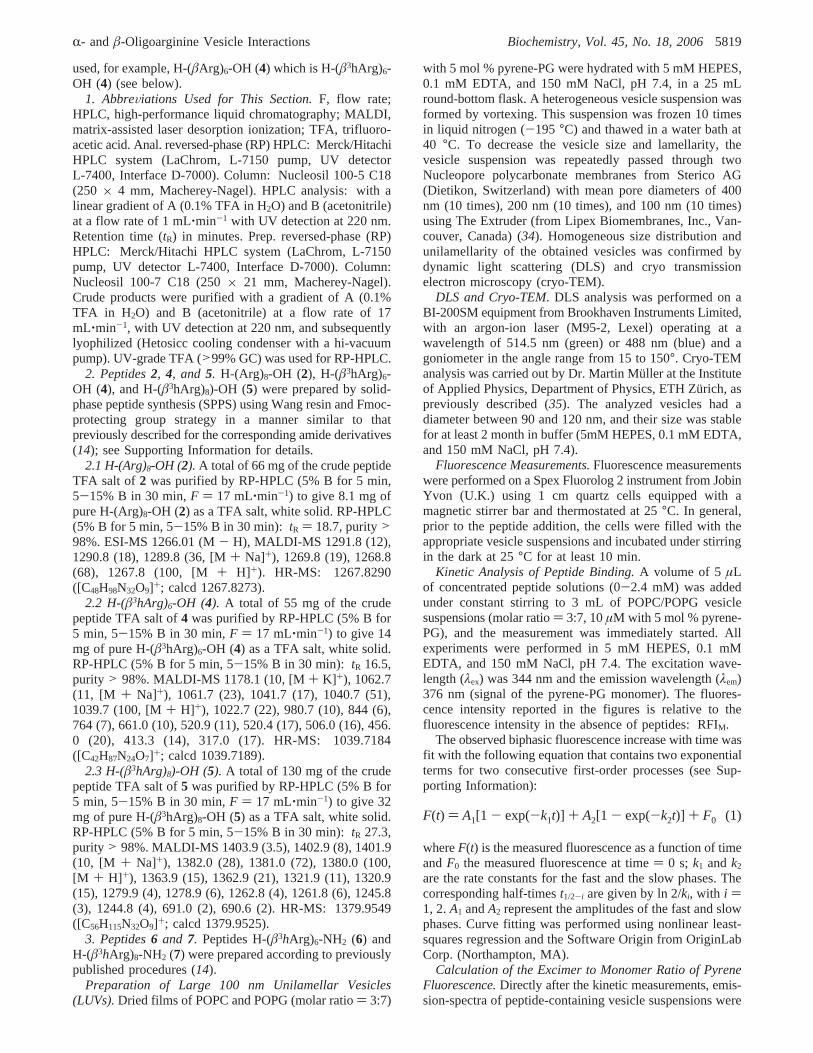
used, for example, H-(âArg)6-OH (4) which is H-(â3hArg)6-OH (4) (see below).
1. AbbreViations Used for This Section.F, flow rate;HPLC, high-performance liquid chromatography; MALDI,matrix-assisted laser desorption ionization; TFA, trifluoro-acetic acid. Anal. reversed-phase (RP) HPLC: Merck/HitachiHPLC system (LaChrom, L-7150 pump, UV detectorL-7400, Interface D-7000). Column: Nucleosil 100-5 C18(250 × 4 mm, Macherey-Nagel). HPLC analysis: with alinear gradient of A (0.1% TFA in H2O) and B (acetonitrile)at a flow rate of 1 mL‚min-1 with UV detection at 220 nm.Retention time (tR) in minutes. Prep. reversed-phase (RP)HPLC: Merck/Hitachi HPLC system (LaChrom, L-7150pump, UV detector L-7400, Interface D-7000). Column:Nucleosil 100-7 C18 (250× 21 mm, Macherey-Nagel).Crude products were purified with a gradient of A (0.1%TFA in H2O) and B (acetonitrile) at a flow rate of 17mL‚min-1, with UV detection at 220 nm, and subsequentlylyophilized (Hetosicc cooling condenser with a hi-vacuumpump). UV-grade TFA (>99% GC) was used for RP-HPLC.
2. Peptides2, 4, and 5. H-(Arg)8-OH (2), H-(â3hArg)6-OH (4), and H-(â3hArg)8)-OH (5) were prepared by solid-phase peptide synthesis (SPPS) using Wang resin and Fmoc-protecting group strategy in a manner similar to thatpreviously described for the corresponding amide derivatives(14); see Supporting Information for details.
2.1 H-(Arg)8-OH (2). A total of 66 mg of the crude peptideTFA salt of 2 was purified by RP-HPLC (5% B for 5 min,5-15% B in 30 min,F ) 17 mL‚min-1) to give 8.1 mg ofpure H-(Arg)8-OH (2) as a TFA salt, white solid. RP-HPLC(5% B for 5 min, 5-15% B in 30 min): tR ) 18.7, purity>98%. ESI-MS 1266.01 (M- H), MALDI-MS 1291.8 (12),1290.8 (18), 1289.8 (36, [M+ Na]+), 1269.8 (19), 1268.8(68), 1267.8 (100, [M + H]+). HR-MS: 1267.8290([C48H98N32O9]+; calcd 1267.8273).
2.2 H-(â3hArg)6-OH (4). A total of 55 mg of the crudepeptide TFA salt of4 was purified by RP-HPLC (5% B for5 min, 5-15% B in 30 min,F ) 17 mL‚min-1) to give 14mg of pure H-(â3hArg)6-OH (4) as a TFA salt, white solid.RP-HPLC (5% B for 5 min, 5-15% B in 30 min): tR 16.5,purity > 98%. MALDI-MS 1178.1 (10, [M+ K] +), 1062.7(11, [M + Na]+), 1061.7 (23), 1041.7 (17), 1040.7 (51),1039.7 (100, [M+ H]+), 1022.7 (22), 980.7 (10), 844 (6),764 (7), 661.0 (10), 520.9 (11), 520.4 (17), 506.0 (16), 456.0 (20), 413.3 (14), 317.0 (17). HR-MS: 1039.7184([C42H87N24O7]+; calcd 1039.7189).
2.3 H-(â3hArg)8)-OH (5). A total of 130 mg of the crudepeptide TFA salt of5 was purified by RP-HPLC (5% B for5 min, 5-15% B in 30 min,F ) 17 mL‚min-1) to give 32mg of pure H-(â3hArg)8-OH (5) as a TFA salt, white solid.RP-HPLC (5% B for 5 min, 5-15% B in 30 min): tR 27.3,purity > 98%. MALDI-MS 1403.9 (3.5), 1402.9 (8), 1401.9(10, [M + Na]+), 1382.0 (28), 1381.0 (72), 1380.0 (100,[M + H]+), 1363.9 (15), 1362.9 (21), 1321.9 (11), 1320.9(15), 1279.9 (4), 1278.9 (6), 1262.8 (4), 1261.8 (6), 1245.8(3), 1244.8 (4), 691.0 (2), 690.6 (2). HR-MS: 1379.9549([C56H115N32O9]+; calcd 1379.9525).
3. Peptides6 and 7. Peptides H-(â3hArg)6-NH2 (6) andH-(â3hArg)8-NH2 (7) were prepared according to previouslypublished procedures (14).
Preparation of Large 100 nm Unilamellar Vesicles(LUVs).Dried films of POPC and POPG (molar ratio) 3:7)
with 5 mol % pyrene-PG were hydrated with 5 mM HEPES,0.1 mM EDTA, and 150 mM NaCl, pH 7.4, in a 25 mLround-bottom flask. A heterogeneous vesicle suspension wasformed by vortexing. This suspension was frozen 10 timesin liquid nitrogen (-195 °C) and thawed in a water bath at40 °C. To decrease the vesicle size and lamellarity, thevesicle suspension was repeatedly passed through twoNucleopore polycarbonate membranes from Sterico AG(Dietikon, Switzerland) with mean pore diameters of 400nm (10 times), 200 nm (10 times), and 100 nm (10 times)using The Extruder (from Lipex Biomembranes, Inc., Van-couver, Canada) (34). Homogeneous size distribution andunilamellarity of the obtained vesicles was confirmed bydynamic light scattering (DLS) and cryo transmissionelectron microscopy (cryo-TEM).
DLS and Cryo-TEM. DLS analysis was performed on aBI-200SM equipment from Brookhaven Instruments Limited,with an argon-ion laser (M95-2, Lexel) operating at awavelength of 514.5 nm (green) or 488 nm (blue) and agoniometer in the angle range from 15 to 150°. Cryo-TEManalysis was carried out by Dr. Martin Mu¨ller at the Instituteof Applied Physics, Department of Physics, ETH Zu¨rich, aspreviously described (35). The analyzed vesicles had adiameter between 90 and 120 nm, and their size was stablefor at least 2 month in buffer (5mM HEPES, 0.1 mM EDTA,and 150 mM NaCl, pH 7.4).
Fluorescence Measurements.Fluorescence measurementswere performed on a Spex Fluorolog 2 instrument from JobinYvon (U.K.) using 1 cm quartz cells equipped with amagnetic stirrer bar and thermostated at 25°C. In general,prior to the peptide addition, the cells were filled with theappropriate vesicle suspensions and incubated under stirringin the dark at 25°C for at least 10 min.
Kinetic Analysis of Peptide Binding.A volume of 5 µLof concentrated peptide solutions (0-2.4 mM) was addedunder constant stirring to 3 mL of POPC/POPG vesiclesuspensions (molar ratio) 3:7, 10µM with 5 mol % pyrene-PG), and the measurement was immediately started. Allexperiments were performed in 5 mM HEPES, 0.1 mMEDTA, and 150 mM NaCl, pH 7.4. The excitation wave-length (λex) was 344 nm and the emission wavelength (λem)376 nm (signal of the pyrene-PG monomer). The fluores-cence intensity reported in the figures is relative to thefluorescence intensity in the absence of peptides: RFIM.
The observed biphasic fluorescence increase with time wasfit with the following equation that contains two exponentialterms for two consecutive first-order processes (see Sup-porting Information):
whereF(t) is the measured fluorescence as a function of timeandF0 the measured fluorescence at time) 0 s; k1 andk2
are the rate constants for the fast and the slow phases. Thecorresponding half-timest1/2-i are given by ln 2/ki, with i )1, 2.A1 andA2 represent the amplitudes of the fast and slowphases. Curve fitting was performed using nonlinear least-squares regression and the Software Origin from OriginLabCorp. (Northampton, MA).
Calculation of the Excimer to Monomer Ratio of PyreneFluorescence.Directly after the kinetic measurements, emis-sion-spectra of peptide-containing vesicle suspensions were
F(t) ) A1[1 - exp(-k1t)] + A2[1 - exp(-k2t)] + F0 (1)
R- andâ-Oligoarginine Vesicle Interactions Biochemistry, Vol. 45, No. 18, 20065819
recorded (λex ) 344 nm, λem ) 360-520 nm). Thefluorescence-intensity ratioI482nm /I376nm was utilized tomonitor the excimer-to-monomer ratio of pyrene in pyrene-PG as a function of the peptide concentration. Presentedexcimer-to-monomer ratios are expressed relative to thesignal in absence of peptide: RFIE/RFIM.
Thermodynamic Analysis with the Complex FormationModel.The maximum of the fluorescence increase observedin the kinetic measurements (∆IFmax) was averaged over 20s for each peptide concentration. These∆IFmax-values wereutilized to monitor the binding onto the anionic POPC/POPGvesicles at equilibrium. We assume that oligoarginines attachonly to negatively charged POPG and only the outer leafletof the bilayer is available for binding (= 54% of the totalPOPG present in 100 nm-vesicles). The apparent constantof binding (Kapp1) was calculated for the following complexformation model (31, 36):
where P stands for peptide, L for the negatively chargedphospholipid POPG, andn for the number of POPG-lipidsbound to one peptide. Therefore, the binding site to whichone peptide binds consists ofn POPG-lipids. P× Ln is thecomplex formed between one peptide andn POPG mol-ecules. The equilibrium constantKapp1(M-1) is given by thefollowing equation:2
It is assumed that the observed increase in pyrene fluores-cence (∆IF) divided by the maximally observed fluorescencechange (∆IFmax) is directly proportional to the concentrationof the binding sites occupied divided by the concentrationof all available binding sites. This leads to the followingrelationship (31):
[P × Ln] corresponds to the concentration of bound peptide,which is equal to the concentration of occupied binding sites.[L n]tot corresponds to the concentration of all accessiblebinding sites and is equal to [L]tot/n. In this study, [L]tot isequal to the POPG concentration in the outer leaflet of thebilayer. The concentration of the unbound peptide [P] andthe concentration of the available (un-occupied) binding sites[L n] during the binding process are given by the followingmass balances:
When eqs 3-6 are combined, it is possible to evaluate (byfitting) Kapp1 and n by measuring∆IF and ∆IFmax and by
knowing [L]tot and [P]tot. [P]tot stands for the total peptideconcentration. [P× Ln] can be calculated fromKapp1 andnfor any given [P]tot and [L]tot (see Supporting Information).Kapp1 describes the binding of the peptides to peptide-freevesicles.
Thermodynamic Analysis with the Partitioning Model.Forquantifying the interaction of the peptides with vesicles thatalready contain some bound peptides, the partitioning modelwas also considered (37). The peptide is assumed to partitionbetween a bulk aqueous phase and a lipidic membrane phase(the outer layer of the vesicles). The degree of binding isdefined as follows (38):
[M] tot is the total concentration of accessible membrane lipids(POPC+ POPG present in the outer layer of the vesicles).Xb was plotted as a function of [P], the equilibriumconcentration of the peptide in the bulk aqueous phase, andthe apparent partition coefficient was calculated for condi-tions under which the vesicle surface was almost saturatedwith peptides, yielding the apparent partition coefficient
For a direct comparison ofKapp2with mole fraction partitioncoefficients,Kapp2has to be multiplied by the factor 55.5 M(39).3
Calculation of the EffectiVe Peptide Charge, zp. The vesiclesurface charge density (σ) of POPC/POPG membranes isgiven by (40)
Wheree0 is the elementary charge,AL is the lateral crosssectional area per lipid molecule (∼72 Å2 (41)), XPG is themole fraction of anionic POPG ([POPG]/([POPC]+ [POPG])) 0.7), XNa+ is the mole fraction of Na+ associated withPOPG,zp is the effective peptide charge,Xb is the degree ofbinding obtained from the complex formation model, andAP is the effective area of the peptide at the membrane surface(assumed to be∼100 Å2 (42)). XNa+ is ≈0, as calculated byassuming a Langmuir adsorption isotherm and an associationconstant of 0.6 M-1 for Na+ to POPG (43); see SupportingInformation.
Assuming that at the lowest total peptide concentration atwhich saturation is observed (no significant further changein fluorescence intensity observed after adding more peptide)the surface charge density (σ) is zero,zp was calculated forthe different peptides (see Supporting Information).
Calculation of the Nonelectrostatic Contribution tothe Peptide Binding.For distinguishing electrostatic from
2 Note that every thermodynamic equilibrium constant is defined by∆G° ) -RT ln K, whereby the equilibrium constantK is without anyunits. However, for the sake of comparison with literature, allequilibrium constants defined in this work have units, e.g., for thepeptide-vesicle binding equilibrium formulated with eq 2, the unit isM-1.
3 The mole fraction partition coefficientKx is defined asXb/Xw, whereXb andXw are the mole fractions of the peptide in the membrane andin water, respectively. In the case ofKapp ) Xb/[P], the equilibriumconcentration of the peptide in water, [P], is given as a mole per litersolution containing 55.5 mol of water as the dominant species.
P + Ln a P× Ln (2)
Kapp1)[P × Ln]
[P] × [Ln](3)
∆IF
∆IFmax)
[P × Ln]
[Ln]tot
(4)
[P] ) [P]tot - [P × Ln] (5)
[Ln] ) [Ln]tot - [P × Ln] (6)
Xb )[P × Ln]
[M] tot
(7)
Kp ) Kapp2 )Xb
sat
[P]sat(8)
σ ) (e0/AL)-XPG(1 - XNa+) + zp × Xb
1 + (Ap/AL) × Xb
(9)
5820 Biochemistry, Vol. 45, No. 18, 2006 Hitz et al.
nonelectrostatic contributions to the binding,∆Gnonel )-RT ln Kapp2was calculated as well as∆Gel ) -RT ln(Kapp1/Kapp2) (38).
Vesicle Leakage.An 80 mM calcein solution in 5 mMHEPES, 0.1 mM EDTA, and 30 mM NaCl, pH 7.4, wasadded to a vacuum-dried lipid film composed of POPC andPOPG (molar ratio) 3:7) and incubated for 5 min withshaking to entrap calcein in the formed vesicles. Vesicleshomogeneous in size (100 nm in diameter on average) andunilamellar were prepared according to the method men-tioned above. Vesicles with entrapped calcein were separatedfrom nonentrapped calcein by size exclusion chromatography(Sepharose 4B from Amersham Biosciences AB, Uppsala,Sweden) using 5 mM HEPES, 0.1 mM EDTA, and 150 mMNaCl, pH 7.4, as eluate buffer. The estimated entrapmentyield was∼1% and the internal volume (Vint) ∼3.7 L mol-1.Leakage experiments were performed by adding 5µL ofpeptide solution to 3 mL 10µM POPC/POPG vesiclesuspension in 5 mM HEPES, 0.1 mM EDTA, and 150 mMNaCl, pH 7.4. Maximal leakage was determined by adding5 µL of Triton X-100. Calcein release was determined bymeasuring the fluorescence intensity at 513 nm (excitation496 nm), and %-leakage was calculated after 1200 s(arbitrarily chosen) according to
where I1200 denotes the fluorescence intensity determinedafter 1200 s,I0 the intensity att ) 0 s, andITriton the intensityafter adding Triton X-100.
CD Spectroscopy.CD-spectra were recorded on a JascoJ-715 spectropolarimeter (Tokyo, Japan) connected to athermostat (Jasco PTC-348WI) with 1 mm quartz cells at25 °C. Spectra were measured with a bandwidth of 1 nm, asensitivity of 10 mdeg, and a response time of 2 s, with ascan speed of 50 nm‚min-1, and were accumulated at leasttwice.
RESULTS
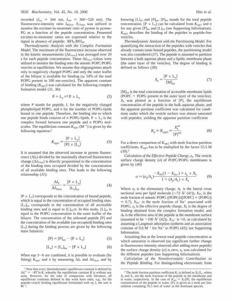
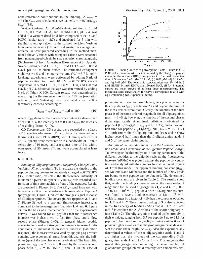
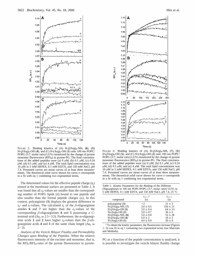
Binding of Oligoarginines onto NegatiVely Charged LipidVesicles: Kinetic Analysis.To investigate the kinetics of thepeptide binding process to negatively charged POPC/POPG(3:7, molar ratio) vesicles, the fluorescence intensity ofmonomeric pyrene in pyrene-PG (RFIM) was recorded as afunction of time after addition of one of the peptides. Resultsare presented in Figures 1-3. The RFIM-signal increases withtime as a result of the peptide-vesicle association. Peptide3(polyarginine, Figure 1) induces the strongest signal responseof all oligoarginines. The octaarginines (peptides2, 5, and7, Figure 3) lead to a stronger fluorescence increase, ascompared to the hexaarginines (peptides1, 4, and6, Figure2). On the basis of a mathematical fit of the experimentalcurves, it was found for all peptides that the fluorescenceincrease was biphasic with a fast first phase and a slowsecond phase (Figures 1-3). In general, higher peptideconcentrations lead to a stronger fluorescence increase. Underconditions of maximal fluorescence increase (saturationresponse), the increase was analyzed by applying eq 1 whichcontains two exponential terms. From this analysis, the half-times (t1/2) of the two phases can be obtained. The fast initialphase witht1/2-1 ) 2-5 s is followed by the slower secondphase witht1/2-2 ) 16-150 s (Table 1). In the case of
polyarginine, it was not possible to give a precise value forthis peptide, ast1/2-1 was below 2 s and beyond the limit ofthe measurement resolution. Clearly, the kinetics of the firstphase is of the same order of magnitude for all oligoarginines(t1/2-1 ) 3-5 s); however, the kinetics of the second phasesdiffer significantly. A minimal half-time is obtained forpeptide4 (H-(âArg)6-OH, t1/2-2 ) 16 ( 3 s), and a maximalhalf-time for peptide7 (H-(âArg)8-NH2, t1/2-2 ) 150 ( 21s). Furthermore theâ-oligoarginine amides6 and 7 showhigher second half-times than theâ-oligoarginine acids4and5 of the same chain length.
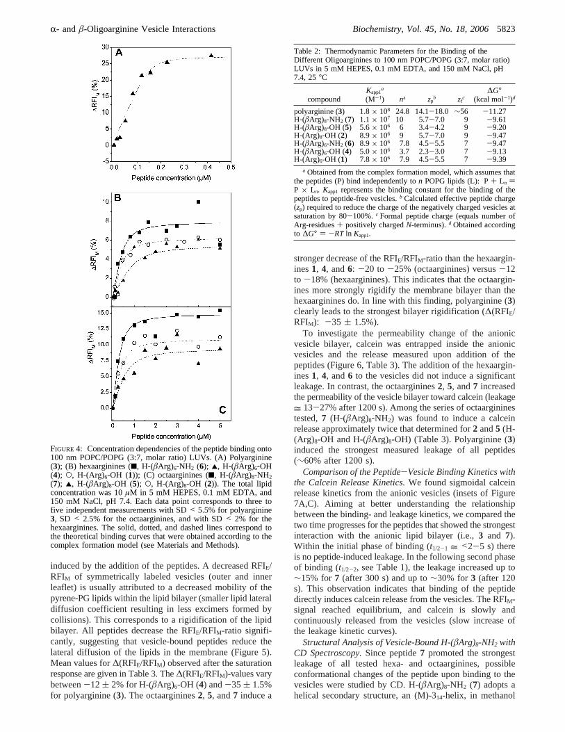
Analysis of the Peptide Binding with the Complex Forma-tion Model and Calculation of the EffectiVe Peptide Charge.To investigate the thermodynamic binding parameters of thedifferent peptides to the anionic vesicles, the fluorescenceincrease (∆RFIM) was plotted against the peptide concentra-tion and analyzed with the complex formation model (Figure4). From this model, the apparent binding constant (Kapp1,see Materials and Methods) and the number of POPG lipids(n) bound to one peptide can be obtained. The determinedbinding constants are given in Table 2. The results showthat, while the binding constants are of the same order ofmagnitude for the short oligoarginines1, 2, and4-7 (5.0×10⁶ to 1.1× 10⁷ M-1), peptide3, with ∼56 arginine residues,was found to have a binding constant of 1.8× 10⁸ M-1,which is larger by a factor of∼10 than the constants obtainedfor 1, 2, and4-7. The stronger binding of3 is also reflectedin the free energy of binding (∆G°) that is∼1.7-2.1 kcalmol-1 lower than the∆G°-values of the shorter oligoargin-ines (Table 2). The oligoarginines studied differ strongly intheir n-values, ranging from 3.7 for peptide4 up to 24.8 forpeptide3. Furthermore, theâ-oligoarginine amides6 and7possess highern-values than theâ-oligoarginine acids4 and5 of the same chain length (∆n = 4). Also, the experimentallydeterminedn-values of theR-oligoarginine acids1 and 2are higher than then-values of the correspondingâ-oli-goarginine acids4 and 5 (∆n = 3-4). This suggests thatR-and â-oligoarginines containing the same number ofguanidinium groups interact differently with anionic vesicles.
FIGURE 1: Binding kinetics of polyarginine3 onto 100 nm POPC/POPG (3:7, molar ratio) LUVs monitored by the change of pyrenemonomer fluorescence (RFIM) in pyrene-PG. The final concentra-tion of 3 was (a) 0µM, (b) 0.05µM, (c) 0.08µM, (d) 0.12µM,and (e) 0.42µM. The total lipid concentration was 10µM in 5mM HEPES, 0.1 mM EDTA, and 150 mM NaCl, pH 7.4. Presentedcurves are mean curves of at least three measurements. Thetheoretical solid curve shown for curve e corresponds to a fit witheq 1 combining two exponential terms.
[(I1200- I0)/(ITriton - I0)] × 100 (10)
R- andâ-Oligoarginine Vesicle Interactions Biochemistry, Vol. 45, No. 18, 20065821
The determined values for the effective peptide charge (zp)sensed at the membrane surface are presented in Table 2. Itwas found that allzp-values are smaller than the correspond-ing number of POPG lipids (n) bound to one peptide andalso smaller than the formal peptide charges (zf). In thiscontext, polyarginine (3) displays the greatest difference inzp- and zf-values. The calculatedzp of the â-oligoarginineamides 6 and 7 are higher than thezp-values of thecorrespondingâ-oligoarginines4 and 5 possessing a C-terminal acid (∆zp = 2.5-3.5). Furthermore, theR-oligoargi-nine acids1 and 2 have higherzp-values than theâ-oli-goarginine acids4 and5 of the same chain length (∆zp =2-3).
Analysis of the Vesicle Bilayer Fluidity and PermeabilityChanges upon Binding of the Peptides.When the relativefluorescence intensity of the excimer and monomer, that is,the RFIE/RFIM-ratio of the pyrene fluorescence in pyrene-
PG as a function of the peptide concentration is analyzed, itis possible to investigate the vesicle bilayer fluidity change
FIGURE 2: Binding kinetics of (A) H-(âArg)6-NH2 (6), (B)H-(âArg)6-OH (4), and (C) H-(Arg)6-OH (1) onto 100 nm POPC/POPG (3:7, molar ratio) LUVs monitored by the change of pyrenemonomer fluorescence (RFIM) in pyrene-PG. The final concentra-tions of the added peptides were (a) 0µM, (b) 0.1 µM, (c) 0.24µM, (d) 0.5 µM, and (e) 4µM. The total lipid concentration was10 µM in 5 mM HEPES, 0.1 mM EDTA, and 150 mM NaCl, pH7.4. Presented curves are mean curves of at least three measure-ments. The theoretical solid curve shown for curve e correspondsto a fit with eq 1 combining two exponential terms.
FIGURE 3: Binding kinetics of (A) H-(âArg)8-NH2 (7), (B)H-(âArg)8-OH (5), and (C) H-(Arg)8-OH (2) onto 100 nm POPC/POPG (3:7, molar ratio) LUVs monitored by the change of pyrenemonomer fluorescence (RFIM) in pyrene-PG. The final concentra-tions of the added peptides were (a) 0µM, (b) 0.1 µM, (c) 0.24µM, (d) 0.5 µM, and (e) 4µM. The total lipid concentration was10 µM in 5 mM HEPES, 0.1 mM EDTA, and 150 mM NaCl, pH7.4. Presented curves are mean curves of at least three measure-ments. The theoretical solid curve shown for curve e correspondsto a fit with eq 1 combining two exponential terms.
Table 1: Kinetic Parameters for the Binding of the DifferentOligoarginines to 100 nm POPC/POPG (3:7, molar ratio) LUVs in5 mM HEPES, 0.1 mM EDTA, and 150 mM NaCl, pH 7.4, 25°C
compoundt1/2-1
a
(s)t1/2-2
(s)
polyarginine (3) <2 21( 3H-(âArg)8-NH2 (7) 3.0( 0.5 150( 21H-(âArg)8-OH (5) 3.0( 0.4 64( 13H-(Arg)8-OH (2) 3.0( 0.7 36( 5H-(âArg)6-NH2 (6) 5.0( 0.8 52( 28H-(âArg)6-OH (4) 5.0( 2 16( 3H-(Arg)6-OH (1) 4.0( 0.9 20( 4
a To obtain the kinetic parameters, the fluorescence increase (Figures1-3) was fit to eq 1 containing two exponential terms (see Materialsand Methods).
5822 Biochemistry, Vol. 45, No. 18, 2006 Hitz et al.
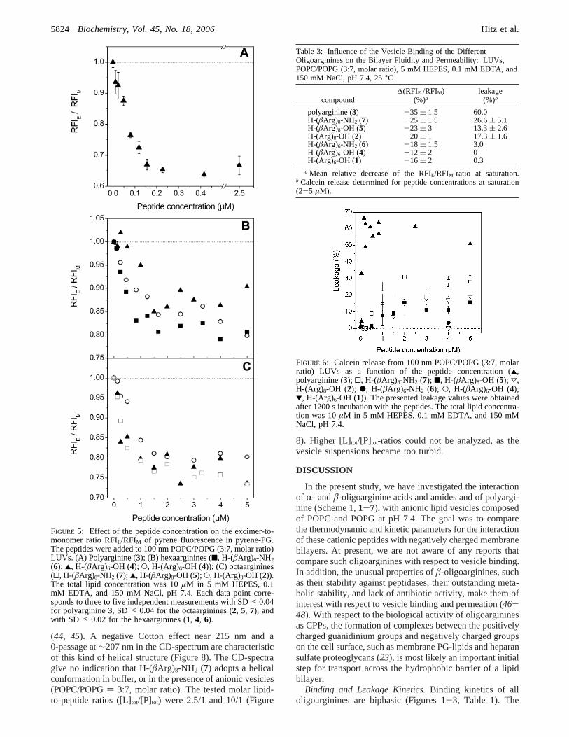
induced by the addition of the peptides. A decreased RFIE/RFIM of symmetrically labeled vesicles (outer and innerleaflet) is usually attributed to a decreased mobility of thepyrene-PG lipids within the lipid bilayer (smaller lipid lateraldiffusion coefficient resulting in less excimers formed bycollisions). This corresponds to a rigidification of the lipidbilayer. All peptides decrease the RFIE/RFIM-ratio signifi-cantly, suggesting that vesicle-bound peptides reduce thelateral diffusion of the lipids in the membrane (Figure 5).Mean values for∆(RFIE/RFIM) observed after the saturationresponse are given in Table 3. The∆(RFIE/RFIM)-values varybetween-12 ( 2% for H-(âArg)6-OH (4) and-35 ( 1.5%for polyarginine (3). The octaarginines2, 5, and7 induce a
stronger decrease of the RFIE/RFIM-ratio than the hexaargin-ines1, 4, and6: -20 to -25% (octaarginines) versus-12to -18% (hexaarginines). This indicates that the octaargin-ines more strongly rigidify the membrane bilayer than thehexaarginines do. In line with this finding, polyarginine (3)clearly leads to the strongest bilayer rigidification (∆(RFIE/RFIM): -35 ( 1.5%).
To investigate the permeability change of the anionicvesicle bilayer, calcein was entrapped inside the anionicvesicles and the release measured upon addition of thepeptides (Figure 6, Table 3). The addition of the hexaargin-ines1, 4, and6 to the vesicles did not induce a significantleakage. In contrast, the octaarginines2, 5, and7 increasedthe permeability of the vesicle bilayer toward calcein (leakage= 13-27% after 1200 s). Among the series of octaargininestested,7 (H-(âArg)8-NH2) was found to induce a calceinrelease approximately twice that determined for2 and5 (H-(Arg)8-OH and H-(âArg)8-OH) (Table 3). Polyarginine (3)induced the strongest measured leakage of all peptides(∼60% after 1200 s).
Comparison of the Peptide-Vesicle Binding Kinetics withthe Calcein Release Kinetics.We found sigmoidal calceinrelease kinetics from the anionic vesicles (insets of Figure7A,C). Aiming at better understanding the relationshipbetween the binding- and leakage kinetics, we compared thetwo time progresses for the peptides that showed the strongestinteraction with the anionic lipid bilayer (i.e.,3 and 7).Within the initial phase of binding (t1/2-1 = <2-5 s) thereis no peptide-induced leakage. In the following second phaseof binding (t1/2-2, see Table 1), the leakage increased up to∼15% for 7 (after 300 s) and up to∼30% for 3 (after 120s). This observation indicates that binding of the peptidedirectly induces calcein release from the vesicles. The RFIM-signal reached equilibrium, and calcein is slowly andcontinuously released from the vesicles (slow increase ofthe leakage kinetic curves).
Structural Analysis of Vesicle-Bound H-(âArg)8-NH2 withCD Spectroscopy.Since peptide7 promoted the strongestleakage of all tested hexa- and octaarginines, possibleconformational changes of the peptide upon binding to thevesicles were studied by CD. H-(âArg)8-NH2 (7) adopts ahelical secondary structure, an (M)-314-helix, in methanol
FIGURE 4: Concentration dependencies of the peptide binding onto100 nm POPC/POPG (3:7, molar ratio) LUVs. (A) Polyarginine(3); (B) hexaarginines (9, H-(âArg)6-NH2 (6); 2, H-(âArg)6-OH(4); O, H-(Arg)6-OH (1)); (C) octaarginines (9, H-(âArg)8-NH2(7); 2, H-(âArg)8-OH (5); O, H-(Arg)8-OH (2)). The total lipidconcentration was 10µM in 5 mM HEPES, 0.1 mM EDTA, and150 mM NaCl, pH 7.4. Each data point corresponds to three tofive independent measurements with SD< 5.5% for polyarginine3, SD < 2.5% for the octaarginines, and with SD< 2% for thehexaarginines. The solid, dotted, and dashed lines correspond tothe theoretical binding curves that were obtained according to thecomplex formation model (see Materials and Methods).
Table 2: Thermodynamic Parameters for the Binding of theDifferent Oligoarginines to 100 nm POPC/POPG (3:7, molar ratio)LUVs in 5 mM HEPES, 0.1 mM EDTA, and 150 mM NaCl, pH7.4, 25°C
compoundKapp1
a
(M-1) na zpb zf
c∆G°
(kcal mol-1)d
polyarginine (3) 1.8× 10⁸ 24.8 14.1-18.0 ∼56 -11.27H-(âArg)8-NH2 (7) 1.1× 10⁷ 10 5.7-7.0 9 -9.61H-(âArg)8-OH (5) 5.6× 10⁶ 6 3.4-4.2 9 -9.20H-(Arg)8-OH (2) 8.9× 10⁶ 9 5.7-7.0 9 -9.47H-(âArg)6-NH2 (6) 8.9× 10⁶ 7.8 4.5-5.5 7 -9.47H-(âArg)6-OH (4) 5.0× 10⁶ 3.7 2.3-3.0 7 -9.13H-(Arg)6-OH (1) 7.8× 10⁶ 7.9 4.5-5.5 7 -9.39
a Obtained from the complex formation model, which assumes thatthe peptides (P) bind independently ton POPG lipids (L): P+ Ln )P × Ln. Kapp1 represents the binding constant for the binding of thepeptides to peptide-free vesicles.b Calculated effective peptide charge(zp) required to reduce the charge of the negatively charged vesicles atsaturation by 80-100%.c Formal peptide charge (equals number ofArg-residues+ positively chargedN-terminus).d Obtained accordingto ∆G° ) -RT ln Kapp1.
R- andâ-Oligoarginine Vesicle Interactions Biochemistry, Vol. 45, No. 18, 20065823
(44, 45). A negative Cotton effect near 215 nm and a0-passage at∼207 nm in the CD-spectrum are characteristicof this kind of helical structure (Figure 8). The CD-spectragive no indication that H-(âArg)8-NH2 (7) adopts a helicalconformation in buffer, or in the presence of anionic vesicles(POPC/POPG) 3:7, molar ratio). The tested molar lipid-to-peptide ratios ([L]tot/[P]tot) were 2.5/1 and 10/1 (Figure
8). Higher [L]tot/[P]tot-ratios could not be analyzed, as thevesicle suspensions became too turbid.
DISCUSSION
In the present study, we have investigated the interactionof R- andâ-oligoarginine acids and amides and of polyargi-nine (Scheme 1,1-7), with anionic lipid vesicles composedof POPC and POPG at pH 7.4. The goal was to comparethe thermodynamic and kinetic parameters for the interactionof these cationic peptides with negatively charged membranebilayers. At present, we are not aware of any reports thatcompare such oligoarginines with respect to vesicle binding.In addition, the unusual properties ofâ-oligoarginines, suchas their stability against peptidases, their outstanding meta-bolic stability, and lack of antibiotic activity, make them ofinterest with respect to vesicle binding and permeation (46-48). With respect to the biological activity of oligoargininesas CPPs, the formation of complexes between the positivelycharged guanidinium groups and negatively charged groupson the cell surface, such as membrane PG-lipids and heparansulfate proteoglycans (23), is most likely an important initialstep for transport across the hydrophobic barrier of a lipidbilayer.
Binding and Leakage Kinetics.Binding kinetics of alloligoarginines are biphasic (Figures 1-3, Table 1). The
FIGURE 5: Effect of the peptide concentration on the excimer-to-monomer ratio RFIE/RFIM of pyrene fluorescence in pyrene-PG.The peptides were added to 100 nm POPC/POPG (3:7, molar ratio)LUVs. (A) Polyarginine (3); (B) hexaarginines (9, H-(âArg)6-NH2(6); 2, H-(âArg)6-OH (4); O, H-(Arg)6-OH (4)); (C) octaarginines(0, H-(âArg)8-NH2 (7); 2, H-(âArg)8-OH (5); O, H-(Arg)8-OH (2)).The total lipid concentration was 10µM in 5 mM HEPES, 0.1mM EDTA, and 150 mM NaCl, pH 7.4. Each data point corre-sponds to three to five independent measurements with SD< 0.04for polyarginine3, SD < 0.04 for the octaarginines (2, 5, 7), andwith SD < 0.02 for the hexaarginines (1, 4, 6).
Table 3: Influence of the Vesicle Binding of the DifferentOligoarginines on the Bilayer Fluidity and Permeability: LUVs,POPC/POPG (3:7, molar ratio), 5 mM HEPES, 0.1 mM EDTA, and150 mM NaCl, pH 7.4, 25°C
compound∆(RFIE /RFIM)
(%)aleakage
(%)b
polyarginine (3) -35 ( 1.5 60.0H-(âArg)8-NH2 (7) -25 ( 1.5 26.6( 5.1H-(âArg)8-OH (5) -23 ( 3 13.3( 2.6H-(Arg)8-OH (2) -20 ( 1 17.3( 1.6H-(âArg)6-NH2 (6) -18 ( 1.5 3.0H-(âArg)6-OH (4) -12 ( 2 0H-(Arg)6-OH (1) -16 ( 2 0.3
a Mean relative decrease of the RFIE/RFIM-ratio at saturation.b Calcein release determined for peptide concentrations at saturation(2-5 µM).
FIGURE 6: Calcein release from 100 nm POPC/POPG (3:7, molarratio) LUVs as a function of the peptide concentration (2,polyarginine (3); 0, H-(âArg)8-NH2 (7); 9, H-(âArg)8-OH (5); 3,H-(Arg)8-OH (2); b, H-(âArg)6-NH2 (6); O, H-(âArg)6-OH (4);1, H-(Arg)6-OH (1)). The presented leakage values were obtainedafter 1200 s incubation with the peptides. The total lipid concentra-tion was 10µM in 5 mM HEPES, 0.1 mM EDTA, and 150 mMNaCl, pH 7.4.
5824 Biochemistry, Vol. 45, No. 18, 2006 Hitz et al.
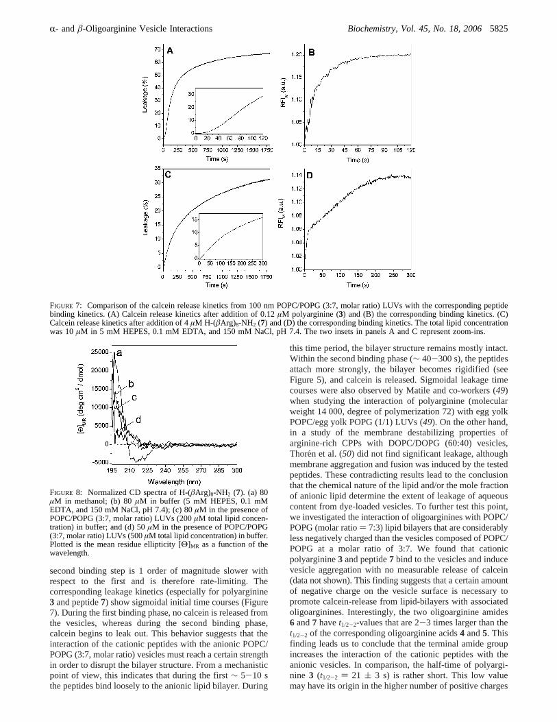
second binding step is 1 order of magnitude slower withrespect to the first and is therefore rate-limiting. Thecorresponding leakage kinetics (especially for polyarginine3 and peptide7) show sigmoidal initial time courses (Figure7). During the first binding phase, no calcein is released fromthe vesicles, whereas during the second binding phase,calcein begins to leak out. This behavior suggests that theinteraction of the cationic peptides with the anionic POPC/POPG (3:7, molar ratio) vesicles must reach a certain strengthin order to disrupt the bilayer structure. From a mechanisticpoint of view, this indicates that during the first∼ 5-10 sthe peptides bind loosely to the anionic lipid bilayer. During
this time period, the bilayer structure remains mostly intact.Within the second binding phase (∼ 40-300 s), the peptidesattach more strongly, the bilayer becomes rigidified (seeFigure 5), and calcein is released. Sigmoidal leakage timecourses were also observed by Matile and co-workers (49)when studying the interaction of polyarginine (molecularweight 14 000, degree of polymerization 72) with egg yolkPOPC/egg yolk POPG (1/1) LUVs (49). On the other hand,in a study of the membrane destabilizing properties ofarginine-rich CPPs with DOPC/DOPG (60:40) vesicles,Thoren et al. (50) did not find significant leakage, althoughmembrane aggregation and fusion was induced by the testedpeptides. These contradicting results lead to the conclusionthat the chemical nature of the lipid and/or the mole fractionof anionic lipid determine the extent of leakage of aqueouscontent from dye-loaded vesicles. To further test this point,we investigated the interaction of oligoarginines with POPC/POPG (molar ratio) 7:3) lipid bilayers that are considerablyless negatively charged than the vesicles composed of POPC/POPG at a molar ratio of 3:7. We found that cationicpolyarginine3 and peptide7 bind to the vesicles and inducevesicle aggregation with no measurable release of calcein(data not shown). This finding suggests that a certain amountof negative charge on the vesicle surface is necessary topromote calcein-release from lipid-bilayers with associatedoligoarginines. Interestingly, the two oligoarginine amides6 and7 havet1/2-2-values that are 2-3 times larger than thet1/2-2 of the corresponding oligoarginine acids4 and5. Thisfinding leads us to conclude that the terminal amide groupincreases the interaction of the cationic peptides with theanionic vesicles. In comparison, the half-time of polyargi-nine 3 (t1/2-2 ) 21 ( 3 s) is rather short. This low valuemay have its origin in the higher number of positive charges
FIGURE 7: Comparison of the calcein release kinetics from 100 nm POPC/POPG (3:7, molar ratio) LUVs with the corresponding peptidebinding kinetics. (A) Calcein release kinetics after addition of 0.12µM polyarginine (3) and (B) the corresponding binding kinetics. (C)Calcein release kinetics after addition of 4µM H-(âArg)8-NH2 (7) and (D) the corresponding binding kinetics. The total lipid concentrationwas 10µM in 5 mM HEPES, 0.1 mM EDTA, and 150 mM NaCl, pH 7.4. The two insets in panels A and C represent zoom-ins.
FIGURE 8: Normalized CD spectra of H-(âArg)8-NH2 (7). (a) 80µM in methanol; (b) 80µM in buffer (5 mM HEPES, 0.1 mMEDTA, and 150 mM NaCl, pH 7.4); (c) 80µM in the presence ofPOPC/POPG (3:7, molar ratio) LUVs (200µM total lipid concen-tration) in buffer; and (d) 50µM in the presence of POPC/POPG(3:7, molar ratio) LUVs (500µM total lipid concentration) in buffer.Plotted is the mean residue ellipticity [Θ]MR as a function of thewavelength.
R- andâ-Oligoarginine Vesicle Interactions Biochemistry, Vol. 45, No. 18, 20065825
(zf ∼ 56) possessed by polyarginine3 with respect to theshorter oligoarginines (zf ) 7-9). This may lead to a fasteroverall association process. Biphasic time courses were alsoreported for the interaction of cationic annexins with anionicPOPC/ POPG vesicles and for the anionic assembly factorP17 (net charge of-7 at pH 7.2) with cationic vesiclescomposed of phosphatidylcholine, phosphatidylethanol-amine, and sphingosine lipids (51, 52). In both cases, thebilayer association was completed after∼ 250-500 s.
In general, it is suggested that the binding of chargedproteins to oppositely charged vesicle membranes is com-posed of at least two steps (53). The initial binding step ismainly driven by electrostatic interactions that result in anoverall charge neutralization at the contact side. This bindingstep takes place near the interface and leads to a thinning ofthe membrane (Shai-Matsuzaki-Huang model) (54). Thesecond step may also involve hydrophobic interactions and/or hydrogen bonding, including a significant disruption ofthe membrane. According to this premise, the first peptidebinding phase reported herein most likely corresponds toelectrostatic attraction that neutralizes the peptide charge nearthe interface, whereas the second binding phase also involvesnonelectrostatic forces, which cause leakage of the bilayer.To further explore this possibility, we analyzed in more detailthe energetic contributions of the binding event to the totalfree energy of binding.
Thermodynamic Analysis.Initial analysis revealed thebinding constant (Kapp1) of polyarginine3 to be approximately1 order of magnitude larger than theKapp1-values calculatedfor the oligoarginines, corresponding to a difference in thefree energy of binding of∼ -1.7 to-2.1 kcal/mol (Table2). CalculatedKapp1 values for the hexa- and octaarginineswere shown to be of the same order of magnitude (∼5 ×10⁶ to 107 M-1), indicating similar binding strengths for theanionic vesicles. A related study has recently been performedfor the association of H-(Arg)9OH with anionic POPC/POPG(7:3, molar ratio) vesicles; a binding constant of 8.2× 10⁴
M-1 was reported (23). This is approximately 2 orders ofmagnitude lower than the binding constants reported hereinfor the association onto POPC/POPG (3:7, molar ratio)vesicles with the opposite lipid ratio. Similar observationshave been made for the binding of heptalysine onto POPC/POPS vesicles. A reduction of POPS from 17% to 4% molarreduced the molar partitioning coefficient by a factor of∼1000 (55).
The stronger peptide-vesicle interaction of polyarginine(3) is also reflected in a higher number of bound POPG lipidsper peptide (n) and in a higher effective peptide charge value(zp) sensed at the membrane surface. This is reasonable, sincethis polyarginine possesses 7-10 times the number ofguanidinium groups per peptide chain as compared to theoligoarginines1, 2, and 4-7. However, the determinedn-value of polyarginine (3) is rather low (∼25). As3 has aformal charge (zf) of ∼ +56, this indicates that only about45% of this charge is electrically neutralized withn = 25.A similar phenomenon was observed by Hartmann and Galla(56) for polylysine. Their investigation showed that onlyevery second lysine-residue was bound to a lipid. Thecalculatedzp of 3 for complete charge neutralization atsaturation is even lower (zp ) 14-18, Table 2). Lowereffective peptide charge (zp ) with respect to the formalcharge (zf) has also recently been observed for the binding
of H-(Arg)9-OH onto POPC/POPG (7:3) vesicles and forother CPPs (23, 42, 57). It is argued that this phenomenonreflects the polyelectrolytic behavior of such cationic pep-tides. In buffer, such components will attract counterions thatwill not be fully released when peptide-vesicle associationoccurs. Accordingly, we find for all oligoarginines thatzp
< zf (Table 2). Interestingly, the difference betweenzp andzf is larger for the investigatedâ-oligoarginine acids4 and5 (zp/zf = 0.45) than forR-oligoarginine acids1 and2 (zp/zf
= 0.78). This finding suggests that eitherâ-oligoargininesmore strongly attract counterions from the aqueous surround-ing and/or R-oligoarginines better expose their positivecharges to the vesicles. This would also explain the highernumber of bound lipids per peptide observed forR-oli-goarginines (see Table 2).
Strong differences concerning then- andzp-values werealso observed betweenâ-oligoarginine acids and amides(Table 2). Theâ-oligoarginine amides6 and7 possess largern- andzp-values than the corresponding acids,4 and5. Thisobservation indicates that the terminal amide group interactswith the membrane surface and/or that the acid group reducesthe number of bound lipids due to charge repulsion withphosphate-anions of POPG. In terms of the hydrogen-bonding properties of amides, the additional interaction bythe amide group may involve hydrogen bonding near theinterface at the membrane hydration layer.
To distinguish electrostatic from nonelectrostatic energeticcontributions to the total free energy of binding (∆G°), wecalculatedKapp2 at the saturation response for all peptides(Table 4). The determinedKapp2-values (8.3× 10⁴ to 6.1×10⁵ M-1) are about 2 orders of magnitude smaller than theKapp1-values, the binding constants describing the associationonto peptide-free vesicles. Assuming that the surface poten-tial is reduced to zero when reaching saturation response,Kapp2 represents the binding constant onto a neutral vesicleand hence thenonelectrostaticenergetic contribution:∆Gnonel
) -RT ln Kapp2. The electrostaticcontribution to the freeenergy of binding can therefore be estimated by the followingequation: ∆Gel ) -RT ln(Kapp1/Kapp2) (38). Data for theenergetic contributions are given in Table 4. Analysis revealsthat only about 25-30% of the total free energy of bindinghas electrostatic origin. A similar value has also recently beenreported for H-(Arg)9-OH binding onto POPC/POPG vesicles
Table 4: Nonelectrostatic and Electrostatic Contributions of theDifferent Oligoarginines to the Total Free Energy of Binding ontoPOPC/POPG (3:7, molar ratio) LUVs in 5 mM HEPES, 0.1 mMEDTA, and 150 mM NaCl, pH 7.4, 25°C
compoundKapp2
(M-1)a∆Gel
(kcal mol-1)b∆Gnonel
(kcal mol-1)c %-∆Geld
polyarginine (3) 6.1× 10⁵ -3.38 -7.88 30H-(âArg)8-NH2 (7) 8.9× 10⁴ -2.86 -6.75 30H-(âArg)8-OH (5) 8.3× 10⁴ -2.49 -6.71 27H-(Arg)8-OH (2) 8.9× 10⁴ -2.73 -6.75 29H-(âArg)6-NH2 (6) 1.0× 10⁵ -2.66 -6.82 28H-(âArg)6-OH (4) 1.0× 10⁵ -2.28 -6.85 25H-(Arg)6-OH (1) 9.4× 10⁴ -2.61 -6.78 27
a Calculated according toKp ) Kapp2 ) Xb/[P]. [P] is the bulkequilibrium concentration.Kapp2was calculated at saturation, where theconcentration directly above the bilayer surface is approximately equalto the bulk equilibrium concentration [P].b The electrostatic free energyof the peptide binding (∆Gel) was calculated according to-RT ln(Kapp1/Kapp2). c The nonelectrostatic contribution (∆Gnonel) can be obtainedaccording to-RT ln Kapp2. d %-∆Gel ) (∆Gel/(∆Gel + ∆Gnonel)) × 100.
5826 Biochemistry, Vol. 45, No. 18, 2006 Hitz et al.
(23). The authors calculated a %-∆Gel of 33% for a peptideconcentration of 1µM. In contrast, studies investigating thebinding of cell-penetrating peptide H-(Arg)7Trp-OH ontoDOPC/DOPG vesicles reported electrostatic contributions ofmore than 50% (57). Strong nonelectrostatic contributionscan be explained by the hydrogen-bonding properties of thebivalent guanidinium group of arginine that allows stronginteraction with the membrane hydration layer, which extends1-3 nm into the aqueous phase (23). On the other hand,high nonelectrostatic contributions are also indicative of atransfer from the aqueous phase into the less polar environ-ment of the lipid bilayer (58).
Analysis of Vesicle Bilayer Destabilization.The investi-gated oligoarginines decrease the RFIE/RFIM-ratio of pyrenefluorescence in pyrene-PG in the following order:3 (∼56Arg residues)> 2, 5, 7 (eight Arg residues)> 1, 4, 6 (sixArg residues). Since the vesicles are labeled symmetrically,it is unlikely that the signal change arises from a flip-flopmechanism of the POPG lipids. It is more likely that theadsorbed peptides decrease the lateral mobility of thephospholipids. Such observations have also been reportedfor other basic peptides and proteins such as polylysine,cytochromeb5, histones, and H-(Lys)19-OH (27, 55, 59).Domain formation of negatively charged POPG lipids wouldrequire an increased RFIE/RFIM-ratio, a phenomenon that wasnot observed in the present study. The membrane rigidifi-cation is directly correlated to the determined leakage (Table3): 3 > 7 > 2 g 5 . 1, 4, 6. None of the hexaargininesinduce significant calcein release, although theirKapp-valuesare of the same order of magnitude as those of theoctaarginines. It is likely that the somewhat stronger mem-brane rigidification induced by the octaarginines is necessaryto induce leakage of the anionic vesicles, possibly bypromoting a positive curvature (60). Interestingly, the calceinrelease data obtained for6 and7 correlate well with the cell-uptake efficiency determined for corresponding fluorescein-labeled derivatives (14): increased calcein release corre-sponds to a more efficient cell uptake. Also of importanceis that â-peptide7, which possesses a C-terminal amidegroup, induces approximately twice as much calcein releaseas â-peptide 5 with C-terminal acid group. This findingsuggests that the ability of the amide-form of theâ-peptide7 to interact with more POPG lipids than the acid form5(see Table 2) leads to a stronger membrane disruption, andtherefore, more calcein is released from the vesicles.
Analysis of Peptide Structure.Recent studies have shownthat cationicâ-peptides containing cyclic amino acid residuesare potent bilayer-disrupting agents and that 14-helix forma-tion in the presence of anionic vesicles is necessary forbilayer disruption (61). However, our CD analysis ofoligoarginine 7 (H-(âArg)8-NH2) in the presence of anionicPOPC/POPG vesicles shows that the formation of a 14-helixconformation is not required for membrane permeabilization(Figure 8). Similar observations have recently been reportedfor the cell-penetratingR-peptide H-(Arg)7Trp-OH, whichwas also shown not to adopt an ordered structure in thepresence of anionic vesicles for an [L]tot/[P]tot of 100:1 (57).
Another interesting aspect of our investigations relates tothe observednonendocytoticuptake of oligoarginines intolive cells (15, 20, 21). Our binding studies show that, if theinteraction is strong enough (Kapp= 105-106 M-1, significantmembrane rigidification), LUVs with a mean diameter of
100 nm become permeable. For polyarginine, efficientleakage (∼60%, Figures 6 and 7) is already observed at a[L] tot/[P]tot-ratio of ∼100 and matches the [L]tot/[P]tot-ratiosof known pore-forming peptides (62). Also, the octaarginineamide 7 induces significant leakage of∼20% at a [L]tot/[P]tot-ratio of 10. As mentioned previously, the interactionof oligoarginines with cell surface GAGs such as heparansulfate is most likely necessary in order to observe efficientinternalization. A possible explanation is the very highaffinity that oligoarginines, such as H-(Arg)9-OH, showtoward heparan sulfate. In recent studies, association con-stants in the range of∼106 M-1 were determined for thebinding to heparin and heparan sulfate. This is in agreementwith the binding constants obtained in this study for thebinding of oligoarginines onto strongly negatively chargedvesicles (POPC/POPG) 3:7, molar ratio). Such strongbinding may lead to significant membrane disruption allow-ing arginine-rich CPPs to cross the vesicle bilayer in anonendocytotic manner. Also, the escape from endocytoticvesicles requires the membrane bilayer to become permeable,thereby permitting the CPP to enter into the cytoplasm ofthe cell (3, 24).
Although it was not the focus of the present work, thethermodynamics of binding of the amino cationic polylysineto anionic POPC/POPG vesicles (7:3, molar ratio) containingpyrene-PG was compared with the binding of the guani-dinium cationic polyarginine3 (see Supporting Information).Although both polypeptides had a similar degree of polym-erization,3 led to a stronger increase of the monomer pyrenefluorescence (∆RFIM) than polylysine (see Figure S-2,Supporting Information). Furthermore, a preliminary analysiswith the complex formation model showed thatKapp1is aboutthree times larger thanKapp1 for polylysine, and3 binds totwice as many POPG molecules than polylysine.
CONCLUSIONS
We have shown, for a range ofR-and â-oligoarginines(see Scheme 1) with C-terminal amide and acid end-groups,clear differences in their interaction with anionic lipidvesicles (POPC/POPG) 3:7, molar ratio): (i) polyarginine(3) binds with an association constant approximately 1 orderof magnitude higher than those of the shorter oligoarginines1, 2, and4-7, and polyarginine (3), leads to the strongestbilayer rigidification and induces the largest membranedisruption (leakage); (ii) all octaarginines (peptides2, 5, and7) induce significant vesicle leakage (13-30%), whereashexaarginines (peptides1, 4, and6) do not; (iii) â-oligoargi-nine amides (peptides6 and7) interact with more lipids andinduce a stronger bilayer disruption than the correspondingacids (peptides4 and 5); and (iv) R-oligoarginine acids(peptides1 and 2) interact with more lipids and expose ahigher effective peptide charge to the anionic vesicles thanthe correspondingâ-oligoarginine acids (peptides4 and5).
We calculate that for all oligoarginines significantly lessthan 50% of the total free energy of binding has electrostaticorigin. Such observations are supported by the finding thatthe stronger vesicle association of polyarginine with respectto the oligoarginines has nonelectrostatic origins. Kineticanalysis of the binding processes shows biphasic timecourses. We suggest that the first fast process (4-10 s)corresponds to weaker electrostatic attraction, whereas the
R- andâ-Oligoarginine Vesicle Interactions Biochemistry, Vol. 45, No. 18, 20065827
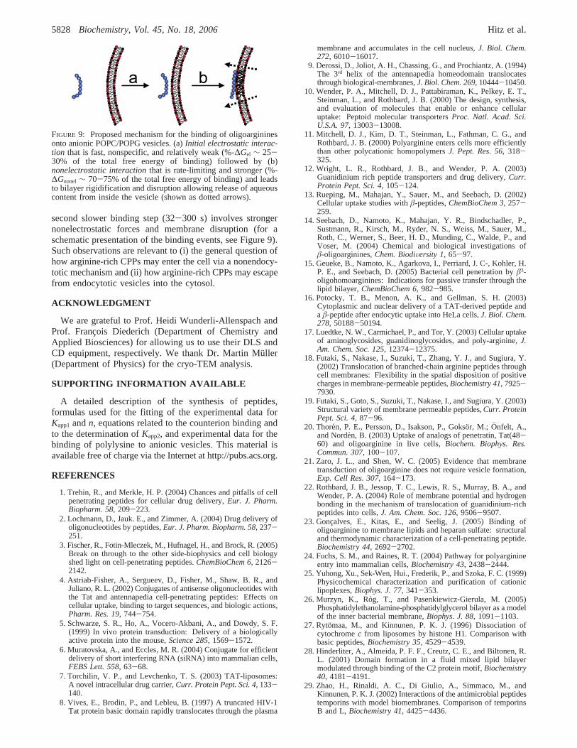
second slower binding step (32-300 s) involves strongernonelectrostatic forces and membrane disruption (for aschematic presentation of the binding events, see Figure 9).Such observations are relevant to (i) the general question ofhow arginine-rich CPPs may enter the cell via a nonendocy-totic mechanism and (ii) how arginine-rich CPPs may escapefrom endocytotic vesicles into the cytosol.
ACKNOWLEDGMENT
We are grateful to Prof. Heidi Wunderli-Allenspach andProf. Franc¸ois Diederich (Department of Chemistry andApplied Biosciences) for allowing us to use their DLS andCD equipment, respectively. We thank Dr. Martin Mu¨ller(Department of Physics) for the cryo-TEM analysis.
SUPPORTING INFORMATION AVAILABLE
A detailed description of the synthesis of peptides,formulas used for the fitting of the experimental data forKapp1andn, equations related to the counterion binding andto the determination ofKapp2, and experimental data for thebinding of polylysine to anionic vesicles. This material isavailable free of charge via the Internet at http://pubs.acs.org.
REFERENCES
1. Trehin, R., and Merkle, H. P. (2004) Chances and pitfalls of cellpenetrating peptides for cellular drug delivery,Eur. J. Pharm.Biopharm. 58, 209-223.
2. Lochmann, D., Jauk. E., and Zimmer, A. (2004) Drug delivery ofoligonucleotides by peptides,Eur. J. Pharm. Biopharm. 58, 237-251.
3. Fischer, R., Fotin-Mleczek, M., Hufnagel, H., and Brock, R. (2005)Break on through to the other side-biophysics and cell biologyshed light on cell-penetrating peptides.ChemBioChem 6, 2126-2142.
4. Astriab-Fisher, A., Sergueev, D., Fisher, M., Shaw, B. R., andJuliano, R. L. (2002) Conjugates of antisense oligonucleotides withthe Tat and antennapedia cell-penetrating peptides: Effects oncellular uptake, binding to target sequences, and biologic actions,Pharm. Res. 19, 744-754.
5. Schwarze, S. R., Ho, A., Vocero-Akbani, A., and Dowdy, S. F.(1999) In vivo protein transduction: Delivery of a biologicallyactive protein into the mouse,Science 285, 1569-1572.
6. Muratovska, A., and Eccles, M. R. (2004) Conjugate for efficientdelivery of short interfering RNA (siRNA) into mammalian cells,FEBS Lett. 558, 63-68.
7. Torchilin, V. P., and Levchenko, T. S. (2003) TAT-liposomes:A novel intracellular drug carrier,Curr. Protein Pept. Sci. 4,133-140.
8. Vives, E., Brodin, P., and Lebleu, B. (1997) A truncated HIV-1Tat protein basic domain rapidly translocates through the plasma
membrane and accumulates in the cell nucleus,J. Biol. Chem.272, 6010-16017.
9. Derossi, D., Joliot, A. H., Chassing, G., and Prochiantz, A. (1994)The 3rd helix of the antennapedia homeodomain translocatesthrough biological-membranes,J. Biol. Chem. 269, 10444-10450.
10. Wender, P. A., Mitchell, D. J., Pattabiraman, K., Pelkey, E. T.,Steinman, L., and Rothbard, J. B. (2000) The design, synthesis,and evaluation of molecules that enable or enhance cellularuptake: Peptoid molecular transportersProc. Natl. Acad. Sci.U.S.A. 97, 13003-13008.
11. Mitchell, D. J., Kim, D. T., Steinman, L., Fathman, C. G., andRothbard, J. B. (2000) Polyarginine enters cells more efficientlythan other polycationic homopolymersJ. Pept. Res. 56, 318-325.
12. Wright, L. R., Rothbard, J. B., and Wender, P. A. (2003)Guanidinium rich peptide transporters and drug delivery,Curr.Protein Pept. Sci. 4, 105-124.
13. Rueping, M., Mahajan, Y., Sauer, M., and Seebach, D. (2002)Cellular uptake studies withâ-peptides,ChemBioChem 3, 257-259.
14. Seebach, D., Namoto, K., Mahajan, Y. R., Bindschadler, P.,Sustmann, R., Kirsch, M., Ryder, N. S., Weiss, M., Sauer, M.,Roth, C., Werner, S., Beer, H. D., Munding, C., Walde, P., andVoser, M. (2004) Chemical and biological investigations ofâ-oligoarginines,Chem. BiodiVersity 1, 65-97.
15. Geueke, B., Namoto, K., Agarkova, I., Perriard, J. C-, Kohler, H.P. E., and Seebach, D. (2005) Bacterial cell penetration byâ3-oligohomoarginines: Indications for passive transfer through thelipid bilayer, ChemBioChem 6, 982-985.
16. Potocky, T. B., Menon, A. K., and Gellman, S. H. (2003)Cytoplasmic and nuclear delivery of a TAT-derived peptide anda â-peptide after endocytic uptake into HeLa cells,J. Biol. Chem.278, 50188-50194.
17. Luedtke, N. W., Carmichael, P., and Tor, Y. (2003) Cellular uptakeof aminoglycosides, guanidinoglycosides, and poly-arginine,J.Am. Chem. Soc. 125, 12374-12375.
18. Futaki, S., Nakase, I., Suzuki, T., Zhang, Y. J., and Sugiura, Y.(2002) Translocation of branched-chain arginine peptides throughcell membranes: Flexibility in the spatial disposition of positivecharges in membrane-permeable peptides,Biochemistry 41, 7925-7930.
19. Futaki, S., Goto, S., Suzuki, T., Nakase, I., and Sugiura, Y. (2003)Structural variety of membrane permeable peptides,Curr. ProteinPept. Sci. 4, 87-96.
20. Thoren, P. E., Persson, D., Isakson, P., Gokso¨r, M.; Onfelt, A.,and Norde´n, B. (2003) Uptake of analogs of penetratin, Tat(48-60) and oligoarginine in live cells,Biochem. Biophys. Res.Commun. 307, 100-107.
21. Zaro, J. L., and Shen, W. C. (2005) Evidence that membranetransduction of oligoarginine does not require vesicle formation,Exp. Cell Res. 307, 164-173.
22. Rothbard, J. B., Jessop, T. C., Lewis, R. S., Murray, B. A., andWender, P. A. (2004) Role of membrane potential and hydrogenbonding in the mechanism of translocation of guanidinium-richpeptides into cells,J. Am. Chem. Soc. 126, 9506-9507.
23. Gonc¸alves, E., Kitas, E., and Seelig, J. (2005) Binding ofoligoarginine to membrane lipids and heparan sulfate: structuraland thermodynamic characterization of a cell-penetrating peptide.Biochemistry 44, 2692-2702.
24. Fuchs, S. M., and Raines, R. T. (2004) Pathway for polyarginineentry into mammalian cells,Biochemistry 43, 2438-2444.
25. Yuhong, Xu., Sek-Wen, Hui., Frederik, P., and Szoka, F. C. (1999)Physicochemical characterization and purification of cationiclipoplexes,Biophys. J. 77, 341-353.
26. Murzyn, K., Ro´g, T., and Pasenkiewicz-Gierula, M. (2005)Phosphatidylethanolamine-phosphatidylglycerol bilayer as a modelof the inner bacterial membrane,Biophys. J. 88, 1091-1103.
27. Rytomaa, M., and Kinnunen, P. K. J. (1996) Dissociation ofcytochromec from liposomes by histone H1. Comparison withbasic peptides,Biochemistry 35, 4529-4539.
28. Hinderliter, A., Almeida, P. F. F., Creutz, C. E., and Biltonen, R.L. (2001) Domain formation in a fluid mixed lipid bilayermodulated through binding of the C2 protein motif,Biochemistry40, 4181-4191.
29. Zhao, H., Rinaldi, A. C., Di Giulio, A., Simmaco, M., andKinnunen, P. K. J. (2002) Interactions of the antimicrobial peptidestemporins with model biomembranes. Comparison of temporinsB and L,Biochemistry 41, 4425-4436.
FIGURE 9: Proposed mechanism for the binding of oligoargininesonto anionic POPC/POPG vesicles. (a)Initial electrostatic interac-tion that is fast, nonspecific, and relatively weak (%-∆Gel ∼ 25-30% of the total free energy of binding) followed by (b)nonelectrostatic interactionthat is rate-limiting and stronger (%-∆Gnonel ∼ 70-75% of the total free energy of binding) and leadsto bilayer rigidification and disruption allowing release of aqueouscontent from inside the vesicle (shown as dotted arrows).
5828 Biochemistry, Vol. 45, No. 18, 2006 Hitz et al.
30. Hinderliter, A., Biltonen, R. L., and Almeida, P. F. F. (2004) Lipidmodulation of protein-induced membrane domains as a mechanismfor controlling signal transduction,Biochemistry 43, 7102-7110.
31. Meers, P., Daleke, D., Hong, K., and Papahadjopoulos, D. (1991)Interactions of annexins with membrane phospholipids,Biochem-istry 30, 2903-2908.
32. Rueping, M., Mahajan, Y. R., Jaun, B., and Seebach, D. (2004)Design, synthesis and structural investigations of aâ-peptideforming a 314-helix stabilized by electrostatic interactions,Chem.sEur. J. 10, 1607-1615.
33. Seebach, D., Beck, A. K., Bierbaum, D. (2004) The wold ofâ-and γ-peptides comprised of homologated proteinogenic aminoacids and other components,Chem. BiodiVersity 1, 1111-1239.
34. Hope, M. J., Nayar, R., Mayer, L. D., and Cullis, P. R. (1993)Reduction of liposome size and preparation of unilamellar vesiclesby extrusion techniques, inLiposome Technology, 2nd ed.,(Gregoriadis, G., Ed.) Vol. I., 124-130, CRC Press, Boca Raton,FL.
35. Berclaz, N., Mu¨ller, M., Walde, P., and Luisi, P. L. (2001) Growthand transformation of vesicles studied by ferritin labeling andcryotransmission electron microscopy,J. Phys. Chem. B 105,1056-1064.
36. Cho, W., Bittova, L., and Stahelin, R. V. (2001) Membrane bindingassays for peripheral proteins,Anal. Biochem. 296, 153-161.
37. White, S. H., Wimley, W. C., Ladokhin, A. S. and Hristova, K.(1998) Protein folding in membranes: determining energetics ofpeptide-bilayer interactions,Methods Enzymol. 295, 62-87.
38. Seelig, J. (2004) Thermodynamics of lipid-peptide interactions,Biochim. Biophys. Acta 1666, 40-50.
39. Lazaridis, T. (2005) Implicit solvent simulations of peptideinteractions with anionic lipid membranes,Proteins: Struct.,Funct., Bioinf. 58, 518-527.
40. Beschiaschvili, G., and Seelig, J. (1990) Peptide binding to lipidbilayers. Binding isotherms andú-potential of a cyclic somatostatinanalogue,Biochemistry 29, 10995-1000.
41. Cornell, B. A., Middlehurst, J. and Separovic, F. (1980) Themolecular packing and stability within highly curved phospholipidbilayers,Biochim. Biophys. Acta 598, 405-410.
42. Ziegler, A., Blatter, X. L., Seelig, A., and Seelig, J. (2003) Proteintransduction domains of HIV-1 and SIV TAT interact with chargedlipid vesicles. Binding mechanism and thermodynamic analysis,Biochemistry 42, 9185-9194.
43. Eisenberg, M., Gresalfi, T., Riccio, T., and McLaughlin, S. (1979)Adsorption of monovalent cations to bilayer membranes containingnegative phospholipids,Biochemistry 18, 5213-5223.
44. Seebach, D., Overhand, M., Ku¨hnle, F. N. M., Martinoni, R.,Oberer, L., Hommel, U., and Widmer, H. (1996) Synthesis byArndt-Eisterthomologation with concomitant peptide coupling.Structure determination by NMR and CD spectroscopy and byX-ray crystallography. Helical secondary structure of aâ-hexapep-tide in solution and its stability towards pepsin,HelV. Chim. Acta79, 913-941.
45. Seebach, D., Ciceri, P., Overhand, M., Juan, B., Rigo, D., Oberer,L., Hommel, U., Amstutz, R., and Widmer, H. (1996) Probingthe helical secondary structure of short-chainâ-peptides,HelV.Chim. Acta 79, 2043-2066.
46. Frackenpohl, J., Arvidsson, P. I., Schreiber, J. V., and Seebach,D. (2001) The outstanding biological stability ofâ- andγ-peptidestoward proteolytic enzymes: An in vitro investigation with fifteenpeptidases,ChemBioChem 2, 445-455.
47. Wiegand, H., Wirz, B., Schweitzer, A., Camenisch, G. P., Perez,M. I. R., Gross, R., Woessner, R., Voges, R., Arvidsson, P. I.,
Frackenpohl, J., and Seebach, D. (2002) The outstanding metabolicstability of a C-14-labeledâ-nonapeptide in rats- in vitro andin vivo pharmacokinetic studies,Biopharm. Drug Dispos. 23,251-262.
48. Arvidsson, P., Ryder, N., Weiss, M., Gross, G., Kretz, O.,Woessner, R., and Seebach, D. (2003) Antibiotic and hemolyticactivity of aâ2/â3-peptide capable of folding into a 12/10-helicalsecondary structure,ChemBioChem 4, 1345-1347.
49. Sakai, N., and Matile, S. (2003) Anion-mediated transfer ofpolyarginine across liquid and bilayer membranes,J. Am. Chem.Soc. 125, 14348-14356.
50. Thoren, E. G., Persson, D., Lincoln, D., and Norde´n, B. (2005)Membrane destabilizing properties of cell-penetrating peptides,Biophys. Chem. 114, 169-179.
51. Ladokhin, A. S., Isas, J. M., Haigler, H. T., and White, S. H. (2002)Determining the membrane topology of proteins: insertionpathway of a transmembrane helix of annexin 12,Biochemistry41, 13617-13626.
52. Holopainen, J. M., Saily, M., Caldentey, J., and Kinnunen, P. K.J. (2000) The assembly factor P17 from bacteriophage PRD1interacts with positively charged lipid membranes,Eur. J. Bio-chem. 267, 6231-6238.
53. Schendel, S. L., and Cramer, W. A. (1994) On the nature of theunfolded intermediate in the in vitro transition of the colicin E1channel domain from the aqueous to the membrane phase,ProteinSci. 3, 2272-2279.
54. Zasloff, M. (2002) Antimicrobial peptides of multicellular organ-isms,Nature 415, 389-395.
55. Kim, J., Mosior, M., Chung, L. A., Wu, H., and McLaughlin, S.(1991) Binding of peptides with basic residues to membranescontaining acidic phospholipids,Biophys. J. 60, 135-148.
56. Hartmann, W., and Galla, H. J. (1978) Binding of polylysine tocharged bilayer membranes: Molecular organization of a lipid-peptide complex,Biochim. Biophys. Acta 509, 474-490.
57. Thoren, P. E. G., Persson, D., Esbjo¨rner, E. K., Gokso¨r, M.,Lincoln, P., and Norde´n, B. (2004) Membrane binding andtranslocation of cell-penetrating peptides,Biochemistry 43, 3471-3489.
58. Murray, D., Arbuzova, A., Hangyas-Mihalyne, G., Gambhir, A.,Ben-Tal, N., Honig, B., and McLaughlin, S. (1999) Electrostaticproperties of membranes containing acidic lipids and adsorbedbasic peptides: Theory and experiment,Biophys. J. 77, 3176-3188.
59. Freire, E., Markello, T., Rigell, C., and Holloway, P. W. (1983)Calorimetric and fluorescence characterization of interactionsbetween cytochrome b5 and phosphatidylcholine bilayers,Bio-chemistry 22, 1675-1680.
60. Matsuzaki, K., Sugishita, K., Ishibe, N., Ueha, M., Nakata, S.,Miyajima, K., and Epand, R. M. (1998) Relationship of membranecurvature to the formation of pores by magainin 2,Biochemistry37, 11856-11863.
61. Epand, R. F., Raguse, T. L., Gellman, S. H., and Epand, R. M.(2004) Antimicrobial 14-helicalâ-peptides: Potent bilayer dis-rupting agents,Biochemistry 43, 9527-9535.
62. Medina, M. L., Chapman, B. S., Bolender, J. P., and Plesniak, L.A. (2002) Transient vesicle leakage initiated by a syntheticapoptotic peptide derived from the death domain of neurotrophinreceptor, p75NTR,J. Pept. Res. 59, 149-158.
BI060285D
R- andâ-Oligoarginine Vesicle Interactions Biochemistry, Vol. 45, No. 18, 20065829


![Anion-π Interactions in Adducts of Anionic Guests …Anion-π Interactions in Adducts of Anionic Guests with Octahydroxy-pyridine[4]arene: Theoretical and Experimental Study (Supplementary](https://static.fdocument.org/doc/165x107/5f48b60517b28731f42f3460/anion-interactions-in-adducts-of-anionic-guests-anion-interactions-in-adducts.jpg)