Institut für Mikrobiologie Forschungszentrum für Milch und...
98
Institut für Mikrobiologie Forschungszentrum für Milch und Lebensmittel Weihenstephan Technische Universität München Functional analysis of heterologous holin proteins in a λ∆Sthf genetic background Nataša Vukov Vollständiger Abdruck der von der Fakultät Wissenschaftszentrum Weihenstephan für Ernährung, Landnutzung und Umwelt der Technischen Universität München zur Erlangung des akademischen Grades eines Doktors der Naturwissenschaften (Dr. rer. nat.) genehmigten Dissertation. Vorsitzender: Univ.-Prof. Dr. Berthold Hock Prüfer der Dissertation 1. Univ.-Prof. Dr. Siegfried Scherer 2. Univ.-Prof. Dr. Dieter Langosch Die Dissertation wurde am 04. 06. 2002 bei der Technischen Universität München eingereicht und durch die Fakultät Wissenschaftszentrum Weihenstephan für Ernährung, Landnutzung und Umwelt am 08. 07. 2002 angenommen.
Transcript of Institut für Mikrobiologie Forschungszentrum für Milch und...

Institut für Mikrobiologie
Forschungszentrum für Milch und Lebensmittel Weihenstephan
Technische Universität München
Functional analysis of heterologous holin proteinsin a λ∆Sthf genetic background
Nataša Vukov
Vollständiger Abdruck der von der Fakultät Wissenschaftszentrum Weihenstephan für
Ernährung, Landnutzung und Umwelt der Technischen Universität München zur Erlangung
des akademischen Grades eines
Doktors der Naturwissenschaften
(Dr. rer. nat.)
genehmigten Dissertation.
Vorsitzender: Univ.-Prof. Dr. Berthold Hock
Prüfer der Dissertation 1. Univ.-Prof. Dr. Siegfried Scherer
2. Univ.-Prof. Dr. Dieter Langosch
Die Dissertation wurde am 04. 06. 2002 bei der Technischen Universität München
eingereicht und durch die Fakultät Wissenschaftszentrum Weihenstephan für Ernährung,
Landnutzung und Umwelt am 08. 07. 2002 angenommen.

II

III
Table of content
Table of content III
List of Tables V
List of Figures VI
Summary VIII
Zusammenfassung X
1. Introduction 12
1. 1 Bacteriophages 12
1. 1. 1 The lytic cycle 13
1. 1. 2 Lysogeny 14
1. 2 Holin-endolysin system of phage induced cell lysis 16
1. 3 Lysis region of bacteriophage λ 17
1. 4 Holins 18
1. 4. 1 S holin 19
2. Material and Methods 23
2. 1 Bacterial strains, phages, plasmids, and culture conditions 23
2. 2 Phage plating 23
2. 3 Standard DNA manipulation 30
2. 3. 1 Isolation of λ DNA 30
2. 3. 2 Plasmid isolation, PCR, agarose electrophoresis, 31
and ligation reactions
2. 3. 3 DNA sequencing 31
2. 3. 4 Preparation of electrocompetent E. coli cells 32
2. 3. 5 Electrotransformation 32
2. 4 Construction of λ∆Sthf 32
2. 5 Growth and lysis kinetics of individual λ∆Sthf::hol 34
2. 6 Cloning heterologous holins into λ∆Sthf 35
2. 7 Membrane protein preparation 36
2. 8 SDS-PAGE electrophoresis and Immunoblotting 37
2. 9 In vitro gene expression 38
2. 10 Primer extention inhibition analysis (toeprinting) 38
2. 11 Construction of λimm434hol118 and λimm434hol500 39
IV
2. 12 Construction of transactivation plasmid pBRT 40
2. 13 Construction of pBSH32-phoA and pBSH32-lacZ 40
3. Results 41
3. 1 Construction of λ∆Sthf 41
3. 2 Complementation of the λ∆Sthf lysis defect with λ S alleles 41
3. 3 Complementation of the λ∆Sthf lysis defect with holins
from T4 and T7 bacteriophages 45
3. 4 Complementation of the λ∆Sthf lysis defect by HolTw
from bacteriophage Twort 47
3. 5 Orf 2 from Listeria monocytogenes bacteriophage A115
does not complement the λ∆Sthf lysis defect 47
3. 6 Analysis of Hol118 from the Listeria monocytogenes
bacteriophage A118 48
3. 6. 1 Hol118 causes late cell lysis in λ∆Sthf phage 51
3. 6. 2 Hol118 inserts into the E. coli membrane 53
3. 6. 3 The first ß-Turn of Ho118 resides in the cytoplasm 53
3. 6. 4 Hol118 has three possible translational starts 55
3. 6. 5 In vitro transcription/translation yielded the expected products 57
3. 6. 6 Hol118 is detected in Listeria cells after infection with A118 57
3. 6. 7 Hol118 (83) inhibits function of Hol118 wt in trans 58
3. 6. 8 Hol118M14L, Hol118M14I and Hol118M14V
are defect in lysis timing 60
3. 6. 9 F84S in Hol118 causes a lysis timing defect 63
3. 7 Analysis of Hol500 from Listeria monocytogenes bacteriophage A500 64
3. 7. 1 Hol500 can be induced prematurely through the dissipation
of the membrane potential 66
3. 7. 2 Mutation in the N-terminus of Hol500 67
3. 7. 3 Membrane insertion of different Hol500 N- terminal variants 68
3. 7. 4 M-14 is used as a translational start in hol500 70
3. 7. 5 Hol118 (83) inhibits Hol500 lysis in trans 70
3. 7. 6 Lysis timing mutant in the third transmembrane
domain of Hol500 73
3. 8 Complementation of λ∆Sthf lysis defect with Hol2438
from a Listeria innocua cryptic prophage 74
V
3. 9 MscL can not complement the lysis defect of λ∆Sthf 75
4. Discussion 76
4. 1 Holins from Echerichia coli phages 76
4. 2 HolTw from Staphylococcus aureus bacteriophage Twort 78
4. 3 Hol118 from Listeria monocytognes bacteriophage A118 79
4. 4 Hol500 from Listeria monocytogenes bacteriophage A500
and differences between Hol118, Hol500, and Hol2438 81
5. General conclusions 83
6. References 85
Appendices 94
Danksagung/Acknowledgements 96
Bibliography 97
Curriculum vitae 98
List of Tables
Table 1. 1 Classification of bacteriophages 12
Table 2. 1 Bacterial strains 24
Table 2. 2 Plasmids 25
Table 2. 3 Bacteriophages 26
Table 2. 4 Primers used for amplification of holins, holin variants
and membrane protein tested in λ∆Sthf 35
Table 2. 5 Primers used for amplification of hol118 genes tested in vitro 38
Table 2. 6 Primers used in construction of pSP-λ∆Shol118
and pSP-λ∆Shol500 39
Table 3. 1 Alleles and mutants of λ S tested in this study 43
Table 3. 2 Lysis timing of induced double lysogens expressing
different hol118 alleles 62
Table 3. 3 Hol500 alleles and mutants tested in this study 69
VI
List of Figures
Figure 1. 1 Schematic presentation of a one-step growth curve 13
Figure 1. 2 Regulatory, λ, genes involved in establishing lysogeny
or the lytic growth mode 15
Figure 1. 3 Map of the λ lysis region 17
Figure 1. 4 Translation initiation region of λ S 20
Figure 1. 5 Membrane topologies of S variants 22
Figure 1. 6 Model for S hole formation 22
Figure 3. 1 Schematic presentation of the construction of λ∆Sthf phage,
and the 5´ - mRNA sequence of the translation initiation region
of S and R 42
Figure 3. 2 Nucleotide sequence of the upstream and downstream region
of the S deletion in λ∆Sthf 43
Figure 3. 3 Permeabilization features of S variants tested in λ∆Sthf 44-45
Figure 3. 4 T and gp 17, 5 have similar lytic features tested in λ∆Sthf 45
Figure 3. 5 Amino acid sequences and secondary domain structure of λ S,
T7 gp 17,5, and T4 T 46
Figure 3. 6 Hol-Tw causes extremly early lysis in λ∆Sthf 48
Figure 3. 7 Amino acid sequences and secondary domain structures
of tested holins from phages infecting Gram - positive bacteria, and
tested proteins for holin function 49-50
Figure 3. 8 Translation control region of λ∆Sthf::hol118 51
Figure 3. 9 Lysis profiles of N-terminal variants of Hol118 52
Figure 3. 10 Holin synthesis in E. coli LE392 cells lysogenized
with λ∆Sthf::hol118 54
Figure 3. 11 Ternary complex formation on the translation region of hol118
and in vitro translation of hol118 56
Figure 3. 12 Detection of Hol118 in the membrane of Listeria cells
following infection with A118 58
Figure 3. 13 Hol118(83) has no lytic activity in λ∆Sthf phage
and causes in trans inhibition of Hol118 function 59
Figure 3. 14 Cell lysis is influenced by Hol118:Hol118(83) ratio 60

VII
Figure 3. 15 Hol118M14V, Hol118M14L, and Hol118M14I
are early lysis mutants 61
Figure 3. 16 Mutations at M14 are dominant over presence of Hol118(83 63
Figure 3. 17 Hol118F84S is an early lysis mutant 64
Figure 3. 18 Translation control region of λ∆Sthf::hol500
and differences in amino acid sequences of Hol118 and Hol500 65
Figure 3. 19 Hol500 can be induced prematurely by dissipation
of the membrane potential 66
Figure 3. 20 Lysis profiles of N-terminal variants of Hol500 68
Figure 3. 21 Membrane insertion of N-terminal variants of Hol500 69
Figure 3. 22 Ternary complex formation on the translational region of hol500 71
Figure 3. 23 M14I change in Hol500 causes accelerated cell lysis 72
Figure 3. 24 Hol118(83) inhibits Hol500 in trans 72
Figure 3. 25 F81L possibly influences oligomerization of Hol500 73
Figure 3. 26 Reduction of the positive charge at the C-terminus
influences lysis timing of Hol500 74
Figure 4. 1 Membrane topology for Hol118(96) and Hol118(83) 81

VIII
Summary
Holins are small hydrophobic proteins causing non-specific membrane lesions at
the end of bacteriophage multiplication, to promote access of the murein hydrolase to
their substrate. We have established a λ∆S genetic system, which enables functional
expression of holins from various phages in an isogenic phage λ background, and allows
qualitative evaluation of their ability to support lysis of Escherichia coli cells. Synthesis of
holins is under control of native λ transcription and translation initiation signals, and the
temperature-sensitive CIts857 repressor. A number of different holins were tested in this
study. The opposing action of phage λ S105 and S107 holin variants in lysis timing could
be confirmed, whereas we found evidence for a functionally non-homologous dual
translational start motif in the Listeria phage Hol500 holin. The largest holin known,
HolTW from a Staphylococcus aureus phage, revealed an early lysis phenotype in the
λ∆Sthf background, which conferred a plaque forming defect due to premature lysis.
Mutant analysis revealed that an altered C-terminus and/or a V52L substitution were
sufficient to delay lysis and enable plaque formation. These results suggest that the
extensively charged HolTW C-teminus may be important in regulation of lysis timing.
Gene 17.5 product of E. coli phage T7, and Gp T from T4 was found to support sudden,
saltatory cell lysis in the λ∆Sthf background, which clearly confirms their holin character.
In conclusion, λ∆Sthf offers a useful genetic tool for studying the structure-function
relationship of the extremely heterogeneous group of holin protein orthologs. MscL, the
mechanosensitive channel forming protein, which shares main structural features with
holins of was unable to complement the lysis defect of λ∆Sthf, confirming specitivity of
holin function.
The functional properties of Hol118 holin from Listeria monocytogenes
bacteriophage A118 were analysed in detail. The gene was cloned into λ∆Sthf, whose
CIts857 repressor allows precise estimation of the cell lysis event mediated by a cloned
holin, through the possibility to heat-induce the lytic cycle in lysogenized E. coli. Native
hol118 caused relatively late cell lysis, beginning at 90 min after induction. Lysis could not
be prematurely triggered with energy poisons, indicating that the energized membrane
does not inhibit permeabilization by this holin. Immunological analyses demonstrated
that Hol118 appears in the inner membrane fraction of infected cells 20 min after phage
multiplication starts in induced E. coli. Hol118 could also be detected in A118-infected
Listeria monocytogenes cells. Since hol118 features a dual start, different N-terminally
modified Hol118 variants were tested for differences in lytic properties. Changing the

IX
ATGs encoding M1 or M4 into CTG had no significant influence on lysis timing, indicating
that these alleles do not assume the effector/inhibitor roles described for S. Toeprinting
assays of hol118 mRNA revealed use of an additional ATG start codon at position 40,
encoding M14. Using in vitro approaches, we were able to demonstrate that a
Hol118(83) variant is actually translated from the hol118 transcripts. This N-terminally
truncated holin lacks the first predicted transmembrane domain. Although it appears in
the cytoplasma membrane, it is functionally deficient and unable to complement R in
λ∆Sthf. Changing the M14-encoding ATG into codons not used as translational starts
(M14I, M14L) resulted in an accelerated, premature lysis phenomenon, pointing to an
inhibitor function of Hol118(83). This hypothesis was further supported by the
observation that hol118(83) expressed in trans also inhibited holin function. This
suggests that the first transmembrane domain is indispensable for the permeablization
process leading to pore formation. Based on our findings, we propose a new model of
holin functional regulation, where the intragenic Hol118(83) acts as an functional inhibitor,
and therefore constitutes a key part of the lysis clock of A118. The strict regulation and
inhibition of poreforming aids to explain the long latend period of Listeria phage A118,
where the onset of lysis under optimal conditions takes approximately 70 min, more than
twice the time needed by phage λ.
The functional properties of Hol500 holin from Listeria monocytogenes
bacteriophage A500 was also analysed, and compared to Hol118 and to Hol2438 from
Listeria innocua. Native hol500 caused cell lysis, beginning at 60 min after induction of
λ∆Sthf::hol500. Here, lysis could be prematurely triggered with energy poisons,
indicating that the energized membrane inhibits permeabilization by this holin. N-
terminally modified Hol500 variants were tested for differences in lytic properties.
Changing M14-encoding ATG into ATT resulted in accelerated cell lysis. Toeprinting
assays on hol500 mRNA revealed use of M14 as a translational start pointing to the
synthesis of a truncated protein from this position. We have shown that Hol118(83), the
intragenic inhibitor of Hol118, can also inhibit Hol500 lysis, which further supports our
model for regulation of lysis timing in these very similar Listeria holins. Hol2438 differs
from Hol500 in the reduced net charge of the C-terminal domain, due to the lack of one
lysine residue at the C-terminal end. This difference had a significant influence on lysis
timing, confirming the crucial role for the distal part of the C-terminus of Listeria holins
tested in this work.

X
Zusammenfassung
In dieser Arbeit wurde die Struktur-Funktionsbeziehung von Holinen,
phagencodierten Membranproteinen in einem einheitlichem genetischen Hintergrund
untersucht. Für den zeitlichen Verlauf und die effektive intrazelluläre Lyse von
phageninfizierten Bakterien ist außer den Endolysinen als Mureinhydrolasen meist noch
ein zusätzliches Protein nötig, welches über Porenbildung in der Cytoplasma-Membran
den Durchtritt der Endolysine an das Zellwand-Substrat ermöglicht. Diese kleinen
hydrophoben Proteine werden aufgrund ihrer Funktion als Holine bezeichnet. Die
Primär-Sequenzen der Holinen sind sehr heterogen; es gibt fast keine signifikanten
Homologien, außer bei einzelnen Phagen von taxonomisch sehr nah verwandten
Bakterien. Hier wurde ein Derivat von dem Phagen λgt11 konstruiert, λ∆Sthf, in dem das
S Holin Gen vollständig deletiert wurde und eine eingeführte EcoRI - Schittstelle die
Einklonierung einen heterologen Holin Gens erlaubte. Die Lysisgen-Kassette mit dem
klonierten Holin Gen konnte mittels Prophagen-Induktion von λ∆Sthf durch Inaktivierung
des temperatur-sensitiven Cits857 Repressors exprimiert werden. Beobachtung und
Verlauf des zeitlichen Verlaufes der Lyse ermöglichte einen Vergleich der Funktion
verschiedener Holine.
Als erstes wurden die in der Funktion unterschiedliche Varianten von λ S getestet.
Die Expression des S105 Effektors führte zu sehr schneller, vorzeitiger Lyse, während
der Inhibitor S107 deutlich schlechter lysierte. Das größte bis jetzt bekannte Holin des
Staphylococcus aureus Phagen Twort verursachte eine vorzeitige Lyse die zu einem
"Plaque Defekt" führte. Die Holine der virulenten E. coli Phagen T4 (gp T) und T7 (gp
17,5) führten zu einer schnellen, abrupten Lyse, die für diese in ihrer Struktur sehr
unterschiedlichen Holinen fast identisch war. Außerdem wurde gezeigt das ein
porenbildendes Membranprotein, MscL, welches den Holinen gemeinsame Sekundär-
Strukturmerkmalen aufweist, in λ∆Sthf keine Lyse verursachte. Das bedeutet, das die
Funktion auch bei der extermen Heterogenität der primären Holinstrukturen spezifisch ist.
Die membranpermeabilisierende Aktivität des Hol118 Holin des Listeria
monocytogenes Bakteriophagen A118 wurde in λ∆Sthf getestet. λ∆Sthf::hol118 lysierte
E. coli Zellen relativ spät, 90 min nach Induktion. Die Lyse konnte auch nicht durch eine
Zerstörung des Membranpotentials vorzeitig induziert werden. Zwanzig Minuten nach
der λ∆Sthf::hol118 Prophagen-Induktion war Hol118 in der inneren Membranfraktion von
E. coli immunologisch nachweisbar. Außerdem wurde das Hol118 Protein in der
Membran der Listeria Zellen nach A118 Infektion nachgewiesen. Um den potentiellen

XI
doppelten Start zu untersuchen, wurden Mutationen im N-Terminus eingeführt und der
Effekt dieser Mutationen getestet. Veränderungen des ersten und vierten ATGs hatten
keinen bedeutenden Einfluß auf den zeitlichen Verlauf der Lyse. "Toeprinting" auf der
hol118 mRNS zeigte ein zusätzliches ATG Start Kodon an Nukleotid-Position 40 in
hol118. In vitro Versuche zeigten daß diese Hol118(83) Variante von hol118 tatsächlich
translatiert wird. Das gekürzte Protein kann nur zwei Transmembrandomänen in der
Membran bilden, ist nicht mehr funktionsfähig und kann R in λ∆Sthf nicht mehr
komplementieren. Diese funktionellen Eigenschaften von Hol118(83) zeigten eindeutig
das die erste Transmembrandomäne für die porenbildende Funktion des Holin essentiell
ist. Ersatz des M14 ATG Kodons in die für die Initiation der Translation in der Regel nicht
verwendeten CTG or ATT (M14L, M14I) führte zu zunehmend schnelleren Lyse. Diese
Ergebnisse deuten auf eine Inhibitor-Funktion für Hol118(83) hin. Hol118(83) inhibierte
auch in trans Hol118 induzierte Lyse. Auf Grund dieser Ergebnisse wird ein neues
Modell postuliert: Hol118(83), das an Nukleotid-Position 40 des hol118 Gens startet
funktioniert als der Inhibitor der Lyse, und hat somit einen Einfluß auf die Regulation der
Dauer der Latenzphase des A118 Bakteriophagen. Die Aminosäuresequenzen der
Holine Hol118 und Hol500 unterscheiden sich nur in sieben Aminosäuren, haben aber
unterschiedliche membranpermeabilisierende Aktivitäten in λ∆Sthf. λ∆Sthf::hol500
lysierte die E. coli Zellen schneller und effizienter als Hol118. Das Membranpotential der
Zelle hatte hier einen inhibitorischen Effekt auf die Lyse; die Inaktivierung des
Membranpotentials führte zu einer vorzeitigen Lyse. Wie bei Hol118 wurden Mutationen
in den N-Terminus eingeführt und getestet. Die Änderungen des vierten Methionins in
Leucin oder Isoleucin verstärkten die lytische Aktivität, während die Inaktivierung des
ersten ATGs zu einer verzögerten Lyse führte. "Toeprinting" auf der hol500 mRNS zeigte
ebenfalls ein zusätzliches intragenes ATG Start Kodon an Position 40. Die Änderung
ATG ATT hatte einen identischen funktionellen Effekt wie in hol118: Beschleunigung
der Lyse. Exprimiert in trans inhibierte Hol118(83) die Hol500-induzierte Lyse. Da die
Unterschiede in der Aminosäuresequenzen zwischen Hol118 und Hol500 minimal sind
kann das postulierte Modell der Lyseinhibition für A118 auf den Phagen A500 erweitert
werden. Hol2438 unterscheidet sich von Hol500 nur in einer Aminosäure des C-
terminalen Bereichs. Das fehlende Lysin reduzierte die positive Ladung des C-Terminus
was im Vergleich zu Hol500 zu einer Beschleunigung der Lyse führte.

12
1. Introduction
1. 1 Bacteriophages
Bacteriophages were discovered independently by Frederik W. Twort and Félix H.
d`Hérelle. Félix d`Hérelle published in 1917 his findings on agents which are obligate
parasite of living bacteria, named them "bacteriophages" and laid foundations for
experimental phage work. Phage research was tidily connected with the birth of
molecular biology, and many basic concepts in this field have been established during
research on molecular aspects of phage multiplication (Ackermann and Dubow, 1987).
Bacteriophages are classified according to morphological characters into six basic
morphotypes (groups) (A-F) (Table 1. 1). Further classification is based on the nucleic
acid type present in phage particles.
Table 1. 1 Classification of bacteriophages
Morphotype
groupePhages dsDNA ssDNA dsRNA ssRNA
A Contractile tail
(Myoviridae)T4, A511
B Long and non
contractile tail
(Siphoviridae)
λ, A118, A500,
187
C Short tail
(Podoviridae)
T7
D Cubic (Corticoviridae)
PM2 (lipid
containing
capsid)
(Tectiviridae)
PRD1 (double
capsid)
(Microviridae)
φX174
(Cystoviridae)
φ6 (envelope)
(Leviviridae)
MS2
E Filamentous fd
F Phleomorphic

13
Host ranges can be very different for various bacteriophages. Listeria phages are
strictly genus specific, while phages infecting Enterobacteria can be polyvalent
(Ackermann and Dubow, 1987). This specificity depends to some degree on receptor
structures on host cells, which are recoginized by phages and used to adsorb and infect
bacterial cells.
Multiplication of bacteriophages in bacterial hosts proceeds in four main steps:
adsorbtion, infection, multiplication, maturation and release (Ackermann and Dubow,
1987). According to the established relationship between bacteriophages and its host
they are grouped into: virulent phages (obligate lethal parasites of bacterial cells), and
temperent phages, which can shuttle between the lytic cycle and lysogeny.
1. 1. 1 The lytic cycle
Phage growth cycles are usually determined using a one-step growth experiment
(Ellis and Delbrück, 1939), in which the production of phage in a synchronous phage
infected cell culture is measured (Fig. 1. 1).
Fig. 1. 1 Schematic presentation of a one - step growth curve
A typical curve (Fig. 1. 1) resulting from an one step growth experiment defines
three major time periods: (i) latent phase, during which the number of phages is not
changed, (ii) rise phase, when new phage particles are liberated into the growth medium,
and (iii), plateau phase. Length of the latent period, and the average number of phage
particles released per infected cell, the burst size, determined as the ratio between the
number of infectious centers during plateau phase and latent phase are parameters
Time
phage/cell
Latent phase
Plateau phase
Rise phase

14
which describe a virus-host system (Ackermann and Dubow, 1987; Birge, 1994). During
the latent phase, phage progeny is synthesized and at the end of the phase, phages are
released, with simultaneous cell lysis of the host. Only filamentous phages are non lytic.
They replicate in harmony with the host bacterium; infected cells are not lysed but
continue to grow during infection and phages extrude from the cell (Sambrook et al.,
1989).
The lenght of the latent phase depends on the nature and physiological condition
of the host, host cell density, and composition of the medium and temperature
(Ackermann and Dubow, 1987; Wang et al., 1996). The timing and regulation of lysis is
coordinated with lenght of the latent period for each bacteriophage.
There are two fundamentally different strategies for host cell lysis, used by
bacteriophages: (i) most double stranded DNA phages synthesize an endolysin which
degrades the peptidoglycan of bacterial cell wall. Additionally, holin proteins are made
which, at a certain time point, allow the endolysin to access the peptidoglycan through its
pore forming ability (Young, 1992); (ii) lytic phages with smaller genomes and single
stranded DNA phages encode proteins which interfere with bacterial enzymes involved in
the peptidoglycan biosynthesis. Cell lysis accurs through the collaps of the bacterial cell
wall from the osmotic pressure from within, influenced by the impaired peptidoglycan
synthesis (Bernhardt et al., 2001).
1. 1. 2 Lysogeny
Phages which are able to establish a symbiotic reationship with bacterial hosts
they infect are designated as temperate phages (Ackermann and Dubow, 1987). After
adsorbtion and infection, the phage genome is either replicated and new particles are
produced, or it intergates into the host chromosome and becomes latent, persisting as a
prophage. Bacteria carrying prophages are described as lysogenic, with the potential to
produce phages and eventually lyse. The equilibrium between host and phage can be
destroyed, naturally through changes in environmental and physiological conditions (UV
induction, host starvation) a process called induction. Events leading to the
establishment of the lysogenic or lytic cycle are best known for bacteriophage λ.
When λ enters E. coli cell, both lytic and lysogenic pathways require expression of
early phage genes. The lytic cycle is followed by expression of late genes and lysogeny
is established if the synthesis of the CI repressor is established. CI binds to the operator
regions of pL (left), and pR (right) promotors, preventing transcription of genes

15
responsible for entering the lytic cycle and at the same time it binds to pRM promotor,
supporting own continued synthesis. Prophage is induced to enter the lytic cycle if the
repressor is inactivated. The control region responsible for lysogeny establishment
determines the immunity of the phage. So lysogenic phages confer immunity to infection
for any other phage which posess the same control region where the repressor can act.
Lambdoid phages φ80, 21, 434 and λ all have unique immunity regions (Lewin, 1997).
A
Head genes Tail genes Recombination Regulation Replication Lysis
lysogeny
lysogeny +lysis
lysogeny
lysis
lysogeny
lysis
B
tL NutL OL/PL PRM OR / PR NutR t PRE
cIII N cI Cro cII
+ - + - + +
+
Fig. 1. 2 Regulatory, λ, genes involved in establishing lysogeny or the lytic growth mode.
Panel A. Clustering of related functions on the lambda map (Lewin, 1997). The influence of
regulatory genes for establishing the lytic or lysogenic mode is indicated. Panel B. Regulatory
(immunity) region of λ. Binding of specific gene products are indicated by arrows (Chauthaiwale
et al., 1992).
AWBCNu3DEFIFIIZUVGTHMLKI J att int xis α β γ cIII N cI cro cII O P Q S R
16
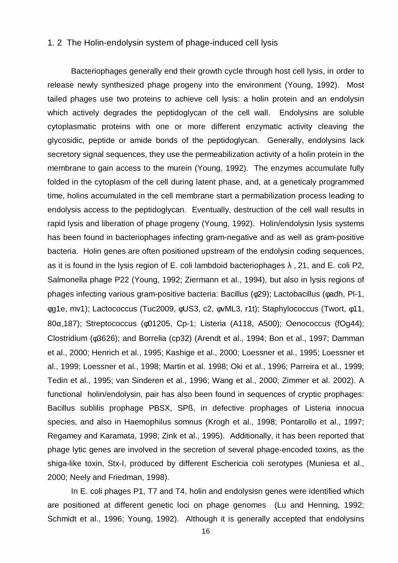
1. 2 The Holin-endolysin system of phage-induced cell lysis
Bacteriophages generally end their growth cycle through host cell lysis, in order to
release newly synthesized phage progeny into the environment (Young, 1992). Most
tailed phages use two proteins to achieve cell lysis: a holin protein and an endolysin
which actively degrades the peptidoglycan of the cell wall. Endolysins are soluble
cytoplasmatic proteins with one or more different enzymatic activity cleaving the
glycosidic, peptide or amide bonds of the peptidoglycan. Generally, endolysins lack
secretory signal sequences, they use the permeabilization activity of a holin protein in the
membrane to gain access to the murein (Young, 1992). The enzymes accumulate fully
folded in the cytoplasm of the cell during latent phase, and, at a geneticaly programmed
time, holins accumulated in the cell membrane start a permabilization process leading to
endolysis access to the peptidoglycan. Eventually, destruction of the cell wall results in
rapid lysis and liberation of phage progeny (Young, 1992). Holin/endolysin lysis systems
has been found in bacteriophages infecting gram-negative and as well as gram-positive
bacteria. Holin genes are often positioned upstream of the endolysin coding sequences,
as it is found in the lysis region of E. coli lambdoid bacteriophages λ , 21, and E. coli P2,
Salmonella phage P22 (Young, 1992; Ziermann et al., 1994), but also in lysis regions of
phages infecting various gram-positive bacteria: Bacillus (φ29); Lactobacillus (φadh, Pl-1,
φg1e, mv1); Lactococcus (Tuc2009, φUS3, c2, φvML3, r1t); Staphylococcus (Twort, φ11,
80α,187); Streptococcus (φ01205, Cp-1; Listeria (A118, A500); Oenococcus (fOg44);
Clostridium (φ3626); and Borrelia (cp32) (Arendt et al., 1994; Bon et al., 1997; Damman
et al., 2000; Henrich et al., 1995; Kashige et al., 2000; Loessner et al., 1995; Loessner et
al., 1999; Loessner et al., 1998; Martin et al. 1998; Oki et al., 1996; Parreira et al., 1999;
Tedin et al., 1995; van Sinderen et al., 1996; Wang et al., 2000; Zimmer et al. 2002). A
functional holin/endolysin, pair has also been found in sequences of cryptic prophages:
Bacillus sublilis prophage PBSX, SPß, in defective prophages of Listeria innocua
species, and also in Haemophilus somnus (Krogh et al., 1998; Pontarollo et al., 1997;
Regamey and Karamata, 1998; Zink et al., 1995). Additionally, it has been reported that
phage lytic genes are involved in the secretion of several phage-encoded toxins, as the
shiga-like toxin, Stx-I, produced by different Eschericia coli serotypes (Muniesa et al.,
2000; Neely and Friedman, 1998).
In E. coli phages P1, T7 and T4, holin and endolysisn genes were identified which
are positioned at different genetic loci on phage genomes (Lu and Henning, 1992;
Schmidt et al., 1996; Young, 1992). Although it is generally accepted that endolysins

17
lack secretory signal sequences, recently secretory lysins have been indentified. It has
been shown that Lys44 from fOg44 has an active N-terminal signal sequence, and can
employ the GSP (general secretory pathway) for passage through the membrane bilayer.
Ply21 endolysin from phage TP21, infecting Bacillus cereus, has also a potential N-
terminal signal peptide, but its function has not been further investigated (Loessner et al.,
1997). In contrast to fOg44 were a holin gene has been found adjacent to Lys44, no
holin genes could be found adjacent to endolysins in phages Bastille, TP21 and, 12826
of Bacillus cereus and adjacent to Ply511 from Listeria bacteriophage A511 (Loessner et
al., 1995; Loessner et al., 1997; Sao-Jose et al., 2000).
1. 3 Lysis region of bacteriophage λ
In bacteriophage λ, three lysis genes (S, R and Rz) were identified which are all
transcribed from the late pR` promotor. PR` is a constitutive promotor activated by the Q
antitermination factor, which is a product of the delayed early genes. Activation of the
promotor beginns about 8-10 min after induction of λ lysogenes. Hence, lysis proteins
are synthesized long before the acctual lysis event starts, 40 min after induction of λ
lysogens. λ lysis can be induced earlier by the addition of energy poissons, called
premature lysis, a phenomenon also described for T even phages (Campbell and Rolfe,
1975; Doermann, 1952; Young, 1992).
Q
tR` S R Rz/Rz1
PR`
44,5 46.5
Fig. 1. 3 Map of the λ lysis region (Young, 1992). S, lysis control gene (holin), R, murein
transglycosylase, Rz, Rz1, outer membrane proteins. Position of the late pR` promotor and tR`
transcriptional terminator is indicated. The Q antitermination factor is acting on this region to
start transcription. The arrow indicate the mRNA of the lysis genes.

18
R encodes for the endolysin, a muralytic transgycosylase, with no secretory signal
sequence. R reaches the periplasm through the action of the S protein, which forms
"holes" or pores, permeabilizing the membrane, a process which actually starts lysis.
The product of the Rz gene is only required for lysis in the presence of divalent cations.
Additionally, a protein named Rz1 is translated out of frame within the sequence of Rz;
Rz and Rz1 are outer membrane proteins and assumed to be auxiliary lysis factors
(Zhang and Young, 1999). Under standard laboratory conditions only S and R proteins
are required for cell lysis. The most frequently used model system for studying lysis is
the use of termosensitive λ lysogenes which allows the vegetative cycle to be initiated
synchronously in an entire culture, so the lysis phenotype can be measured very
precisely.
1. 4 Holins
S protein was the first membrane permeabilizing protein identified which led to a
definition of a new class of membrane protein, making "holes" into the membrane,
therefore designated as holins (Young, 1992). Holin proteins have two main functions in
cell lysis: (i) they form "holes" through an oligomerization process in the membrane that
enable the endolysin to reach the murein and, (ii) they have a timing function
"programmed" into the structure of the protein that regulates the process of cell lysis to
start at a defined timepoint. Because of these properties, holins are thought to be the
simplest "molecular clocks". Triggering of the clock ends the latent period, a period
when the phage replicates and new phage particles are assembled. All holin proteins
have some structural features in common: at least one transmembrane domain, short,
mostly charged, N-terminal sequence and, a highly hydrophilic, positively charged C-
terminal tail. Holins are grouped into two classes, according to the nummber of potential
transmembrane domains. Class I holins (90-125 aa) have the potential to form three
transmembrane domains. The prototype of this class is the well studied λ S holin. Class
II holins (57-185 aa) can form only two transmembrane domain, it is represented by the
S21 holin from lambdoid bacteriophage 21. There are also holins which do not fit into
these two classes, such as gpT from bacteriophage T4 having only one transmembrane
domain. More than 100 known and putative holin protein constitute the most structurally
diverse functional group of proteins, presently classified into 34 different families (Wang
et al., 2000). Most of them were identified only by primary and secondary structural
features of protein sequences. Holin function is nonspecific for endolysin activity, which

19
enables testing of novel holins in a λ genetic background, by complementation of S
mutants either from transactivation plasmids or directly, from holin genes inserted into
λ∆Sthf phage (Vukov et al., 2000; Wang et al., 2000).
The timing of cell lysis is an as equally important function as the pore forming
ability of a holin. The major determinant of timing is intrinsic to the structure of the
transmembrane domains, as was shown at least for λ S (Johnson-Boaz et al., 1994).
This timing is modulated by charged amino acids in the N- and C- terminal domains, and
by the expression of "inhibitors" or "antiholins" (Bläsi et al., 1989; Bläsi et al., 1990;
Ramanculov and Young, 2001c; Steiner and Bläsi, 1993).
Some of the holin genes from classes I and II have a dual start motif which
permits translation of two proteins with different N-termini and "opposite" functions
(Barenboim et al., 1999; Bläsi and Young, 1996; Nam et al., 1990; Tedin et al., 1995).
The dual-start motif is found among holins from bacteriophages infecting both gram-
positive and gram-negative bacteria, and which belong to class I or class II holins. In all
cases were this has been investigated, the longer product was found to be an inhibitor of
the shorter product, which is the main effector of lysis. Experimental evidence indicated
that S can form homo-oligomers but the exact nature of the hole is still not known (Wang
et al., 2000; Zagotta and Wilson, 1990). There is biochemical evidence for dimerization
between the effector S105 protein and the S107 inhibitor leading to heterodimer
formation (Gründling et al., 2000c). Apart from intragenic holin inhibitors found in S or
S21, separate genes are described coding for the holin and antiholin protein in P1, P2
and T4 bacteriophage (Ramanculov and Young, 2001c; Schmidt et al., 1996; Ziermann
et al., 1994). Recently, the product of the rI gene was described as the specific antiholin
in T4. It was shown that RI binds to T and forms heterodimers, as demonstrated for
S107 and S105 (Ramanculov and Young, 2001c). It is accepted that the dual start motif
represents fine-tuning regulation of holin function. Moreover, it is assumed that the
effector / inhibitor ratio is actively regulated under physiological conditions of the cell, but
experimental evidences for such regulation have still not been reported (Young et al.,
2000).
1. 4. 1 S holin
S was first defined as the control protein of the lysis process in λ infected cells.
Inactivation of S leads to continuing respiration and macromolecular synthesis past the
normal lysis timing, which results in virion and endolysin accumulation in the cell to very
20
high levels. The possiblity to complement this defect by the addition of CHCl3 revealed
that the acctual function of S is to permeabilize the cytoplasmic membrane (Young,
1992). Premature lysis through energy poissons in λ-infected cell is entirely dependent
on S. λ S null mutants could not be triggered prematurely, so the timing of lysis has
been correlated exclusively to the function of S protein (Campbell and Rolfe, 1975).
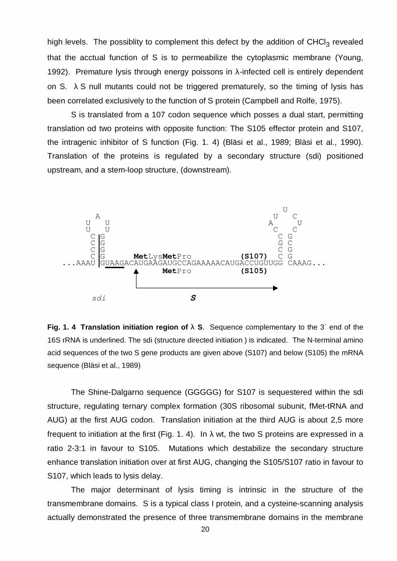
S is translated from a 107 codon sequence which posses a dual start, permitting
translation od two proteins with opposite function: The S105 effector protein and S107,
the intragenic inhibitor of S function (Fig. 1. 4) (Bläsi et al., 1989; Bläsi et al., 1990).
Translation of the proteins is regulated by a secondary structure (sdi) positioned
upstream, and a stem-loop structure, (downstream).
U A U C U U A U U U C C C G C G C G G C C G C G C G MetLysMetPro (S107) C G ...AAAU GUAAGACAUGAAGAUGCCAGAAAAACAUGACCUGUUGG CAAAG... MetPro (S105)
sdi S
Fig. 1. 4 Translation initiation region of λ S. Sequence complementary to the 3´ end of the
16S rRNA is underlined. The sdi (structure directed initiation ) is indicated. The N-terminal amino
acid sequences of the two S gene products are given above (S107) and below (S105) the mRNA
sequence (Bläsi et al., 1989)
The Shine-Dalgarno sequence (GGGGG) for S107 is sequestered within the sdi
structure, regulating ternary complex formation (30S ribosomal subunit, fMet-tRNA and
AUG) at the first AUG codon. Translation initiation at the third AUG is about 2,5 more
frequent to initiation at the first (Fig. 1. 4). In λ wt, the two S proteins are expressed in a
ratio 2-3:1 in favour to S105. Mutations which destabilize the secondary structure
enhance translation initiation over at first AUG, changing the S105/S107 ratio in favour to
S107, which leads to lysis delay.
The major determinant of lysis timing is intrinsic in the structure of the
transmembrane domains. S is a typical class I protein, and a cysteine-scanning analysis
actually demonstrated the presence of three transmembrane domains in the membrane

21
(Gründling et al., 2000b). An early lysis S variant was selected, with a A52G change in
the middle of the second transmembrane domain. The mutant had a dominant effect
over S wt, a finding that revealed that this position is crucial for intermolecular
interactions between membrane-spanning domains of S (Johnson-Boaz et al., 1994).
Charged amino acids in the N - and C - terminal tails influence the lysis timing set by the
intrinsic lysis clock (Bläsi et al., 1999; Steiner and Bläsi, 1993). On the molecular level,
charged amino acids in the N-terminal tail influence the topology of S. The holin needs
the N-terminus positioned in the periplasm for pore formation (Graschopf and Bläsi,
1999b). This Nout - Cin topology of S is inhibited by an energized membrane to a
different degree for S105 and S107 proteins (Fig. 1. 5). Passage of the S107 N-terminus
is inhibited by an additional positive charged residue, which retards electrophoretic
translocation of the N- terminus through the membrane (Graschopf and Bläsi, 1999).
The inhibitory function of S107 is based on its different N-terminus and the possibility to
form heterodimers with S105 (Gründling et al., 2000c) (Fig. 1. 5, Fig. 1. 6) However, as
soon as the membrane is depolarized, the N- terminus of S107 protein reaches the
periplasm, takes on the active Nout - Cin topology, and contributes to the pool of active
pore forming proteins, resulting in a very rapid onset of cell lysis.
The C-terminal domain of S is highly hydrophilic, with an overall positive net
charge. Mutation analysis in this region revealed that at least one basic residue must be
present to retain function (Bläsi et al., 1999). Excess positive charge in the C-terminus
led to lysis delay, compared to S wt. S protein which, features a deletion of its last 15 aa
can still oligomerize in the cell membrane; a finding which indicated that this region is not
involved in intermolecular interaction (Rietsch et al., 1997). It is assumed that the C-
terminus is a regulatory domain with little structural features.
The simplest currently proposed model for holin function, based on extensive
genetical and biochemical analyses of λ S, is the critical-concentration model. It is
assumed that a two-dimensional precipitation of holin proteins occurs in the membrane
when their amount exceeds a critical level in the fluid bilayer. It has been shown for S
that the precipitation event is inhibited by the energized state of the membrane
(Campbell and Rolfe, 1975). The permeabilization process supposedly starts at a locus
in the membrane were the mechanical integrity is locally disrupted. After the
electrochemical potential collapses, all available holin would suddenly precipitate,
resulting in massive membrane disruption. The model is build upon experiments that
showed that the energy state of the cell is completely intact until just before the actual
pore forming event takes place (Gründling et al., 2001).

22
S105 Periplasm S107 S107/S105
+ + + N + + + + + + + + + + + + + + + - - N + + - - -
- - - - + + + +
- - - - - - - - - - - - - - - - - - - + + + +- -
C N C C
Cytoplasm Membrane depolarization
Fig. 1. 5 Membrane topologies of S variants. S107 protein assumes the Nin-Cin conformation
in the cytoplasmic membrane when the membrane is energized. Upon depolarization, both
proteins assume the same topology (Graschopf and Bläsi, 1999).
S1 S2 S2a Sn hole
1 1a 2 3
Fig. 1. 6 Model for S hole formation. At least three steps, are required for hole formation: (1)
dimerization, (2) oligomerization and, (3) conformation or triggering. S monomers are depicted as
open (S105) or filled (S107) bars. According to the model, S105/S107 heterodimers (1) can not
lead to hole formation. The model is presented on physiological conditions when the cell
membrane is energized. Upon depolarization both monomers contribute to pore formation
(Gründling et al., 2000a; Gründling et al., 2000c)
Cytoplasmicmembrane

23
2. Material and Methods
2. 1 Bacterial strains, phages, plasmids, and culture conditions
All bacterial strains used in this work are listed in Table 2. 1. Plasmids and phages
constructed are listed in Tables 2. 2 and Table 2. 3. Escherichia coli strains were grown
in Luria Bertani, LB (10 g Tryptone, 5 g Yeast extract, 5 g NaCl for 1 l) medium at 37°C.
For propagation of the λ phages, media were supplemented with 0,2% maltose and 10
mM MgSO4. XL1-Blue and DH5∆MCR were used for plasmid propagation. HB101 was
used for plating of λgt11. LE392 was used as a general strain for propagation and
lysogenization with λ∆Sthf. For efficient lysogenization in recombination experiments,
C600hfl was used. Plasmid-bearing cells were selected on LB medium supplemented
with 80 or 100 µg ml-1 ampicilin, 7 µg ml-1 chloramphenicol, 12 µg ml-1 tetracyclin,
depending on the plasmid. E. coli lysogenized with λ∆Sthf was selected on LB
supplemented with 30 µg ml-1 amplicilin or 30 µg ml-1 kanamycin, were appropriate. For
the destrution of the energized state of the cell membrane CCCP (carbonyl cyanide m-
chlorophenyl-hydrazone) was added to induced cultures to a final concentration of 50µM.Strain CC118 (phoA-), was used for detection of alkaline phosphatase activity on
LB media supplemented with the chromogenic substrate XP (5-bromo-4chloro-3-indolyl
phosphate, Sigma) at the final concentration of 50 µg ml-1. Beta- galatosidase activity
was selected on the same media supplemented with X-gal (5-bromo-4-chloro-3-indolyl-ß-
D-galactosidase, Sigma) the chromogenic substrate for beta-galactosidase at the final
concentration of 40 µgml-1.
Listeria monocytogenes WSLC 1001 and Listeria monocytogenes WSLC 1042
were grown in TEB (20 g Tryptose, 1 g glucose, 5 g NaCl, 0.005 g Thiamine for 1 l, ph
7.3-7.4) medium at 30°C. The strains were used for propagation of A118 and A500
phages, respectively.
2. 2 Phage plating
Concentration of phages in suspentions were determined by phage plating,
determing the number of plaques formed on plates with host bacteria. In order to
perform plaque assays serial dilutions of bateriophage was performed in SM buffer (5.8 g
NaCl, 2 g MgSO4 x 7 H2O, 50 ml 1 M Tris-HCl (pH 7.5) in 1L). Aliquots (0.1 ml) of each

24
dilution were mixed with plating bacteria (0.1 ml) in 3 ml molten (0.6%) top agar. The
entire mixture was poured onto a agar plate surface. Plates were incubated overnight,
(42°C) for λ∆Sthf and (30°C) for Listeria bacteriophages.
Table 2. 1 Bacterial strains
Strain GenotypeSource or
Reference
Listeria monocytogenes
WSLC 1042
Wild type, serovar 4b ATCC 23074
Listeria monocytogenes
WSLC 1001
Wild type, serovar 1/2 c ATCC 19112
E. coli DH5∆MCR F- mcrA ∆(mrr-hsdRMS-mcrBC) φ80dlacZ∆M15
∆(lacZYA-argF) U169 deoR recA1 endA1
supE44 thi-1 gyrA96 relA1
In vitrogen
E. coli C600hfl supE44, hsdR, thi-1, thr-1, leuB6, lacY1, tonA21,
hflA150[chr :: Tn10(tetr)]
Laboratory
stock
E. coli XL1-Blue supE44 hsdR17 recA1 endA1 gyrA46 thi relA1 lac
F´[proAB+ lacIq lacZ∆M15 Tn10(tet)
Stratagene
E. coli LE392 RF- hsdR574(rk-, mk+)supE44 supF58 lacY1 galK2
galT22 metB1 trpR55
(phage host; permissive for λgt11)
Promega
E. coli HB101 F- hsdS20 supE44 recA13 ara-14 proA2 lacY1 galK2
rpsL20 xyl-5 mtl-1 (phage host; non-permissive for
λgt11)
Promega
E. coli Y1090 F- ∆(lacU169), proA+, ∆(lon), araD139,strA, supF,
[trpC22:Tn10(tetr)], (pMC9), hsdR(rk-, mk+)
Promega
E. coli CC118 ara D 139 ∆(ara-leu) 7697 ∆(lac) X74 ∆(phoA) 20 galE
galK thi rpsE rpoB argE(Am) recA1
(Manoil and
Beckwith,
1985)
25
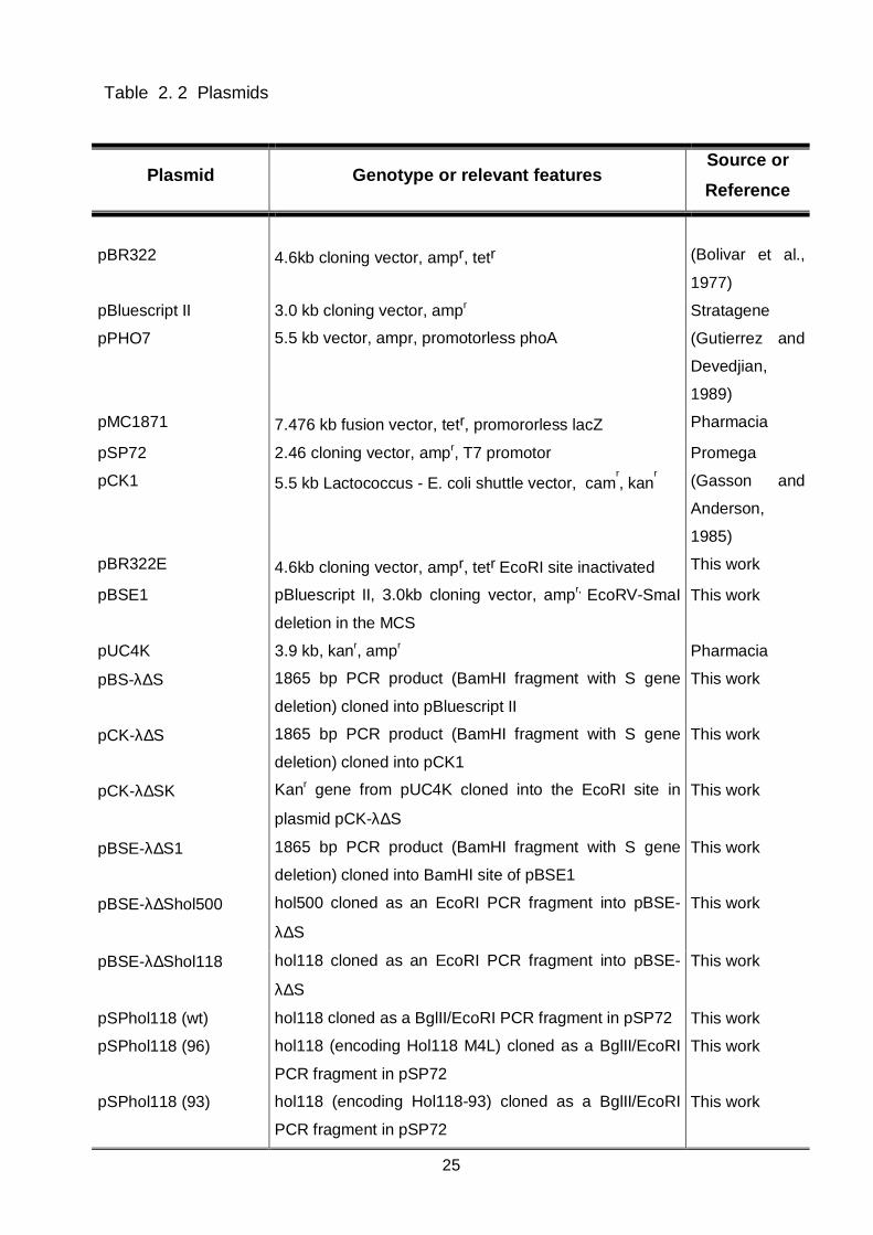
Table 2. 2 Plasmids
Plasmid Genotype or relevant featuresSource or
Reference
pBR322 4.6kb cloning vector, ampr, tetr (Bolivar et al.,
1977)
pBluescript II 3.0 kb cloning vector, ampr Stratagene
pPHO7 5.5 kb vector, ampr, promotorless phoA (Gutierrez and
Devedjian,
1989)
pMC1871 7.476 kb fusion vector, tetr, promororless lacZ Pharmacia
pSP72 2.46 cloning vector, ampr, T7 promotor Promega
pCK1 5.5 kb Lactococcus - E. coli shuttle vector, camr, kan
r(Gasson and
Anderson,
1985)
pBR322E 4.6kb cloning vector, ampr, tetr EcoRI site inactivated This work
pBSE1 pBluescript II, 3.0kb cloning vector, ampr, EcoRV-SmaI
deletion in the MCS
This work
pUC4K 3.9 kb, kanr, ampr Pharmacia
pBS-λ∆S 1865 bp PCR product (BamHI fragment with S gene
deletion) cloned into pBluescript II
This work
pCK-λ∆S 1865 bp PCR product (BamHI fragment with S gene
deletion) cloned into pCK1
This work
pCK-λ∆SK Kanr gene from pUC4K cloned into the EcoRI site in
plasmid pCK-λ∆S
This work
pBSE-λ∆S1 1865 bp PCR product (BamHI fragment with S gene
deletion) cloned into BamHI site of pBSE1
This work
pBSE-λ∆Shol500 hol500 cloned as an EcoRI PCR fragment into pBSE-
λ∆S
This work
pBSE-λ∆Shol118 hol118 cloned as an EcoRI PCR fragment into pBSE-
λ∆S
This work
pSPhol118 (wt) hol118 cloned as a BglII/EcoRI PCR fragment in pSP72 This work
pSPhol118 (96) hol118 (encoding Hol118 M4L) cloned as a BglII/EcoRI
PCR fragment in pSP72
This work
pSPhol118 (93) hol118 (encoding Hol118-93) cloned as a BglII/EcoRI
PCR fragment in pSP72
This work
26
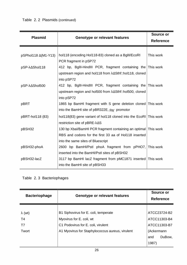
Table 2. 2 Plasmids (continued)
Plasmid Genotype or relevant featuresSource or
Reference
pSPhol118 ∆(M1-Y13) hol118 (encoding Hol118-83) cloned as a BglII/EcoRI
PCR fragment in pSP72
This work
pSP-λ∆Shol118 412 bp, BglII-HindIII PCR, fragment containing the
upstream region and hol118 from λ∆Sthf::hol118, cloned
into pSP72
This work
pSP-λ∆Shol500 412 bp, BglII-HindIII PCR, fragment containing the
upstream region and hol500 from λ∆Sthf::hol500, cloned
into pSP72
This work
pBRT 1865 bp BamHI fragment with S gene deletion cloned
into the BamHI site of pBR322E, pR` promotor
This work
pBRT-hol118 (83) hol118(83) gene variant of hol118 cloned into the EcoRI
restriction site of pBRE-λ∆S
This work
pBSH32 130 bp XbaI/BamHI PCR fragment containing an optimal
RBS and codons for the first 33 aa of Hol118 inserted
into the same sites of Bluescript
This work
pBSH32-phoA 2600 bp BamHI/PstI phoA fragment from pPHO7,
inserted into the BamHI/PstI sites of pBSH32
This work
pBSH32-lacZ 3117 bp BamHI lacZ fragment from pMC1871 inserted
into the BamHI site of pBSH33
This work
Table 2. 3 Bacteriophages
Bacteriophage Genotype or relevant featuresSource or
Reference
λ (wt) B1 Siphovirus for E. coli, temperate ATCC23724-B2
T4 Myovirus for E. coli, wt ATCC11303-B4
T7 C1 Podovirus for E. coli, virulent ATCC11303-B7
Twort A1 Myovirus for Staphylococcus aureus, virulent (Ackermann
and DuBow,
1987)
27
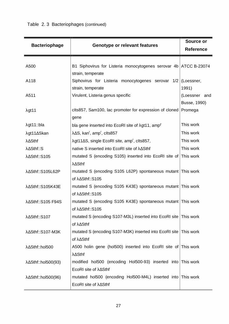
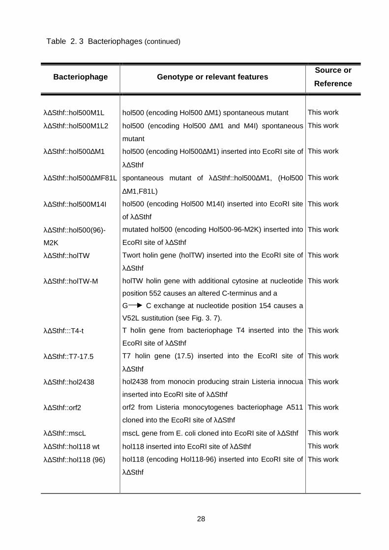
Table 2. 3 Bacteriophages (continued)
Bacteriophage Genotype or relevant featuresSource or
Reference
A500 B1 Siphovirus for Listeria monocytogenes serovar 4b
strain, temperate
ATCC B-23074
A118 Siphovirus for Listeria monocytogenes serovar 1/2
strain, temperate
(Loessner,
1991)
A511 Virulent, Listeria genus specific (Loessner and
Busse, 1990)
λgt11 cIts857, Sam100, lac promoter for expression of cloned
gene
Promega
λgt11::bla bla gene inserted into EcoRI site of λgt11, ampr This work
λgt11∆Skan λ∆S, kanr, ampr, cIts857 This work
λ∆Sthf λgt11∆S, single EcoRI site, ampr, cIts857, This work
λ∆Sthf::S native S inserted into EcoRI site of λ∆Sthf This work
λ∆Sthf::S105 mutated S (encoding S105) inserted into EcoRI site of
λ∆Sthf
This work
λ∆Sthf::S105L62P mutated S (encoding S105 L62P) spontaneous mutant
of λ∆Sthf::S105
This work
λ∆Sthf::S105K43E mutated S (encoding S105 K43E) spontaneous mutant
of λ∆Sthf::S105
This work
λ∆Sthf::S105 F94S mutated S (encoding S105 K43E) spontaneous mutant
of λ∆Sthf::S105
This work
λ∆Sthf::S107 mutated S (encoding S107-M3L) inserted into EcoRI site
of λ∆Sthf
This work
λ∆Sthf::S107-M3K mutated S (encoding S107-M3K) inserted into EcoRI site
of λ∆Sthf
This work
λ∆Sthf::hol500 A500 holin gene (hol500) inserted into EcoRI site of
λ∆Sthf
This work
λ∆Sthf::hol500(93) modified hol500 (encoding Hol500-93) inserted into
EcoRI site of λ∆Sthf
This work
λ∆Sthf::hol500(96) mutated hol500 (encoding Hol500-M4L) inserted into
EcoRI site of λ∆Sthf
This work
28
Table 2. 3 Bacteriophages (continued)
Bacteriophage Genotype or relevant featuresSource or
Reference
λ∆Sthf::hol500M1L hol500 (encoding Hol500 ∆M1) spontaneous mutant This work
λ∆Sthf::hol500M1L2 hol500 (encoding Hol500 ∆M1 and M4I) spontaneous
mutant
This work
λ∆Sthf::hol500∆M1 hol500 (encoding Hol500∆M1) inserted into EcoRI site of
λ∆Sthf
This work
λ∆Sthf::hol500∆MF81L spontaneous mutant of λ∆Sthf::hol500∆M1, (Hol500
∆M1,F81L)
This work
λ∆Sthf::hol500M14I hol500 (encoding Hol500 M14I) inserted into EcoRI site
of λ∆Sthf
This work
λ∆Sthf::hol500(96)-
M2K
mutated hol500 (encoding Hol500-96-M2K) inserted into
EcoRI site of λ∆Sthf
This work
λ∆Sthf::holTW Twort holin gene (holTW) inserted into the EcoRI site of
λ∆Sthf
This work
λ∆Sthf::holTW-M holTW holin gene with additional cytosine at nucleotide
position 552 causes an altered C-terminus and a
G C exchange at nucleotide position 154 causes a
V52L sustitution (see Fig. 3. 7).
This work
λ∆Sthf:::T4-t T holin gene from bacteriophage T4 inserted into the
EcoRI site of λ∆Sthf
This work
λ∆Sthf::T7-17.5 T7 holin gene (17.5) inserted into the EcoRI site of
λ∆Sthf
This work
λ∆Sthf::hol2438 hol2438 from monocin producing strain Listeria innocua
inserted into EcoRI site of λ∆Sthf
This work
λ∆Sthf::orf2 orf2 from Listeria monocytogenes bacteriophage A511
cloned into the EcoRI site of λ∆Sthf
This work
λ∆Sthf::mscL mscL gene from E. coli cloned into EcoRI site of λ∆Sthf This work
λ∆Sthf::hol118 wt hol118 inserted into EcoRI site of λ∆Sthf This work
λ∆Sthf::hol118 (96) hol118 (encoding Hol118-96) inserted into EcoRI site of
λ∆Sthf
This work

29
Table 2. 3 Bacteriophages (continued)
Bacteriophage Genotype or relevant featuresSource or
Reference
λ∆Sthf::hol118 (93) hol118 (encoding Hol118-93) inserted into EcoRI site of
λ∆Sthf
This work
λ∆Sthf::hol118M14I hol118 (encoding Hol118 M14I) inserted into EcoRI site
of λ∆Sthf
This work
λ∆Sthf::hol118M14L hol118 (encoding Hol118 M14L) inserted into EcoRI site
of λ∆Sthf
This work
λ∆Sthf::hol118M14V spontaneous mutant phage, A G transition causing
M14V substitution in Hol118
This work
λ∆Sthf::hol118M1L4 hol118 (encoding Hol118 M1L, F84S) holin variant,
spontaneous mutant
This work
λimm434cI Swt cIts, Swt (Bläsi et al.,
1990)
λimm434cI::hol118 recombinant λimm434cIts phage, S recombinated for
hol118 from pBSE-λ∆Shol118
This work
λimm434cI::hol500 recombinant λimm434cIts phage, S recombinated for
hol500 from pBSE-λ∆Shol500
This work
30
2. 3 Standard DNA manipulations
2. 3. 1 Isolation of λ DNA
λ DNA was isolated from λ∆Sthf LE392 lysogens. 500 ml E. coli liquid culture was
grown to an OD600 about 0.5 (32°C). Phages were induced (30 min, 42°C), and the
culture further incubated (60 min, 37°C). Cells were disrupted, treating the culture with
20 ml of CHCl3. The obtained lysate was either filtrated through a 0.2 µm pore filter, or
treated with DNAse and RNAse (1µg/ml). After centrifugation (2 X 15 min, 9600 x g at
RT, JA-14, Beckman), phages obtained in the supernatant were precipitated with
polyethylene glycol (PEG 8000, 15%) and NaCl (1M), over/night (4°C). Precipitated
phages were recoverd by centrifugation (20 min, 9600 x g RT, JA-14, Beckman).
Bacteriophage pellets were resuspended in 5 ml SM buffer (per 1 l: 5.8 g NaCl, 2 g
MgSO4 x 7 H2O and 50 ml 1 M Tris HCl (pH 7.5)). Phages were purified by using
stopped CsCl gradient centrifugation (Sambrock et al., 1989). A discontinuous CsCl
gradient was formed (from the bottom to the top of the centrifugation tube: 2ml CsCl ρ=
2.0; 2ml CsCl ρ= 1.50; 2ml CsCl ρ=1.45; 2ml CsCl ρ=1.40; 2ml CsCl ρ=1.30; 2.5ml CsCl
ρ=1.20), and the phage particle solution layed on top. After centrifugation (15 hours,
90.000 x gmax, 4°C, SW-28.1, Beckman), a bluish band of bacteriophage particles was
visible. The particles were collected by puncturing the sides of centrifugation tubes with
a needle, and stored on 4°C. The CsCl suspension of phage particle was diluted with
SM buffer and centrifuged (90 min, 150.000 x g at 15°C, SW-60Ti, Beckman). Phage
pellet was resuspended in EPS buffer (20 mM EDTA,
50 µgml-1 Proteinase K and 0.5% SDS), and the suspension incubated (60 min, 50°C)
for digestion of phage proteins. DNA was further extracted with Phenol-Chlorophorm,
repeating the procedure several times. After extraction, the DNA was precipitated with
1/10 Vol 3M NaAc and 2 Vol Ethanol.
Alternatively, DNA was isolated from λ∆Sthf phages using the Qiagen Lambda
Mini Kit (Qiagen). The clear lysate, obtained after liberation of phages from cells through
CHCl3 and centrifugation, was treated with DNAse and RNAse. 10 ml of this lysate was
taken as the starting volume to isolate DNA following the steps of instructions given by
the manufacturer. DNA was dissolved in TE buffer (10 mM Tris HCl pH 8.0, 1 mM EDTA
pH 8.0).

31
2. 3. 2 Plasmid isolation, PCR, agarose electrophoresis, and ligation reactions
Plasmid DNA was isolated from plasmid-carrying E. coli cultures, after overnight
incubation in liquid culture. Three to five ml aliquotes were taken for isolation of plasmid
DNA and further proceeded according to instructions for Plasmid Mini-Prep kit (Qiagen)
or GeneEluteTM Plasmid Mini-prep Kit (Sigma).
All PCR reactions were performed using the Taq DNA polymerase (Qiagen), and
the corresponding reaction buffer with the addition of a dNTP mix containing eqimolar
concentration of all four nucleotides (2 mM). The following PCR conditions were used:
(1X [120s, 94°C], (30X [90s, 52°C (or different anealing temperature depending on the
primer set), 50s, 72°C, 60s, 94°C], 1X [120s, 52°C], 1X [5 min, 72°C]). All PCR products
used for further manipulations were purified using the QIAquick PCR Purification Kit
(Qiagen).
DNA molecules isolated from λ phages, plasmids, PCR products, and DNA
fragments generated after digestions with restriction enzymes were separated by
agarose gel electropheresis. Agarose gels varying in concentration between 0.7%-1.3%
(SeaKem LE, FMC, BioProducts) were run in 1 X TAE buffer (0.04 M Tris-acetat, 0.001
M EDTA). After electrophoresis, gels were stained with ethidium-bromide EtBr (0.5 µg
ml-1), and visualized by UV-transilumination using the ImageMaster® VDS (Pharmacia).
In order to extract DNA fragments from agarose gels, the QIAquick Gel Extraction Kit
(Quiagen) was used
Ligation reactions of different DNA restriction fragments with plasmid or phage
were performed using T4 DNA ligase (Boeringer), and the corresponding T4 ligation
buffer. Ligation reactions were incubated over night (8°C-16°C), and desalted prior to
transformation into E. coli cells by electroporation.
2. 3. 3 DNA sequencing
All holin genes cloned into λ∆Sthf phages were sequenced, in a region 105 bp
upstream and 40 bp downstream of the insert. The fragment to be sequenced was first
amplified using For2 (5'-GCCCGTGCATATCGGTCACG-3') and Rev2 (5'-
ACCACGCCAGCATATCGAGG-3') primers. The obtained PCR fragments were
sequenced with the identical IRD-800 labeled primers. Reactions were performed using
the SequiTherm EXELTM II DNA Sequencing Kit-LC (for 66-cm gels), sequencing in the

32
presence of dideoxy termination nucleotides GTP, ATP, TTP and CTP. The mixes were
resolved on a 7.5% PAA gel, in a LI-COR automated DNA sequencer.
2. 3. 4 Preparation of electrocompetent E. coli cells
500 ml LB medium was inoculated with 1ml over night growing cultures of E. coli
cells. The culture was grown to an OD between 0.5-0.6. Cells were collected by
centrifugation (15 min, 9600 x g, 4°C, JA-14). The obtained cell pellet was washed twice
with pure water, with subsequent centrifugation under the conditions mentioned above.
Finally, cells were washed in 10% glycerol and, after centrifugation and resuspension in
1/100 Vol., stored in 40 µl aliquotes at -70°C.
2. 3. 5 Electrotransformation
Transformation of E. coli cells was carryied out by electroporation using
electrocompetent cell (Dower et al., 1988). A BioRad Gene Pulser was used, and 2-mm
electroporation cuvettes (EquiBio) under the following conditions: 200 Ω Resistance, 25
µFD capacitance, 12.000 V/cm fiels strenght. After regeneration in LB medium,
transformants were selected on medium supplemented with the appropriate antibiotic,
and further tested by PCR or plasmid isolation.
2. 4 Construction of λ∆Sthf
λ∆Sthf phage was constructed by recombination between λgt11 in the lysis region
of the phage and a fragment carring the ∆S deletion on plasmid. First the ampicillin
resistance gene (bla) was inserted into λgt11. The ampicillin resistance gene (bla) was
amplified using primer (MunI sites are underlined, start and stop codons are in boldface):
bla-5' (5'-ATATCAATTGTAAAGGAGATTTATTATGAGTATTCAACATTTCCGTGTCG
CCCTT-3') and bla-3' (5'-ATATCAATTGTTACCAATGCTTAATCAGT GAGGCACCTA-
3'), using pBluescript as a template. The 860 bp product was cloned into the EcoRI site
within lacZ in λgt11, and thereby destroyed this recognition sequence. The ligation
mixture was packaged into λ particles according to the instruction of the manufacturer
(Packagene system, Stratagene), and aliquots containing the recombinant phages plated

33
on Y1090. Phage clones containing bla gave colorless plaques in a softagar overlayer
supplemented with ampicillin, X-Gal, and IPTG.
Deletion of the S gene and introduction of a new EcoRI site upstream of R in place
of S in the intermediate construct λgt11::bla was achieved by homologous recombination
of this region with a corresponding fragment on a plasmid. For this purpose, two λ DNA
fragments were amplified using λgt11 DNA as a template: (1) a 1053 bp fragment
(nuleotide coordinates 44129 to 45182 on wt λ DNA) using primer pair A (5'-
ATATGGATCCGTGGTGTGGCAAAGCTTGAAG-3') and B (5'-ATATGAATTC
TTCCCCCCCAATAAGGGGATTTGCTCTATTTAATTAG-3´) and (2), a 812 bp fragment
(coordinates 45482-46294) using primer pair C (5'-ATATGAATTC
AGGAGTAGAAGATGGTAGAAATCAA-3´) and D (5'-ATATGGATCCCCGGAGGC
GGTG GTGGCTTCACGCA-3'). BamHI and EcoRI sites are underlined, changed
nucleotides are shown in italics, and the start codon is shown boldfaced. After EcoRI
digestion, both fragments were ligated to yield a 1865 bp fragment with BamHI sites on
each end and an EcoRI site in the middle. For easier handling, this construct was initially
cloned into BamHI-digested pBluescript, to yield pBS-λ∆S. Then, the entire fragment
was excised and subcloned into the BamHI site of pCK1 (Gasson and Anderson, 1985),
a vector that replicates through a rolling-circle (sigma) mechanism, and is well suited for
homologous recombination. pCK-λ∆S was electroporated into E. coli LE392, and
recombinants selected by chloramphenicol. In order to introduce an additional selective
marker for recombination into gt11::bla, the kanamycin resistance gene kan was
obtained (on an EcoRI fragment) from the pUC4K vector, and ligated into the unique
EcoRI site replacing S within the λ DNA fragment. Homologous recombination was
performed as follows: LE392 (pCK-λ∆SK) cells were infected with λgt11::bla in a volume
of 10 ml and at a multiplicity of infection (m.o.i.) of approximately 10, and grown for 1 h at
37°C. In the absence of S, R-mediated lysis was induced by addition of a few drops of
CHCl3, and, after 15 min, debris removed by centrifugation at 6000 x g for 10 min. The
resulting lysate containing the recombinant phages was used to lysogenize E. coli
C600hfl cells, and, in oder to enrich for double-crossover mutants, the infected culture
immediately plated on agar containing both ampicillin and chloramphenicol. Following
incubation, ampr/camr colonies were picked and pooled, grown at 30°C, thermally
induced (42°C, 20 min), and aerated at 37°C for 1 h. Phages from the cleared lysate
were again used to lysogenize C600hfl cells, followed by plating on medium containing

34
kanamycin and ampicillin. Approximately 100 clones (i.e., colonies) were then tested for
chloramphenicol sensitivity, to exclude plasmid co-integrates resulting from single-
crossover events. Using PCR, kanr/cams clones were checked for presence of kan in
the EcoRI site. Then, the lysogenic clones were tested for lytic competence after
temperature shift, and a single clone with a non-lytic phenotype was selected. Phage
λgt11∆Skan particles were isolated from the cells by chloroform-induced lysis as
described above. Following purification of the virus particles by banding in CsCl, their
DNA was extracted (Sambrook et al., 1989), and digested with EcoRI to remove the kan
fragment. The generated arms (approximate sizes 40.7 and 3.0 kbp) were separated by
agarose gel electrophoresis, recovered from the gel, and religated. After packaging,
aliquots containing the recombinant viruses were again used to lysogenize C600hfl cells,
and plated on LB plates containing ampicillin. The correct sequence of the altered holin
gene region in λ∆Sthf was confirmed by nucleotide sequencing using an automated DNA
sequencer (Li-Cor model 4200) and the primers For2 (5'-
GCCCGTGCATATCGGTCACG-3') and Rev2 (5'-ACCACGCCAGCATATCGAGG-3').
Finally, high-titre phage stocks were prepared as decribed above, and λ∆Sthf DNA was
extracted and purified according to standard methods (Sambrook et al., 1989).
2. 5 Growth and lysis kinetics of individual λ∆Sthf::hol lysogens
In order the test holins inserted into λ∆Sthf, LE392 cells were lysogenized with
different λ∆Sthf::hol phages. Phages were mixed with cells grown to an OD600 ~ 0.2
and, after incubation (60 min, RT), the cell/phage mixture was poured on LB plates
supplemented with 30 µg ml-1 ampicilin, selecting for colonies of λ∆Sthf lysogens.
The permeabilization feature of each inserted holin or membrane protein in λ∆Sthf
was tested in liquid culture, measuring the lysis kinetics, after inducing the phages to lytic
growth cycle. Lysogens carrying λ∆Sthf::hol were grown to OD600 0.150- 0.250 at
preinduction temperature (32°C), induced (20 min, 42°C) and further grown (37°C) for
the rest of the experiment. The OD600 was measured in intervals of 10 min or less to
follow cell lysis.

35
2. 6. Cloning heterologous holins into λ∆Sthf
λ∆Sthf DNA was digested with EcoRI, and two fragments (arms) treated with
shrimp alkaline phosphatase (United States Biochemical). DNA fragments encoding
various native and mutated holin genes were amplified from intact phage DNA by PCR,
using the primers listed in Table 2. 4. The products were digested with EcoRI, and
directly ligated into the λSthf arms (approximately 0.2 µg). After phage packaging,
(Packagene system, Stratagene), phage particles were plated on LE392 cells. Individual
plaques from the cell lawn were picked and phages eluted. Aliquots containing
recombinant phages were then used to lysogenize LE392 cells. Lysogenic clones could
be directly selected by their resistance to ampicillin. Presence and identity of individual
holin genes was checked by PCR amplification and nucleotide sequencing.
Table 2. 4 Primer used for amplification of holins, holin variants, and membrane
proteins tested in λ∆Sthf
Primer Sequence (5´ 3´)a
S-5´ ATCAGAATTCATGAAGATGCCAGAAAAACATGAC
S105-5´ ATCAGAATTCATGCCAGAAAAACATGACCTGTTG
S107-5´ ATCAGAATTCATGAAGCTGCCAGAAAAACATGAC
S107-M3K-5´ ATCAGAATTCATGAAGAAGCCAGAAAAACATG
S-3´ ATCAGAATTCTTATTGATTTCTACCATCTTCTACTCC
Hol500-5´ ATCAGAATTCATGATGAAAATGGAGTTTGGAAAAGAG
Hol500M1L ATCAGAATTCCTGATGAAAATGGAGTTTGGAAAAGAG
Hol500∆M1 ATCAGAATTCATGAAAATGGAGTTTGGAAAAGAG
Hol500-96-5´ ATACGAATTCATGATGAAACTGGAGTTTGGAAAAGAG
Hol500-93-5´ ATCAGAATTCATGGAGTTTGGAAAAGAGTTACTAGTT
Hol500-M2K-5´ ATCAGAATTCATGAAGAAACTGGAGTTTGGAAAAGAG
Hol500-3´ ATCAGAATTCTTATTTATCATCCTTTCCATATTTTTTAGC
HolTw-5´ ATCAGAATTCATGGATAAAAAAGATAAAACACCTACA
HolTw-3´ ATCAGAATTCTTAATGTAGGACTCTGCTTGT
T7 gp17.5-5´ ATCAGAATTCATGCTATCATTAGACTTTAAC
T7 gp17.5-3´ ATCAGAATTCTCACTCCTTATTGGCTTT
T4R ATCAGAATTCTTATTTAGCCCTTCCTAATATTCTG
T4F ATCAGAATTCATGGCAGCACCTAGAATA

36
Table 2. 4 Primer used for amplification of holins, holin variants, and membrane
proteins tested in λ∆Sthf (continued)
Primer Sequence (5´ 3´)a
Li2438R ATCAGAATTCTTAATCATCCTTTCCATATTTTTTAGC
Hol118F ATCAGAATTCATGATAGAAATGGAGTTTGGAAAA
Hol118(96)-5´ ATCAGAATTCATGATAGAACTGGAGTTTGGAAAA
Hol118(93)-5´ ATCAGAATTCATGGAGTTTGGAAAA
Hol118H-3´ ATCAAAGCTTTTATTTATCATCCTCTC
Hol118∆M14-5´ ATCAGAATTCATGACATTTTTAGTAGTTGTAACACCTGTG
Hol118M14I-5´ ATACGAATTCATGATAGAAATGGAGTTTGGAAAAGAGTTACTAGTTT
ACATTACATTTTTAGTA
Hol118M14L-5´ ATACGAATTCATGATAGAAATGGAGTTTGGAAAAGAGTTACTAGTTT
ACCTGACATTTTTAGTA
Hol500M14I-5´ ATACGAATTCATGATGAAAATGGAGTTTGGAAAAGAGTTACTAGTTT
ATATTACATT
orf2F ATACGAATTCATGCACGATAATGAATTTGAA
orf2R ATACGAATTCTTATTTTATTACACTATTTAC
mscLF ATATGAATTCATGAGCATTATTAAAGAATTTCGCGAA
mscLR ATATGAATTCTTAAGAGCGGTTATTCTGCTCTTTCAG
a Start codons are indicated in bold letters and introduced mutations are underlined.
2. 7 Membrane protein preparation
Membrane protein samples were isolated by a modification of a procedure
described earlier (Chang et al., 1995). E. coli LE392 lysogenized with λ∆Sthf::hol118 or
λ∆Sthf::hol500, and a control strain lysogenized with λ∆Sthf, were grown at 32°C to an
OD600 of 0.2, induced for 20 min (42°C), and further grown at 37°C. The sample
aliquots (25 ml) were taken at various times after induction, as indicated in the legends of
the figures. Samples were centrifuged, (10.300 x g, 4°C) and cells disrupted in 1 ml FP
buffer (0.1 M Sodium phosphate pH 7.0, 0.1 M KCL, 5 mM EDTA, 1 mM DTT, 1 mM
PMSF) by sonication with 3 pulses, 30 s each, 25% power setting (Sonoplus; Bandelin).
Membrane fractions were collected by ultracentrifugation (100.000 x g, 60 min, 18°C).
Membrane pellets were solubilized in 1/100 Vol ME buffer (20 mM Tris-HCl, 1% Triton-X,
10% glycerol, 0.5 M NaCl, 35 mM MgCl2), overnight at room temperature, with agitation.

37
Detergent-insoluble material was removed by centrifugation (50.000 x g, 20°C, 60 min,)
to obtain a soluble preparation of inner membrane proteins. Protein concentration was
determined using a modified Bradford assay (Nanoquant; Roth).
Infection of log-phase Listeria monocytogenes strain 1001 (OD 600, 0.150) with phage
A118 was done at a m. o. i. of 10, in a total volume of 70 ml. Phages were allowed to
adsorb to cells for 15 min at room temperature. Infected Listeria cells were incubated at
30°C, without shaking. A sample (20 ml) was taken 90 min after incubation for isolation
of membrane protein fraction, mixed with 5 ml 4X FP buffer (1X FP buffer: 0,1M Sodium
phosphate pH 7.0, 0,1 M KCL, 5 mM EDTA, 1 mM DTT, 1 mM PMSF), and 300 µl
recombinant Ply118 endolysin (Loessner et al., 1996). Cells were incubated with the
enzyme 30 min at room temperature and centrifuged (12.000 x g, 5 min). The
supernatant was further processed as described above for isolation of membrane
proteins from E. coli.
2. 8 SDS-PAGE electrophoresis and Immunoblotting
In order to separate and detect holin proteins in the isolated inner membrane
fractions, SDS-PAGE (sodium dodecyl sulfate-polyacrylamide gel electrophoresis) and
immunoblotting was performed. For SDS-PAGE, membrane fractions were diluted 1:2
with sample buffer (62.5 mM Tris-Hcl, pH6,8, 2% SDS, 25% glycerol, 0.01%
Bromophenole Blue, 350 mM DTT), and boiled at 100°C for 10 min. Protein samples
were resolved on a 16.5% Tris/Tricin precast gel, (Criterion system, Biorad), which is
especially suited for separation of proteins below 20 kDa (Schägger and von Jagow,
1987). Electrophoresis was performed at a constant voltage (125 V), at a starting current
of 140 mA in Tris/Tricin buffer (100 mM Tris, 100 mM Tricin, 0.1% SDS). After
electrophoresis, proteins were transfered to a 0.45 µm PVDF (polyvinylidene di-flouride)
membrane (Gelman), using a semidry-blotting method applying a discontinuous buffer
system (Anode I (0.3 M Tris, 20% (v/v) methanol), Anode II ( 25 mM Tris 20% (v/v)
methanol), cathode (40 mM 6-aminohexanoic acid, 0.01% SDS (g/v), 20% (v/v)
methanol) (Kyhse-Andersen, 1984). In order to detect Hol118, antibodies were raised in
rabbits against a synthetic 23 aa peptide of the C-terminal tail of the protein
(AGGTGLFEQFTNRSKKYGEDDK). A cysteine residue was added to the N-terminal
end, and the peptide conjugated to KLH (keyhole limpet hemocyanin). The conjugate
was used to immunize rabbits in a standard 70 day protocol, and serum from the final
bleed was harvested used for immunological detection (Genosys, Biotechnologies). The
38
serum was used at a dilution of 1:500, and the secondary antibody (anti-rabbit
immunoglobulin G conjugated to horseradish peroxidase, Pierce) was used at a dilution
of 1:5000. After incubation, blocking, and washing procedures, signal detection was
performed using the chemiluminescent substrate for HRP according to the manufactures
instructions (Chemiluminescent Western Blotting Kit; Roth).
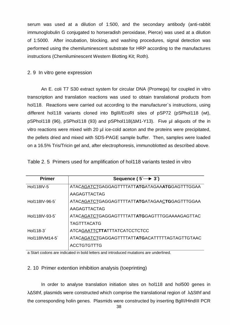
2. 9 In vitro gene expression
An E. coli T7 S30 extract system for circular DNA (Promega) for coupled in vitro
transcription and translation reactions was used to obtain translational products from
hol118. Reactions were carried out according to the manufacturer`s instructions, using
different hol118 variants cloned into BglII/EcoRI sites of pSP72 (pSPhol118 (wt),
pSPhol118 (96), pSPhol118 (93) and pSPhol118(∆M1-Y13). Five µl aliquots of the in
vitro reactions were mixed with 20 µl ice-cold aceton and the proteins were precipitated,
the pellets dried and mixed with SDS-PAGE sample buffer. Then, samples were loaded
on a 16.5% Tris/Tricin gel and, after electrophoresis, immunoblotted as described above.
Table 2. 5 Primers used for amplification of hol118 variants tested in vitro
Primer Sequence ( 5´ 3´)
Hol118IV-5´ ATACAGATCTGAGGAGTTTTATTATGATAGAAATGGAGTTTGGAA
AAGAGTTACTAG
Hol118IV-96-5´ ATACAGATCTGAGGAGTTTTATTATGATAGAACTGGAGTTTGGAA
AAGAGTTACTAG
Hol118IV-93-5´ ATACAGATCTGAGGAGTTTTATTATGGAGTTTGGAAAAGAGTTAC
TAGTTTACATG
Hol118-3´ ATCAGAATTCTTATTTATCATCCTCTCC
Hol118IVM14-5´ ATACAGATCTGAGGAGTTTTATTATGACATTTTTAGTAGTTGTAAC
ACCTGTGTTTG
a Start codons are indicated in bold letters and introduced mutations are underlined.
2. 10 Primer extention inhibition analysis (toeprinting)
In order to analyse translation initiation sites on hol118 and hol500 genes in
λ∆Sthf, plasmids were constructed which comprise the translational region of λ∆Sthf and
the corresponding holin genes. Plasmids were constructed by inserting BglII/HindIII PCR

39
fragments with the upstream translational region of λ∆Sthf phage and hol118 or hol500
gene into pSP72. Primers used in the construction of pSP-λ∆Shol118 and pSP-
λ∆Shol500 are listed in Table 2. 6. pSP-λ∆Shol118 and pSP-λ∆Shol500 served as a
source for mRNA preparation. After linearization with HindIII, in vitro transcription with
T7 RNA polymerase was performed, and the run-off transcripts were used for in vitro
toeprinting studies. The 32P-5´-end-labeled primer K16 5´-CCGCCAGCTCCTGC-3´,
which is complementary to hol118 and hol500 mRNA sequences downstream of the start
codon, was used in the toeprinting reactions performed essentially as described earlier
(Tedin et al., 1997). After annealing of the primer, 0.04 pmol mRNA were incubated with
4 pmol 30S ribosomes and 20 pmol fMet-tRNA for 10´ at 37°C, in a final volume of 10 µl
VD-buffer (10 mM Tris-Cl pH 7.3, 60 mM NH4Cl, 10 mM MgAc, 6 mM ß-
Mercaptoethanol), to allow formation of the ternary complex. Then, 16 units MMLV
reverse transcriptase premixed with dNTPs were added to each reaction, and cDNA
sythesis was performed by incubation of the mix for 15´ on 37°C. Reactions were
stopped by the addition of the loading dye (10 M urea, 10xTBE, traces of bromphenol
blue and xylencyanol). After heating the samples for 5´at 95°C, aliquots were loaded on
a 8%PAA-8 M urea gel. The gel was dried under vacuum (65°C), and exposed to a
radiographic film for detection followed by exposure to a Phosphoimager screen
Molecular Dynamics
Table 2. 6 Primers used in construction of pSP-λ∆Shol118 and pSP-λ∆Shol500
Primer Sequence ( 5` 3`)
LDSFT-5´ ACAGATCTCCCGTGCATATCGGTCACG
Hol118-3´ ATCAGAATTCTTATTTATCATCCTCTCC
Hol500-3´ ATCAGAATTCTTATTTATCATCCTTTCCATATTTTTTAGC
a Stop codons are indicated in bold letters
2. 11 Construction of λ imm434hol118 and λimm434hol500
The λimm434hol118/λimm434hol500 phages were constructed by recombination
within the lysis region between plasmid pBSE-λ∆Shol118 or pBSE-λ∆Shol500, and
phage λimm434cI Swt. First, hol118 or hol500 were inserted into the EcoRI site of
pBSE-λ∆S1. Orientation of the product and integrity were confirmed by sequencing.
40
Recombination occured in C600 hfl cells carrying pBSE-λ∆Shol118 or pBSE-λ∆Shol500:
cells were infected with the phages (m. o. i. ≈ 1) and after 15 min of adsorption ot room
temperature, incubation temperature was rised to 42°C for 20 min in order to induce
phage replication, followed by incubating for 60 min at 37°C. Liberated phages were
used to lysogenize LE392 cells. Ampicilin-resistant clones were selected, which carried
recombinant phages originated from a single recombination event between the plasmid
and phage. A second recombination event was provoked through induction of the
lysogenized phages. Liberated phages were again used to lysogenize LE392 cells.
Ampicilin-sensitive LE392 lysogens were selected, and checked by PCR and DNA
sequencing for the presence of hol118 or hol500 in λimm434hol118 and
λimm434hol500.
2. 12 Construction of the transactivation plasmid pBRT
pBRT was constructed in a pBR322E backbone. The single EcoRI site was
inactivated through restriction digestion, blunt-end filling with the Klenow fragment
(Boehringer), and ligation. A 1865 bp BamHI fragment with the S gene deletion from
pBS-λ∆S was cloned into the BamHI site of pBR322E, forming pBRT. The plasmid has
a single EcoRI restriction site within the λ sequence, which can be used for inserting
holin genes.
2. 13 Construction of pBSH32- phoA and pBSH32-lacZ plasmids
The gene fragment comprising the first 32 amino acids of Hol118 was amplified
using 5´HolF1 (5´-ATATTCTAGAATAAGAGGAGTTTTATTATGATAGAAATGG
AGTTTGGAAAAGAGTTA-3´) and 3´HolF1 (5´-ATATGGATCCTCGTCTTCTTAA
TCGCCTGAACAAACAG-3´) primers. The product was inserted into XbaI/BamHI of
pBluescript, forming pBSH32. LacZ and phoA genes were fused to the holin fragment at
the BamHI site in pBSH32 plasmid generating pBSH32-lacZ and pBSH32- phoA,
respectively. The fusion genes were expressed under the lac promotor upon induction
with 5 mM IPTG on agar plates.

41
3. Results
3. 1 Construction of λ∆Sthf phage
In order to compare the functional properties of different holin protein in the same
genetic background, we have established the λ∆Sthf lysis deficient phage vector (Fig. 3.
1), whose lytic cycle can be induced by inactivating the temperature-sensitive CIts857
repressor. Using homologous recombination, the holin gene S was removed in phage
λgt11, and substituted by a single EcoRI restriction site placed immediately downstream
of the ribosome binding site initiating S translation (Fig. 3. 1, Fig. 3. 2). After insertion of
heterologous holin genes, the cloned genes are expressed under control of the native λ
pR late promoter, and the translation signals provided on the cloned fragments. In λ wt,
the translational stop signal for the holin overlaps the translational start region of the R
endolysin gene. In the λ∆Sthf phage, the translational start for the R gene is
independent of the translation of the cloned holin gene (Fig. 3. 2). Within the region of
the lacZ gene in λgt11, an ampicilin resistance gene (bla) was cloned into the EcoRI site
thereby inactivating this site. The presence of the ampicilin resistance marker on the
phage allows convenient selection of E. coli cells lysogenized with the phage, by
selecting for ampicilin resistant E. coli cells.
3. 2 Complementation of the λ∆Sthf lysis defect with λ S alleles
The S holin gene encodes two protein starting at two ATG codons separated by a
codon endoding lysine. The presence of the additional positive charge determines
different lytic characteristics in the process of pore formation, S107 and S105 (Fig. 3. 1)
(Bläsi et al., 1989; Bläsi et al., 1990). To test whether λ∆Sthf can be used as a model
system for functional analysis of holins, different λ S alleles with lytic properties that have
been previously decribed were evaluated in the λ∆Sthf background (Table 3. 1). The
S105 effector and the S107 inhibitor were tested for lytic competence in λ∆Sthf. Both
alleles were amplified using PCR from the λ(wt) phage. Additionally, an allele was tested
which has one more positive charge on the N-terminus of S.

42
Fig. 3. 1. Schematic presentation of the construction of λ∆Sthf phage, and the 5´- mRNA
sequence of the translation initiation region of S and R . The S holin and the R endolysin
genes in λ wildtype, λ∆Sthf, and λ∆Sthf::hol are underlined. The bent vertical lines indicate the
changes introduced by deletions and insertions. Altered nucleotides are underlined, and the
single EcoRI site in λ∆Sthf is shown in italics. Sdi denotes the secondary structure involved in
regulation of translation initiation (Bläsi et al., 1989). The N-terminal amino acid sequences of
the two S variants are given above (S107) and below (S105) the nucleotide sequence of λ (wt).
A sdi U U U U C G λ(wt) C G C G C G U G MetLysMet- - (S107) AGCAAA UAAGACAUGAAGAUG - S - CGGAGUAGAAGAUG.....UAA - R Met- - (S105)
A U U U U C G λ∆Sthf C G C G C G U G AGCAAA GAAGAAUUC AGGAGUAGAAGAUG - R
A U U U U C Gλ∆Sthf::hol C G C G C G U G AGCAAA GAAGAAUUC AUG - Holin - UAA GAATTCAGGAGUAGAAGAUG - R

43
GAATTGTAAT ACGACTCACT ATAGGGCGAA TTGGGCCCTC TAGATGCATG CTCGAGCGGC
CGCCAGTGTG ATGGATATCT GCAGAATTCG CCCTTAGCCA GTGCTCTTTC CGTTGTGCTG
AATTAAGCGA ATACCGGAAG CAGAACCGGA TCACCAAATG CGTACAGGCG TCATCGCCGC
CCAGCAACAG CACAACCCAA ACTGAGCCGT AGCCACTGTC TGTCCTAATT AATTAGTAAT
AGTTACGCTG CGGCCTTTTA CACATGACCT TCGTGAAAGC GGGTGGCAGG AGGTCGCGCTLDSFT-5´
AACAACCTCC TGCCGTTTTG CCCGTGCATA TCGGTCACGA CAAATCTGAT TACTAAACAC
For 2
AGTAGCCTGG ATTTGTTCTA TCAGTAATCG ACCTTATTCC TAATTAAATA GAGCAAATCC
R
CCTTATTGGG GGGAAGAATT CAGGAGTAGA AGATGGTAGA AATCAATAAT CAACGTAAGG
CGTTCCTCGA TATGCTGGCG TGGTCGAAGG GCGAATTCCA GCACACTGGC GGCCGTTACT
Rev 2
AGTGGATCCG AGCTCGGTAC CAAGCTTGGC GTAATCATGG TCATAGCTGT TTCCTGTGTG
AAATTGTTAT CCGCTCACAA TTCCACACAA CATACGAGCC GGAAGCATAA AGTGTAAAGC
Fig. 3. 2 Nucletide sequence of the upstream and downstream region of the S deletion in
λ∆Sthf phage. The single EcoRI restriction site is shown in italics and the ATG start codon for
R, endolysin gene is underlined The arrows indicate the position of For2 and Rev2 primers,
which were used for amplification and sequencing and LDSFT-5´ primer, used for amplification of
fragments used in toeprinting assays.
Table 3. 1 Alleles and mutants of λ S tested in this study
Holin N-terminal amino acid sequencesa
S + - +MKMPEK
S105 - +- - MPEK
S107 + -+MKLPEK
S107-M3K + + - +MKKPEK
a Translation start codons for λ∆S are indicated by bold letters, charged amino-acids by + and -
above the sequence, and mutated amino acids are underlined

44
Induction of λ∆Sthf::S (wt) resulted in lysis beginning at 45 min after induction (Fig. 3.
3A). Expression of only S105 led to accelerated, saltatory lysis which was completed in
only 30 min, whereas the S107 allele substantially delayed the course of cell lysis
(starting at 50 min). The mutated S107-M3K holin resulted in a completely non-lytic
phenotype showing no signs of growth inhibition. These results confirm that the
functional difference between effector and inhibitor can be detected in the λ∆Sthf vector
used, and that λ∆Sthf::S (wt) behaves essentially as in the λ wt (Raab et al., 1988).
A
Fig. 3. 3 Panel A. Permeabilization features of S variants tested in λ∆Sthf. Panel A. Lysis
profiles following thermal induction of E. coli LE392 lysogenized with λ∆Sthf carrying different
alleles of S holin as indicated in the legend. λ∆Sthf, negative control is shown as LDS.
Additionally, three different S105 mutant were analysed. All were isolated as
spontaneous S105 mutants (Fig. 3. 3B). The F94S and K43E changes influenced lysis
timing, delaying the onset of lysis for 20 min compared to S105 wt, while L62P did not
result in a significantly different lysis behaviour.
0 15 30 45 60 75 90 105 120
Time [min] after induction
0,01
0,1
1
OD
600
S107M3K
LDS
S107
S
S105

45
Fig. 3. 3 Fig. 3. 4
Fig. 3. 3 Panel B. Lysis profiles following thermal induction of E. coli LE392 lysogenized with
λ∆Sthf carrying variants of S105 causing lysis delay.
Fig. 3. 4 T and Gp 17, 5 have similar lytic features tested in λ∆Sthf. Lysis profile observed
following thermal induction of E. coli LE392 lysogenized with λ∆Sthf carrying gp T holin from
bacteriophage T4 and Gp17,5 holin from bacteriophage T7, λ∆Sthf, negative control is shown as
LDS.
3. 3. Complementation of the λ∆Sthf lysis defect with holins from T4 and T7
bacteriophages
Two very dissimilar holins (T and Gp17.5) were tested for their properties to
complement the lysis defect of λ∆Sthf (Fig. 3. 4). Lysogens carrying λ∆Sthf::17,5 showed
a sudden, saltatory onset of cell lysis at about 30 min after induction (Fig. 3. 4). Within
60-70 min, the culture was completely clear. An almost indentical lysis onset was
observed for λ∆Sthf::t. For both holins, the rate of lysis is very similar to that observed
when the native λ S allele is expressed in this system (compare to curve in Fig. 3. 3A).
Apparently, these two holin protein have very similar functional features, despite their
very different structural characteristics (Fig. 3. 5). Both proteins have a high probability
to form only one transmembrane domain in the cytoplasmic membrane, according to the
prediction made by TMHMM 2. 0 (Fig. 3. 5).
0 10 20 30 40 50 60
Time [min] after induction
0,01
0,1
1
OD
600
S105 (L62P)
S105 (F94S)
S105 (w t)
S105 (K43E)
0 20 30 40 60 80 100 120
Time [min] after induction
0,01
0,1
1
OD
600
LDS
T7 gp17.5
T4 T

46
λ S
+ -+ - +- + + + - +- - - ++ ++ -- +MKMPEKHDLLAAILAAKEQGIGAILAFAMAYLRGRYNGGAFTKTVIDATMCAIIAWFIRDLLDFAGLSSNLAYITSVFIGYIGTDSIGSLIKRFAAKKAGVEDGRNQ **************** S107 S105
T7 gp17,5
- - + - + - + -+ - ++ +-MLSLDFNNELIKAAPIVGTGVADVSARLFFGLSLNEWFYVAAIAYTVVQIGAKVVDKMIDWKKANKE ***************
T4 T
+ - -+ +- + + + - - + + - - -+-+ + - - - - + +MAAPRISFSPSDILFGVLDRLFKDNATGKVLASRVAVVILLFIMAIVWYRGDSFFEYYKQSKYETYSEIIEKERTARFESVALEQLQIVHISSEADFSAVYSFRPKNLNY
- - + -+ -+ -- + + ++ - - + - -+ - + + +FVDIIAYEGKLPSTISEKSLGGYPVDKTMDEYTVHLNGRHYYSNSKFAFLPTKKPTPEINYMYSCPYFNLDNIYAGTITMYWYRNDHISNDRLESICAQAARILGRAK
Fig. 3. 5 Amino acid sequences and secondary, domain structures of λ S, T7, Gp 17,5, and T4 T holins. Charged residues are indicated by +
and - signs above the sequence, potential transmembrane domains, according to predictions made by TMHMM 2.0, are underlined. The double
translational start motiv for S107 and S105 of λ S holin is indicated by boldface letters.
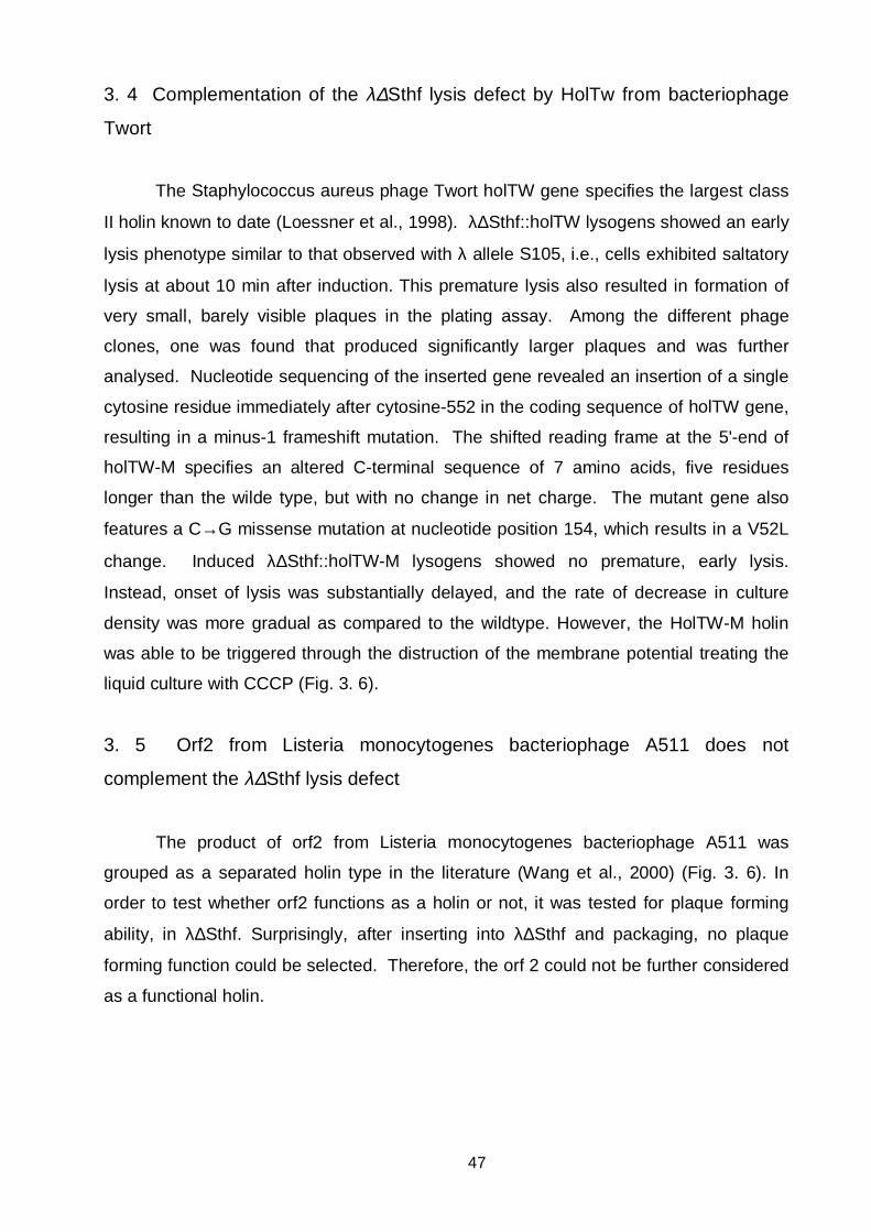
47
3. 4 Complementation of the λ∆Sthf lysis defect by HolTw from bacteriophage
Twort
The Staphylococcus aureus phage Twort holTW gene specifies the largest class
II holin known to date (Loessner et al., 1998). λ∆Sthf::holTW lysogens showed an early
lysis phenotype similar to that observed with λ allele S105, i.e., cells exhibited saltatory
lysis at about 10 min after induction. This premature lysis also resulted in formation of
very small, barely visible plaques in the plating assay. Among the different phage
clones, one was found that produced significantly larger plaques and was further
analysed. Nucleotide sequencing of the inserted gene revealed an insertion of a single
cytosine residue immediately after cytosine-552 in the coding sequence of holTW gene,
resulting in a minus-1 frameshift mutation. The shifted reading frame at the 5'-end of
holTW-M specifies an altered C-terminal sequence of 7 amino acids, five residues
longer than the wilde type, but with no change in net charge. The mutant gene also
features a C→G missense mutation at nucleotide position 154, which results in a V52L
change. Induced λ∆Sthf::holTW-M lysogens showed no premature, early lysis.
Instead, onset of lysis was substantially delayed, and the rate of decrease in culture
density was more gradual as compared to the wildtype. However, the HolTW-M holin
was able to be triggered through the distruction of the membrane potential treating the
liquid culture with CCCP (Fig. 3. 6).
3. 5 Orf2 from Listeria monocytogenes bacteriophage A511 does not
complement the λ∆Sthf lysis defect
The product of orf2 from Listeria monocytogenes bacteriophage A511 was
grouped as a separated holin type in the literature (Wang et al., 2000) (Fig. 3. 6). In
order to test whether orf2 functions as a holin or not, it was tested for plaque forming
ability, in λ∆Sthf. Surprisingly, after inserting into λ∆Sthf and packaging, no plaque
forming function could be selected. Therefore, the orf 2 could not be further considered
as a functional holin.

48
Fig. 3. 6 HolTw causes extremly early lysis in λ∆Sthf. Lysis profiles observed following
thermal induction of E. coli LE392 lysogenized with λ∆Sthf::holTw and λ∆Sthf::holTw-M. Lysis
of λ∆Sthf::holTw-M lysogens was induced prematurely with CCCP, indicated by an arrow on the
panel.
3. 6 Analysis of Hol118 from the Listeria monocytogenes bacteriophage A118
Listeria monocytogenes bacteriophage A118 causes cell lysis by a combined
action of the Hol118 holin and the Ply118 endolysin, having a unique L-alanoyl-D-
glutamate specificity (Loessner et al., 1995). The gene coding for Hol118 precedes the
endolysin gene in a different reading frame with a overlapping stop codon for the holin
and the start codon for endolysin. Hol118 belongs to the class I holin proteins which
have three transmembrane domains. (Fig 3. 7). In order to test its permeabilization
features the λ∆Sthf vector was employed, by insertion of hol118 into the EcoRI site
immediately upstream of R. N- terminally different Hol118 variants were tested for lysis
timing, in order to detect possible functional similarities to the λ S. During this study,
toeprinting assays, in vitro translation, and immunological methods were used to
provide evidence for synthesis of a truncated protein starting at M14 codon in hol118.
Site directed mutagenesis of this codon in hol118 and in trans expression of Hol118(83)
protein were performed to reveal the exact function of the protein.
0 15 30 45 60 75 90 105 120
Time [min] after induction
0
0,1
0,2
0,3
0,4
0,5
OD
600 HolTw
LDS
HolTw M
HolTw M CCCP
49
Fig. 3. 7 Amino acid sequences and secondary domain structures of tested holins from phages infecting Gram-positive bacteria, and tested
proteins for holin function. Charged residues are indicated by +and - signs above the sequence, potential transmembrane domains, according to the
prediction made by TMHMM 2.0, are underlined. The position for the start for Hol118(96) and Hol118(83) are indicated. The proximal positively charged
C-terminal domain is underlined by asteriskes. (A) Holins: HolTw (S. aureus Twort) (Loessner et al., 1998). Altered amino acids in the mutant HolTW-
M are indicated by bold letters. Hol118, Hol500 (L. monocytogenes A118, A500), and Hol2438 (L. innocua monocine producing strain) (Loessner et al.,
1995; Zink et al., 1995). Hol3626 (C. perfringens φ3626), (Zimmer et al., 2002). Different amino acid positions in Hol500 in respect to Hol118 are
indicted in bold letters. (B) Potential holin: Orf 2 (L. monocytogenes A511). (C) Mscl E. coli (Chang et al., 1998)
A
HolTw
-++-+ + + + - - - + +- - +- +- -+MDKKDKTPTVVGGINLSTRFKSKTFWVATVSALAVFTNQLTGAFGLDYSSQVEQGVNIVGSILTLLAGLGILVDNNTKGIKDSVIVQTDYHEPRDSKDPEKHolTw-M L
HolTw
- + -- - + +- --- - - - --- +- + -- - + - +-- -- - +ALTWEHNHNKVEDNVVDNTKTPKEYGTDEDFSDVDPHVYDADEEPFKDQAKPYGVNWDETVDTTKNIDTKEEGVIDENPETSRVLHHolTw-M PTLRIQE
Hol118
- - +- ++ - + - - + ++ ---+MIEMEFGKELLVYMTFLVVVTPVFVQAIKKTELVPSKWLPTVSILIGAILGALATFLDGSGSLATMIWAGALAGAGGTGLFEQFTNRSKKYGEDDK
Hol118(96) Hol118(83) ***************
Hol500
+ - +- ++ - + - - + ++ +--+MMKMEFGKELLVYMTFLVVVTPVFVQAIKKTELIPSKWLPTVSILVGAILGALATSLDGSGSLATMIWAGALAGAGGTGLFEQFTNRAKKYGKDDK Hol500(96) Hol500(83) ****************

50
Hol2438
+ - +- ++ - + - - + ++ +--MMKMEFGKELLVYMTFLVVVTPVFVQAIKKTELIPSKWLPTVSILVGAILGALATSLDGSGSLATMIWAGALAGAGGTGLFEQFTNRAKKYGKDD. *************
Hol3626
+ - + - + + + + ++ +- + - - + +- - - - -MFKFIPEVISWLLVLYIGFKIIDMILGVLKTIKNKNYRSRKMRDGIIRWVAELMAIAFVLILDMFLGLKFTVIGVTLALFAYKEAGSIVENLGECGVELPE -+ - + + +- ++-IVSEKLEVLNKNNKNKEGFNKKEN
B
Orf 2
- - - - - -+ +- + -+ + - +- + + -- - + - ++ + + -+ -+ +-+MHDNEFEVPESVINASATPFEAPDRTKDTYQSSLKNWLDRVKSVGDAGMSREFVIRIPAKALEENNPEATLFLKQCNEIKRNPKCNLPIKIEKTVEKYKDK+ - + - - +KHLMLDLVFTYSNKTDIDAVNSVIK
C
MscL
+- +- + - + - - + +- - - + +MSIIKEFREFAMRGNVVDLAVGVIIGAAFGKIVSSLVADIIMPPLGLLIGGIDFKQFAVTLRDAQGDIPAVVMHYGVFIQNVFDFLIVAFAIFMAIKLINK
+++-- +-- - +- +- +LNRKKEEPAAAPAPTKEEVLLTEIRDLLKEQNNRS

51
3. 6. 1 Hol118 causes late cell lysis in λ∆Sthf phage
Hol118 variants, hol118 wt, (starting from ATG(1)), hol118 (93) (starting from
ATG(2)), and hol118 (96) (starting from ATG(1), with ATG(2) mutated into CTG) were
inserted into λ∆Sthf and tested for lytic competence (Fig. 3. 8 and Fig. 3. 9A).
λ∆Sthf::hol118 wt, λ∆Sthf::hol118 (96) and λ∆Sthf:hol118 (93) were induced over
lysogenic LE392 E. coli cultures (Fig. 3. 9A), and lysis profiles monitored in time.
Hol118 wt caused relatively late cell lysis, 90 min after induction, whereas Hol118 (96)
and Hol118 (93) variants, resulted in growth inhibition rather then sudden lysis (Fig. 3.
9A). It was of interest whether Hol118 could be triggered prematurely by the destruction
of the intact membrane potential by addition of CCCP to induced cultures at different
time points (Fig. 3. 9B) As shown in Fig. 9B, a slight effect can be seen when the
cultures were treated 70 min after thermal induction. In contrast, treating the culture
with CHCl3, 30 min and 50 min after induction resulted in immediate lysis, and indicated
that sufficient cytoplasmatic endolysin was present for degradation of the peptidoglycan.
Hol118 could not be triggered prematurely with energy poisons in the manner
demonstrated for the λ S holin. These results suggested that the energized membrane
does not play a similar role in inhibiting the permeabilization of Hol118 holin.
Fig. 3. 8 Translation control region of λ∆Sthf::hol118. mRNA region governing the
translation of the hol118 gene in λ∆Sthf. Ribosome binding region experimentally shown for λ
wt (Bläsi et al., 1989) and a putative RBS in front of the AUG(3) are underlined.
A U U sdi U U C G C G C G C G U G.AGCAAA GAAGAATTCAUG(1)AUAGAAAUG(2)GAGUUUGGAAAAGAGUUACUAGUUUACAUG(3)ACAU..

52
A
B
Fig. 3. 9 Lysis profiles of N-terminal variants of Hol118. Panel A. Lysis profiles of Hol118
wt, Hol118 (93), and Hol118 (96) inserted into λ∆Sthf. Panel B. Lysis profile of λ∆Sthf::hol118
following dissipation of membrane potential, or disruption of membranes. CCCP was added to
the cells 30, 50, 70 min after induction. At the same time points, CHCl3 was added to different
cultures.
0 20 40 60 80 100
Time [min] after induction
0
0,1
0,2
0,3
0,4
0,5
0,6
OD
600
Hol118 w t
Hol118 (96)
Hol118 (93)
0 20 40 60 80 100
Time after induction [min]
0
0,1
0,2
0,3
0,4
0,5
0,6
0,7
OD
600
Hol118 30 Hol118 50 Hol118 70
Hol118 30 Hol118 50 Hol118 70
53
3. 6. 2 Hol118 inserts into the E. coli membrane
The observed late cell lysis caused by the Listeria phage holin in the λ∆Sthf
genetic background claimed for the inspection whether Hol118 actually inserts into the
membrane of E. coli. Inner membrane fractions of E. coli cells were isolated after
induction of λ∆Sthf phages with three N-terminal different Hol118 variants (Fig. 3. 10A)
All samples were taken 55 min after induction (Fig. 3. 10A). After SDS-PAGE and
immunoblotting specific Hol118 protein bands were detected for Hol118 wt, Hol118 (96)
and Hol118 (93). Our results indicate that Hol118 wt and Hol118 (96) insert the E. coli
membrane efficiently in contrast to Hol118 (93). The observed lysis curve of Hol118
(93) may therefore result from insufficient insertion of the protein into the membrane
(Fig. 3. 10A).
Further, membrane fractions of E.coli cells were isolated from cell culture after
induction of lysogenic λ∆Sthf::hol118 wt phages, were aliquots were taken at different
time points after induction (Fig. 3. 10B). The Hol118 specific band can be detected in
the inner membrane fraction from 20 min following induction, and from the band
intensities an increase in holin concentration over time was observed. In the Western
Blot experiments performed on Listeria holin analysed, a faint band below the specific
Hol118 (marked by blocked arrows on Fig. 3. 10A and Fig. 3. 10B) is observed. This
band is most distinct in the lane corresponding the the culture sample taken 20 min after
induction of λ∆Sthf::hol118 phage (Fig. 3. 10B), and corresponds to Hol118(83).
3. 6. 3 The first ß-Turn of Ho118 resides in the cytoplasm
In order to experimentally elucidate the position of the N-terminal portion of
Hol118, two fusions were constructed containing the first 32 amino acids of Hol118 and
alkaline phosphatase or ß-glactosidase reporter molecules (Fig. 3. 7A). The approach
is based on a reciprocal activity of the two proteins in the individual cell compartments.
LacZ is active only if the protein remains in the cytoplasm; in contrast to alkaline
phosphatase which is active only in the periplasm (Manoil, 1990). In pBSH32-phoA and
pBSH32-lacZ plasmids, Hol32-phoA and Hol32-lacZ fusion proteins were synthesized
after IPTG induction, and the activity of the reporter enzymes tested on agar plates
supplemented with chromogenic substrates. E. coli cells expressing Hol32-lacZ fusion
were blue coloured on plates with X-gal, whereas alkaline phoshatase activity could not
be detected.

54
A
B
Fig. 3. 10 Holin synthesis in E. coli LE392 cells lysogenized with λ∆Sthf::hol118. Panel
A. Immunological analysis of inner membrane fraction of E. coli cells after induction of
temperent λ∆Sthf carrying different Hol118 variants. Lane 1, λ∆Sthf; λ∆Sthf::hol118 wt, lane 2,
λ∆Sthf::hol118 (93); lane 3, λ∆Sthf::hol118 (96); lane 4. Panel B Time-resolved appearance of
Hol118 in the inner membrane of E. coli after induction of λ∆Sthf::hol118. The samples were
taken at 20 min, (lane 1), 35 min, (lane 2), 45 min, (lane 3) and 55 min, (lane 4) after induction.
Lane 5 is a control with λ∆Sthf taken 60 min after induction.
1 2 3 4 5
1 2 3 4

55
detected upon production of Hol32-phoA fusion protein. The result indicated that the N-
terminal part of the fusion protein (first 32 aa of Hol118) positioned the reporter protein
in the cytoplasm of the cell.
3. 6. 4 Hol118 has three possible translational starts
Hol118 starts with two Met codons separated by codons for the amino acids
isoleucine (I) and glutamic acid (E) (Fig. 3. 7, Fig. 3. 8). Direct evidence for utilization of
AUG(1) and AUG(2) in λ∆Sthf as start signals for translation was obtained using
toeprinting analysis. This method employs primer extention inhibition, and indicates the
sites were 30S ribosomal subunits are bound on the mRNA in ternary complexes (Hartz
et al., 1988). The polymerase usually stops 15 nucleotides downstream of the AUG
codon were the ternary complex is positioned, and a toeprinting signal is formed by
accumuation of labelled product on a polyacrylamide gel.
Toeprinting reactions were performed using a run-off transcript from pSP-
λ∆Shol118. The obtained signals suggested the presence of 3 initiation sites (Fig. 3.
11A): The first one is located 15 nucleotides downstream from the AUG(1) (see Fig. 3.
8); the second 15 nucleotides downstream from AUG(2), and a third toeprinting band
was located 15 nucleotides apart from a third AUG codon, located 40 nt downstream of
the 5´ end of hol118. A strong (but unspecific) signal formed inbetweeen AUG(2) and
AUG(3), which however, did not correspond to any canonical start codon and was
therefore not analysed further.
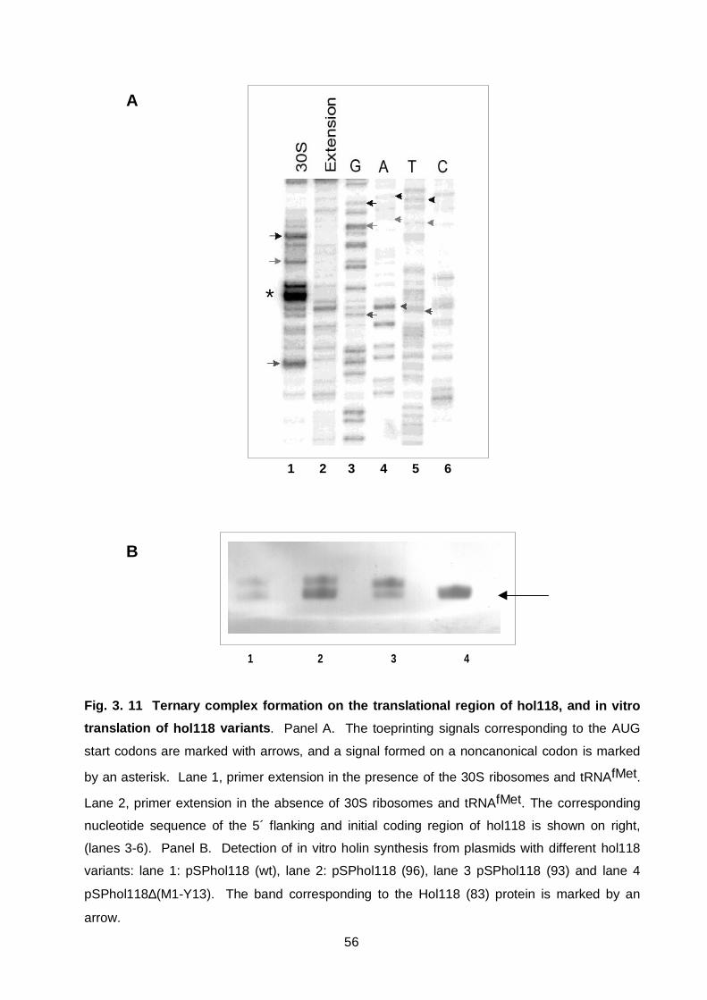
56
A
B
Fig. 3. 11 Ternary complex formation on the translational region of hol118, and in vitro
translation of hol118 variants. Panel A. The toeprinting signals corresponding to the AUG
start codons are marked with arrows, and a signal formed on a noncanonical codon is marked
by an asterisk. Lane 1, primer extension in the presence of the 30S ribosomes and tRNAfMet.
Lane 2, primer extension in the absence of 30S ribosomes and tRNAfMet. The corresponding
nucleotide sequence of the 5´ flanking and initial coding region of hol118 is shown on right,
(lanes 3-6). Panel B. Detection of in vitro holin synthesis from plasmids with different hol118
variants: lane 1: pSPhol118 (wt), lane 2: pSPhol118 (96), lane 3 pSPhol118 (93) and lane 4
pSPhol118∆(M1-Y13). The band corresponding to the Hol118 (83) protein is marked by an
arrow.
1 2 3 4 5 6
*
1 2 3 4

57
3. 6. 5 In vitro transcription/translation yielded the expected products
Toeprinting indicated that translation starts from not only the first two "expected"
codons, but also from an additional start, AUG(3) (Fig. 3. 8, Fig. 3. 11A). To
experimentally demonstrate that this occurs under physiological conditions, an in vitro
reaction was performed using a coupled transcription-translational system. Ho118 and
variant genes with mutated start codons were transcribed from a T7 promotor. An
optimized ribosome-binding site (GAGGAG) with a spacer of 7 nucleotides was
positioned upstream of the gene. The products of the in vitro reactions were separated
by SDS-PAGE electrophoresis and the protein bands visualized by Western analysis
using antiHol118 polyclonal antibodies. Four different variants were analysed: hol118
wt, hol118 (96), hol118 (93) and hol118 (83). In all reactions, two bands appeared,
were the lower band corresponded to Hol118 (83) protein (Fig. 3. 11B). It should be
noticed that proteins starting at AUG(1) and AUG(2) form one band, they cannot be
resolved on the electrophoresis system used here.
3. 6. 6 Hol118 is detected in Listeria cells after infection with A118
In order to reveal the lenght of the latent period, lysis profile of A118 in infected
Listeria cells was analysed by performing a one-step growth curve (Fig. 3. 12). Cell
lysis started 60 min after infection, and was complete after about 120 min. To detect
the presence of the Hol118 protein in Listeria cell membranes, a sample was taken 120
min after infection with A118 and processed as described above. Proteins were
separated by SDS-PAGE electrophoresis visualized as described above. Two bands
were observed, indistinguishable from the bands detected in E. coli cell samples, and by
in vitro translation of hol118 (Fig. 3. 10B and Fig. 3.11B).
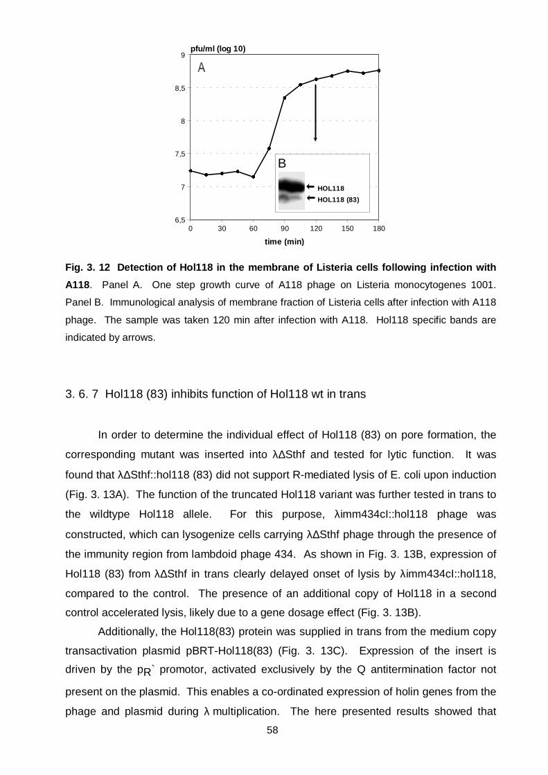
58
Fig. 3. 12 Detection of Hol118 in the membrane of Listeria cells following infection with
A118. Panel A. One step growth curve of A118 phage on Listeria monocytogenes 1001.
Panel B. Immunological analysis of membrane fraction of Listeria cells after infection with A118
phage. The sample was taken 120 min after infection with A118. Hol118 specific bands are
indicated by arrows.
3. 6. 7 Hol118 (83) inhibits function of Hol118 wt in trans
In order to determine the individual effect of Hol118 (83) on pore formation, the
corresponding mutant was inserted into λ∆Sthf and tested for lytic function. It was
found that λ∆Sthf::hol118 (83) did not support R-mediated lysis of E. coli upon induction
(Fig. 3. 13A). The function of the truncated Hol118 variant was further tested in trans to
the wildtype Hol118 allele. For this purpose, λimm434cI::hol118 phage was
constructed, which can lysogenize cells carrying λ∆Sthf phage through the presence of
the immunity region from lambdoid phage 434. As shown in Fig. 3. 13B, expression of
Hol118 (83) from λ∆Sthf in trans clearly delayed onset of lysis by λimm434cI::hol118,
compared to the control. The presence of an additional copy of Hol118 in a second
control accelerated lysis, likely due to a gene dosage effect (Fig. 3. 13B).
Additionally, the Hol118(83) protein was supplied in trans from the medium copy
transactivation plasmid pBRT-Hol118(83) (Fig. 3. 13C). Expression of the insert is
driven by the pR` promotor, activated exclusively by the Q antitermination factor not
present on the plasmid. This enables a co-ordinated expression of holin genes from the
phage and plasmid during λ multiplication. The here presented results showed that
0 30 60 90 120 150 180
time (min)
6,5
7
7,5
8
8,5
9pfu/ml (log 10)
B
HOL118
HOL118 (83)

59
Hol118 (83) inhibited cell lysis by the λ∆Sthf::hol118 phage (Fig. 4C), which further
supports the assumption that the N-terminally truncated protein acts as the inhibitor of
lysis.
A
B C
Fig 3. 13 Hol118 (83) has no lytic activity in λ∆Sthf phage, and causes in trans inhibition
of Hol118 function. Panel A. Lysis profiles of E. coli LE392 cells lysogenized with
λ∆Sthf::hol118 (83), λ∆Sthf::hol118 wt. Panel B. Lysis profiles of E. coli LE392 double
lysogens with λimm434cI::hol118 wt and λ∆Sthf::hol118 (83), or λ∆Sthf::hol118 and λ∆Sthf,
respectively. Panel C. Lysis profiles of E. coli LE392 cells lysogenized with λ∆Sthf::hol118 wt,
induced in the presence of the transactivation plasmid pBRT-hol118 (83).
0 20 40 60 80 100
Time [min] after induction
0
0,2
0,4
0,6
0,8
OD
600
Hol118 (83)
Hol118
0 20 40 60 80 100
Time [min] after induction
0
0,2
0,4
0,6
OD
600
D Hol118/Hol118 (83)
D Hol118/Hol118
D Hol118/LDS
0 30 60 90 120 150
Time [min] after induction
0
0,2
0,4
0,6
0,8
OD
600
Hol118/pBRT-hol118 (83)
Hol118
LDS/pBRT-hol118 (83)
LDS

60
A B
Fig. 3. 14 Cell lysis is influenced by the Hol118:Hol118 (83) ratio. Panel A. Lysis profiles of
different E. coli double lysogens (1 and 3) carrying: λimm434cI::hol118 and λ∆Sthf::hol118(83),
expressed at different levels after induction. Panel B. PCR performed on liberated phages in
the medium after thermal induction. The lower band corresponds to hol118(83) in λ∆Sthf and
the upper band to hol118 in λimm434cI. Lane 1. PCR products from clone 1. Lane 2. PCR
products from clone 3. Lane 3. PCR on phage λ∆Sthf::hol118wt. Lane 3. 100bp DNA ladder.
This hypothesis is further strengthened by the observation that overexpression of
Hol118 (83) to Hol118 in double lysogenes expressing λ∆Sthf::hol118 (83) and
λimm434::hol118, led also to cell lysis inhibition (Fig. 3. 14A and Fig. 3. 14B).
3. 6. 8 Hol118M14L, Hol118M14I and Hol118M14V are defect in lysis timing
In order to inhibit translation initiation at AUG(3), this start codon was changed to
codon CTG or ATT, and the effect of the hol118M14I and hol118M14L alleles tested in
λ∆Sthf (Fig. 3. 15). All mutants showed accelerated lysis. λ∆Sthf::hol118M14L had a
very short lysis timing, cell density started to decrease 30 min after induction, while
λ∆Sthfhol118M14I phage lysed later, 50 min after induction. Obviously, the impaired
translation starts for Hol118 (83) protein in hol118M14I and hol118M14L led to
0 30 60 90 120
Time [min] after induction
0
0,2
0,4
0,6
OD
600
D Immhol118/ hol118 (83) 1
D Immhol118/ hol118 (83) 3
1 2 3 M
-800bp-700bp-600bp-500bp

61
accelerated cell lysis in both cases, again strengthening the hypothesis that Hol118(83)
is the inhibitor of Hol118 function. However, the observed difference in lysis timing of
Hol118M14I and Hol118M14L, suggests that the phenotypic change associated with
this mutation can not be solely attributed to the lack of the inhibitor function.
Fig. 3. 15 Hol118M14V, Hol118M15L and, Hol118M14I are early lysis mutants. Lysis
profiles of E. coli LE392 cells lysogenized with λ∆Sthf expressing hol118M14I, hol118M14L, and
hol118M14V and induced by heat shock.
The character of these mutations was further tested by expressing them in trans
to Hol118 wt allele in order to observe its dominant or recessive behaviour over the wild
type. Lysis timing assays of the tested double lysogenes are summarized in Table 3. 1.
0 30 50 70 90
Time after induction [min]
0
0,2
0,4
0,6
OD
600
Hol118 (w t)
Hol118 (M14V)
Hol118 (M14I)
Hol118 (M14L)

62
Table 3. 2 Lysis timing of induced double lysogens expressing different hol118 alleles
E.coli strain LE392: Hol118 allelesLysis time
[min]a
λimm434cI::hol118/λ∆Sthf Hol118/--- 90
λimm434cI::hol118/λ∆Sthf::hol118 Hol118/Hol118 60
λimm434cI::hol118/λ∆Sthf::hol118M14I Hol118/Hol118M14I 70
λimm434cI::hol118/λ∆Sthf::hol118M14V Hol118/Hol118M14V 20
λimm434cI::hol118/λ∆Sthf::hol118M14L Hol118/Hol118M14L 20
λimm434cI::hol118/λ∆Sthf::hol118 (83) Hol118/Hol118-(83) <110
a Lysis time is defined as the time in minutes between the shift to 42°C and onset of lysis.
Hol118M14L, hol118M14V, and hol118M14I mutants had a dominant effect over
the presence of Hol118 wt. A finding which indicated that residue 14 in the first
transmembrane domain is important for the oligomerization process of the holin in the
membrane. Since Hol118 (83) protein, can not complement the lysis defect of λ∆Sthf,
and from the dominant character of M14 mutations, it may be concluded that the first
transmembrane domain is cructial for the permeabilization process leading to pore
formation.
Further, Hol118 (83) was expressed in trans to Hol118M14I, Hol118M14L and
Hol118M14V from pBRT plasmid in cells lysogenized with λ∆Sthf::hol118M14V,
λ∆Sthf::hol118M14I or λ∆Sthf::hol118M14L phages. We observed a dominant effect of
the mutations at M14 to the presence of the inhibitor expressed in trans. It may be
speculated that this position within the first transmembrane domain is involved in the
interaction between Hol118(96) and Hol118(83) variants.

63
Fig. 3. 16 Mutation at M14 are dominant over the presence of Hol118(83). Lysis profiles
observed follwing thermal induction of E. coli LE392 lysogenized with λ∆Sthf::hol118M14V,
panel A; λ∆Sthf::hol118M14L, panel B; λ∆Sthf::hol118M14I, panel C; induced in the presence of
pBRT-hol118(83).
3. 6. 9 F84S in Hol118 causes a lysis timing defect
Hol118M1L,F84S mutant was isolated as a spontaneous, plaque forming, mutant
of λ∆Sthf::hol118M1L which formed extreamly small plaques. Hol118M1L, F84S
mutant, caused rappid cell lysis 20min after induction (Fig. 3. 17) The presence of the
single M1L mutation in Hol118 had no significant influence on lysis, so the extreamly
early lysis of the double Hol118 mutant could be attributed only to the F84S change in
the C-terminal part of the protein.
0 20 40 60 80 100
Time [min] after induction
0
0,1
0,2
0,3
0,4
Hol118M14L/pBRT-Hol118(83)
Hol118M14L
0 20 40
Time [min] after induction
0
0,1
0,2
0,3
0,4
OD
600
Hol118M14V
Hol118M14V/pBRT-Hol118(83)
A B
0 20 40 60 80 100
Time [min] after induction
0
0,1
0,2
0,3
0,4
OD
600
Hol118M14IHol118M14I/pBRT-Hol118(83)
C

64
Fig. 3. 17 Hol118F84S is an early lysis mutant. Lysis profiles observed follwing thermal
induction of E. coli LE392 lysogenized with λ∆Sthf::hol118 phage and λ∆Sthf::hol118M1L,F84S.
3. 7 Analysis of Hol500 from Listeria monocytogenes bacteriophage A500
The lysis gene casette of Listeria monocytogenes phage A500 contains the dual
start hol500 gene, preceding the endolysin gene in a different reading frame, with
overlapping start/stop codons (Loessner et al., 1995). Inserting hol500 into λ∆Sthf
complemented the lysis defect of the used phage. λ∆Sthf::hol500 phage formed bigger
plaque compared to λ∆Sthf::hol118. The significant difference in lysis properties of this
two inserted holins was surprising, since they differ in only seven amino acids (Fig. 3.
7). Both holins differ in the organization of the N-terminus. Hol500 has a typical dual
start motif, two M codon separated by a codon for a positively charged amino acids.
Further changes are in the first ß-turn and the second transmembrane domain of the
protein. The C-terminus of Hol500 differs in two amino acid and has an increased
positive charge compared to Hol118.
0 20 40 60 80 100
Time after induction [min]
0
0,1
0,2
0,3
0,4
0,5
0,6
OD
600
Hol118
Hol118M1L,F84S

65
Fig. 3. 18 Translation control region of λ∆Sthf::hol500 and amino acid sequences of
Hol118 and Hol500. Panel A. mRNA region governing the translation of the hol500 gene.
Single EcoRI restriction site in λ∆Sthf is shown in italics. Ribosom binding site (sdi) region
experimentally shown for λ wt (Bläsi et al 1989) and a putative RBS in front of the AUG(4) are
underlined. The four AUG start codons that gave signals in the toeprinting reaction are
indicated. Panel B. Amino acid sequences of Hol118 and Hol500. Amino acids differing in
Hol500 and Hol118 are underlined. Translational starts detected in the toeprinting reactions are
marked by an asteriskes below the amino acids.
A
A U U sdi U U C G C G C G C G U G ..AGCAAA GAAGAAUUCAUG(1)AUG(2)AAAAUG(3)GAGUUUGGAAAAGAGUUACUAGUUUAUAUG(4)ACA.
λ∆Sthf::hol500
B
Hol118 - - +- ++ - + - - + ++ - - -+MIEMEFGKELLVYMTFLVVVTPVFVQAIKKTELVPSKWLPTVSILIGAILGALATFLDGSGSLATMIWAGALAGAGGTGLFEQFTNRSKKYGEDDK* * *
Hol500 + - +- + + - + - - + ++ +- -+MMKMEFGKELLVYMTFLVVVTPVFVQAIKKTELIPSKWLPTVSILVGAILGALATSLDGSGSLATMIWAGALAGAGGTGLFEQFTNRAKKYGKDDK** * *

66
3. 7. 1 Hol500 can be induced prematurely through the dissipation of the
membrane potential
We evaluated the lytic properties of both the longer and the shorter possible
translation products of hol500. The three individual alleles (native hol500, hol500 (96),
hol500(93), were tested for lytic competence (Fig. 3. 19A). Cells in which native Hol500
is produced start to lyse 60 min following induction of λ∆Sthf::hol500 phage. Synthesis
of only the longer Hol500(96) polypeptide (M4L mutation) led to complete lysis, and
production of Hol500(93) also enabled membrane disruption and gpR-mediated lysis.
These results are clearly different from those obtained with λ S, but also different to the
lysis by Hol118 variants (see Fig. 3. 7). Moreover, although the presence of an
additional amino-terminal positive charge in the Hol500-96-M2K mutant was found to
decrease the lysis-supporting function of this holin, the effect seems somewhat similar
to inhibition oberserved with the corresponding mutation in S107-M3K.
A B
Fig. 3. 19 Hol500 can be induced prematurely by dissipation of the membrane potential.
Panel A. Lysis profiles observed follwing thermal induction of E. coli LE392 lysogenized with
λ∆Sthf carrying N-terminally different hol500 gene variants. Panel B. Lysis profiles observed
follwing thermal induction of E. coli LE392 lysogenized with λ∆Sthf carrying different hol500
gene variants as specified in the legend, treated with CCCP 40 min after induction (black arrow)
0 20 40 60 80 100
Time after induction [min]
0,1
0,2
0,3
0,4
0,5
OD
600
Hol500 w t
Hol500 (96)
Hol500 (93)
LDS
Hol500-M2K
0 20 40 60 80 100
Time after induction [min]
0
0,1
0,2
0,3
OD
600
Hol500 w t
Hol500 (96)
Hol500 (93)
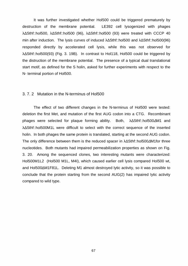
67
It was further investigated whether Hol500 could be triggered prematurely by
destruction of the membrane potential. LE392 cell lysogenized with phages
λ∆Sthf::hol500, λ∆Sthf::hol500 (96), λ∆Sthf::hol500 (93) were treated with CCCP 40
min after induction. The lysis curves of induced λ∆Sthf::hol500 and λ∆Sthf::hol500(96)
responded directly by accelerated cell lysis, while this was not observed for
λ∆Sthf::hol500(93) (Fig. 3. 19B). In contrast to Hol118, Hol500 could be triggered by
the distruction of the membrane potential. The presence of a typical dual translational
start motif, as defined for the S holin, asked for further experiments with respect to the
N- terminal portion of Hol500.
3. 7. 2 Mutation in the N-terminus of Hol500
The effect of two different changes in the N-terminus of Hol500 were tested:
deletion the first Met, and mutation of the first AUG codon into a CTG. Recombinant
phages were selected for plaque forming ability. Both, λ∆Sthf::hol500∆M1 and
λ∆Sthf::hol500M1L were difficult to select with the correct sequence of the inserted
holin. In both phages the same protein is translated, starting at the second AUG codon.
The only difference between them is the reduced spacer in λ∆Sthf::hol500∆M1for three
nucleotides. Both mutants had impaired permeabilization properties as shown on Fig.
3. 20. Among the sequenced clones, two interesting mutants were characterized:
Hol500M1L2 (Hol500 M1L, M4I), which caused earlier cell lysis compared Hol500 wt,
and Hol500∆M1F81L. Deleting M1 almost destroyed lytic activity, so it was possible to
conclude that the protein starting from the second AUG(2) has impaired lytic activity
compared to wild type.

68
Fig. 3. 20 Lysis profiles of N-terminal variants of Hol500. Lysis profiles observed follwing
thermal induction of E. coli LE392 lysogenized with λ∆Sthf carrying N-terminally different hol500
gene variants as specified in the legend of the figure.
3. 7. 3 Membrane insertion of different Hol500 N- terminal variants
In order to correlate the lytic properties of Hol500 variants in λ∆Sthf with the
presence of the proteins in the membrane, inner membrane fraction of E. coli cells
lysogenized with phages λ∆Sthfhol500wt, λ∆Sthfhol500(93), λ∆Sthfhol500(96),
λ∆Sthfhol500M1L, λ∆Sthfhol500M1L2, λ∆Sthfhol500∆M1 and λ∆Sthf were isolated 55
min after induction (Fig. 3. 21). Membrane proteins were resolved by SDS-PAGE
electrophoresis, and immunoblotted using Hol118 antiserum. A correlation between the
band intensity observed in the Western blot and lytic efficiency of holins was observed
(Fig. 3. 20, Fig. 3. 21). Variants with accelerated cell lysis all had increased band
intensities compared to Hol500 wt. The changes intoduced into the N-terminus of
Hol500 influenced membrane insertion of the protein, but did not reveal functional
differences for proteins starting at the double translational start of Hol500 as observed
for λ S.
0 20 40 60 80 100 120
Time [min] after induction
0
0,1
0,2
0,3
0,4
0,5
OD
600
Hol500 w t
Hol500M1L2
Hol500M1L
Hol500DM1

69
Fig. 3. 21 Membrane insertion of N-terminal variants of Hol500. Western blot analysis of
inner membrane fractions of E. coli cells after induction of prophages: Lane 1. λ∆Sthf::hol500;
Lane 2. λ∆Sthf::hol500 (93); Lane 3. λ∆Sthf::hol500(96); Lane 4. λ∆Sthf::hol500M1L; Lane 5.
λ∆Sthf::hol500M1L2; Lane 6. λ∆Sthf::hol500∆M1; Lane 7. λ∆Sthf. Arrow indicate the Hol500
specific band.
Table 3. 3 Hol500 alleles and mutants tested in this study
Holin N-terminal amino acid sequencea Lysis time[min]b
Hol500 wt + - + -MMKMEFGKE 65-70
Hol500 (93) - + - - - - MEFGKE 65-70
Hol500 (96) + - +-MMKLEFGKE 65-70
Hol500-M2K + + - + -MKKLEFGKE no lysis
Hol500∆M1 + - + -- MKMEFGKE 80
Hol500M1L + - + -- MKMEFGKE 80
Hol500M1L2 + - + -- MKIEFGKE 60
a Translation start codons for hol500 are indicated by bold letters, charged amino-acids by +
and - above the sequence, and mutated amino acids are shown in italics
b Lysis time is defined as the time in minutes between the shift to 42°C and onset of lysis.
1 2 3 4 5 6 7

70
3. 7. 4 M14 is used as a translational start in hol500
In order to see which start codons in hol500 are actually used as translational
starts, primer extention inhibition (toeprinting) analysis was performed. The upstream
nucleotide sequence which comprises the sdi structure was amplified together with the
hol500 sequence from λ∆Sthf::hol500 wt and the fragment inserted into pSP72 under
the control of T7 promotor. The toeprinting reaction was carried out using a run-off
transcript from pSP-λ∆Shol500 (Fig. 3. 22, Fig. 3. 11). The obtained signals suggested
the presence of 3 initiation sites immediately at the 5´end of the gene. A fourth
toeprinting band was located 15 nucleotides apart from AUG(4), located 40 nt
downstream of the 5´ end of hol500. A strong (but unspecific) signal formed inbetween
AUG(3) and AUG(4), which however, did not correspond to any canonical start codon
and was therefore not analysed further.
In order to inhibit translation initiation at AUG(4), this start codon was changed
into ATT. Hol500M14I was tested in the λ∆Sthf phage and compared to the
permeabilization properties of the Hol500 wt (Fig. 3. 23). Toeprinting analysis revealed
that M14 codon in hol500, is used as a translation start for a truncated protein starting
within the gene. Changing this start codon into ATT led to accelerated cell lysis, as
observed in hol118. Due to the overall homology between hol118 and hol500 it can be
concluded that the same type of inhibitor is translated starting from M14 position in the
gene.
3. 7. 5 Hol118 (83) inhibits Hol500 lysis in trans
In order to detect the effect of Hol118 (83) protein on Hol500, the protein was
expressed in trans to λimm434::hol500 phage. Clear inhibition of lysis could be
observed when Hol118(83) is expressed to Hol500 (Fig. 3. 24). Hol118 (83) and the
postulated Hol500 (83) protein would differ in only four amino acid positions, so the in
trans effect of Hol500(83) should not be very different compared to the effect of
Hol118(83). From these results it was concluded that the same type of inhibitor is
involved in the regulation of lysis timing in A500 as observed for bacteriophage A118.
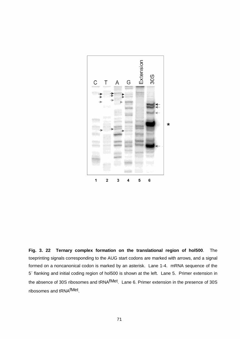
71
Fig. 3. 22 Ternary complex formation on the translational region of hol500. The
toeprinting signals corresponding to the AUG start codons are marked with arrows, and a signal
formed on a noncanonical codon is marked by an asterisk. Lane 1-4. mRNA sequence of the
5´ flanking and initial coding region of hol500 is shown at the left. Lane 5. Primer extension in
the absence of 30S ribosomes and tRNAfMet. Lane 6. Primer extension in the presence of 30S
ribosomes and tRNAfMet.

72
Fig. 3. 23 M14I change in Hol500 causes accelerated cell lysis. Lysis profiles observed
follwing thermal induction of E. coli LE392 lysogenized with λ∆Sthf::hol500 wt and
λ∆Sthf::hol500M14I.
Fig. 3. 24 Hol118(83) inhibits Hol500 in trans. Lysis profiles observed follwing thermal
induction of E. coli LE392 lysogenized with λimm434::hol500, and of an E. coli double lysogen
carrying λimm434::hol500 and λ∆Sthf::hol118(83) phage.
0 20 40 60 80 100
Time [min after induction
0
0,1
0,2
0,3
0,4
0,5
OD
600
Hol500
Hol500M14I
0 30 60 90 120 150
Time after induction [min]
0
0,1
0,2
0,3
0,4
0,5
OD
600
D Hol500/Hol118(83)
Imm Hol500 w t

73
3. 7. 6 Lysis timing mutant in the third transmembrane domain of Hol500
A λ∆Sthf::hol500∆M1F81L was isolated as a spontaneous mutant during the
construction of λ∆Sthf::hol500∆M1 phage (Fig. 3. 19. and Fig. 3. 20). This mutation in
the third transmembrane domain revelead a very early lysis timing phenotype compared
Hol500 wt.
Fig. 3. 25 F81L possibly influences oligomerization. Lysis profiles observed follwing
thermal induction of E. coli LE392 lysogenized with λ∆Sthf::hol500∆M1,
λ∆Sthf::hol500∆M1F81L and double lysogen carrying λimm434::hol500 and
λ∆Sthf::hol500∆M1F81L.
The phenotype of the mutant was further tested by expressing it in double
lysogens to Hol500 wt in λimm434::hol500. This change had a clear dominant effect
over the Hol500 wt (Fig. 3. 25) indicating its possible influence on oligomerization. The
F81L change created a LL motif of Leucins at the end of the third transmembrane
protein. This motif has been found in all holins, except Gp 17,5, tested in this work and
can therfore be of functional and structural importance for this protein (Fig. 3. 5, Fig. 3.
7) group.
0 30 60 90 120
Time after induction [min]
0
0,1
0,2
0,3
OD
600 Hol500DM1
Hol500DM1F81L
D Hol500/Hol500DM1F81L

74
3. 8 Complementation of the λ∆Sthf lysis defect with Hol2438 from a Listeria
innocua cryptic prophages
The Hol2438 gene was discovered in a cloned fragment of DNA from Listeria
innocua, which showed lytic activity against Listeria cells (Zink et al., 1995). The gene
is located upstream of cpl2438 endolysin. The only difference between Hol2438 and
Hol500 is the lack of the last amino acid residue, a positively charged lysine (K) (Fig. 3.
7), thereby reducing the positive charge of the C-terminus. Here, hol2438 was amplified
by PCR using Listeria chromosomal DNA as a template, and inserted into λ∆Sthf.
Recombinant phages were selected for plaque forming ability, and phage-carrying cells
were further tested for lysis timing.
Fig. 3. 26 Reduction of the positive charge at the C-terminus influences lysis timing of
Hol500. Lysis profiles observed follwing thermal induction of E. coli LE392 lysogenized with
λ∆Sthf::hol500 and λ∆Sthf::hol2438 phages.
It was surprising to find that only the lack of one K residue had such a profound effect
on the lysis timing of the holin (Fig. 3. 26). These results are in accordance with results
obtained from extensive analysis of the C-terminal region of λ S holin which revealed
that the reduction of positive charges on the C-terminus accelerated cell lysis.
0 20 40 60 80 100
Time [min] after induction
0
0,1
0,2
0,3
0,4
0,5
OD
600
Hol500
Hol2438

75
3. 9 MscL can not complement the lysis defect of λ∆Sthf
MscL, the large mechanosensitive channel belonges to the group of high-
conductance ion channels gated by mechanical forces on cell membranes, and
functions in osmoregulation. These proteins protect bacteria from lysis upon osmotic
shock. The mscL gene codes for a 15 kd protein present in the cytoplasmic membrane.
Homologous genes have been identified both in gram-negative and gram-positive
bacteria (Blount et al., 1999). MscL has two transmembrane domains, and the N-
terminus and C-teminus positioned in the cytoplasm of the cell (Blount et al., 1996;
Chang et al., 1998). The exact conformation of MscL homologous channel from
Mycobacterium tuberculosis has been determined by x-ray crystallography. It has been
shown that the protein forms a homopentamer channel in the membrane (Chang et al.,
1998). Further, hypotonic shock causes MscL channel to pass solutes, including small
proteins as thioredoxin but also elongation factor Tu and DnaK proteins through the
cytoplasmic membrane (Ajouz et al., 1998; Berrier et al., 2000). The topology of MscL
protein is similar to the topology of the class II holin proteins: N-terminus and C-
terminus are positioned in the cytoplasm, with two transmembrane domains traversing
the cytoplasmic membrane. The fact that, through the MscL channel, proteins can be
transferred from the cytoplasm to the periplasm of the bacterial cell promted to test
whether MscL can complement the lysis defect of λ∆Sthf.
For this purpose, a gene fragment encoding MscL was inserted into λ∆Sthf,
phages packaged, and plated to test for plaque forming ability. However, no plaque
forming ability for MscL could be detected. The recombinant λ∆Sthf::mscL phage was
then selected by direct lysogenization of LE392 cells. No lysis could be observed
following induction of λ∆Sthf::mscl lysogens.
It was shown here that a very similar protein to holins, in the organization of basic
structural domains, was unable to complement the lysis defect of λ∆Sthf (Fig. 3. 7).
Therefore, holin function seems to be specific, in spite of the great diversity of holin
protein sequences yet discovered

76
4. Discussion
The last compilation of holins and putative holin sequences revealed more than
100 protein sequences (Wang et al., 2000). Most genes coding for putative holins are
found upstream of adjacent endolysin genes. They specify small membrane proteins
having at least one transmembrane domain flanked by charged and hydrophilic N- and
C- terminal domains. Not very many holins and putative holin protein were
experimentally tested for their function. Holins start a permeabilization process, "hole"
formation, in the membrane which enables the endolysin to pass through the membrane
and reach the periplasm to degrade the peptidoglycan. Holins form pores in the
membrane in a time dependent manner and the whole process assumes a crucial
timing function. This function can be fulfilled by very different proteins, which are
currently classified (according to sequence similarities into at least 34 holin families
(Young et al., 2000). A novel genetic system is described in this work, which allows to
assess at least qualitatively the functional properties of various holins of all classes, in
E. coli λ∆Sthf::hol lysogens. Induction of a lysogen permits observation of a
synchronized lytic cycle, and allows reasonably precise assessment of the course and
timing of lysis.
In this study, holins from phages infecting both gram-negative and gram-positive
bacteria where analysed, with a special focus on holins from Listeria monocytogenes
bacteriophages A118 and A500. Two proteins were also tested which do not belong to
holins per se: Gp orf 2 from Listeria monocytogenes bacteriophage A511, and MscL,
the mechanosensitive channel from E. coli. Both proteins were unable to complement
the lysis defect of λ∆Sthf. It was demonstrated that λ∆Sthf can be used to specifically
select a protein with holin function.
4. 1 Holins from Escherichia coli phages
Three different holins from E. coli phages were tested here: λ S, and two holins
from non-lambdoid phages T7 and T4. S belongs to the class I, while Gp 17.5 was
classified as class II, and T4, T as a completely different holin type (Wang et al., 2000).
Function of S is post-transcriptionally controlled by a dual start motif, which
permits synthesis of two N-terminally different polypeptides, S107 and S105 with
different functions (Bläsi and Young, 1996; Blasi et al., 1989). The timing and rate of
cell lysis established here for wild-type S and S105 were similar to the values reported

77
earlier (Raab et al., 1988). However, it was reported that lysis in an induced λ lysogen
expressing only S107 was almost absent. This is in contrast to here reported results,
where induction of λ∆Sthf::S107 resulted in lysis starting at approximately 60 min. This
may be explained by the different translation initiation sites present in the two
constructs. In native λ, a relatively inefficient Shine-Dalgarno sequence with a very
short spacer directs the ribosome to the first start codon, whereas the layout of λ∆Sthf
enables identical translation conditions for all the cloned holin genes, since they all
exhibit a start codon immediately downstream of the EcoRI site. In terms of
comparative holin analysis, this can be regarded as an advantage.
The difference in lytic capacity of S105 and S107 is determined by the
distribution of amino-terminal charged residues, and the total amino-terminal net charge
(Steiner and Bläsi, 1993). The here presented finding that the S107-M3K protein
resulted in a complete non-lytic phenotype confirms results obtained using a similar
mutant (Graschopf and Bläsi, 1999). It has been observed that presence of additional
positive charge in the N-terminus in S hindered it from translocating through the
membrane, and therefore prevented formation of the active Nout-Cin topology
(Graschopf and Bläsi, 1999). Our results are in excellent agreement with these data,
and justify the conclusion that λ∆Sthf represents a quick and reliable system for
evaluation of holin function in an easy-to-use λ background.
Three lysis genes have been identified in bateriophage T7 encoding: Gp 3,5 a
murein hydrolase which can also bind and inhibit the fuction of T7 RNA polymerase, Gp
17,5 and Gp 18,5 which is homologous to the λ Rz (Young, 1992). It has been
suggested that Gp 17,5 and Gp 3,5 have the same function in T7 as S and R in λ
phage. Tested in λ∆Sthf, Gp 17,5 caused a sudden and saltatory onset of lysis 30 min
after induction, which confirmed its holin function. Gp 17,5 was initially classified as a
class II holin (Wang et al., 2000; Young, 1992). However, analysis for potential
transmembrane domains using the TMHMM 2.0 software predicts only one
transmembrane domain to be formed and a high probability for a Nout-Cin topology for
the holin (Fig. 3. 5).
It has been shown that T from phage T4 can complement the S gene defect of λ
and it was postulated that T is a functional equivalent of S (Lu and Henning, 1992). In a
λ hybrid were T was inserted in the place of the S, T causes an abrupt cell lysis starting
about 20 min after the induction (Ramanculov and Young, 2001a). Lysis timing of
λ∆Sthf::t is about 30 min and this difference reflects different expression conditions in

78
these two phages. The most interesting observation was the finding that Gp 17,5 and
Gp T have almost identical lysis timing tested in λ∆Sthf. T has a single transmembrane
domain near the N-terminus of the protein with a large C-terminal part positioned in the
periplasm of the cell (Ramanculov and Young, 2001b). This topology was established
using gene fusions with phoA (alkaline phosphatase) and lacZ (ß-galactosidase) genes
and is in perfect accordance with the prediction for transmembrane domains made by
TMHMM 2.0. Experimental data for gp 17,5 toplogy of are not available. However,
according to the prediction for transmembrane domains T and Gp 17, 5 would group
into the same class of holins which posess only one transmembrane domain. This
similar behaviour of these very different holins (S, T, Gp 17, 5) of E. coli phages tested
in an identical E. coli-dependent λ∆Sthf system is not based on similarity of protein
sequences. (Fig. 3. 5). It may be hypothesized that holin function (pore formation and
timing) relies on some underlying structural principles in the sequences of the
transmembrane domains of holin proteins.
4. 2 HolTw from Stapylococcus aureus bacteriophage Twort
HolTw from S. aureus bacteriophage Twort belongs to the class II holins (see
Fig. 3. 7), has a positively charged N-terminus, and an extensive, negatively charged C-
terminus (Loessner et al., 1998). λ∆Sthf::holTW showed an "early lysis" phenotype
and, as a consequence, conferred a plaque forming defect. Surprisingly, the mutant
polypeptide HolTW-M in λ∆Sthf::holTW-M delayed the lysis event as compared to the
native protein. This difference is due to the presence of 5 additional C-terminal residues
in the mutant protein, and a V52L substitution. It is not entirely clear which of the two
changes is responsible for the observed effect. The V52 residue is located within the
predicted ß-turn that separates the two possible membrane spanning domains, and
substitution by leucine was not predicted to alter folding properties and protein structure.
However, the addition of two charged residues at the C-terminus increases
hydrophilicity of this domain. Important findings in λ S were that the hydrophilic C-
terminal domain is not essential for oligomerization (Rietsch et al., 1997), but represents
a regulatory domain that interacts with the membrane in order to schedule lysis timing
(Bläsi et al., 1999). These results obtained with the mutated HolTW-M protein suggest
that (i) the extensively charged C-teminus somehow schedules lysis timing, and (ii)
show that mutations which add charged residues without affecting net charge can
influence holin function. Although it seems obvious that the primary amino-acid

79
structure of this class II holin determines a Nin - Cin topology (TMHMM 2.0) of the
protein in the membrane, the exact function of the large C terminus needs to be further
elucidated.
4. 3 Hol118 from Listeria monocytogenes bacteriophage A118
Holins of λ and A118 are structurally similar but functionally different. Hol118
belongs to the same holin class as the λ S holin. Analysis for potential transmembrane
domains (TMHMM 2.0) (Sonnhammer et al., 1998) identified three transmembrane
domains and indicated a high probability for a Nout- Cin topology (Fig. 4. 1). The activity
of a Hol32-lacZ fusion protein over the lack of Hol32-phoA fusion protein activity
provided experimental evidence for the Nout position of the N-terminal domain of
Hol118. This domain has a negative charge of -2, and two potential translational starts
(Fig. 3. 7A). However, these do not seem to have an important role in regulating its
function, since no significant difference in lysis could be seen among the different N-
terminal variants. Changing AUG(2) codon into CTG had no major effect, and
changing of AUG(1) into CTG did also not influence lysis. The Hol118(93) protein,
(truncated for the first three amino acids) can complement the lysis defect of λ∆Sthf but
inserts into the membrane of E. coli less efficiently compared to Hol118(96). It was
surprising to find that disruption of the membrane potential had no premature activating
effect on Hol118. Due to the overall negative charge of the N-terminal portion of
Hol118, it is reasonable to conclude that the energized membrane plays an active role
in positioning the N-terminus in the periplasm of the cell (Cao et al., 1995). Negatively
charged amino acids in the N- terminal domains together with hydrophobic forces are
important for the proper orientation of proteins in the membrane depending on the intact
membrane potential (Kiefer and Kuhn, 1999; Kiefer et al., 1997).
Hol118 appeared in the cytoplasmic membrane of E. coli 20 min after induction of
λ∆Sthf::hol118, so that the lysis process must be delayed to the point determined by the
"lysis clock" of Hol118. Here a model is proposed in which Hol118(83), translated from
the AUG(3) start codon, acts as the intragenic inhibitor of Hol118 permeabilization.
A translational start at M14 was detected in hol118, and in vitro translation of
hol118 yieled two bands were the smaller corresponds to Hol118(83). Western blotting
detected two identical Hol118 polypeptides in membrane fractions of induced E. coli
(λ∆Sthf::hol118) lysogens, and in Listeria monocytogenes infected with A118.

80
Expressed in λ∆Sthf, Hol118(83) had no lytic activity by itself, and recombinant
λ∆Sthf::hol118(83) was unable to form plaques. Hol118 (83) was expressed in trans
from λ∆Sthf and from a transactivation plasmid. In double lysogenes carrying
λ∆Sthf::hol118(83) and λimm434::hol118, Hol118(83) caused delayed lysis. Cell lysis
interference by Hol118(83) was also observed when Hol118(83) was made in trans from
pBRT-Hol118(83). Taken together, all these data strongly suggest that the lysis
process is inhibited by Hol118(83). In spite of the lack of a permeabilization function
which leads to pore formation, Hol118(83) had toxic effect on cells, when expressed
from a plasmid. In fact, it was not possible to clone hol118(83) on any high copy
plasmid (data not shown).
The start codon for the Hol118(83) holin is positioned within the first
transmembrane domain of the protein. In order to prevent translation initiation, the M14
codon, was changed into CTG or ATT. Both mutations resulted in accelerated cell lysis,
but to a different degree. Changes in the start codon for Hol118(83) also influenced the
amino acid sequence of the protein starting upstream. It is important to emphasize that
three mutations changing the AUG(3) codon into GTG, ATT or CTG had a dominant
effect on lysis timing when expressed in trans to wild type Hol118. It was reported
earlier that dominant mutations in holins point to a possible influence on the
oligomerization process, leading to "hole" formation (Raab et al., 1988). From the
character of changes at this position, it is obvious that M14 position forms part of the
intrinsic lysis clock of Hol118, besides being a start codon for Hol118(83) protein. For
Hol118(83), TMHMM 2.0 software predicts only two transmembrane domains to be
formed, which correspond to the second and third transmembrane domain of Hol118
(Fig. 4. 1). The protein could not complement the lysis defect of λ∆Sthf, but it was able
to delay lysis timing of Hol118 in trans. Further, the mutation in M14 influenced the
interaction with Hol118(83). We have observed that lysis timing could not be delayed in
cells expressing Hol118(83) from pBRT to induced λ∆Sthf::hol118M14I,
λ∆Sthf::hol118M14V, or λ∆Sthf::hol118M14L phages. Hence, Hol118(83) had a
recessive effect on lysis timing of Hol118M14I, Hol118M14L and Hol118M14V variants,
but was able to delay the lysis timing of the wildtype holin. From the present results, it
can be postulated that Hol118(83) constitutes part of the lysis clock of Hol118 and is
superimposed on the intrinsic lysis clock. Results also clearly demonstrated that the
first transmembrane domain is crucial for permeabilization function and influences the
interaction with Hol118(83). The actual effector/inhibitor ratio and the molecular basis of

81
lysis inhibition and its regulation has to be further elucidated in the A118 genetic
background.
Periplasm
Hol118(96) Hol118(83)
+ + + + N + + + + + + + + + + + + + + + + + + + + + + + + +
11
- - - - - - - - - - - - - - - N - - - - - - - - - - - - - - - - - - C Cytoplasm C
Fig. 4. 1 Membrane topology for Hol118(86), and Hol118(83). Membrane topology was
depicted according to a bioinformatic analysis by TMHMM 2.0 software
(http://www.cbs.dtu.dk/krogh/TMHMM/).
4. 4 Hol500 from Listeria monocytogenes bacteriophage A500 and differences
between Hol118, Hol500, and Hol2438
Hol500, Hol118 and Hol2438 have almost identical amino acid sequences (Fig.
3. 7). The N- terminus of Hol500 has an overall neutral charge; in the first ß-turn a L34I
change is present; the second transmembrane domain differs in two amino acids: I46V
and F56S, and the C- terminus of Hol500 is different in the overall charge, caused by
the E93K substitution and through a S88A change. The only difference of Hol2438 to
Hol500 is the lack of the last lysine in the C-terminal tail of the protein. In spite of these
small changes in the amino acid sequences, their detected lytic properties in λ∆Sthf
were significantly different.
Hol500 has a possible dual translational motif, two start codon separated by a
codon for lysine. The hol500 dual start alleles that were tested in this work were
defined by their similarity to λS and to the corresponding translational initiation sites in
hol118 gene of Listeria monocytogenes bacteriophage A118. However, the gpR-
dependent cell lysis mediated by the individual hol500 alleles is different. The longer
Hol500(96) polypeptide enabled reasonably quick cell lysis. The protein starting at M4,
Cytoplasmicmembrane
82
Hol500(93), could not be defined as the effector of lysis, and changing the AUG(4)
codon into a CTG, Hol500(96) led to an abrupt cell lysis not observed for other Hol500
alleles Cell lysis mediated by Hol500 holin could be triggered prematurely through the
destruction of the intact membrane potential of the cell, so that the lysis process is
inhibited by the energized membrane. The presence of aditional positive charge in
Hol500-M2K mutant inhibits cell lysis which is in accordance with the properties of S
mutants were the excess of positive charge on the N- terminus completely blocks the
cell lysis event (Graschopf and Bläsi, 1999). Western blot analysis showed a
correlation between protein concentration in the membrane of the E. coli cells and the
observed lysis pattern. Mutants in the N-terminus, which accelerated lysis had, in the
same time point, more holin proteins present in the membrane. These results confirm
the postulated critical-concentation model, in which lysis time depends on the
concentration level of the protein in the membrane of the bacterial cell (Gründling et al.,
2001). All three M codons at the beginning of hol500 were used as transational starts in
λ∆Sthf, and M14 within the first transmembrane domain. As observed for Hol118,
mutations in the first M codons did not significantly influence the lytic property of hol500.
However, changing AUG(4) codon into AUU led to accelerated cell lysis of Hol500; the
same mutation had a similar effect on lysis in Hol118. Hol118(83) was able to inhibit in
trans cell lysis mediated by Hol500. The effect of the cognate Hol500(83) has to be
tested separately in order to see the in trans effect of this protein. Due to few
differences in the amino acid composition of Hol118(83) and Hol500(83), the inhibitory
function for Hol500(83) could be extrapolated from the function of Hol118(83). At any
rate, it would be interesting to test the inhibitory effect of Hol500(83) in an experiment
because of the I46V and F56S changes in the second transmembrane domains
between the two Listeria holins. Both changes could have an effect on the interaction
between transmembrane domains; valine through its branched aliphatic chain, and
serine through the hydroxyl group which was found to stabilize interactions between
membrane spanning domains (Lemmon et al., 1997; Zhou et al., 2000).
It was surprising to find that the influence of a reduced net charge of +1 (Hol2438
compared to Hol500) led to such a pronounced effect, causing lysis to start 30 min
before Hol500. The C-terminal domain of Hol118 is also reduced in its positive charge
in comparision to Hol500, but lysis timing is not influenced by this feature, based on the
observation that the lysis timing is much delayed for Hol118 compared to Hol500.
Therefore it may be possible to conclude that other amino acids in the C-terminal
domain or in other domains compensate this charge difference in the C-terminal
83
domains of Hol118 and Hol500. It was found that the single effect of charged amino
acids in the C-terminal domain, as observed in Hol2438 has the same influence on
timing regulation as observed in λ. Excess positive charge in the C-terminus delayed
lysis timing.
5. General conclusions
Holins are major regulatory proteins of the lysis process mediated by
bacteriophages which use the dual protein strategy in order to lyse bacterial cells at the
end of the lytic growth cycle (Young, 1992). These small membrane proteins
permeabilize the membrane in a time dependent manner, allow endolysin access to the
peptidoglycan of the cell wall which leads to its degradation and subsequent cell lysis.
Holin constitute the largest group of functional homologous protein with almost no
similarity on the level of primary protein structure. In order to investigate the
relationship between the structure and function within holins, λ∆Sthf phage vector has
been constructed which was used to asses qualitatively functional properties of holins
from very different bacteriophages and with various structural features. Holin function
has been defined as the possibility of a membrane protein to complement the lysis
defect of λ∆Sthf phage, which has a deletion of it own holin gene. From the here
presented results, it is possible to conclude that holin function is specific; membrane
proteins such as MscL with a similar domain structure to holins can not complement the
lysis defect of the phage. This finding indicates that the postulated common domain
structure (hydrophylic N-terminal tail, two or more transmembrane domains, highly
charged and hydrophylic C-terminal tail) is not enough to fulfill holin function. Lytic
properties of holins from bacteriophage T7, (Gp 17.5), T4, (T) and A118, (Hol118)
revealed that at least one transmembrane domain is necessary and sufficient for the
permeabilization function. Gp 17.5 and T holin have a different organization of
structural domains but very similar lytic properties. The structural basis for this similar
function has to be sought in the features of the transmembrane domains of T and Gp
17, 5. Holins oligomerize in the membrane of the cell in the process of membrane
permeabilization (Zagotta and Wilson, 1990). Oligomerization depends on specific and
unspecific interactions between membrane spanning domains of membrane proteins
(Lemmon et al. 1997). Therefore, the key structural determinants for holins must reside
within this segments.

84
Hol118 and Hol500 from Listeria bacteriophages A118 and A500 have the same
overall organization of the basic structural domains as λ S holin: charged N-terminal tail,
three potential transmembrane domains and hydrophylic, charged C-terminal tail.
However, functional analysis of Hol118 revealed difference in lytic features to S holin.
It has here been demonstarted that the potential double translational starts present at
the 5´ end of hol118 and hol500 is fuctionally non-homologous to the double start of S.
Differences in the lenght and amino acid composition of the N-terminal portion of
Hol118 and Hol500 influenced insertion of proteins in the cytoplasmic membrane. Lysis
timing of Hol118/Hol500 was influenced by mutations in the region of the first and third
transmembrane domain and the C-terminal tail. A model has been proposed in which a
protein starting at the fourteenth codon (M14), within the gene, Hol118(83) or
Hol500(83), is acting as the inhibitor of lysis and regulates the lysis timing
superimposed on the intrinsic lysis timing which is determined by the features of the
transmembrane domains. Experimental evidences have been presented for this
proposed model. Further experiments which are designed to elucidate the interaction
between the truncated inhibitor Hol118(83) and the effector in the membrane of Listeria
cells are desirable, in order to unambiquously prove the novel mode of holin inhibition
function proposed here.
85
6. References
Ackermann, H.W., and Dubow, M.S. (1987) General properties of bacteriophages..In:
Viruses of procaryotes:. Boca Raton, Florida: CRC Press.
Ackermann, H.W., and DuBow, M.S. (1987) Viruses of procaryotes. Boca Raton,
Florida, USA: CRC Press.
Ajouz, B., Berrier, C., Garrigues, A., Besnard, M., and Ghazi, A. (1998) Release of
Thioredoxin via the Mechanosensitive channel Mscl during osmotic downshock
of Escherichia coli cells. J Biol Chem 273: 26670-26674.
Arendt, E.K., Daly, C., Fitzgerald, G.F., and van de Guchte, M. (1994) Molecular
characterization of Lactococcal bacteriophage Tuc2009 and identification and
analysis of genes encoding lysin, a putative holin, and two structural proteins.
Appl Environ Microbiol 60: 1875-1883.
Barenboim, M., Chang, C.Y., dib Hajj, F., and Young, R. (1999) Characterization of the
dual start motif of a class II holin gene. Mol Microbiol 32: 715-727.
Bernhardt, G., T., Wang, I-N., Struck, K., D., and Young, R. (2001) A protein antibiotic in
the phage Qb virion: Diversity in the lysis targets. Science 292: 2326-2329.
Berrier, C., Garrigues, A., Richarme, G., and Ghazi, A. (2000) Elongation factor Tu and
Dnak are transferred from the cytoplasm to the periplasm of Escherichia coli
during osmotic downshock presumably via the Mechanosensitive channel Mscl. J
Bacteriol 182: 248-251.
Birge, A.E. (1994) Bacterial and bacteriophage genetics: Springer-Verlag.
Bläsi, U., and Young, R. (1996) Two beginnings for a single purpose: The dual-start
holins in the regulation of phage lysis. Mol Microbiol 21: 675-682.
Bläsi, U., Chang, C.Y., Zagotta, M.T., Nam, K.B., and Young, R. (1990) The lethal λ S
gene encodes its own inhibitor. EMBO J 9: 981-989.
86
Bläsi, U., Fraisl, P., Chang, C.Y., Zhang, N., and Young, R. (1999) The C-terminal
sequence of the λ holin constitutes a cytoplasmic regulatory domain. J Bacteriol
181: 2922-2929.
Bläsi, U., Nam, K., Hartz, D., Gold, L., and Young, R. (1989) Dual translational
initiation sites control function of the λ S gene. EMBO J 8: 3501-3510.
Blount, P., Sukharev, S.I., Moe, P.C., Martinac, B., and Kung, C. (1999)
Mechanosensitive channels of bacteria. Methods Enzymol 294: 458-482.
Blount, P., Sukharev, S.I., Moe, P.C., Schroeder, M.J., Guy, H.R., and Kung, C. (1996)
Membrane topology and multimeric structure of a Mechanosensitive channel
protein of Escherichia coli. EMBO J 15: 4798-4805.
Bolivar, F., Rodriguez, R.L., Greene, P.J., Betlach, M.C., Heyneker, H.L., and Boyer,
H.W. (1977) Construction and characterization of new cloning vehicles. II. A
multipurpose cloning system. Gene 2: 95-113.
Bon, J., Mani, N., and Jayaswal, R.K. (1997) Molecular analysis of lytic genes of
bacteriophage 80 α of Staphylococcus aureus. Can J Microbiol 43: 612-616.
Campbell, J.H., and Rolfe, B.G. (1975) Evidence for a dual control of the initiation of
host-cell lysis caused by phage Lambda. Mol Gen Genet 139: 1-8.
Cao, G., Kuhn, A., and Dalbey, R.E. (1995) The translocation of negatively charged
residues across the membrane is driven by the electrochemical potential:
Evidence for an electrophoresis-like membrane transfer mechanism. EMBO J 14:
866-875.
Chang, C.Y., Nam, K., and Young, R. (1995) S gene expression and the timing of lysis
by bacteriophage Lambda. J Bacteriol 177: 3283-3294.
Chang, G., Spencer, R.H., Lee, A.T., Barclay, M.T., and Rees, D.C. (1998) Structure of
the Mscl homolog from Mycobacterium tuberculosis: A gated mechanosensitive
ion channel. Science 282: 2220-2226.
87
Chauthaiwale, V.M., Therwath, A., and Deshpande, V.V. (1992) Bacteriophage Lambda
as a cloning vector. Microbiol Rev 56: 577-591.
Damman, C.J., Eggers, C.H., Samuels, D.S., and Oliver, D.B. (2000) Characterization
of Borrelia burgdorferi Blya and Blyb proteins: A prophage-encoded holin-like
system. J Bacteriol 182: 6791-6797.
Doermann, A.H. (1952) The intracellular growth of bacteriophages. I. Liberation of
intracellular bacteriophage T4 by premature lysis with another phage or with
cyanide. J. Gen. Physiol. 35: 645-656.
Dower, W.J., Miller, J.F., and Ragsdale, C.W. (1988) High efficiency transformation of
E. coli by high voltage electroporation. Nucleic Acids Res 16: 6127-6145.
Ellis, E.L., and Delbrück, M. (1939) The growth of bacteriophage. J. Gen. Phisiology 23:
365-384.
Gasson, M.J., and Anderson, P.H. (1985) High copy number plasmid vectors for use in
Lactic streptococci. FEMS Microbiol Lett 30: 193-196.
Graschopf, A., and Bläsi, U. (1999) Molecular function of the dual-start motif in the λ S
holin. Mol Microbiol 33: 569-582.
Gründling, A., Bläsi, U., and Young, R. (2000a) Genetic and biochemical analysis of
dimer and oligomer interactions of the λ S holin. J Bacteriol 182: 6082-6090.
Gründling, A., Bläsi, U., and Young, R. (2000b) Biochemical and genetic evidence for
three transmembrane domains in the class I holin, λ S. J Biol Chem 275: 769-
776.
Gründling, A., Manson, M.D., and Young, R. (2001) Holins kill without warning. Proc
Natl Acad Sci U S A 98: 9348-9352.
Gründling, A., Smith, D.L., Bläsi, U., and Young, R.Y. (2000c) Dimerization between the
holin and holin inhibitor of phage λ. J Bacteriol 182: 6075-6081.

88
Gutierrez, C., and Devedjian, J.C. (1989) A plasmid facilitating in vitro construction of
phoA gene fusions in Escherichia coli. Nucleic Acids Res 17: 3999.
Hartz, D., McPheeters, D.S., Traut, R., and Gold, L. (1988) Extension inhibition analysis
of translation initiation complexes. Methods Enzymol 164: 419-425.
Henrich, B., Binishofer, B., and Bläsi, U. (1995) Primary structure and functional
analysis of the lysis genes of Lactobacillus gasseri bacteriophage φ adh. J
Bacteriol 177: 723-732.
Johnson-Boaz, R., Chang, C.Y., and Young, R. (1994) A dominant mutation in the
bacteriophage lambda S gene causes premature lysis and an absolute defective
plating phenotype. Mol Microbiol 13: 495-504.
Kashige, N., Nakashima, Y., Miake, F., and Watanabe, K. (2000) Cloning, sequence
analysis, and expression of Lactobacillus casei phage Pl-1 lysis genes. Arch Virol
145: 1521-1534.
Kiefer, D., and Kuhn, A. (1999) Hydrophobic forces drive spontaneous membrane
insertion of the bacteriophage Pf3 coat protein without topological control.
EMBO J 18: 6299-6306.
Kiefer, D., Hu, X., Dalbey, R., and Kuhn, A. (1997) Negatively charged amino acid
residues play an active role in orienting the Sec-independent Pf3 coat protein in
the Escherichia coli inner membrane. EMBO J 16: 2197-2204.
Krogh, S., Jorgensen, S.T., and Devine, K.M. (1998) Lysis genes of the Bacillus subtilis
defective prophage PBSX. J Bacteriol 180: 2110-2117.
Kyhse-Andersen, J. (1984) Electroblotting of multiple gels: A simple apparatus without
buffer tank for rapid transfer of proteins from polyacrylamide to nitrocellulose. J.
Biochem Biophs Methods 10: 203-209.
Lehninger, L. A. (1982) Principles of Biochemistry. New York: Worth Publishers, Inc

89
Lemmon, A., Mark, MacKenzie, R., Kevin, Arkin, T., Isiah, and Engelman M., D.
(1997) Transmembrane α- helix interactions in folding and oligomerization of
integral membrane proteins. In Membrane protein assembly. Heijne, G.v. (ed): R.
G. Landes Company.
Lewin, B. (1997) Gene VI. New York: Oxford University Press.
Loessner, M.J. (1991) Improved procedure for bacteriophage typing of Listeria strains
and evaluation of new phages. Appl Environ Microbiol 57: 882-884.
Loessner, M.J., and Busse, M. (1990) Bacteriophage typing of Listeria species. Appl
Environ Microbiol 56: 1912-1918.
Loessner, M.J., Wendlinger, G., and Scherer, S. (1995) Heterogeneous endolysins in
Listeria monocytogenes bacteriophages: A new class of enzymes and evidence
for conserved holin genes within the siphoviral lysis cassettes. Mol Microbiol 16:
1231-1241.
Loessner, M.J., Schneider, A., and Scherer, S. (1996) Modified Listeria bacteriophage
lysin genes (ply) allow efficient overexpression and one-step purification of
biochemically active fusion proteins. Appl Environ Microbiol 62: 3057-3060.
Loessner, M.J., Gaeng, S., and Scherer, S. (1999) Evidence for a holin-like protein
gene fully embedded out of frame in the endolysin gene of Staphylococcus
aureus bacteriophage 187. J Bacteriol 181: 4452-4460.
Loessner, M.J., Maier, S.K., Daubek-Puza, H., Wendlinger, G., and Scherer, S. (1997)
Three Bacillus cereus bacteriophage endolysins are unrelated but reveal high
homology to cell wall hydrolases from different bacilli. J Bacteriol 179: 2845-
2851.
Loessner, M.J., Gaeng, S., Wendlinger, G., Maier, S.K., and Scherer, S. (1998) The
two-component lysis system of Staphylococcus aureus bacteriophage Twort: A
90
large TTG-start holin and an associated amidase endolysin. FEMS Microbiol Lett
162: 265-274.
Lu, M.J., and Henning, U. (1992) Lysis protein T of bacteriophage T4. Mol Gen Genet
235: 253-258.
Manoil, C., and Beckwith, J. (1985) TnphoA: A transposon probe for protein export
signals. Proc Natl Acad Sci U S A 82: 8129-8133.
ópez, R., and Garc
system of the Pneumococcal phage Cp-1 in homologous and heterologous host
cells. J Bacteriol 180: 210-217.
Muniesa, M., Recktenwald, J., Bielaszewska, M., Karch, H., and Schmidt, H. (2000)
Characterization of a shiga toxin 2e-converting bacteriophage from an
Escherichia coli strain of human origin. Infect Immun 68: 4850-4855.
Nam, K., Bläsi, U., Zagotta, M.T., and Young, R. (1990) Conservation of a dual-start
motif in P22 lysis gene regulation. J Bacteriol 172: 204-211.
Neely, M.N., and Friedman, D.I. (1998) Functional and genetic analysis of regulatory
regions of coliphage H-19B: Location of shiga-like toxin and lysis genes suggest
a role for phage functions in toxin release. Mol Microbiol 28: 1255-1267.
Oki, M., Kakikawa, M., Yamada, K., Taketo, A., and Kodaira, K.I. (1996) Cloning,
sequence analysis, and expression of the genes encoding lytic functions of
bacteriophage φ g1e. Gene 176: 215-223.
Parreira, R., Sao-Jose, C., Isidro, A., Domingues, S., Vieira, G., and Santos, M.A.
(1999) Gene organization in a central DNA fragment of Oenococcus oeni
bacteriophage fOg44 encoding lytic, integrative and non-essential functions.
Gene 226: 83-93.

91
Pontarollo, R.A., Rioux, C.R., and Potter, A.A. (1997) Cloning and characterization of
bacteriophage-like DNA from Haemophilus somnus homologous to phages P2
and Hp1. J Bacteriol 179: 1872-1879.
Raab, R., Neal, G., Sohaskey, C., Smith, J., and Young, R. (1988) Dominance in
lambda S mutations and evidence for translational control. J Mol Biol 199: 95-
105.
Ramanculov, E., and Young, R. (2001a) Functional analysis of the phage T4 holin in a λ
context. Mol Genet Genomics 265: 345-353.
Ramanculov, E., and Young, R. (2001b) Genetic analysis of the T4 holin: Timing and
topology. Gene 265: 25-36.
Ramanculov, E., and Young, R. (2001c) An ancient player unmasked: T4 rI encodes a
T-specific antiholin. Mol Microbiol 41: 575-583.
Regamey, A., and Karamata, D. (1998) The N-acetylmuramoyl-l-alanine amidase
encoded by the Bacillus subtilis 168 prophage SPß. Microbiology 144: 885-893.
Rietsch, A., Fraisl, P., Graschopf, A., and Bläsi, U. (1997) The hydrophilic C-terminal
part of the lambda S holin is non-essential for intermolecular interactions. FEMS
Microbiol Lett 153: 393-398.
Sambrook, J., Maniatis, T., and Frisch, E.F. (1989) Molecular cloning: A laboratory
manual. Cold Spring Harbour, NY: Cold Spring Harbor Laboratory Press.
Sao-Jose, C., Parreira, R., Vieira, G., and Santos, M.A. (2000) The N-terminal region of
the Oenococcus oeni bacteriophage fOg44 lysin behaves as a bona fide signal
peptide in Escherichia coli and as a cis-inhibitory element, preventing lytic activity
on Oenococcal cells. J Bacteriol 182: 5823-5831.
Schägger, H., and von Jagow, G. (1987) Tricine-sodium dodecyl sulfate-polyacrylamide
gel electrophoresis for separation of proteins in the range from 1 to 100 kda.
Analytical Biochemistry 166: 368-379.

92
Schmidt, C., Velleman, M., and Arber, W. (1996) Three functions of bacteriophage P1
involved in cell lysis. J Bacteriol 178: 1099-1104.
Sonnhammer, E.L., von Heijne, G., and Krogh, A. (1998) A Hidden markov model for
predicting transmembrane helices in protein sequences. Proc Int Conf Intell Syst
Mol Biol 6: 175-182.
Steiner, M., and Bläsi, U. (1993) Charged amino-terminal amino acids affect the lethal
capacity of Lambda lysis proteins S107 and S105. Mol Microbiol 8: 525-533.
Tedin, K., Resch, A., Steiner, M., and Bläsi, U. (1995) Dual translational start motif
evolutionarily conserved in the holin gene of Bacillus subtilis phage φ 29. Virology
206: 479-484.
van Sinderen, D., Karsens, H., Kok, J., Terpstra, P., Ruiters, M.H., Venema, G., and
Nauta, A. (1996) Sequence analysis and molecular characterization of the
temperate Lactococcal bacteriophage r1t. Mol Microbiol 19: 1343-1355.
Vukov, N., Scherer, S., Hibbert, E., and Loessner, M.J. (2000) Functional analysis of
heterologous holin proteins in a λ∆S genetic background. FEMS Microbiol Lett
184: 179-186.
Wang, I., Dykhuizen, E.D., and Slobodkin, B.L. (1996) The evolution of phage lysis
timing. Evolutionary ecology 10: 545-558.
Wang, I.N., Smith, D.L., and Young, R. (2000) Holins: The protein clocks of
bacteriophage infections. Annu Rev Microbiol 54: 799-825.
Young, I., Wang, I., and Roof, W.D. (2000) Phages will out: Strategies of host cell lysis.
Trends Microbiol 8: 120-128.
Young, R. (1992) Bacteriophage lysis: Mechanism and regulation. Microbiol Rev 56:
430-481.
Zagotta, M.T., and Wilson, D.B. (1990) Oligomerization of the bacteriophage lambda S
protein in the inner membrane of Escherichia coli. J Bacteriol 172: 912-921.
93
Zhang, N., and Young, R. (1999) Complementation and characterization of the nested
Rz and Rz1 reading frames in the genome of bacteriophage lambda. Mol Gen
Genet 262: 659-667.
Zhou, F.X., Cocco, M.J., Russ, W.P., Brunger, A.T., and Engelman, D.M. (2000)
Interhelical hydrogen bonding drives strong interactions in membrane proteins.
Nat Struct Biol 7: 154-160.
Ziermann, R., Bartlett, B., Calendar, R., and Christie, G.E. (1994) Functions involved in
bacteriophage P2-induced host cell lysis and identification of a new tail gene. J
Bacteriol 176: 4974-4984.
Zimmer, M., Scherer, S., and Loessner, J. M. (2002) Genome Analysis of Clostridium
perfringens bacteriophage φ3626, which integrates into guaA and possibly
affects sporulation. J. Bacteriol 184: 4359-4368.
Zink, R., Loessner, M.J., and Scherer, S. (1995) Characterization of cryptic prophages
(monocins) in Listeria and sequence analysis of a holin/endolysin gene.
Microbiology 141: 2577-2584.

94
Appendices
Appendix A Table of the degenerated genetic code (Lewin, 1997)
First base Second base
U C A GUUU
UUC Phe
UAU
UAC Tyr
UGU
UGC CysU
UUA
UUG Leu
UCU Ser
UCC
UCA
UCG
UAA
UAG Stop
UGA Stop
UGG Trp
CAU
CAC HisC CUU
CUC Leu
CUA
CUG
CCU
CCC Pro
CCA
CCG
CAA
CAG Gln
CGU
CGC Arg
CGA
CGG
AUU
AUC Ile
AAU
AAC Asn
AGU
AGC SerA
AUA
AUG Met (Start)
ACU
ACC Thr
ACA
ACG
AAA
AAG Lys
AGA
AGG Arg
GAU
GAC AspG GUU
GUC Val
GUA
GUG
GCU
GCC Ala
GCA
GCGGAA
GAG Glu
GGU
GGC Gly
GGA
GGG

95
Appendix B Amino Acid Shorthand (Lehninger, 1982)
Amino acid Three - letterabbreviation
One - lettersymbol
Alanine Ala A
Arginine Arg R
Asparagine Asn N
Aspartic acid Asp D
Cysteine Cys C
Glutamine Gln Q
Glutamic acid Glu E
Glycine Gly G
Histidine His H
Isoleucine Ile I
Leucine Leu L
Lysine Lys K
Methionine Met M
Phenylalanine Phe F
Proline Pro P
Serine Ser S
Threonine Thr T
Tryptophan Trp W
Tyrosine Tyr Y
Valine Val V

96
Danksagung/Acknowledgements
Die vorliegende Arbeit wurde unter der Leitung von Prof. Dr. Siegfried Scherer am
Institut für Mikrobiologie, FML Weihenstephan der Technischen Universität München in
Freising durchgeführt.
Ich möchte mich bei Prof. Dr. Siegfried Scherer für die Möglichkeit am Institut für
Mikrobiologie zu promovieren, für dieses wunderschöne Thema, das ich in meiner
Dissertation bearbeiten konnte, seine jahrelange Unterstützung und Vertrauen herzlich
bedanken.
PD Dr. Martin Loessner möchte ich danken das er mich in seine Arbeitsgruppe
aufgenommen hat, für die Betreuung der Arbeit in allen Phasen, und Unterstützung.
Ebenso bedanke ich mich für die Korrektur dieser Arbeit und der wissenschaftlichen
Veröffentlichungen.
Prof. Dr. Udo Bläsi, PD Dr. Martin Loessner und Prof. Dr. Siegfried Scherer für die
Möglichkeit einige Zeit im Institut für Mikrobiologie und Genetik des Biozentrum´s der
Universität Wien verbrinngen zu können. Prof. Dr. Udo Bläsi danke ich für die
Anregung zu Analysen der Translationsinitiationspostionen (Toeprints) an den Listeria
Holin Genen und hilfreiche Disskusionen und Dr. Isabella Moll für die experimentelle
Durchführung der Toeprints die in dieser Arbeit vorgestellt sind und für ihr Interesse und
Zusammenarbeit an diesem Thema.
Prof. Dr. Erhard Bremer und Dr. Claude Gutierrez bedanke ich mich für die Plasmide
und Stämme für die Protein-Fusion Analysen mit der Alkalischen Phosphatase (PhoA).
Prof. Dr. Dieter Langosch möchte ich für die Übernahme des Korreferates danken.
Allen Mitarbeitern des Instituts für Mikrobiologie am FML, Weihenstephan danke ich für
eine freundliche und herzliche Atmosphäre und große Hilfsbereitschaft während meiner
Arbeit. Besonders möchte ich mich bei Markus Zimmer, Dr. Susanne Gäng und Patrick
Schiwek bedanken für die Zusammenarbeit im Labor und ihre Hilfe.

97
Bibliography
1. Nataša Vukov and Branka Vasiljevic. (1998) Analysis of plasmid pMZ1 from
Micromonospora zionenzis. FEMS Microbiol Lett 162: 317-23.
2. Nataša Vukov, Siegfried Scherer, Edward Hibbert and Martin J. Loessner.
(2000) Functional analysis of heterologous holin proteins in a λ∆S genetic background.
FEMS Microbiol Lett 184: 179-186.
3. Markus Zimmer, Nataša Vukov, Siegfried Scherer and Martin J. Loessner.
(2002) The murein hydrolase of the bacteriophage φ3626 dual lysis system is active
against all Clostridium perfringens cells (submitted).
4. Nataša Vukov, Isabella Moll, Udo Bläsi, Siegfried Scherer, and Martin J.
Loessner. (2002) Functional regulation of the Listeria monocytogenes bacteriophage
A118 holin by an intragenic inhibitor lacking the first transmembrane domain
(submitted).
5. Nataša Vukov, Isabella Moll, Siegfried Scherer, and Martin J. Loessner. (2002)
Comparision of three ralated Listeria bacteriophage holins: minor changes cause major
differences (in preparation).

98
Curriculum vitae
Name Nataša Vukov
Date of birth 25. 03. 1967 in Senta, Serbia, Yugoslavia
Education:
1985 Final exam in Chemistry, Secondary School with
Nature-Mathematical education, Senta, Serbia,
Yugoslavia
1985-1992 Graduate studies in Molecular Biology and Physiology,
Faculty of Biology, University of Beograd, Yugoslavia
1992 Diploma Thesis in Molecular Biology and Physiology:
Analysis of plasmid pMZ1 from Micromonospoa
zionenzis
1992-1997 Postgraduate studies in Molecular Genetics and
Genetic engineering
1997 Master Thesis in Biology: Analysis of plasmid pMZ1
from Micromonospoa zionenzis
1997-2002 Ph. D. student at the Institute for Microbiology, FML,
Weihenstephan at the Technical University, München
2002 Ph. D. Thesis: Functional analysis of heterologous
holins in a λ∆S genetic background
Working experiences:
1993-1997 Research assistant at the Institute of Molecular Genetics and
Genetic Engineering, Beograd, Serbia, Yugoslavia
1997-2002 Research at the Institute for Microbiology, FML,
Weihenstephan at Technische Universität, München,
financially supported by a fellowship from the "Vereinigung
der Freunde und Förderer des FML Weihenstephan"