Influence of sulfur-nitrosyl iron complexes of “µ-S...
7
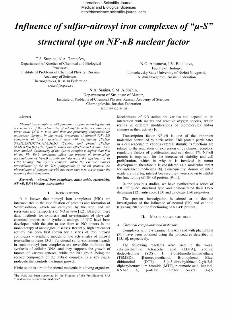
Influence of sulfur-nitrosyl iron complexes of “µ-S” structural type on NF-κB nuclear factor T.S. Stupina, N.A. Terent’ev, Departament of Kinetics of Chemical and Biological Processes, Institute of Problems of Chemical Physics, Russian Academy of Sciences, Chernogolovka, Russian Federation, [email protected] N.O. Antonova, I.V. Balalaeva, Faculty of Biology, Lobachevsky State University of Nizhni Novgorod, Nizhni Novgorod, Russian Federation N.A. Sanina, S.M. Aldoshin, Departament of Structure of Matter, Institute of Problems of Chemical Physics, Russian Academy of Sciences, Chernogolovka, Russian Federation [email protected] Abstract Nitrosyl iron complexes with functional sulfur-containing ligands are mimetics of the active sites of nitrosyl ferredoxines, donors of nitric oxide (NO) in vivo, and they are promising compounds for anticancer therapy. In this work, properties of nitrosyl [2Fe-2S] complexes of “µ-S” structural type with cysteamine [Fe2(µ- S(CH2)2NH3)2(NO)4]⋅2.5H2O (CysAm) and phenyl [Fe2(µ- SC6H5)2(NO)4] (Ph) ligands, which are effective NO donors, have been studied. Cytotoxicity of the CysAm complex is higher than that of the Ph. Both complexes affect the process of intranuclear accumulation of NF-κB protein and decrease the efficiency of its DNA binding. The CysAm complex, unlike the Ph one, induces nitrosylation of the 65 kDa polypeptide of NF-κB protein. No nitrosylation of polypeptide p50 has been shown to occur under the action of these complexes. Keywords – nitrosyl iron complexes, nitric oxide, cytotoxicity, NF-κB, DNA binding, nitrosylation I. INTRODUCTION It is known that nitrosyl iron complexes (NIC) are intermediates in the modification of proteins and formation of S-nitrosothiols, which are catalyzed by the iron, and are reservoirs and transporters of NO in vivo [1,2]. Based on these data, methods for synthesis and investigation of physical- chemical properties of synthetic analogs of NIC have been developed, with the aim to use them as NO donors in the monotherapy of oncological diseases. Recently, high anticancer activity has been first shown for a series of iron nitrosyl complexes – synthetic models of the active sites of nitrosyl iron-sulfur proteins [3-5]. Functional sulfur-containing ligands in such nitrosyl iron complexes are reversible inhibitors for synthesis of cellular DNA, and they suppress the growth of tumors of various geneses, while the NO group, being the second component of the hybrid complex, is a key signal molecule that controls the tumor growth. Nitric oxide is a multifunctional molecule in a living organism. Mechanisms of NO action are various and depend on its interaction with metals and reactive oxygen species, which results in different modifications of biomolecules and/or changes in their activity [6]. Transcription factor NF-κB is one of the important molecules controlled by nitric oxide. This protein participates in a cell response to various external stimuli; its functions are related to the regulation of expression of cytokines, receptors, regulatory factors of proliferation and cell death. [7]. NF-κB protein is important for the increase of viability and cell proliferation, which is why it is involved in tumor development; therefore it is considered as a molecular target for anticancer medicines [8]. Consequently, donors of nitric oxide are of a big interest because they were shown to inhibit the functioning of NF-κB protein. [9-11]. In the previous studies, we have synthesized a series of NIC of “µ-S” structural type and demonstrated their DNA damaging [12], anticancer [13] and cytotoxic [14] properties. The present investigation is aimed at a detailed investigation of the influence of neutral (Ph) and cationic (CysAm) NIC on the functioning of NF-κB protein. II. MATERIALS AND METHODS A. Chemical compounds and materials. Complexes with cysteamine (CysAm) and with phenylthiyl (Ph) have been obtained using the procedures described in [15,16], respectively. The following reactants were used in the work: ethylenediamine tetraacetic acid (EDTA), sodium dodecylsulfate (SDS), 1, 2-bis(dimethylamino)ethane (ТЕМЕD), 1β-mercaptoethanol, Bromophenol Blue, dithiotreitol (DTT), 3-(4,5-dimethylthiazol-2-yl)-2,5- diphenyltetrazolium bromide (MTT), p-cumaric acid, luminal, RNAse А, protease inhibitor cocktail (4-(2- The work has been supported by the Program of the Presidium of RAS "Fundamental sciences for medicine". International Scientific Journal Medical and Biological Sciences http://bioscience.scientific-journal.com
Transcript of Influence of sulfur-nitrosyl iron complexes of “µ-S...
Influence of sulfur-nitrosyl iron complexes of “µ-S”
structural type on NF-κB nuclear factor
T.S. Stupina, N.A. Terent’ev, Departament of Kinetics of Chemical and Biological
Processes, Institute of Problems of Chemical Physics, Russian
Academy of Sciences, Chernogolovka, Russian Federation,
N.O. Antonova, I.V. Balalaeva,
Faculty of Biology, Lobachevsky State University of Nizhni Novgorod,
Nizhni Novgorod, Russian Federation
N.A. Sanina, S.M. Aldoshin, Departament of Structure of Matter,
Institute of Problems of Chemical Physics, Russian Academy of Sciences, Chernogolovka, Russian Federation
Abstract
Nitrosyl iron complexes with functional sulfur-containing ligands are mimetics of the active sites of nitrosyl ferredoxines, donors of nitric oxide (NO) in vivo, and they are promising compounds for anticancer therapy. In this work, properties of nitrosyl [2Fe-2S] complexes of “µ-S” structural type with cysteamine [Fe2(µ-S(CH2)2NH3)2(NO)4]⋅2.5H2O (CysAm) and phenyl [Fe2(µ-SC6H5)2(NO)4] (Ph) ligands, which are effective NO donors, have been studied. Cytotoxicity of the CysAm complex is higher than that of the Ph. Both complexes affect the process of intranuclear accumulation of NF-κB protein and decrease the efficiency of its DNA binding. The CysAm complex, unlike the Ph one, induces nitrosylation of the 65 kDa polypeptide of NF-κB protein. No nitrosylation of polypeptide p50 has been shown to occur under the action of these complexes.
Keywords – nitrosyl iron complexes, nitric oxide, cytotoxicity, NF-κB, DNA binding, nitrosylation
I. INTRODUCTION It is known that nitrosyl iron complexes (NIC) are
intermediates in the modification of proteins and formation of S-nitrosothiols, which are catalyzed by the iron, and are reservoirs and transporters of NO in vivo [1,2]. Based on these data, methods for synthesis and investigation of physical-chemical properties of synthetic analogs of NIC have been developed, with the aim to use them as NO donors in the monotherapy of oncological diseases. Recently, high anticancer activity has been first shown for a series of iron nitrosyl complexes – synthetic models of the active sites of nitrosyl iron-sulfur proteins [3-5]. Functional sulfur-containing ligands in such nitrosyl iron complexes are reversible inhibitors for synthesis of cellular DNA, and they suppress the growth of tumors of various geneses, while the NO group, being the second component of the hybrid complex, is a key signal molecule that controls the tumor growth.
Nitric oxide is a multifunctional molecule in a living organism.
Mechanisms of NO action are various and depend on its interaction with metals and reactive oxygen species, which results in different modifications of biomolecules and/or changes in their activity [6].
Transcription factor NF-κB is one of the important molecules controlled by nitric oxide. This protein participates in a cell response to various external stimuli; its functions are related to the regulation of expression of cytokines, receptors, regulatory factors of proliferation and cell death. [7]. NF-κB protein is important for the increase of viability and cell proliferation, which is why it is involved in tumor development; therefore it is considered as a molecular target for anticancer medicines [8]. Consequently, donors of nitric oxide are of a big interest because they were shown to inhibit the functioning of NF-κB protein. [9-11].
In the previous studies, we have synthesized a series of NIC of “µ-S” structural type and demonstrated their DNA damaging [12], anticancer [13] and cytotoxic [14] properties.
The present investigation is aimed at a detailed investigation of the influence of neutral (Ph) and cationic (CysAm) NIC on the functioning of NF-κB protein.
II. MATERIALS AND METHODS
A. Chemical compounds and materials. Complexes with cysteamine (CysAm) and with phenylthiyl
(Ph) have been obtained using the procedures described in [15,16], respectively.
The following reactants were used in the work: ethylenediamine tetraacetic acid (EDTA), sodium dodecylsulfate (SDS), 1, 2-bis(dimethylamino)ethane (ТЕМЕD), 1β-mercaptoethanol, Bromophenol Blue, dithiotreitol (DTT), 3-(4,5-dimethylthiazol-2-yl)-2,5-diphenyltetrazolium bromide (MTT), p-cumaric acid, luminal, RNAse А, protease inhibitor cocktail (4-(2-
The work has been supported by the Program of the Presidium of RAS "Fundamental sciences for medicine".
International Scientific Journal Medical and Biological Sciences http://bioscience.scientific-journal.com
aminoethyl)benzenesulfonylfluoride hydrochloride, aprotinin, bestatin, E-64, leupeptin, pepstatin), anti-β-Actin primary (rabbit) antibody (Sigma, USA), 2-[4-(2-hydroxyethyl)piperazine-1-yl]ethanesulfonic acid (HEPES), Tris, Nonidet P-40 (NP-40) (Amresco, USA), acrylamide (Serva, Germany), primary rabbit antibodies against p53 protein and C-terminal fragment of poly (ADP-ribose) polymerase (PARP), anti-rabbit IgG secondary (goat) antibody conjugated with the horseradish peroxidase (Santa Cruz Biotechnology, USA), 4-(1,1,3,3-tetramethylbutyl)phenyl-poly(ethylene glycol) (Triton X-100), polyoxyethylene(20)sorbitan monolaurate (Tween-20), glycine («Panreac», Spain), bovine serum albumin (BSA), N,N'-methylene-bis-acrylamide, ammonium persulfate (APS), phenylmethanesulfonyl fluoride (PMSF) (Dia-M, Russia), incubation medium DMEM (Institute of Poliomyelitis and Viral Encephalitis of the Russian Academy of Sciences, Russia), fetal calf serum (BioWest, France), propidium iodide (Applichem. Germany).
B. Cell culture The experiments were carried out on the cell line M-HeLa
(Institute of Cytology, RAS). Cells were maintained at 37°С in atmosphere containing 5% CO2 in EMEM with addition of 10% fetal calf serum.
C. Investigation of cytotoxic effect and determination of IC50 dose Cytotoxicity was studied using the MTT assay. The cells
were plated in 24-well plates (1.5•104 cells per well) in the standard incubation medium. The CysAm complex was dissolved in DMSO before use. It was added into the incubation medium in the concentration of 1 to 15 µM in 24 h after the plating. The final concentration of DMSO was 0.1%. In 24 h after the addition of the compounds into the incubation medium, MTT was added to the final concentration of 0.45 mg·mL-1, and the cells were stained in the incubator for 4 h. After the staining, the incubation medium was removed, and formed crystals of MTT formazane were dissolved in the acidified alcohol (50% isopropyl alcohol, 0.05 M HCl). The staining intensity was determined at 570 nm. The staining intensity of the cells treated with solvents (DMSO) was taken as 100%. The IC50 values were calculated using the median effect analysis [Chou & Talalay 1984].
D. Flow cytofluorimetry The cells were plated in Petri dishes in the standard
incubation medium (106 cells per point). In 24 h after the plating, the compounds under study were added into the incubation medium at the concentrations corresponding to IC50. DMSO was added at concentration 0.1%.
For studying the cell cycle, in 24 h after adding the studied compounds, the cells were collected by trypsinization, washed thrice with PBS (137 mM NaCl, 2.68 mM KCl , 4.29 mM Na2HPO4, 1.47 mM KH2PO4, pH 7.4), and then fixed and permeabilized by a dropwise addition of 70% aqueous ethanol cooled to -20°С. The samples were kept for 12 h at 4°C. Ethanol from the fixed samples was removed by triple washing with a solution of BSA (1%) in PBS. The washed cells were
resuspended in PBS containing 1% of BSA, propidium iodide (0.1 mg·mL-1) and RNAse (1 mg·mL-1) and incubated for 1 h at 37°С.
After the staining, the samples were subjected to the flow cytofluorimetry on a FACSCalibur instrument (Becton Dickinson, USA) with an excitation laser (λ 488 nm). Readings of propidium iodide were carried out at 585nm.
E. Preparation of cell lysates and nuclear extracts The cells were plated in Petri dishes in the standard
incubation medium (106 cells per point). The studied compounds were added into the incubation medium in 24 h after the plating. At different time intervals after the addition of the compounds, the incubation medium was aspirated, and the cells were washed three times with the PBS buffer.
For the preparation of the lysates, the cells were lysed in the buffer containing 500 mM HEPES (рН 7.5), 150 mM NaCl, 1 mM EDTA, 25 mM NaF, 10 µМ ZnCl2, glycerol (10%), 1% Triton X-100, 1 mM DTT, 1 mM PMSF, and a protease inhibitor cocktail (Sigma-Aldrich, USA) on ice for 20 min. The lysates were clarified by centrifugation at 10000 g for 15 min.
For the preparation of nuclear extracts, the cells were lysed in the buffer containing 10 mМ HEPES (pH 7.9), 1.5 mM MgCl2, 10 mM KCl, 0.1% NP-40, 1 mM DTT, 1 mM PMSF and the proteas inhibitor cocktail on ice for 20 min. After 20 min of centrifugation at 10000 g, the pellet of nuclei was washed with the same buffer and then resuspended in the buffer containing 20 mМ HEPES (pH 7.9), glycerol (25%), 420 mM NaCl, 1.5 mM MgCl2, 0.2 mM EDTA, 1 mM DTT, 1 mM PMSF and the proteases inhibitors cocktail. The suspension was stirred for 45 min at 4°С. The nuclear extracts were separated from the pellet by centrifugation for 20 min at 10000 g.
The protein content in the lysates and nuclear extracts was measured according to the Lowry method [17].
F. Immunoprecipitation All the procedures were performed at 4°С. Before
immunoprecipitation, the studied compounds were added into the obtained nuclear extracts to the final concentration of 1 µM or 10 µM, while DMSO was added to the control sample. The final concentration of DMSO in all samples was 0.1%. The nuclear extracts were incubated for 1 h in the presence of the studied compounds.
After the incubation, 0.5µg of antibody against NF-κB protein p50 (Thermo Scientific, USA) was added to each sample, and extracts were incubated for 1 h with permanent stirring, then Protein G Sepharose (Sagma-Aldrich, USA) was added to samples and they were incubated for 1 h with permanent stirring. Sepharose grains were collected by a short centrifugation, washed with PBS three times, then with water. The washed precipitate was mixed with 20 µL of the SDS-PAGE loading buffer containing 125 мМ Tris (pH 6.8), SDS (4.1%), glycerol (20%), β-mercaptoethanol (0.2%) and Bromophenol Blue (0.0005%), and then heated at 96°C for 3-5 min for the further denaturing electrophoresis.
International Scientific Journal Medical and Biological Sciences http://bioscience.scientific-journal.com
G. Immunoblotting The proteins of lysates, nuclear extracts or
immunoprecipitates were separated with 10% SDS PAGE and transferred on a Hybond-C Extra membrane (Amercham-GE Healthcare, USA) in the transfer buffer (25 mM Tris, 19.3 mM glycine, 20% methanol) at 4°C at the current strength of 300 mA for 3 h. After transferring, the membrane was incubated for 1 h at room temperature in a blocking solution containing 5% BSA and 0.02% NaN3 in the ТBST buffer (100 mM Tris, pH 7.5, 150 mM NaCl, 0.1% Tween-20). The membrane was incubated for 1 h with anti-S-nitrosylated cysteine (AbCam, UK), anti-NF-κB p50 (Thermo Scientific, USA) or anti-actin (Sigma-Aldrich, USA) primary antibody diluted at the 1:1000 ratio in the blocking solution and washed three times with the TBST buffer, then the membrane was incubated with secondary HRP-conjugated antibody (R&D Systems, UK) diluted at the 1:15000 ratio in the blocking solution without NaN3 and washed three times with the TBST buffer. The chemiluminescence reaction was carried out using a mixture of solutions as follows: solution A (140 µL) containing 0.68 mM p-cumaric acid in DMSO, and solution B (14 mL) containing 100 mM Tris, (pH 8.5), 1.25 mM luminol, and 30% H2O2 (5 µL). After 1 min of incubation in this mixture, the membrane was exposed on an X-ray film.
H. Electrophoretic mobility shift assay Two-chained oligonucleotide corresponding to the
consensus sequence of NF-κB binding element (Promega, USA): 5’-AGTTGAGGGGACTTTCCCAGGC-3’ was used for analysis of DNA binding. DNA labeling was carried out using Gel Shift Assay Core System kit (Promega, USA) according to the manufacturer's instructions with γ-[32P]-ATP (IMB RAS, Russia); purification from a non-bound nucleotide was performed on the columns BioSpin30 (Bio-Rad, USA) according to the manufacturer's instructions.
The studied compounds were added to the nuclear extracts to the final concentration of 1 µM or 10 µM, and DMSO was added to the control sample. The final concentration of DMSO in all samples was 10%. The samples were incubated for 1 h at 4°С.
The DNA binding reaction was carried out in a mixture containing 15 mМ HEPES (pH 7.9), 1 mМ MgCl2, 50 mМ NaCl, 1 mМ DTT, glycerol (6%) and poly-dIdC (Sigma, USA) (at 0.25 µg per 10 µL of the mixture). 3-5 µg of the nuclear extract was added into the reaction mixture. The samples were incubated for 15 min at room temperature, then labelled DNA (25.000-50.000 CPM) was added. The samples were incubated for 30 min at room temperature, and then applied to the electrophoretic separation gel. Electrophoresis was carried out in 4 % PAGE containing 0.5-fold ТВЕ (44.5 mМ Tris, 44.5 mМ H3BO3, 1 mМ EDTA, pH 8.3). 0.5-fold TBE was used as the electrode buffer. Electrophoresis was carried out at electric field strength 25 V/cm for 30-40 min at 4°С. After electrophoresis, the gels were transferred on filtration paper and exposed on X-ray film "Retina" using X-ray cassette with enhancing iodine shield at -50°С.
III. RESULTS
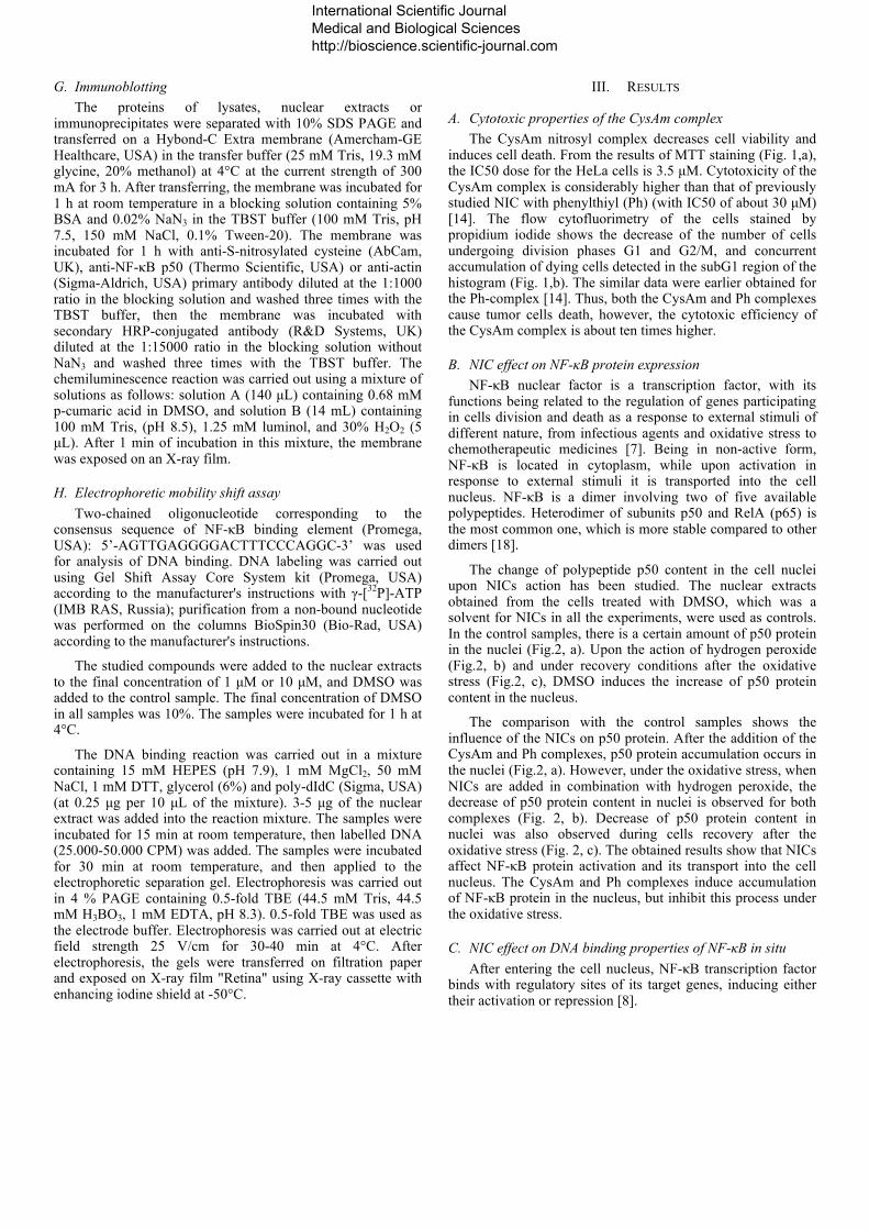
A. Cytotoxic properties of the CysAm complex The CysAm nitrosyl complex decreases cell viability and
induces cell death. From the results of MTT staining (Fig. 1,a), the IC50 dose for the HeLa cells is 3.5 µM. Cytotoxicity of the CysAm complex is considerably higher than that of previously studied NIC with phenylthiyl (Ph) (with IC50 of about 30 µM) [14]. The flow cytofluorimetry of the cells stained by propidium iodide shows the decrease of the number of cells undergoing division phases G1 and G2/M, and concurrent accumulation of dying cells detected in the subG1 region of the histogram (Fig. 1,b). The similar data were earlier obtained for the Ph-complex [14]. Thus, both the CysAm and Ph complexes cause tumor cells death, however, the cytotoxic efficiency of the CysAm complex is about ten times higher.
B. NIC effect on NF-κB protein expression NF-κB nuclear factor is a transcription factor, with its
functions being related to the regulation of genes participating in cells division and death as a response to external stimuli of different nature, from infectious agents and oxidative stress to chemotherapeutic medicines [7]. Being in non-active form, NF-κB is located in cytoplasm, while upon activation in response to external stimuli it is transported into the cell nucleus. NF-κB is a dimer involving two of five available polypeptides. Heterodimer of subunits p50 and RelA (p65) is the most common one, which is more stable compared to other dimers [18].
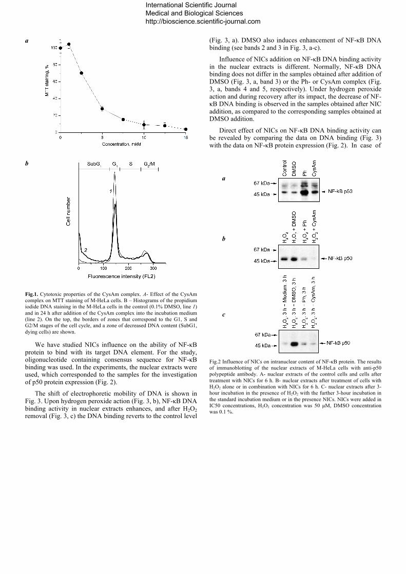
The change of polypeptide p50 content in the cell nuclei upon NICs action has been studied. The nuclear extracts obtained from the cells treated with DMSO, which was a solvent for NICs in all the experiments, were used as controls. In the control samples, there is a certain amount of p50 protein in the nuclei (Fig.2, a). Upon the action of hydrogen peroxide (Fig.2, b) and under recovery conditions after the oxidative stress (Fig.2, c), DMSO induces the increase of p50 protein content in the nucleus.
The comparison with the control samples shows the influence of the NICs on p50 protein. After the addition of the CysAm and Ph complexes, p50 protein accumulation occurs in the nuclei (Fig.2, a). However, under the oxidative stress, when NICs are added in combination with hydrogen peroxide, the decrease of p50 protein content in nuclei is observed for both complexes (Fig. 2, b). Decrease of p50 protein content in nuclei was also observed during cells recovery after the oxidative stress (Fig. 2, c). The obtained results show that NICs affect NF-κB protein activation and its transport into the cell nucleus. The CysAm and Ph complexes induce accumulation of NF-κB protein in the nucleus, but inhibit this process under the oxidative stress.
C. NIC effect on DNA binding properties of NF-κB in situ After entering the cell nucleus, NF-κB transcription factor
binds with regulatory sites of its target genes, inducing either their activation or repression [8].
International Scientific Journal Medical and Biological Sciences http://bioscience.scientific-journal.com
a
b
Fig.1. Cytotoxic properties of the CysAm complex. А- Effect of the CysAm complex on MTT staining of M-HeLa cells. B – Histograms of the propidium iodide DNA staining in the M-HeLa cells in the control (0.1% DMSO, line 1) and in 24 h after addition of the CysAm complex into the incubation medium (line 2). On the top, the borders of zones that correspond to the G1, S and G2/M stages of the cell cycle, and a zone of decreased DNA content (SubG1, dying cells) are shown.
We have studied NICs influence on the ability of NF-κB protein to bind with its target DNA element. For the study, oligonucleotide containing consensus sequence for NF-κB binding was used. In the experiments, the nuclear extracts were used, which corresponded to the samples for the investigation of p50 protein expression (Fig. 2).
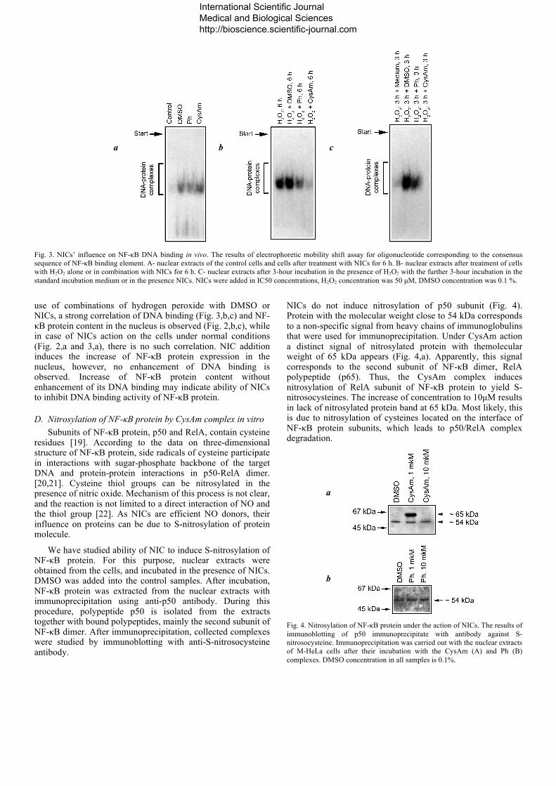
The shift of electrophoretic mobility of DNA is shown in Fig. 3. Upon hydrogen peroxide action (Fig. 3, b), NF-κB DNA binding activity in nuclear extracts enhances, and after H2O2 removal (Fig. 3, c) the DNA binding reverts to the control level
(Fig. 3, a). DMSO also induces enhancement of NF-κB DNA binding (see bands 2 and 3 in Fig. 3, a-c).
Influence of NICs addition on NF-κB DNA binding activity in the nuclear extracts is different. Normally, NF-κB DNA binding does not differ in the samples obtained after addition of DMSO (Fig. 3, a, band 3) or the Ph- or CysAm complex (Fig. 3, a, bands 4 and 5, respectively). Under hydrogen peroxide action and during recovery after its impact, the decrease of NF-κB DNA binding is observed in the samples obtained after NIC addition, as compared to the corresponding samples obtained at DMSO addition.
Direct effect of NICs on NF-κB DNA binding activity can be revealed by comparing the data on DNA binding (Fig. 3) with the data on NF-κB protein expression (Fig. 2). In case of
a
b
c
Fig.2 Influence of NICs on intranuclear content of NF-κB protein. The results of immunoblotting of the nuclear extracts of M-HeLa cells with anti-p50 polypeptide antibody. A- nuclear extracts of the control cells and cells after treatment with NICs for 6 h. B- nuclear extracts after treatment of cells with H2O2 alone or in combination with NICs for 6 h. C- nuclear extracts after 3-hour incubation in the presence of H2O2 with the further 3-hour incubation in the standard incubation medium or in the presence NICs. NICs were added in IC50 concentrations, H2O2 concentration was 50 µM, DMSO concentration was 0.1 %.
International Scientific Journal Medical and Biological Sciences http://bioscience.scientific-journal.com
a
b
c
Fig. 3. NICs’ influence on NF-κB DNA binding in vivo. The results of electrophoretic mobility shift assay for oligonucleotide corresponding to the consensus sequence of NF-κB binding element. A- nuclear extracts of the control cells and cells after treatment with NICs for 6 h. B- nuclear extracts after treatment of cells with H2O2 alone or in combination with NICs for 6 h. C- nuclear extracts after 3-hour incubation in the presence of H2O2 with the further 3-hour incubation in the standard incubation medium or in the presence NICs. NICs were added in IC50 concentrations, H2O2 concentration was 50 µM, DMSO concentration was 0.1 %.
use of combinations of hydrogen peroxide with DMSO or NICs, a strong correlation of DNA binding (Fig. 3,b,c) and NF-κB protein content in the nucleus is observed (Fig. 2,b,c), while in case of NICs action on the cells under normal conditions (Fig. 2,a and 3,а), there is no such correlation. NIC addition induces the increase of NF-κB protein expression in the nucleus, however, no enhancement of DNA binding is observed. Increase of NF-κB protein content without enhancement of its DNA binding may indicate ability of NICs to inhibit DNA binding activity of NF-κB protein.
D. Nitrosylation of NF-κB protein by CysAm complex in vitro Subunits of NF-κB protein, p50 and RelA, contain cysteine
residues [19]. According to the data on three-dimensional structure of NF-κB protein, side radicals of cysteine participate in interactions with sugar-phosphate backbone of the target DNA and protein-protein interactions in p50-RelA dimer. [20,21]. Cysteine thiol groups can be nitrosylated in the presence of nitric oxide. Mechanism of this process is not clear, and the reaction is not limited to a direct interaction of NO and the thiol group [22]. As NICs are efficient NO donors, their influence on proteins can be due to S-nitrosylation of protein molecule.
We have studied ability of NIC to induce S-nitrosylation of NF-κB protein. For this purpose, nuclear extracts were obtained from the cells, and incubated in the presence of NICs. DMSO was added into the control samples. After incubation, NF-κB protein was extracted from the nuclear extracts with immunoprecipitation using anti-p50 antibody. During this procedure, polypeptide p50 is isolated from the extracts together with bound polypeptides, mainly the second subunit of NF-κB dimer. After immunoprecipitation, collected complexes were studied by immunoblotting with anti-S-nitrosocysteine antibody.
NICs do not induce nitrosylation of p50 subunit (Fig. 4). Protein with the molecular weight close to 54 kDa corresponds to a non-specific signal from heavy chains of immunoglobulins that were used for immunoprecipitation. Under CysAm action a distinct signal of nitrosylated protein with themolecular weight of 65 kDa appears (Fig. 4,a). Apparently, this signal corresponds to the second subunit of NF-κB dimer, RelA polypeptide (p65). Thus, the CysAm complex induces nitrosylation of RelA subunit of NF-κB protein to yield S-nitrosocysteines. The increase of concentration to 10µM results in lack of nitrosylated protein band at 65 kDa. Most likely, this is due to nitrosylation of cysteines located on the interface of NF-κB protein subunits, which leads to p50/RelA complex degradation.
a
b
Fig. 4. Nitrosylation of NF-κB protein under the action of NICs. The results of immunoblotting of p50 immunoprecipitate with antibody against S-nitrosocysteine. Immunoprecipitation was carried out with the nuclear extracts of M-HeLa cells after their incubation with the CysAm (A) and Ph (B) complexes. DMSO concentration in all samples is 0.1%.
International Scientific Journal Medical and Biological Sciences http://bioscience.scientific-journal.com
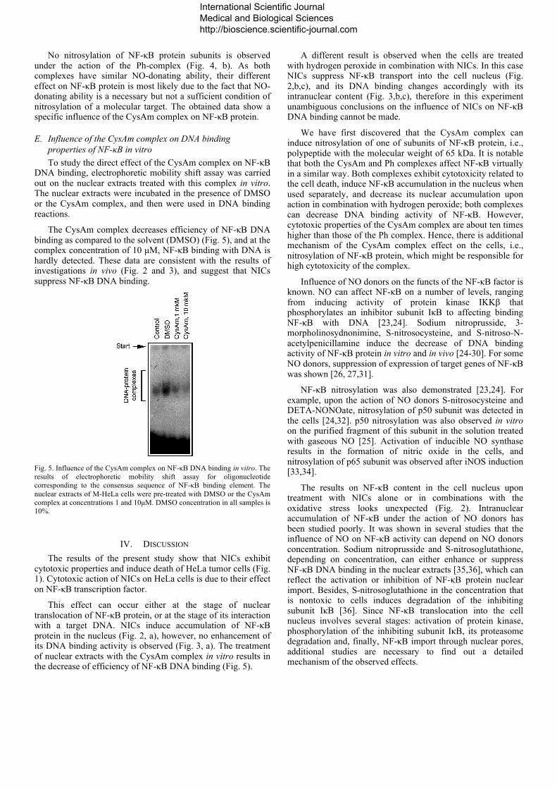
No nitrosylation of NF-κB protein subunits is observed under the action of the Ph-complex (Fig. 4, b). As both complexes have similar NO-donating ability, their different effect on NF-κB protein is most likely due to the fact that NO-donating ability is a necessary but not a sufficient condition of nitrosylation of a molecular target. The obtained data show a specific influence of the CysAm complex on NF-κB protein.
E. Influence of the CysAm complex on DNA binding properties of NF-κB in vitro To study the direct effect of the CysAm complex on NF-κB
DNA binding, electrophoretic mobility shift assay was carried out on the nuclear extracts treated with this complex in vitro. The nuclear extracts were incubated in the presence of DMSO or the CysAm complex, and then were used in DNA binding reactions.
The CysAm complex decreases efficiency of NF-κB DNA binding as compared to the solvent (DMSO) (Fig. 5), and at the complex concentration of 10 µM, NF-κB binding with DNA is hardly detected. These data are consistent with the results of investigations in vivo (Fig. 2 and 3), and suggest that NICs suppress NF-κB DNA binding.
Fig. 5. Influence of the CysAm complex on NF-κB DNA binding in vitro. The results of electrophoretic mobility shift assay for oligonucleotide corresponding to the consensus sequence of NF-κB binding element. The nuclear extracts of M-HeLa cells were pre-treated with DMSO or the CysAm complex at concentrations 1 and 10µM. DMSO concentration in all samples is 10%.
IV. DISCUSSION The results of the present study show that NICs exhibit
cytotoxic properties and induce death of HeLa tumor cells (Fig. 1). Cytotoxic action of NICs on HeLa cells is due to their effect on NF-κB transcription factor.
This effect can occur either at the stage of nuclear translocation of NF-κB protein, or at the stage of its interaction with a target DNA. NICs induce accumulation of NF-κB protein in the nucleus (Fig. 2, a), however, no enhancement of its DNA binding activity is observed (Fig. 3, a). The treatment of nuclear extracts with the CysAm complex in vitro results in the decrease of efficiency of NF-κB DNA binding (Fig. 5).
A different result is observed when the cells are treated with hydrogen peroxide in combination with NICs. In this case NICs suppress NF-κB transport into the cell nucleus (Fig. 2,b,c), and its DNA binding changes accordingly with its intranuclear content (Fig. 3,b,c), therefore in this experiment unambiguous conclusions on the influence of NICs on NF-κB DNA binding cannot be made.
We have first discovered that the CysAm complex can induce nitrosylation of one of subunits of NF-κB protein, i.e., polypeptide with the molecular weight of 65 kDa. It is notable that both the CysAm and Ph complexes affect NF-κB virtually in a similar way. Both complexes exhibit cytotoxicity related to the cell death, induce NF-κB accumulation in the nucleus when used separately, and decrease its nuclear accumulation upon action in combination with hydrogen peroxide; both complexes can decrease DNA binding activity of NF-κB. However, cytotoxic properties of the CysAm complex are about ten times higher than those of the Ph complex. Hence, there is additional mechanism of the CysAm complex effect on the cells, i.e., nitrosylation of NF-κB protein, which might be responsible for high cytotoxicity of the complex.
Influence of NO donors on the functs of the NF-κB factor is known. NO can affect NF-κB on a number of levels, ranging from inducing activity of protein kinase IKKβ that phosphorylates an inhibitor subunit IκB to affecting binding NF-κB with DNA [23,24]. Sodium nitroprusside, 3-morpholinosydnonimine, S-nitrosocysteine, and S-nitroso-N-acetylpenicillamine induce the decrease of DNA binding activity of NF-κB protein in vitro and in vivo [24-30]. For some NO donors, suppression of expression of target genes of NF-κB was shown [26, 27,31].
NF-κB nitrosylation was also demonstrated [23,24]. For example, upon the action of NO donors S-nitrosocysteine and DETA-NONOate, nitrosylation of p50 subunit was detected in the cells [24,32]. p50 nitrosylation was also observed in vitro on the purified fragment of this subunit in the solution treated with gaseous NO [25]. Activation of inducible NO synthase results in the formation of nitric oxide in the cells, and nitrosylation of p65 subunit was observed after iNOS induction [33,34].
The results on NF-κB content in the cell nucleus upon treatment with NICs alone or in combinations with the oxidative stress looks unexpected (Fig. 2). Intranuclear accumulation of NF-κB under the action of NO donors has been studied poorly. It was shown in several studies that the influence of NO on NF-κB activity can depend on NO donors concentration. Sodium nitroprusside and S-nitrosoglutathione, depending on concentration, can either enhance or suppress NF-κB DNA binding in the nuclear extracts [35,36], which can reflect the activation or inhibition of NF-κB protein nuclear import. Besides, S-nitrosoglutathione in the concentration that is nontoxic to cells induces degradation of the inhibiting subunit IκB [36]. Since NF-κB translocation into the cell nucleus involves several stages: activation of protein kinase, phosphorylation of the inhibiting subunit IκB, its proteasome degradation and, finally, NF-κB import through nuclear pores, additional studies are necessary to find out a detailed mechanism of the observed effects.
International Scientific Journal Medical and Biological Sciences http://bioscience.scientific-journal.com
Despite a possible stimulation of NF-κB intranuclear accumulation, NICs suppress its activity at the stage of DNA binding. Therefore, both under normal conditions and under the oxidative stress, NICs suppress NF-κB functions. Because of the role of NF-κB factor in the development of oncological diseases and its role as a molecular target for anticancer therapy [8], NICs can be considered as a new type of NF-κB inhibitors, which can become a basis for the development of anticancer chemotherapeutic medicines.
Acknowledgements
The work has been supported by the Program of the Presidium of RAS "Fundamental sciences for medicine".
References [1] Butler A.R., Megson I.L., “Non-heme iron nitrosyls in biology”, Chem
Rev., 2002, vol. 102(4), pp. 1155-1166. [2] Lewandowska H., Kalinowska M., Brzóska K., Wójciuk K., Wójciuk G.,
Kruszewski M., “Nitrosyl iron complexes--synthesis, structure and biology”, Dalton Trans. 2011, vol. 40(33), pp. 8273-8289.
[3] Sanina N.A., Аldoshin S.M., “Structure and properties of iron nitrosyl complexes with functionalized sulfur-containing ligands”, Russ Chem Bull,. 2011, vol. 60(7), pp. 1223-1251.
[4] Sanina N.A., Zhukova O.S., Аldoshin S.M., Emel’yanova N.S., Gerasimova G.K, “Use of tetranitrosyl iron complex with thiophenol as a antitumor drug”, patent RU 2429242 (2011).
[5] Sanina N.A., Lysenko K.A., Zhukova O.S., Roudneva T.N., Emel’yanova N.S., Aldoshin S.M., “Water-soluble binuclear nitrosyl iron complexes with natural aliphatic thiolyls possessing cytotoxic, apoptotic and NO-donor activity”, patent US 8,067,628 B2 (2011).
[6] Wink D.A., Mitchell J.B., “Chemical biology of nitric oxide: Insights into regulatory, cytotoxic, and cytoprotective mechanisms of nitric oxide”, Free Radic Biol Med., 1998, vol. 25(4-5), pp. 434-456.
[7] Lin Y., Bai L., Chen W., Xu S., “The NF-κB activation pathways, emerging molecular targets for cancer prevention and therapy”, Expert Opin Ther Targets, 2010, vol. 14(1), pp. 45–55.
[8] Bonavida B., Baritaki S., “Dual role of NO donors in the reversal of tumor cell resistance and EMT: Downregulation of the NF-κB/Snail/YY1/RKIP circuitry”, Nitric Oxide, 2011, vol. 24(1), pp. 1-7.
[9] Marshall H.E., Hess D.T., Stamler J.S., “S-nitrosylation: physiological regulation of NF-kB”, PNAS, 2004, vol. 101(24), pp.8841-8842.
[10] Colasanti M., Persichini T., “Nitric oxide: an inhibitor of NF-κB/Rel system in glial cells”, Brain Res Bull., 2000, vol. 52(3), pp. 155-161.
[11] Vasilieva S.V., Moschkovskaya E.Ju., Terekhov A.S., Sanina N.A., Aldoshin S.M., ”Intracellular iron ions regulate the genetic activity of NO-donating agents”, Russian Journal of Genetics, 2006, No.7, pp. 737-743.
[12] Sanina, N.A., Rudneva, T.N., Sulimenkov, I.V., Kono valova, N.P., Sashenkova, T.E. and Aldoshin, S.M. (2009) Antitumor activity of iron nitrosyl complexes: New donors of nitrogen monoxide. Rossiiskii Khimicheskii Zhurnal, 53(1), pp.164-171.
[13] Stupina T.S., Parkhomenko I.I., Balalaeva I.V., Kostyuk G.V., Sanina N.A., Terent’ev A.A. “Cytotoxic properties of the nitrosyl iron complex with phenylthiyl”, Russ. Chem. Bull., 2011, vol. 60(7), pp. 1488-1494.
[14] Rudneva Т.N., Sanina N.А., Lysenko К.А., Aldoshin S.М., Antipin М.Y., Оvanesyan N.S., “Synthesis and structure of water-soluble nitrosyl iron complex with cysteinamine ligand [Fe2(S(CH)2NH3)2(NO)4]SO4 · 2.5H2O”, Mendeleev communications, 2009, vol.19, pp. 253-255.
[15] Sanina N.A., Emel'Yanova N.S., Chekhlov A.N., Shestakov A.F., Aldoshin S.M., Sulimenkov I.V., “Structure and properties of µ2-S-[bis(benzenethiolato)tetranitrosyldiiron] in solution”, Russian Chemical Bulletin, 2010, vol. 59(6), pp. 1126-1136.
[16] Lowry O.H., Rosenbrough N.J., Farr A.L., Randall R.J., “Protein measurement with the Folin phenol reagent”, J Biol Chem., 1951, vol. 193(1), pp. 265-275.
[17] Ghosh G., Wang V.Y., Huang D.B., Fusco A., “NF-κB regulation: lessons from structures”, Immunol Rev., 2012, vol. 246(1), pp. 36-58.
[18] Ruben S.M., Dillon P.J., Schreck R., Henkel T., Chen C.-H., Maher M., Baeuerle P.A., Rosen C.A., “Isolation of a rel-related human cDNA that potentially encodes the 65-kD subunit of NF-κB”, Science, 1991, vol. 251(5000), pp. 1490-1493.
[19] Chen F.E., Huang D.B., Chen Y.Q., Ghosh G., “Crystal structure of p50/p65 heterodimer of transcription factor NF-κB bound to DNA” Nature, 1998, vol. 391(6665), pp. 410-413.
[20] Stroud J.C., Oltman A., Han A., Bates D.L., Chen L., “Structural basis of HIV-1 activation by NF-κB – a higher-order complex of p50:RelA bound to the HIV-1 LTR”, J Mol Biol., 2009, vol. 393(1), pp. 98-112.
[21] Mannick J.B., “Regulation of apoptosis by protein S-nitrosylation”, Amino Acids, 2007, vol. 32(4), P.523-526.
[22] Perkins N.D. “Post-translational modifications regulating the activity and function of the nuclear factor kappa B pathway” Oncogene, 2006, vol. 25(51), pp. 6717-6730.
[23] Marshall H.E., Stamler J.S., “Inhibition of NF-κB by S-nitrosylation”, Biochemistry, 2001, vol. 40(6), pp. 1688-1693.
[24] Matthews J.R., Botting C.H., Panico M., Morris H.R., Hay R.T., “Inhibition of NF-κB DNA binding by nitric oxide”, Nucleic Acids Res., 1996, vol. 24(12), pp. 2236-2242.
[25] Lee S.K., Kim J.H., Yang W.S., Kim S.B., Park S.K., Park J.S., “Exogenous nitric oxide inhibits VCAM-1 expression in human peritoneal mesothelial cells. Role of cyclic GMP and NF-κB”, Nephron, 2002, vol. 90(4), pp. 447-454.
[26] Paradkar P.N., Roth J.A., “Nitric oxide transcriptionally down-regulates specific isoforms of divalent metal transporter (DMT1) via NF-κB”, J Neurochem., 2006, vol. 96(6), pp. 1768-1777.
[27] Mendes A.F., Carvalho A.P., Caramona M.M., Lopes M.C., “Role of nitric oxide in the activation of NF-κB, AP-1 and NOS II expression in articular chondrocytes”, Inflamm Res., 2002, vol. 51(7), pp. 369-375.
[28] Rishi L., Dhiman R., Raje M., Majumdar S., “Nitric oxide induces apoptosis in cutaneous T cell lymphoma (HuT-78) by downregulating constitutive NF-κB”, Biochim Biophys Acta, 2007, vol. 1770(8), pp. 1230-1239.
[29] Ibe W., Bartels W., Lindemann S., Grosser T., Buerke M., Boissel J.P., Meyer J., Darius H., “Involvement of PKC and NF-κB in nitric oxide induced apoptosis in human coronary artery smooth muscle cells” Cell Physiol Biochem., 2001, vol. 11(5), pp. 231-240.
[30] Jiang M.Z., Tsukahara H., Ohshima Y., Todoroki Y., Hiraoka M., Maeda M., Mayumi M., “Effects of antioxidants and nitric oxide on TNF-alpha-induced adhesion molecule expression and NF-κB activation in human dermal microvascular endothelial cells”, Life Sci., 2004, vol. 75(10), pp. 1159-1170.
[31] Huerta-Yepez S., Vega M., Jazirehi A., Garban H., Hongo F., Cheng G., Bonavida B., “Nitric oxide sensitizes prostate carcinoma cell lines to TRAIL-mediated apoptosis via inactivation of NF-κB and inhibition of Bcl-xl expression”, Oncogene, 2004, vol. 23(29), pp. 4993-5003.
[32] Kelleher, Z.T., Matsumoto, A., Stamler, J.S., and Marshall H.E., “NOS2 regulation of NF-κB by S-nitrosylation of p65”, J Biol Chem., 2007, vol. 282(42), pp. 30667-30672.
[33] Nishida M., Kitajima N., Saiki S., Nakaya M., Kurose H., “Regulation of Angiotensin II receptor signaling by cysteine modification of NF-kB”, Nitric Oxide, 2011, vol. 25(2), pp. 112-117.
[34] Siednienko J., Nowak J., Moynagh P.N., Gorczyca W.A., “Nitric oxide affects IL-6 expression in human peripheral blood mononuclear cells involving cGMP-dependent modulation of NF-κB activity”, Cytokine, 2011, vol. 54(3), pp. 282-288.
[35] von Knethen A., Callsen D., Brüne B., “NF-kappaB and AP-1 activation by nitric oxide attenuated apoptotic cell death in RAW 264.7 macrophages”, Mol Biol Cell, 1999, vol. 10(2), pp. 361-372.
International Scientific Journal Medical and Biological Sciences http://bioscience.scientific-journal.com