Inclusion Complexation of 2,6-Diphenylphenol with ...
8
www.ijapbc.com IJAPBC – Vol. 1(3), Jul- Sep, 2012 ISSN: 2277 - 4688 _____________________________________________________________________________________ 354 INTERNATIONAL JOURNAL OF ADVANCES IN PHARMACY, BIOLOGY AND CHEMISTRY Research Article ABSTRACT Inclusion complexation of 2,6-dipenylphenol with β-cyclodextrin and its photoprototropic behavior has been studied with absorption and fluorescence spectra. 2,6-Diphenylphenol forms a 1:1 inclusion complex with a binding constant of 272.12 M -1 . It is found to be show more acidic characteristics in β-cyclodextrin. Contrary to the observation of stretched sigmoidal fluorimetric titration curves in aqueous solution, the fluorimetric titration curves for the neutral – monoanion equilibrium of 2,6-diphenylphenol in β-cyclodextrin are sharp and they meet at the middle of inflection showing that the prototropic equilibrium is attained in the S 1 state. Keywords: Cyclodextrin, Photoprototropism, Inclusion complex, Binding constant. INTRODUCTION Host-guest association of two or more species, assembled together without a covalent bond, is the basis for a new type of photo-response, in which the photophysics and photochemistry of the guest are modified and made unique 1-4 . Crown ethers 5,6 , cyclophanes 7 , calixarenes 8-10 , zeolites 11-13 , and cyclodextrins are some of the host structures which draw considerable attention by the researchers for the study of host-guest association. Among the potential hosts, cyclodextrin (CD) seems to be the important one due to the following reasons: (i) CDs are semi natural products, produced from starch by a simple enzymatic conversion, (ii) CDs may form stable host- guest systems 14-16 , (iii) CD can provide a good miniature model for enzyme-substrate complexes, (iv) the reduced polarity and restricted space provide by the CD can incorporate large number of molecules and can change and modify the properties of the guest molecules, and (v) CDs are non-toxic and can be consumed by humans. Cyclodextrins are all- purpose molecular containers for organic, inorganic, organo-metallic, and metallo organic compounds that may be neutral, cationic, anionic or radical 17,18 . The spatial conformation of cyclodextrins gives them the ability to include various guest molecules on the condition that the size of the guest molecules is compatible with the cavity size of the host molecules. The spectroscopic parameters of an organic fluorophore (species with characteristic fluorescence property) often change dramatically upon inclusion in CDs allowing one to monitor the inclusion phenomenon through this change 19 . Though a number of techniques such as conductometry 20 , calorimetry 21 , NMR 22 , FT-IR 23 and induced circular dichroism 24 have been reported for the characterization of inclusion complexes, fluorescence spectroscopy is found to be very sensitive and suitable for the study of inclusion complexes and excited state proton transfer. Fluorescence enhancement of benzene derivatives due to inclusion in β-cyclodextrin has been studied extensively 25 . Since the initial study on excited state proton transfer was done, the excited state proton transfer has been extensively studied in aqueous solution. It has been reported that the excited state proton transfer rates or the excited state pK a values were affected by the inclusion complexation 26 . The solvatochromic and prototropic behavior of substituted biphenyls in aqueous solution have been extensively studied 26-28 . One way to develop a molecular switch is to activate a competing guest in a designed host-guest system through the addition or removal of electrons, protons, or ions to create Inclusion Complexation of 2,6-Diphenylphenol with -Cyclodextrin: Fluorescence Characteristics and the Effect of pH Israel V.M.V. Enoch * and Y. Sameena Department of Chemistry, Karunya University, Coimbatore – 641 114, Tamil Nadu, India.
Transcript of Inclusion Complexation of 2,6-Diphenylphenol with ...

www.ijapbc.com IJAPBC – Vol. 1(3), Jul- Sep, 2012 ISSN: 2277 - 4688 _____________________________________________________________________________________
354
INTERNATIONAL JOURNAL OF ADVANCES IN PHARMACY,
BIOLOGY AND CHEMISTRY
Research Article
ABSTRACT Inclusion complexation of 2,6-dipenylphenol with β-cyclodextrin and its photoprototropic behavior has been studied with absorption and fluorescence spectra. 2,6-Diphenylphenol forms a 1:1 inclusion complex with a binding constant of 272.12 M-1. It is found to be show more acidic characteristics in β-cyclodextrin. Contrary to the observation of stretched sigmoidal fluorimetric titration curves in aqueous solution, the fluorimetric titration curves for the neutral – monoanion equilibrium of 2,6-diphenylphenol in β-cyclodextrin are sharp and they meet at the middle of inflection showing that the prototropic equilibrium is attained in the S1 state. Keywords: Cyclodextrin, Photoprototropism, Inclusion complex, Binding constant.
INTRODUCTION Host-guest association of two or more species, assembled together without a covalent bond, is the basis for a new type of photo-response, in which the photophysics and photochemistry of the guest are modified and made unique1-4. Crown ethers5,6, cyclophanes7, calixarenes8-10, zeolites11-13, and cyclodextrins are some of the host structures which draw considerable attention by the researchers for the study of host-guest association. Among the potential hosts, cyclodextrin (CD) seems to be the important one due to the following reasons: (i) CDs are semi natural products, produced from starch by a simple enzymatic conversion, (ii) CDs may form stable host-guest systems14-16, (iii) CD can provide a good miniature model for enzyme-substrate complexes, (iv) the reduced polarity and restricted space provide by the CD can incorporate large number of molecules and can change and modify the properties of the guest molecules, and (v) CDs are non-toxic and can be consumed by humans. Cyclodextrins are all-purpose molecular containers for organic, inorganic, organo-metallic, and metallo organic compounds that may be neutral, cationic, anionic or radical17,18. The spatial conformation of cyclodextrins gives them the ability to include various guest molecules on the condition that the size of the guest molecules is
compatible with the cavity size of the host molecules. The spectroscopic parameters of an organic fluorophore (species with characteristic fluorescence property) often change dramatically upon inclusion in CDs allowing one to monitor the inclusion phenomenon through this change19. Though a number of techniques such as conductometry20, calorimetry21, NMR22, FT-IR23 and induced circular dichroism24 have been reported for the characterization of inclusion complexes, fluorescence spectroscopy is found to be very sensitive and suitable for the study of inclusion complexes and excited state proton transfer. Fluorescence enhancement of benzene derivatives due to inclusion in β-cyclodextrin has been studied extensively25. Since the initial study on excited state proton transfer was done, the excited state proton transfer has been extensively studied in aqueous solution. It has been reported that the excited state proton transfer rates or the excited state pKa values were affected by the inclusion complexation26. The solvatochromic and prototropic behavior of substituted biphenyls in aqueous solution have been extensively studied26-28. One way to develop a molecular switch is to activate a competing guest in a designed host-guest system through the addition or removal of electrons, protons, or ions to create
Inclusion Complexation of 2,6-Diphenylphenol with
-Cyclodextrin: Fluorescence Characteristics and the Effect of pH
Israel V.M.V. Enoch* and Y. Sameena
Department of Chemistry, Karunya University, Coimbatore – 641 114, Tamil Nadu, India.
www.ijapbc.com IJAPBC – Vol. 1(3), Jul- Sep, 2012 ISSN: 2277 - 4688 _____________________________________________________________________________________
355
detectable spectroscopic changes between the two different states of the molecule. The inclusion complexation and prototropic behavior of 2-hydroxybiphenyl and 4-hydroxybiphenyl in β-cyclodextrin have been reported29-31. But the studies of excited state proton transfer (ESPT) in the presence of aqueous cyclodextrin solution are limited. The present work presents the effect of β-CD on the fluorescence and photoprotropic behavior of 2,6-diphenylphenol. MATERIALS AND METHODS Chemicals and instrumentation β-cyclodextrin obtained from S.D.Fine chemicals were used as received. 2,6-diphenylphenol purchased from Aldrich was purified by repeated re-crystallization from methanol. The purity of DPP was confirmed by their sharp melting point, thin layer chromatography and its fluorescence emission spectrum in suitable solvents with different excitation wavelengths. Spectral grade solvents viz., dioxane, acetonitrile, and methanol (Qualigens) were used for the study of absorption and fluorescence spectral measurements. Absorption spectra were recorded with a JASCO – 7800 spectrophotometer and steady – state fluorescence measurements were made using a JASCO FP – 750 spectrofluorimeter. pH values in the range of 3.0 to 10.0 were measured on ELICO pH meter (model LI – 10T). Fluorescence lifetimes were measured using a time – resolved single photon counting picosecond spectrofluorimeter (Tsunami, Spectraphysics) with a mode locked, synchronously pumped cavity – dumped dye laser as the excitation source. Preparation of solutions Owing to its poor solubility in water, 2,6-diphenylphenol stock solution was prepared in methanol. The concentrations of the solutions used for taking spectral measurements were of the order of 10-5 M in 3% methanol-water mixture. Triply distilled water was used for the preparation of test solutions for spectral measurements. The fluorophore (2,6-diphenylphenol) was added to the β-CD solution and stirred for 6 hours using a magnetic stirrer and spectral measurements were taken immediately after the process. Solutions in the pH range of 2-10 were prepared by adding appropriate amount of sodium hydroxide and phosphoric acid (A.R. grade, Qualigens). A modified Hammet’s acidity scale32 was
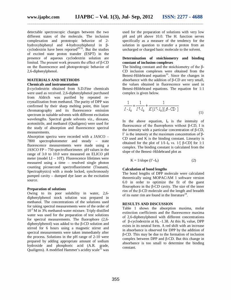
used for the preparation of solutions with very low pH and pH above 10.0. The H- function serves specifically as a measure of the tendency for the solution in question to transfer a proton from an uncharged or charged basic molecule to the solvent. Determination of stoichiometry and binding constant of inclusion complexes The binding constant and the stoichiometry of the β-CD inclusion complexes were obtained from the Benesi-Hildebrand equation33. Since the changes in absorbance with the addition of β-CD are very small, the values obtained in fluorescence were used in Benesi-Hildebrand equations. The equation for 1:1 complex is given below.
(1) In the above equation, I0 is the intensity of fluorescence of the fluorophores without β-CD, I is the intensity with a particular concentration of β-CD, I’ is the intensity at the maximum concentration of β-CD used and K is the binding constant. Linearity is obtained for the plot of 1/I–I0 vs. 1/[ β-CD] for 1:1 complex. The binding constant is calculated from the slope of the Benesi–Hildebrand plot as
K = 1/slope (I’–I0) (2) Calculation of bond lengths The bond lengths of DPP molecule were calculated theoretically using MOPAC/AM 1 software version 6.0 in order to optimize the fit of the guest flourophore in the β-CD cavity. The size of the inner rim of the β-CD molecule and the length and breadth of its outer rim are found in the literature34. RESULTS AND DISCUSSION Table 1 shows the absorption maxima, molar extinction coefficients and the fluorescence maxima of 2,6-diphenylphenol with different concentrations of β-cyclodextrin at H0 -1.38. At this H0 value, DPP exists in its neutral form. A red shift with an increase in absorbance is observed for DPP by the addition of β-CD. This may be due to the formation of inclusion complex between DPP and β-CD. But this change in absorbance is too small to determine the binding constant.
www.ijapbc.com IJAPBC – Vol. 1(3), Jul- Sep, 2012 ISSN: 2277 - 4688 _____________________________________________________________________________________
356
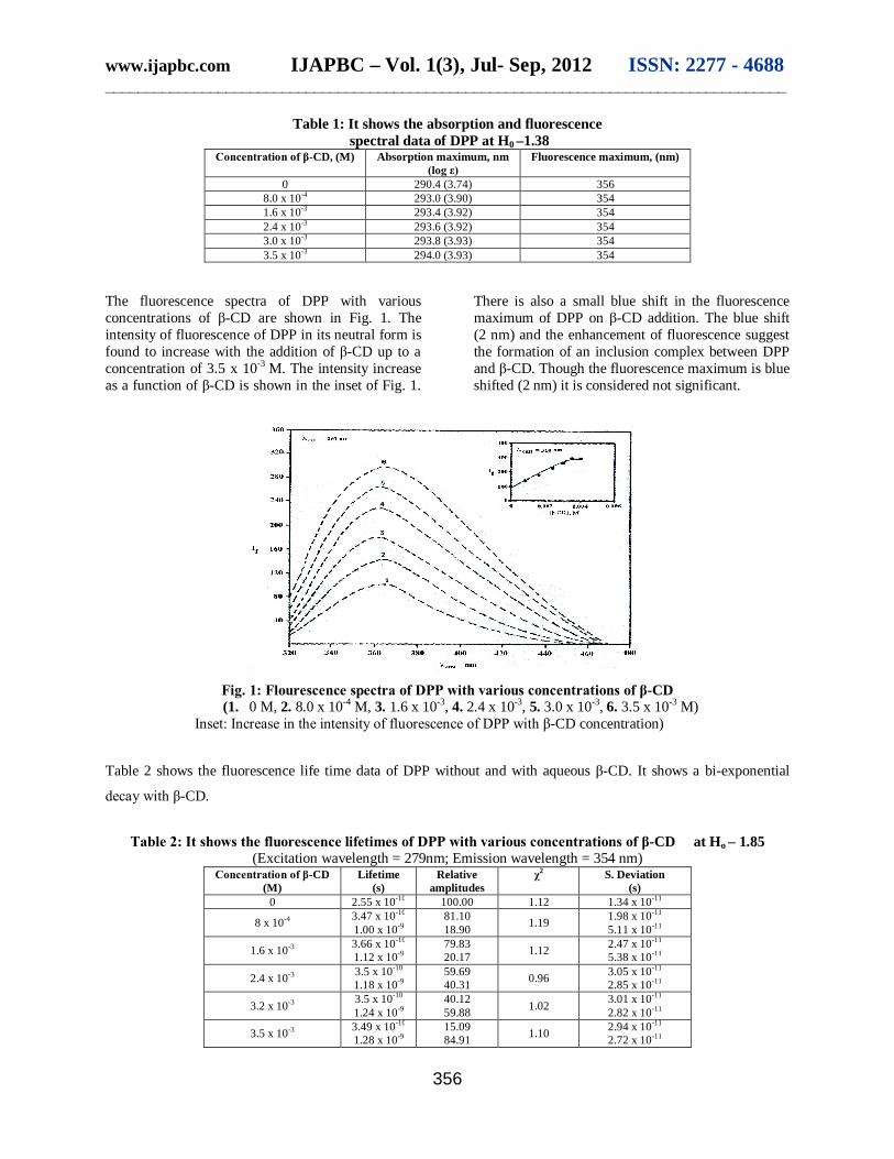
Table 1: It shows the absorption and fluorescence spectral data of DPP at H0 –1.38
Concentration of β-CD, (M) Absorption maximum, nm (log ε)
Fluorescence maximum, (nm)
0 290.4 (3.74) 356 8.0 x 10⁻⁴ 293.0 (3.90) 354 1.6 x 10⁻³ 293.4 (3.92) 354 2.4 x 10⁻³ 293.6 (3.92) 354 3.0 x 10⁻³ 293.8 (3.93) 354 3.5 x 10⁻³ 294.0 (3.93) 354
The fluorescence spectra of DPP with various concentrations of β-CD are shown in Fig. 1. The intensity of fluorescence of DPP in its neutral form is found to increase with the addition of β-CD up to a concentration of 3.5 x 10⁻³ M. The intensity increase as a function of β-CD is shown in the inset of Fig. 1.
There is also a small blue shift in the fluorescence maximum of DPP on β-CD addition. The blue shift (2 nm) and the enhancement of fluorescence suggest the formation of an inclusion complex between DPP and β-CD. Though the fluorescence maximum is blue shifted (2 nm) it is considered not significant.
Fig. 1: Flourescence spectra of DPP with various concentrations of β-CD (1. 0 M, 2. 8.0 x 10⁻⁴ M, 3. 1.6 x 10⁻³, 4. 2.4 x 10⁻³, 5. 3.0 x 10⁻³, 6. 3.5 x 10⁻³ M)
Inset: Increase in the intensity of fluorescence of DPP with β-CD concentration)
Table 2 shows the fluorescence life time data of DPP without and with aqueous β-CD. It shows a bi-exponential
decay with β-CD.
Table 2: It shows the fluorescence lifetimes of DPP with various concentrations of β-CD at Ho – 1.85
(Excitation wavelength = 279nm; Emission wavelength = 354 nm) Concentration of β-CD
(M) Lifetime
(s) Relative
amplitudes χ2 S. Deviation
(s) 0 2.55 x 10⁻¹⁰ 100.00 1.12 1.34 x 10⁻¹¹
8 x 10⁻⁴ 3.47 x 10⁻¹⁰
1.00 x 10⁻⁹ 81.10 18.90 1.19 1.98 x 10⁻¹¹
5.11 x 10⁻¹¹
1.6 x 10⁻³ 3.66 x 10⁻¹⁰
1.12 x 10⁻⁹ 79.83 20.17 1.12 2.47 x 10⁻¹¹
5.38 x 10⁻¹¹
2.4 x 10⁻³ 3.5 x 10⁻¹⁰
1.18 x 10⁻⁹ 59.69 40.31 0.96 3.05 x 10⁻¹¹
2.85 x 10⁻¹¹
3.2 x 10⁻³ 3.5 x 10⁻¹⁰
1.24 x 10⁻⁹ 40.12 59.88 1.02 3.01 x 10⁻¹¹
2.82 x 10⁻¹¹
3.5 x 10⁻³ 3.49 x 10⁻¹⁰
1.28 x 10⁻⁹ 15.09 84.91 1.10 2.94 x 10⁻¹¹
2.72 x 10⁻¹¹
www.ijapbc.com IJAPBC – Vol. 1(3), Jul- Sep, 2012 ISSN: 2277 - 4688 _____________________________________________________________________________________
357
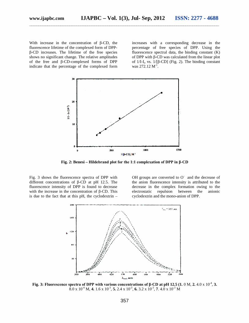
With increase in the concentration of β-CD, the fluorescence lifetime of the complexed form of DPP- β-CD increases. The lifetime of the free species shows no significant change. The relative amplitudes of the free and β-CD-complexed forms of DPP indicate that the percentage of the complexed form
increases with a corresponding decrease in the percentage of free species of DPP. Using the fluorescence spectral data, the binding constant (K) of DPP with β-CD was calculated from the linear plot of 1/I-Io vs. 1/[β-CD] (Fig. 2). The binding constant was 272.12 M-1.
Fig. 2: Benesi – Hildebrand plot for the 1:1 complexation of DPP in β-CD
Fig. 3 shows the fluorescence spectra of DPP with different concentrations of β-CD at pH 12.5. The fluorescence intensity of DPP is found to decrease with the increase in the concentration of β-CD. This is due to the fact that at this pH, the cyclodextrin –
OH groups are converted to O– and the decrease of the anion fluorescence intensity is attributed to the decrease in the complex formation owing to the electrostatic repulsion between the anionic cyclodextrin and the mono-anion of DPP.
Fig. 3: Fluorescence spectra of DPP with various concentrations of β-CD at pH 12.5 (1. 0 M, 2. 4.0 x 10⁻⁴, 3.
8.0 x 10⁻⁴ M, 4. 1.6 x 10⁻³, 5. 2.4 x 10⁻³, 6. 3.2 x 10⁻³, 7. 4.0 x 10⁻³ M
www.ijapbc.com IJAPBC – Vol. 1(3), Jul- Sep, 2012 ISSN: 2277 - 4688 _____________________________________________________________________________________
358
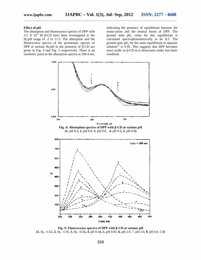
Effect of pH The absorption and fluorescence spectra of DPP with 3.5 X 10⁻³ M β-CD have been investigated in the H0/pH range of -2 to 11.5. The absorption and the fluorescence spectra of the prototropic species of DPP at various H0/pH in the presence of β-CD are given in Fig. 4 and Fig. 5 respectively. There is an isosbestic point in the absorption spectra at 294.4 nm,
indicating the presence of equilibrium between the mono-anion and the neutral forms of DPP. The ground state pKa value for this equilibrium is calculated spectrophotometrically to be 8.5. The ground state pKa for the same equilibrium in aqueous solution35 is 9.95. This suggests that DPP becomes more acidic in β-CD as it dissociates under less basic condition.
Fig. 4: Absorption spectra of DPP with β-CD at various pH
(1. pH 8.4, 2. pH 8.8, 3. pH 9.0, 4. pH 9.4, 5. pH 9.8)
Fig. 5: Fluorescence spectra of DPP with β-CD at various pH
(1. H0 –1.62, 2. H0 –1.05, 3. H0 –0.56, 4. pH 0.44, 5. pH 0.83, 6. pH 2.0, 7. pH 3.0, 8. pH 4.0, 5.0)
www.ijapbc.com IJAPBC – Vol. 1(3), Jul- Sep, 2012 ISSN: 2277 - 4688 _____________________________________________________________________________________
359
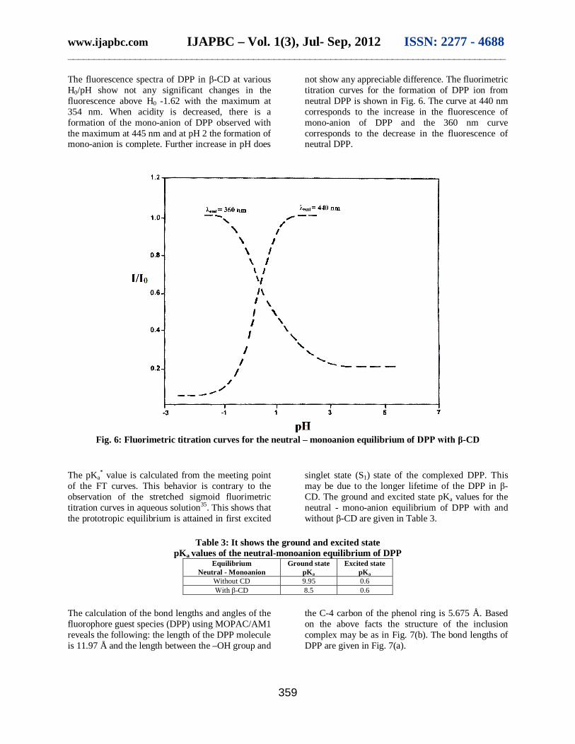
The fluorescence spectra of DPP in β-CD at various H0/pH show not any significant changes in the fluorescence above H0 -1.62 with the maximum at 354 nm. When acidity is decreased, there is a formation of the mono-anion of DPP observed with the maximum at 445 nm and at pH 2 the formation of mono-anion is complete. Further increase in pH does
not show any appreciable difference. The fluorimetric titration curves for the formation of DPP ion from neutral DPP is shown in Fig. 6. The curve at 440 nm corresponds to the increase in the fluorescence of mono-anion of DPP and the 360 nm curve corresponds to the decrease in the fluorescence of neutral DPP.
Fig. 6: Fluorimetric titration curves for the neutral – monoanion equilibrium of DPP with β-CD
The pKa
* value is calculated from the meeting point of the FT curves. This behavior is contrary to the observation of the stretched sigmoid fluorimetric titration curves in aqueous solution35. This shows that the prototropic equilibrium is attained in first excited
singlet state (S1) state of the complexed DPP. This may be due to the longer lifetime of the DPP in β-CD. The ground and excited state pKa values for the neutral - mono-anion equilibrium of DPP with and without β-CD are given in Table 3.
Table 3: It shows the ground and excited state
pKa values of the neutral-monoanion equilibrium of DPP Equilibrium
Neutral - Monoanion Ground state
pKa Excited state
pKa Without CD 9.95 0.6 With β-CD 8.5 0.6
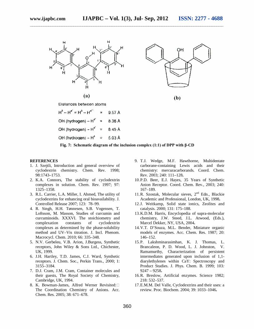
The calculation of the bond lengths and angles of the fluorophore guest species (DPP) using MOPAC/AM1 reveals the following: the length of the DPP molecule is 11.97 Å and the length between the –OH group and
the C-4 carbon of the phenol ring is 5.675 Å. Based on the above facts the structure of the inclusion complex may be as in Fig. 7(b). The bond lengths of DPP are given in Fig. 7(a).
www.ijapbc.com IJAPBC – Vol. 1(3), Jul- Sep, 2012 ISSN: 2277 - 4688 _____________________________________________________________________________________
360
Fig. 7: Schematic diagram of the inclusion complex (1:1) of DPP with β-CD
REFERENCES 1. J. Szejtli, Introduction and general overview of
cyclodextrin chemistry. Chem. Rev. 1998; 98:1743–1753.
2. K.A. Connors, The stability of cyclodextrin complexes in solution. Chem. Rev. 1997; 97: 1325–1358.
3. R.L. Carrier, L.A. Miller, I. Ahmed, The utility of cyclodextrins for enhancing oral bioavailability. J. Controlled Release 2007; 123: 78–99.
4. R. Singh, H.H. Tønnesen, S.B. Vogensen, T. Loftsson, M. Masson, Studies of curcumin and curcuminoids. XXXVI. The stoichiometry and complexation constants of cyclodextrin complexes as determined by the phase-solubility method and UV–Vis titration. J. Incl. Phenom. Macrocycl. Chem. 2010; 66: 335–348.
5. N.V. Gerbeleu, V.B. Arion, J.Burgess, Synthetic receptors, John Wiley & Sons Ltd., Chichester, UK, 1999.
6. J.H. Hartley, T.D. James, C.J. Ward, Synthetic receptors. J. Chem. Soc., Perkin Trans., 2000; 1: 3155–3184.
7. D.J. Cram, J.M. Cram, Container molecules and their guests, The Royal Society of Chemistry, Cambridge, UK, 1994.
8. K. Bowman-James, Alfred Werner Revisited: The Coordination Chemistry of Anions. Acc. Chem. Res. 2005; 38: 671–678.
9. T.J. Wedge, M.F. Hawthorne, Multidentate carborane-containing Lewis acids and their chemistry: mercuracarborands. Coord. Chem. Rev. 2003; 240: 111–128.
10. P.D. Beer, E.J. Hayes, 35 Years of Synthetic Anion Receptor. Coord. Chem. Rev., 2003; 240: 167–189.
11. R. Szostak, Molecular sieves, 2nd Edn., Blackie Academic and Professional, London, UK, 1998.
12. J. Weitkamp, Solid state ionics, Zeolites and catalysis. 2000; 131: 175–188.
13. K.D.M. Harris, Encyclopedia of supra-molecular chemistry, J.W. Steed, J.L. Atwood, (Eds.), Marcel Dekker, NY, USA, 2004.
14. V.T. D’Souza, M.L. Bender, Miniature organic models of enzymes. Acc. Chem. Res. 1987; 20: 146–152.
15. P. Lakshminarasimhan, K. J. Thomas, L. Brancaleon, P. D. Wood, L. J. Johnston, V. Ramamurthy, Characterization of persistent intermediates generated upon inclusion of 1,1-diarylethylenes within CaY: Spectroscopy and Product Studies. J. Phys. Chem. B. 1999; 103: 9247 – 9258.
16. R. Breslow, Artificial enzymes. Science 1982; 218: 532–537.
17. E.M.M. Del Valle, Cyclodextrins and their uses: a review. Proc. Biochem. 2004; 39: 1033–1046.
www.ijapbc.com IJAPBC – Vol. 1(3), Jul- Sep, 2012 ISSN: 2277 - 4688 _____________________________________________________________________________________
361
18. N.T. Southall, K.A. Dill, A.D.J. Haymet, A View of the Hydrophobic Effect. J. Phys. Chem. 2002; 106: 521–533.
19. T. Ogoshi, A. Harada, Chemical Sensors Based on Cyclodextrin Derivatives. Sensors 2008; 8(8): 4961–4982.
20. B. Schazmann, N. Alhashimy, D.J. Diamond, Chloride selective calix[4]arene optical sensor combining urea functionality with pyrene excimer transduction. J. Am. Chem. Soc. 2006; 128(26): 8607–8614.
21. Y. Liu, Z. Li, H.Y. Zhang, H. Wang, C.J. Li, Colorimetric sensor and inverse fluorescent behavior of anions by calix[4]arene possessing imidazo[4,5-f]-1,10-phenanthroline groups and Its Ru(II) complex. Supramol. Chem., 2008; 20 (4): 419–426.
22. S.S. Braga, T.A. Ree, K. Imamura, P. Vertut, I. B. Palheiros, W. Senger, J.J.C.T. Dias, Structure of the beta-cyclodextrin center dot p-hydroxybenzaldehyde inclusion complex in aqueous solution and in the crystalline state. J. Incl. Phenom. Macrocycl. Chem., 2002; 43: 115–125.
23. A. Farcas, N. Jarrouxb, A-M. Farcas, V. Harabagiu, P. Guegan, Synthesis and characterization of furosemide complex in β-cyclodextrin. Digest J. Nanomat. Biostruct. 2006; 1(2): 55–60.
24. F. Zsila, Z. Bikádi, I. Fitos, M. Simonyi, Probing protein binding sites by circular dichroism spectroscopy. Curr. Drug Discov. Technol., 2004; 1: 133-153.
25. M. Hoshino, M. Imamura, K. Ikehara, Y. Hama, Fluorescence enhancement of benzene derivatives by forming inclusion complexes with -cyclodextrin in aqueous solutions. J. Phys. Chem., 1981; 85(13): 1820–1823.
26. I.V.M.V. Enoch, M. Swaminathan, Fluorimetric Study on Molecular Recognition of β-cyclodextrin with 2-amino-9-fluorenone. J. Fluoresc. 2006; 16: 501–510.
27. I.V.M.V. Enoch, M. Swaminathan, Flourimetric and prototropic studies on the inclusion
complexation of 2-amino and 4-aminodiphenyl ethers with β-cyclodextrin: Unusual behavior of 4-aminodiphenyl ether. J. Luminesc., 2007; 127: 713–720.
28. I.V.M.V. Enoch, R. Rajamohan, M. Swaminathan, Fluorimetric and prototropic studies on the inclusion complexation of 3,3'-diaminodiphenylsulphone with beta-cyclodextrin and its unusual behavior. Spectrochim. Acta: A. 2010; 77(2): 473–477.
29. Pietro Bortolus, Giancarlo Marconi, Sandra Monti, Bernd Mayer, Gottfried Köhler, Gottfried Grabner, Interaction of 4-Hydroxybiphenyl with Cyclodextrins: Effect of Complex Structure on Spectroscopic and Photophysical Properties. Chem. Eur. J. 2001; 7(23): 4993.
30. P. Bortolus, G. Marconi, S. Monti, G. Grabner, B. Mayer, Structures and excited state properties of 2- and 3-hydroxybiphenyl complexed with cyclodextrins. Phys. Chem. Chem. Phys. 2000; 2: 2943–2949.
31. L. Liu, X.S. Liu, K.S. Song, Q.X. Guo, PM3 studies on the complexation of -cyclodextrin with benzaldehyde and acetophenone. J. Molec. Structure (Theochem). 2000; 531: 127–134.
32. Keithy Ates, Stanley A. Shapiro, A Critical Test of the Hammett Acidity Function. Canadian J. Chem. 1972; 50: 581–583.
33. D.W. Cho, Y.H. Kim, S.G. Kang, M. Yoon, D. Kim, Cyclodextrin effects on intramolecular charge transfer of 2-biphenylcarboxylic acid: a pre-twisted molecule. J. Chem. Soc., Farad. Trans. II, 1996; 92: 29–34.
34. Zarzycki PK, Ohta H, Saito Y, Jinno K. Interaction of native alpha-cyclodextrin, beta-cyclodextrin and gamma-cyclodextrin and their hydroxypropyl derivatives with selected organic low molecular mass compounds at elevated and subambient temperature under RP-HPLC conditions. Anal Bioanal Chem. 2008; 391(8): 2793–2801.
35. S. Kothainayaki, M. Swaminathan, Unusual luminescence characteristics of aminobiphenyls. Spectrochim. Acta: A, 2002; 58(13): 2931–2940.