Inauguraldissertation - unibas.ch · Inauguraldissertation zur Erlangung der Würde eines Doktors...
99
NADPH oxidase (NOX) in the heart: The interplay of NOX‐derived ROS in β 1 ‐integrin‐induced survival signalling Inauguraldissertation zur Erlangung der Würde eines Doktors der Philosophie vorgelegt der Philosophisch‐Naturwissenschaftlichen Fakultät der Universität Basel von Berit Ines Rosc‐Schlüter aus Österreich (AT) Basel, 2011
Transcript of Inauguraldissertation - unibas.ch · Inauguraldissertation zur Erlangung der Würde eines Doktors...
NADPH oxidase (NOX) in the heart:
The interplay of NOX‐derived ROS in β1‐integrin‐induced survival signalling
Inauguraldissertation zur
Erlangung der Würde eines Doktors der Philosophie vorgelegt der
Philosophisch‐Naturwissenschaftlichen Fakultät der Universität Basel
von
Berit Ines Rosc‐Schlüter
aus
Österreich (AT)
Basel, 2011
2
Genehmigt von der Philosophisch‐Naturwissenschaftlichen Fakultät
auf Antrag von
Dr. Gabriela M. Kuster
Prof. Dr. Markus A. Rüegg
Prof. Dr. Karl G Hofbauer
Basel, den 22. Februar 2011
Prof. Dr. Martin Spiess
3
INDEX
1.) Abbreviations, acronyms and synonyms ........................................................................................... 6
2.) Abstract .............................................................................................................................................. 9
3.) Introduction ..................................................................................................................................... 10
3.1.) General Introduction ................................................................................................................. 10
3.1.1) Classification of heart cells .................................................................................................. 12
3.1.2.) The cardiac interstitium ..................................................................................................... 12
3.2.) Cell adhesion receptors ............................................................................................................. 16
3.2.1.) Integrins ............................................................................................................................. 16
3.2.1.1.) Integrin structure ........................................................................................................ 18
3.2.1.2.) Integrin signalling ........................................................................................................ 20
3.2.1.3.) β1‐integrin (CD29)........................................................................................................ 23
3.2.1.3.1.) β1 integrin‐induced survival and signalling .......................................................... 23
3.2.1.3.2.) Effects of CD29 engagement on JNK/SAPK .......................................................... 24
3.2.1.3.3.) Src family kinase and protein kinase C are likely upstream partners in CD29‐induced survival signalling ..................................................................................................... 25
3.3.) Reactive Oxygen Species (ROS) ................................................................................................. 26
3.4.) NADPH oxidase .......................................................................................................................... 29
3.5.) ROS, NOX and signalling ............................................................................................................ 31
3.5.1.) ROS generation and NOX in cardiomyocytes ..................................................................... 31
3.6.) Aims of this study ...................................................................................................................... 33
4.) Materials & Methods ....................................................................................................................... 34
4.1.) Reagents .................................................................................................................................... 34
4.2.) Cell culture ................................................................................................................................ 37
4.2.1.) Isolation of neonatal rat ventricular myocytes (NRVM) .................................................... 37
4.2.2.) Isolation of neonatal mouse ventricular myocytes (NMVM) ............................................. 38
4.2.3.) Cell line: H9C2 .................................................................................................................... 38
4.2.3.1.) Defreezing ................................................................................................................... 38
4.2.3.2.) Passaging ..................................................................................................................... 39
4.2.2.3.) Cryopreservation ......................................................................................................... 39
4.3.) Treatments ................................................................................................................................ 40
4.4.) Viral infection ............................................................................................................................ 41
4.5.) Sample preparation ................................................................................................................... 41
4.6.) Sodium dodecyl sulfate‐polyacrylamide gelelectrophoresis (SDS‐PAGE) ................................. 41
4
4.6.1.) Western blotting ................................................................................................................ 41
4.6.2.) Stripping for reprobing ....................................................................................................... 42
4.7.) Flow Cytometry ......................................................................................................................... 43
4.8.) RNA ............................................................................................................................................ 44
4.8.1.) RNA isolation ...................................................................................................................... 44
4.8.2.) Deoxyribonuclease I (DNase I) treatment of RNA and cDNA synthesis ............................. 45
4.8.3.) Primers ............................................................................................................................... 45
4.8.4.) Gradient PCR ...................................................................................................................... 46
4.8.5.) Optimization of PCR cycles ................................................................................................. 47
4.8.6.) Semi‐quantitative PCR ........................................................................................................ 47
4.9.) Statistical analyses .................................................................................................................... 48
5.) Results .............................................................................................................................................. 49
5.1.) CD29 activation induces phosphorylation of ERK1/2 (Thr202/Tyr204), Akt (Ser473) and GSK‐3β (Ser9) in newborn cardiomyocytes .............................................................................................. 49
5.2.) CD29 activation induces a time‐dependent oxidative burst ..................................................... 51
5.2.1.) The CD29‐induced oxidative burst is impaired by apocynin and diphenylene iodonium in cardiomyocytes ............................................................................................................................. 52
5.3.) CD29‐induced pro‐survival signalling is ROS‐dependent .......................................................... 53
5.4.) CD29‐induced pro‐survival signalling is inhibited by apocynin and DPI ................................... 55
5.5.) gp91phox‐ and p47phox‐deficient cardiomyocytes show loss of α‐CD29‐induced pro‐survival signals ................................................................................................................................................ 56
5.6.) The CD29‐induced oxidative burst is impaired in p47phox LOF, but not in NOX2 knock‐out cardiomyocytes ................................................................................................................................. 58
5.7.) Effects of CD29 activation on SAPK/JNK phosphorylation ........................................................ 60
5.7.1.) Localization of SAPK/JNK1/2 in CD29‐induced signalling ................................................... 60
5.7.2.) Effect of JNK inhibition on GSK‐3β phosphorylation .......................................................... 61
5.7.3.) Effect of apocynin and the flavoenzyme inhibitor DPI on SAPK/JNK phosphorylation ..... 62
5.8.) Src tyrosine kinase is upstream of both MEK/ERK and PI3K/PKB ............................................. 62
5.8.1.) Src family kinase is ROS dependent ................................................................................... 65
5.8.2.) Protein kinase C inhibits CD29‐induced GSK‐3β phosphorylation ..................................... 66
5.8.3.) Effects of the Src kinase inhibitor PP2, SU and PKC inhibitor on ROS generation ............. 66
5.9.) Preliminary gene expression data ............................................................................................. 68
5.9.1.) NOX expression .................................................................................................................. 68
5.9.2.) Expression patterns of stress responsive and anti‐apoptotic genes ................................. 69
6.) Discussion ......................................................................................................................................... 71
6.1.) CD29 and the oxidative burst .................................................................................................... 71

5
6.2.) CD29‐induced signalling depends on NOX2‐derived ROS ......................................................... 72
6.3.) Kinases potentially up‐ and downstream of NOX ..................................................................... 75
6.4.) NOX and NOX‐derived ROS in the heart ................................................................................... 76
6.5.) The physiological relevance of NOX and ROS ........................................................................... 79
6.6.) The spatiotemporal distribution and subcellular localization of NOX ...................................... 80
6.7.) Conclusions ............................................................................................................................... 82
7.) References ........................................................................................................................................ 83
8.) Acknowledgements .......................................................................................................................... 94
9.) Curriculum Vitae ............................................................................................................................... 96
6
1.) ABBREVIATIONS, ACRONYMS AND SYNONYMS
ANF: atrial natriuretic factor ang II: angiotensin II AMI: acute myocardial infarction aPKC: atypical PKC apo: apocynin AT1R: angiotensin II type 1 receptor Bcl‐2: B‐cell lymphoma 2 BrdU: 5‐bromo‐2‐deoxyuridine BSA: bovine serum albumin CAM: cell adhesion molecules cat: catalase CD29: β1‐integrin CHD: congenital heart disease CGD: chronic granulomatous disease CMC: cardiomyocytes
CM‐H2DCF‐DA: dichlorodihydrofluorescein diacetate acetyl ester CVD: cardiovascular disease cPKC: Classical PKC CR: counter‐receptors DCF: dihydrodichlorofluorescein diacetate ddH2O: double destilled H2O DDR2: discoidin domain receptor 2 DEPC: diethylpyrocarbonate DMEM: Dulbecco’s modified Eagle's medium DMSO: dimethylsulfoxide dNTP: deoxyribonucleotide DPI: diphenylene iodonium EC: endothelial cells EDTA: ethylenediaminetetraacetic acid ECL: enhanced chemiluminescense EGF(R): epidermal growth factor (receptor) ECM: extracellular matrix ER: endoplasmatic reticulum FACS: Fluorescence Activated Cell Sorter FAK: focal adhesion kinase FB: fibroblasts FLIM: fluorescence lifetime imaging microscopy FN: fibronectin FRET: fluorescence resonance energy transfer FCS‐DMEM: fetal calf serum‐ Dulbecco’s modified Eagle's medium
7
GAPs: GTPase activating proteins GEFs: eight guanine‐nucleotide exchange factors GPCR: G‐protein coupled receptor GPX: glutathione peroxidise GR: glutathione reductase GSH/GSSG: glutathione GSK: glycogen synthase kinase HBSS: Hank’s Buffered Salt Solution HO‐1: haem oxygenase 1 HRP: horseradish peroxidase Ig: immunoglobulin Ig‐CAM: immunoglobulin‐cell adhesion molecule IL‐1β: interleukin‐1 β ILK: integrin linked kinase JNK/SAPK: c Jun terminal kinase/stress activated protein kinase k.o.: knock‐out LN: laminin LOF: loss of function LOX: lipoxygenase MAPKKKK: mitogen activated protein kinase kinase kinase kinase MAPK: mitogen‐activated protein kinase MEK: mitogen‐activated protein kinase/extracellular signal‐ regulated kinase MMP: metalloproteinases MHC: myosin heavy chain MIDAS: metal‐ion‐dependent adhesive site MnT: manganese(III) tetrakis(1‐methyl‐4‐pyridyl)porphyrin MST2 Mammalian sterile 20‐like kinase NFκB: nuclear factor kappa B NMVM: neonatal mouse ventricular myocytes NOX: NADPH oxidase NADP+/NADPH: nicotinamide adenine dinucleotide phosphate NGF: nerve growth factor nPKC: novel PKCs NRVM: neonatal rat ventricular myocytes PBS: phosphate buffered saline PKB: protein kinase B (also known as Akt) phox: phagocytic oxidase PI3K: phosphoinositide (PI)‐3‐OH kinase PKC: protein kinase C PKN: PKC related kinases PMA: phorbol myrestate acetate prf‐DMEM: phenolred free‐ Dulbecco’s modified Eagle's medium PSI: plexin‐semaphorin‐integrin domain redox: reduction/oxidation ROI: reactive oxygen intermediates ROS: reactive oxygen species
8
ROM: reactive oxygen mediators RT‐PCR: reverse transcriptase‐polymerase chain reaction PP2: pyrazolopyrimidin P/S: penicillin/streptomycin PVDF: polyvineleden fluoride RSK: ribosomal S6 kinase S6K: S6 protein kinase SDS‐PAGE sodiumdodecylsulfate‐polyacrylamide gelelectrophoresis SF‐DMEM: serum free‐ Dulbecco’s modified Eagle's medium SH2: Src homology 2 domain SOD: superoxide dismutase SOO: superoxide oxidase SOR: superoxide reductase STAT3: signal transducer and activator of transcription 3 SU: SU6656 SU: subunit TAE: Tris‐acetate EDTA TBS: Tris‐buffered saline TBST: Tris‐buffered saline and Tween 20 TNFα: tumour necrosis factor α TM: transmembrane UV: ultraviolet VLA: very late antigen VSMC: vascular smooth muscle cells wt: wild‐type
9
2.) ABSTRACT
Moderate levels of reactive oxygen species (ROS) act as mediators in cellular signalling
processes. An important source of cardiac ROS is the highly expressed NADPH oxidase (NOX)
family isoform NOX2. However, little is known about whether NOX‐derived ROS are
protective in the heart.
In this study we show that CD29 (β1‐integrin), a cell adhesion receptor highly expressed on
cardiac muscle cells, induces NOX‐dependent ROS. CD29 is known to be mandatory in cell
growth and survival, and non‐functional CD29 causes severe heart disease. We demonstrate
that NOX2‐derived ROS are essential for CD29‐induced survival signalling, including the
PI3K/PKB and MEK/ERK pathways. Furthermore, CD29‐induced NOX‐derived ROS are
indispensible in the inhibition of the pro‐apoptotic kinase GSK‐3β, which we uncovered as a
downstream target of both the ERK and PKB survival pathways in cardiac muscle cells. These
findings clearly add to the growing body of evidence suggesting that moderate ROS levels
are beneficial to the cell and highlight the crucial role of NOX2‐derived ROS for cell survival
in the heart.
10
3.) INTRODUCTION
3.1.) General Introduction
Cardiovascular disease (CVD) is the leading cause of death globally with estimated 17.1
million people dying per year according to the WHO1. About 80% of these deaths occurred in
the developing countries [Smith Jr., Jackson et al. 2004]. CVD encompass hypertensive heart
disease (due to high blood pressure), coronary heart disease (including myocardial
infarction), valvular heart disease, congenital heart disease and idiopathic cardiomyopathies,
cerebrovascular (including stroke) and peripheral‐vascular diseases, most of which occur
with no geographic, gender or socio‐economic boundaries2.
The heart is the first organ formed in the embryo [Olson 2004]. It requires precise
functionality and is responsible for blood circulation, thereby supplying the organism with
the required oxygen and nutrients. Heart formation is closely regulated, and any subtle
perturbation of this process might have devastating consequences such as congenital heart
disease (CHD) [Srivastava and Olson 2000]. Here, cardiac malformation or abnormalities
occur due to mutations in control genes involved in heart development. Besides the
developing heart, also the adult heart is prone to malfunction. Conditions of haemodynamic
stress such as myocardial infarction, hypertension, aortic stenosis and valvular dysfunction,
cause injury of the heart. This leads to the development of adaptive (compensatory or
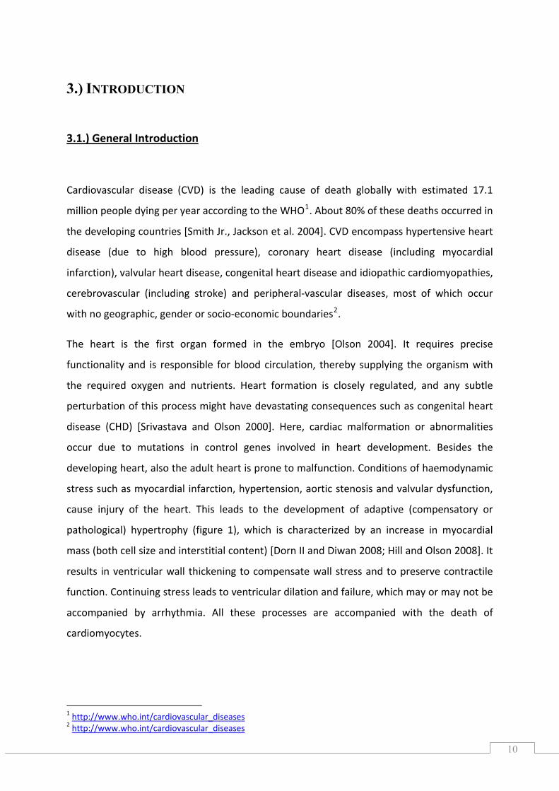
pathological) hypertrophy (figure 1), which is characterized by an increase in myocardial
mass (both cell size and interstitial content) [Dorn II and Diwan 2008; Hill and Olson 2008]. It
results in ventricular wall thickening to compensate wall stress and to preserve contractile
function. Continuing stress leads to ventricular dilation and failure, which may or may not be
accompanied by arrhythmia. All these processes are accompanied with the death of
cardiomyocytes.
1 http://www.who.int/cardiovascular_diseases 2 http://www.who.int/cardiovascular_diseases
11
Figure 1. Cardiac remodelling due to environmental stimuli. The heart adapts (remodels) in
response to environmental conditions causing the heart to shrink (A; Atrophy) or grow (B and C;
hypertrophy). While normal remodelling occurs during exercise, pregnancy, and postnatal growth
(physiological hypertrophy), pathologic remodelling due to neurohumoral activation, hypertension,
and myocardial injury lead to pathologic hypertrophy. The initial adaptation in pathologic
hypertrophy is also known as compensated (adaptive) hypertrophy which will develop to
decompensated (maladaptive) hypertrophy in response to continuous stress. This adverse cardiac
remodelling leads to heart failure. Figure was taken and adapted from [Hill and Olson 2008]
A) B) C)
12
3.1.1) Classification of heart cells
The main cell types in the myocardium (figure 2) are cardiomyocytes (CMC) and non‐
myocytes including cardiac fibroblasts (FB), vascular smooth muscle cells (VSMC) and
endothelial cells (EC). In the past, CMC and FB were thought to account for 30% and 70% of
the heart, respectively [Weber 1989; Baudino, Carver et al. 2006]. This view has changed in
the last couple of years, because more specific markers are now available for the
identification of FB. These markers include the discoidin domain receptor (DDR2, FB) or α‐
MHC (CMC). DDR2, which belongs to the family of collagen‐specific receptor tyrosine
kinases, is capable of transmitting extracellular signals into the cell [Schlessinger 1997;
Goldsmith, Hoffman et al. 2004]. Using FACS analysis, Banerjee and colleagues recently
confirmed that the number and type of cardiac cells in the heart varies depending on the
developmental stage, the physiological or pathophysiological conditions and also the
species. Neonatal murine hearts (day 1) were composed of 10 % FB versus 70% CMC
compared to neonatal rat hearts (day 1) with 30% FB and 62 % CMC [Banerjee, Yekkala et al.
2006; Banerjee, Fuseler et al. 2007]. Cardiac FB produce various proteins found in the
extracellular matrix (ECM) including collagen fibres of type I and III, elastin, growth factors
and matrix metalloproteinases (MMP) [Weber and Brilla 1991; MacKenna, Summerour et al.
2000; Spinale 2002; Bowers, Banerjee et al. 2010]. Type I collagen, which accounts for 85%
of the collagen matrix, mainly forms thick fibres that have substantial strength; in contrast,
type III collagen fibres (11%) that maintain structural integrity and distensibility of the 3‐
dimensional network are thin and resilient [Weber 1989].
3.1.2.) The cardiac interstitium
The cardiac interstitium (extracellular matrix, ECM) consists of the collagen network that
envelopes and connects muscle cells and the vasculature. A functional network of cells and
their ECM is characterized by a) direct or b) indirect cell adhesions. In a) cells adhere to one
another by cell‐cell adhesion through so‐called cell adhesion molecules (CAM). These cluster
in cell junctions such as tight junctions, gap junctions or desmosomes. In case b) cells
13
connect to surrounding components of the ECM through plasma membrane adhesion
receptors. These include polysaccharides and proteins, which are secretion products of the
present cells [Lodish, Berk et al. 2008]. The cell adhesion receptor classes involved in group
a) are cadherins and immunoglobulin (Ig)‐superfamily CAM, both of which generate
homophilic (self) interactions. The receptors of group b) include integrins which bind to
multiadhesive ECM proteins such as laminin (LN), and selectins which bind to carbohydrates
of the counter‐receptors (glycoproteins) [Hynes 1999; Juliano 2002]. Collagens,
proteoglycans and structural glycoproteins (also known as multiadhesive matrix proteins)
are the three classes of ECM components distinguished in vertebrates. Collagens are highly
abundant structural components forming fibres of type I, II and III, or sheets and networks
like basement collagen type IV [Aumailley and Gayraud 1998]. The multiadhesive matrix
proteins LN and fibronectin (FN), due to their structural flexibility, bind to various collagen‐
types, ECM or signalling proteins, polysaccharides, and cell adhesion receptors. By doing so,
these matrix proteins control, regulate and organize cell adhesion processes, migration, and
cell shape [Martin and Timpl 1987; Lodish, Berk et al. 2008]
Overall, the heart’s interstitium (figure 2) can be subdivided into three parts: epimysium,
perimysium, and endomysium. The epimysium surrounds the entire muscle and is located
along the epicardial and endocardial surfaces. The perimysial collagen fibres cover groups of
cardiomyocytes, organized in laminae of two to five myocytes in thickness. Further,
permysial strands are located between muscle bundles to connect to adjacent collagen
weaves. Endomysial collagen fibres which are formed from the perimysium either surround
individual cardiomyocytes, or connect adjoining myocytes to another or to capillaries by
perpendicular attachments [Weber 1989; Goldsmith and Borg 2002; Pope, Sands et al.
2008]. FBs lie near or between cardiomyocytes; connecting either with them or with one
another, enmeshed in the connective collagen tissue network [Goldsmith, Hoffman et al.
2004].
14
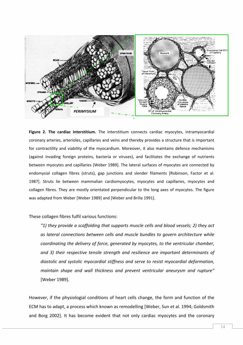
Figure 2. The cardiac interstitium. The interstitium connects cardiac myocytes, intramyocardial
coronary arteries, arterioles, capillaries and veins and thereby provides a structure that is important
for contractility and viability of the myocardium. Moreover, it also maintains defence mechanisms
(against invading foreign proteins, bacteria or viruses), and facilitates the exchange of nutrients
between myocytes and capillaries [Weber 1989]. The lateral surfaces of myocytes are connected by
endomysial collagen fibres (struts), gap junctions and slender filaments [Robinson, Factor et al.
1987]. Struts lie between mammalian cardiomyocytes, myocytes and capillaries, myocytes and
collagen fibres. They are mostly orientated perpendicular to the long axes of myocytes. The figure
was adapted from Weber [Weber 1989] and [Weber and Brilla 1991].
These collagen fibres fulfil various functions:
“1) they provide a scaffolding that supports muscle cells and blood vessels; 2) they act
as lateral connections between cells and muscle bundles to govern architecture while
coordinating the delivery of force, generated by myocytes, to the ventricular chamber,
and 3) their respective tensile strength and resilience are important determinants of
diastolic and systolic myocardial stiffness and serve to resist myocardial deformation,
maintain shape and wall thickness and prevent ventricular aneurysm and rupture”
[Weber 1989].
However, if the physiologial conditions of heart cells change, the form and function of the
ECM has to adapt, a process which known as remodelling [Weber, Sun et al. 1994; Goldsmith
and Borg 2002]. It has become evident that not only cardiac myocytes and the coronary
PERIMYSIUM
15
vasculature are mandatory for the contractility and viability of the myocardium, but so is the
ECM, too. Alterations of the interstitium results in loss of normal structural support and
changes in myocardial geometry and function [Spinale 2002], which plays an important role
in the progression to hear failure[Miner and Miller 2006]. Indeed, Gustafsson et al. (2003)
pointed out that:
“changes in the ECM components which are constantly being remodelled, degraded,
and resynthesized locally, can modulate their interactions of a cell with its
environment. The matrix also serves as a reservoir for many extracellular signalling
molecules that control cell growth and differentiation. In addition the matrix provides a
lattice through or on which cells can move, particularly in the early stage of tissue
assembly. Morphogenesis ‐ the stage of embryonic development in which tissues,
organs and body parts are formed by cell movements and re‐arrangements ‐ also is
critically dependent on cell matrix adhesion as well as cell‐cell adhesion. [..] Disruption
in cell‐matrix or cell‐cell interactions can have devastating consequences for the
development of tissues, as is seen in the dramatic changes in the skeletal system of
embryonic mice when the genes of either the two key ECM molecules, collagen II or
perlecan, are inactivated.” [Gustafsson, Aszódi et al. 2003]
Depletion of hormones or growth factors from the ECM leads to apoptosis [Meredith, Fazeli
et al. 1993; Schwartz 2010]. When mouse strains with null mutations in the FN gene (which
is recessive and embryonic‐lethal) were intercrossed, the resulting strains had embryonic
defects such as a deficit in mesoderm or neuronal tube formation [George, George‐
Labouesse et al. 1993], as well as defects in heart and blood vessel development. It was
observed that embryos developed either no primitive hearts or abnormal hearts, dependent
on the genetic background of the mice used to create FN null strains. [George, Baldwin et al.
1997]. These and further studies (reviewed by Hynes 1996) clearly show that per se, the ECM
represents a survival signal [Hynes 1996]. Furthermore, disruption of the connection
between cells and the ECM leads to cell detachment‐induced apoptosis (also referred to as
anoikis) [Frisch and Francis 1994].
16
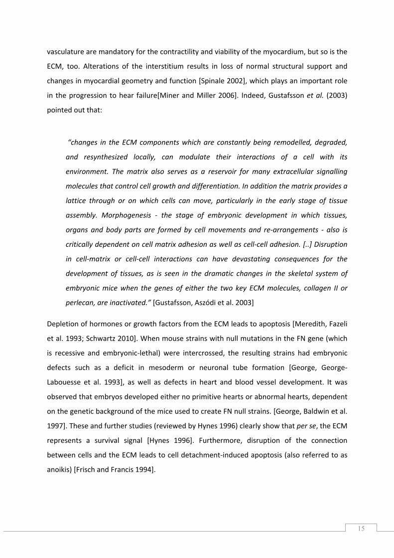
3.2.) Cell adhesion receptors
Receptors that maintain connections a) among cells and b) between cells and the ECM
represent the large family of cell adhesion receptors. Integrins, cadherins, selectins and IgG
superfamily (immunoglobulin‐cell adhesion molecules: Ig‐CAMs) are members of this family
(figure 3) [Aplin, Howe et al. 1998; Hynes 1999; Juliano 2002]. Among these, integrins
represent the major receptor group in the ECM in cardiomyocytes [Ross and Borg 2001].
Early studies in neonatal rat ventricular myocytes (NRVM) showed a tight binding of this cell
type to collagen IV, LN, collagen I and III, all of which are found in the interstitium [Borg,
Rubin et al. 1984].
Figure 3. Families of cell adhesion receptors associated with their typical binding partner. While integrins normally bind components of the ECM, they can also bind counter‐receptors (CR) such as Ig CAMs or selectins. Figure taken from [Rojas and Ahmed 1999].
3.2.1.) Integrins
Integrins have been identified from sponges to chicken to mammals, but are absent from
plants, fungi or bacteria [Hynes and Zhao 2000; Takada, Ye et al. 2007]. In mammals,
connections to ECM components are essential for the survival and the structural and
17
functional integrity of CMC [Meredith, Fazeli et al. 1993; Hynes 1996]. In particular, these
transmembrane cell surface adhesion receptors bind ECM proteins such as LN, collagen and
FN [Hynes 1992; Ross and Borg 2001]. Integrins are heterodimeric transmembrane receptors
composed of different α‐ and β‐subunits, which can form 24 distinct receptors; these
receptors are in a bent conformation in quiescent and in an extended conformation in
activated adherent cells [Askari, Tynan et al. 2010].
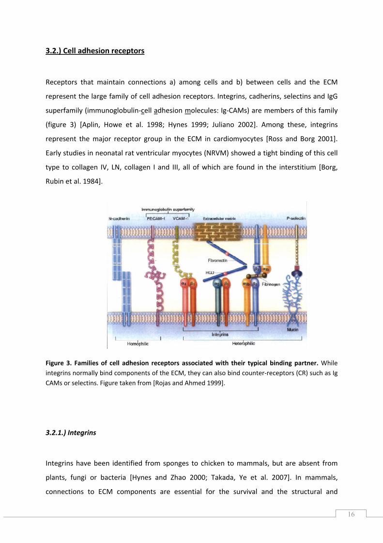
Based on their binding affinity, the integrin family can be grouped (figure 4,.for an
informative review see [Barczyk, Carracedo et al. 2010]) into RGD‐binding, LN‐binding,
collagen binding‐receptors, and leukocyte specific receptors [Hynes 1987; Hynes 2002]. As
their name implies, the RGD binding group binds to the short tripeptide sequence Arg‐Gly‐
Asp, one or more of which are found in many ECM proteins such as FN, fibrinogen or
vitronectin [Hynes 1987; Hynes 2002]. Although many integrin ligands have a RGD binding
sequence, not all of them are actually involved in binding.
Figure 4. Integrin subunits and their binding partners. The coloured α bars represent the ligand
specificity: where α3, α6 and α7 represent LN binding receptors (turquoise), α1, α2, α10, and α11 are
collagen binding (yellow), α5, αv and α8 (grey pentagon) represent RGD binding receptors. β7 (green
sun) and β2 integrins (not depicted here; pairs with αL, αM, αX, and αD) are restricted to leukocytes
[Hynes 2002]. A given β‐subunit (SU) binds several α SUs, and some α‐SU can bind several β‐SU. α
and β form non‐covalently linked receptors. β1 integrin, for instance, pairs with 10 different α SU,
whereas β3 has αV as a partner and αV combines with 4 different β SU. Adapted from [Hynes 2002;
Brakebusch, Hirsch et al. 1997]
18
Also, different RGD binding integrins have different affinities to the RGD sequence [Hynes
1992]. While α5β1, αIbβ3 and αvβ integrins recognize RGD, αIIbβ3 integrins additionally
recognize the sequence KQAGDV in fibrinogen; α2β1 binds the sequence DGEA in type I
collagen; α4β1 binds EILDV in alternatively spliced FN; and αvβ2 binds GPRP in fibrinogen
[Loike, Sodeik et al. 1991]. Ligand binding by integrins requires divalent cations [D`Souza,
Ginsberg et al. 1991]. Collagen‐binding integrins recognize the sequence GFP*GER (where P*
stands for hydroxyproline) [Knight, Morton et al. 1998]. These findings clearly indicate that
integrin binding to RGD sequences is not sufficient to explain the specificity of ligand
binding. It is likely that integrin splice variants in the extracellular and cytosolic part add to
the binding specificity of integrins (reviewed by Melker 1999). For example, the cytoplasmic
domain is essential in integrin heterodimerization and membrane expression [de Melker and
Sonnenberg 1999]. Five splice variants of β1 integrin were found in the cytoplasmic domain
(β1A β1B, β1C, β1C‐2 and β1D), which can activate different signalling cascades resulting in
different cellular responses. β1A and β1D splice variants differ only by 13 amino acids in their
cytoplasmic domain. β1D for instance is specifically found in adult skeletal muscle and
strongly expressed in the heart (adult and postnatal) [Van der Fllier, Gaspar et al. 1997]. β1D
integrins show a high affinity to the actin cytoskeleton and extracellular ligands [de Melker
and Sonnenberg 1999]. β1A is downregulated during myoblast differentiation as compared to
β1D and binds actin less tightly than β1D. Baudoin (1998) reported that β1A is more efficient in
transducing mechanical signals to the cell than β1D [Baudoin, Goumans et al. 1998].
3.2.1.1.) Integrin structure
Both, α and β subunits are comprised of a large extracellular domain (700‐1100 amino acids)
followed by a hydrophobic membrane spanning part and a short cytosolic domain (20‐60
amino acids) [Ruoslahti and Pierschbacher 1987]. Integrins bind to an array of distinct
proteins [McDonald 1989; Plow, Haas et al. 2000; Van der Fllier and Sonnenberg 2001], and
can be bound by anti‐integrin antibodies [Diamond and Springer 1994]. The α‐ and β‐integrin
subunits consist of several characteristic domains as illustrated in figure 5A and B. The
extracellular part of the integrin subunits can be divided into “head” and “leg” parts. The
“head” of α integrin is composed of two domains: the β propeller domain, and in some cases
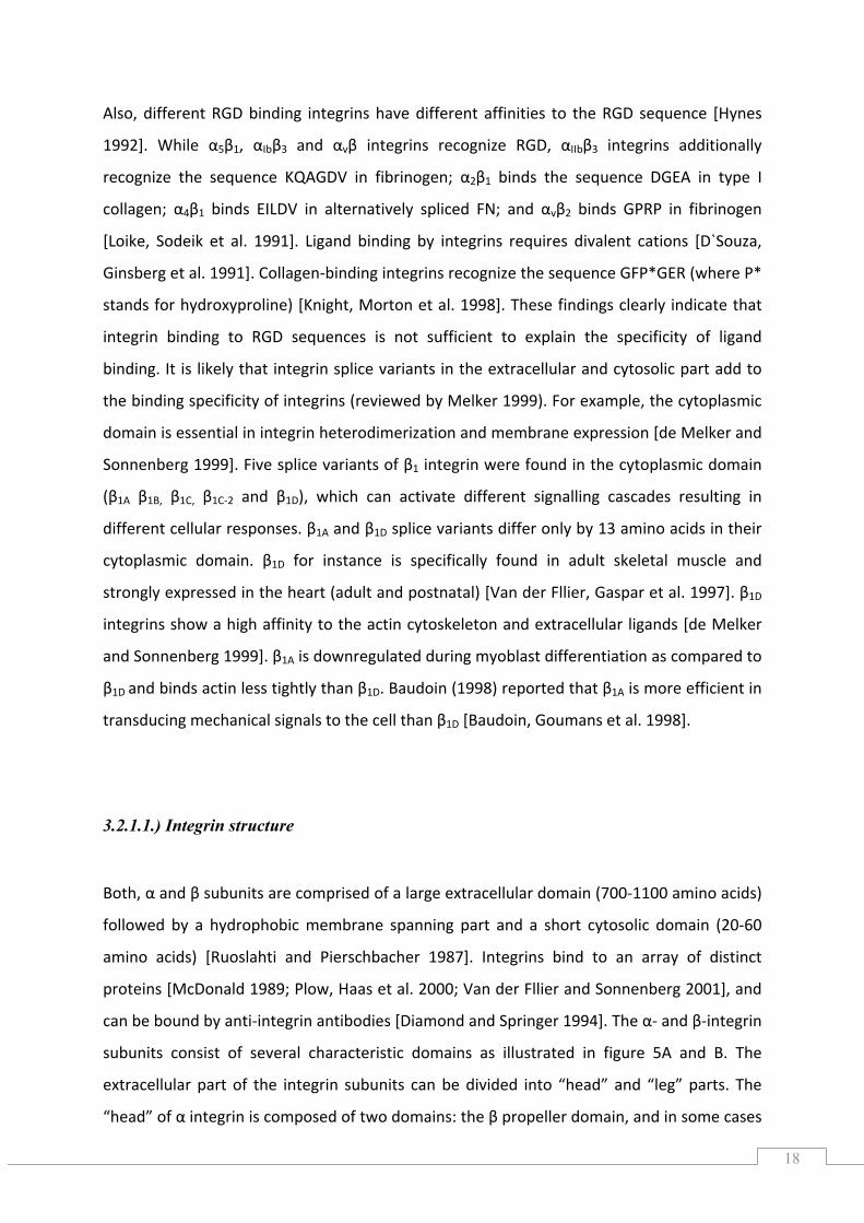
19
an αA domain. This αA domain is also known as I domain and is located between the β‐
sheets 2 and 3 of the β propeller domain; it contains a metal‐ion‐dependent adhesive site
(MIDAS, occupied by a divalent cation) for binding of negatively charged ligand residues. The
“leg” domain consists of the Ig‐like “thigh” domain, the “knee” domain (genu) and 2 β
sandwiches known as “calf” domains.
Figure 5. Common integrin structure with organized regions. TM: transmembrane domain; cyto:
cytoplasmic domain; genu: knee; PSI: Plexin‐semaphorin‐integrin domain. The head part contains the
I domain or van Willebrand factor A domain, which is also referred to as A domain. Figures were
taken from [Luo and Springer 2006; Byron, Humphries et al. 2009].
The β‐integrin subunit contains 11 domains which can be also subdivided into “head” and
“leg”. The head is composed of the βA domain (or β I‐like domain) which is inserted in the
hybrid domain and the PSI domain. Additionally the top face of the βA domain has also a
MIDAS site. The leg of β integrin is comprised of a hybrid, a PSI and a knee domain, followed
by four EGF‐like domains and a β tail domain.
The RGD binding site is located between the β propeller of the α subunit and the A domain
of the β subunit [Shimaoka, Takagi et al. 2002; Luo and Springer 2006; Arnaout, Goodman et
al. 2007; Takada, Ye et al. 2007; Byron, Humphries et al. 2009]. Activation of integrins and
A) B)
20
their associated conformational changes have been studies extensively. Several models have
been developed to describe signalling, including data from the investigation of the
localization of integrin transmembrane and cytoplasmic domains [Xiong, Stehle et al. 2003;
Arnaout, Goodman et al. 2007].
Integrins are not constitutively active, persisting in an inactive, bent conformation. It is
hypothesised that this closed conformation embodies a low affinity state for ligand binding
[Arnaout, Goodman et al. 2007]. The knee region keeps the integrin receptors of quiescent,
non‐activated cells in the bent conformation. Upon activation, the conformation of the
receptor changes and the “leg” swings open into a state primed for activation [Luo and
Springer 2006], and then an activated, extended state with an open headpiece, finally
resulting in integrin clustering with high apparent affinity (or avidity) [Rocco, Rosano et al.
2008; Byron, Humphries et al. 2009]. Some models imply a separation of the transmembrane
and cytosolic domains [Wegener and Campbell 2008], whereas Rocco et al. (2009) proposes
that tail domains stay in contact [Rocco, Rosano et al. 2008] during inside‐out signalling,
whereas outside‐in signalling may be independent of this separation.
Recently, Askari et al. (2010) showed that α5β1 integrin can be directly activated at focal
adhesions of adherent cells in response to binding of stimulating anti‐β1 and anti‐α5
monoclonal antibodies directed to several epitopes of the extracellular part of a mutated
α5β1 integrin receptor [Askari, Tynan et al. 2010]. In that study, the integrin α‐subunit and β‐
subunit were locked together through a disulphide bond. By use of the FLIM (fluorescence
lifetime imaging microscopy) based FRET (fluorescence resonance energy transfer)
technology, α5β1 conformational changes were observed in human foreskin FB.
3.2.1.2.) Integrin signalling
Integrins can signal in two directions: a) from the outside into the cell (outside‐in signalling)
and b) from inside to the outside (inside‐out signalling). They signal in combination with
growth factor receptors, cytokine receptors or G‐protein coupled receptors, and in an array
21
of different cell types, including cardiac cells [Arnaout, Mahalingam et al. 2005]. Outside‐in
signalling happens synergistically with co‐stimulators, ligands, and/or cations (Mn2+),
however, it can be also triggered experimentally by so‐called activating antibodies [Bazzoni,
Ma et al. 1998]. Since cytosolic integrin domains do not have kinase activity, they have to
recruit functional proteins to focal adhesion sites in order for signalling to occur [Giancotti
and Ruoslahti 1999]. Integrin signalling is a complex matter, because these receptors bind to
an array of proteins and lipids. Zaidel‐Bar et al. (2007) illustrated the complex network of
integrins and their interconnected partners [Zaidel‐Bar, Itzkovitz et al. 2007] derived from
published experimental studies from different cell types. Their model revealed a signalling
interaction network of 690 links of which 213 were activating and 98 were inhibitory (figure
6).
22
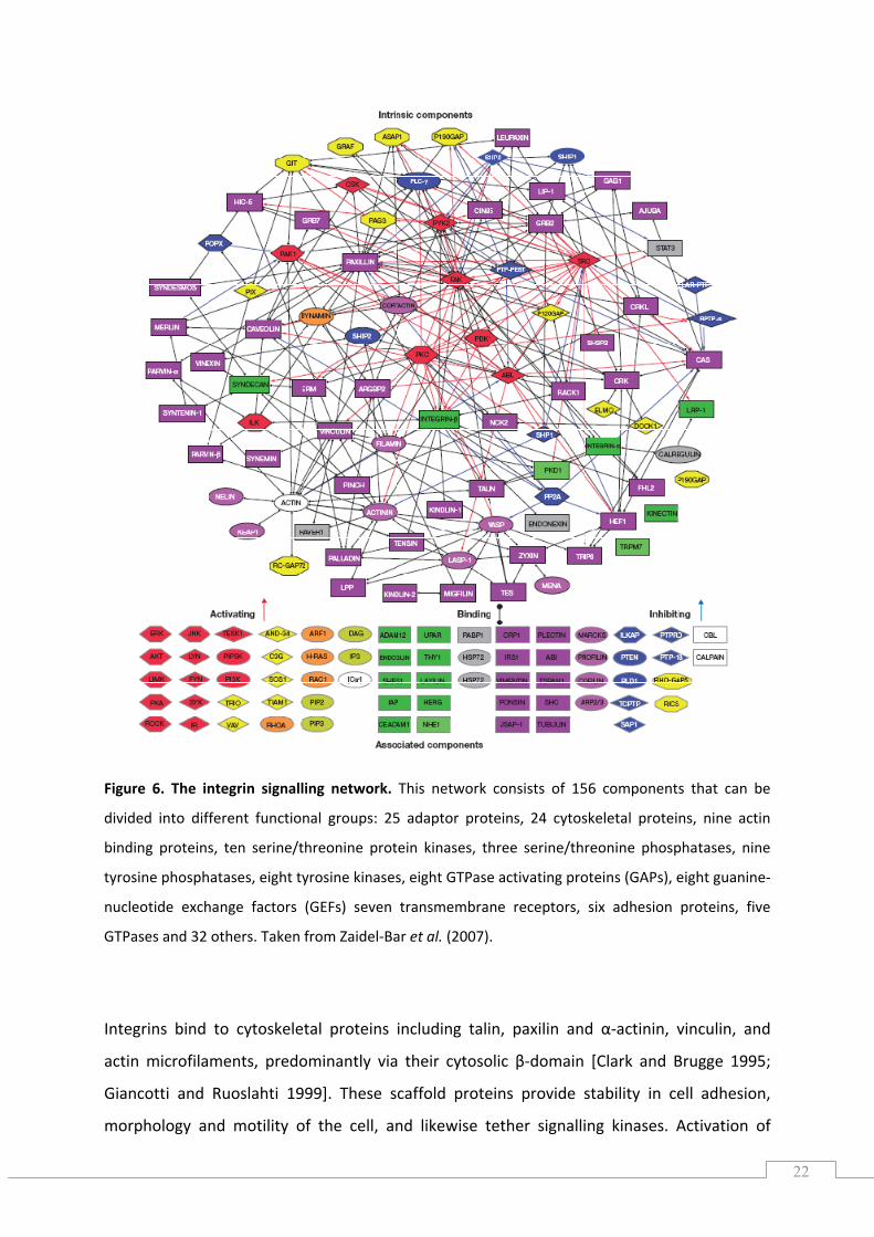
Figure 6. The integrin signalling network. This network consists of 156 components that can be
divided into different functional groups: 25 adaptor proteins, 24 cytoskeletal proteins, nine actin
binding proteins, ten serine/threonine protein kinases, three serine/threonine phosphatases, nine
tyrosine phosphatases, eight tyrosine kinases, eight GTPase activating proteins (GAPs), eight guanine‐
nucleotide exchange factors (GEFs) seven transmembrane receptors, six adhesion proteins, five
GTPases and 32 others. Taken from Zaidel‐Bar et al. (2007).
Integrins bind to cytoskeletal proteins including talin, paxilin and α‐actinin, vinculin, and
actin microfilaments, predominantly via their cytosolic β‐domain [Clark and Brugge 1995;
Giancotti and Ruoslahti 1999]. These scaffold proteins provide stability in cell adhesion,
morphology and motility of the cell, and likewise tether signalling kinases. Activation of
23
various kinases upon integrin engagement has been reported. Among the first kinases that
are recruited are the tyrosine kinase Src, focal adhesion kinase (FAK), and integrin linked
kinase (ILK). The latter was shown to bind to the cytosolic β integrin domain, activating GSK‐
3β, mTOR or NFκB as substrates, besides other kinases [Legate, Montañez et al. 2006]. But
also serine/threonine kinase families (such as protein kinase C, PKC and MAPK) are recruited
[Clark and Brugge 1995]. The lipid kinase phosphoinositide (PI)‐3‐OH kinase (PI3K)
phosphorylates its substrate, the protein kinase B (PKB, also known as Akt) either in a FAK‐
Src dependent or independent manner [Giancotti 1997]. The latter case was described by
Velling (2004) in a β1B integrin transfected cell line (GD25) that lacks endogenous β1 integrin
expression [Velling, Nilsson et al. 2004].
3.2.1.3.) β1-integrin (CD29)
3.2.1.3.1.) β1 integrin-induced survival and signalling
The β1 integrin subunit (also known as CD29, very late antigen or VLA in humans) [Hemler,
Huang et al. 1987; Hynes 1987], represents the most abundant isoform expressed in foetal
and postnatal rat heart [Carver, Price et al. 1994; de Melker and Sonnenberg 1999], where it
can dimerize with different α subunits [Ross 2002]. The expression of the α integrins is
altered in neonatal development and hypertrophied hearts [Terracio, Rubin et al. 1991; Ross
2002]. Neonatal cardiomyocytes mainly interact with the subunits α1, α3, α5 and α6 [Terracio,
Rubin et al. 1991; Maitra, Flink et al. 2000; Ross 2002]. Integrin expression and signalling are
of tremendous importance to myocyte growth and survival [Kuppuswamy 2002], as well as
in cardiomyocyte remodelling [Ross 2002].
For instance, a Chinese ovary hamster cell line that expressed α5β1 integrin was protected
from apoptosis by Bcl‐2 upregulation [Zhang, Vuori et al. 1995]. Lei et al. (2008) showed that
endothelial CD29 expression is needed for postnatal vascular remodelling. Homozygous
deletion of CD29 in the endothelium led to embryonic death whereas heterozygous
endothelial CD29 deletion did not diminish foetal or postnatal survival but reduced CD29
expression in endothelial cells of adult mice [Lei, Liu et al. 2008]. Deletions of CD29 are
embryonic lethal [Fässler and Meyer 1995; Fässler, Rohwedel et al. 1996; Keller, Shai et al.
24
2001]. Moreover, cardiac‐specific knock‐outs or inhibition lead to disturbed cardiac function,
impaired cardiac differentiation, fibrosis and heart failure [Fässler and Meyer 1995; Shai,
Harpf et al. 2002]. Deficiency in CD29 is reported to cause increased myocardial dysfunction
after myocardial infarction in mice [Krishnamurthy, Subramanian et al. 2006].
In vitro, CD29 is directly activated by the binding of ECM components or activating
antibodies [Diamond and Springer 1994], which causes the rearrangement of the actin
cytoskeleton and recruitment of signalling kinases such as FAK, PI3K, PKB, MEK1/2, ERK1/2
or talin to the cytosolic part of the integrin receptor [Hynes 1992; Clark and Brugge 1995].
Besides activating anti‐CD29 antibodies also inhibiting anti‐CD29 antibodies were developed
[Byron, Humphries et al. 2009]. Through activation of above signalling kinases, CD29
participates in the regulation of CMC growth and survival [Ross and Borg 2001]. Specifically,
CD29 mediates α‐adrenergic receptor‐stimulated hypertrophy [Ross, Pham et al. 1998] and
inhibits β‐adrenergic receptor‐induced apoptosis [Communal, Singh et al. 2003; Menon,
Singh et al. 2005].
3.2.1.3.2.) Effects of CD29 engagement on JNK/SAPK
The JNK (c‐Jun N‐terminal kinase) /SAPK (stress‐activated protein kinase) belongs to the
MAPK (mitogen‐activated protein kinase) superfamily, besides ERK and the p38 MAP kinases
[Davis 2000]. Stress and inflammatory signals activate JNK and p38 MAPK. The JNK family
includes; JNK1 (four isoforms), JNK2 (four isoforms), and JNK3 (two isoforms). JNK has been
implicated in both survival [Dougherty, Kubasiak et al. 2002; Scuteri, Galimberti et al. 2010]
and death pathways [Lin 2002]. Its involvement in cell survival is context‐dependent, varying
according to stimulus, tissue and environmental factors. It has been observed that persistent
but not transient activation of JNK is linked to apoptosis [Andreka, Zang et al. 2001; Lin
2002]. ROS have been assumed to cause sensitive and permanent activation of JNK [Nakano,
Nakajima et al. 2006]. Reinhard et al. (1997) showed that TNFα‐induced JNK activation
occurred independently of the death pathway [Reinhard, Shamoon et al. 1997]. Whereas
insulin‐like growth factor protects human neuroblastoma cells from apoptosis through the
PI3K/PKB/GSK‐3β pathway by inhibiting JNK [Wang, Yang et al. 2010]; integrin induced
25
survival signalling was shown to be mediated by FAK and JNK activation in rabbit synovial FB
[Almeida, Ilić et al. 2000].
3.2.1.3.3.) Src family kinase and protein kinase C are likely upstream partners in CD29-
induced survival signalling
Integrin engagement results in the recruitment of several protein kinases to propagate
signals, including upstream protein tyrosine kinases such as Src (Src family kinases), FAK, as
well as a serine/threonine kinase ILK [Giancotti 1997; Giancotti and Ruoslahti 1999]. Upon
auto‐phosphorylation of FAK at residue Tyr397, a binding site for Src’s Src homology 2
domain (SH2) is created in chicken embryo cells [Schaller, Hildebrand et al. 1994]. Based on
reports that inhibition of PI3K by Wortmannin or Ly inhibited phosphorylation of the
regulatory NADPH oxidase (NOX) subunit p47phox, and hence suppressing NOX activity in
neutrophils [Ding, Vlahos et al. 1995], Chen et al. (2003) showed that PKB is capable of
activating and phosphorylating p47phox; it therefore regulates human gp91phox [Chen, Powell
et al. 2003]. Beside PKB and ERK [Dewas, Fay et al. 2000], also PKC, specifically PKCζ, has
been implicated in the independent activation of the NADPH oxidase by phosphorylation of
p47phox, [Dang, Fontayne et al. 2001]. and gp91phox [Raad, Paclet et al. 2009]. Protein kinases
C can be subdivided into 4 distinct groups, namely atypical, classical, novel, and PKC related
kinases. PKCζ belongs to the subclass of atypical or aPKC, together with PKCι (also known as
PKCλ in mice) [Rosse, Linch et al. 2010]. Classical PKC (cPKC) proteins including PKCα, PKCβ,
PKCγ, novel PKCs (nPKC) PKCδ, PKCε, PKCη, PKCθ and PKC related kinases (PKN) include
PKN1, PKN2 and PKN3. It is of note that Src kinase interacts with PKCζ in nerve growth factor
(NGF)‐stimulated PC12 cells (a cell line derived from a pheochromocytoma ‐ a
neuroendocrine tumour ‐ of the rat adrenal medulla) [Greene and Tischler 1976]. Tyrsosine
residues of PKCζ were shown to be only phosphorylated in the presence of Src kinase in
PC12 cells [Seibenhener, Roehm et al. 1999]. Also, PKCδ was shown to be a target of Src
tyrosine kinase phosphorylation [Gschwendt, Kielbasse et al. 1994]. In 2009, Gupte and co‐
workers showed that Src kinase mediated PKC‐induced NOX2 activation and peroxide
production upon phorbol 12, 13‐butryate treatment in coronary artery smooth muscle cells
[Gupte, Kaminski et al. 2009]. Moreover, intracellular lipoxygenase (LOX)‐derived ROS
activated Src kinase during cell adhesion [Giannoni, Buricchi et al. 2005] upstream of
26
epidermal growth factor receptor (EGFR), ERK, PKB and Bim [Giannoni, Buricchi et al. 2008]
in mouse embryonic FB (NIH‐3T3 cell line).
3.3.) Reactive Oxygen Species (ROS)
The term ROS or oxygen‐derived species comprises radicals (superoxide anion, O2•‐; hydroxyl
radicals, etc.) and non‐radicals (hydrogen peroxide, H2O2). However, other terms, such as
reactive oxygen intermediates (ROI) or reactive oxygen mediators (ROM), are also used in
the literature. Radicals are defined as any molecule species having one or more unpaired
electrons, and participating in one‐electron transfer reactions [Halliwell and Gutteridge
1985]. ROS are generated as by‐products in many metabolic processes, or as products of
enzymatically catalysed processes.
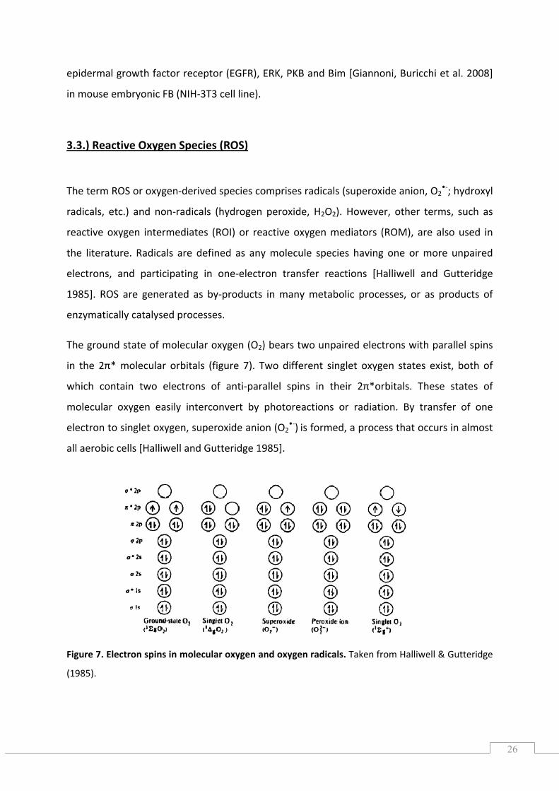
The ground state of molecular oxygen (O2) bears two unpaired electrons with parallel spins
in the 2π* molecular orbitals (figure 7). Two different singlet oxygen states exist, both of
which contain two electrons of anti‐parallel spins in their 2π*orbitals. These states of
molecular oxygen easily interconvert by photoreactions or radiation. By transfer of one
electron to singlet oxygen, superoxide anion (O2•‐) is formed, a process that occurs in almost
all aerobic cells [Halliwell and Gutteridge 1985].
Figure 7. ll & utteridge
(1985).
Electron spins in molecular oxygen and oxygen radicals. Taken from Halliwe G
27
For instance, superoxide is formed by one‐electron transfer from the electron transport
chains of mitochondria, chloroplasts or the endoplasmic reticulum (ER), but also by the
respiratory (oxidative) burst oxidase in phagocytes [Hancock, Desikan et al. 2001] and
xanthine oxidase [Halliwell and Gutteridge 1985]. H2O2 has no unpaired electrons and is
either spontaneously formed at low pH or in a reaction catalysed by the enzyme superoxide
dismutase (SOD), which was uncovered by McCord & Fridovich in 1969 [McCord and
Fridovich 1969]. The enzyme catalase (cat) subsequently reduces H2O2 to H2O. Other
detoxification mechanisms include glutathione peroxidise (GPX), as well as non enzymatic
compounds like ascorbic acid, α‐tocopherol or glutathione [Dröge 2002].
Four classes of SOD enzymes can be distinguished, containing either divalent Cu/Zn ions or
Mn, nickel (Ni) or iron (Fe) ions. In humans, three SOD subtypes can be distinguished, a)
extracellular SOD (ecSOD or SOD3, homotetrameric and glycosylated, and heparin binding),
b) intracellular, homodimeric SOD (Cu/Zn‐SOD or SOD1, found in the cytosol, nucleolus,
lysosomes and the intermembrane space of mitochondria) and c) homotetrameric SOD (Mn‐
SOD or SOD2, in the mitochondrial matrix) [Fridovich March 2009]. Mn‐SOD obtains its main
superoxide anions from the mitochondrial respiratory chain. Lack of Mn‐SOD results in
dilated cardiomyopathy and neonatal lethality in knock‐out mice [Matés 2000]. TNF
application in mice “selectively induces Mn‐SOD expression and human Mn‐SOD expressed
in transgenic mice protects against oxygen induced pulmonary injury and adriamycin‐
induced cardiac toxicity” [Matés 2000]. Molecular oxygen induces SOD in prokaryotes such
as Escherichia coli and Streptococcus faecalis, as well as in eukaryotes [Gregory and Fridovich
1973; Gregory, Goscin et al. 1974]. Notably, Cu/Zn‐SOD can be inactivated by H2O2, due to
reduction/oxidation processes of the cofactor Cu2+ at the active site of the enzyme [Liochev
and Fridovich 2002]. Aside from having a dismutase activity, Cu/Zn‐SOD also displays
superoxide reductase (SOR) and a superoxide oxidase (SOO) activities [Liochev and Fridovich
2000]. By contrast, human Mn‐SOD does not seem to have any additional activities [Liochev
and Fridovich 2000].
Although oxygen is inherently toxic to aerobic organisms, efficient systems have evolved to
handle oxygen in living systems ranging from bacteria to mammals. Irrespective of ROS‐
induced toxic effects including lipid‐peroxidation, DNA damage (strand breaks), and
damaging effects on phospholipids, peptides, nucleotides, carbohydrates, also beneficial
28
effects of ROS have been investigated [Hensley, Robinson et al. 2000; Bochkov, Kadl et al.
2002; Blüml, Rosc et al. 2008]. High levels of ROS are damaging and are often referred to as
oxidative stress, whereas low level of ROS may act as second messengers in redox signalling
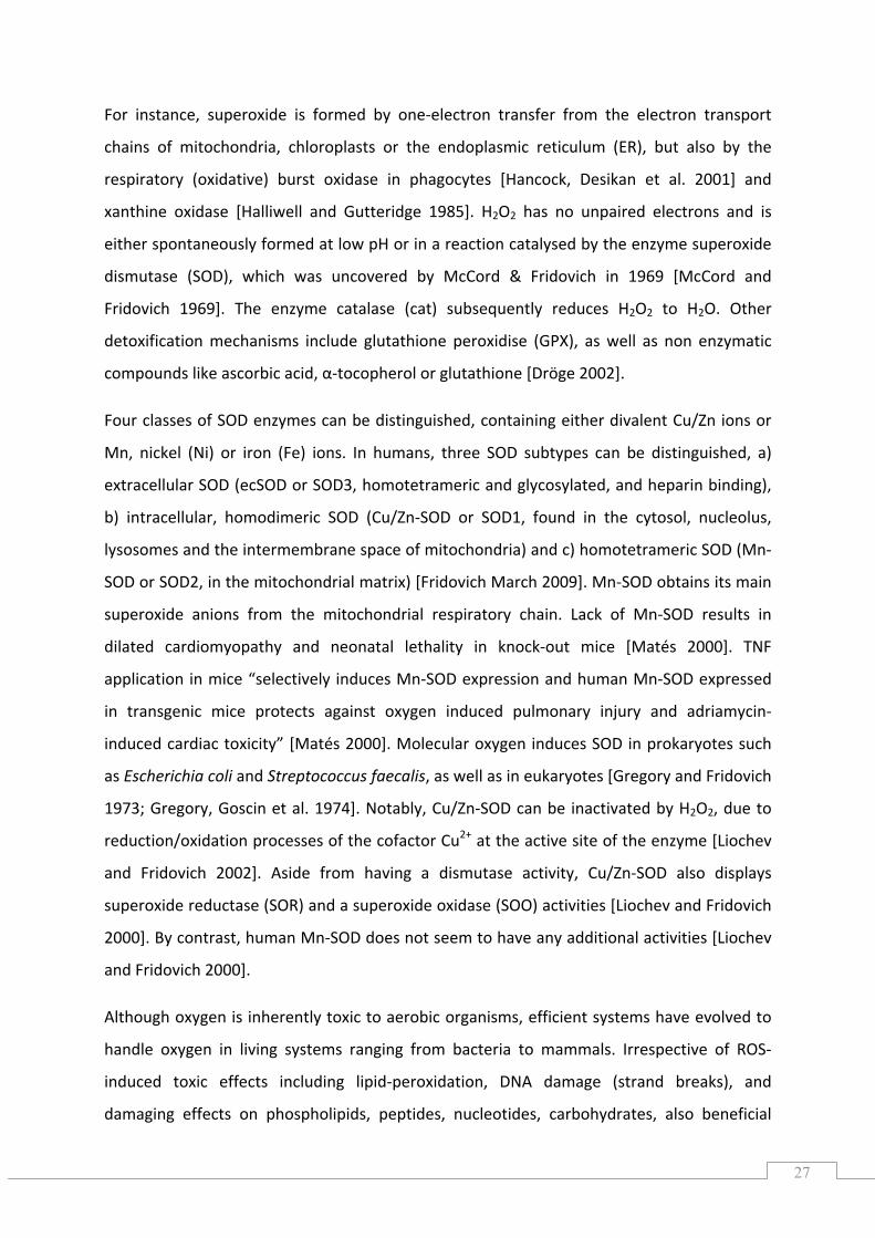
[Rhee 2006], activating signalling cascades that have been implicated in cell survival and
growth. Sources of such ROS (figure 8) include xanthine oxidase, mitochondria, 5‐
lipoxygenase, and NOX, the latter having been implicated as an important source of cardiac
ROS, generating ROS in a highly regulated fashion [Dröge 2002; Sawyer, Siwik et al. 2002;
Lambeth 2004; Cave, Brewer et al. 2006; Chiarugi and Fiaschi 2007].
Figure 8. Generation and clearance of ROS. glutathione peroxidase (GPX), glutathione reductase
(GR); 5‐lipoxygenase (5‐LOX). Adapted from Dröge (2002).
NOX
xanthine oxidase
O2 O2•‐ H2O2 H2O
mitochondria
GSH GSSG
GPX
NADPH NADP+
GR
SOD cat
5‐LOX
29
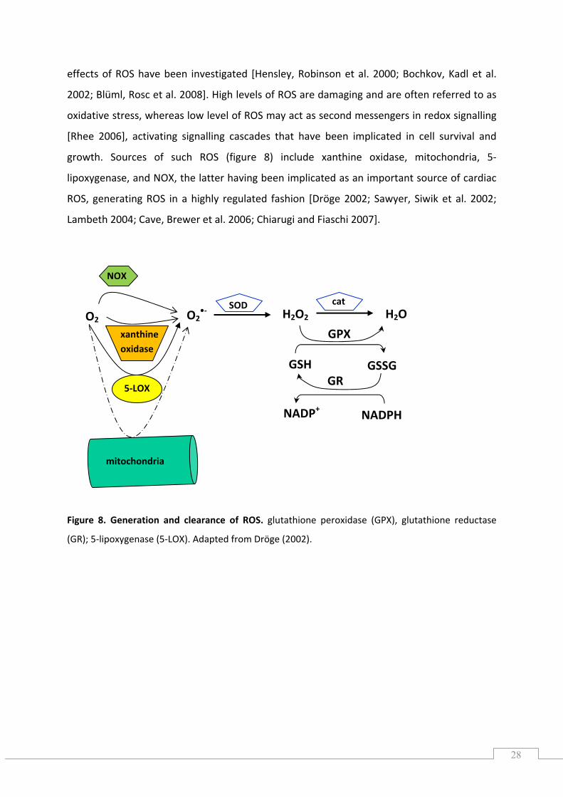
3.4.) NADPH oxidase
The NADPH oxidase is a transmembrane, multi‐component protein that produces a surge of
ROS upon stimulation [Petry, Weitnauer et al. 2010]. Initially, this enzyme was referred to as
“O2‐ generating enzyme” [Babior, Curnutte et al. 1976] or phox (phagocytic oxidase) [Babior
1999], but in the recent literature it is termed NOX (NADPH oxidase). NOX generates oxygen
radicals radicals in what is termed a respiratory or oxidative burst. This phenomenon was
first observed in phagocytes by Sbarra and Karnovsky [Sbarra and Karnovsky 1959;
Karnovsky and Sbarra 1960].The catalytic entity of NOX is comprised of gp91phox and p22phox..
These are also referred to as cytochrome b558, and lie embedded in the plasma membrane
[De Leo, Ulman et al. 1996; Vignais 2002]. p47phox, p67phox, p40phox and rac1/2 make up the
regulatory subunits. Studies from patients suffering from inherent chronic granulomatous
disease (CGD) showed their incapability to deal with bacterial infections due to loss of
functional NOX. In most cases (66.6%), CGD could explained by a defective CYBB (gp91phox)
gene, followed by defective CYBA (p22phox), NCF1 (p47phox) or NCF2 (p67phox) genes [Segal
and Shatwell 1997]. NOX is not restricted to leukocytes; several NOX homologues have been
identified in non‐phagocytes including NOX1‐5 and, DUOX1‐2 [Geiszt and Leto 2004], and a
NOX2 splice variant NOX2S [Heidari, Shah et al. 2004] in various tissues and species
[Lambeth, Cheng et al. 2000; Geiszt and Leto 2004; Kawahara, Quinn et al. 2007]. In the
heart, NOX2 has been reported as the predominant isoform, but also NOX4 and NOX1 have
been shown to be expressed in rat CMC [Hingtgen, Tian et al. 2006]. NOX2 and NOX1 both
depend on regulatory subunits such as the NOX organizers p47phox (for NOX2) and its
equivalent NOXO1 (for NOX1). NOX4 is active even in the absence of regulatory subunits, as
depicted in figure 9 [Brown and Griendling 2009].
Figure 9. NOX1, NOX2 and NOX4, with and without regulatory subunits. Taken and adapted from
[Brown and Griendling 2009]
30
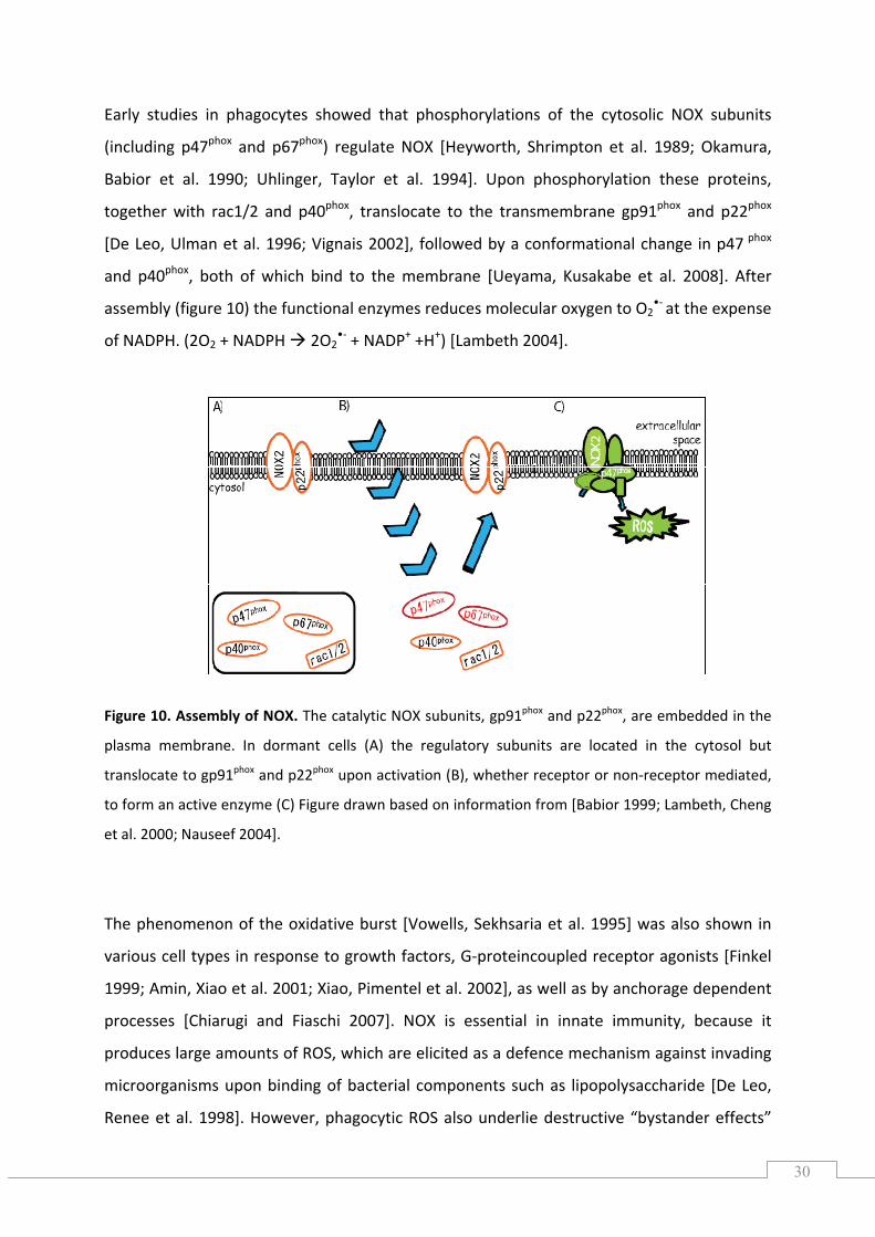
Early studies in phagocytes showed that phosphorylations of the cytosolic NOX subunits
(including p47phox and p67phox) regulate NOX [Heyworth, Shrimpton et al. 1989; Okamura,
Babior et al. 1990; Uhlinger, Taylor et al. 1994]. Upon phosphorylation these proteins,
together with rac1/2 and p40phox, translocate to the transmembrane gp91phox and p22phox
[De Leo, Ulman et al. 1996; Vignais 2002], followed by a conformational change in p47 phox
and p40phox, both of which bind to the membrane [Ueyama, Kusakabe et al. 2008]. After
assembly (figure 10) the functional enzymes reduces molecular oxygen to O2•‐ at the expense
of NADPH. (2O2 + NADPH 2O2•‐ + NADP+ +H+) [Lambeth 2004].
Figure 10. Assembly of NOX. The catalytic NOX subunits, gp91phox and p22phox, are embedded in the
plasma membrane. In dormant cells (A) the regulatory subunits are located in the cytosol but
translocate to gp91phox and p22phox upon activation (B), whether receptor or non‐receptor mediated,
to form an active enzyme (C) Figure drawn based on information from [Babior 1999; Lambeth, Cheng
et al. 2000; Nauseef 2004].
The phenomenon of the oxidative burst [Vowells, Sekhsaria et al. 1995] was also shown in
various cell types in response to growth factors, G‐proteincoupled receptor agonists [Finkel
1999; Amin, Xiao et al. 2001; Xiao, Pimentel et al. 2002], as well as by anchorage dependent
processes [Chiarugi and Fiaschi 2007]. NOX is essential in innate immunity, because it
produces large amounts of ROS, which are elicited as a defence mechanism against invading
microorganisms upon binding of bacterial components such as lipopolysaccharide [De Leo,
Renee et al. 1998]. However, phagocytic ROS also underlie destructive “bystander effects”
31
that can damage surrounding tissue (rheumatoid arthritis). For every tissue expressing NOX ‐
including heart, lung and the central nervous system ‐ NOX‐derived ROS have been
implicated in physiological as well as pathophysiological processes [Quinn, Ammons et al.
2006].
3.5.) ROS, NOX and signalling
NOX‐derived H2O2 is typically thought to be the most likely ROS‐derived signalling molecule
in physiological processes, promoting normal physiological processes, which although some
authors only attribute a minor role to NOX [Chiarugi and Fiaschi 2007; Forman, Maiorino et
al. 2010] Cells use H2O2 as signalling modulators regulating peroxyredoxin signalling
[Toledano, Planson et al. 2010] and activating protein tyrosine kinases. Gianonni et al. (2005)
showed that in NIH 3T3 cells, Src is activated through H2O2‐mediated oxidation of cysteine
residues and subsequent phosphorylation upon cell adhesion [Giannoni, Buricchi et al.
2005]. In CMC survival was determined by activation of ERK through Src and Ras as well as by
activation of the PI3K/PKB pathway in response to H2O2 [Aikawa, Komuro et al. 1997;
Aikawa, Nawano et al. 2000]. ROS have also been implicated as mandatory mediators during
cell adhesion [Chiarugi, Pani et al. 2003]. Further, increased ROS levels have been observed
upon excessive exercise [Ristow, Zarse et al. 2009]. If those ROS are scavenged by
antioxidants, the beneficial effects of exercise‐induced ROS are lost [Ristow, Zarse et al.
2009].
3.5.1.) ROS generation and NOX in cardiomyocytes
Various stimuli including growth factors and G‐protein coupled receptor (GPCR) agonists
induce intracellular ROS [Finkel 1998; Finkel 1999; Xiao, Pimentel et al. 2002]. NOX plays a
decisive role in myocardial ROS formation [Bendall, Cave et al. 2002; Sawyer, Siwik et al.
2002]. By contrast, previous studies (Chirugi, 2003; Giannoni, 2008) concluded that the
integrin requirement for growth factor signalling depends mainly on lipoxygenase‐derived
ROS, based on the use of synthetic inhibitors. As noted before, at moderate levels, NOX‐
32
derived ROS act as second messengers in redox‐signalling, activating several signalling
kinases implicated in cell survival and growth [Sawyer, Siwik et al. 2002; Rhee 2006].
Although NOX or NOX‐derived ROS are generally implicated in physiological and
pathophysiological processes [Bedard and Krause 2007], they are mainly associated with
pro‐apoptotic processes in the heart [Akki, Zhang et al. 2009], which may be due to the lack
of studies in morphologically normal CMC, of comprehensive studies, and the choice of
stimulus and working model. NOX was implicated as a mediator of ischaemia and GPCR‐
induced apoptosis in H9C2 cells based on the use of synthetic inhibitors [Qin, Patel et al.
2006]. Likewise, NOX was shown to be involved in cardiac hypertrophy and fibrosis. While
NOX2 was reported to be involved in GPCR‐dependent myocardial hypertrophy, it is
dispensable in pressure overload‐induced hypertrophy [Maytin, Siwik et al. 2004]. On the
other hand, the lack of NOX2 in ischaemic preconditioning prevented NOX‐associated pro‐
survival signalling and protection against ischaemia and reperfusion injury [Bell, Cave et al.
2005]. Irrespective of the number of studies performed to elucidate the role of cardiac NOX
or NOX‐derived ROS, the respective protective pathways involved in CMCs remain poorly
characterized.
33
3.6.) Aims of this study
The highly abundant cell adhesion receptor CD29 is essential for CMC growth and survival.
Its deletion is embryonic lethal and its loss of function or disruption causes severe heart
diseases. CD29‐induced survival signalling cascades in healthy, morphologically normal
cardiomyocytes are barely investigated. Further, several lines of evidence suggest a potential
role of ROS in the regulation of cell survival, with NOX having been reported to act as a
source of ROS in the heart. Due to the emerging role of ROS as regulators of cell survival, and
the crucial role of NOX as a potent source of ROS in the heart, we addressed the question if
CD29‐induced pro‐survival signalling depends on NOX‐derived ROS in neonatal CMC.
Therefore, the involvement of ROS in the survival signalling hierarchy was dissected, with a
special focus on the PI3K/PKB and the MAPK pathways. It was also investigated whether
GSK‐3β, which is reported to be a direct substrate of PKB, could be involved in pro‐survival
signalling. Further, likely upstream kinases of PI3K/PKB, MAPK and/or NOX were
investigated. Additionally we wanted to explore whether CD29 induces ROS formation in
newborn rat CMC, and in mouse CMC deficient in functional NOX subunits.
A minor aim was to investigate which NOX gene homologues are expressed in neonatal rat
cardiomyocytes in response to CD29 stimulation, and whether the genes encoding the ROS
sensor NFκB, the O2•‐ converting enzyme SOD, or the anti‐apoptotic protein Bcl2 were
expressed upon CD29 engagement.
34
4.) MATERIALS & METHODS
4.1.) Reagents
Materials for cell culture: Reagents from GIBCO/Invitrogen: Dubelco’s Modified Eagels Medium
(DMEM) for cardiomyocytes; low glucose, 1g/L, for cell line; high glucose, 4.5g/L; phenol red free
(prf)‐DMEM, low glucose, 1 g/L (cardiac myocytes); high glucose, 4.5 g/L (H9c2), HBSS, Hepes, 0.05%
trypsin‐EDTA, PBS, D‐PBS (+ Ca2+), L‐glutamine (GlutaMAX), penicillin (P, 100 U/mL), streptomycin (S,
100 µg/mL) and FCS; 5‐bromo‐2‐deoxyuridine (BrdU, 200 mmol/L) and trypan blue were purchased
from Sigma Aldrich, Collagenase was from Worthington, Accutase from PAA Laboratories (The cell
culture company). Culture flasks T75 and T25, as well as sterile 5 mL, 10 mL and 25 mL serological
plastic pipettes, 1.5 mL and 2.0 mL micro tubes, syringe filter (Filtropour S; nonpyrogenic/sterile
0.20µM; No.: 83.1286.001) were purchased from Sarstedt Cell culture dishes easy grip (standard
tissue culture) 35 × 10 mm and 60 × 15 mm, cell strainer (100µM; Ref. No.: 353260) were from BD
Falcon™. 50 mL tubes were from both suppliers Sarstedt and BD Falcon.
Treatments: Wortmannin (#W1628), U0126 (#U120), apocynin ([4′‐hydroxy‐3′‐
methoxyacetophenone], acetovanillone, #W508454) DPI (diphenyleneiodonium chloride, #D2926),
PMA [12‐O‐tetradecanoylphorbol 13‐acetate, 4β,9α,12β,13α,20‐pentahydroxytiglia‐1,6‐dien‐3‐one
12‐tetradecanoate 13‐acetate, #P1585], phenylephrine (PE, phenylephrine hydrochloride, #P6126)
were purchased from Sigma‐Aldrich; LY294002 (LY, [2‐(4‐morpholinyl) 8‐phenyl‐4H‐1‐benzopyran‐4‐
one], #440202‐5MG ) Akti1/2 (PKB inhibitor VIII; isozyme selective, [1,3‐dihydro‐1‐(1‐((4‐(6‐phenyl‐
1H‐imidazo[4,5‐g]quinoxalin‐7‐yl)phenyl)methyl)‐4‐piperidinyl)‐2H‐benzimidazol‐2‐on]; #124018‐
1MG), PP2 ([4‐amino‐5‐(4‐chlorophenyl)‐7‐(t‐butyl)pyrazol[3,4‐d]pyrimidine], #529573‐1MG),
SU6656 (Src family kinase inhibitor, #572635‐1MG) and MnTMPyP ([Mn(III)tatrakis(1‐methyl‐4‐
pyridyl) porphyrin pentachloride], #475872‐25MG was stored desiccated in Drierite® from Fluka at ‐
20°C) were purchased from Merck. H3232 (PKC inhibitor; 19‐31, #H‐3232.0001) was from Bachem.
Activating anti‐β1‐integrin (α‐CD29, #610468) antibody was from BD Biosciences, inhibitory α‐CD29
from BioLegend (#102209) and rabbit IgG from Sigma Aldrich.
Viral infection: The following viral constructs were purchased from the Gene Transfer Vector Core,
University of Iowa: Ad5CMVCatalase, Ad5CMVSOD1 and Ad5CMVcytoLacZ.
Flow cytometry: 5‐(and‐6)‐chloromethyl‐2’,7’‐DCF‐DA (dichlorodihydrofluorescein diacetate acetyl
ester, CM‐H2DCFDA, # C6827) was purchased from Invitrogen.
35
Sample preparation: Cell lysis buffer (Cell Signaling Technology, Inc., Danvers, MA, USA): 10×; 20 mM
Tris‐HCl (pH 7.5), 150 mmol/L NaCl, 1 mmol/L Na2EDTA, 1 mmol/L EGTA, 1% Triton‐X100, 2.5 mmol/L
sodium pyrophosphate, 1 mmol/L β‐glycerophosphate, 1 mmol/L Na3VO4, 1 μg/mL leupeptin; 1
mmol/L Pefabloc SC Plus (Roche Diagnostics GmbH, Mannheim, Germany) and 100 × phosphatase
inhibitor cocktail 1 (Sigma‐Aldrich).
Sodium dodecylsulfate polyacrylamide gel electrophoresis (SDS‐PAGE) and Western Blotting
company Product #
Acrylamide / Bis‐acrylamide 30% solution
Sigma A3699‐100G
Acrylamide / Bis‐acrylamide 37.5:1 (Rotiopherese gel 30)
Roth 3029.1 (1 litre)
ammonium persulfate (APS) Sigma A3678‐25G
blotting paper; pure cellulose; extra thick, size 7x10cm
Whatman/Sigma
P7796‐100EA
bovine serum albumin (BSA) Sigma Aldrich A7906
enhanced chemiluminescence Western Lightening Plus
Perkin Elmer
glycine Fluka 50050 (5kg)
HCl (fuming) 37% Fluka 84422 (1 ltr.)
Dual colour protein loading buffer pack
Fermentas R1011
Methanol (CHROMASOLV® for HPLC; ≥99.9%)
Sigma 34860‐2.5L‐R
milk‐powder; low fat Migros or COOP
PVDF (polyvinyliden fluoride)‐ membrane (HybondTM‐ P)
Amersham/GE Healthcare RPN303F
NaCl Fluka 7138 (1kg)
sodium dodecyl sulphate (SDS) Fluka 05030 (500ml)
TEMED Fluka 87689
Tris base, Trizma® base Sigma T1503‐1KG
Tween 20 Qbiogene, Sigma TWEEN201 (500ml)
loading tips Costar to order via Vitaris 4853 (2 boxes/ cardboard box)
Blue X‐ray films ThermoFischer Scientific
Table 1 Compounds for Western blot
36
Dual Color Protein Loading Buffer Pack (4 × Loading Buffer: 0.25 M Tris‐HCl (pH 8.5), 8% SDS, 1.6 M
EDTA, 0.024% pyronin Y, 0.04% bromophenol blue and 40% glycerol; 20 ×Reducing Agent: 2 M DTT;
stored at ‐20°C), and Page RulerTM Prestained Protein Ladder (SM0671) from Fermentas were used.3
Antibodies: Anti‐phospho‐ERK1/2 [p44/42‐MAPK, (Thr202/Tyr204)], anti‐phospho‐MEK1/2
(Ser217/221), monoclonal anti‐phospho‐Akt [(Ser437) (193H12)], anti‐phospho‐GSK‐3β (Ser9),
phospho‐src tyrosine kinase (Tyr416), non‐phospho src (Tyr 527), phospho PKCζ/λ/δ (Thr410/403)
and corresponding anti‐total p44/42 MAP kinase, MEK1/2, Akt, monoclonal GSK‐3β, Src, PKCζ rabbit
antibodies were from Cell Signaling. Secondary antibody [horseradish peroxidise (HRP)‐conjugated
AffiniPure goat α‐rabbit‐IgG] was purchased from Jackson Immuno Research Laboratories.
Stripping buffer: For one litre abcam® buffer 15 g glycine and 1 g SDS dissolved in 900 mL ddH2O. 10
mL Tween 20 were added and pH adjusted to 2.2. Restore™ Plus Western Blot stripping Buffer (#
46430) from Thermo Scientific was used.
RNA‐isolation: TRIzol or TRI reagent was purchased from Invitrogen or Sigma, respectively.
Chloroform and isopropanol (2‐propanol) were from Fluka, nuclease‐free water (#R0582), RNA
loading dye from Fermentas. EtOH absolute (96%) was from Sigma. Equipment needed a cooling
centrifuge (Hettich), a chemical hood, a thermoblock (Eppendorf), a Nanodrop ND‐1000
spectrophotometer, an agarose gel apparatus and power supply both from Biorad, a balance, a Gel
documantation system (Gel doc).
DNase I treatment: DNase I, RNase‐free (#EN0521) supplied with 10× reaction buffer with MgCl2
(100mM Tris, pH 7.5 at 25°C, 25mM MgCl2, 1 mM CaCl2) and RNase inhibitor (RiboLock™; #EO0381)
were from Fermentas. EDTA was from Fluka.
cDNA synthesis: Oligo dT‐primer were purchased from Microsynth. dNTP (10 mM), RNAse inhibitor
(Riboblock; # EO0381), reverse transcriptase (RevertAid™ H Minus M‐MuLV RT; #EP0452), Reverse
Transcriptase/RevertAid 5x buffer Buffer were from Fermentas. Equipment: Thermal cycler/PCR
machine.
PCR: Nuclease free water (Fermentas, #R0582), Sigma REDTaq® ReadyMix™ PCR reaction mix with
MgCl2 (#R2523) and DreamTaq™ Green PCR Master Mix (2X; #K1081) from Fermentas were used;
Equipment: Thermal cycler/PCR machine.
3 www.thermofischer.com/fermentas
37
4.2.) Cell culture
4.2.1.) Isolation of neonatal rat ventricular myocytes (NRVM)
NRVM were isolated from 1‐3 day old pup hearts as previously described [Thaik, Calderone
et al. 1995; Lim, Zuppinger et al. 2004]. Hearts were removed from Wistar rat pups sacrificed
by decapitation. After ventricles were dissected from the heart, they were washed in HBSS
and cut in equal pieces. Ventricle pieces were digested in 0.05% trypsin‐EDTA in a 50 mL
tube (Sarstedt) for 14 hours with gentle agitation in a square box at 4°C, to ensure controlled
rolling of the tube. The following day trypsin‐EDTA was removed and pieces were washed
once with 10 mL 7% FCS‐DMEM (low glucose) by shaking gently in a pre‐warmed water bath
at 37°C for 4 minutes. After having removed the FCS‐containing DMEM, a step‐wise
digestion with 7 mL 0.07% (w/v) collagenase in 50 mL HBSS followed for 1 to 8 minutes, in
which the first step lasted 5 minutes. Each digestion step was stopped when the
collagenase‐cell suspension turned cloudy. Aliquots were collected in 50 mL tubes containing
10 mL 7% FCS‐DMEM that were kept on ice. Since with each digestion step (7 mL
collagenase‐HBSS) the ventricle pieces release cardiomyocytes and non‐myocytes, the
ventricular tissue left gets slurry and its transfer to the collected cells must be avoided. After
the 7th digestion step, first the collected cell suspension and second the tissue left in
collagenase‐HBSS solution were filtered with a 100 µm cell strainer to remove tissue parts,
the tubes rinsed with 7% FCS‐DMEM, equilibrated in volume and centrifuged for 8 minutes
at 800 rpm and 4°C. Pellets were resuspended in 10 mL 7%‐FCS‐DMEM. Cell suspensions
were pre‐cultured at 37°C, 5% CO2 in T75 culture flasks (Sarstedt) for 2 hours to allow FB to
attach, and after 1 hour of incubation the cell suspension was transferred to new T75 culture
flasks.
The cell number was estimated using a Neubauer haemocytometer counting chamber with
trypan blue as an exclusion dye for dead cells. The yield of viable cells was estimated as
follows: Mean (total number of cells in 4 grids/ 4) × 2 × 104 × (cell suspension volume). 2x10⁶
cells were plated in 7% FCS‐DMEM (with P/S, 25 mM Hepes and 100 µM BrdU; BrdU was
used to prevent proliferation of non‐myocytes) on a 60 mm plastic dish (P60, BD Falcon).
NRVM were kept for 24 hours at 37 °C and 5% CO2 before changing to serum‐free (SF)‐
DMEM (without FCS, but with Hepes and P/S) for an additional 16 hours.
38
4.2.2.) Isolation of neonatal mouse ventricular myocytes (NMVM)
NMVM were isolated from 1‐2 days old pup hearts of wild‐type (C57/BL6), p47phox loss‐of‐
function (LOF, homozygous Ncf1m1J) and NOX2 (gp91phox, homozygous Cybbtm1Din) knock‐out
strains as described previously [Springhorn and Claycomb 1989; Xiang, Sakata et al. 2006].
Collected ventricular heart pieces were digested in 0.05% trypsin‐EDTA for 45 minutes with
gentle agitation at 4°C, followed by a wash step with 10% FCS‐DMEM, and serial digestions
with 0.0686% (w/v) collagenase in HBSS, 6 mL at each step for 4 to 8 minutes. As soon as the
solution turned cloudy, cells were collected in pre‐cooled 10 mL 10%‐FCS‐DMEM. The cell
suspension was filtered through a 100 µm filter strainer. After centrifugation at 800 rpm at
room temperature for 5 minutes, the pellet was resuspended in 12 mL 10% FCS‐DMEM and
transferred to T75 Sarstedt‐flasks. Non‐myocytes were removed by pre‐plating of these
flasks at 37 °C and 5% CO2 for 2 h. The cell number was estimated using a Neubauer
haemocytometer counting chamber [yield= Mean (total number of cells in 4 grids/ 4) × 104 ×
(cell suspension volume)]. 10% FCS‐DMEM (with P/S, Hepes and BrdU) was used to plate
cells of each strain on LN pre‐coated (70 µg/mL) 35 mm dishes (P35) or 12‐well plates.
Cardiomyocytes were seeded at a density of 0.9x10⁶ (12‐well plate) or 1.8x10⁶ (P35) and
kept at 37°C and 5% CO2 for 24 h. The serum containing medium was replaced by SF‐DMEM
16 to 18 hours prior to treatment.
4.2.3.) Cell line: H9C2
H9c2 are cardiac myoblasts from rat [Kimes and Brandt 1976; Hescheler, Meyer et al. 1991]
and were handled as described previously [Masters and Stacey 2007]. Passages 12‐20 were
used.
4.2.3.1.) Defreezing
Cells were thawed at room temperature, and transferred to a Sarstedt tube containing 19
mL pre‐warmed FCS‐DMEM (4.5g/L glucose) supplemented with 10% FCS. The cell
39
suspension was centrifuged for 5 minutes at 1300 rpm and room temperature. The pellet
was resuspended in 7 mL 10% FCS‐DMEM (high glucose), transferred to T25 culture flasks
and incubated at 37°C with 5 % CO2.
4.2.3.2.) Passaging
Cells were split 1:4 to 1:6 twice a week when sub‐confluent (70‐80%). After having removed
the medium, cells were washed twice with 5 mL pre‐warmed HBSS. For detaching cells 5 mL
0.05% Trypsin‐EDTA were used and cells incubated for about 2 minutes at 37°C and 5 CO2.
While incubating, a 50 ml tube supplemented with 3 mL 10% FCS high glucose DMEM was
prepared. Cells were transferred from the T75 flasks to the 50 mL tubes and rinsed with a
few mL 10% FCS DMEM (4.5g/L glucose) and cells centrifuged at 1300 rpm for 5 minutes.
Resuspend pellets in 25 mL 10% high glucose DMEM, in case of 1:5 splitting. 5 mL cell
suspension were mixed with 15 mL high glucose medium and transferred to new T75 flasks
and kept at 37°C and 5% CO2. The cell density was controlled after 74 h. If cells were sub‐
confluent, they were split again to new T75 flaks or seeded on P60s for experiments, with
300000 cells per P60 (used within the next 48 h) and 200000 cells (for experiments in the
following 74 h). For estimating the cell number, a Neubauer counting chamber with trypan
blue as an exclusion dye for dead cells was used.
4.2.2.3.) Cryopreservation
The total volume of the cell suspension to be frozen was estimated; cells were collected,
counted and re‐suspended in 10% FCS‐DMEM supplemented with 10% FCS and 20% DMSO;
one T75 flask equals 4 cryotubes with 1 mL volume each. A cryofreezing container
“Mr.Frosty”4 was used, supplemented with 100% isopropyl alcohol to guarantee
cryopreservation with 1°C/min.
4 http://www.nalgenelabware.com/products/productDetail.asp?product_id=405
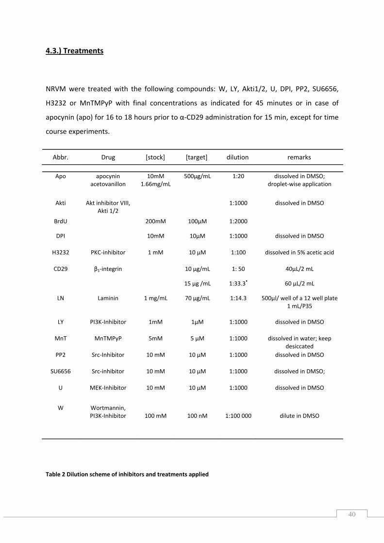
40
4.3.) Treatments
NRVM were treated with the following compounds: W, LY, Akti1/2, U, DPI, PP2, SU6656,
H3232 or MnTMPyP with final concentrations as indicated for 45 minutes or in case of
apocynin (apo) for 16 to 18 hours prior to α‐CD29 administration for 15 min, except for time
course experiments.
Table 2 Dilution scheme of inhibitors and treatments applied
Abbr. Drug [stock] [target] dilution remarks
Apo apocynin acetovanillon
10mM1.66mg/mL
500µg/mL 1:20 dissolved in DMSO; droplet‐wise application
Akti Akt inhibitor VIII, Akti 1/2
1:1000 dissolved in DMSO
BrdU 200mM 100µM 1:2000
DPI 10mM 10µM 1:1000 dissolved in DMSO
H3232 PKC‐inhibitor 1 mM 10 µM 1:100 dissolved in 5% acetic acid
CD29 β1‐integrin 10 µg/mL 1: 50 40µL/2 mL
15 µg /mL 1:33.3• 60 µL/2 mL
LN Laminin 1 mg/mL 70 µg/mL 1:14.3 500µl/ well of a 12 well plate1 mL/P35
LY PI3K‐Inhibitor 1mM 1µM 1:1000 dissolved in DMSO
MnT MnTMPyP 5mM 5 µM 1:1000 dissolved in water; keep desiccated
PP2 Src‐Inhibitor 10 mM 10 µM 1:1000 dissolved in DMSO
SU6656 Src‐inhibitor 10 mM 10 µM 1:1000 dissolved in DMSO;
U MEK‐Inhibitor 10 mM 10 µM 1:1000 dissolved in DMSO
W Wortmannin, PI3K‐Inhibitor
100 mM 100 nM 1:100 000
dilute in DMSO
41
4.4.) Viral infection
After cardiomyocytes were changed to SF‐DMEM, they were infected with viral constructs
bearing either SOD (Ad5CMVSOD1) [Zwacka, Zhou et al. 1998], catalase (Ad5CMVCatalase)
[Lam, Zwacka et al. 1999] or β‐galactosidase (Ad5CMVcytoLacZ) [Davidson, Allen et al. 1993]
for 36 h. Cardiomyocytes were washed twice with SF‐DMEM before supplemented with SF‐
DMEM for additional 14‐16 h, followed by CD29 treatment for 15 minutes.
4.5.) Sample preparation
All signalling samples were washed twice with ice‐cold PBS and scraped in 70 µL (P35) or 120
µL (P60) cell lysis buffer, centrifuged at 15000 rpm for 15 min at 4 °C, supernatants were
collected in new micro tubes (Eppendorf). Cell lysis buffer (1:10 in ddH2O) was
supplemented with 1 mM Pefabloc SC Plus and phosphatase cocktail 1 (1:100). Sample
protein concentrations were estimated using a Pierce protein BCA kit (Thermo Fischer
Scientific) [Kurien and Scofield 2003]. 10µg (ERK), 25µg (PKB) and 35µg (GSK‐3β, Src, PKC)
protein were prepared according to Fermentas DualColor protein loading buffer pack
instructions (1 µL DTT and 5 µL loading buffer per 20µL total volume including protein
sample and ddH2O; incubated for 5 minutes at 95‐100 °C with 300 rpm mixing on a thermo‐
block; spun a few seconds in a microcentrifuge).
4.6.) Sodium dodecyl sulfate‐polyacrylamide gelelectrophoresis (SDS‐PAGE)
SDS‐PAGE was performed as previously described [Rehm 2002]. Briefly, a 12% resolving gel
was prepared. Gels polymerized within 30 minutes. To obtain an even gel a thin layer of 70%
ethanol (300‐500 µL) covered the top of the gel, which was removed before the 5% stacking
gel was prepared.
4.6.1.) Western blotting
Western blotting was performed as previously described [Kurien and Scofield 2006]. The gel
apparatus was set up (Mini‐PROTEAN® Tetra Electrophoresis System, Bio‐Rad) and filled with
pre‐cooled running buffer. Protein samples and prestained protein ladder ran with constant
42
Volts (90 V) till the tracking dye reached the bottom of the gel. Next, fibre pads, blotting
paper (Whatmann, extra thick, pure cellulose) and PVDF‐ membranes were soaked in 1×
used transfer buffer, whereas PVDF‐ membranes were first activated for 10 second in
methanol (Chromasolv®, Sigma) and 5 minutes in ddH2O. The transfer apparatus was
assembled (electrophoresis blotting module, 2 gel holder cassettes and the Bio‐IceTM cooling
unit; pre‐cooled transfer buffer; Bio‐Rad), placed in a styrofoam box surrounded with ice
cubes (a stir bar inside the buffer tank). The tank transfer system was plugged to a BioRad
Power Pac 3000 for 2 hours with constant Volts (100 V). After transfer, membranes were
reactivated in methanol and blocked in 5% milk/TBST (TBS with 0.1% Tween‐20, Sigma) for
GSK‐3β (Ser9) or 4% BSA/TBST for 30 to 60 minutes. Effectiveness of protein transfer was
checked by either staining gels with Coomassie Blue R (Sigma) or staining the membranes
with Ponceau S solution (Sigma).
Membranes were incubated with the primary antibody overnight, while gently shaking at
4°C (phospho and total ERK 1:5000, phospho and total PKB, total GSK3β 1:2500, phospho‐
GSK‐3β, phospho‐Src, phospho‐PKCζ dilutions 1:1500,). Membranes were washed 3 times
with TBST for 15 minutes. HRP‐conjugated secondary antibody (in 4% BSA/TBST) was added
and left for 45 min with gentle shaking at room temperature. Finally membranes were
washed 3 × with TBST for 15 min, ECL was prepared according to the manufacturer’s
protocols (Perkin Elmer Biosciences; Oxidizing Reagent Plus and Enhanced Luminol Reagent
Plus in ratio 1:1) to detect immunoreactive bands with a blue X‐ray film (Fisher Thermo
Scientific) and a Curix Developer. The bands were analysed using Image J v1.3725 [Burger
and Burge 2007].
4.6.2.) Stripping for reprobing
Membranes that were re‐used to probe for different antibodies were stripped either with
mild stripping buffer protocol provided by abcam®6 or by use of Restore™ Plus Western Blot
stripping buffer according to manufacturers’ instructions.
5 http://rsb.info.nih.gov/ij/ 6 www.abcam.com/index.html?pageconfig=resource&rid=11353
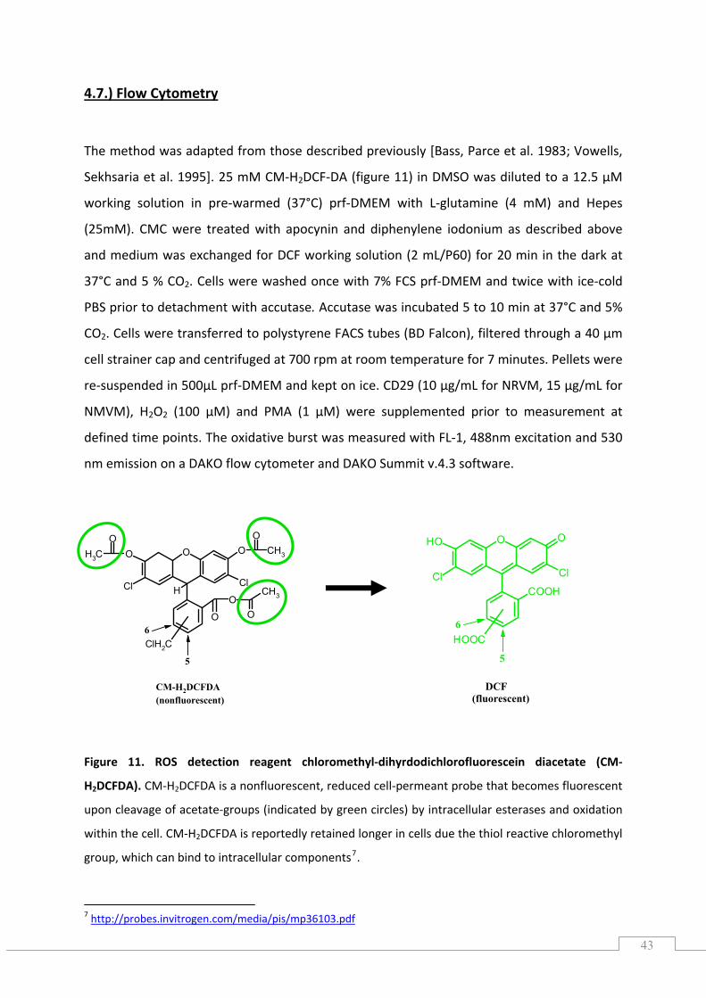
43
O O
COOH
OH
Cl Cl
HOOC
5
6
DCF(fluorescent)
4.7.) Flow Cytometry
The method was adapted from those described previously [Bass, Parce et al. 1983; Vowells,
Sekhsaria et al. 1995]. 25 mM CM‐H2DCF‐DA (figure 11) in DMSO was diluted to a 12.5 µM
working solution in pre‐warmed (37°C) prf‐DMEM with L‐glutamine (4 mM) and Hepes
(25mM). CMC were treated with apocynin and diphenylene iodonium as described above
and medium was exchanged for DCF working solution (2 mL/P60) for 20 min in the dark at
37°C and 5 % CO2. Cells were washed once with 7% FCS prf‐DMEM and twice with ice‐cold
PBS prior to detachment with accutase. Accutase was incubated 5 to 10 min at 37°C and 5%
CO2. Cells were transferred to polystyrene FACS tubes (BD Falcon), filtered through a 40 µm
cell strainer cap and centrifuged at 700 rpm at room temperature for 7 minutes. Pellets were
re‐suspended in 500µL prf‐DMEM and kept on ice. CD29 (10 µg/mL for NRVM, 15 µg/mL for
NMVM), H2O2 (100 µM) and PMA (1 µM) were supplemented prior to measurement at
defined time points. The oxidative burst was measured with FL‐1, 488nm excitation and 530
nm emission on a DAKO flow cytometer and DAKO Summit v.4.3 software.
Figure 11. ROS detection reagent chloromethyl‐dihyrdodichlorofluorescein diacetate (CM‐
H2DCFDA). CM‐H2DCFDA is a nonfluorescent, reduced cell‐permeant probe that becomes fluorescent
upon cleavage of acetate‐groups (indicated by green circles) by intracellular esterases and oxidation
within the cell. CM‐H2DCFDA is reportedly retained longer in cells due the thiol reactive chloromethyl
group, which can bind to intracellular components7.
7 http://probes.invitrogen.com/media/pis/mp36103.pdf
O OO
CH3
O
H
OO
CH3
Cl Cl
OCH3
O
ClH2C
5
6
CM-H2DCFDA(nonfluorescent)
44
4.8.) RNA
4.8.1.) RNA isolation
RNA isolation using Trizol reagent was performed as recommended by manufacturer (Invitrogen)8
Total RNA was extracted from tissue cultured cells (NRVM). Cells were washed with ice‐cold
PBS prior to lysing with 1.4 mL Trizol (Invitrogen) or TRI® reagent (Sigma) on each P60
culture dish. Cell lysates were passed several times through a pipette, scraped with a cell
scraper and collected in 2 mL microtubes (Sarstedt). Homogenised cells (NRVM) were stored
at ‐80°C.
Trizol samples were centrifuged for 10 min at 12000 g and 4°C in a precooled centrifuge
(Hettich MIKRO 22R). 1000 µL were transferred to fresh, sterilized 1.5 microtube, 200 µL ice‐
cold chloroform was applied. The two solutions were mixed by inversion, left at room
temperature for 5 minutes and centrifuged for 15 min at 12000 g and 4°C. The aqueous
phase (containing RNA) was transferred to new micro tubes. RNA was precipitated by adding
the same volume ice‐cold 2‐propanol, and left at room temperature for 10 minutes,
followed by a centrifugation step for 15 min at 12000 g and 4°C.
After discarding the supernatant the RNA pellet was washed with 75% ethanol in RNase‐free
(DEPC‐treated) water, and centrifuged for 5 minutes at 7400 g and 4°C. Ethanol was
discarded and the pellet was air dried at room temperature for 10 minutes.
Pellets were re‐suspended in 50 µL RNase‐free water and dissolved for 10 min at 60°C with
gentle mixing (Eppendorf Thermoblock, 300 rpm). Afterwards, RNA was chilled on ice.
Total RNA concentrations were measured with a NanoDrop9 UV/VIS spectrophotometer ND‐
1000 according to the manufacturer’s protocol, loaded with 1.2 to 1.5 µL total RNA extract
on the lower measurement pedestal. RNA quality was assessed by loading an RNA : loading
dye mix on 1% agarose gel, which ran for 30 minutes at 100 V. Visualization under UV
revealed two strong rRNA bands on the gel.
8 http://products.invitrogen.com/ivgn/product/15596026 9 http://www.peqlab.de/wcms/nanodrop/nanodrop‐service.php
45
4.8.2.) Deoxyribonuclease I (DNase I) treatment of RNA and cDNA synthesis
DNase I treatment and cDNA synthesis were performed as described by the supplier
(Fermentas)10.
1.5 µg total RNA were supplemented with 0.25 µL 40u/µL RNase inhibitor, 1 µL 10x DNase I
buffer with MgCl2 and 1 u DNase I in a final volume of 10 µL. Samples incubated for 30 min
at 37°C followed by application of 25 mM EDTA and incubation at 65°C for10 minutes.
Samples were kept on ice until use for cDNA synthesis.
RevertAid™ H Minus Reverse Transcriptase (Fermentas) was used, which is a genetically
modified Moloney Murine Leukaemia Virus reverse transcriptase, with an mutated and non‐
functional RNase H. Briefly, 1.5 µg total RNA was used, pre‐mixed with 1 µL 100µM dT18
primer (~0.5 µg/µL) and incubated for 5 min at 70°C. Further, 4 µL 5x RevertAid Buffer, 2 µL
10 mM (each) dNTP, 0.5 µL 40 u/µL RNase Inhibitor, and finally 1 µL 200 u/µL RevertAid H‐
M‐MuLV RT t were added. RNA was incubated for 5 min at 37°C followed by 1.5 h at 42°C
and 10 min at 70°C. cDNA samples were stored at 4°C for use in RT‐PCR to avoid freeze thaw
damage.
4.8.3.) Primers
Primer pairs (Table 2) were designed using the NCBI primer‐BLAST11 tool, except for primers marked * and §.
*primers were taken from [Cao, Dai et al. 2007]; § primer was taken from[Kutty, Kutty et al. 1995].
10 http://www.fermentas.com/en/products/all/modifying‐enzymes 11 http://www.ncbi.nlm.nih.gov/tools/primer‐blast/index.cgi
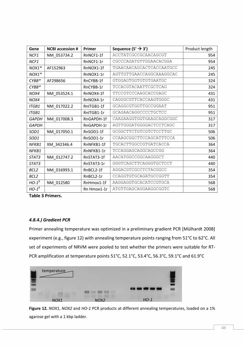
46
Table 3 Primers.
4.8.4.) Gradient PCR
Primer annealing temperature was optimized in a preliminary gradient PCR [Mülhardt 2008]
experiment (e.g., figure 12) with annealing temperature points ranging from 51°C to 62°C. All
set of experiments of NRVM were pooled to test whether the primers were suitable for RT‐
PCR amplification at temperature points 51°C, 52.1°C, 53.4°C, 56.3°C, 59.1°C and 61.9°C
Figure 12. NOX1, NOX2 and HO‐1 PCR products at different annealing temperatures, loaded on a 1%
agarose gel with a 1 kbp ladder.
Gene NCBI accession # Primer Sequence (5' 3') Product length NCF1 NM_053734.2 RnNCF1‐1f ACCTATCGCCGCAACAGCGT 954NCF1 RnNCF1‐1r CGCCCAGATGTTGGAACACGGA 954NOX1* AF152963 RnNOX1‐1f TGAACAACAGCACTCACCAATGCC 245NOX1* RnNOX1‐1r AGTTGTTGAACCAGGCAAAGGCAC 245CYBB* AF298656 RnCYBB‐1f GTGGAGTGGTGTGTGAATGC 324CYBB* RnCYBB‐1r TCCACGTACAATTCGCTCAG 324NOX4 NM_053524.1 RnNOX4‐1f TTCCGTCCCAAGCACCGAGC 431NOX4 RnNOX4‐1r CAGGGCGTTCACCAAGTGGGC 431ITGB1 NM_017022.2 RnITGB1‐1f GCAGGCGTGGTTGCCGGAAT 951ITGB1 RnITGB1‐1r GCAGAACAGGCCCCTGCTCC 951GAPDH NM_017008.3 RnGAPDH‐1f CAAGAAGGTGGTGAAGCAGGCGGC 317GAPDH RnGAPDH‐1r AGTTGGGATGGGGACTCCTCAGC 317SOD1 NM_017050.1 RnSOD1‐1f GCGGCTTCTGTCGTCTCCTTGC 506SOD1 RnSOD1‐1r CCAAGCGGCTTCCAGCATTTCCA 506NFKB1 XM_342346.4 RnNFKB1‐1f TGCACTTGGCCGTGATCACCA 364NFKB1 RnNFKB1‐1r TCCAGGAGCAGGCAGCCGG 364STAT3 NM_012747.2 RnSTAT3‐1f AACATGGCCGGCAAGGGCT 440STAT3 RnSTAT3‐1r GGGTCAGCTTCAGGGTGCTCCT 440BCL2 NM_016993.1 RnBCL2‐1f AGGACGTCGCCTCTACGGCC 354BCL2 RnBCL2‐1r CCAGGTGTGCAGATGCCGGTT 354HO‐1§ NM_012580 RnHmox1‐1f AAGGAGGTGCACATCCGTGCA 568HO‐1§ Rn Hmox1‐1r ATGTTGAGCAGGAAGGCGGTC 568
temperature
NOX1 NOX2 HO‐1
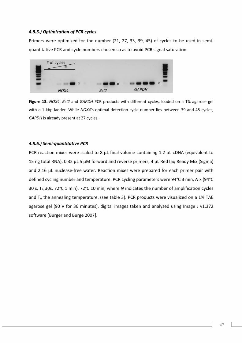
47
4.8.5.) of PCR cycles
Primers were optimized for the number (21, 27, 33, 39, 45) of cycles to be used in semi‐
quantitative PCR and cycle numbers chosen so as to avoid PCR signal saturation.
with a 1 kbp ladder. While NOX4‘s optimal detection cycle number lies betwee
GAPDH is already present at 27 cycles.
4.8.6.) Semi‐quantitative PCR
PCR reaction mixes were scaled to 8 µL final volume containing 1.2 µL cDNA (equivalent to
ng total RNA), 0.32 µL 5 µM forward and reverse primers, 4 µL RedTaq Ready Mix (Sigma)
nd 2.16 µL nuclease‐free water. Reaction mixes were prepared for each primer pair with
defined cycling number and temperature. PCR cycling parameters were 94°C 3 min, N x (94°C
30 s, TA 30s, 72°C 1 min), 72°C 10 min, where N indicates the number of amplification cycles
Optimization
Figure 13. NOX4, Bcl2 and GAPDH PCR products with different cycles, loaded on a 1% agarose gel
n 39 and 45 cycles,
15
a
and TA the annealing temperature. (see table 3). PCR products were visualized on a 1% TAE
agarose gel (90 V for 36 minutes), digital images taken and analysed using Image J v1.372
software [Burger and Burge 2007].
# o
Bcl2 GAPDH
f cycles
NOX4
×××
48
Primer pair Annealing temperature # of cycles
NFκB 57°C 33
STAT3 57°C 31
SOD1 57°C 32
CD29 57°C 35
NCF1 59°C 34
NOX4 57°C 40
Bcl2 57°C 35
GAPDH 57°C 28
NOX1 56.5°C 31
5
NOX2 56.5°C 40
HO‐1 6.5°C 40
Table 4 Prim imization
4.9.) Statis nalyses
er opt
tical a
All statistica yses were performed in G ad Prism Version 4.03 and below
0.05 were ed to indicate significan rences. Significance was tested in repeated
measures 1 NOVA with post‐test Bonferroni (for sample sizes ≤ 4) o wman Keuls
(for > 4 sam compare) correction.
l anal raphp p‐values
consider t diffe
‐way A r Ne
ples to
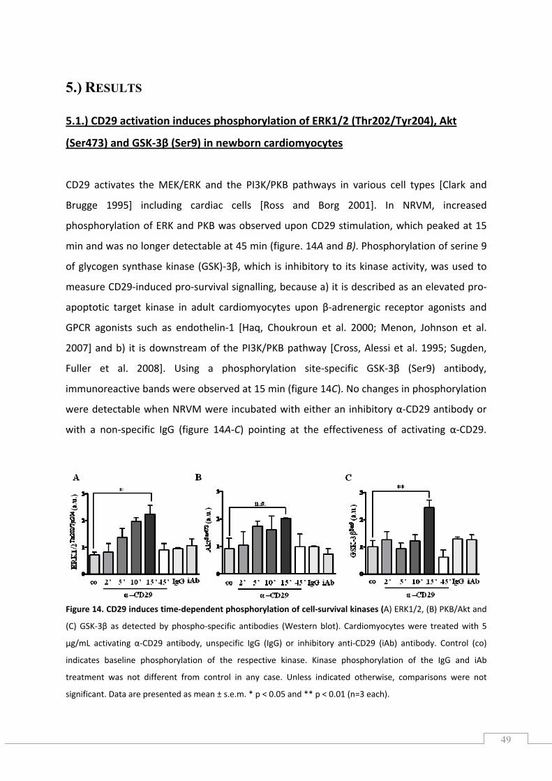
49
5.) RESULTS
5.1.) CD29 activation induces phosphorylation of ERK1/2 (Thr202/Tyr204), Akt
(Ser473) and GSK‐3β (Ser9) in newborn cardiomyocytes
CD29 activates the MEK/ERK and the PI3K/PKB pathways in various cell types [Clark and
Brugge 1995] including cardiac cells [Ross and Borg 2001]. In NRVM, increased
phosphorylation of ERK and PKB was observed upon CD29 stimulation, which peaked at 15
min and was no longer detectable at 45 min (figure. 14A and B). Phosphorylation of serine 9
of glycogen synthase kinase (GSK)‐3β, which is inhibitory to its kinase activity, was used to
measure CD29‐induced pro‐survival signalling, because a) it is described as an elevated pro‐
apoptotic target kinase in adult cardiomyocytes upon β‐adrenergic receptor agonists and
GPCR agonists such as endothelin‐1 [Haq, Choukroun et al. 2000; Menon, Johnson et al.
2007] and b) it is downstream of the PI3K/PKB pathway [Cross, Alessi et al. 1995; Sugden,
Fuller et al. 2008]. Using a phosphorylation site‐specific GSK‐3β (Ser9) antibody,
immunoreactive bands were observed at 15 min (figure 14C). No changes in phosphorylation
were detectable when NRVM were incubated with either an inhibitory α‐CD29 antibody or
with a non‐specific IgG (figure 14A‐C) pointing at the effectiveness of activating α‐CD29.
Figure 14. CD29 induces time‐dependent phosphorylation of cell‐survival kinases (A) ERK1/2, (B) PKB/Akt and
(C) GSK‐3β as detected by phospho‐specific antibodies (Western blot). Cardiomyocytes were treated with 5
µg/mL activating α‐CD29 antibody, unspecific IgG (IgG) or inhibitory anti‐CD29 (iAb) antibody. Control (co)
indicates baseline phosphorylation of the respective kinase. Kinase phosphorylation of the IgG and iAb
treatment was not different from control in any case. Unless indicated otherwise, comparisons were not
significant. Data are presented as mean ± s.e.m. * p < 0.05 and ** p < 0.01 (n=3 each).
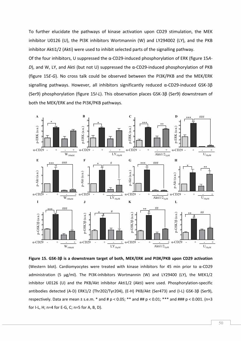
50
0
1
2
3
4
5
0
1
2
3
p-A
kt (a
.u.)
p-A
kt (a
.u.)
p-A
kt (a
.u.)
E F G H
0
1
2
3
0
1
2
3
0
1
2
3
p-A
kt (a
.u.)
****** ### ###* #* **
α-CD29 - + - +α-CD29 - + - +
p-G
SK3β
(a.u
.)
p-G
SK3β
(a.u
.)
p-G
SK3β
(a.u
.)
I J K L
0
1
2
3
4
5
0
1
2
3
4
5
p-G
SK3β
(a.u
.)
α-CD29 - + - + α-CD29 - + - + α-CD29 - + - + α-CD29 - + - +W100nM LY10μM Akti1/25μM U10μM
****
****
### #####
0
1
2
3
4
5
α-CD29 - + - +α-CD29 - + - +W100nM LY10μM Akti1/25μM U10μM
0
1
2
3
4
5
0
1
2
3
4
5 * *
p-ER
K (a
.u.)
α-CD29 - + - + α-CD29 - + - + α-CD29 - + - +α-CD29 - + - +
p-ER
K (a
.u.)
A B C D
W100nM LY10μM Akti1/25μM U10μM
0
1
2
3
4
5*** **
p-ER
K (a
.u.)
0
1
2
3
4
5
6
7
###***
p-ER
K (a
.u.)
To further elucidate the pathways of kinase activation upon CD29 stimulation, the MEK
), the PI3K inhibitors Wortmannin (W) and LY294002 (LY), and the PKB
Of the four inhibitors, U suppressed the α‐CD29‐induced phosphorylation of ERK (figure 15A‐
D), and W, LY, and Akti (but not U) suppressed the α‐CD29‐induced phosphorylation of PKB
(figure 15E‐G). No cross talk could be observed between the PI3K/PKB and the MEK/ERK
signalling pathways. However, all inhibitors significantly reduced α‐CD29‐induced GSK‐3β
(Ser9) phosphorylation (figure 15I‐L). This observation places GSK‐3β (Ser9) downstream of
both the MEK/ERK and the PI3K/PKB pathways.
Figure 15. GSK‐3β is a downstream target of both, MEK/ERK and PI3K/PKB upon CD29 activation
PKB/Akt (I K
respectively. Data are mean ± s.e.m. * and # p < 0.05; ** and ## p < 0.01; *** and ### p < 0.001. (n=3
I‐L, H; n=4 for E‐G, C; n=5 for A, B, D).
inhibitor U0126 (U
inhibitor Akti1/2 (Akti) were used to inhibit selected parts of the signalling pathway.
(Western blot). Cardiomyocytes were treated with kinase inhibitors for 45 min prior to α‐CD29
administration (5 µg/ml). The PI3K‐inhibitors Wortmannin (W) and LY29400 (LY), the MEK1/2
inhibitor U0126 (U) and the PKB/Akt inhibitor Akti1/2 (Akti) were used. Phosphorylation‐specific
antibodies detected (A‐D) ERK1/2 (Thr202/Tyr204), (E‐H) (Ser473) and ‐L) GS ‐3β (Ser9),
for
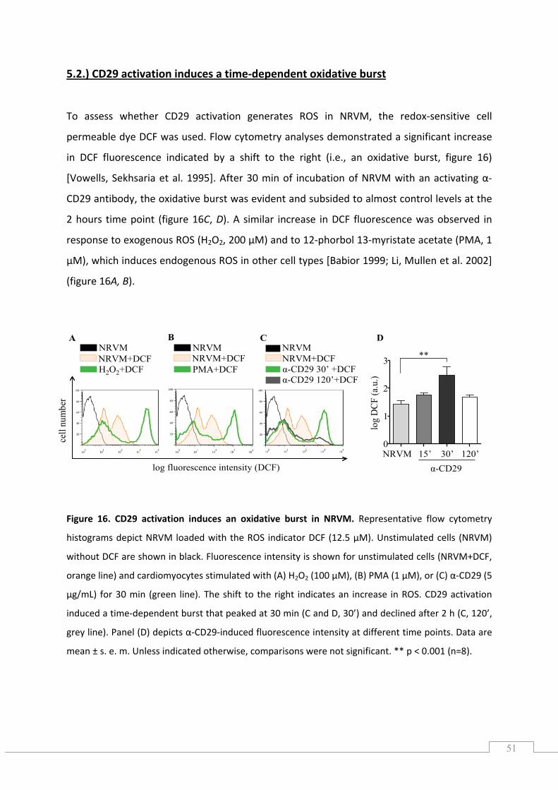
51
5.2.) CD29 activation induces a time‐dependent oxidative burst
To assess whether CD29 activation generates ROS in NRVM, the redox‐sensitive cell
to 12‐phorbol 13‐myristate acetate (PMA, 1
M), which induces endogenous ROS in other cell types [Babior 1999; Li, Mullen et al. 2002]
igure 16A, B).
igure 16. CD29 activation induces an oxidative burst in NRVM. Representative flow cytometry
istograms depict NRVM loaded with the ROS indicator DCF (12.5 µM). Unstimulated cells (NRVM)
ithout DCF are shown in black. Fluorescence intensity is shown for unstimulated cells (NRVM+DCF,
range line) and cardiomyocytes stimulated with (A) H2O2 (100 µM), (B) PMA (1 µM), or (C) α‐CD29 (5
permeable dye DCF was used. Flow cytometry analyses demonstrated a significant increase
in DCF fluorescence indicated by a shift to the right (i.e., an oxidative burst, figure 16)
[Vowells, Sekhsaria et al. 1995]. After 30 min of incubation of NRVM with an activating α‐
CD29 antibody, the oxidative burst was evident and subsided to almost control levels at the
2 hours time point (figure 16C, D). A similar increase in DCF fluorescence was observed in
response to exogenous ROS (H2O2, 200 µM) and
µ
(f
F
h
w
o
µg/mL) for 30 min (green line). The shift to the right indicates an increase in ROS. CD29 activation
induced a time‐dependent burst that peaked at 30 min (C and D, 30’) and declined after 2 h (C, 120’,
grey line). Panel (D) depicts α‐CD29‐induced fluorescence intensity at different time points. Data are
mean ± s. e. m. Unless indicated otherwise, comparisons were not significant. ** p < 0.001 (n=8).
NRVMNRVM+DCFα-CD29 30’ +DCFα-CD29 120’+DCF
D
NRVM 15’ 30’ 120’α-CD29
log
DCF
(a.u
.)
NRVMNRVM+DCFH2O2+DCF
log fluorescence intensity (DCF)
cell
num
ber
CA BNRVMNRVM+DCFPMA+DCF
0
1
2
3 **
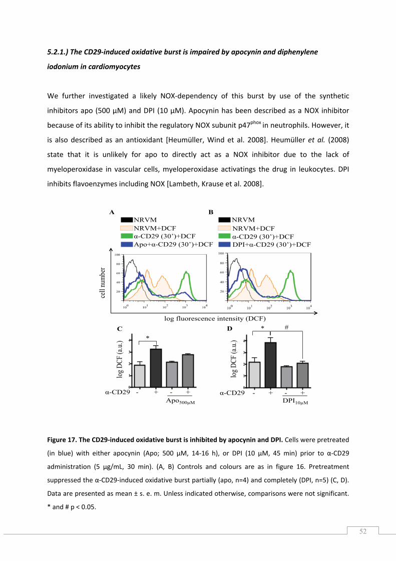
52
log fluorescence intensity (DCF)
cell
numb
er
NRVM
Apo+α-CD29 (30’)+DCF
NRVM+DCFα-CD29 (30’)+DCF
NRVM
DPI+α-CD29 (30’)+DCF
NRVM+DCFα-CD29 (30’)+DCF
A B
#D
0
1
2
3
4
α-CD29 - + - +DPI10μM
*
log D
CF (a
.u.)
C
0
1
2
3
4
α-CD29 - + - +Apo500μM
*
log D
CF (a
.u.)
5.2.1.) The CD29‐induced oxidative burst is impaired by apocynin and diphenylene
donium in cardiomyocytes
nt
uppressed the α‐CD29‐induced oxidative burst partially (apo, n=4) and completely (DPI, n=5) (C, D).
ata are presented as mean ± s. e. m. Unless indicated otherwise, comparisons were not significant.
* and # p < 0.05.
io
We further investigated a likely NOX‐dependency of this burst by use of the synthetic
inhibitors apo (500 µM) and DPI (10 µM). Apocynin has been described as a NOX inhibitor
because of its ability to inhibit the regulatory NOX subunit p47phox in neutrophils. However, it
is also described as an antioxidant [Heumüller, Wind et al. 2008]. Heumüller et al. (2008)
state that it is unlikely for apo to directly act as a NOX inhibitor due to the lack of
myeloperoxidase in vascular cells, myeloperoxidase activatings the drug in leukocytes. DPI
inhibits flavoenzymes including NOX [Lambeth, Krause et al. 2008].
Figure 17. The CD29‐induced oxidative burst is inhibited by apocynin and DPI. Cells were pretreated
(in blue) with either apocynin (Apo; 500 µM, 14‐16 h), or DPI (10 µM, 45 min) prior to α‐CD29
administration (5 µg/mL, 30 min). (A, B) Controls and colours are as in figure 16. Pretreatme
s
D
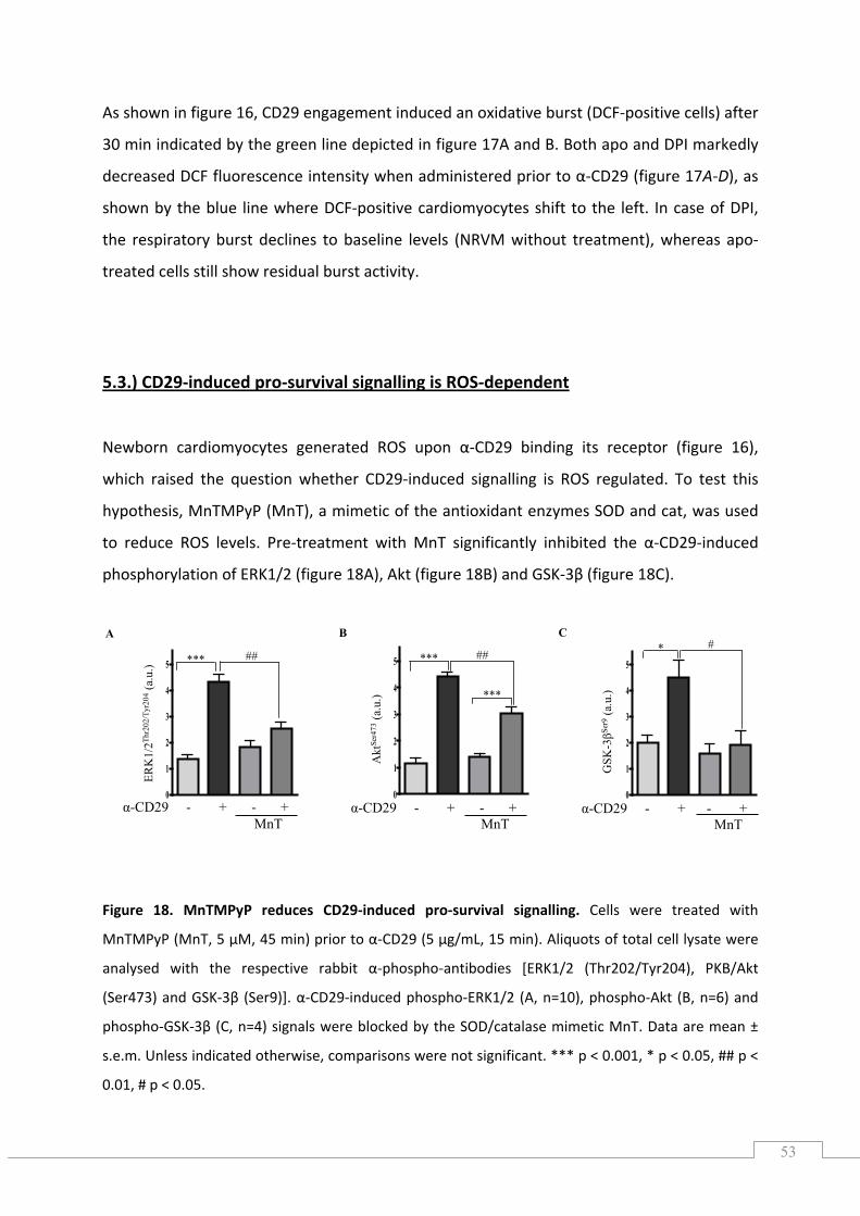
53
A B C
α-CD29 - + - +MnT
GSK
-3βSe
r9(a
.u.)
0
1
2
3
4
5* #
α-CD29 - + - +MnT
Akt
Ser4
73(a
.u.)
0
1
2
3
4
5 ***
***
##
α-CD29 - + - +MnT
ERK
1/2Th
r202
/Tyr
204
(a.u
.)
0
1
2
3
4
5 *** ##
As shown in figure 16, CD29 engagement induced an oxidative burst (DCF‐positive ce
30 min indicated by the green
lls) after
line depicted in figure 17A and B. Both apo and DPI markedly
ecreased DCF fluorescence intensity when administered prior to α‐CD29 (figure 17A‐D), as
5.3.) CD29‐induced pro‐survival signalling is ROS‐dependent
d
shown by the blue line where DCF‐positive cardiomyocytes shift to the left. In case of DPI,
the respiratory burst declines to baseline levels (NRVM without treatment), whereas apo‐
treated cells still show residual burst activity.
cardiomyocytes generated ROS upon α‐CD29 binding its receptor (figure 16),
hich raised the question whether CD29‐induced signalling is ROS regulated. To test this
ypothesis, MnTMPyP (MnT), a mimetic of the antioxidant enzymes SOD and cat, was used
reduce ROS levels. Pre‐treatment with MnT significantly inhibited the α‐CD29‐induced
hosphorylation of ERK1/2 (figure 18A), Akt (figure 18B) and GSK‐3β (figure 18C).
Figure 18. MnTMPyP reduces CD29‐induced pro‐survival signalling. Cells were treated with
MnTMPyP (MnT, 5 µM, 45 min) prior to α‐CD29 (5 µg/mL, 15 min). Aliquots of total cell lysate were
analysed with the respective rabbit α‐phospho‐antibodies [ERK1/2 (Thr202/Tyr204), PKB/Akt
(Ser473) and GSK‐3β (Ser9)]. α‐CD29‐induced phospho‐ERK1/2 (A, n=10), phospho‐Akt (B, n=6) and
phospho‐GSK‐3β (C, n=4) signals were blocked by the SOD/catalase mimetic MnT. Data are mean ±
s.e.m. Unless indicated otherwise, comparisons were not significant. *** p < 0.001, * p < 0.05, ## p <
0.01, # p < 0.05.
Newborn
w
h
to
p
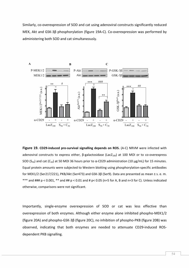
54
Similarly, co‐overexpression of SOD and cat using adenoviral constructs significantly reduced
MEK, Akt and GSK‐3β phosphorylation (figure 19A‐C). Co‐overexpression was performed by
administering both SOD and cat simultaneously.
Figure 19. CD29‐induced pro‐survival signalling depends on ROS. (A‐C) NRVM were infected with
denoviral constructs to express either, β‐galactosidase (LacZ100) at 100 MOI or to co‐overexpress
OD (S50) and cat (C50) at 50 MOI 36 hours prior to α‐CD29 administration (10 µg/mL) for 15 minutes.
Equal protein amounts were subjected to Western blotting using phosphorylation‐specific antibodies
MEK1/2 (Ser217/221), PKB/Akt (Ser473) and GSK‐3β (Ser9). Data are presented as mean ± s. e. m.
** and ### p < 0.001, ** and ## p < 0.01 and # p< 0.05 (n=5 for A, B and n=3 for C). Unless indicated
otherwise, comparisons were not significant.
a
S
for
*
Importantly, single‐enzyme overexpression of SOD or cat was less effective than
overexpression of both enzymes. Although either enzyme alone inhibited phospho‐MEK1/2
(figure 20A) and phospho‐GSK‐3β (figure 20C), no inhibition of phospho‐PKB (figure 20B) was
observed, indicating that both enzymes are needed to attenuate CD29‐induced ROS‐
dependent PKB signalling.
0
1
2
3
4
5
P-MEK1/2MEK1/2
P-AktAkt
P-GSK-3βGSK-3β
0
1
2
3
4
5
0
1
2
3
4
5
S50 + C50
#**
LacZ100
α-CD29 - + - +
MEK
1/2Se
r217
/221
(a.u
.)
**
*** ###
α-CD29 - + - +LacZ100 S50 + C50
Akt
Ser4
73(a
.u.)
## ***
GSK
-3βSe
r9(a
.u.)
α-CD29 - + - + LacZ100 S50 + C50
A B C
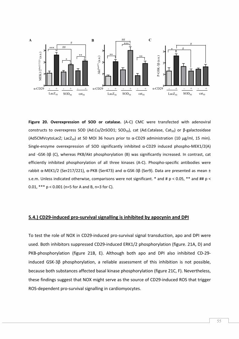
55
Figure 20. Overexpression of SOD or catalase. (A‐C) CMC were transfected with adenoviral
constructs to overexpress SOD (Ad.Cu/ZnSOD1; SOD50), cat (Ad.Catalase, Cat50) or β‐galactosidase
(Ad5CMVcytoLacZ; LacZ50) at 50 MOI 36 hours prior to α‐CD29 administration (10 µg/ml, 15 min).
Single‐enzyme overexpression of SOD significantly inhibited α‐CD29 induced phospho‐MEK1/2(A)
and ‐GSK‐3β (C), whereas PKB/Akt phosphorylation (B) was significantly increased. In contrast, cat
efficiently inhibited phosphorylation of all three kinases (A‐C). Phospho‐specific antibodies were
rabbit α‐MEK1/2 (Ser217/221), α‐PKB (Ser473) and α‐GSK‐3β (Ser9). Data are presented as mean ±
s.e.m. Unless indicated otherwise, comparisons were not significant. * and # p < 0.05, ** and ## p <
5.4.) CD29‐induced pro‐survival signalling is inhibited by apocynin and DPI
0.01, *** p < 0.001 (n=5 for A and B, n=3 for C).
‐survival signal transduction, apo and DPI were
used. Both inhibitors suppressed CD29‐induced ERK1/2 phosphorylation (figure. 21A, D) and
PKB‐phosphorylation (figure 21B, E). Although both apo and DPI also inhibited CD‐29‐
To test the role of NOX in CD29‐induced pro
induced GSK‐3β phosphorylation, a reliable assessment of this inhibition is not possible,
because both substances affected basal kinase phosphorylation (figure 21C, F). Nevertheless,
these findings suggest that NOX might serve as the source of CD29‐induced ROS that trigger
ROS‐dependent pro‐survival signalling in cardiomyocytes.
0
1
2
3
A
α-CD29 - + - + - +LacZ50 SOD50
##***
*
#M
EK1/
2Ser2
17/2
21(a
.u.)
0
1
2
3
B
Akt
Ser4
73(a
.u.)
LacZ50 SOD50
***
** **
##
0
1
2
3
C
P-G
SK-3
β(a
.u.)
*
α-CD29 - + - + - +
##
α-CD29 - + - + - +
**
cat50 cat50 LacZ50 SOD50 cat50
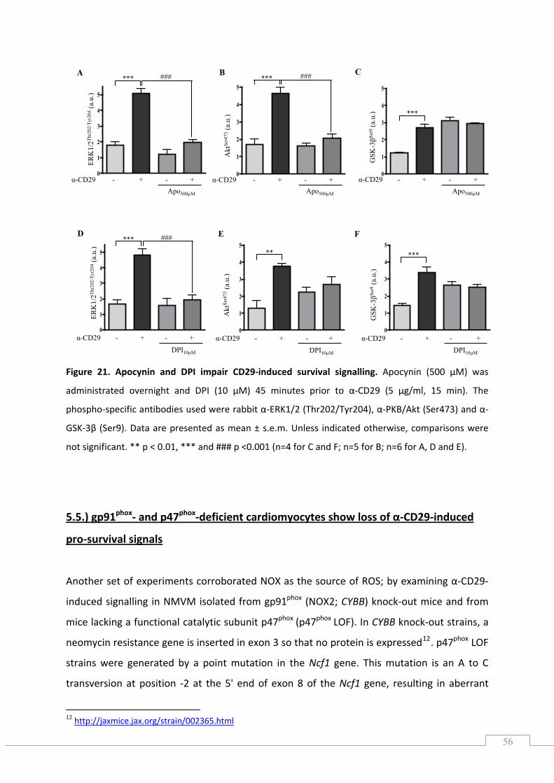
56
D E F
Apo500μM
B
α-CD29 - + - +
Akt
Ser4
73(a
.u.)
0
1
2
3
4
5*** ###
α-CD29 - + - +Apo500μM
A
0
1
2
3
4
5
*** ###
ERK
1/2Th
r202
/Tyr
204
(a.u
.)
0
1
2
3
4
5
***
DPI10μM
###
α-CD29 - + - +
ERK
1/2Th
r202
/Tyr
204
(a.u
.)
0
1
2
3
4
5**
DPI10μM
α-CD29 - + - +
Akt
Ser4
73(a
.u.)
0
1
2
3
4
5***
DPI10μM
α-CD29 - + - +
GSK
-3βSe
r9(a
.u.)
Apo500μM
C
α-CD29 - + - +
GSK
-3βSe
r9(a
.u.)
0
1
2
3
4
5
***
gnalling.
e.m. Unless indicated otherwise, comparisons were
significant. ** p < 0.01, *** and ### p <0.001 (n=4 for C and F; n=5 for B; n=6 for A, D and E).
5.5.) gp91 ‐ and p47 ‐deficient cardiomyocytes show loss of α‐CD29‐induced
Figure 21. Apocynin and DPI impair CD29‐induced survival si Apocynin (500 µM) was
administrated overnight and DPI (10 µM) 45 minutes prior to α‐CD29 (5 µg/ml, 15 min). The
phospho‐specific antibodies used were rabbit α‐ERK1/2 (Thr202/Tyr204), α‐PKB/Akt (Ser473) and α‐
GSK‐3β (Ser9). Data are presented as mean ± s.
not
phox phox
pro‐survival signals
Another set of experiments corroborated NOX as the source of ROS; by examining α‐CD29‐
induced signalling in NMVM isolated from gp91 (NOX2; CYBB) knock‐out mice and from
mice lacking a functional catalytic subunit p47 (p47 LOF). In CYBB knock‐out strains, a
neomycin resistance gene is inserted in exon 3 so that no protein is expressed . p47 LOF
strains were generated by a point mutation in the Ncf1
phox
phox phox
12 phox
gene. This mutation is an A to C
ansversion at position ‐2 at the 5' end of exon 8 of the Ncf1 gene, resulting in aberrant
tr
12 http://jaxmice.jax.org/strain/002365.html

57
P-ERK1/2 P-Akt P-GSK-3βAkt GSK-3β
D FE
ERK1/2
wt p47LOF wt p47LOF wt p47LOF
ERK
1/2Th
r202
/Tyr
204 (a
.u.)
P-ERK1/2 P-Akt P-GSK-3βAkt GSK-3β
CA B
ERK1/2
0
1
2
3
4
5
0
1
2
3
4
5
α-CD29 - + - +wt NOX2k.o.
α- CD29 - + - + α- CD29 - + - +wt NOX2k.o. wt NOX2k.o.
* *# ###
0
1
2
3
4
5 ** ###
GSK
-3βSe
r9(a
.u.)
Akt
Ser4
73(a
.u.)
ERK
1/2Th
r202
/Tyr
204
(a.u
.)
0
1
2
3
4
0
1
2
3
4
0
1
2
3
4
*****
*** ### ##
GSK
-3βSe
r9(a
.u.)
Akt
Ser4
73(a
.u.)
α-CD29 - + - + α- CD29 - + - + α- CD29 - + - +
P-ERK1/2 P-Akt P-GSK-3βAkt GSK-3β
D FE
ERK1/2
wt p47LOF wt p47LOF wt p47LOF
ERK
1/2Th
r202
/Tyr
204 (a
.u.)
P-ERK1/2 P-Akt P-GSK-3βAkt GSK-3β
CA B
ERK1/2
0
1
2
3
4
5
0
1
2
3
4
5
α-CD29 - + - +wt NOX2k.o.
α- CD29 - + - + α- CD29 - + - +wt NOX2k.o. wt NOX2k.o.
* *# ###
0
1
2
3
4
5 ** ###
GSK
-3βSe
r9(a
.u.)
Akt
Ser4
73(a
.u.)
ERK
1/2Th
r202
/Tyr
204
(a.u
.)
0
1
2
3
4
0
1
2
3
4
0
1
2
3
4
*****
*** ### ##
GSK
-3βSe
r9(a
.u.)
Akt
Ser4
73(a
.u.)
α-CD29 - + - + α- CD29 - + - + α- CD29 - + - +
splicing of the Ncf1 transcript, no intact p47phox protein being detectable by Western blotting
cells from these mice13. Upon CD29 stimulation, significantly lower levels of phospho‐
RK1/2, phospho‐PKB and phospho‐GSK‐3β were observed in NOX2‐knock‐out NMVM as
ompared to wild‐type (wt) (figure 22A‐C). In p47phox LOF NMVM, CD29 activation failed to
phosphorylation of PKB (Ser473) (figure 22E) and GSK‐3β(Ser9) (figure 22F). By
contrast, CD29‐induced phosphorylation of ERK1/2 (figure 22D), was unaffected by the loss
functional p47phox, suggesting the activation of a p47phox‐independent and/or
ompensatory pathway.
Figure 22. CD29‐induced signal transduction require ‐derived ROS. α‐CD29 administration (10
2 k out
. U
in
E
c
induce
of
c
s NOX2
µg/mL, 15 min) induced phosphorylation of ERK1/2 (p44/42), PKB/Akt and GSK‐3β in wt NMVM (A‐F).
α‐CD29 induced phosphorylation was absent in NOX nock‐ (NOX2k.o.) cardiomyocytes (A‐C); and
partially diminished in p47phox LOF (p47LOF) NMVM (D‐F). Data are presented as mean ± s. e. m nless
indicated otherwise, comparisons were not significant. * p < 0.05; ** and ## p < 0.01; *** and ### p
< 0.001 (n=5 for A, C, D‐F; n=6 for B).
13 http://jaxmice.jax.org/strain/004742.html
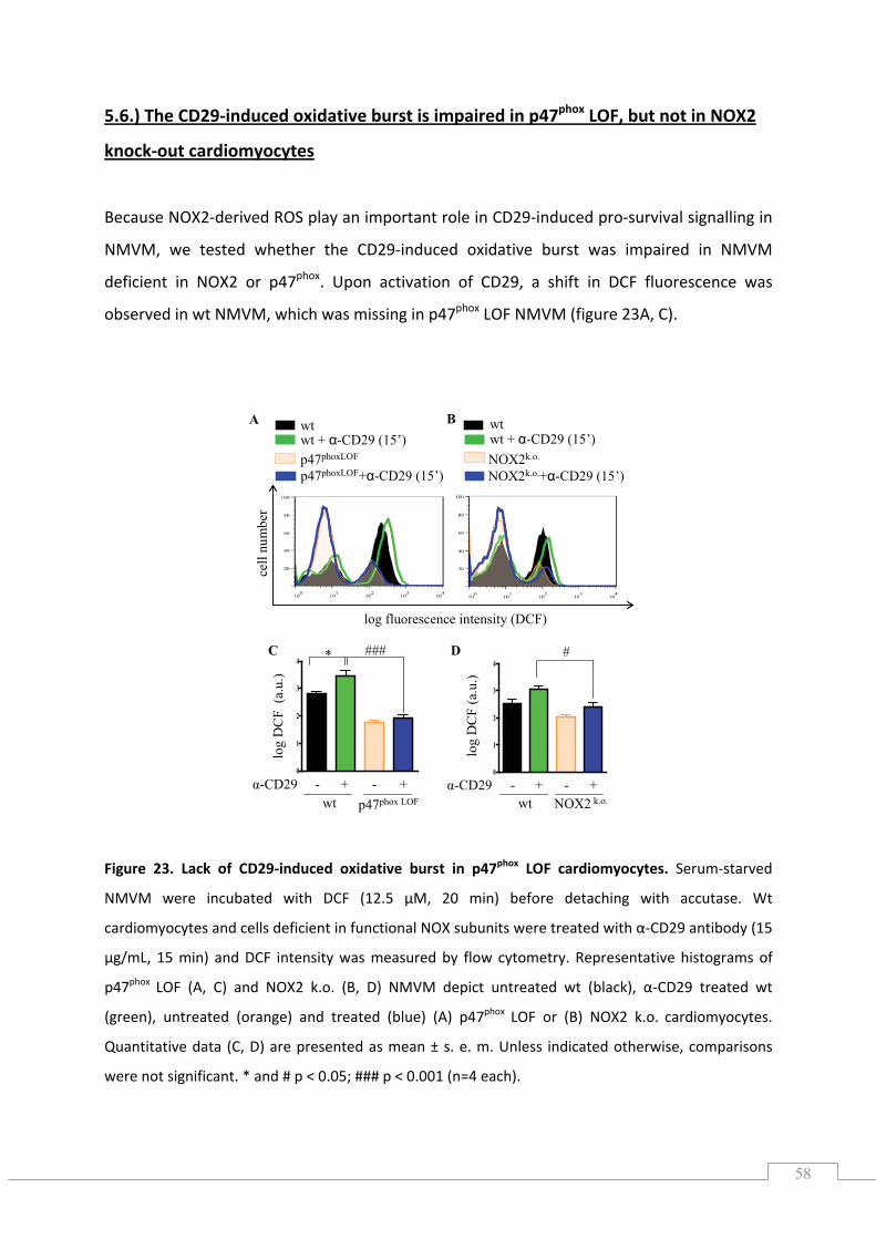
58
0
1
2
3
4
0
1
2
3
4
log fluorescence intensity (DCF)
cell
num
ber
wt
p47phoxLOF+α-CD29 (15’)
wt + α-CD29 (15’)p47phoxLOF
wt
NOX2k.o.+α-CD29 (15’)
wt + α-CD29 (15’)NOX2k.o.
A B
C D #
log
DC
F (a
.u.)
wt NOX2 k.o.α-CD29 - + - +
wt p47phox LOF
α-CD29 - + - +
* ###
log
DC
F (a
.u.)
5.6.) The CD29‐induced oxidative burst is impaired in p47phox LOF, but not in NOX2
knock‐out cardiomyocytes
Because NOX2‐derived ROS play an important role CD29‐induced pro‐survival signalling in
NMVM, we tested whether the CD29‐induced oxidative burst was impaired in NMVM
deficient in NOX2 or p47phox. Upon activation of CD29, a shift in DCF fluorescence was
observed in wt which was missing i p47phox LOF NMV (figure 23A, C).
in
NMVM, n M
igure 23. Lack of CD29‐induced oxidative burst in p47phox LOF cardiomyocytes. Serum‐starved
MVM were incubated with DCF (12.5 µM, 20 min) before detaching with accutase. Wt
phox
phox
# p < 0.001 (n=4 each).
F
N
cardiomyocytes and cells deficient in functional NOX subunits were treated with α‐CD29 antibody (15
µg/mL, 15 min) and DCF intensity was measured by flow cytometry. Representative histograms of
p47 LOF (A, C) and NOX2 k.o. (B, D) NMVM depict untreated wt (black), α‐CD29 treated wt
(green), untreated (orange) and treated (blue) (A) p47 LOF or (B) NOX2 k.o. cardiomyocytes.
Quantitative data (C, D) are presented as mean ± s. e. m. Unless indicated otherwise, comparisons
were not significant. * and # p < 0.05; ##
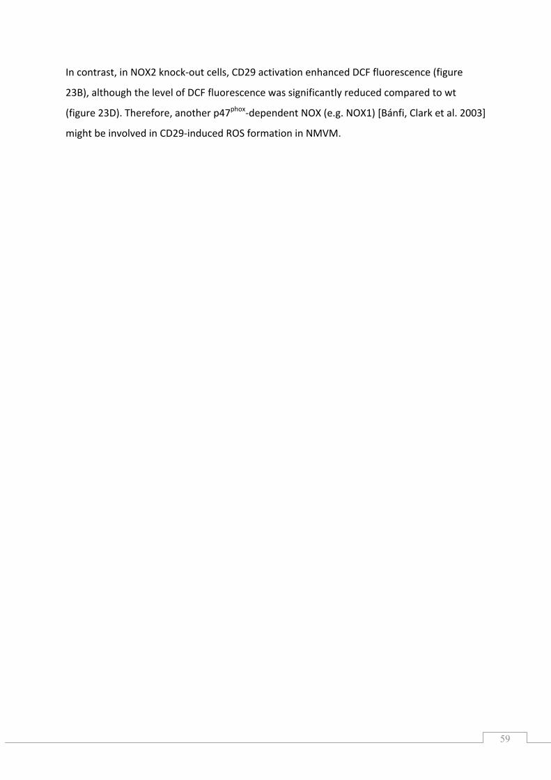
59
In contrast, in NOX2 knock‐out cells, CD29 activation enhanced DCF fluorescence (figure
23B), although the level of DCF fluorescence was significantly reduced compared to wt
igure 23D). Therefore, another p47phox‐dependent NOX (e.g. NOX1) [Bánfi, Clark et al. 2003] (f
might be involved in CD29‐induced ROS formation in NMVM.
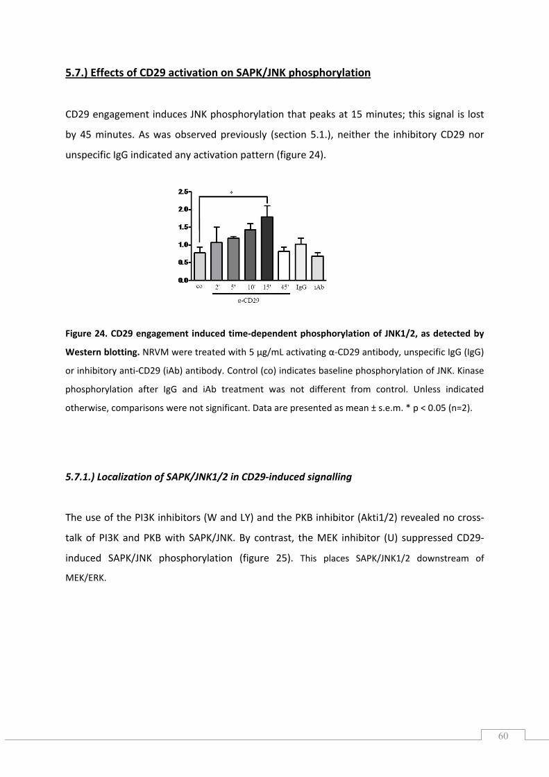
60
5.7.) Effects of CD29 activation on SAPK/JNK phosphorylation
CD29 engagement induces JNK phosphorylation that peaks at 15 minutes; this signal is lost
by 45 minutes. As was observed previously (section 5.1.), neither the inhibitory CD29 nor
IgG indicated any activation pattern (figure 24).
Figure 24. CD29 engagement induced time‐dependent phosphorylation of JNK1/2, as detected by
Western blotting. NRVM were treated with 5 µg/mL activating α‐CD29 antibody, unspecific IgG (IgG)
or inhibitory anti‐CD29 (iAb) antibody. Control (co) indicates baseline phosphorylation of JNK. Kinase
phosphorylation after IgG and iAb treatment was not different from control. Unless indicated
otherwise, comparisons were not significant. Data are presented as mean ± s.e.m. * p < 0.05 (n=2).
5.7.1.) Localization of SAPK/JNK1/2 in CD29‐induced signalling
The use of the PI3K inhibitors (W and LY) and the PKB inhibitor (Akti1/2) revealed no cross‐
talk of PI3K and PKB with SAPK/JNK. By contrast, the MEK inhibitor (U) suppressed CD29‐
induced SAPK/JNK phosphorylation (figure 25). This places SAPK/JNK1/2 downstream of
MEK/ERK.
unspecific
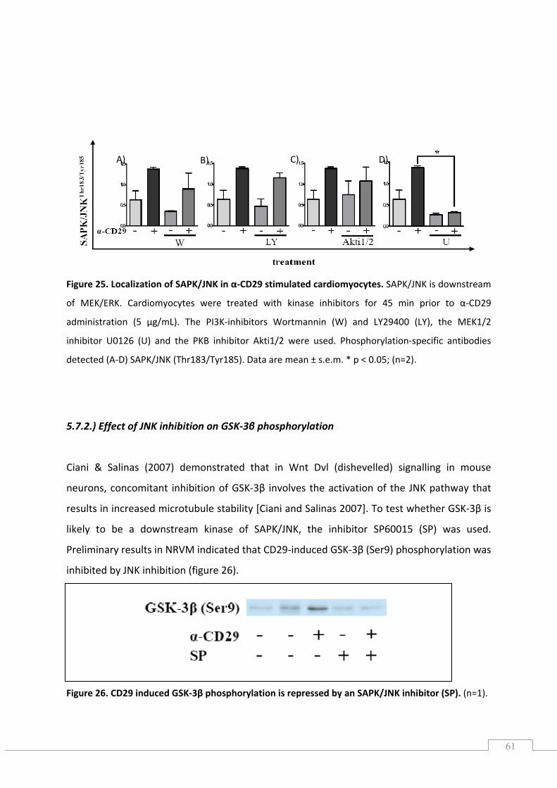
61
Figure 25. Localization of SAPK/JNK in α‐CD29 stimulated cardiomyocytes. SAPK/JNK is downstream
for 45 min prior to α‐CD29
Effect of JNK inhibition on GSK‐3β phosphorylation
Ciani & Salinas (2007) demonstrated that in Wnt Dvl (dishevelled) signalling in mouse
that
‐3β is
igure 26. CD29 induced GSK‐3β phosphorylation is repressed by an SAPK/JNK inhibitor (SP). (n=1).
of MEK/ERK. Cardiomyocytes were treated with kinase inhibitors
administration (5 µg/mL). The PI3K‐inhibitors Wortmannin (W) and LY29400 (LY), the MEK1/2
inhibitor U0126 (U) and the PKB inhibitor Akti1/2 were used. Phosphorylation‐specific antibodies
detected (A‐D) SAPK/JNK (Thr183/Tyr185). Data are mean ± s.e.m. * p < 0.05; (n=2).
5.7.2.)
neurons, concomitant inhibition of GSK‐3β involves the activation of the JNK pathway
esults in increased microtubule stability [Ciani and Salinas 2007]. To test whether GSKr
likely to be a downstream kinase of SAPK/JNK, the inhibitor SP60015 (SP) was used.
Preliminary results in NRVM indicated that CD29‐induced GSK‐3β (Ser9) phosphorylation was
inhibited by JNK inhibition (figure 26).
F
A) B) C) D)
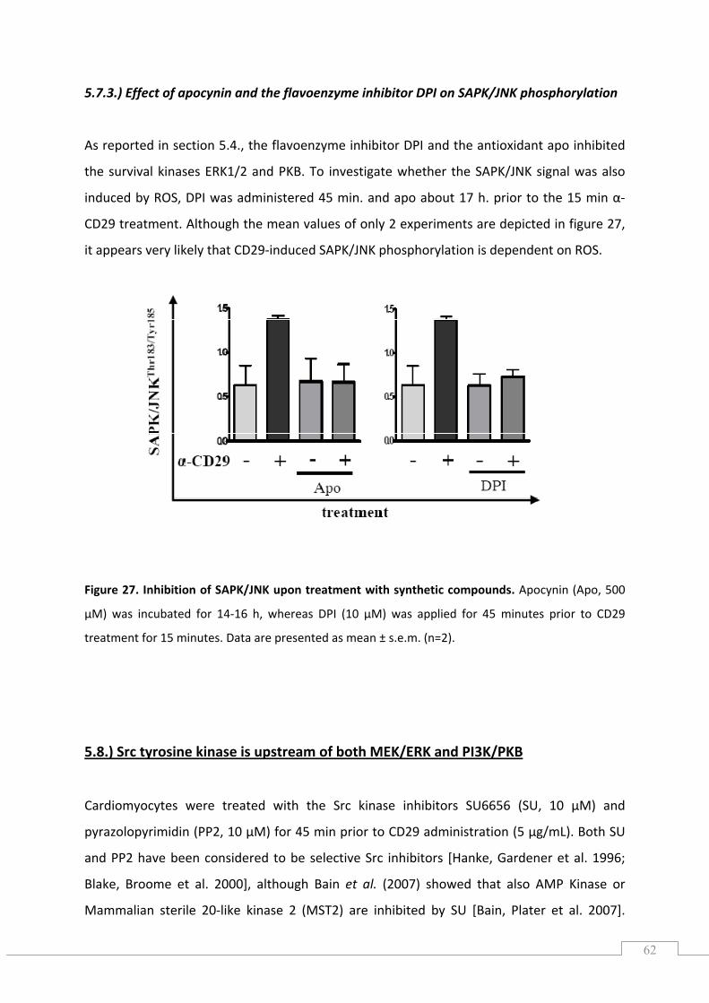
62
5.7.3.) Effect of apocynin and the flavoenzyme inhibitor DPI on SAPK/JNK phosphorylation
s reported in section 5.4., the flavoenzyme inhibitor DPI and the antioxidant apo inhibited
the survival kinases ERK1/2 and PKB. To investigate whether the SAPK/JNK signal was also
induced by S, DPI was administered 45 min. and apo about 17 h. prior to the 15 min α‐
CD29 treatment. Although the mean values of only 2 experiments are depicted in figure 27,
it appears very likely that CD29‐induced SAPK/JNK phosphorylation is dependent on ROS.
treatment for 15 minutes. Data are presented as mean ± s.e.m. (n=2).
.8.) Src tyrosine kinase is upstream of both MEK/ERK and PI3K/PKB
A
RO
Figure 27. Inhibition of SAPK/JNK upon treatment with synthetic compounds. Apocynin (Apo, 500
µM) was incubated for 14‐16 h, whereas DPI (10 µM) was applied for 45 minutes prior to CD29
5
ardiomyocytes were treated with the Src kinase inhibitors SU6656 (SU, 10 µM) and
yrazolopyrimidin (PP2, 10 µM) for 45 min prior to CD29 administration (5 µg/mL). Both SU
nd PP2 have been considered to be selective Src inhibitors [Hanke, Gardener et al. 1996;
Mammalian sterile 20‐like kinase 2 (MST2) are inhibited by SU [Bain, Plater et al. 2007].
C
p
a
Blake, Broome et al. 2000], although Bain et al. (2007) showed that also AMP Kinase or
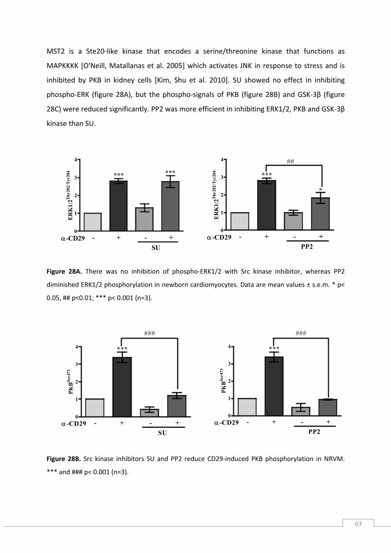
63
0
1
2
3
4
- + - +α-CD29SU
*** ***
ER
K1/
2Thr2
02/T
yr20
4
0
1
2
3
4
- + - +α -CD29PP2
***
*
##
ER
K1/
2Thr2
02/T
yr20
4
0
1
2
3
4
- + - +α -CD29PP2
***
###
PKB
Ser4
73
0
1
2
3
4
- + - +α -CD29SU
***
###
PKB
Ser4
73
APKKKK [O'Neill, Matallanas et al. 2005] which activates JNK in response to stress and is
igure 28A. There was no inhibition of phospho‐ERK1/2 with Src kinase inhibitor, whereas PP2
iminished ERK1/2 phosphorylation in newborn cardiomyocytes. Data are mean values ± s.e.m. * p<
.05, ## p<0.01; *** p< 0.001 (n=3).
MST2 is a Ste20‐like kinase that encodes a serine/threonine kinase that functions as
M
inhibited by PKB in kidney cells [Kim, Shu et al. 2010]. SU showed no effect in inhibiting
phospho‐ERK (figure 28A), but the phospho‐signals of PKB (figure 28B) and GSK‐3β (figure
28C) were reduced significantly. PP2 was more efficient in inhibiting ERK1/2, PKB and GSK‐3β
kinase than SU.
F
d
0
Figure 28B. Src kinase inhibitors SU and PP2 reduce CD29‐induced PKB phosphorylation in NRVM.
*** and ### p< 0.001 (n=3).
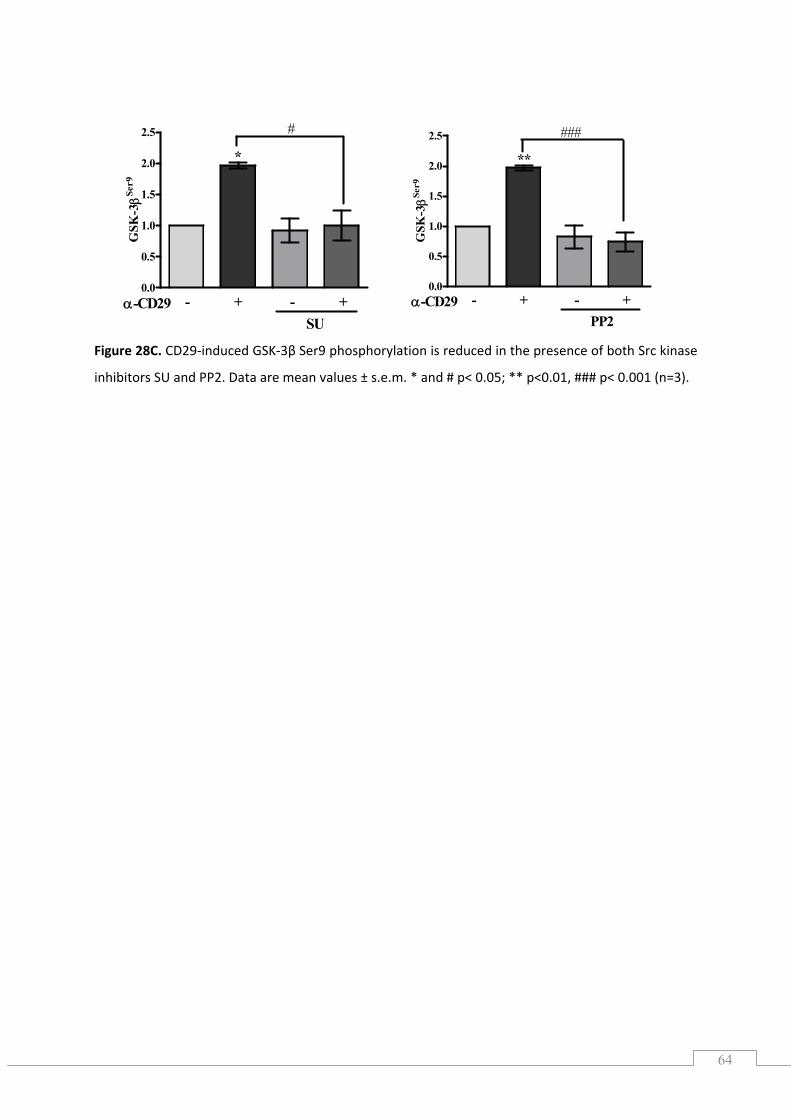
64
0.0
0.5
1.0
1.5
2.0
2.5
- + - +α-CD29SU
*
#G
SK-3βSe
r9
0.0
0.5
1.0
1.5
2.0
2.5
- + - +α-CD29PP2
**
###
GS
Figure 28C. CD29‐induced GSK‐3β Ser9 phosphorylation is reduced in the presence of both Src kinase
hibitors SU and PP2. Data are mean values ± s.e.m. * and # p< 0.05; ** p<0.01, ### p< 0.001 (n=3).
K-3βSe
r9
in
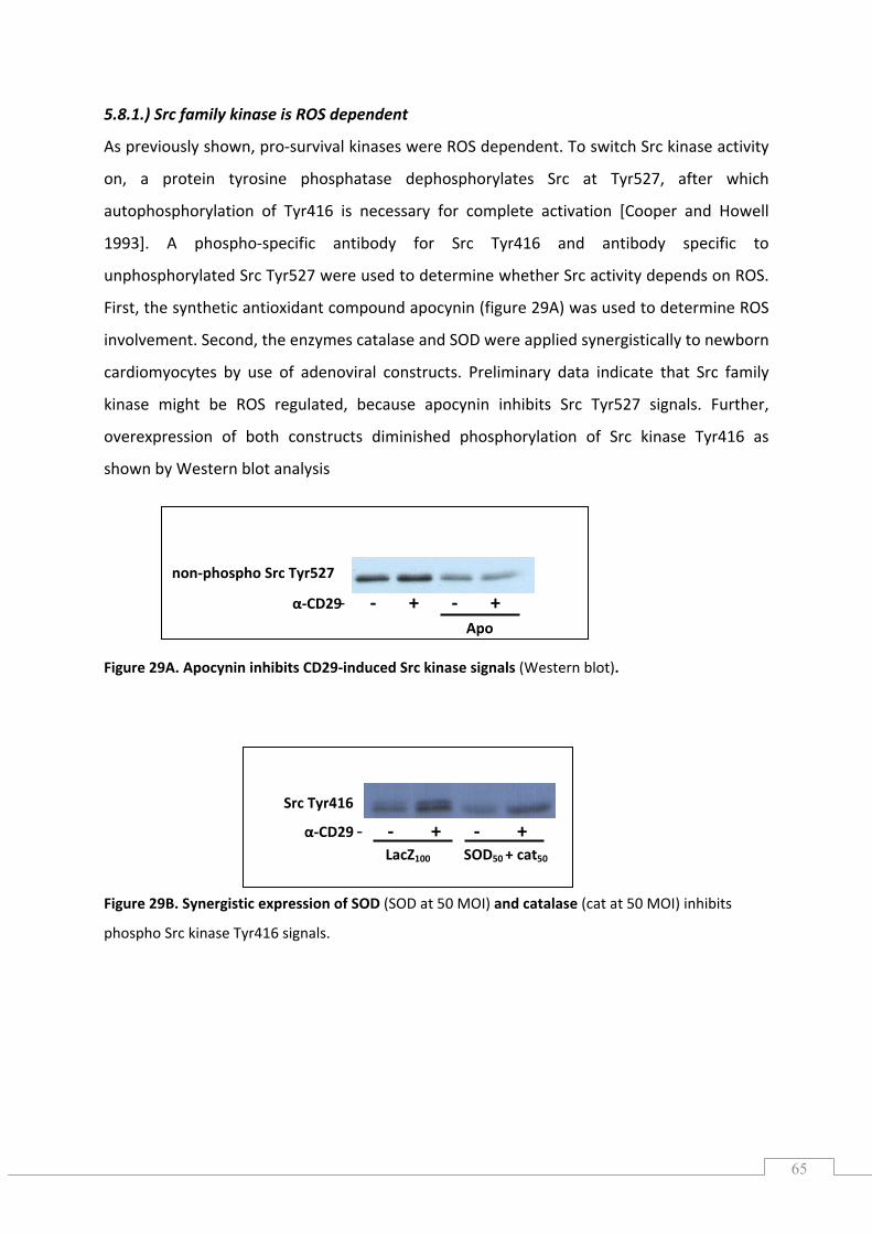
65
5.8.1.) Src family kinase is ROS dependent
previously shown, pro‐survival kinases were ROS dependent. To switch Src kinase activity
on, a protein tyrosine phosphatase dephosphorylates Src at Tyr527, after which
utophosphorylation of Tyr416 is necessary for complete activation [Cooper and Howell
993]. A phospho‐specific antibody for Src Tyr416 and antibody specific to
unphosphorylated Src Tyr527 were used to determine whether Src activity depends on ROS.
irst, the synthetic antioxidant compound apocynin (figure 29A) was used to determine ROS
volvement. Second, the enzymes catalase and SOD were applied synergistically to newborn
cardiomyocytes by use of adenoviral constructs. Preliminary data indicate that Src family
overexpression of both constructs diminished phosphorylation of Src kinase Tyr416 as
hown by Western blot analysis
Figure 29A. Apocynin inhibits CD29‐induced Src kinase signals (Western blot).
Figure 29B. Synergistic expression of SOD (SOD at 50 MOI) and catalase (cat at 50 MOI) inhibits
phospho Src kinase Tyr416 signals.
As
a
1
F
in
kinase might be ROS regulated, because apocynin inhibits Src Tyr527 signals. Further,
s
‐ ‐ + ‐ +Apo
α‐CD29
non‐phospho Src Tyr527
‐ ‐ + ‐ +α‐CD29 LacZ100 SOD50 + cat50
Src Tyr416

66
0
50
100
150
200
- + - +α-CD29H3232
A)
DC
F fl
uore
san
)ce
nce
(geo
me
0
50
100
150
200
- + - +α -CD29
H3232
###*
§
B)
DC
F fl
uore
scen
ce (g
)eo
mea
n
5.8.2.) Protein kinase C inhibits CD29‐induced GSK‐3β phosphorylation
s Hexha .
30. H3232 inhibition of the GSK3β signal in NRVM (A) and H9c2 (B) cells is shown.
S generation
cardiac myoblasts, DCF fluorescence was clearly reduced after treatment with the PKC
inhibitor H3232, as compared to control NRVM. Both datasets (figure 31A and B) hint at the
volvement of PKC in ROS formation (figure 31A) and at being located upstream of NOX
.
igure 31. H3232 inhibits the CD29‐induced burst in NRVM (A) and H9c2 (B). Data are presented as
mean values ± s.e.m. (n=2) * p< 0.05, ### p< 0.001; § p< 0.05 vs. H9c2 without α‐ CD29 treatment.
To investigate whether GSK‐3β is a target of PKC, NRVM and rat cardiomyoblasts were
treated with the PKC inhibitor H3232 for 45 minutes prior to CD29 administration (15
minutes). H3232 is a synthetic peptide which binds to the pseudosubstrate domain of PKC
and inhibit its kinase activity. [Alexander, m et al 1989; Jakab, Weiger et al.
1996].Similar patterns as in NRVM were observed in H9c2 cells, namely H3232 inhibited
CD29 induced GSK‐3β (Ser9) inactivation (figure 30).
Figure
5.8.3.) Effects of the Src kinase inhibitor PP2, SU and PKC inhibitor on RO
In
in
(figure 31B)
F
GSK‐3β Ser9
A) B)
α‐CD29 ‐ + ‐ + ‐ + ‐ + H3232 H3232
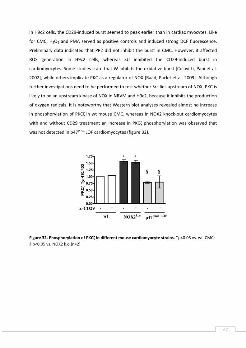
67
In H9c2 cells, the CD29‐induced burst seemed to peak earlier than in cardiac myocytes. Like
hether Src lies upstream of NOX, PKC is
likely to be an upstream kinase of NOX in NRVM and H9c2, because it inhibits the production
of oxygen radicals. It is teworthy that Western blot analyses revealed almost no increase
phosphor of PKCζ in wt mouse CMC, whereas in NOX2 knock‐out cardiomyocytes
and without that
was not detected in p47phox LOF cardiomyo igure 32).
igure 32. Phosphorylation of PKCζ in different mouse cardiomyocyte strains. *p<0.05 vs. wt CMC; § p<0.05 vs. NOX2 k.o.(n=2)
for CMC, H2O2 and PMA served as positive controls and induced strong DCF fluorescence.
Preliminary data indicated that PP2 did not inhibit the burst in CMC. However, it affected
ROS generation in H9c2 cells, whereas SU inhibited the CD29‐induced burst in
cardiomyocytes. Some studies state that W inhibits the oxidative burst [Colavitti, Pani et al.
2002], while others implicate PKC as a regulator of NOX [Raad, Paclet et al. 2009]. Although
further investigations need to be performed to test w
no
in ylation
with CD29 treatment an increase in PKCζ phosphorylation was observed
cytes (f
F
0.00
0.25
0.50
0.75
1.00
1.25
1.50
1.75
- + - + - +α-CD29
wt NOX2k.o. p47phox LOF
* *
§ §
Tyr
410/
403
PKCζ
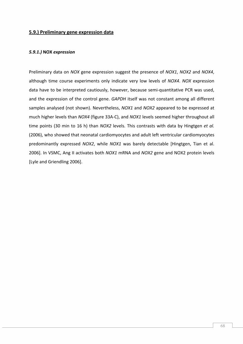
68
5.9.) Preliminary gene expression data
5.9.1.) NOX expression
Preliminary data on NOX gene expression suggest the presence of NOX1, NOX2 and NOX4,
although time course experiments only indicate very low levels of NOX4. NOX expression
data have to be interpreted cautiously, however, because semi‐quantitative PCR was used,
and the expression of the control gene. GAPDH itself was not constant among all different
samples analysed (not shown). Nevertheless, NOX1 and NOX2 appeared to be expressed at
much higher levels than NOX4 (figure 33A‐C), and NOX1 levels seemed higher throughout all
time points (30 min to 16 h) than NOX2 levels. This contrasts with data by Hingtgen et al.
(2006), who showed that neonatal cardiomyocytes and adult left ventricular cardiomyocytes
redominantly expressed NOX2, while NOX1 was barely detectable [Hingtgen, Tian et al.
2006]. In VSMC, Ang II activates both NOX1 mRNA and NOX2 gene and NOX2 protein levels
[Lyle and Griendling 2006].
p
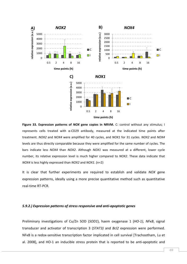
69
0
1000
2000
3000
4000
5000
0.5 2 4 8 16relative
expression (a.u.)
time points (h)
NOX1
C
I
0500
0.5 2 4 8 16
relative
time points (h)
10001500200025003000
expression
(a.u.)
NOX4
C
I
Figure 33. Expression pattern
s of NOX gene copies in NRVM. C: control without any stimulus; I
represents cells treated with α‐CD29 antibody, measured at the indicated time points after
treatment. NOX2 and NOX4 were amplified for 40 cycles, and NOX1 for 31 cycles. NOX2 and NOX4
vels are thus directly comparable because they were amplified for the same number of cycles. The
bars indicate less NOX4 than NOX2. Although NOX1 was measured at a different, lower cycle
umber, its relative expression level is much higher compared to NOX2. These data indicate that
OX4 is less highly expressed than NOX2 and NOX1. (n=2)
is clear that further experiments are required to establish and validate NOX gene
expression patterns, ideally using a more precise quantitative method such as quantitative
eal‐time RT‐PCR.
.9.2.) Expression patterns of stress responsive and anti‐apoptotic genes
Preliminary investigations of Cu/Zn SOD (SOD1), haem oxygenase 1 (HO‐1), NFκB, signal
transducer and activator of transcription 3 (STAT3) and Bcl2 expression were performed.
FκB is a redox‐sensitive transcription factor implicated in cell survival [Trachootham, Lu et
al. 2008], and HO‐1 an inducible stress protein that is reported to be anti‐apoptotic and
le
n
N
It
r
5
N
0
1000
0.5 2 4 8 16
lative
time points (h)
2000
3000
5000re
expression (a.u.)
4000
NOX2
C
I
C)
A) B)
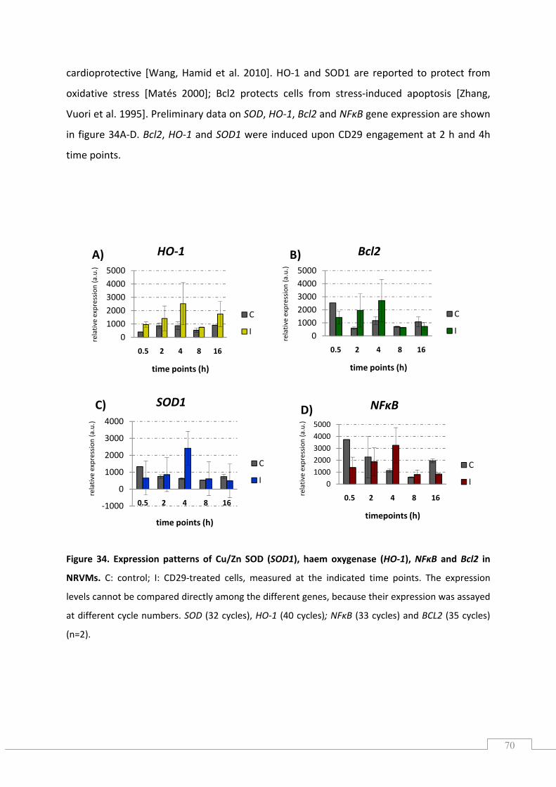
70
‐1000
0
4000
relativ
e a.u.)
SOD1
1000
2000
3000
0.5 2 4 8 16
expression
(
time points (h)
C
I 0
4000
0.5 2 4 8 16
expression
(
100020003000
5000
relativ
e a.u.)
NFκB
timepoints (h)
C
I
cardioprotective [Wang, Hamid et al. 2010]. HO SOD1 are reported to protect from
oxidative stress [Matés 2000]; Bcl2 protects cells from stress‐induced apoptosis [Zhang,
Vuori et al. 1995]. Preliminary data on SOD, HO‐1, Bcl2 and NFκB gene expression are shown
in figure 34A‐D. Bcl2, HO‐1 and SOD1 were indu ed upon CD29 engagement at 2 h and 4h
time points.
n patterns of Cu/Zn SOD (SOD1), haem oxygenase (HO‐1), NFκB and Bcl2 in
RVMs. C: control; I: CD29‐treated cells, measured at the indicated time points. The expression
levels cannot be compared directly among the different genes, because their expression was assayed
BCL2 (35 cycles)
=2).
‐1 and
c
Figure 34. Expressio
N
at different cycle numbers. SOD (32 cycles), HO‐1 (40 cycles); NFκB (33 cycles) and
(n
010002000300040005000
0.5 2 4 8 16
relativ
e expression
(a.u.)
Bcl2
time points (h)
C
I0
10002000300040005000
0.5 2 4 8 16
relativ
e expression
(a.u.)
HO‐1
time points (h)
C
I
A) B)
D)C)
71
6.) DISCUSSION
6.1.) CD29 and the oxidative burst
It has become evident that moderate or low levels of ROS act as mediators in cellular
ignalling processes. The production and removal of ROS are tightly regulated by enzymatic
(e.g., SOD, catalase) and non‐enzymatic (e.g., ascorbic acid, α‐tocopherol) systems that
ensure a balanced redox‐system. While under physiological conditions, low levels of ROS act
transiently and locally, excessive ROS levels ar armful to bystander cells and adjacent
tissue by damaging proteins, lipids, DNA and carbohydrates, resulting in injury and
pathophysiological processes. H2O2 levels greater than 0.1 mM promote apoptosis in
neonatal and adult cardiomyocytes [Aikawa, Komuro et al. 1997; Kwon, Pimentel et al.
2003]. The latter became necrotic when exposed to levels greater than 1 mM [Aikawa,
Komuro et al. 1997].
However, in the experiments described in this thesis, the generated oxygen radicals and
non‐radicals seemed to be present at low levels her than in excessive amounts. We
showed that CD29‐induced ROS generation occurred within a one‐hour time interval. This
burst was impaired by DPI and apo. Interestingly, these findings are in agreement with DPI‐
based data implicating NOX in CD29‐induced ROS production in the colic adenocarcinoma
cell line (caco‐2) [Honoré, Kovacic et al. 2003], but not with findings from mouse embryonic
FB (NIH‐3T3), where growth factors and integrin derived ROS were mostly 5‐lipoxygenase (5‐
LOX)‐dependent [Chiarugi, Pani et al. 2003]. At hours, the level of ROS declined to baseline
levels, but we observed a second wave of ROS generation at three to four hours after
activation of the integrin receptor. Using NOX subunit‐deficient mouse strains, we showed
that NOX is responsible for ROS generation in cardiomyocytes. The finding that inhibition of
ment of another functional NOX member (i.e., not NOX2). NOX4 differs from NOX2
and NOX1 by being independent of regulatory subunits like p47phox, only requiring the
004] for activity. The most likely
candidate therefore is NOX1, since its activity also depends on phagocytic p47phox regulatory
subunits in certain tissues [Chen, Craige et al. 2009]. In general, NOX1 functions with the
s
e h
rat
‐
2
ROS formation is more pronounced in p47phox LOF than in NOX2 knock‐out cells implies the
involve
transmembrane p22phox subunit [Ambasta, Kumar et al. 2
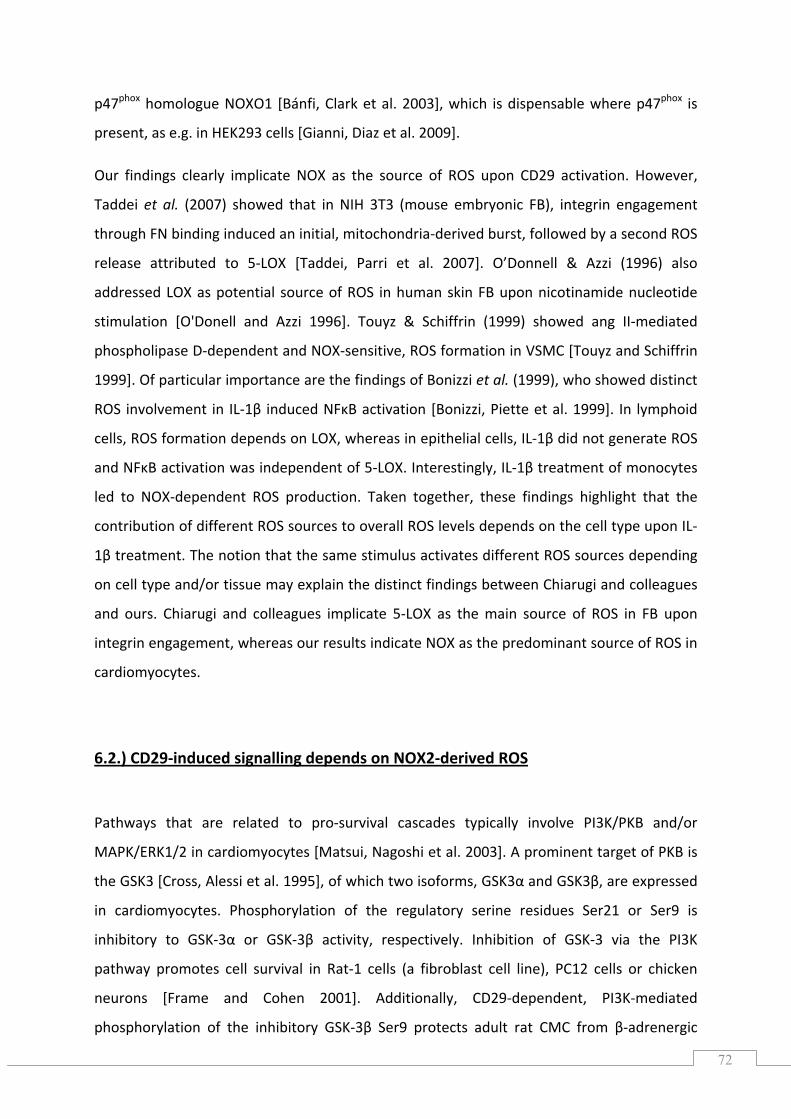
72
p47phox homologue NOXO1 [Bánfi, Clark et al. 2003], which is dispensable where p47phox is
293 cells [Gianni, Diaz et al. 2009].
the source of ROS upon CD29 activation. However,
addei et al. (2007) showed that in NIH 3T3 (mouse embryonic FB), integrin engagement
t ROS production. Taken together, these findings highlight that the
contribution of different ROS sources to overall ROS levels depends on the cell type upon IL‐
present, as e.g. in HEK
Our findings clearly implicate NOX as
T
through FN binding induced an initial, mitochondria‐derived burst, followed by a second ROS
release attributed to 5‐LOX [Taddei, Parri et al. 2007]. O’Donnell & Azzi (1996) also
addressed LOX as potential source of ROS in human skin FB upon nicotinamide nucleotide
stimulation [O'Donell and Azzi 1996]. Touyz & Schiffrin (1999) showed ang II‐mediated
phospholipase D‐dependent and NOX‐sensitive, ROS formation in VSMC [Touyz and Schiffrin
1999]. Of particular importance are the findings of Bonizzi et al. (1999), who showed distinct
ROS involvement in IL‐1β induced NFκB activation [Bonizzi, Piette et al. 1999]. In lymphoid
cells, ROS formation depends on LOX, whereas in epithelial cells, IL‐1β did not generate ROS
and NFκB activation was independent of 5‐LOX. Interestingly, IL‐1β treatment of monocytes
led to NOX‐dependen
1β treatment. The notion that the same stimulus activates different ROS sources depending
on cell type and/or tissue may explain the distinct findings between Chiarugi and colleagues
and ours. Chiarugi and colleagues implicate 5‐LOX as the main source of ROS in FB upon
integrin engagement, whereas our results indicate NOX as the predominant source of ROS in
cardiomyocytes.
6.2.) CD29‐induced signalling depends on NOX2‐derived ROS
o
inhibitory to GSK‐3α or GSK‐3β activity, respectively. Inhibition of GSK‐3 via the PI3K
pathway promotes cell survival in Rat‐1 cells (a fibroblast cell line), PC12 cells or chicken
neurons [Frame and Cohen 2001]. Additionally, CD29‐dependent, PI3K‐mediated
Pathways that are related to pro‐survival cascades typically involve PI3K/PKB and/or
MAPK/ERK1/2 in cardiomyocytes [Matsui, Nag shi et al. 2003]. A prominent target of PKB is
the GSK3 [Cross, Alessi et al. 1995], of which two isoforms, GSK3α and GSK3β, are expressed
in cardiomyocytes. Phosphorylation of the regulatory serine residues Ser21 or Ser9 is
phosphorylation of the inhibitory GSK‐3β Ser9 protects adult rat CMC from β‐adrenergic
73
receptor‐induced apoptosis [Menon, Johnson et al. 2007]. We showed that in NRVM the
PI3K/PKB and MEK/ERK pathways are both activated by CD29; here, GSK3β was uncovered
as a target of both pathways. Even though GSK3β is reportedly directly phosphorylated by
PKB, several other kinases such as S6K or RSK (a kinase target of ERK1/2) have been
implicated in GSK3β inhibition in the heart [Sugden, Fuller et al. 2008]. Furthermore, we
showed that CD29 induced the stress responsive kinase JNK, which acts upstream of GSK‐3β,
although JNK activation occurred at a much lower level than for the other investigated
kinases. JNK activation has been demonstrated to mediate cytoprotection in NRVM after
oxidative stress (hypoxia reoxygenation) [Dougherty, Kubasiak et al. 2002] and in nitric
oxide‐induced apoptosis [Andreka, Zang et al. 2001]. In conclusion, we found that CD29‐
induced survival signalling in cardiomyocytes includes the phosphorylation of ERK1/2, PKB,
JNK and GSK‐3β, all of which were further examined for their ROS dependency.
by adenoviral overexpression. In the presence of both SOD and catalase,
ROS is cleared and α‐CD29‐induced MEK1/2‐phosphorylation, ERK1/2‐phosphorylation, PKB
phosphorylation, and GSK‐3β phosphorylation were inhibited (see also figure 35). Siwik et al.
ion of Cu/Zn SOD with
iethyldithiocarbamic acid (DDC) resulted in apoptosis when DDC was applied at higher
l f w
At moderate levels, ROS may serve as signalling molecules participating in the regulation of
cell survival [Finkel 1998; Chiarugi 2009; Groeger, Quiney et al. 2009], because of their ability
to activate pro‐survival related cascades, including PI3K/PKB in VSMC upon Ang II treatment
[Ushio‐Fukai, Alexander et al. 1999] and/or ERK1/2 in various cell types [Dröge 2002; Forman
and Torres 2002], including neonatal cardiomyocytes [Aikawa, Komuro et al. 1997]. We
showed the aforementioned survival kinases to be ROS‐dependent by the combined use of
the enzymes Cu/Zn SOD and catalase, both of which were investigated pharmacologically
(MnTMPyP) and
(1999) showed that in neonatal rat cardiomyocytes, the inhibit
d
concentrations, whereas lower concentrations of DDC mediated cell growth [Siwik, Tzortzis
et al. 1999]. High DDC levels led to high intrecellular superoxide anion levels that were
cleared by Euk8, a SOD/cat mimetic. In this respect, our experiments showed that single‐
enzyme overexpression of either enzyme was not as efficient as the combination of both in
inhibiting CD29‐induced signalling in NRVM. Therefore, both O2•‐ and H2O2 are implicated as
second messengers in CD29‐induced pro‐survival signal transduction in NRVM. Recently,
beneficia effects o ROS ere shown in vitro and also in humans. In particular, Groeger et al.
(2009) indicated the necessity of H2O2 for cell survival in retina cells [Groeger, Quiney et al.
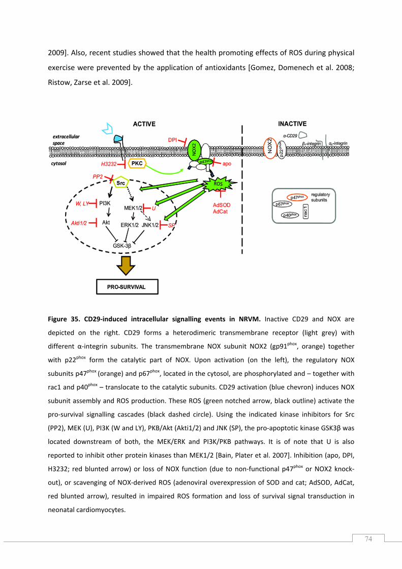
74
2009]. Also, recent studies showed that the health promoting effects of ROS during physical
exercise were prevented by the application of antioxidants [Gomez, Domenech et al. 2008;
Ristow, Zarse et al. 2009].
Figure 35. CD29‐induced intracellular signalling events in NRVM. Inactive CD29 and NOX are
H3232; red blunted arrow) or loss of NOX function (due to non‐functional p47 or NOX2 knock‐
out), or scavenging of NOX‐derived ROS (adenoviral overexpression of SOD and cat; AdSOD, AdCat,
depicted on the right. CD29 forms a heterodimeric transmembrane receptor (light grey) with
different α‐integrin subunits. The transmembrane NOX subunit NOX2 (gp91phox, orange) together
with p22phox form the catalytic part of NOX. Upon activation (on the left), the regulatory NOX
subunits p47phox (orange) and p67phox, located in the cytosol, are phosphorylated and – together with
rac1 and p40phox – translocate to the catalytic subunits. CD29 activation (blue chevron) induces NOX
subunit assembly and ROS production. These ROS (green notched arrow, black outline) activate the
pro‐survival signalling cascades (black dashed circle). Using the indicated kinase inhibitors for Src
(PP2), MEK (U), PI3K (W and LY), PKB/Akt (Akti1/2) and JNK (SP), the pro‐apoptotic kinase GSK3β was
located downstream of both, the MEK/ERK and PI3K/PKB pathways. It is of note that U is also
reported to inhibit other protein kinases than MEK1/2 [Bain, Plater et al. 2007]. Inhibition (apo, DPI, phox
red blunted arrow), resulted in impaired ROS formation and loss of survival signal transduction in
neonatal cardiomyocytes.
75
Finally the role of NOX in CD29‐activated cells was investigated. DPI and apocynin both
abolished CD29‐induced ERK1/2 phosphorylation, PKB phosphorylation, and JNK
phosphorylation, whereas the evaluation of GSK‐3β phosphorylation is hampered because
DPI and apo induced kinase phosphorylation. In any event, these findings suggest an
involvement of NOX. Subsequently, mice carrying either a knock‐out of the transmembrane
NOX subunit NOX2, or a LOF of the regulatory subunit p47phox were used to assess whether
NOX2 determines CD29‐activation‐mediated survival signal transduction in NMVM. As
expected, NOX2 knock‐out mouse cardiomyocytes showed reduced levels of CD29‐induced
ERK1/2 phosphorylation. Similarly, the levels of phosphorylation of PKB and GSK‐3β were
significantly reduced in both NOX2‐ and p47phox‐LOF cardiomyocytes, highlighting the central
role of NOX2‐ and p47phox‐derived ROS in CD29‐induced survival signalling. The remaining
phospho‐ERK1/2 signal in p47phox LOF NMVM indicates a NOX independent and/or
compensatory pathway. As discussed before (section 6.1), p47phox can be replaced by its
homologue NOXO1 in the NOX homologue NOX1, which might signal through ERK1/2 in
p47phox LOF cardiomyocytes. Taken together these data clearly indicate that CD29‐induced
survival signalling depends on NOX‐derived ROS.
6.3.) Kinases potentially up‐ and downstream of NOX
in integrin
In detail, Giannoni and co‐workers proposed that in cell suspension, Src kinase is inactive
and in a reduced state with only Tyr527 phosphorylated. Upon contact to the ECM in “early
tive. In the following “late adhesion/spreading” phase, Tyr527 is still
dephosphorylated due to ECM binding and LOX‐derived ROS production. Tyr416 is still
We sought for likely upstream kinases of the PI3K/PKB and the MEK/ERK pathway and/ or of
NOX. Primarily, Src family kinase involvement was investigated by use of the inhibitors SU
and PP2, both of which did place Src family kinase upstream of both PI3K/PKB and MEK/ERK
signalling pathways. Preliminary data on the inhibition of Src family kinase by the antioxidant
apo and the combined SOD/cat overexpression point at Src being ROS‐regulated. Giannoni et
al. (2005) reported that ROS activated Src tyrosine kinase through redox regulation during
cell adhesion and anchorage‐dependent cell growth [Giannoni, Buricchi et al. 2005]. Src was
oxidized and activated response to ECM attachment and thus following activation.
adhesion”, Tyr527 is dephosphorylated and Tyr416 autophosphorylated. Src kinase is still
reduced, but partially ac
76
phosphorylated, and then Src kinase is oxidized and becomes completely active. After the
application of reducing agents, ROS formation is dimin shed, Src kinase becomes reduced
and inactive, Tyr416 is dephosphorylated and Tyr527 phosphorylated. Three years later, the
same research group presented a model in which integrin engagement due to cell
attachment induced Src activity via oxidation of the kinase [Giannoni, Buricchi et al. 2008]. In
that study, this led to inhibition of the pro‐apoptotic kinase Bim through EGFR activation
followed by ERK and PKB activation, and protected against anoikis in a human endothelial
cell line derived from the bladder (ECV304). Also, another study demonstrated Src kinase
activation upon Ang II treatment, which transactivated EGFR, Rac and NOX1 [Lyle and
Griendling 2006]. Thus in short, our data indicate that Src activity is impaired upon ROS
removal and it appears very likely that NOX2 promotes Src’s activity through redox‐
regulation.
Secondly, we explored whether PKC could be involved in the investigated signalling
cascade(s), hypothesizing an involvement upstream of NOX. In neutrophils, PKC has been
suggested to activate the regulatory NOX subu
i
nit p47phox by phosphorylation [Fontayne,
ang et al. 2002]. In VSMC the effectiveness of NOX activation depends on PKC (among
other kinases). By administering the PKC inhibitor H3232 to newborn cardiomyocytes or to
cardiac myoblasts, ROS formation was attenuated, indicating that PKC might indeed regulate
NOX. Interestingly, in the NOX2 knock‐out strain, PKCζ is highly phosphorylated compared to
wild type or the p47 LOF strain. It is of note that a CD29‐induced increase in PKCζ‐
phosphorylation is absent in wild‐type CMC, which leaves the possibility that PKCζ is not
inducible by CD29. Nevertheless, we showed that PKC lies upstream of NOX, and highly
activated PKCζ could be suggestive of the activity of another NOX homologue.
D
phox
6.4.) NOX and NOX‐derived ROS in the heart
Data on the involvement of NOX in different processes in the cardiovascular system,
including cell survival and apoptosis, is partly contradictory. Findings suggesting beneficial
roles of NOX in physiological processes are scarce. Such findings include NOX participating in
77
oxygen sensing or regulating the vascular muscle tone [Cave, Brewer et al. 2006]. However,
in the myocardium, NOX and NOX‐derived ROS have previously mainly been implicated in
pathophysiological processes [Akki, Zhang et al. 2009].
Peng et al. (2005) showed that NOX2 contributed to myocardial dysfunction when
endotoxins (lipopolysaccharides) were present in the blood [Peng, Lu et al. 2005]. Increased
NOX2 protein expression was shown in human CMC after acute myocardial infarction (AMI)
[Krijnen, Meischl et al. 2003]. In that study, infarcted myocardial tissue of patients who died
of AMI was investigated. High levels of NOX2 were detected, localized at the plasma
membrane and in the cytosol of cardiomyocytes. NOX2 was expressed in both, normal and
diseased human cardiomyocytes, but was increased in sections after AMI. Similarly, Looi et
al. (2008) demonstrated the involvement of NOX2 in adverse cardiac remodelling after
myocardial infarction in NOX2 knock‐out mice [Looi, Grieve et al. 2008]. In contrast, NOX2‐
derived ROS have been also implicated as mediators of cardiomyocyte protection against
ischaemia/reperfusion injury conferred by ischaemic preconditioning [Bell, Cave et al. 2005].
NOX2 is involved in the hypertrophic response to Ang II [Bendall, Cave et al. 2002], and ROS
production was increased in wild‐type cardiomyocytes upon Ang II treatment compared to
the basal level in knock‐out mice. The hypertrophic markers ANF (atrial natriuretic factor;
inhibits maladaptive hypertrophy), β‐MHC, were significantly induced in wild type
cardiomyocytes but not in knock‐out CMC, as was the heart to body weight ratio in wild‐type
animals. These findings indicate a pathphysiological role of NOX2 in Ang II‐induced
hypertrophy. A follow‐up study by the same group investigated the role of NOX upon
pressure overload in left ventricular hypertrophy in NOX2 knock‐out mice [Byrne, Grieve et
al. 2003]. Byrne et al. used chronic Ang II infusion or aortic constriction as a study system;
while Ang II failed to induce an increase in NOX activity, ANF expression and cardiac mass in
NOX2 knock‐out mice, aortic constriction did induce NOX activity. Furthermore, expression
f NOX4 was upregulated at both protein and gene levels, hinting at contrasting roles of NOX
as these findings in the literature mainly
oint to damaging functions of NOX2 in the heart under pathophysiological conditions, our
data indicate that under physiological conditions, NOX2 is essential for survival.
o
isoforms in response to different stimuli. Where
p
NOX4 has been reported to be important during cardiac differentiation, in regulating MAPK
activation, and several transcription factors [Li, Stouffs et al. 2006]. NOX4 knock‐down
78
impairs cardiac differentiation, which can be rescued by application of exogenous H2O2 in
the nM range. Recently, NOX4 has been implicated as major source of mitochondrial
oxidative stress causing cardiac dysfunction upon pressure overload in the mouse heart, as
shown by use of a cardiac specific NOX4 knock‐out [Kuroda, Ago et al. 2010]. They also
showed that NOX4 overexpression promotes the proliferation of cardiac FB. Although other
NOX isoforms were not upregulated in cardiac‐specific NOX4 knock‐out mice, its catalytic
partner subunit p22 was dramatically downregulated at the protein but not at the mRNA
level[Kuroda, Ago et al. 2010]. Similar observations were made in systemic NOX2 knock‐outs.
NOX4 is particularly interesting with respect to its ROS formation: While it can generate H O
without regulatory NOX subunits, it depends on p22 in HEK293, Hela, H292 and COS cells
[Martyn, Frederick et al. 2006]. Additionally, it has been emphasized that NOX4 activity is
regulated at the gene expression level [Serrander, Cartier et al. 2007]. Interestingly, Zhang et
al. (2010) showed that NOX4 protects the heart from failure [Zhang, Brewer et al. 2010]. In
that study, NOX4 null mice were generated by targeted deletion of the translation initiation
site and exon 1 and 2 of NOX4, which resulted in total loss of NOX4 protein without affecting
myocardial protein levels of NOX2 or p22 . Upon pressure overload, NOX4 knock‐out
animals developed contractile impairment, ventricular dilation, as well as exaggerated
cardiac hypertrophy and increased interstitial fibrosis as compared to NOX4 competent litter
mates [Zhang, Brewer et al. 2010]. These findings demonstrate that NOX4 protects against
pressure overload‐induced heart failure. Secondly, Zhang et al. generated a cardiomyocyte‐
specific NOX4 transgenic mouse to increase NOX4 protein levels. These mice showed
increased NOX4 gene‐and p22 protein expression whereas NOX2 levels remained
constant between strains. NOX4 transgenic mouse hearts did not show basal dysfunction,
and they showed less hypertrophy and interstitial fibrosis in response to pressure overload.
Additionally, upon pressure overload these transgenic mice exhibited an enhanced
myocardial capillary density and release of angiogenic factors promoting angiogenesis
[Zhang, Brewer et al. 2010]. In conclusion, contrasting roles have been attributed to NOX4 in
the literature. NOX4 seems mandatory in cardiac differentiation but also in the failing heart,
whereas it is also a cause of cardiac dysfunction. Since the effect of NOX4 appears to depend
on context, this raises the possibility that NOX2 may display a similar context dependency.
phox
2 2
phox
phox
phox
Therefore, its study in different laboratory model systems may be fruitful and lead to the
elucidation of further protective effects of NOX2.
79
6.5.) The physiological relevance of NOX and ROS
O2•‐and H2O2 are the prevalent oxygen‐derived molecules implicated in redox signalling.
More than a decade ago, these ROS were implicated in a range of cellular outcomes
including apoptosis, senescence, and survival. Several cell surface receptors, transmembrane
agonists, and extracellular signals, including peptide growth factors, cytokines, cell
attachment or phorb esters mediate ROS generation [Rhee 1999; Mehdi, Azar et al. 2007].
H2O2 modulates protein tyrosine kinases and phosphatases by reduction or oxidation, all of
which are essential in cell signalling [Rhee 2006]. H2O2 acts locally and within a defined time‐
frame [Mishina, Tyurin‐Kuzmin et al. 2011]. A numbe of studies suggest NOX as the source
of ROS involved in physiological processes (see comprehensive review by [Bedard and Krause
2007]).
In particular, NOX2‐derived ROS are essential in neutrophils, in host defence and cellular
signalling. Conversely, the lack or loss of function of NOX2 has been implicated in the severe
disease CGD, which is accompanied by infections and inflammations [Lambeth 2004]. NOX2‐
derived ROS are also anti‐inflammatory and prevent autoimmune disease [Hultqvist, Olsson
et al. 2009]. Accumulating evidence implies NOX2‐derived ROS in cell survival, as shown by
the studies of Maraldi et al. (2009) and Groeger et al. (2009). In the human leukaemic
megacaryocytic cell line M07e, NOX2 was implicated as the source of ROS in response to
interleukin (IL) 3 treatment [Maraldi, Prata et al. 2009]. In that study, NOX2‐mediated
survival was mainly validated by use of DPI and apocynin, although NOX2 siRNA was also
used in initial experiments. DCF fluorescence and viability were significantly reduced
compared to IL3‐treated cells and cell death markers such as caspase 3 or apoptotic nuclei
were studied with the synthetic inhibitors apocynin and DPI [Maraldi, Prata et al. 009]. In a
previous study by the same group, ROS were shown to be anti‐apoptotic by use of several
ROS scavenging compounds such as EUK‐134 [Maraldi, Prata et al. 2009]. EUK‐134 is a
SOD/catalase mimetic and inhibited phosphorylation of PKB, ERK, and Src, but did not affect
JNK phosphorylation. Based on their study, suggesting that transient H2O2 generation
induces survival [Mackey, Sanvicens et al. 2008], Groeger et al. (2009) later showed that
NOX2 and NOX4 are the source of these ROS in a retina‐derived photoreceptor cell line
(661W) [Groeger, Mackey et al. 2009]. A possible
ol
r
2
physiological relevance was examined in
80
mouse retinal explants by use of DCF fluorescence‐based time‐lapse imaging to detect H2O2
eneration. Knock‐down of NOX2 and NOX4 by siRNA decreased oxidative burst activity by g
70%. These findings indicate pro‐survival roles of NOX outside of the heart and thus make
pro‐survival roles of NOX in the heart appear more plausible.
6.6.) The spatiotemporal distribution and subcellular localization of NOX
Although several ROS sources besides NOX are present in each cell, recent reports
demonstrated NOX acting in a spatio‐temporally defined manner. NOX is distributed in
several compartments including endosomes, the endoplasmic reticulum, lipid rafts, nuclei,
or at cell migrations sites [Ushio‐Fukai 2006; Ushio‐Fukai 2009]. Oakley et al. (2009) also
suggested that so‐called redoxosomes exist. These redox‐active endosomes contain NOX, as
well as other ROS processing proteins such as SOD1. In response to stimuli such as IL‐1 or
TNFα, superoxide anions are produced in the endosomal lumen facilitated by NOX1 and/or
NOX2 [Oakley, Abbott et al. 2009]. The different localizations of NOX reviewed above might
explain that NOX isoforms such as NOX2 and NOX4 can participate either in distinct [Byrne,
Grieve et al. 2003; Anilkumar, Weber et al. 2008] or similar processes [Petry, Djordevjevij et
al. 2006; Pendayala, Gorshkova et al. 2009; Kim, Choi et al. 2010].
In a model of specialized microdomains (caveolae), angiotensin II receptor type I (AT1R)
stimulation by ang II resulted in partial internalization of the receptor. This was accompanied
with activation of NOX1 and recruitment of caveolin 1 to the receptor, which promoted
transactivation of EGFR through ROS‐dependent Src activation. Both, tyrosine‐
phosphorylated EGFR and caveolin 1, are subsequently released to the site of focal adhesion.
There they colocalize with another NOX family member, namely NOX4, as well as paxillin and
integrin, resulting in redox signalling leading to VSMC hypertrophy [Ushio‐Fukai 2009].
Pendayala et al. (2009) localized NOX4 in the nucleus under normoxia. In response to
hyperoxia, NOX4 translocates to the cytosol and endoplasmic reticulum. They also described
the reciprocal compensation of NOX2 and NOX4 in lung endothelial cells: They showed
increased expression and regulation of NOX2 proteins and transcripts, in siRNA‐mediated
NOX4‐deficient lung endothelial cells. Vice Versa, NOX4 proteins and transcripts are
81
upregulated in NOX2 knock‐down cells [Pendayala, Gorshkova et al. 2009]. In rat and human
VSMC, Hilenski et al. (2004) investigated the subcellular localization of NOX1 and NOX4
[Hilenski, Clempus et al. 2004]. Laser scanning confocal microscopy revealed that NOX1 was
localized to the surface and at cellular margins, whereas NOX4 was found at focal adhesions,
i.e. where cells contact the underling matrix. NOX4 co‐localized with vinculin, a marker for
focal adhesions and NOX1 was co‐localized with caveolin in VSMC. Distinct cellular redox
transfected
EK293 cells [Anilkumar, Weber et al. 2008]. Anilkumar et al. showed NOX2 was localized at
the plasma membrane, while NOX4 was found at the endoplasmic reticulum, co‐localizing
with calnexin. Under non‐treated conditions, NOX4 overexpression increased superoxide
production – which was not the case when NOX2 was overexpressed [Anilkumar, Weber et
al. 2008]. Likewise, phospho‐JNK and ERK1/2 levels were elevated in NOX4 but not in NOX2
expressing cells. Constant phospho‐PKB and phospho‐GSK‐3β signals were observed in both
NOX2‐ and NOX4‐expressing HEK293 cells. Interestingly however, p38 phosphorylation was
increased in NOX2 expressing cells. Ang II‐induced O generation was driven by NOX2 and
not by NOX4. Insulin‐induced O production was both NOX2‐ and NOX4‐driven, whereas
TNFα‐treated cells only induced ROS in NOX2‐transfected cells [Anilkumar, Weber et al.
2008].
9, Ushio‐Fukai2006], it is
clear that NOX‐derived ROS may act in a context‐dependent fashion, and may also
contribute to cell survival. This also implies that a) at the same time, several NOX isoforms
may be activated in response to one defined stimulus; b) the activated NOX isoform may act
either in close proximity or at distant compartments relative to the stimulus, and c) one
stimulus may activate different NOX isoforms in different physiological states. For example,
under pathological conditions in the heart, NOX2 activity may be harmful while NOX4
hardly expressed in the cardiomyocytes we investigated. Also, another functional NOX
homologue dependent on p47 is activated under conditions when NOX2 activity is
impaired. Therefore, it appears very likely that several NOX homologues are activated
depending on the physiological conditions, the stimulus, tissue and the cell type.
responses due to different responses were also assigned to NOX2‐ and NOX4‐
H
2•‐
2•‐
Due to the distinct spatiotemporal NOX distributions [Oakley200
activity is not. However, our data implicate NOX2 in cell survival processes, while NOX4 is
phox
82
6.7.) Conclusions
In my thesis, I have provided novel insights into the mechanisms of CD29 induced pro‐
survival signalling in primary cardiomyocytes (see figure 35). The pro‐survival‐related
pathways PI3K/PKB and MEK/ERK were activated upon CD29 engagement. Src family kinase
was placed upstream of the aforementioned survival pathways and PKC was placed
upstream of NOX. With the exception of PKC, all signalling kinases investigated were
regulated by ROS. GSK3β was identified as a common anti‐apoptotic kinase when
phosphorylated upon PKB/Akt, MEK/ERK, JNK and PKC activation. NOX2 was identified as the
source of the regulatory ROS involved. Furthermore CD29 was shown to induce a transient
oxidative burst that is mediated by a p47phox‐dependent NOX. Thus, NOX1 could serve as an
additional oxidase involved in generating ROS. Preliminary expression data hint at the
availability of this NOX isoform in ventricular cardiomyocytes. These data clearly add to the
growing body of evidence that NOX‐derived ROS (also) have a protective function in the
heart.
83
7.) REFERENCES
Aikawa, R., I. Komuro, et al. (1997). "Oxidative stress activates extracellular signal‐regulated kinasses
through Src and Ras in cultured cardiac myocytes of neonatal rats." Journal of Clinical Investigation 100: 1813‐1821.
Aikawa, R., M. Nawano, et al. (2000). "Insulin prevents cardiomyocytes from oxidative stress‐induced apoptosis through activation of PI3 Kinase/Akt." Circulation 102: 2873‐2879.
Akki, A., M. Zh et (2009). "NADPH oxidase signaling cardiac myocyte function." Journal of ang, al. and Molecular and Cellular Cardiology 47: 15‐22.
Alexander, D. R., J. M. Hexham, et al. (1989). "A pro ein kinase C pseudosubstrate peptide inhibits
Biochem J.
tphosphorylation of the CD3 antigen in streptolysin‐O‐permeabilized human T lymphocytes."
260: 893‐901. Almeida, E. A. C., D. Ilić, et al. (2000). "Matrix survival signaling: from fibronectin via focal adhesion
kinase to c‐Jun NH2‐terminal kinase." The Journal of Cell Biology 149: 741‐754. Ambasta, R. K., P. Kumar, et al. (2004). "Direct interaction of the novel Nox proteins with p22phox is
required for the formation of a functionally active NADPH oxidsae." The Journal of Biological Chemistry 279: 45935‐45941.
Amin, J. K., L. Xiao, et al. (2001). "Reactive oxygen species mediate alpha‐adrenergic receptor‐stimulated hypertrophy in adult ventricular myocytes." Journal of Molecular & Cellular Cardiology 33: 131‐139.
Andreka, P., J. Zang, et al. (2001). "Cytoptrotection by Jun kinase during nitric oxide‐induced cardiac myocte apoptosis." Circulation Research 88: 305‐312.
Anilkumar, N., R. Weber, et al. (2008). "Nox4 and Nox2 NADPH oxidases mediate distinct cellular redox signaling responses to agonist stimulation." Arteriosclerosis, Thrombosis and Vascular Biology 28: 1347‐1354.
plin, A. E., A. Howe, et al. (1998). "Signal transduction and signal modulation by cell adhesion receptors: the role of integrins, cadherins, immunoglobulin‐cell adhesion molecules and selectins." Pharmacological Reviews
A
50: 197‐263. d mechanics of integrin‐based cell
adhesion." Current Opinion in Cell BiologyArnaout, M. A., S. L. Goodman, et al. (2007). "Structure an
19: 495‐507. Arnaout, M. A., B. Mahalingam, et al. (2005). "Integrin structure, allostery, and bidirectional
signaling." Annual Reviews of Cell and Developmental Biology 21: 381‐410. Askari, J. A., C. J. Tynan, et al. (2010). "Focal adhesions are sites of integrin extension." The Journal of
Cell Biology 188: 891‐903. Aumailley, M. and B. Gayraud (1998). "Structure and biological activity of the extracellular matrix."
Journal of Molecular Medicine 76: 253‐265. Babior, B. M. (1999). "NADPH oxidase: an update." Blood 93: 1464‐1476. Babior, B. M., J. T. Curnutte, et al. (1976). "The particulate superoxide‐forming system from human
neutrophils." The Journal of Clinical Investigation 58: 989‐996. Bain, J., L. Plater, et al. (2007). "The selectivity of protein kinase inhibitors: a further update."
Biochemical Journal 408: 297‐315. Banerjee, I., J. W. Fuseler, et al. (2007). "Determination of cell types and numbers during cardiac
development in the neonatal and adult rat and mouse." American Journal of Physiology‐ Heart & Circulatory Physiology 293: H1883‐H1891.
Banerjee, I., K. Yekkala, et al. (2006). "Dynamic interactions between myocytes, fibroblasts, and extracellular matrix." Annals of New York Academy of Sciences. 1080: 76‐84.
Bánfi, B., R. A. Clark, et al. (2003). "Two novel proteins activate superoxide generation by the NADPH oxidase NOX1." Journal of Biological Chemistry 278: 3510‐3513.
Barczyk, M., S. Carracedo, et al. (2010). "Integrins." Cell and Tissue Research 339: 269‐280.
84
Bass, D., J. Parce, et al. (1983). "Flow cytometric studies of oxidative product formation by ded response to membrane stimulation." The Journal of Immunologyneutrophils: a gra 130:
audino, T. A., W. Carver, et al. (2006). "Cardiac fibroblasts: friend or foe?" American Journal of1910‐1917.
B Physiology‐ Heart & Circulatory Physiology 291: H1015‐H1026.
Baudoin, C., M.‐J. Goumans, et al. (1998). "Knockout and knockin of the β1 exon D define distinct roles of integrin splice variants in heart funtion and embryonic development." Genes & Development 12: 1202‐1216.
Bazzoni, G., L. Ma, et al. (1998). "Divalent cations and ligands induce conformational changes that are highly divergent among β integrins." The Journal of Biological Chemistry1 273: 6670‐6678.
Bedard, K. and K.‐H. Krause (2007). "The NOX family of ROS‐generating NADPH oxidases: physiology and pathophysiology." Physiological Review 87: 245‐313.
Bell, R. M., A. C. Cave, et al. (2005). "Pivotal role of NOX2‐containing NADPH oxidase in early ischemic preconditioning." The FASEB Journal 19: 2037‐2039. J. K., A. C. Cave, et al.Bendall, (2002). "Pivotal role of gp91phox‐containing NADPH oxidse in angiotensin II‐induced cardiac hypertrophy in mice." Circulation 105: 293‐296. . A., M. A. Broome, et al. (2000). "SU6656, a selective Src family kinase inhiBlake, R bitor, used to probe growth factor signaling." Molecular and Cellular Biology 20: 9018‐9027. ., B. Rosc, et al. (2008). "The oxidation state of phospholipids controls the oxidative burst in neutrophil granulocytes." The Journal
Blüml, S of Immunology 181: 4347‐4353.
Bochkov, V. N., A. Kadl, et al. (2002). "Protective role of phospholipid oxidation products in endotoxin induces tissue damage." Nature 419: 77‐81. G., J. Piette, et al. (199Bonizzi, 9). "Reactive oxygen intermediate‐dependent NF‐κB activation by interleukin‐1β requires 5‐lipoxygenase or NADPH oxidase activity." Molecular and Cellular Biology 19: 1950‐1960.
Borg, T. K., K. Rubin, et al. (1984). "Recognition of extracellular matrix components by neonatal and adult cardiac myocytes." Developmental Biology 104: 86‐96. S., I. Banerjee, et al. (2010).Bowers, "The extracellular matrix: at the center of all." Journal of itMolecular and Cellular Cardiology 48: 474‐482. sch, C., E. Hirsch, et al. (1997). "Genetic analysis of β1 integrin function: confirmed, new and revised roles for a crucial family of cell adhesion
Brakebu molecules." Journal of Cell Science 110:
2895‐2904. D. I. and K. K. Griendling (2009). "NOX proteins in signal Brown, transduction." Free Radical Biology & Medicine 47: 1239‐1253. W. and M. Burge (2007). Digital imaging processing: An algorithmic introduBurger, ction using Java, Springer‐Verlag New York. J. A., D. J. Grieve, et al. (2Byrne, 003). "Contrasting roles of NADPH oxidase isoforms in pressure overload versus angiotensin II ‐ induced cardiac hypertrophy." Circulation Research 93: 802‐805.
Byron, A., J. D. Humphries, et al. (2009). "Anti‐integrin monoclonal antibodies." Journal of Cell Science 122: 4009‐4011. X. Dai, et al. (2007). "Differential regulation of NADPH oxidasCao, X., e in sympathetic and sensory ganglia in deoxycorticosterone acetate salt hypertension." Hypertension 50: 663‐671.
Carver, W., R. L. Price, et al. (1994). "Distruption of β‐1 integrin in the developing heart." The Journal of Histochemistry and Cytochemistry 42: 167‐175. . C., A. C. Brewer, et al. (2006). "NADPH oxidases in cardiovascular health and disease." Cave, AAntioxidants & Redox Signaling 8: 691‐728.
Chen, K., S. E. Craige, et al. (2009). "Downstream targets and intracellular compartmentalizations in Nox signaling." Antioxidants & Redox Signaling 11: 2467‐2480.
Chen, Q., D. W. Powell, et al. (2003). "Akt phosphorylates p47phox and mediates respiratory burst activity in human neutrophils." The Journal of Immunology 170: 5302‐5308.
85
Chiarugi, P. (2009). "Survival or death: the redox parodox." Antioxidants & Redox Signaling 11: 2651‐2654. i, P. and T. FiaschiChiarug (2007). "Redox signalling in anchorage‐dependent cell growth." Cellular Signalling 19: 672‐682. i, P., G. Pani, et al. (2003). "Reactive oxygen species as essentiaChiarug l mediators of cell adhesion: the oxidative inhibition of a FAK tyrosine phosphatase is required for cell adhesion." The Journal of Cell Biology 161: 933‐944. and P. C. Salinas (2007). "c‐JuCiani, L. n N‐terminal kinase (JNK) cooperates with GSK3β to regulate Dishevelled‐mediated microtubule stability." BMC Cell Biology 8: 27. . A. and J. S. Brugge (1995). "Integrins and signal transduction pathways: the road takenClark, E " Science 268: 233‐239. i, R., G. Pani, et al. (2002). "Reactive oxygen species as doColavitt wnstream mediators of angiogenic signaling by vascular endothelial growth factor receptor‐2/KDR." The Journal of Biological Chemistry 277: 3101‐3108.
Communal, C., M. Singh, et al. (2003). "β1 integrins expression in adult rat ventricular myocytes and its role in the regulation of β‐adrenergic receptor stimulated apoptosis." Journal of Cellular Biochemistry 89: 381‐388. J. A. and B. Howell (1993). "The when and how of Src regulation." CellCooper, 73: 1051‐1054.
Cross, D. A. E., D. R. Alessi, et al. (1995). "Inhibition of glycogen synthase‐3 by insulin mediated by protein kinase B." Nature 378: 785‐789.
D'Souza, S. E., M. H. Ginsberg, et al. (1991). "Arginyl‐glycyl‐aspartic acid (RGD): a cell adhesion motif." TRENDS in Biochemical Sciences 16: 246‐250.
urst." The Journal of Immunology
Dang, P. M.‐C., A. Fontayne, et al. (2001). "Protein kinase C ζ phosphorylates a subset of selective sites of the NADPH oxidase component p47phox and participates in formyl peptide‐mediated neutrophil respiratory b 166: 1206‐1213.
Davidson, B., E. Allen, et al. (1993). "A model system for in vivo gene transfer into the central nervous system using an adenoviral vector." Nature Genetics 3: 219‐223.
Davis, R. J. (2000). "Signal transduction by the JNK group of MAP kinases." Cell 103: 239‐252. F. R., J. Renee, et al. (1998). "Neutrophils exposDe Leo, ed to bacterial lipopolysaccharide upregulate NADPH oxidase assembly." The Journal of Clinical Investigation 101: 455‐463. F. R., K. V. Ulman, et al. (1996). "Assembly of the human neutrophil NADPH oxidase involves binding of p
De Leo,67phox and flavocytochrome b to a common functional domain in p47phox." The
Journal of Biological Chemistry 271(29): 17013‐17020. lker, A. A. and A. SonnenDe Me berg (1999). "Integrins: alternative splicing as a mechanism to regulate ligand binding and integrin signaling events." Bio Essays 21: 499‐509. C., M. Fay, et al. (2000). "ThDewas, e mitogen‐activated protein kinase extracellular signal‐regulated kinase 1/2 pathway is involved in formyl‐methionyl‐leucyl‐phenylalanine‐induced p47phox phosphorylation in human neutrophils." The Journal of Immunology 165: 5238‐5244. d, M. Diamon S. and T. A. Springer (1994). "The dynamic regulation of integrin adhesiveness." Current Biology 4: 506‐517. C. J. Vlahos, et al. (1995Ding, J., ). "Antagonists of phosphatidylinositol 3‐kinase block activation of several protein kinases in neutrophils." The Journal of Biological Chemistry 270: 11684‐11691.
Dorn II, G. W. and A. Diwan (2008). "The rationale for cardiomyocyte resusitation in myocardial salvage." Journal of Molecular Medicine 86: 1085‐1095.
Biochemical JournalDougherty, C. J., L. A. Kubasiak, et al. (2002). "Activation of c‐Jun N‐terminal kinase promotes survival
of cardiac myocytes after oxudative stress." 362: 561‐571. Dröge, W. (2002). "Free radicals in the physiological control of cell function." Physiological Review 82:
47‐95. Fässler, R. and M. Meyer (1995). "Consequences of lack of β1 integrin gene expression in mice."
Genes & Development 9: 1896‐1908.
86
Fässler, R., J. Rohwedel, et al. (1996). "Differnetiation and integrity of cardiac muscle cells are impaired in the absence of β1 integrin." Journal of Cell Science 109: 2989‐2999.
Finkel, T. (1998). "Oxygen radicals and signaling." Current Opinion Cell Biology 10: 248‐253. T. (1999). "Signal transduFinkel, ction by reactive oxygen species in non‐phagocytic cells." Journal of Leukocyte Biology 65: 337‐340. e, A., P. M.‐C. Dang, et al. (2002). "Phosphorylation of p47phox sites by PKC α, βII, δ, and ζ:effect on binding to p22phox and on
Fontayn NADPH oxidase activation." Biochemistry 41: 7743‐
Forman ctive oxygen species." 7750. , H. J., M. Maiorino, et al. (2010). "Signaling functions of reaBiochemistry 49: 835‐842. , H. J. and M. Torres (20Forman 02). "Reactive oxygen species and cell signaling." American Journal of Respiratory and Critical Care Medicine 166: S4‐S8. S. and P. Cohen (2001). "GSK3 takes centre stage more than 20 years after its discovery." Biochemical Journal
Frame, 359: 1‐16.
Fridovich, I. (March 2009). "Oxidative stress." Encyclopedia of Life Sciences. . M. and H. Francis (1994). "Disruption of epithelial cell‐matrix inteaction induces apoptosis." The Journal of Cell Biology
Frisch, S 124: 619‐626.
." The Geiszt, M. and T. L. Leto (2004). "The Nox family of NADP(H) oxidases: host defense and beyondJournal of Biological Chemistry 279: 51715‐51718. E. L., H. S. Baldwin, et al. (1997). "FibGeorge, ronectins are essential for heart and blood vessel morphogenesis but are dispensable for initial specification of precursor cells." Blood 90: 3073‐3081.
George, E. L., E. N. George‐Labouesse, et al. (1993). "Defects in mesoderm, neuronal tube and vascular development in mouse embryos lacking fibronectin." Development 119: 1079‐1091. ti, F. G. (1997). "Integrin signaling: specifity and control of cell survivGiancot al and cell cycle progression." Current Opinion in Cell Biology 9: 691‐700. ti, F. G. and E. Ruoslahti (1999). "Integrin signaling." ScienceGiancot 285: 1028‐1032.
oxidase Gianni, D., B. Diaz, et al. (2009). "Novel p47phox‐related organizers regulate localized NADPH 1 (Nox1) activity." Science Signaling 2: ra54 (1‐11). i, E., F. Buricchi, et al. (2008). "Redox regulation of anoikis: reactive oxyGiannon gen species as essential mediators of cell survival." Cell Death and Differentiation 15: 867‐878. i, E., F. Buricchi, et al. (2005). "Intracellular reactive oxygen species activates Src tyrosine kinase during cell adhesion and anchorage‐dependent
Giannon cell growth." Molecular and Cellular
Biology 25: 6391‐6403. ith, E. C. and T. K. Borg (2002). "The dynamic interaction of the extracellular mGoldsm atrix in cardiac remodeling." Journal of Cardiac Failure 8: S314‐S318. ith, E. C., A. Hoffman, et al. (2004). "Organization of fibroblsats in the heart." Developmental GoldsmDynamics 230: 787‐794.
Gomez, C., Mari‐Carmen, E. Domenech, et al. (2008). "Oral administration of vitamin C decreases muscle mitochondrial biogenesis and hampers training‐induced adatations in endurance performance." The American Journal of Clinical Nutrition 87: 142‐149. L. A. and A. S. Tischler (1976). "Establishment of a noradrenergic clonal cell line of rat adrenal
Greene,pheochromocytoma cells which respond to nerve growth factor." The Proceedings of
the National Academy of Sciences USA 73: 2424‐2428. , E. M. and I. Fridovich (1973). "Induction of superoxidGregory e dismutase by molecular oxygen." Journal of Bacteriology 114: 543‐548. , E. M., S. A. Goscin, et al. (1974). "Superoxide dismutase and oxygen toxicity inGregory a eukaryote." Journal of Bacteriology 117: 456‐460. r, G., A. Groege M. Mackey, et al. (2009). "Stress‐induced activation of NOX contributes to cell survival signalling via production of hydrogen peroxide." Journal of Neurochemistry 109: 1544‐1554. r, G., C. Quiney, et al. (2009). "HydrGroege ogen peroxide as a cell survival signaling molecule." Antioxidants & Redox Signaling 11: 2655‐2671.
87
Gschwendt, M., K. Kielbasse, et al. (1994). "Tyrosine phosphorylation and stimulation of protein kinase Cδ from porcine spleen by src in vitro." FEBS Letters 347: 85‐89.
Gupte, S. A., P. M. Kaminski, et al. (2009). "Peroxide generation by p47phox‐Src activation of Nox2 has a key role in protein kinase C‐induced smooth muscle contraction." American Journal of Physiology‐ Heart & Circulatory Physiology 296: H1048‐H1057.
Gustafsson, E., A. Aszódi, et al. (2003). "Role of collagen type II and perlecan in skeletal development." Annals of New York Academy of Sciences 995: 140‐150. ll, B. anHalliwe d J. M. Gutteridge (1985). "The importance of free radicals and catalytic metal ions in human disease." Molecular Aspects in Medicine 8: 89‐193. k, J. T., R. Desikan, et al. (20Hancoc 01). "Role of reactive oxygen species in cell signalling pathways." Biochemical Society Transaction 29: 345‐350. J. H., J. P. Gardener, et al. (1996). "Discovery of aHanke, novel, potentm and src family‐selective tyrosine kinase inhibitor." The Journal of Biological Chemistry 271: 695‐701. G. Choukroun, et al. (2000).Haq, S., "Glycogen Synthase Kinase‐3β is a negative regulator of cardiomyocyte hypertrophy." The Journal of Cell Biology 151: 117‐129.
Heidari, Y., A. M. Shah, et al. (2004). "NOX‐2S is a new member of the NOX family of NADPH oxidases." Gene 335: 133‐140.
Hemler, M. E., C. Huang, et al. (1987). "The VLA protein family." The Journal of Biological Chemistry 262: 3300‐3309.
Hensley, K., K. A. Robinson, et al. (2000). "Reactive oxygen species, cell signaling, and cell injury." Free Radical Biology & Medicine 28: 1456‐1462. ler, J., R. MHesche eyer, et al. (1991). "Morphological, biochemical, and electrophysiological characterization of a clonal cell (H9C2) line from rat heart." Circulation Research 69: 1476‐1486.
Heumüller, S., S. Wind, et al. (2008). "Apocynin is not an inhibitor of vascular NADPH oxidases but an antioxidant." Hypertension 51: 211‐217.
Heyworth, P. G., C. F. Shrimpton, et al. (1989). "Localization of the 47 kDa phosphoprotein involved in the respiratory‐burst NADPH oxidase of phagocytic cells." Biochemical Journal 260: 243‐248. i, L. L., R. E. Clempus, et al. (2004). "Distinct subcHilensk ellular localization of Nox1 and Nox4 in vascular smooth muscle cells." Arteriosclerosis, Thrombosis and Vascular Biology 24: 677‐683.
Hill, J. A. and E. N. Olson (2008). "Cardiac plasticity." The New England Journal of Medicine 358: 1370‐1380. n, S. D., X. Tian, et al. (2Hingtge 006). "Nox2‐containing NADPH oxidase and Akt activation play a key role in angiotensin II ‐ induced cardimyocyte hypertrophy." Physiological Genomics 26: 180‐191.
Honoré, S., H. Kovacic, et al. (2003). "α2β1‐integrin signaling by itself control G1/S transition in a human adenocarcinoma cell line (Caco‐2) implication of NADPH oxidase‐dependent production of ROS." Experimental Cell Research 285: 59‐71. t, M., L. M. Olsson, et al. (2009). "The protective role of ROS in autoimmune disaese." TRENDS in Immunology
Hultqvis 30: 201‐208.
Hynes, R. (1992). "Integrins: versatility, modulation, and signaling in cell adhesion." Cell 69: 11‐25. R. (1996). "Targeted mutations in cell adhesion genes: what have we learned from them?" Developmental Biology
Hynes, 180: 402‐412.
Hynes, R. O. (1987). "Integrins: a family of cell surface receptors." Cell 48: 549‐554. R. O. (1999). "Cell adhesion: old and neHynes, w questions." TRENDS in Cell Biology 9: M33‐M37.
Hynes, R. O. (2002). "Integrins: bidirectional, allosteric signaling machines." Cell 110: 673‐687. R. O. and Q. Zhao (2000). "The evolutHynes, ion of cell adhesion." The Journal of Cell Biology 150:
Jakab, M F89‐F95. ., T. M. Weiger, et al. (1996). "Ethanol activates maxi Ca2+‐activated K+ channels of clonalpituitary (GH3) cells." The Journal of Membrane Biology. 157: 237‐245.
88
Juliano, R. L. (2002). "Signal transduction by cell adhesion receptors and the cytoskeleton: functions of integrins, cadherins, selectins, and immunoglobulin‐superfamily members." Annual Review of Pharmacology and Toxicology 42: 283‐323. ky, M. L. and A. J. Sbarra (1960). "Metabolic changes accompanying the ingestion of particulate matter by cells." American Journal of Clinical Nutritio
Karnovsn 8: 147‐155.
ogyKawahara, T., M. T. Quinn, et al. (2007). "Molecular evolution of the reactive oxygen‐generating
NADPH oxidase (NOX/DUOX) family of enzymes." BMC Evolutionary Biol 7: 109.
formance." American Journal of Keller, R. S., S.‐Y. Shai, et al. (2001). "Disruption of integrin function in the murine myocardium leads
to perinatal lethality, fibrosis, and abnormal cardiac perPathology 158: 1079‐1090. S. Shu, et al. (2010). "Regulation of proapoptKim, D., otic mammalian ste20‐like kinase MST2 by the IGF1‐Akt pathway." PloS One 5: e9616. S., H. W. Choi, et al. (2010). "Reactive oxygen species generated by NADPHKim, K. oxidase 2 and 4 are required for chondrogenic differentiation." The Journal of Biological Chemistry 285: 40294‐40302.
Kimes, B. W. and B. L. Brandt (1976). "Properties of a clonal muscle cell line from rat heart." Experimental Cell Research 98: 367‐381.
essental GER sequence." The Journal of Biological ChemistryKnight, C. G., L. F. Morton, et al. (1998). "Identification in collagen type I of an integrin α2β1‐binding
site containing an 273: 33287‐
Krijnen, 2 expression in human cardiomyocytes after 33294. P. A. J., C. Meischl, et al. (2003). "Increased Noxacute myocardial infarction." Journal of Clinical Pathology 56: 194‐199. murthy, P., V. Subramanian, et al. (2006). "Deficiency of β1integrins results in increased myoca
Krishnardial dysfunction after myocardial dysfunction after myocardial infarction." Heart 92:
Kuppus grin signaling in myocyte growth and survival." 1309‐1315. wamy, D. (2002). "Importance of inteCirculation Research 90: 1240‐1242. B. and R. Scofield (2003). "Protein blotting: a review." The Journal of Immunological MethodsKurien,
Kurien, 274: 1‐15. B. T. and R. H. Scofield (2006). "Western blotting." Methods 38: 283‐293. J., T. Kuroda, Ago, et al. (2010). "NADPH oxidase (NOX4) is a major source of oxidative stress in the failing heart." The Proceedings of the National Academy of Sciences USA 107: 15565‐15570. . K., G.Kutty, R Kutty, et al. (1995). "Induction of heme oxygenase 1 in the retina by intense visible light: suppression by the antioxidant demethylthiourea." The Proceedings of the National Academy of Sciences USA 92: 1177‐1181. S. H.,Kwon, D. R. Pimentel, et al. (2003). "H2O2 regulates cardiac myocyte phenotype via concentration‐dependent activation of distinct kinase pathways." Journal of Molecular and Cellular Cardiology 35: 615‐621. R. Zwacka, et al. (1999). "Effects of antioxidant enzyme Lam, E., overexpression on the invasive phenotype of hamster cheek pouch carcinoma cells." Free Radiacals & Biological Medicine 27: 572‐579.
Lambeth, J. D. (2004). "NOX enzymes and the biology of reactive oxygen " Nature Reviews Immunology 4: 181‐189. h, J. D., G. Cheng, et al. (2000). "Novel homolLambet ogs of gp91phox." Frontlines S 25: 459‐461.
g development." Lambeth, J. D., K.‐H. Krause, et al. (2008). "NOX enzymes as novel targets for druSemin Immunopathol 30: 339‐363.
Legate, K. R., E. Montañez, et al. (2006). "ILK, PINCH and parvin: the tiPP of integrin signalling." Nature Reviews Molecular Cell Biology 7: 20‐31. . Liu, et al.Lei, L., D (2008). "Endothelial expression of β1 integrin is required for embryonic vascular patterning and postnatal vascular remodeling." Molecular and Cellular Biology 28: 794‐802.
89
Li, J.‐M., A. Mullen, et al. (2002). "Essential role of the NADPH oxidase subunit p47phox in endothelial cell superoxide production in response to phorbol esters and tumor necrosis factor‐α." Circulation Research 90: 143‐150.
Li, J., M. Stouffs, et al. (2006). "The NADPH oxidase NOX4 drives cardiac differentiation: role in regulating cardiac transcription factors and MAP kinase activation " Molecular Biology of the Cell 17: 3978‐3988. C. Zuppinger, et al. (2004). "Anthracyclines induce calpain‐dependent titin proteoLim, C., lysis and necrosis in cardiomyocytes." The Journal of Biological Chemistry 279: 8290‐8299. (2002). "Activation of the JNK signaling pathway: breaking the brake on apopotosis." Bio Lin, A. Essays 25: 17‐24.
Liochev, S. I. and I. Fridovich (2000). "Copper‐ and Zinc‐containing superoxide dismutase can act as a superoxide reductase and a superoxide oxidase." Journal of Biological Chemistry 275: 38482‐
Liochev,38485. S. I. and I. Fridovich (2002). "Copper, zinc superoxide dismutase and H2O2." The Journal of Biological Chemistry 277: 34674‐34678.
Lodish, H., A. Berk, et al. (2008). Integrating cells into tissues. Molecular Cell Biology: 801‐846. D., B. Sodeik, et al. (1991). "CD11c/CDLoike, J. 18 on neutrophils recognizes a domain at the N terminus of the Aα chain of fibrinogen." The Proceedings of the National Academy of Sciences USA 88: 1044‐1048. H., D. J.Looi, Y. Grieve, et al. (2008). "Involvement of Nox2 NADPH oxidase in adverse cardiac remodeling after myocardial infarction." Hypertension 51: 319‐325. ‐H. and T. A. Springer (2006). "Integrins structures and conformationLuo, B. al signaling." Current Opinion in Cell Biology 18: 579‐586. N. and K. K. Griendling (2006). "Modulation of vascular smooth muscle signaling by reactive oxygen speci
Lyle, A.es." Physiology 21: 269‐280.
synthesis." Cardiovascular ResearchMacKenna, D., S. R. Summerour, et al. (2000). "Role of mechanical factors in modulating cardiac
fibroblast function and extracellular matrix 46: 257‐263.
DifferentiationMackey, A. M., N. Sanvicens, et al. (2008). "Redox survival signalling in retina‐derived 661W cells."
Cell Death and 15: 1291‐1303. al differentiation of Maitra, N., I. L. Flink, et al. (2000). "Expression of α and β integrings during termin
cardiomyocytes." Cardiovascular Research 47: 715‐725. , T., C. Prata, et al. (2009). "NAD(P)H oxidase isoform Nox2 plays a prosurvival role in hMaraldi uman leukaemia cells." Free Radical Research 43: 1111‐1121. , T., C. Prata, et al. (2009). "Induction of apoptosis in human leukemic cell line via reactive oxygen species modulation by antioxida
Maraldints." Free Radical Biology & Medicine 46: 244‐252.
Martin, G. R. and R. Timpl (1987). "Laminin and other basement mebrane components." Annual Review of Cell Biology 3: 57‐85. K. D., L. M. Frederick, et al.Martyn, (2006). "Functional analysis of Nox4 reveals unique characteristics compared to other NADPH oxidases." Cellular Signalling 18: 69‐82. , J. R. and G. N. Stacey (2007). "Changing medium and passaging cells." Nature ProtocolsMasters 2: 2276‐2284.
Matés, J. M. (2000). "Effects of antioxidant enzymes in the molecular control of reactive oxygen species toxicology." Toxicology 153: 83‐104.
and Matsui, T., T. Nagoshi, et al. (2003). "Akt and PI3‐kinase signaling in cardiomyocyte hypertrophysurvival." Cell Cycle 2: 220‐223. M., D. A. Siwik, et al. (2004). "PreMaytin, ssure overload‐induced myocardial hypertrophy in mice does not require gp91phox." Circulation 109: 1168‐1171. , J. M. and I. Fridovich (1969). "Superoxide dismMcCord utase." The Journal of Biological Chemistry
McDon f 244: 6049‐6055. ald, J. A. (1989). "Receptors for extracellular matrix components." American Journal oPhysiology 257: L331‐L337.
90
Mehdi, M. Z., Z. M. Azar, et al. (2007). "Role of receptor and nonreceptor protein tyrosine kinases in H2O2‐induced PKB and ERK1/2 signaling." Cell Biochemistry and Biophysics 47: 1‐10. B., J. Johnson, et al. (2007). "GlycMenon, ogen synthase kinase‐3β plays a pro‐apoptotic role in β‐adrenergic receptor‐stimulated apoptosis in adult rat ventricular myocytes: role of β1‐integrin." Journal of Molecular and Cellular Cardiology 42: 653‐661. B., M. Singh, et alMenon, . (2005). "β‐adrenergic receptor‐stimulated apoptosis in adult cardiac myocytes involves MMP2‐mediated disruption of β1 integrin signaling and mitochondrial pathway." American Journal of Physiology Cell Physiology 290: 254‐261.
Meredith, J., B. Fazeli, et al. (1993). "The extracellular matrix as a cell survival factor." Molecular Biology of Cell 4: 953‐961.
Miner, E. C. and W. L. Miller (2006). "A look between cardiomyocytes: the extracellular matrix in heart failure." Mayo Clinic Proceedings 81: 71‐76. , N. M., Mishina P. A. Tyurin‐Kuzmin, et al. (2011). "Does cellular hydrogen peroxide diffuse or act locally?" Antioxidants & Redox Signaling 14: 1‐7. dt, C. (2008). Der Experimentator: MoMülhar lekularbiologie/Genomics, Spektrum Akademischer Verlag.
Nakano, H., A. Nakajima, et al. (2006). "Reactive oxygen species mediate crosstalk between NFκB and JNK." Cell Death and Differentiation 13: 730‐737. f, W. M. (2004). "AsseNausee mbly of the phagocyte NADPH oxidase." Histochemistry and Cell Biology
O'Done generation by cultured 122: 277‐291. ll, V. B. and A. Azzi (1996). "High rates of extracellular superoxidehuman fibroblasts: involvement of a lipid‐metabolizing enzyme." Biochemical Journal 318: 805‐812.
O'Neill, E. W., D. Matallanas, et al. (2005). "Mammalian sterile 20‐like kinases in tumor suppression: an emerging pathway." Cancer Research 65: 5485‐5487.
Oakley, F. D., D. Abbott, et al. (2009). "Signaling components of redox active endosomes: the redoxosomes." Antioxidants & Redox Signaling 11: 1313‐1333.
Okamura, N., B. M. Babior, et al. (1990). "The p67‐phox cytosolic peptide of the respiratory burst oxidase from human neutrophils." Journal of Clinical Investigation 85: 1583‐1587.
Olson, E. N. (2004). "A decade of discoveries in cardiac biology." Nature Medicine 10: 467‐474. ala, S., I. Gorshkova, et al. (2009). "Role of Nox4 and Penday Nox2 in hyperoxia‐induced reactive oxygen species generation and migration of human lung endothelial cells." Antioxidants & Redox Signaling 11: 747‐764.
Peng, T., X. Lu, et al. (2005). "Pivotal role of gp91phox‐containing NADH oxidase in lipopolysaccharide‐induced tumor necrosis factor‐α expression and myocyrdial depression." Circulation 111:
Petry, ). "NOX2 and NOX4 mediate proliferative responses in 1637‐1644. A., T. Djordevjevij, et al. (2006endothelial cells." Antioxidants & Redox Signaling 8: 1473‐1484. ., M. Weitnauer, et al. (2010). "Receptor activation of NADPH oxidases." AntiPetry, A oxidants & Redox Signaling 13: 467‐487. F., T. A. HaaPlow, E. s, et al. (2000). "Ligand binding to integrins." Journal of Biological Chemistry 275:
Pope, A transmural organization of permysal collagen in 21785‐21788. ., G. Sands, et al. (2008). "Three‐dimensional the heart." American Journal of Physiology Heart & Circulatory Physiology 295: H1243‐H1252.
Qin, F., R. Patel, et al. (2006). "NADPH oxidase is involved in angiotensin II‐induced apoptosis in H9C2 cardiac muscle cells: effects of apocynin." Free Radical Biology & Medicine 40: 236‐246.
r just agents of death and destruction." Clinical ScienceQuinn, M. T., M. C. B. Ammons, et al. (2006). "The expanding role of NADPH oxidases in health and
disease: no longe 111: 1‐20.
x/NOX2 by protein kinase C enhances diaphorase activity and binding to Rac2, p67phox, and p47phox." The FASEB Journal
Raad, H., M.‐H. Paclet, et al. (2009). "Regulation of the phagocyte NADPH oxidase activity: phosphorylation of gp91pho
23: 1011‐1022.
91
Rehm, H. (2002). Der Experimentator: Proteinbiochemie/Proteomics, Spektrum‐Akademischer Verlag.
Reinhard, C., B. Shamoon, et al. (1997). "Tumor necrosis factor α‐induced activation of c‐jun N‐terminal kinase is mediated by TRAF2." The EMBO Journal 16: 1080‐1092. (2006). "H2O2, a necessary evil for cell signaling." ScienceRhee, S. 312: 1882‐1883.
Rhee, S. G. (1999). "Redox signaling: hydrogen peroxide as intracellular messenger." Experimental and Molecular Medicine 31: 53‐59. M., K. Zarse, et al. (2009). "Antioxidants prevent health‐promoting Ristow, effects of physical exercises in humans." The Proceedings of the National Academy of Sciences USA 106: 8665‐8670. n, T. F., S. M. Factor, et al. (198Robinso 7). "Morphology, composition, and function of struts between cardiac myocytes of rat and hamster." Cell and Tissue Research 249: 247‐255. M., C. Rosano, et al. (2008). "Integrin conformatiRocco, onal regulation: uncoupling extension/tail separation from changes in the head region by a multiresolution approach." Structure 16: 954‐964.
Rojas, A. I. and A. R. Ahmed (1999). "Adhesion receptors in health and disease." Critical Reviews in Oral Biology & Medicine 10: 337‐358.
Ross, R. (2002). "The extracellular connections: the role of integrins in myocardial remodeling." Journal of Cardiac Failure 8: S326‐S331.
Ross, R., C. Pham, et al. (1998). "β1 integrins participate in the hypertrophic response of rat ventricular myocytes." Circulation Research 82: 1160‐1172.
Ross, R. S. and T. K. Borg (2001). "Integrins and the myocardium." Circulation Research 88: 1112‐1119. ., M. LinchRosse, C , et al. (2010). "PKC and the control of localized signal dynamics." Nature Reviews Molecular Cell Biology 11: 103‐112. ti, E. and M. D. Pierschbacher (1987). "New perspectives Ruoslah in cell adhesion: RGD and integrins." Science 238: 491‐497. D., D. Siwik, et al. (2002). "Role of oxidative stress in myocarSawyer, dial hypertrophy and failure." Journal of Molecular and Cellular Cardiology 34: 379‐388. A. J. and M. L. Karnovsky (1959). "The biochemical basis of phagocytosis." The Sbarra, Journal of Biological Chemistry 234: 1355‐1362.
Schaller, M. D., J. D. Hildebrand, et al. (1994). "Autophosphorylation of the focal adhesion kinase, pp125FAK, directs SH2‐dependent binding of pp60src." Molecular and Cellular Biology 14: 1680‐1688.
Schlessinger, J. (1997). "Direct binding and activation of receptor tyrosine kinases by collagen." Cell 91: 869‐872. tz, M. A. (201Schwar 0). "Remembrance of dead cells past: discovering that the extracellular matrix is a cell survival factor." Molecular Biology of the Cell 21: 499‐500. A., A. Galimberti, et al. (2010). "NGF protects dorsal root gangliScuteri, on neurons from oxaliplatin by modulating JNK/Sapk and ERK1/2." Neuroscience Letters 486: 141‐145. . W. and K. P. Shatwell (1997).Segal, A "The NADPH oxidase of phagocytic leukocytes." Annals of New York Academy of Sciences. 832: 215‐222. ener, M. L., J. RoeSeibenh hm, et al. (1999). "Identification of Src as a novel atypical protein kinase C‐interacting protein." Molecular Cell Biology Research Communications 2: 28‐31. er, L., L. Cartier, et al. (2007). "NOX4 activity is determined by mRNA levels and reveals a unique pa
Serrandttern of ROS generation." Biochemical Journal 406: 105‐114.
Shai, S.‐Y., A. E. Harpf, et al. (2002). "Cardiac myocyte‐specific excission of the β1 integrin gene results in myocardial fibrosis and cardia failure." Circulation Research 90: 458‐464.
Shimaoka, M., J. Takagi, et al. (2002). "Conformational regulation of integrin structure and function." Annual Reviews Biophysical and Biomolecular Structure 31: 485‐516.
Siwik, D. A., J. D. Tzortzis, et al. (1999). "Inhibition of copper‐zinc superoxide dismutase induces cell growth, hypertrophic phenotype, and apoptosis in neonatal cardiac myocytes in vitro." Circulation Research 85: 147‐153.
92
Smith Jr., S. C., R. Jackson, et al. (2004). "Principles for national and regional guidlines on cardiovascular disease prevention." Circulation 109: 3112‐3121.
Spinale, F. G. (2002). "Matrix metalloproteinases: regulation and dysregulation in the failing heart." Circulation Research 90: 520‐530.
Springhorn, J. and W. Claycomb (1989). "Preproenkephalin mRNA expression in developing rat heart and in cultured ventricular cardiac muscle cells." Biochemical Journal 258: 73‐78. va, D. and E. N. Olson (2000). "A gSrivasta enetic blueprint for cardiac development." Nature 407:
Sugden, 221‐226. S., S. Fuller, et al. (2008). "Glycogen synthase kinase 3 (GSK3) in the heart: a point ofintegration in hypertrophic signalling and a therapeutic target? A critical analysis." British Journal of Pharmacology 153: S137‐S153.
Taddei, M. L., M. Parri, et al. (2007). "Integrin‐mediated cell adhesion and spreading engage different sources of reactive oxygen species." Antioxidants & Redox Signaling 9: 469‐481. Y., X. Ye, etTakada, al. (2007). "The integrins." Genome Biology 8: 215.1‐215.9.
ation ResearchTerracio, L., K. Rubin, et al. (1991). "Expression of collagen binding integrins during cardiac
development and hypertrophy." Circul 68: 734‐744.
Clinical InvestigationThaik, C., A. Calderone, et al. (1995). "Interleukin‐1b modulates the growth and phenotype of
neonatal rat cardiac myocytes." Journal of 96: 1093‐1099. Toledano, M. B., A.‐G. Planson, et al. (2010). "Reining in H2O2 for safe signaling." Cell 140: 454‐456.
R. M. and E. L. Schiffrin (1999). "Ang II‐stimulated superoTouyz, xide production is mediated via phospholipase D in human vascular smooth muscle cells." Hypertension 34: 976‐982. tham, Trachoo D., W. Lu, et al. (2008). "Redox regulation of cell survival." Antioxidants & Redox Signaling 10: 1343‐1374. , T., T. Kusakabe, et al. (2008). "SequUeyama ential binding of cytosolic phox comples to phagosomes
of phogocytosis." The Journal of Immunologythrough regulated adaptor proteins: evaluation using the novel monomeric Kusabira‐green system and life imaging 181: 629‐640.
rnal of Biological ChemistryUhlinger, D. J., K. L. Taylor, et al. (1994). "p67‐phox enhances the binding of p47‐phox to the human
neutrophil respiratory burst oxidase complex." The Jou 269:
Ushio‐F ase‐derived ROS." Science STKE22095‐22098. ukai, M. (2006). "Localizing NADPH oxid re8.
Ushio‐Fukai, M. (2009). "Compartmentalization of redox signaling through NADPH oxidase‐derived ROS." Antioxidants & Redox Signaling 11: 1289‐1299. ukai, M., R. W.Ushio‐F Alexander, et al. (1999). "Reactive oxyhen species mediate the activation of Akt/protein kinase B by angiotensin II in vascular smooth muscle cells." The Journal of Biological Chemistry 274: 22699‐22704.
Van der Fllier, A., A. C. Gaspar, et al. (1997). "Spatial and temporal expression of the β1D integrin during mouse development." Developmental Dynamics 210: 472‐486.
Van der Fllier, A. and A. Sonnenberg (2001). "Function and interactions of integrins." Cell Tissue Research 305: 285‐298.
Velling, T., S. Nilsson, et al. (2004). "β1‐integrins induce phosphorylation of Akt on serine 473 idependently of focal adhesion kinase and Src family kinases." EMBO reports 5: 901‐905.
Vignais, P. V. (2002). "The superoxide‐generating NADPH oxidase: structural aspects and activation mechanism." Cellular and Molecular Life Sciences 59: 1428‐1459.
Vowells, S. J., S. Sekhsaria, et al. (1995). "Flow cytometric analysis of the granulocyte respiratory burst: a comparison study of fluorescent probes." Journal of Immunological Methods 178:
Wang, G nase‐1 in 89‐97. ., T. Hamid, et al. (2010). "Cardioprotective and antapototic effects of heme oxygethe failing heart." Circulation 121: 1912‐1925. ., H.‐J. Yang, et al. (2010). "Insulin like growth factor 1 protects humWang, L an neuroblastoma cells SH‐EP1 against MPP+‐induced apoptosis by Akt/GSK‐3β/JNK signaling." Apoptosis 15: 1470‐1479.
93
Weber, K. (1989). "Cardiac interstitium in health and disease: the fibrillar collagen network." Journal of the American College of Cardiology 13: 1637‐1652.
Weber, K. and C. Brilla (1991). "Pathological hypertrophy and cardiac interstitium." Circulation 83: 1849‐1865.
Weber, K., Y. Sun, et al. (1994). "Collagen network of the myocardium: function, structural remodeling and regulatory mechanisms." Journal of Molecular & Cellular Cardiology 26: 279‐
Wegene d I. D. Campbell (2008). "Transmembrane and cytoplasmic domains in integrin 292. r, K. L. anactivation and protein‐protein interactions." Molecular Membrane Biology 25: 376‐387. ., Y. Sakata, et al. (2006). "Transcription factor CHF1/Hey2 suppresses cardiac hypertrophy through an inhibitory interaction with G
Xiang, FATA4." American Journal of Physiology‐ Heart &
Circulatory Physiology 290: 1997‐2006. D. Pimentel, et al. (2002). "Role of reactive oxygen species and NAD(P)H oxXiao, L., idase in α1‐
nal of Physiology Cell adrenoreceptor signaling in adult rat cardiac myocytes." American JourPhysiology 282: C926‐C934. .‐P., T. Stehle, et al. (2003). "New insights into the structural basXiong, J is of integrin activation." Blood 102: 1155‐1159. ar, R., S. Itzkovitz, et al. (2007). "Functional atlas of the integrin adhesome.Zaidel‐B " Nature Cell Biology 9: 858‐867.
Zhang, M., A. C. Brewer, et al. (2010). "NADPH oxidase‐4 mediates protection against chronic load induced stress in mouse hearts by enhancing angiogenesis." The Proceedings of the National Academy of Sciences USA 107: 18121‐18126. Z., K. Vuori, et al. (1995). "TheZhang, α5β1 integrin supports survival of cells on fibronectin and up‐regulates Bcl‐2 expression." The Proceedings of the National Academy of Sciences USA 92: 6161‐6165. R., W. Zhou, et al. (1998). "Redox gene therapy for ischemic/reperfusion of the liverZwacka, reduces AP1 and NFκB activation." Nature Medicine 4: 698‐704.

94
8.) A
I thank
thank P
Furthe
and sci
meetin
mu
ena Angmann, Roberto Brera, Kathrin Bühler , Kaethi Dembinski, Frederique Doublouz;
ederica Facciotti, Maria Filippova, Steffi Fritz, Jean Grisouard, Stéphanie Häuselmann, Heike
immelreich, Heidi Hoyermann, Roc Humar, Vreni Jäggin, Linde John, Vera Lorenz; Silvia
eili Butz, Karolin Möller; Christian Morandi, Lucia Mori De Liebero; Ottmar Pfister, Isabelle
laisance, Tanja Radimerski; Pankaj Shende; Gaby Zenhäusern; Ueli Schneider and team,
seniya Maslova, Emanuelle Traunecker, and all those I probably forgot to mention here.
specially want to thank Hiroko Asano, Alex Boyko, Alok Gupta, Ueli Grosniklaus, Monica and
kip Schauer; Meret Huber and Shuqing Xu, Sara Manafzadeh and Yannick Städler, Simone &
lorian Schiestl for sophisticated and motivating chats, for having a good time at apéros,
BQ, fondue (meat), dinner parties or simply sitting together; and for always being welcome
t Ueli’s partys and lab outings.
I would like to hank my parents, who from childhood on thought me to ask
difficult questions and for encouraging me in my interest in natural sciences. Right from the
beginning, they challenged me as a biologist, having to identify Picea and Abies.
CKNOWLEDGEMENTS
Dr Gabriela Kuster Pfister for the great experience and the challenge to work in the
field of cardiac cell biology. I would like thank Prof. Markus Rüegg and Prof. Karl Hofbauer
for taking part in the thesis committee and their critical comments and questions. Likewise, I
rof. Ruth Chiquet‐Ehrismann for chairing my thesis committee.
rmore I thank Prof. Marijke Brink for asking stimulating and critical questions at
gs. meetin
I also would like to thank the following people (in alphabetical order) for a helping hand,
g the elevator, a sstoppin mile for the day, apéros, a PC, good discussions, critical thoughts
entific help, providing antibodies or primers as well as all kinds of chemicals or tools,
for the help with FlowJo, the flow cytometer, the knock‐out mice, support in talks, joint
gs, stopping by for a coffee or lunch together, for cake or a glass of wine, being
listener in phases when experiments did not work at all and when the challenges seemed
ch to take. too
L
F
H
M
P
K
I
S
F
B
a
Furthermore, t

95
Finally I would like to express my gratitude to my greatly beloved Philipp, who always
sonally and scientifically. He always encouraged me to
llow my way in times I thought I would lose it. I thank him for his critical advice, for
supported me in every situation, per
fo
keeping the “Schmäh”, picking me from the station whenever experiments ran very late, and
for making it possible to work out scientific questions.
96
9.) CURRICULUM VITAE
PERSONAL DATA:
Name: Berit Ines Rosc‐Schlüter
Academic degree: Mag. rer. nat. (Graz, 2006)
Date of birth: 23. 09. 1977
Place of birth: Graz, Austria
Nationality: Austrian
Marital status: Married
EDUCATION:
1992‐1996: BORG (grammar school; natural sciences) in Deutschlandsberg, Austria
June 1996: University entrance qualification (Matura)
1996‐2000: Studies of Biology in Graz with intermission
2000‐2006: Studies of Microbiology at the University and Technical University in Graz with intermission
2003‐2006: Advanced courses and practicals in immunology, oncology and genetics at the Medical University of Vienna and at the University of Vienna
04 2004‐04 2005: Diploma thesis in immunology (funded via a BMT research grant) at the Medical University of Vienna: Induction and regulation of innate,antimicrobial defence mechanisms in phagocytes. Advisor: Prof. Johannes Stöckl
02.2007‐01.2011 PhD studies at the Department of Biomedicine at the University and University Hospital of Basel: NADPH oxidase (NOX) in the heart: The interplay of NOX‐derived ROS and β1‐integrin‐induced survival signalling in the heart. Advisor: Dr. med. Gabriela M. Kuster
97
PUBLICATIONS:
erl, S. Kirchberger, O. Oskolkova, V. N. Bochkov, O. Majdic, E. igeti, J. Stöckl; The oxidation state of phospholipids controls the oxidative burst in neutrophil granulocytes; The Journal of Immunology, 2008, 181; 4347‐4353
Gabriela M. Kuster, Stéphanie P. Häuselmann, Berit I. Rosc‐Schlüter, Vera Lorenz, and Otmar Pfister; eactive oxygen/ nitrogen species and the myocardial cell homeostasis: an ambiguous relationship;
nts & searc 9‐1910
RATION:
S. Blüml, B. Rosc, A. Lorincz, M. SeyL
RAntioxida Re h Signaling, 2010, 13; 189
MANUSCRIPTS SUBMITTED AND IN PREPA
S. P , V. Lorenz, F. Facciotti, O. Pfister; G. M. Kuster; NOX2‐derived reactive oxygen species are crucial for CD29‐induced pro‐survival signaling in cardiomyocytes. in prep.
. I. R ter, V. Lorenz, I. Plaisance, M. Brink, O. Pfister and G. M. Kuster; β1‐ is up‐regulated via Rac1‐dependent reactive oxygen species as part of the hypertrophic
ardiomyocyte response; submitted to Free Radical Biology and Medicine.
ATIONS:
B. I. Rosc‐Schlüter, . Häuselmann
S. P. Häuselmann, B osc‐SchlüIntegrinc
ORAL PRESENT
hlü er, S. Mori De Libero, O. Pfister, G. M. Kuster; SGK‐Lausanne (freie Mitteilungen) June 2009; Beta1‐integrin‐mediated pro‐survival
en s on N
hlüter, S. PKuster; Cardiovascular Meeting, Muntelier October 2009; Beta1‐integrin‐mediated anti‐apoptotic signalling depends on NOX2‐derived reactive oxygen species;
B. I. Rosc‐Schlüter, S. Kardiologie Assistenten Journal Club, University Hospital Basel, September 2010; Reactive Oxygen Species:
t of t
POSTER PRESENTATIONS:
B. I. Rosc‐Sc t P. Häuselmann, V. Lorenz, F. Facciotti, L.
signalling dep d OX2‐derived reactive oxygen species;
B. I. Rosc‐Sc . Häuselmann, V. Lorenz, F. Facciotti, L. Mori De Libero, O. Pfister and G. M. Biology and Clinical Implications
P. Häuselmann, V. Lorenz, F. Facciotti, O. Pfister, G. M. Kuster;
Getting to the hear he matter;
V.phagocyte functions; AnGraz, Austria, 2005
I. Rosc‐Schlüter, S. P. Häuselmann, V. Lorenz, F. Facciotti, L. Mori De Libero, O. Pfister, G. M. uster; Beta 1 integrin induced anti‐apoptotic signalling requires NOX2 derived reactive oxygen pecies; Heart Failure Congress, Nice, France, 2009
B. I. Rosc, S. Blüml, Bochkov, O. Majdic, J. Stöckl; Phospholipids novel regulators of human nual Meeting of the Austrian Society for Allerlogy and Immunology (ÖGAI),
B.Ks

98
B. I. Rosc‐Schlüter, S. P. Häuselmann, V. Lorenz, F. Facciotti, L. Mori De Libero, O. Pfister, G.M Kuster; NOX2‐derived ROS play a key role in beta1‐integrin‐induced pro‐survival signalling; Biovalley Life Science Week 2009; Basel, 13th ‐ 16th October 2009

99