Design, Synthesis and in Vitro Evaluation of Antimicrobial ...
www.wjpr.net Vol 6, Issue 15, 2017. 1194
Syed et al. World Journal of Pharmaceutical Research
IN VITRO FREE RADICLE SCAVENGING, ANTIDIABETIC AND
ANTIBACTERIAL POTENTIAL OF SCHIFF BASE COMPLEXES OF
COPPER(II) AND NICKEL(II) DERIVED FROM β-DIKETONE WITH
AROMATIC DIAMINE
Syed Tajudeen S.a*
, Karthik D.a, Ibrahim Sheriff A. K.
a, Gayathri M.
b, Nidhi Sharon
Dasb, Tamoghna Das
b and Paridhi Pillai
b
aPG & Research Department of Chemistry, C. Abdul Hakeem College (Autonomous),
Melvisharam, Vellore District, Tamil Nadu, India.
bDepartment of Biotechnology, School of Bio-Sciences and Technology, VIT University,
Vellore, Tamil Nadu, India.
ABSTRACT
Synthesis of a novel tetradentate β-diketone based ligand, two
copper(II) and nickel(II) complexes [CuL2(Bipy)] and
[NiL2(Bipy)H2O] is describe here. The complexes were synthesized by
simple reaction of copper(II) and nickel(II) metal salts with
tetradentate β-diketone based ligand under the solvothermal conditions
and characterized by several techniques such as elemental analyses,
molar conductivity measurements, magnetic moments, spectral (UV-
Vis, FT-IR, 1H NMR,
13C NMR) techniques. The catecholase activity
of the copper(II) and nickel(II) complexes were carried out using
pyrocatechol as the substrate. Among the two complexes the copper(II)
catalyst exhibited remarkable activity. The synthesized ligand and
complexes were also screened for their anti oxidant, in vitro
antidiabatic and in vitro anitbacterical activity studies.
KEYWORDS: β-diketone, aromatic diamine, catecholase activity, anti oxidant, in vitro
antidiabatic activity, in vitro anitbacterical activity.
INTRODUCTION
The design of new coordination supramolecules and polymers based on transition metal
compounds and multidentate organic ligands has attracted much interest in recent years.[1]
World Journal of Pharmaceutical Research SJIF Impact Factor 7.523
Volume 6, Issue 15, 1194-1209. Research Article ISSN 2277– 7105
*Corresponding Author
Syed Tajudeen S.
PG & Research Department
of Chemistry, C. Abdul
Hakeem College
(Autonomous),
Melvisharam, Vellore
District, Tamil Nadu, India.
Article Received on
01 Oct. 2017,
Revised on 21 Oct. 2017,
Accepted on 12 Nov. 2017
DOI: 10.20959/wjpr201715-10200
www.wjpr.net Vol 6, Issue 15, 2017. 1195
Syed et al. World Journal of Pharmaceutical Research
Transition metal complexes of tetradentate Schiff base ligands find applications as models of
certain metal enzymes and in catalysis and materials chemistry.[2]
Metal catalysts with Schiff base ligands were mostly used in the field of catalytic
hydrogenation, addition polymerisation, epoxidation reaction, bionic catalytic oxidation,
etc.[3]
1,3–Diphenyl-1,3-propanedione a class of chelating compound called β–diketone. β–
diketones have played and continue to play a key role in coordination compounds that have
found wide application in several fields, from new materials[4]
to catalysts,[5]
as precursors for
CVD in the microelectronic industry[6]
and as potential antitumourals.[7]
Due to variety of
applications of this class of ligand, the studies on metal complexes of these derivatives were
the subject of many researchers including ours. We herein report the novel metal complexes
of this class of new ligands with copper(II) and nickel(II) metal ion.
EXPERIMENTAL
MATERIALS AND METHODS
All chemicals employed in the present study were of analytical grade and were used without
purification.
The purity of compounds were checked routinely by TLC (0.5 mm thickness) using silica
gel-G coated aluminium plates (Merck) and spots were visualized by exposing the dry plates
to iodine vapours or by exposing UV light.
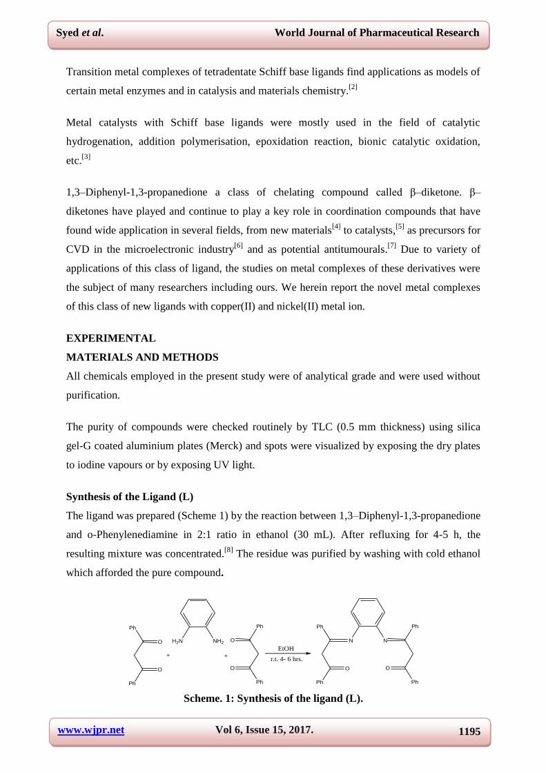
Synthesis of the Ligand (L)
The ligand was prepared (Scheme 1) by the reaction between 1,3–Diphenyl-1,3-propanedione
and o-Phenylenediamine in 2:1 ratio in ethanol (30 mL). After refluxing for 4-5 h, the
resulting mixture was concentrated.[8]
The residue was purified by washing with cold ethanol
which afforded the pure compound.
H2N NH2
++
O
O
Ph
Ph
O
O
Ph
Ph
EtOH
r.t. 4- 6 hrs.
N N
O
Ph
Ph
O
Ph
Ph
Scheme. 1: Synthesis of the ligand (L).
www.wjpr.net Vol 6, Issue 15, 2017. 1196
Syed et al. World Journal of Pharmaceutical Research
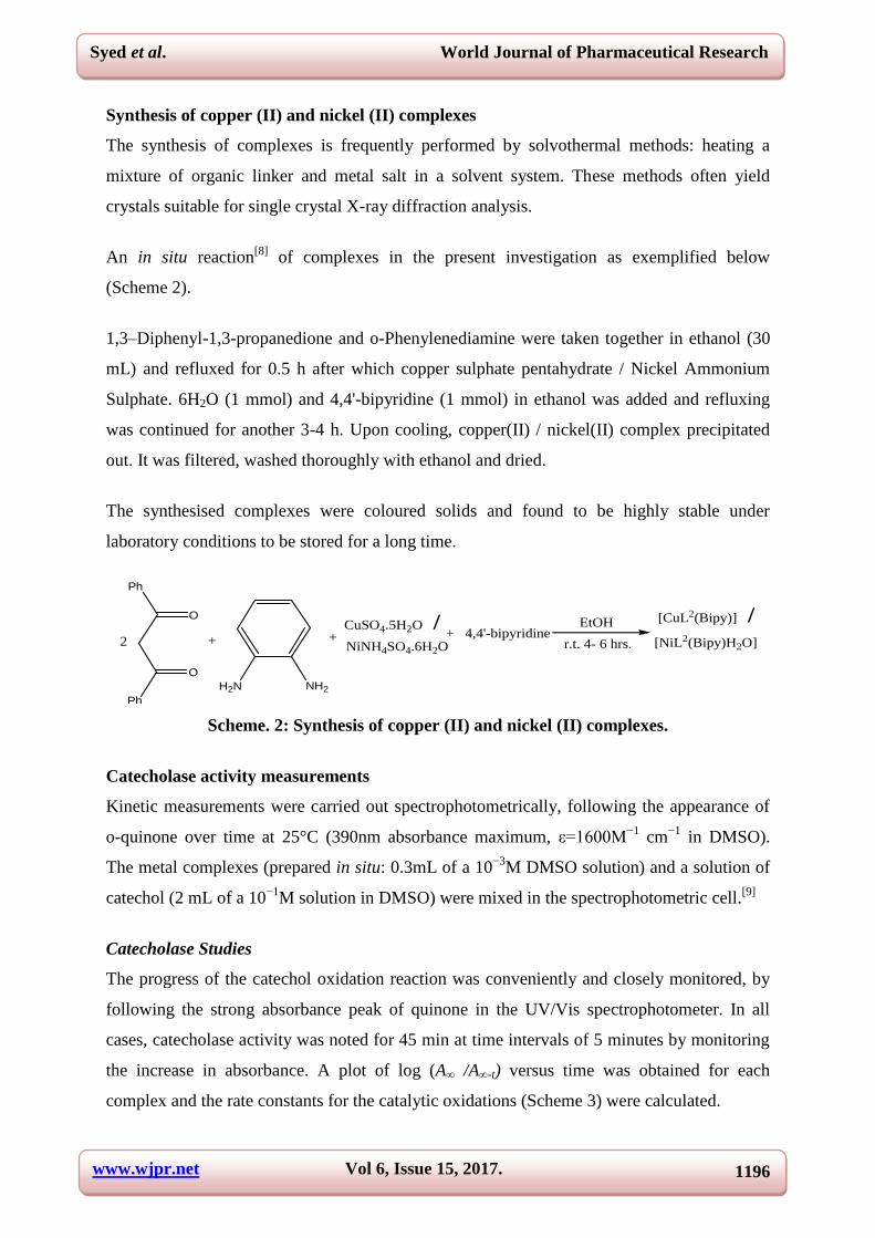
Synthesis of copper (II) and nickel (II) complexes
The synthesis of complexes is frequently performed by solvothermal methods: heating a
mixture of organic linker and metal salt in a solvent system. These methods often yield
crystals suitable for single crystal X-ray diffraction analysis.
An in situ reaction[8]
of complexes in the present investigation as exemplified below
(Scheme 2).
1,3–Diphenyl-1,3-propanedione and o-Phenylenediamine were taken together in ethanol (30
mL) and refluxed for 0.5 h after which copper sulphate pentahydrate / Nickel Ammonium
Sulphate. 6H2O (1 mmol) and 4,4'-bipyridine (1 mmol) in ethanol was added and refluxing
was continued for another 3-4 h. Upon cooling, copper(II) / nickel(II) complex precipitated
out. It was filtered, washed thoroughly with ethanol and dried.
The synthesised complexes were coloured solids and found to be highly stable under
laboratory conditions to be stored for a long time.
H2N NH2
++
O
O
Ph
Ph
EtOH
r.t. 4- 6 hrs.
CuSO4.5H2O /NiNH4SO4.6H2O
[CuL2(Bipy)] /
[NiL2(Bipy)H2O]+ 4,4'-bipyridine
2
Scheme. 2: Synthesis of copper (II) and nickel (II) complexes.
Catecholase activity measurements
Kinetic measurements were carried out spectrophotometrically, following the appearance of
o-quinone over time at 25°C (390nm absorbance maximum, ε=1600M−1
cm−1
in DMSO).
The metal complexes (prepared in situ: 0.3mL of a 10−3
M DMSO solution) and a solution of
catechol (2 mL of a 10−1
M solution in DMSO) were mixed in the spectrophotometric cell.[9]
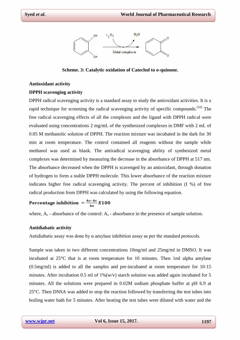
Catecholase Studies
The progress of the catechol oxidation reaction was conveniently and closely monitored, by
following the strong absorbance peak of quinone in the UV/Vis spectrophotometer. In all
cases, catecholase activity was noted for 45 min at time intervals of 5 minutes by monitoring
the increase in absorbance. A plot of log (A∞ /A∞-t) versus time was obtained for each
complex and the rate constants for the catalytic oxidations (Scheme 3) were calculated.
www.wjpr.net Vol 6, Issue 15, 2017. 1197
Syed et al. World Journal of Pharmaceutical Research
Scheme. 3: Catalytic oxidation of Catechol to o-quinone.
Antioxidant activity
DPPH scavenging activity
DPPH radical scavenging activity is a standard assay to study the antioxidant activities. It is a
rapid technique for screening the radical scavenging activity of specific compounds.[10]
The
free radical scavenging effects of all the complexes and the ligand with DPPH radical were
evaluated using concentrations 2 mg/mL of the synthesized complexes in DMF with 2 mL of
0.05 M methanolic solution of DPPH. The reaction mixture was incubated in the dark for 30
min at room temperature. The control contained all reagents without the sample while
methanol was used as blank. The antiradical scavenging ability of synthesized metal
complexes was determined by measuring the decrease in the absorbance of DPPH at 517 nm.
The absorbance decreased when the DPPH is scavenged by an antioxidant, through donation
of hydrogen to form a stable DPPH molecule. This lower absorbance of the reaction mixture
indicates higher free radical scavenging activity. The percent of inhibition (I %) of free
radical production from DPPH was calculated by using the following equation.
where, Ac - absorbance of the control: As - absorbance in the presence of sample solution.
Antidiabatic activity
Antidiabatic assay was done by α amylase inhibition assay as per the standard protocols.
Sample was taken in two different concentrations 10mg/ml and 25mg/ml in DMSO. It was
incubated at 25°C that is at room temperature for 10 minutes. Then 1ml alpha amylase
(0.5mg/ml) is added to all the samples and pre-incubated at room temperature for 10-15
minutes. After incubation 0.5 ml of 1%(w/v) starch solution was added again incubated for 5
minutes. All the solutions were prepared in 0.02M sodium phosphate buffer at pH 6.9 at
25°C. Then DNSA was added to stop the reaction followed by transferring the test tubes into
boiling water bath for 5 minutes. After heating the test tubes were diluted with water and the
www.wjpr.net Vol 6, Issue 15, 2017. 1198
Syed et al. World Journal of Pharmaceutical Research
total volume was made up to 10 ml to dilute the solutions and then the optical density of
absorbance values was taken at 540nm in a Shimazdu UV-Vis spectrophotometer.[11,12]
Total of three controls were used in the assay one is the total blank having nothing only
buffer and DNSA, the other was enzyme blank where leaving enzyme (α amylase) everything
was present and lastly the substrate blank having everything except substrate (starch). Total
blank was used for calibrating the instrument and the other two blanks were used as controls
to nullify the effect of both enzyme and substrate.
Calculations
Actual O.D was used to calculate the results.
Actual O.D. = O.D of sample - (Enzyme blank + substrate blank)
%inhibition= ((Actual O.D.)*100) / (Enzyme blank+ Substrate blank)
In vitro antibacterial activity
Materials and Method
To test the in-vitro anti-bacterial activity of the given complexes and ligands, well diffusion
method was followed as stated in Kirby Bauer Protocol. Bacillus subtilis (MTCC1168) and
Escherichia coli (MTCC1886) were the pathogens used for testing based on their clinical
importance in accordance with reference.[13]
DMSO was used due to its high solvency. The
stock cultures were stored at 4°C.
Antibacterial Susceptibility Test
The in vitro analysis of the culture was done by the agar well diffusion method using Muller
Hinton Agar, as used in reference.[14]
The bacterial strains E.coli (gram negative bacilli) and
B. subtilis (gram positive bacilli) have been used. The anti-bacterial activity was carried out
by inoculating the strains by spread plate method using sterile cotton swabs. Standard wells
were created by using sterilized borers in each plate. A concentration of 1mg in 1ml was
prepared from the complexes and ligands and Ciprofloxacin was used as the positive control,
as used in reference.[15]
100µl was pipetted into each of wells. Duplicates were performed for
both complexes and ligands to achieve standardized results. The zone of inhibition was
observed and measured after 18-24 hours of incubation at 37°C. The effectiveness of the
complexes and ligand were measured through the diameters (zone of inhibition). This
procedure was performed in three replicate plates for each organism.
www.wjpr.net Vol 6, Issue 15, 2017. 1199
Syed et al. World Journal of Pharmaceutical Research
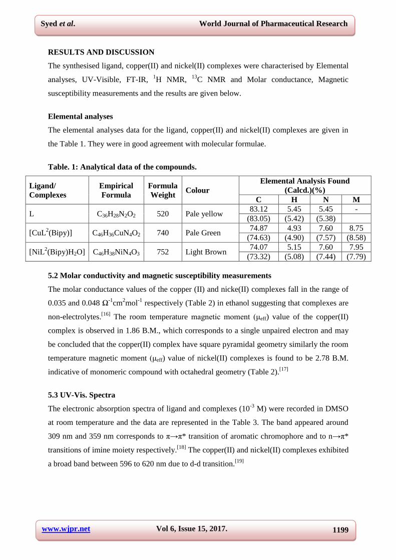
RESULTS AND DISCUSSION
The synthesised ligand, copper(II) and nickel(II) complexes were characterised by Elemental
analyses, UV-Visible, FT-IR, 1H NMR,
13C NMR and Molar conductance, Magnetic
susceptibility measurements and the results are given below.
Elemental analyses
The elemental analyses data for the ligand, copper(II) and nickel(II) complexes are given in
the Table 1. They were in good agreement with molecular formulae.
Table. 1: Analytical data of the compounds.
Ligand/
Complexes
Empirical
Formula
Formula
Weight Colour
Elemental Analysis Found
(Calcd.)(%)
C H N M
L C36H28N2O2 520 Pale yellow 83.12 5.45 5.45 -
(83.05) (5.42) (5.38)
[CuL2(Bipy)] C46H36CuN4O2 740 Pale Green
74.87 4.93 7.60 8.75
(74.63) (4.90) (7.57) (8.58)
[NiL2(Bipy)H2O] C46H38NiN4O3 752 Light Brown
74.07 5.15 7.60 7.95
(73.32) (5.08) (7.44) (7.79)
5.2 Molar conductivity and magnetic susceptibility measurements
The molar conductance values of the copper (II) and nicke(II) complexes fall in the range of
0.035 and 0.048 Ω-1
cm2mol
-1 respectively (Table 2) in ethanol suggesting that complexes are
non-electrolytes.[16]
The room temperature magnetic moment (μeff) value of the copper(II)
complex is observed in 1.86 B.M., which corresponds to a single unpaired electron and may
be concluded that the copper(II) complex have square pyramidal geometry similarly the room
temperature magnetic moment (μeff) value of nickel(II) complexes is found to be 2.78 B.M.
indicative of monomeric compound with octahedral geometry (Table 2).[17]
5.3 UV-Vis. Spectra
The electronic absorption spectra of ligand and complexes (10-3
M) were recorded in DMSO
at room temperature and the data are represented in the Table 3. The band appeared around
309 nm and 359 nm corresponds to π→π* transition of aromatic chromophore and to n→π*
transitions of imine moiety respectively.[18]
The copper(II) and nickel(II) complexes exhibited
a broad band between 596 to 620 nm due to d-d transition.[19]
www.wjpr.net Vol 6, Issue 15, 2017. 1200
Syed et al. World Journal of Pharmaceutical Research
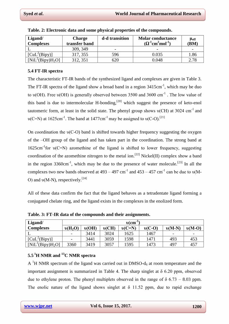
Table. 2: Electronic data and some physical properties of the compounds.
Ligand/
Complexes
Charge
transfer band
d-d transition Molar conductance
(Ω-1
cm2mol
-1)
μeff
(BM)
L 309, 349 - - -
[CuL2(Bipy)] 317, 355 596 0.035 1.86
[NiL2(Bipy)H2O] 312, 351 620 0.048 2.78
5.4 FT-IR spectra
The characteristic FT-IR bands of the synthesized ligand and complexes are given in Table 3.
The FT-IR spectra of the ligand show a broad band in a region 3415cm-1
, which may be duo
to υ(OH). Free υ(OH) is generally observed between 3500 and 3600 cm-1
. The low value of
this band is due to intermolecular H-bonding,[20]
which suggest the presence of keto-enol
tautomeric form, at least in the solid state. The phenyl group shows υ(CH) at 3024 cm-1
and
υ(C=N) at 1625cm-1
. The band at 1477cm-1
may be assigned to υ(C-O).[21]
On coordination the υ(C-O) band is shifted towards higher frequency suggesting the oxygen
of the –OH group of the ligand and has taken part in the coordination. The strong band at
1625cm-1
for υ(C=N) azomethine of the ligand is shifted to lower frequency, suggesting
coordination of the azomethine nitrogen to the metal ion.[22]
Nickel(II) complex show a band
in the region 3360cm-1
, which may be due to the presence of water molecule.[23]
In all the
complexes two new bands observed at 493 – 497 cm-1
and 453 – 457 cm-1
can be due to υ(M-
O) and υ(M-N), respectively.[24]
All of these data confirm the fact that the ligand behaves as a tetradentate ligand forming a
conjugated chelate ring, and the ligand exists in the complexes in the enolized form.
Table. 3: FT-IR data of the compounds and their assignments.
Ligand/
Complexes
υ(cm-1
)
υ(H2O) υ(OH) υ(CH) υ(C=N) υ(C-O) υ(M-N) υ(M-O)
L - 3414 3024 1625 1467 - -
[CuL2(Bipy)] - 3441 3059 1598 1471 493 453
[NiL2(Bipy)H2O] 3360 3419 3057 1595 1473 497 457
5.5 1
H NMR and 13
C NMR spectra
A 1H NMR spectrum of the ligand was carried out in DMSO-d6 at room temperature and the
important assignment is summarized in Table 4. The sharp singlet at δ 6.20 ppm, observed
due to ethylene proton. The phenyl multiplets observed in the range of δ 6.73 – 8.03 ppm.
The enolic nature of the ligand shows singlet at δ 11.52 ppm, due to rapid exchange
www.wjpr.net Vol 6, Issue 15, 2017. 1201
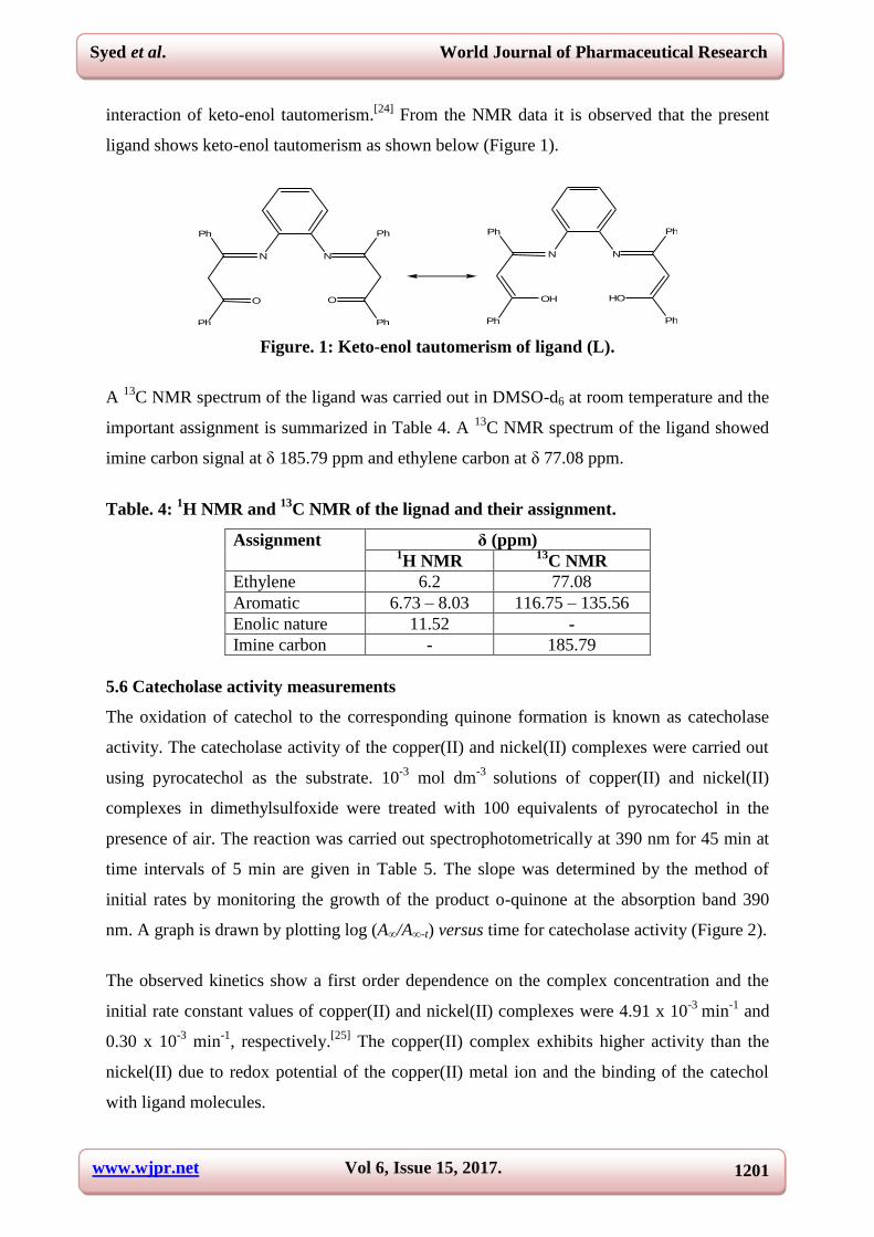
Syed et al. World Journal of Pharmaceutical Research
interaction of keto-enol tautomerism.[24]
From the NMR data it is observed that the present
ligand shows keto-enol tautomerism as shown below (Figure 1).
N N
O
Ph
Ph
O
Ph
Ph
N N
OH
Ph
Ph
HO
Ph
Ph
Figure. 1: Keto-enol tautomerism of ligand (L).
A 13
C NMR spectrum of the ligand was carried out in DMSO-d6 at room temperature and the
important assignment is summarized in Table 4. A 13
C NMR spectrum of the ligand showed
imine carbon signal at δ 185.79 ppm and ethylene carbon at δ 77.08 ppm.
Table. 4: 1H NMR and
13C NMR of the lignad and their assignment.
Assignment δ (ppm) 1H NMR
13C NMR
Ethylene 6.2 77.08
Aromatic 6.73 – 8.03 116.75 – 135.56
Enolic nature 11.52 -
Imine carbon - 185.79
5.6 Catecholase activity measurements
The oxidation of catechol to the corresponding quinone formation is known as catecholase
activity. The catecholase activity of the copper(II) and nickel(II) complexes were carried out
using pyrocatechol as the substrate. 10-3
mol dm-3
solutions of copper(II) and nickel(II)
complexes in dimethylsulfoxide were treated with 100 equivalents of pyrocatechol in the
presence of air. The reaction was carried out spectrophotometrically at 390 nm for 45 min at
time intervals of 5 min are given in Table 5. The slope was determined by the method of
initial rates by monitoring the growth of the product o-quinone at the absorption band 390
nm. A graph is drawn by plotting log (A∞/A∞-t) versus time for catecholase activity (Figure 2).
The observed kinetics show a first order dependence on the complex concentration and the
initial rate constant values of copper(II) and nickel(II) complexes were 4.91 x 10⁻³
min-1
and
0.30 x 10⁻³
min-1
, respectively.[25]
The copper(II) complex exhibits higher activity than the
nickel(II) due to redox potential of the copper(II) metal ion and the binding of the catechol
with ligand molecules.

www.wjpr.net Vol 6, Issue 15, 2017. 1202
Syed et al. World Journal of Pharmaceutical Research
Table. 5: Catecholase activity of copper(II) and nickel(II) complexes.
Time (min) log (A∞ /A∞-t )
[CuL2(Bipy)] [NiL
2(Bipy)H2O]
0 0 0
5 0.009 0.0005
10 0.017 0.0011
15 0.029 0.0020
20 0.046 0.0028
25 0.058 0.0035
30 0.069 0.0043
35 0.081 0.0052
40 0.092 0.0057
45 0.101 0.0063
0 10 20 30 40 50
0.00
0.02
0.04
0.06
0.08
0.10
log
(A /
A - t
)
Time (min)
Cu(II)
Ni(II)
Figure. 2: Catecholase activity of complexes.
5.7 Antioxidant activity
An antioxidant can be defined as any substance that when present at low concentrations,
compared with those of the oxidizable substrate, significantly delays or inhibits oxidation of
that substrate. To study the in vitro antioxidant activity of the ligand and complexes the
DPPH method was adopted. The results revealed all the synthesized compounds showed
negative antioxidant activity. The DPPH scavenging activity of standard antioxidant α-
tocopherol was also assayed for comparison.[26]
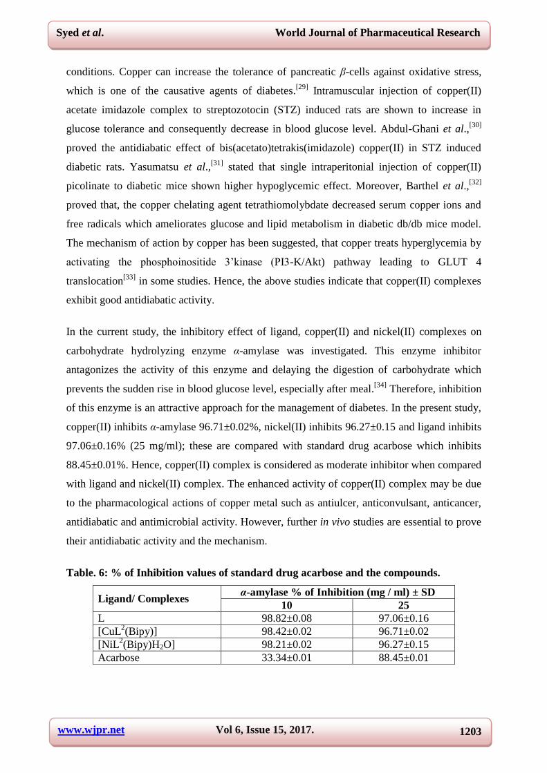
5.8 In vitro antidiabatic activity: The percentage inhibition of α-amylase exhibited by the
ligand and complexes are depicted in Table 6 and are compared with standard drug acarbose.
Many researchers have focused on in vivo antidiabatic activities of copper complexes.[27]
Walter et al.,[28]
hypothesized that copper metabolism encounters the diabetic pathological
www.wjpr.net Vol 6, Issue 15, 2017. 1203
Syed et al. World Journal of Pharmaceutical Research
conditions. Copper can increase the tolerance of pancreatic β-cells against oxidative stress,
which is one of the causative agents of diabetes.[29]
Intramuscular injection of copper(II)
acetate imidazole complex to streptozotocin (STZ) induced rats are shown to increase in
glucose tolerance and consequently decrease in blood glucose level. Abdul-Ghani et al.,[30]
proved the antidiabatic effect of bis(acetato)tetrakis(imidazole) copper(II) in STZ induced
diabetic rats. Yasumatsu et al.,[31]
stated that single intraperitonial injection of copper(II)
picolinate to diabetic mice shown higher hypoglycemic effect. Moreover, Barthel et al.,[32]
proved that, the copper chelating agent tetrathiomolybdate decreased serum copper ions and
free radicals which ameliorates glucose and lipid metabolism in diabetic db/db mice model.
The mechanism of action by copper has been suggested, that copper treats hyperglycemia by
activating the phosphoinositide 3’kinase (PI3-K/Akt) pathway leading to GLUT 4
translocation[33]
in some studies. Hence, the above studies indicate that copper(II) complexes
exhibit good antidiabatic activity.
In the current study, the inhibitory effect of ligand, copper(II) and nickel(II) complexes on
carbohydrate hydrolyzing enzyme α-amylase was investigated. This enzyme inhibitor
antagonizes the activity of this enzyme and delaying the digestion of carbohydrate which
prevents the sudden rise in blood glucose level, especially after meal.[34]
Therefore, inhibition
of this enzyme is an attractive approach for the management of diabetes. In the present study,
copper(II) inhibits α-amylase 96.71±0.02%, nickel(II) inhibits 96.27±0.15 and ligand inhibits
97.06±0.16% (25 mg/ml); these are compared with standard drug acarbose which inhibits
88.45±0.01%. Hence, copper(II) complex is considered as moderate inhibitor when compared
with ligand and nickel(II) complex. The enhanced activity of copper(II) complex may be due
to the pharmacological actions of copper metal such as antiulcer, anticonvulsant, anticancer,
antidiabatic and antimicrobial activity. However, further in vivo studies are essential to prove
their antidiabatic activity and the mechanism.
Table. 6: % of Inhibition values of standard drug acarbose and the compounds.
Ligand/ Complexes α-amylase % of Inhibition (mg / ml) ± SD
10 25
L 98.82±0.08 97.06±0.16
[CuL2(Bipy)] 98.42±0.02 96.71±0.02
[NiL2(Bipy)H2O] 98.21±0.02 96.27±0.15
Acarbose 33.34±0.01 88.45±0.01
www.wjpr.net Vol 6, Issue 15, 2017. 1204
Syed et al. World Journal of Pharmaceutical Research
Statistical analysis
The results were expressed as mean ± S.E.M. Statistical analysis of the data were carried out
using Student’s t-test and the results were considered significant when P < 0.05.[35]
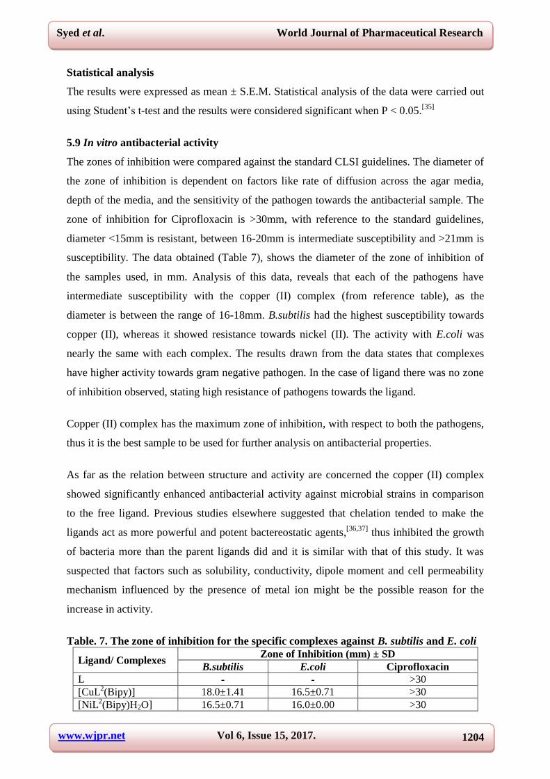
5.9 In vitro antibacterial activity
The zones of inhibition were compared against the standard CLSI guidelines. The diameter of
the zone of inhibition is dependent on factors like rate of diffusion across the agar media,
depth of the media, and the sensitivity of the pathogen towards the antibacterial sample. The
zone of inhibition for Ciprofloxacin is >30mm, with reference to the standard guidelines,
diameter <15mm is resistant, between 16-20mm is intermediate susceptibility and >21mm is
susceptibility. The data obtained (Table 7), shows the diameter of the zone of inhibition of
the samples used, in mm. Analysis of this data, reveals that each of the pathogens have
intermediate susceptibility with the copper (II) complex (from reference table), as the
diameter is between the range of 16-18mm. B.subtilis had the highest susceptibility towards
copper (II), whereas it showed resistance towards nickel (II). The activity with E.coli was
nearly the same with each complex. The results drawn from the data states that complexes
have higher activity towards gram negative pathogen. In the case of ligand there was no zone
of inhibition observed, stating high resistance of pathogens towards the ligand.
Copper (II) complex has the maximum zone of inhibition, with respect to both the pathogens,
thus it is the best sample to be used for further analysis on antibacterial properties.
As far as the relation between structure and activity are concerned the copper (II) complex
showed significantly enhanced antibacterial activity against microbial strains in comparison
to the free ligand. Previous studies elsewhere suggested that chelation tended to make the
ligands act as more powerful and potent bactereostatic agents,[36,37]
thus inhibited the growth
of bacteria more than the parent ligands did and it is similar with that of this study. It was
suspected that factors such as solubility, conductivity, dipole moment and cell permeability
mechanism influenced by the presence of metal ion might be the possible reason for the
increase in activity.
Table. 7. The zone of inhibition for the specific complexes against B. subtilis and E. coli
Ligand/ Complexes Zone of Inhibition (mm) ± SD
B.subtilis E.coli Ciprofloxacin
L - - >30
[CuL2(Bipy)] 18.0±1.41 16.5±0.71 >30
[NiL2(Bipy)H2O] 16.5±0.71 16.0±0.00 >30
www.wjpr.net Vol 6, Issue 15, 2017. 1205
Syed et al. World Journal of Pharmaceutical Research
6. CONCLUSION
In the present study tetradentate ligand and copper(II), nickel(II) complexes have been
synthesized using β - diketones with aromatic amines, and characterized by various physico-
chemical and spectral analyses. The molar conductance of all the complexes suggested their
nonelectrolytic nature. The coordinating mode of the donor atoms of the ligand was
confirmed by FT-IR spectra. Magnetic susceptibility measurements substantiate the
paramagnetic nature of all the complexes. Based on the studies square pyramidal geometry
has been proposed for the copper(II) complex and octahedral geometry has been proposed for
nickel(II) complex.
The catecholase activity of the copper(II) and nickel(II) complexes were carried out using
pyrocatechol as the substrate. The copper(II) complex exhibits higher activity than the
nickel(II) due to redox potential of the copper(II) metal ion and the binding of the catechol
with ligand molecules.
In vitro antidiabatic studies of the ligand and complexes were much higher when compared
with standard drug such as acarbose. Since the ligand and complexes showed significant
inhibiting activities, the current results provide a lead for the in vivo studies to establish the
possibility of these complexes as antidiabatic agent.
In vitro antibacterial and antioxidant studies were carried out. The results revealed that
among the ligand and complexes, complexes were found to exhibit very good antibacterial
activity against B.subtilis and E.coli.
Among the synthesized ligand and complexes copper(II) complex was found to have good
antidiabatic and antibacterial activity. The results revealed all the synthesized compounds
showed negative antioxidant activity. Based on the above results, the tentative structures of
the complexes can be formulated as shown in Figure 3 and 4.

www.wjpr.net Vol 6, Issue 15, 2017. 1206
Syed et al. World Journal of Pharmaceutical Research
NN
OH HO
Cu
N
N
Figure. 3: The tentative structure of the copper (II) complex.
NN
OH HO
Ni
N
N
OH2
Figure. 4: The tentative structure of the nickel (II) complex.
REFERENCES
1. (a) Xu Y, Yuan D, Wu B, Han L, Wu M, Jiang F, Hong M, Cryst. Growth Des, 2006; 6:
1168-1174;(b) Eddaoudi M, Moler D.B, Li H, Chen B, Reineke T.M, O’Keefee M, Yaghi
O.M, Acc. Chem. Res., 2001; 34: 319-330.
2. Garoufis A, Kasselouri S., Mitsopoulou C.A, Sletten J, Papadimitriou C and Hadjiliadis
N, Polyhedron, 1999; 18: 39.
3. Yan J, Li J.Z., Li K.B, Zhou B, Zeng W., Qin S.Y, Transit. Met. Chem, 2006; 31: 286;
Zhang Z.S, Yu X.M, Fong L.K, Margerum L.D, Inorg. Chim. Acta, 2001; 317: 72; Li J.Z,
Li H.B, Zhou B, Zeng W, Qin S.Y, Li S.X, Xie J.Q, Transit. Met. Chem. 2005; 30: 278;
Wang Y, Hu W, Li M.J., Jiang W.D., Hu C.W., Chem. Res. Appl, 2006; 18: 1393.
www.wjpr.net Vol 6, Issue 15, 2017. 1207
Syed et al. World Journal of Pharmaceutical Research
4. (a) de Sa’ GF, Malta OL, De Mello Donega’ C, Simas AM, Longo RL, Santa-Cruz PA, sa
Silva Jr EF, Coord Chem Rev., 2000; 196: 165; (b) McGehee MD, Bergstedt T, Zhang C,
Saab AP, O’Regan MB, Bazan GC, VI. Srdanov, Heeger AJ, Adv Mater, 1999; 11: 1349;
(c) Elbanowski M, Makowska B, Staninski K, Kaczmarek M, J Photochem Photobiol (A):
Chem, 2000; 130: 75; (d) Vicentini G, Zinner LB, Zukerman-Schpector J, Zinner K,
Coord Chem Rev., 2000; 196: 353.
5. (a) Dobrzynski P, Kasperczylk J, Bero M, Macromolecules, 1999; 32: 4735; (b) Bero M,
Kasperczylk J, Dobrzynski P, Polym Bull, 1999; 42: 131.
6. (a) Maverick A, Griffin GL 1994 In: TT. Kodas, MJ. Hampden-Smith (eds) The
chemistry of metal CVD. VCH, Weinheim (Chapter 4); (b) Hampden-Smith MJ, Kodas
TT, Polyhedron, 1995; 14: 699.
7. Keppler BK, Friesen C, Vongerichten H, Vogel E, In: Keppler BK (ed) Metal complexes
in cancer chemotherapy. VCH, Weinheim, 1993; 297–323.
8. Khlood S. Abou-Melha, Spectrochimica Acta Part A, 2008; 70: 162-170; Syed Tajudeen
S, Santhalakshmi S, Kannappan Geetha, Journal of Pharmacy Research, 2010; 3(11):
2759-2760; Syed Tajudeen S, Kannappan Geetha, Journal of Pharmacy Research, 2013;
7: 534-539; Syed Tajudeen S, Kannappan Geetha, Indian Journal of Advances in
Chemical Science, 2016; 4(1): 40-48.
9. Malachowski M. R, Davidson M. G, Inorg. Chim. Acta, 1989; 162: 199-204.
10. GökhnCeyhn; CumaliCelik; SerhanUrus; Ibrahim Demirtas; MahfuzElmastas; Mehmet
Tümer, Spectrochimica Acta Part A: Mol. Biomol. Spec., 2011; 81(1): 184-198; Manjula
SN, Kenganora M, Parihar VK, Kumar S, Nayak PG, Kumar N, Pai KSR, Rao CN,
Pharmaceutical Biology, 2010; 48(6): 690-696.
11. Bhutkar M. A and Bhise S. B, In vitro assay of alpha amylase inhibitory activity of some
indigenous plants.
12. Sindhu. S. Nair, Vaibhavi Kavrekar and Anshu Mishra, In vitro studies on alpha amylase
and alpha glucosidase inhibitory activities of selected plant extracts.
13. Nayan R. Bhalodia and Shukla V. J: Antibacterial and antifungal activities from leaf
extracts of cassia fistula l.: an ethno medicinal plant, 2011; 2(2): 104–109.
14. Multi-laboratory assessment of the linezolid spectrum of activity using the kirby-bauer
disk diffusion method: report of the zyvox® antimicrobial potency study (zaps) in the
United States Ronald n. Jonesa, et.al, 2001; 40: 59–66
15. Study of the antimicrobial activity of metal complexes and their ligands through
bioassays applied to plant extracts: Antonio f. Santos, et. al., 2014; 24(3).

www.wjpr.net Vol 6, Issue 15, 2017. 1208
Syed et al. World Journal of Pharmaceutical Research
16. Robert Boggess K, David Zatko A, J. Chem. Edn., 1975; 52(10): 649- 665.
17. Franco E, Lopez-Torres E, Mendiola M, Sevilla M, Polyhedron, 2000; 19: 441; Dipti
Shingnapurkar, et al., Bioorg. Med. Chem. Let., 2012; 22: 3172-3176; Uday Sandbhor, et
al., J. Inorg. Biochem., 2002; 90: 127-136; Banerjea D, Coordination Chemisty. Tata
McGraw-Hill Pub., New Delhi, 1998.
18. Dehad J, Jordanov F, Keck A, Mosset J.J, Bonnet J, J. Inorg. Chem., 1979; 18: 1543.
19. Kalia SB, Lumba K, Kaushal G, Sharma M, Int. J. Chem. Res., 2007; 46A: 1233-1239;
Atabay NMA, Dulger B, Gucin F, Eur. J. Med. Chem., 2005; 40: 1096-1102.
20. Reddy PM, Prasad AVSS, Shanker K, Ravinder V, Spectrochim. Acta. A., 2007; 68:
1000-1006; Silverstein R.M, Bassler G.C, Morrill T.C, Spectrometric identification of
Organic Compounds, 5th
ed., Wiley, New York, 1991.
21. Jadeja R. N, Shah J.R, Suresh E, Paul P, Polyhedron, 2004; 23: 2465.
22. Patel Y.M, Shah J.R, Indian J. Chem., 1985; 24A(9): 8000.
23. Kiran R. Surati, Thaker B.T., Spectrochim. Acta. A., 2010; 75: 235-242.
24. Nakamoto K, Infrared and Raman Spectra of Inorganic and Coordination Compounds,
5th Edition, A Wiley-Interscience Publication, New York, 1978.
25. Bhardwaj V. K, Aliaga-Alcalde N, Corbella M, Hundal G, Inorg. Chim. Acta, 2010;
363: 97.
26. Malik A, Kushnoor A, Saini V, Singhal S, Kumar S, Chand Yadav Y, J. Chem. Pharm.
Res., 2011; 3(3): 659-665.
27. Vanˇco J, Marek J, Travniˇck Z, Raˇcanska E, Muselik J, Švajlenova O, J. Inorg.
Biochem., 2008; 102: 595.
28. Walter J R M, Uriu-Hare J Y, Olin K L, Oster M H, Anawalt B D and Critchfield J W,
Diabetes Care, 1991; 14: 1050.
29. Kubisch H M, Wang J, Luche R, Carlson E, Bray T M and Epstein C J, Proc. Natl. Acad.
Sci., 1994; 91: 9956.
30. Abdul-Ghani A S, Abu-Hijleh A L, Nahas N and Amin R, Biol. Trac. Ele. Res., 1996; 54:
143.
31. Yasumatsu N, Yoshikawa Y, Adachi Y and Sakurai H, Bioorg. Med. Chem., 2007; 15:
4917.
32. Barthel A, Ostrakhovitch E.A, Walter P.L, Kampkotter A, Klotz L.O, Arch. Biochem.
Biophy., 2007; 463: 175.
33. Ostrakhovitch E.A, Cherian M.G, Arch. Biochem. Biophy. 2004; 423: 351.
34. Koyasu M, Ishili H, Watarai M, Takemoto K, Y. Inden, Clin. Theor., 2010; 32: 1610.
www.wjpr.net Vol 6, Issue 15, 2017. 1209
Syed et al. World Journal of Pharmaceutical Research
35. Sabu M.C, Ramadasan Kuttan, Anti-diabetic activity of medicinal plants and its
relationship with their antioxidant property; Perez R. M, Zavala M. A, S. Perez G. C.
Perez G, Antidiabatic effect of compounds isolated from plants.
36. Chohan Z.H, Praveen M., Appl. Organomet. Chem., 2001; 15: 617-625.
37. A. Chohan Z.H, Supran C.T, Scozzafava , J. Enz. Inhib. Med. Chem., 2004; 19: 79-84.