In vitro evaluation of the α-glucosidase inhibitory ...
9
RESEARCH ARTICLE Open Access In vitro evaluation of the α-glucosidase inhibitory potential of methanolic extracts of traditionally used antidiabetic plants Astha Bhatia 1 , Balbir Singh 2 , Rohit Arora 3 and Saroj Arora 1* Abstract Background: Different plant parts of Roylea cinerea (D. Don) Baill. (Lamiaceae), Clematis grata Wall. (Ranunculaceae), Cornus capitata Wall. (Cornaceae) are traditionally used in the management of diabetes and various other diseases. Method: The air-dried plant parts from different plants were coarsely powdered and macerated in methanol to obtain their crude extracts. The crude extracts were evaluated for their α-glucosidase inhibitory activity. On the basis of results obtained, the methanolic crude extract of Cornus capitata Wall. was further sequentially fractionated in hexane, diethyl ether, ethyl acetate, n-butanol. Fractions obtained were also evaluated for their α-glucosidase inhibitory potential. The kinetic study was performed using Lineweaver Burk plot to evaluate the type of inhibition. Furthermore, in silico analysis was also carried with active sites of the enzyme (PDB ID: 3WY1) using Autodock4. Results: Among all the plant extracts, Cornus capitata extract showed maximum inhibitory activity. Therefore its methanolic extract was further fractionated with the help of different solvents and the maximum activity was shown by the ethyl acetate fraction (IC 50 50 μg/mL). Kinetic analysis indicated that Vmax and Km were increased indicating a competitive type of inhibition. In docking studies, among different constituents known in this plant, betulinic acid showed minimum binding energy (- 10.21 kcal/mol). The kinetic and docking studies have strengthened the observation made in the present study regarding the α-glucosidase inhibitory activity of Cornus capitata. Conclusion: The study provided partial evidence for pharmacological basis regarding clinical applications of Cornus capitata in the treatment of diabetes suggesting it to be a suitable candidate for the treatment of postprandial hyperglycemia. Keywords: α-Glucosidase, Clematis grata wall., Cornus capitata wall., Roylea cinerea (D. Don) Baill Background Diabetes is the world’ s fastest-growing metabolic endo- crine disorder with compromised carbohydrate and lipid metabolism. The defected metabolism can be attributed to the impaired insulin secretion, insulin action or both. Alpha-glucosidase inhibitors are one of the effective class of antidiabetic therapeutics capable of ameliorating hyper-glycemia especially postprandial hyperglycemia over alpha-amylase inhibitors [1]. Postprandial hypergly- cemia is a sudden elevation in glucose levels in the blood after a meal. Major constituents of the human diet are comprised of carbohydrates such as starch and sucrose. The membrane-bound α-glucosidase enzyme, localized in the epithelium of small intestine hastens the digestion of oligosaccharides and disaccharides into simple glucose, after which it gets absorbed and enter into the bloodstream [2]. Inhibition of α-glucosidase enzyme can help in delaying digestion of carbohydrates, thereby re- ducing the levels of glucose in blood [3]. Several drugs are available as α-glucosidase inhibitors such as acarbose and miglitol that inhibit the absorption of carbohydrates from the gut. These inhibitors can prevent or mask the manifestation of the disease. There are several studies reported for such inhibitors being beneficial to prevent © The Author(s). 2019 Open Access This article is distributed under the terms of the Creative Commons Attribution 4.0 International License (http://creativecommons.org/licenses/by/4.0/), which permits unrestricted use, distribution, and reproduction in any medium, provided you give appropriate credit to the original author(s) and the source, provide a link to the Creative Commons license, and indicate if changes were made. The Creative Commons Public Domain Dedication waiver (http://creativecommons.org/publicdomain/zero/1.0/) applies to the data made available in this article, unless otherwise stated. * Correspondence: [email protected] 1 Department of Botanical and Environmental Sciences, Guru Nanak Dev University, Amritsar, Punjab 143005, India Full list of author information is available at the end of the article Bhatia et al. BMC Complementary and Alternative Medicine (2019) 19:74 https://doi.org/10.1186/s12906-019-2482-z
Transcript of In vitro evaluation of the α-glucosidase inhibitory ...
RESEARCH ARTICLE Open Access
In vitro evaluation of the α-glucosidaseinhibitory potential of methanolic extractsof traditionally used antidiabetic plantsAstha Bhatia1, Balbir Singh2, Rohit Arora3 and Saroj Arora1*
Abstract
Background: Different plant parts of Roylea cinerea (D. Don) Baill. (Lamiaceae), Clematis grata Wall. (Ranunculaceae),Cornus capitata Wall. (Cornaceae) are traditionally used in the management of diabetes and various other diseases.
Method: The air-dried plant parts from different plants were coarsely powdered and macerated in methanol toobtain their crude extracts. The crude extracts were evaluated for their α-glucosidase inhibitory activity. On thebasis of results obtained, the methanolic crude extract of Cornus capitata Wall. was further sequentially fractionatedin hexane, diethyl ether, ethyl acetate, n-butanol. Fractions obtained were also evaluated for their α-glucosidaseinhibitory potential. The kinetic study was performed using Lineweaver Burk plot to evaluate the type of inhibition.Furthermore, in silico analysis was also carried with active sites of the enzyme (PDB ID: 3WY1) using Autodock4.
Results: Among all the plant extracts, Cornus capitata extract showed maximum inhibitory activity. Therefore itsmethanolic extract was further fractionated with the help of different solvents and the maximum activity wasshown by the ethyl acetate fraction (IC50 50 μg/mL). Kinetic analysis indicated that Vmax and Km were increasedindicating a competitive type of inhibition. In docking studies, among different constituents known in this plant,betulinic acid showed minimum binding energy (− 10.21 kcal/mol). The kinetic and docking studies havestrengthened the observation made in the present study regarding the α-glucosidase inhibitory activity ofCornus capitata.
Conclusion: The study provided partial evidence for pharmacological basis regarding clinical applications ofCornus capitata in the treatment of diabetes suggesting it to be a suitable candidate for the treatmentof postprandial hyperglycemia.
Keywords: α-Glucosidase, Clematis grata wall., Cornus capitata wall., Roylea cinerea (D. Don) Baill
BackgroundDiabetes is the world’s fastest-growing metabolic endo-crine disorder with compromised carbohydrate and lipidmetabolism. The defected metabolism can be attributedto the impaired insulin secretion, insulin action or both.Alpha-glucosidase inhibitors are one of the effectiveclass of antidiabetic therapeutics capable of amelioratinghyper-glycemia especially postprandial hyperglycemiaover alpha-amylase inhibitors [1]. Postprandial hypergly-cemia is a sudden elevation in glucose levels in the blood
after a meal. Major constituents of the human diet arecomprised of carbohydrates such as starch and sucrose.The membrane-bound α-glucosidase enzyme, localizedin the epithelium of small intestine hastens the digestionof oligosaccharides and disaccharides into simpleglucose, after which it gets absorbed and enter into thebloodstream [2]. Inhibition of α-glucosidase enzyme canhelp in delaying digestion of carbohydrates, thereby re-ducing the levels of glucose in blood [3]. Several drugsare available as α-glucosidase inhibitors such as acarboseand miglitol that inhibit the absorption of carbohydratesfrom the gut. These inhibitors can prevent or mask themanifestation of the disease. There are several studiesreported for such inhibitors being beneficial to prevent
© The Author(s). 2019 Open Access This article is distributed under the terms of the Creative Commons Attribution 4.0International License (http://creativecommons.org/licenses/by/4.0/), which permits unrestricted use, distribution, andreproduction in any medium, provided you give appropriate credit to the original author(s) and the source, provide a link tothe Creative Commons license, and indicate if changes were made. The Creative Commons Public Domain Dedication waiver(http://creativecommons.org/publicdomain/zero/1.0/) applies to the data made available in this article, unless otherwise stated.
* Correspondence: [email protected] of Botanical and Environmental Sciences, Guru Nanak DevUniversity, Amritsar, Punjab 143005, IndiaFull list of author information is available at the end of the article
Bhatia et al. BMC Complementary and Alternative Medicine (2019) 19:74 https://doi.org/10.1186/s12906-019-2482-z
or delay impaired glucose tolerances in diabetes. Thoughbeing effective, these drugs have been reported to causesevere side effects such as gastrointestinal discomfortsincluding diarrhea and flatulence [4].Natural products of plant origin are known for their
dual role both as preventive and therapeutic agents,combined with their less toxicity and side effects.Traditionally different plants are used throughout theworld by the local population for the treatment ofvarious ailments [5]. Cornus capitata Wall. (Cornaceae)is widely available in different regions of the world suchas China and India. Studies have shown that it hasseveral constituents present in its aerial parts such ascornin and phlorin from leaves, others includen-hentriacontane, 7-hydrocadeline, triacontanol, stig-mastranone, lupeol, triacontanoic acid, betulin, epibetu-lin, betulinic acid, epibetulinic acid, tetracosanoic acid,maslinic acid, arjunolic acid, sitosterol-β-D-glucoside,gallic acid isolated from stem [6, 7]. It is used as fuel, foranimal bedding and its fruits are edible [8]. It has beenreported for its traditional use in the management ofdiabetes in Mandi district of Himachal Pradesh [9].Clematis grata Wall. (Ranunculaceae), is used by indi-genous people of Bana valley (Jammu and Kashmir) forits antioxidant, antimicrobial, and antidiabetic activities[10]. Its crushed leaves are used as a paste for the treat-ment of skin diseases such as eczema, warts, carbuncles[11] and boils [9]. Two important constituent viz.,clematoside-S and triterpenoids saponins have beenreported from the roots of Clematis grata Wall. [12].Roylea cinerea (D. Don) Baill. (Lamiaceae) found inWestern Himalayas had been used by the local popula-tion in the management of various diseases includingdiabetes, fever, skin diseases [9, 13]. Rutin, isoquercetin,nicotiflorin and martynoside are some constituents re-ported from its aerial parts [13]. Thus, the present studywas conducted with an objective to unravel theα-glucosidase inhibitory effect of Cornus capitata Wall.(leaves), Clematis grata Wall. (whole plant), Royleacinerea (D. Don) Baill.(leaves and stem) based on theirtraditional use for the management of diabetes [9, 10].Kinetic study and in silico analysis was also carried outto further investigate the mechanism of inhibition.
MethodsPlant extractionThe entire plant or its different parts can be used in theform of a whole plant, crude extract, and aqueous de-coctions. In the present study, the whole plant was se-lected in case of herbs, leaves and stem in case of shrubsand leaves in case of trees on the basis of availability ofphytoconstituents [14]. Plant materials viz., (Cornuscapitata Wall. (leaves), Clematis grata Wall. (Wholeplant), Roylea cinerea (D. Don) Baill. (Leaves and stem)
were collected, identified and submitted as a voucherspecimen (Accession no. 7373, 7374 and 7376 respect-ively) for authenticity in Herbarium, Department ofBotanical and Environmental Sciences, Guru Nanak DevUniversity, Amritsar, Punjab (Additional file 1: Table S1).The identification was done by taxonomist Dr. AmarjitSingh Soodan and Mr. Ram Prasad (Senior lab techni-cian), of Department of Botanical and EnvironmentalSciences, Guru Nanak Dev University, Amritsar, Punjab.Each plant material was air dried, coarsely powderedand subjected to maceration for 48 h in methanol. Themethanolic extracts were further concentrated with a ro-tary evaporator (IKA® RV 10). The extracts were storedat − 20 °C until use. Crude methanolic extract wasdissolved in distilled water. Further, an equal volume ofHexane was added and the mixture was shaken vigor-ously. The mixture was allowed to separate into layers.The upper layer (Hexane) was collected and concen-trated on rotary evaporator to obtain Hexane fraction.Similarly, the aqueous layer was further subjected tofractionation via series of solvents on the basis ofpolarity viz., Diethyl-ether<Ethyl acetate<n-butanol.Polar solvents help in the extraction of hydrophiliccompounds and non-polar solvents aids in the extrac-tion of lipophilic compounds [15, 16].
α-Glucosidase inhibitory assayPlant extracts were evaluated for α-glucosidase inhibi-tory activity according to the method given by PistiaBrueggeman and Hollingsworth, 2001 [17] with slightmodifications. Plant extracts (50 μL) at varying concen-trations (12.5 to 400 μg/mL) was incubated with 10 μLof the α-glucosidase (maltase) ex. Yeast (Sisco ResearchLaboratories Pvt. Ltd.) enzyme solution (1 U/mL) for 20min at 37 °C with an additional 125 μL of 0.1Mphosphate buffer (pH 6.8). After 20 min, the reactionwas started with the addition of 20 μL of 1M pNPG(substrate) and the mixture was incubated for 30 min.The reaction was terminated with the addition of 0.1 Nof Na2CO3 (50 μL) and final absorbance was measuredat 405 nm using Biotek multi-well plate reader. Acarbosewas used as a positive control at varying concentrations(12.5 to 400 μg/mL). Enzyme activity was calculated as:
(ODBLANK-ODSAMPLE)/ODBLANK x 100One unit of the enzyme can be defined as the amount ofenzyme (α-glucosidase) required for the formation ofone μmol of product (p-Nitrophenol) from the substrate(p-nitrophenyl-α-D-glucopyranoside) per minute. IC50
(concentration required to inhibit 50% of the enzymeactivity) was calculated using regression equationobtained through plotting concentration in the range12.5–400 μg/mL (x-axis) and %inhibition (y-axis) fordifferent extracts and fractions.
Bhatia et al. BMC Complementary and Alternative Medicine (2019) 19:74 Page 2 of 9
Kinetic studyLineweaver-Burk plot was plotted for methanolic extractof Cornus capitata Wall. with IC50 of 12.45 μg/mL. Thesubstrate p-nitrophenyl-α-D-glucoside was used invarying concentrations (2 mM, 1 mM, 0.5 mM, 0.25 mMand 0.125 mM). Enzyme concentration was keptconstant (1 U/mL) and molar extinction coefficient forcalculating enzyme activity was 17.4 mM− 1 [3].Inhibition type was determined with Lineweaver-Burkplots and calculated using the Michaelis Mentenequation.
Vo ¼ Vmax S½ �Kmþ S½ � ð1Þ
Rearranging the equation for Line-weaver Burk plot(linear, y = ax+b).
1Vo
¼ Kmþ S½ �Vmax S½ � ¼
KmVmax
1S½ � þ
1Vmax
ð2Þ
Where V0 is the initial rate of reaction, Km is theMichaelis Menten constant, Km/Vmax is the slope, andthe y-intercept (1/Vmax) was calculated using Eq. 2.
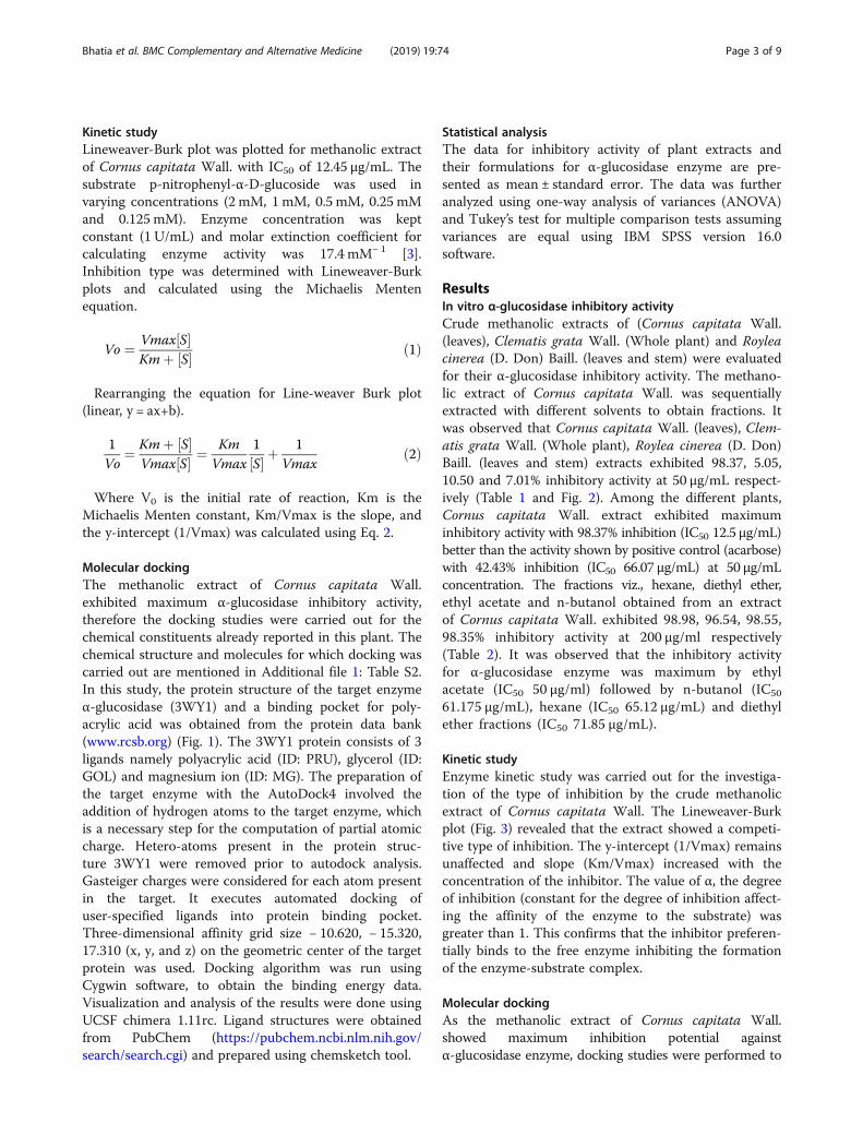
Molecular dockingThe methanolic extract of Cornus capitata Wall.exhibited maximum α-glucosidase inhibitory activity,therefore the docking studies were carried out for thechemical constituents already reported in this plant. Thechemical structure and molecules for which docking wascarried out are mentioned in Additional file 1: Table S2.In this study, the protein structure of the target enzymeα-glucosidase (3WY1) and a binding pocket for poly-acrylic acid was obtained from the protein data bank(www.rcsb.org) (Fig. 1). The 3WY1 protein consists of 3ligands namely polyacrylic acid (ID: PRU), glycerol (ID:GOL) and magnesium ion (ID: MG). The preparation ofthe target enzyme with the AutoDock4 involved theaddition of hydrogen atoms to the target enzyme, whichis a necessary step for the computation of partial atomiccharge. Hetero-atoms present in the protein struc-ture 3WY1 were removed prior to autodock analysis.Gasteiger charges were considered for each atom presentin the target. It executes automated docking ofuser-specified ligands into protein binding pocket.Three-dimensional affinity grid size − 10.620, − 15.320,17.310 (x, y, and z) on the geometric center of the targetprotein was used. Docking algorithm was run usingCygwin software, to obtain the binding energy data.Visualization and analysis of the results were done usingUCSF chimera 1.11rc. Ligand structures were obtainedfrom PubChem (https://pubchem.ncbi.nlm.nih.gov/search/search.cgi) and prepared using chemsketch tool.
Statistical analysisThe data for inhibitory activity of plant extracts andtheir formulations for α-glucosidase enzyme are pre-sented as mean ± standard error. The data was furtheranalyzed using one-way analysis of variances (ANOVA)and Tukey’s test for multiple comparison tests assumingvariances are equal using IBM SPSS version 16.0software.
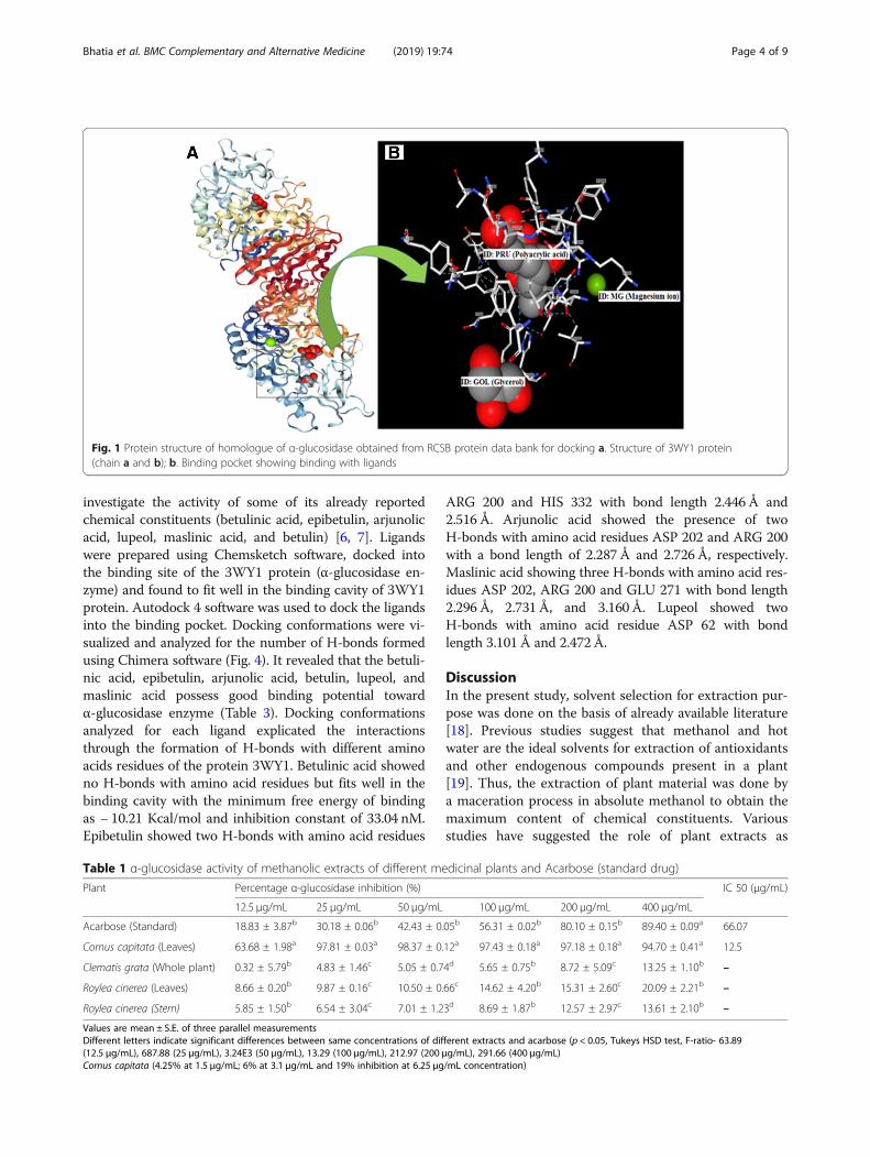
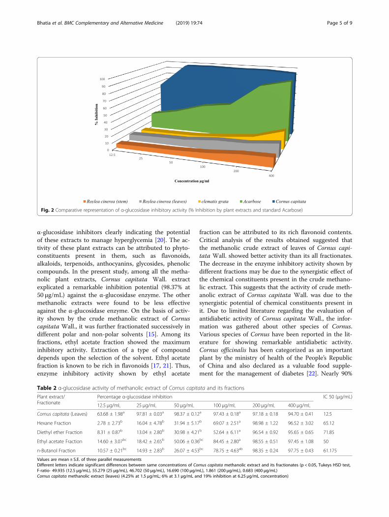
ResultsIn vitro α-glucosidase inhibitory activityCrude methanolic extracts of (Cornus capitata Wall.(leaves), Clematis grata Wall. (Whole plant) and Royleacinerea (D. Don) Baill. (leaves and stem) were evaluatedfor their α-glucosidase inhibitory activity. The methano-lic extract of Cornus capitata Wall. was sequentiallyextracted with different solvents to obtain fractions. Itwas observed that Cornus capitata Wall. (leaves), Clem-atis grata Wall. (Whole plant), Roylea cinerea (D. Don)Baill. (leaves and stem) extracts exhibited 98.37, 5.05,10.50 and 7.01% inhibitory activity at 50 μg/mL respect-ively (Table 1 and Fig. 2). Among the different plants,Cornus capitata Wall. extract exhibited maximuminhibitory activity with 98.37% inhibition (IC50 12.5 μg/mL)better than the activity shown by positive control (acarbose)with 42.43% inhibition (IC50 66.07 μg/mL) at 50 μg/mLconcentration. The fractions viz., hexane, diethyl ether,ethyl acetate and n-butanol obtained from an extractof Cornus capitata Wall. exhibited 98.98, 96.54, 98.55,98.35% inhibitory activity at 200 μg/ml respectively(Table 2). It was observed that the inhibitory activityfor α-glucosidase enzyme was maximum by ethylacetate (IC50 50 μg/ml) followed by n-butanol (IC50
61.175 μg/mL), hexane (IC50 65.12 μg/mL) and diethylether fractions (IC50 71.85 μg/mL).
Kinetic studyEnzyme kinetic study was carried out for the investiga-tion of the type of inhibition by the crude methanolicextract of Cornus capitata Wall. The Lineweaver-Burkplot (Fig. 3) revealed that the extract showed a competi-tive type of inhibition. The y-intercept (1/Vmax) remainsunaffected and slope (Km/Vmax) increased with theconcentration of the inhibitor. The value of α, the degreeof inhibition (constant for the degree of inhibition affect-ing the affinity of the enzyme to the substrate) wasgreater than 1. This confirms that the inhibitor preferen-tially binds to the free enzyme inhibiting the formationof the enzyme-substrate complex.
Molecular dockingAs the methanolic extract of Cornus capitata Wall.showed maximum inhibition potential againstα-glucosidase enzyme, docking studies were performed to
Bhatia et al. BMC Complementary and Alternative Medicine (2019) 19:74 Page 3 of 9
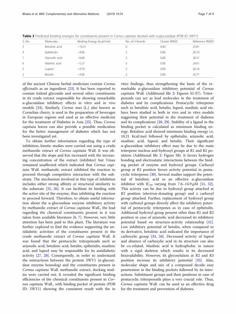
investigate the activity of some of its already reportedchemical constituents (betulinic acid, epibetulin, arjunolicacid, lupeol, maslinic acid, and betulin) [6, 7]. Ligandswere prepared using Chemsketch software, docked intothe binding site of the 3WY1 protein (α-glucosidase en-zyme) and found to fit well in the binding cavity of 3WY1protein. Autodock 4 software was used to dock the ligandsinto the binding pocket. Docking conformations were vi-sualized and analyzed for the number of H-bonds formedusing Chimera software (Fig. 4). It revealed that the betuli-nic acid, epibetulin, arjunolic acid, betulin, lupeol, andmaslinic acid possess good binding potential towardα-glucosidase enzyme (Table 3). Docking conformationsanalyzed for each ligand explicated the interactionsthrough the formation of H-bonds with different aminoacids residues of the protein 3WY1. Betulinic acid showedno H-bonds with amino acid residues but fits well in thebinding cavity with the minimum free energy of bindingas − 10.21 Kcal/mol and inhibition constant of 33.04 nM.Epibetulin showed two H-bonds with amino acid residues
ARG 200 and HIS 332 with bond length 2.446 Å and2.516 Å. Arjunolic acid showed the presence of twoH-bonds with amino acid residues ASP 202 and ARG 200with a bond length of 2.287 Å and 2.726 Å, respectively.Maslinic acid showing three H-bonds with amino acid res-idues ASP 202, ARG 200 and GLU 271 with bond length2.296 Å, 2.731 Å, and 3.160 Å. Lupeol showed twoH-bonds with amino acid residue ASP 62 with bondlength 3.101 Å and 2.472 Å.
DiscussionIn the present study, solvent selection for extraction pur-pose was done on the basis of already available literature[18]. Previous studies suggest that methanol and hotwater are the ideal solvents for extraction of antioxidantsand other endogenous compounds present in a plant[19]. Thus, the extraction of plant material was done bya maceration process in absolute methanol to obtain themaximum content of chemical constituents. Variousstudies have suggested the role of plant extracts as
Table 1 α-glucosidase activity of methanolic extracts of different medicinal plants and Acarbose (standard drug)
Plant Percentage α-glucosidase inhibition (%) IC 50 (μg/mL)
12.5 μg/mL 25 μg/mL 50 μg/mL 100 μg/mL 200 μg/mL 400 μg/mL
Acarbose (Standard) 18.83 ± 3.87b 30.18 ± 0.06b 42.43 ± 0.05b 56.31 ± 0.02b 80.10 ± 0.15b 89.40 ± 0.09a 66.07
Cornus capitata (Leaves) 63.68 ± 1.98a 97.81 ± 0.03a 98.37 ± 0.12a 97.43 ± 0.18a 97.18 ± 0.18a 94.70 ± 0.41a 12.5
Clematis grata (Whole plant) 0.32 ± 5.79b 4.83 ± 1.46c 5.05 ± 0.74d 5.65 ± 0.75b 8.72 ± 5.09c 13.25 ± 1.10b –
Roylea cinerea (Leaves) 8.66 ± 0.20b 9.87 ± 0.16c 10.50 ± 0.66c 14.62 ± 4.20b 15.31 ± 2.60c 20.09 ± 2.21b –
Roylea cinerea (Stem) 5.85 ± 1.50b 6.54 ± 3.04c 7.01 ± 1.23d 8.69 ± 1.87b 12.57 ± 2.97c 13.61 ± 2.10b –
Values are mean ± S.E. of three parallel measurementsDifferent letters indicate significant differences between same concentrations of different extracts and acarbose (p < 0.05, Tukeys HSD test, F-ratio- 63.89(12.5 μg/mL), 687.88 (25 μg/mL), 3.24E3 (50 μg/mL), 13.29 (100 μg/mL), 212.97 (200 μg/mL), 291.66 (400 μg/mL)Cornus capitata (4.25% at 1.5 μg/mL; 6% at 3.1 μg/mL and 19% inhibition at 6.25 μg/mL concentration)
Fig. 1 Protein structure of homologue of α-glucosidase obtained from RCSB protein data bank for docking a. Structure of 3WY1 protein(chain a and b); b. Binding pocket showing binding with ligands
Bhatia et al. BMC Complementary and Alternative Medicine (2019) 19:74 Page 4 of 9
α-glucosidase inhibitors clearly indicating the potentialof these extracts to manage hyperglycemia [20]. The ac-tivity of these plant extracts can be attributed to phyto-constituents present in them, such as flavonoids,alkaloids, terpenoids, anthocyanins, glycosides, phenoliccompounds. In the present study, among all the metha-nolic plant extracts, Cornus capitata Wall. extractexplicated a remarkable inhibition potential (98.37% at50 μg/mL) against the α-glucosidase enzyme. The othermethanolic extracts were found to be less effectiveagainst the α-glucosidase enzyme. On the basis of activ-ity shown by the crude methanolic extract of Cornuscapitata Wall., it was further fractionated successively indifferent polar and non-polar solvents [15]. Among itsfractions, ethyl acetate fraction showed the maximuminhibitory activity. Extraction of a type of compounddepends upon the selection of the solvent. Ethyl acetatefraction is known to be rich in flavonoids [17, 21]. Thus,enzyme inhibitory activity shown by ethyl acetate
fraction can be attributed to its rich flavonoid contents.Critical analysis of the results obtained suggested thatthe methanolic crude extract of leaves of Cornus capi-tata Wall. showed better activity than its all fractionates.The decrease in the enzyme inhibitory activity shown bydifferent fractions may be due to the synergistic effect ofthe chemical constituents present in the crude methano-lic extract. This suggests that the activity of crude meth-anolic extract of Cornus capitata Wall. was due to thesynergistic potential of chemical constituents present init. Due to limited literature regarding the evaluation ofantidiabetic activity of Cornus capitata Wall., the infor-mation was gathered about other species of Cornus.Various species of Cornus have been reported in the lit-erature for showing remarkable antidiabetic activity.Cornus officinalis has been categorized as an importantplant by the ministry of health of the People’s Republicof China and also declared as a valuable food supple-ment for the management of diabetes [22]. Nearly 90%
Fig. 2 Comparative representation of α-glucosidase inhibitory activity (% Inhibition by plant extracts and standard Acarbose)
Table 2 α-glucosidase activity of methanolic extract of Cornus capitata and its fractions
Plant extract/Fractionate
Percentage α-glucosidase inhibition IC 50 (μg/mL)
12.5 μg/mL 25 μg/mL 50 μg/mL 100 μg/mL 200 μg/mL 400 μg/mL
Cornus capitata (Leaves) 63.68 ± 1.98a 97.81 ± 0.03a 98.37 ± 0.12a 97.43 ± 0.18a 97.18 ± 0.18 94.70 ± 0.41 12.5
Hexane Fraction 2.78 ± 2.73b 16.04 ± 4.78b 31.94 ± 5.17b 69.07 ± 2.51a 98.98 ± 1.22 96.52 ± 3.02 65.12
Diethyl ether Fraction 8.31 ± 0.87b 13.04 ± 2.80b 30.98 ± 4.21b 52.64 ± 6.11a 96.54 ± 0.92 95.65 ± 0.65 71.85
Ethyl acetate Fraction 14.60 ± 3.07bc 18.42 ± 2.65b 50.06 ± 0.36bc 84.45 ± 2.80a 98.55 ± 0.51 97.45 ± 1.08 50
n-Butanol Fraction 10.57 ± 0.21bc 14.93 ± 2.83b 26.07 ± 4.53bc 78.75 ± 4.63ab 98.35 ± 0.24 97.75 ± 0.43 61.175
Values are mean ± S.E. of three parallel measurementsDifferent letters indicate significant differences between same concentrations of Cornus capitata methanolic extract and its fractionates (p < 0.05, Tukeys HSD test,F-ratio- 49.935 (12.5 μg/mL), 55.279 (25 μg/mL), 46.702 (50 μg/mL), 16.690 (100 μg/mL), 1.861 (200 μg/mL), 0.683 (400 μg/mL)Cornus capitata methanolic extract (leaves) (4.25% at 1.5 μg/mL; 6% at 3.1 μg/mL and 19% inhibition at 6.25 μg/mL concentration)
Bhatia et al. BMC Complementary and Alternative Medicine (2019) 19:74 Page 5 of 9
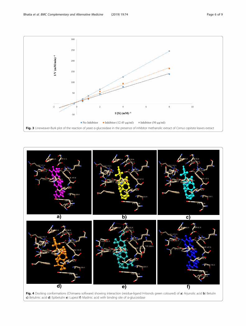
Fig. 3 Lineweaver-Burk plot of the reaction of yeast α-glucosidase in the presence of inhibitor methanolic extract of Cornus capitata leaves extract
Fig. 4 Docking conformations (Chimaera software) showing interaction (residue-ligand H-bonds green coloured) of a) Arjunolic acid b) Betulinc) Betulinic acid d) Epibetulin e) Lupeol f) Maslinic acid with binding site of α-glucosidase
Bhatia et al. BMC Complementary and Alternative Medicine (2019) 19:74 Page 6 of 9
of the ancient Chinese herbal medicines contain Cornusofficinalis as an ingredient [23]. It has been reported tocontain iridoid glycoside and several other constituentsin its crude extract responsible for showing remarkableα-glucosidase inhibitory effects in vitro and in vivomodels [24]. Similarly, Cornus mas (L.) also known asCornelian cherry, is used in the preparation of beveragesin European regions and used as an effective medicinefor the treatment of Diabetes in Asia [25]. Thus, Cornuscapitata leaves can also provide a possible medicationfor the better management of diabetes which has notbeen investigated yet.To obtain further information regarding the type of
inhibition, kinetic studies were carried out using a crudemethanolic extract of Cornus capitata Wall. It was ob-served that the slope and Km increased with the increas-ing concentration of the extract (inhibitor) but Vmaxremained unaffected which indicated that Cornus capi-tata Wall. methanolic extract inhibited the reaction toproceed through competitive interaction with the sub-strate. The mechanism involved in this type of inhibitionincludes either strong affinity or structural similarity tothe substrate [25, 26]. It can facilitate its binding withthe active site of the enzyme, thus inhibiting the reactionto proceed forward. Therefore, to obtain useful informa-tion about the α-glucosidase enzyme inhibitory activityby methanolic extract of Cornus capitata Wall., the leadregarding the chemical constituents present in it wastaken from available literature [6, 7]. However, very littleattention has been paid to this plant. The literature wasfurther explored to find the evidence supporting the an-tidiabetic activities of the constituents present in thecrude methanolic extract of Cornus capitata Wall. Itwas found that the pentacyclic triterpenoids such asarjunolic acid, betulinic acid, betulin, epibetulin, maslinicacid, and lupeol may be responsible for its antidiabeticactivity [27, 28]. Consequently, in order to understandthe interactions between the protein 3WY1 (α-glucosi-dase enzyme homolog) and the constituents present inCornus capitata Wall. methanolic extract, docking stud-ies were carried out. It revealed the significant bindingefficiencies of the chemical constituents present in Cor-nus capitata Wall., with binding pocket of protein (PDBID: 3WY1) showing the consistent result with the in
vitro findings, thus strengthening the basis of the re-markable α-glucosidase inhibitory potential of Cornuscapitata Wall. (Additional file 2: Figures S1-S7). Triter-penoids can act as lead molecules in the treatment ofdiabetes and its complications. Pentacyclic triterpenessuch as betulinic acid, betulin, lupeol, maslinic acid etc.have been studied in both in vivo and in vitro modelssuggesting their potential in the treatment of diabetesand its complications [28, 29]. Stability of a ligand in thebinding pocket is calculated as minimum binding en-ergy. Betulinic acid showed minimum binding energy i.e.10.21 Kcal/mol followed by epibetulin, arjunolic acid,maslinic acid, lupeol, and betulin. Their significantα-glucosidase inhibitory effect may be due to the maintriterpene nucleus and hydroxyl groups at R1 and R2 po-sitions (Additional file 2: Figure S8). It favors hydrogenbonding and electrostatic interactions between the bind-ing pocket of enzyme and hydroxyl groups. Carboxylgroup at R3 position favors activity potential in penta-cyclic triterpenes [30]. Several studies support the poten-tial of betulinic acid as an effective α-glucosidaseinhibitor with IC50 varying from 7.6–14.9 μM [31, 32].This activity can be due to hydroxyl group attached atR2 position (electron-donating group) and a carboxylgroup attached. Further, replacement of hydroxyl groupwith carbonyl groups directly affect the inhibitory poten-tial of pentacyclic triterpenes as in case of epibetulin.Additional hydroxyl group present other than R1 and R2position in case of arjunolic acid decreased its inhibitorypotential based on structure-activity relationship [31].Low inhibitory potential of betulin, when compared toits derivative, betulinic acid indicated the importance ofcarboxylic group [33, 34]. Decreased activity of lupeoland absence of carboxylic acid in its structure can alsobe co-related. Maslinic acid is hydrophobic in naturewith a rigid skeleton which results in its decreasedbioavailability. However, its glycosylation at R2 and R3position increase its inhibitory potential [35]. Also,molecular shape and size of a compound decide easypenetration in the binding pockets followed by its inter-actions. Substituent groups and their positions in case ofpentacyclic triterpenoids plays a very crucial role. Thus,Cornus capitata Wall. can be used as an effective drugfor the treatment and prevention of diabetes.
Table 3 Predicted binding energies for constituents present in Cornus capitata docked with α-glucosidase (PDB ID: 3WY1)
S. No. Molecules Binding Energy (kcal/mol) No. of H-bonds Cluster RMSD Reference RMSD
3 Betulinic acid −10.21 0 0.00 25.81
4 Epibetulin −9.06 2 0.00 26.10
1 Arjunolic acid −6.60 2 0.00 26.51
6 Maslinic acid −5.27 3 0.00 24.01
5 Lupeol −4.79 2 0.00 26.16
2 Betulin −4.00 2 0.00 25.71
Bhatia et al. BMC Complementary and Alternative Medicine (2019) 19:74 Page 7 of 9
ConclusionIn conclusion, our study demonstrated the potentα-glucosidase inhibitory potential of methanolic extractof leaves of Cornus capitata Wall. among different plantextracts and its fractions, suggesting that the activity wasdue to the synergistic potential of chemical constituentspresent in it. It can interfere or delay the absorptionprocess of dietary carbohydrates leading to suppressionof increase in levels of glucose after a meal. The studyprovided partial evidence for pharmacological basisregarding clinical applications of Cornus capitata in thetreatment of diabetes. Further, kinetic analysis and dock-ing studies supported the substantially strong inhibitoryactivity shown by the crude methanolic extract of Cor-nus capitata Wall. (leaves). However, further researchincluding in vivo studies and isolation of the active con-stituents will be interesting to explore to confirm the ef-ficacy of Cornus capitata Wall. leaves extract before itsextensive use in suppressing hyperglycemia in diabeticpatients.
Additional files
Additional file 1: Table S1. Plants used in study with their parts, familywith accession numbers. Table S2. Chemical structures of Reportedchemical constituents from Cornus capitata prepared via Chemsketch forDocking a) Arjunolic acid b) Betulin c) Betulinic acid d) Epibetulin e)Lupeol f) Maslinic acid. (DOCX 86 kb)
Additional file 2: Figure S1. Docking conformation showing binding of3WY1 protein with Betulinic acid. Figure S2. Docking conformationshowing binding of 3WY1 protein with Epibetulin. Figure S3. Dockingconformation showing binding of 3WY1 protein with Arjunolic acid.Figure S4. Docking conformation showing binding of 3WY1 protein withMaslinic acid. Figure S5. Docking conformation showing binding of3WY1 protein with Lupeol. Figure S6. Docking conformation showingbinding of 3WY1 protein with Betulin. Figure S7. Docking conformationshowing binding of 3WY1 protein with Acarbose. Figure S8. Mainstructure of penta-cyclic triterpenoid. (DOCX 6269 kb)
AbbreviationsARG: Arginine; ASP: Aspartic acid; GLU: Glutamic acid; HIS: Histidine; IC50: 50%inhibitory concentration; PDB ID: Protein data bank ID; pNPG: p-nitrophenyl-α-D-glucopyranoside
AcknowledgmentsThe authors duly acknowledge the financial assistance granted by theUniversity Grants Commission (UGC), New Delhi for instrumentation facilityunder UGC CPEPA and UPE program. The authors are thankful to Head,Department of Botanical and Environmental Sciences, Guru Nanak DevUniversity, Amritsar for providing research facilities.
FundingThe present study was supported by the University Grants Commission(UGC), New Delhi under the Rajiv Gandhi National Fellowship scheme to thefirst author (vide grant no. 201415-RGNF-2014-15-SC-PUN-68052).
Availability of data and materialsThe additional datasets used and/or analyzed during the current study areavailable from the corresponding author on reasonable request.
Authors contributionsAB: formal analysis, investigation, methodology, data curation, writing-original draft. RA: writing- reviewing and editing. SA: conceptualization,supervision and project administration, resources. BS: conceptualization,methodology, supervision. All authors read and approved the finalmanuscript.
Ethics approval and consent to participateNot applicable.
Consent for publicationNot applicable.
Competing interestsThe authors declare that they have no competing interests.
Publisher’s NoteSpringer Nature remains neutral with regard to jurisdictional claims inpublished maps and institutional affiliations.
Author details1Department of Botanical and Environmental Sciences, Guru Nanak DevUniversity, Amritsar, Punjab 143005, India. 2Department of PharmaceuticalSciences, Guru Nanak Dev University, Amritsar, Punjab 143005, India.3Department of Biochemistry, Sri Guru Ram Das University of HealthSciences, Amritsar, Punjab 143501, India.
Received: 14 November 2018 Accepted: 12 March 2019
References1. Joshi SR, Standl E, Tong N, Shah P, Kalra S, Rathod R. Therapeutic potential
of α-glucosidase inhibitors in type 2 diabetes mellitus: an evidence-basedreview. Expert Opin Pharmacother. 2015;16(13):1959–81.
2. AG HB. Pharmacology of α-glucosidase inhibition. Eur J Clin Investig.1994;S3:3–10.
3. Van de Laar FA, Lucassen PL, Akkermans RP, Van de Lisdonk EH, De Grauw WJ.Alpha-glucosidase inhibitors for people with impaired glucose tolerance orimpaired fasting blood glucose. Cochrane Libr. 2006;4:CD005061-CD005061.
4. DiNicolantonio JJ, Bhutani J, O'Keefe JH. Acarbose: safe and effective forlowering postprandial hyperglycaemia and improving cardiovascularoutcomes. Open heart. 2015;2(1):e000327.
5. Kapoor LD. Handbook of Ayurvedic medicinal plants: herbal referencelibrary. Newyork: Routledge; 2017.
6. Bhakuni RS, Shukla YN, Thakur RS. Constituents of Cornus capitata. J NatProd. 1986;49(4):714.
7. Bhakuni RS, Shukla YN, Thakur RS. Triterpenoids from Cornus capitata.Phytochemistry. 1987;26(9):2607–10.
8. Pant S, Samant SS. Ethnobotanical observations in the Mornaula reserveforest of Komoun, west Himalaya. India Ethnobot Leaflets. 2010;2010(2):8.
9. Sidhu MC, Thakur S. Documentation of antidiabetic medicinal plants indistrict Mandi of Himachal Pradesh (India). Int J PharmaTech Res.2015;8:164–9.
10. Amjad MS. Ethnobotanical profiling and floristic diversity of Bana Valley,Kotli (Azad Jammu and Kashmir), Pakistan. Asian Pac J Trop Biomed.2015;5(4):292–9.
11. Abbasi AM, Khan MA, Ahmad M, Zafar M, Jahan S, Sultana S.Ethnopharmacological application of medicinal plants to cure skin diseasesand in folk cosmetics among the tribal communities of North-West FrontierProvince, Pakistan. J Ethnopharmacol. 2010;128(2):322–35.
12. Sati OP, Uniyal SK, Bahuguna S, Kikuchi T. Clematoside-S, a triterpenoidsaponin from the roots of Clematis grata. Phytochemistry. 1990;29(11):3676–8.
13. Rawat R, Vashistha DP. Roylea cinerea (D. Don) Baillon: a traditional curativeof diabetes, its cultivation prospects in Srinagar Valley of Uttarakhand.Int J Adv Pharm Biol Chem. 2013;2(2):372–5.
14. Kumari KL, Abeysinghe DC, Dharmadasa RM. Distribution of phytochemicalsand bioactivity in different parts and leaf positions of Stevia Rebaudiana(Bertoni) Bertoni-a non-caloric, natural sweetener. World. 2016;4(6):162–5.
15. Khan S, Khan T, Shah AJ. Total phenolic and flavonoid contents andantihypertensive effect of the crude extract and fractions of Calaminthavulgaris. Phytomedicine. 2018;47:174–83.
Bhatia et al. BMC Complementary and Alternative Medicine (2019) 19:74 Page 8 of 9
16. Nureye D, Assefa S, Nedi T, Engidawork E. In vivo antimalarial activity of the80 Methanolic root bark extract and solvent fractions of Gardenia ternifoliaSchumach. & Thonn.(Rubiaceae) against Plasmodium berghei. Evid BasedComplement Alternat Med. 2018;2018.
17. Pistia-Brueggeman G, Hollingsworth RI. A preparation and screeningstrategy for glycosidase inhibitors. Tetrahedron. 2001;57(42):8773–8.
18. Murugan R, Parimelazhagan T. Comparative evaluation of differentextraction methods for antioxidant and anti-inflammatory properties fromOsbeckia parvifolia Arn.–an in vitro approach. JKSUS. 2014;26(4):267–75.
19. Sultana B, Anwar F, Przybylski R. Antioxidant activity of phenoliccomponents present in barks of Azadirachta indica, Terminalia arjuna, Acacianilotica, and Eugenia jambolana lam. Trees. Food Chem. 2007;104(3):1106–14.
20. Nair SS, Kavrekar V, Mishra A. In vitro studies on alpha amylase and alphaglucosidase inhibitory activities of selected plant extracts. Euro J Exp Bio.2013;3(1):128–32.
21. Sobreira F, Hernandes LS, Vetore-Neto A, Díaz IE, Santana FC, Mancini-FilhoJ, Bacchi EM. Gastroprotective activity of the hydroethanolic extract andethyl acetate fraction from Kalanchoe pinnata (lam.) Pers. Braz J Pharm Sci.2017;53(1).
22. He K, Song S, Zou Z, Feng M, Wang D, Wang Y, Li X, Ye X. The hypoglycemicand synergistic effect of Loganin, Morroniside, and Ursolic acid isolated fromthe fruits of Cornus officinalis. Phytother Res. 2016;30(2):283–91.
23. He LH. Comparative study for α-glucosidase inhibitory effects of total iridoidglycosides in the crude products and the wine-processed products fromCornus officinalis. Yakugaku Zasshi. 2011;131(12):1801–5.
24. JX W, Wu XH, Li CQ, NF C, WY K. Study on inhibitive effect of extracts fromCornus officinalis on α-glucosidase [J]. Chinese J Exper Trad Med Form. 2011;5:024.
25. Jayaprakasam B, Olson LK, Schutzki RE, Tai MH, Nair MG. Amelioration ofobesity and glucose intolerance in high-fat-fed C57BL/6 mice byanthocyanins and ursolic acid in cornelian cherry (Cornus mas).J Agric Food Chem. 2006;54(1):243–8.
26. Chiba S. Molecular mechanism in α-glucosidase and glucoamylase. BiosciBiotechnol Biochem. 1997;61(8):1233–9.
27. Khatun MH, Nesa ML, Islam R, Ripa FA, Kadir S. Antidiabetic andantidiarrheal effects of the methanolic extract of Phyllanthus reticulatusleaves in mice. Asian Pac J Reprod. 2014;3(2):121–7.
28. Alqahtani A, Hamid K, Kam A, Wong KH, Abdelhak Z, Razmovski-NaumovskiV, Chan K, Li KM, Groundwater PW, Li GQ. The pentacyclic triterpenoids inherbal medicines and their pharmacological activities in diabetes anddiabetic complications. Curr Med Chem. 2013;20(7):908–31.
29. Kumar S, Narwal S, Kumar V, Prakash O. α-Glucosidase inhibitors from plants:a natural approach to treat diabetes. Pharmacogn Rev. 2011;5(9):19.
30. Uddin G, Rauf A, Al-Othman AM, Collina S, Arfan M, Ali G, Khan I.Pistagremic acid, a glucosidase inhibitor from Pistacia integerrima.Fitoterapia. 2012;83(8):1648–52.
31. Mbaze LM, Poumale HM, Wansi JD, Lado JA, Khan SN, Iqbal MC, Ngadjui BT,Laatsch H. α-Glucosidase inhibitory pentacyclic triterpenes from the stembark of Fagara tessmannii (Rutaceae). Phytochemistry. 2007;68(5):591–5.
32. Zhang BW, Xing Y, Wen C, Yu XX, Sun WL, Xiu ZL, Dong YS. Pentacyclictriterpenes as α-glucosidase and α-amylase inhibitors: structure-activityrelationships and the synergism with acarbose. Bioorganic Med Chem lett.2017;27(22):5065–70.
33. Matsuda H, Li Y, Murakami T, Matsumura N, Yamahara J, Yoshikawa M.Antidiabetic principles of natural medicines. III. Structure-related inhibitoryactivity and action mode of oleanolic acid glycosides on hypoglycemicactivity. Chem Pharm Bull. 1998;46(9):1399–403.
34. Yoshikawa M, Matsuda H, Harada E, Murakami T, Wariishi N, Yamahara J,Murakami N. Elatoside E, a new hypoglycemic principle from the rootcortex of Aralia elata seem.: structure-related hypoglycemic activity ofoleanolic acid glycosides. Chem Pharm Bull. 1994;42(6):1354–6.
35. Xu J, Nie X, Hong Y, Jiang Y, Wu G, Yin X, Wang C, Wang X. Synthesis ofwater soluble glycosides of pentacyclic dihydroxytriterpene carboxylic acidsas inhibitors of α-glucosidase. Carbohydr Res. 2016;424:42–53.
Bhatia et al. BMC Complementary and Alternative Medicine (2019) 19:74 Page 9 of 9