In vitro antioxidant and α-amylase inhibitory properties ...
Copyright © 2018 The Authors; exclusive licensee Bio-protocol LLC. 1
DOI:10.21769/BioProtoc.2878
www.bio-protocol.org/e2878
In vitro Chaperone Activity Assay Using α-Amylase as Target Protein
Jeeshma Nambidi Parambath, Gayathri Valsala, Karthik Menon and Shiburaj Sugathan*
Division of Microbiology, Jawaharlal Nehru Tropical Botanic Garden and Research Institute, Palode,
Karimancode PO, Trivandrum, Pin- 695 562, Kerala, India
*For correspondence: [email protected]
[Abstract] Small heat shock proteins (sHSP) are stress proteins which are ubiquitously found in almost
all living organisms. They function as molecular chaperones, which assist in protein folding during
translation and in the prevention of irreversible protein aggregation under denaturing conditions. This
protocol describes the use of α-amylase as target protein in assessing the chaperone activity of wild
and mutant recombinant small heat shock proteins of Mycobacterium leprae. Chaperone activity of
these proteins, along with α-crystallin, a standard sHSP was demonstrated using a new method
employing their protective effect against heat denaturation of α-amylase from porcine pancreas. The
regained enzymatic activity of the α-amylase was demonstrated on starch agar plates stained with
Iodine-Potassium Iodide (I2-KI) solution.
Keywords: Small heat shock proteins, sHSP18, α-amylase, Chaperone assay, Heat inactivation [Background] Heat shock proteins (HSPs) are a conserved group of proteins which are induced when
cells are exposed to external stress including heat and cold stress. Most of the members in this group
are functionally related and are involved in the protein folding and unfolding mechanism. Small heat
shock proteins (sHSPs) are a subset of HSPs with a molecular size ranging from 12 to 43 kDa and a
conserved C-terminal region, called ‘α-crystalline domain’. The sHSPs show ATP independent
molecular chaperone activity by binding to partially unfolded proteins and preventing their complete
denaturation. There are several methods for demonstrating in vitro chaperone activity of sHSPs, using
various substrate proteins like RuBisCO (Goloubinoff et al., 1989), rhodanese (Mendoza et al., 1992),
insulin (Farahbakhsh et al., 1995), lysozyme (Rozema and Gellman, 1996), malate dehydrogenase
(Lee et al., 1997), citrate synthase (Grallert et al., 1998), xylose reductase (Rawat and Rao, 1998),
sorbitol dehydrogenase (Marini et al., 2000) and luciferase (Bepperling et al., 2012) etc. In these
assays, the protective activity of sHSPs or other molecular chaperones is demonstrated based on their
efficiency in refolding and prevention of aggregation during heat or chemical denaturation. These
substrates have different quaternary structures, different rates of folding, and different tendencies to
undergo irreversible side reactions during denaturation and HSP assisted renaturation. It has also been
shown that protection against heat-inactivated restriction enzymes like NdeI and SmaI can be used to
demonstrate the chaperone activity of α-crystallin in vitro (Hess and FitzGerald, 1998; Santhoshkumar
and Sharma, 2001). HSP18 is one of the major immunodominant antigens of Mycobacterium leprae,
and has been functionally characterized as an sHSP (Lini et al., 2008). In this protocol, we describe a
simple method to assay chaperone activity of small heat shock proteins using heat inactivated
Copyright © 2018 The Authors; exclusive licensee Bio-protocol LLC. 2
DOI:10.21769/BioProtoc.2878
www.bio-protocol.org/e2878
α-amylase. The efficiency of sHSPs in preventing heat denaturation of α-amylase is demonstrated on
starch agar plates after staining with an I2-KI solution. The iodine reagent binds to the starch polymer to
form a dark blue color in the starch agar plates. Clear halos surrounding each well with sample are
indicative of active amylase enzyme, which digests the starch in the agar plate (Atlas et al., 1995). The
advantage of this method is its simplicity and the empirical demonstration of results on starch agar
plates.
Materials and Reagents
1. Conical flasks 250 ml (BOROSIL, catalog number: 4980021)
2. Dialysis tubing cellulose membrane (14 kDa cut-off, Sigma-Aldrich, catalog number: D9652)
3. Microcentrifuge tubes 1.5 ml, 2 ml, 200 µl (Tarsons, catalog number: 500010)
4. Parafilm (HiMedia, catalog number: LA017)
5. Petri dishes 100 mm, plastic (Tarsons, catalog number: 461030), 150 mm, glass (BOROSIL,
catalog number: 3160081)
6. Pipette tips 1000, 200, 10 µl (Tarsons, catalog numbers: 521020, 521010, 520010)
7. Polypropylene columns (6 ml) (QIAGEN, catalog number: 34924)
8. Test tubes (BOROSIL, catalog number: 9820U04)
9. Whatman No. 1 filter paper (GE Healthcare, Whatman, catalog number: 1001090)
10. 96-well polystyrene flat-bottomed microplate (Tarsons, catalog number: 941196)
11. Strains: E. coli strain C41 DE3 (pLysS: F – ompT hsdSB (rB- mB-) gal dcm (DE3) pLysS (CmR)
Lucigen was utilized for recombinant protein expression (Dumon-Seignovert et al., 2004)
12. Vector: pQE31 (QIAexpress Type IV Kit, QIAGEN, catalog number: 32149)
13. Molecular chaperones: α-crystallin from bovine eye lens (Sigma-Aldrich, catalog number:
C4163), recombinant wild and mutant sHSP18 of M. leprae (Lini et al., 2008)
14. Sterile water (Distilled)
15. Ampicillin sodium salt (Sigma-Aldrich, catalog number: A9518)
16. Isopropyl-β-D-thiogalactoside (IPTG) (Sigma-Aldrich, catalog number: I6758)
17. Lysozyme from chicken egg white (Sigma-Aldrich, catalog number: L6876)
18. α-amylase enzyme from porcine pancreas (Sigma-Aldrich, catalog number: A3176)
19. Ethanol (70%)
20. Starch soluble (HiMedia Laboratories, catalog number: GRM3029)
21. Bradford reagent (Bio-Rad protein assay dye reagent concentrate, Bio-Rad Laboratories,
catalog number: 5000006)
22. Tryptone (HiMedia Laboratories, catalog number: RM9111)
23. Yeast extract (HiMedia Laboratories, catalog number: RM027)
24. Agar powder (HiMedia Laboratories, catalog number: GRM026P)
25. Sodium chloride (NaCl) (HiMedia Laboratories, catalog number: GRM3954)
Copyright © 2018 The Authors; exclusive licensee Bio-protocol LLC. 3
DOI:10.21769/BioProtoc.2878
www.bio-protocol.org/e2878
26. di-Sodium hydrogen phosphate dehydrate (Na2HPO4·2H2O) (HiMedia Laboratories, catalog
number: RM257)
27. Potassium dihydrogen orthophosphate, monobasic (KH2PO4) (HiMedia Laboratories, catalog
number: GRM249)
28. Sodium dihydrogen phosphate, monohydrate (NaH2PO4·H2O) (HiMedia Laboratories, catalog
number: GRM3963)
29. Potassium chloride (KCl) (HiMedia Laboratories, catalog number: RM698)
30. Imidazole (HiMedia Laboratories, catalog number: RM1864)
31. Potassium iodide (KI) (HiMedia Laboratories, catalog number: GRM1086)
32. Iodine (HiMedia Laboratories, catalog number: GRM1064)
33. TGX FastCast Acrylamide Kit (12%) (Bio-Rad Laboratories, catalog number: 1610175)
34. Lysogeny broth (LB) (see Recipes)
35. Phosphate buffer saline (PBS, pH 7.4) (see Recipes)
36. Lysis buffer (see Recipes)
37. Starch agar (see Recipes)
38. Starch solution (see Recipes)
39. Iodine-potassium iodide (I2-KI) solution (see Recipes)
Equipment
1. Analytical balance (Sartorius, model: BSA423S)
2. Autoclave (Labline, India)
3. Cooling Centrifuge (HERMLE Labortechnik, model number: Z 323 K)
4. Deep freezer -80 °C (Labline, model: Ultrafreez)
5. Digital camera (Nikon, model: D5100)
6. Incubator (Thermo Scientific Heratherm, model number: IGS60)
7. Laminar air flow system (Genesys, model: GS LAF 4X2)
8. Magic Mixer (TOPSCIEN, model: TMM-5L)
9. Magnetic stirrer (IKA, model: C-MAG HS 7)
10. Cork-borer, nickel-plated brass, 6 mm diameter (Sigma-Aldrich, model number: Z165220)
11. Milli-Q Integral Water Purification System for Ultrapure Water (Merck, catalog number:
ZRXQ003WW)
12. Pipettes (1,000 µl, 100 µl, 10 µl and 2.5 µl) (Eppendorf, model: Research® plus)
13. SDS-PAGE Electrophoresis apparatus (Bio-Rad Laboratories, model: Mini-PROTEAN® 3 Cell)
14. Spectrophotometer (JASCO, model: V-730)
15. Water bath (DAIHAN LABTECH, model number: LWB 211D)
16. Microplate reader (Tecan Trading, model: Infinite® M200 PRO)
Copyright © 2018 The Authors; exclusive licensee Bio-protocol LLC. 4
DOI:10.21769/BioProtoc.2878
www.bio-protocol.org/e2878
Procedure A. Expression and Purification of recombinant sHSP18 from E. coli C41 strains
1. Inoculate a single colony of recombinant E. coli C41 cells, transformed with pQE31/M.
leprae/sHSP18S or sHSP18C to 5 ml LB broth (Recipe 1) containing 100 µg/ml ampicillin in a
test tube (Lini et al., 2008). 2. Incubate the tubes overnight at 37 °C with constant shaking at 120 rpm. 3. Inoculate 1 ml of the overnight culture to fresh 100 ml LB broth containing ampicillin (100 µg/ml)
and incubate at 37 °C in a shaker at 120 rpm. 4. Induce recombinant protein expression by adding isopropyl thio-β-D-galactoside (IPTG) to a
final concentration of 0.4 mM, when the culture reaches A600 of 0.6. 5. Harvest the induced cells after 4 h of growth by centrifugation at 18,000 x g for 10 min at 4 °C
and wash once with PBS (Recipe 2). 6. Lyse the cells by resuspending the cell pellet in lysis buffer (Recipe 3) supplemented with 100 µl
lysozyme (10 mg/ ml). 7. Purify the histidine-tagged recombinant protein from the cell lysate by Ni-NTA affinity
chromatography (Bornhorst and Falke, 2000; Lini et al., 2008). 8. Check the purity of the eluted protein samples using 12% SDS-PAGE (Laemmli, 1970).
9. Dialyze the purified protein samples against 500 ml 1x PBS buffer at 4 °C overnight, with an
intermittent buffer change, and quantify the protein concentration using Bradford method
(Bradford, 1976).
10. Store the protein samples at -80 °C.
B. In vitro chaperone assays with α-amylase enzyme demonstrated on starch agar plates 1. Pour freshly prepared 100 ml molten starch agar (Recipe 4) evenly into a glass Petri dish (150
mm diameter) under sterile conditions and allow to solidify.
2. Dip the cork borer in 70% ethanol, sterilize by flame using a Bunsen burner and allow it to cool.
3. Cut equal sized wells (6 mm diameter) on the agar plate using the sterile cork borer, keeping
3.5 cm distance between each well.
4. Dissolve 50 mg (750 units) of α-amylase enzyme in 10 ml PBS by thorough mixing in a Magic
Mixer for 30 min.
Note: Remove undissolved particles (if any) by filtration using a Whatman No. 1 filter paper.
5. Prepare reaction mixtures with different concentrations of sHSP18S, sHSP18C or α-crystallin
(0.02 µg, 0.04 µg, 0.4 µg) along with 10 U of α-amylase and make up the final volume to 50 µl
with 1x PBS in 200 µl PCR tubes (in triplicates).
6. Prepare a positive control using unheated α-amylase and a negative control using heated
α-amylase without sHSP.
Note: Prepare positive and negative controls with 10 U amylase and then make up to 50 µl with
1x PBS in 200 µl PCR tubes (for positive control no heat inactivation).
Copyright © 2018 The Authors; exclusive licensee Bio-protocol LLC. 5
DOI:10.21769/BioProtoc.2878
www.bio-protocol.org/e2878
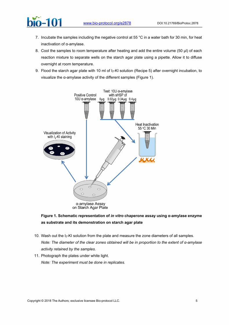
7. Incubate the samples including the negative control at 55 °C in a water bath for 30 min, for heat
inactivation of α-amylase.
8. Cool the samples to room temperature after heating and add the entire volume (50 µl) of each
reaction mixture to separate wells on the starch agar plate using a pipette. Allow it to diffuse
overnight at room temperature.
9. Flood the starch agar plate with 10 ml of I2-KI solution (Recipe 5) after overnight incubation, to
visualize the α-amylase activity of the different samples (Figure 1).
Figure 1. Schematic representation of in vitro chaperone assay using α-amylase enzyme as substrate and its demonstration on starch agar plate
10. Wash out the I2-KI solution from the plate and measure the zone diameters of all samples.
Note: The diameter of the clear zones obtained will be in proportion to the extent of α-amylase
activity retained by the samples.
11. Photograph the plates under white light.
Note: The experiment must be done in replicates.
Copyright © 2018 The Authors; exclusive licensee Bio-protocol LLC. 6
DOI:10.21769/BioProtoc.2878
www.bio-protocol.org/e2878
Data analysis
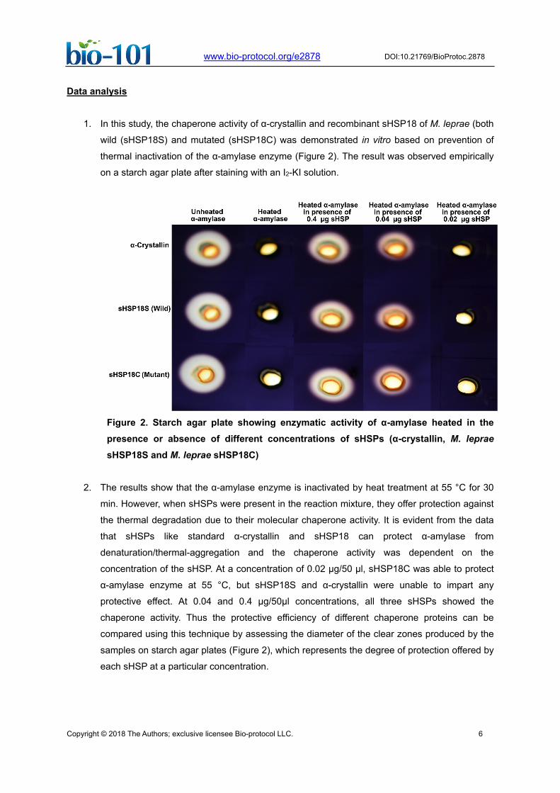
1. In this study, the chaperone activity of α-crystallin and recombinant sHSP18 of M. leprae (both
wild (sHSP18S) and mutated (sHSP18C) was demonstrated in vitro based on prevention of
thermal inactivation of the α-amylase enzyme (Figure 2). The result was observed empirically
on a starch agar plate after staining with an I2-KI solution.
Figure 2. Starch agar plate showing enzymatic activity of α-amylase heated in the presence or absence of different concentrations of sHSPs (α-crystallin, M. leprae sHSP18S and M. leprae sHSP18C)
2. The results show that the α-amylase enzyme is inactivated by heat treatment at 55 °C for 30
min. However, when sHSPs were present in the reaction mixture, they offer protection against
the thermal degradation due to their molecular chaperone activity. It is evident from the data
that sHSPs like standard α-crystallin and sHSP18 can protect α-amylase from
denaturation/thermal-aggregation and the chaperone activity was dependent on the
concentration of the sHSP. At a concentration of 0.02 µg/50 µl, sHSP18C was able to protect
α-amylase enzyme at 55 °C, but sHSP18S and α-crystallin were unable to impart any
protective effect. At 0.04 and 0.4 µg/50µl concentrations, all three sHSPs showed the
chaperone activity. Thus the protective efficiency of different chaperone proteins can be
compared using this technique by assessing the diameter of the clear zones produced by the
samples on starch agar plates (Figure 2), which represents the degree of protection offered by
each sHSP at a particular concentration.
Copyright © 2018 The Authors; exclusive licensee Bio-protocol LLC. 7
DOI:10.21769/BioProtoc.2878
www.bio-protocol.org/e2878
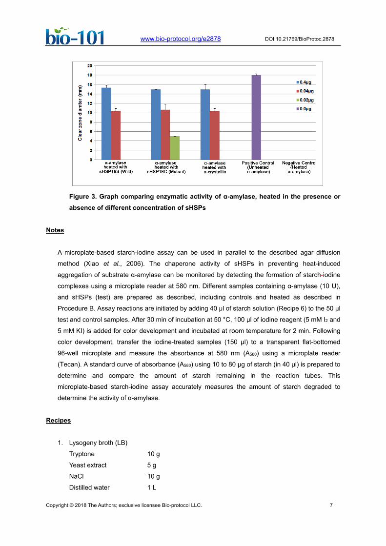
Figure 3. Graph comparing enzymatic activity of α-amylase, heated in the presence or absence of different concentration of sHSPs
Notes
A microplate-based starch-iodine assay can be used in parallel to the described agar diffusion
method (Xiao et al., 2006). The chaperone activity of sHSPs in preventing heat-induced
aggregation of substrate α-amylase can be monitored by detecting the formation of starch-iodine
complexes using a microplate reader at 580 nm. Different samples containing α-amylase (10 U),
and sHSPs (test) are prepared as described, including controls and heated as described in
Procedure B. Assay reactions are initiated by adding 40 µl of starch solution (Recipe 6) to the 50 µl
test and control samples. After 30 min of incubation at 50 °C, 100 µl of iodine reagent (5 mM I2 and
5 mM KI) is added for color development and incubated at room temperature for 2 min. Following
color development, transfer the iodine-treated samples (150 µl) to a transparent flat-bottomed
96-well microplate and measure the absorbance at 580 nm (A580) using a microplate reader
(Tecan). A standard curve of absorbance (A580) using 10 to 80 µg of starch (in 40 µl) is prepared to
determine and compare the amount of starch remaining in the reaction tubes. This
microplate-based starch-iodine assay accurately measures the amount of starch degraded to
determine the activity of α-amylase.
Recipes
1. Lysogeny broth (LB)
Tryptone 10 g
Yeast extract 5 g
NaCl 10 g
Distilled water 1 L
Copyright © 2018 The Authors; exclusive licensee Bio-protocol LLC. 8
DOI:10.21769/BioProtoc.2878
www.bio-protocol.org/e2878
Adjust pH to 7.4 and sterilize by autoclaving
2. PBS (1x)
KH2PO4 0.204 g
Na2HPO4·2H2O 1.157g
NaCl 8.01 g
KCl 0.201 g
Dissolve in 800 ml distilled water, adjust pH to 7.4 and make up the volume to 1 L
Sterilize by autoclaving
3. Lysis buffer
NaH2PO4·H2O 6.90 g
NaCl 17.53 g
Imidazole 0.68 g
Adjust pH to 8 and make up to 1 L
4. Starch agar
1% soluble starch
2% agar
Add 1 g starch to 80 ml distilled water in a conical flask and dissolve by placing in boiling water.
Once the starch has dissolved, make up the volume to 100 ml and add 2 g agar. Sterilize by
autoclaving
5. Iodine-Potassium iodide (I2-KI) solution
0.2% Iodine
2% Potassium iodide
Dissolve 2 g potassium iodide and 0.2 g Iodine in distilled water and make up the volume to
100 ml. Store in a brown bottle at room temperature (can be stored up to 2 weeks)
6. Starch solution
Starch soluble 0.2 g
Distilled water 100 ml
Add 0.2 g starch to 80 ml distilled water in a conical flask and dissolve by placing in boiling
water. Once the starch has dissolved, make up the volume to 100 ml with distilled water.
Acknowledgments
This work was supported by grants from the Kerala State Council for Science Technology and
Environment (KSCSTE, Govt. of Kerala), under Science Research Scheme and Senior Research
Fellowship to Jeeshma Nambidi Parambath. Gayathri Valsala and Karthik Menon acknowledge
Council for Scientific and Industrial Research (CSIR, Govt. of India) for financial aid in the form of
Senior Research Fellowships. The authors have no conflict of interest or competing interest to
declare.
Copyright © 2018 The Authors; exclusive licensee Bio-protocol LLC. 9
DOI:10.21769/BioProtoc.2878
www.bio-protocol.org/e2878
References
1. Atlas, R. M., Parks, L. C. and Brown, A. E. (1995). Laboratory manual of experimental
microbiology. Mosby-Year Book Inc., St Louis.
2. Bepperling, A., Alte, F., Kriehuber, T., Braun, N., Weinkauf, S., Groll, M., Haslbeck, M. and
Buchner, J. (2012). Alternative bacterial two-component small heat shock protein systems.
Proc Natl Acad Sci U S A 109(50): 20407-20412.
3. Bornhorst, J. A. and Falke, J. J. (2000). Purification of proteins using polyhistidine affinity tags.
Methods Enzymol 326: 245-254.
4. Bradford, M. M. (1976). A rapid and sensitive method for the quantitation of microgram
quantities of protein utilizing the principle of protein-dye binding. Anal Biochem 72: 248-254.
5. Dumon-Seignovert, L., Cariot, G. and Vuillard, L. (2004). The toxicity of recombinant proteins in
Escherichia coli: a comparison of overexpression in BL21(DE3), C41(DE3), and C43(DE3).
Protein Expr Purif 37(1): 203-206.
6. Farahbakhsh, Z. T., Huang, Q. L., Ding, L. L., Altenbach, C., Steinhoff, H. J., Horwitz, J., and
Hubbell, W. L. (1995). Interaction of α-crystallin with spin-labeled peptides. Biochemistry
34:509-516.
7. Goloubinoff, P., Gatenby, A. A. and Lorimer, G. H. (1989). GroE heat-shock proteins promote
assembly of foreign prokaryotic ribulose bisphosphate carboxylase oligomers in Escherichia
coli. Nature 337(6202): 44-47.
8. Grallert, H., Rutkat, K. and Buchner, J. (1998). GroEL traps dimeric and monomeric unfolding
intermediates of citrate synthase. J Biol Chem 273(50): 33305-33310.
9. Hess, J. F. and FitzGerald, P. G. (1998). Protection of a restriction enzyme from heat
inactivation by α-crystallin. Mol Vis 4: 29.
10. Laemmli, U. K. (1970). Cleavage of structural proteins during the assembly of the head of
bacteriophage T4. Nature 227(5259): 680-685.
11. Lee, G. J., Roseman, A. M., Saibil, H. R. and Vierling, E. (1997). A small heat shock protein
stably binds heat-denatured model substrates and can maintain a substrate in a
folding-competent state. EMBO J 16(3): 659-671.
12. Lini, N., Rehna, E. A., Shiburaj, S., Maheshwari, J. J., Shankernarayan, N. P. and
Dharmalingam, K. (2008). Functional characterization of a small heat shock protein from
Mycobacterium leprae. BMC Microbiol 8: 208.
13. Marini, I., Moschini, R., Del Corso, A. and Mura, U. (2000). Complete protection by α-crystallin
on lens sorbitol dehydrogenase undergoing thermal stress. J Biol Chem 275:32559-32565.
14. Mendoza, J. A., Lorimer, G. H. and Horowitz, P. M. (1992). Chaperonin cpn60 from Escherichia
coli protects the mitochondrial enzyme rhodanese against heat inactivation and supports
folding at elevated temperatures. J Biol Chem 267(25): 17631-17634.
Copyright © 2018 The Authors; exclusive licensee Bio-protocol LLC. 10
DOI:10.21769/BioProtoc.2878
www.bio-protocol.org/e2878
15. Rawat, U. and Rao, M. (1998). Interactions of chaperone α-crystallin with the molten globule
state of xylose reductase. Implications for reconstitution of the active enzyme. J Biol Chem
273(16): 9415-9423.
16. Rozema, D. and Gellman, S. H. (1996). Artificial chaperone-assisted refolding of
denatured-reduced lysozyme: modulation of the competition between renaturation and
aggregation. Biochemistry 35(49): 15760-15771.
17. Santhoshkumar, P. and Sharma, K. K. (2001). Analysis of α-crystallin chaperone function using
restriction enzymes and citrate synthase. Mol Vis 7: 172-177.
18. Xiao, Z., Storms, R. and Tsang, A. (2006). A quantitative starch-iodine method for measuring
alpha-amylase and glucoamylase activities. Anal Biochem 351(1): 146-148.