Immunological response after stopping NUCsregist2.virology-education.com/presentations/2019/... ·...
30
Immunological response after stopping NUCs Andre Boonstra, PhD Erasmus Medical Center Rotterdam, the Netherlands Nov 6, 2019
Transcript of Immunological response after stopping NUCsregist2.virology-education.com/presentations/2019/... ·...
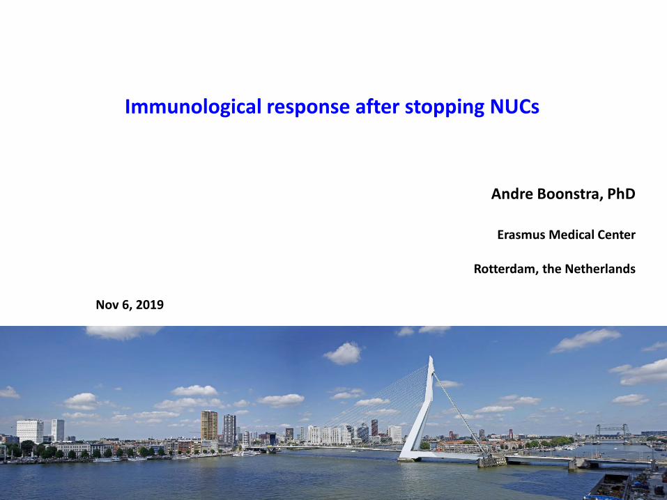
Immunological response after stopping NUCs
Andre Boonstra, PhD
Erasmus Medical Center
Rotterdam, the Netherlands
Nov 6, 2019
Disclosures
Research funding: Gilead, Roche, Fujirebio, Viroclinics, Janssen
www.escalon.eu
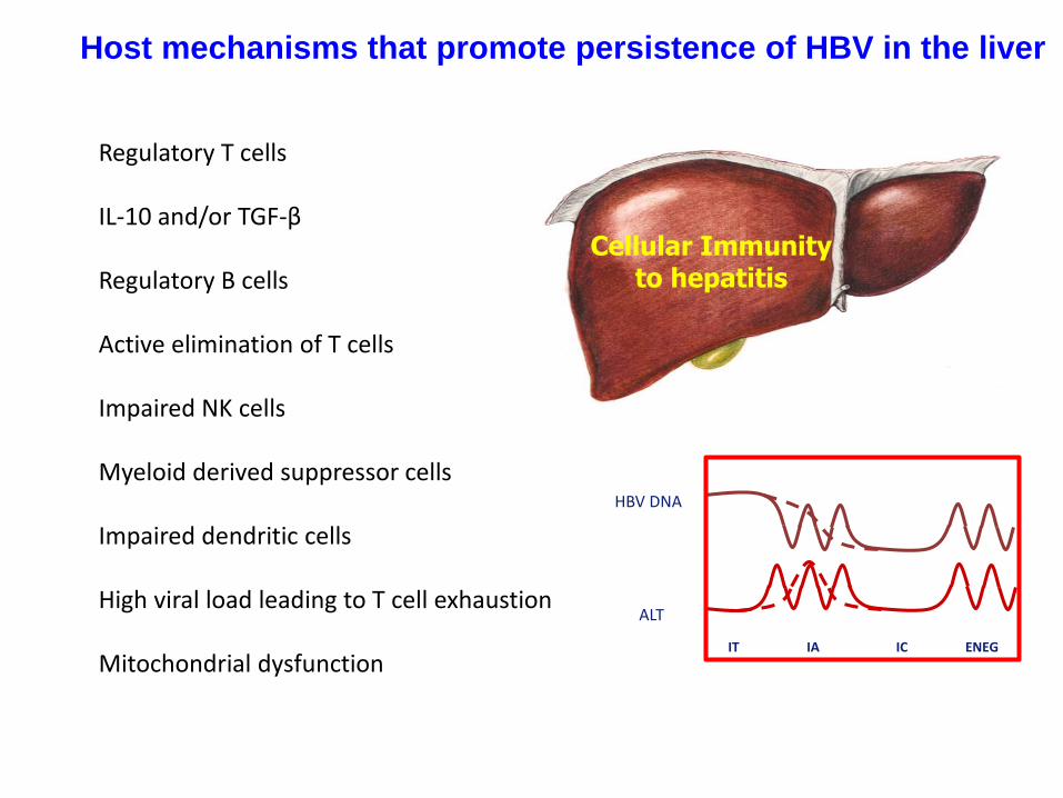
Host mechanisms that promote persistence of HBV in the liver
Regulatory T cells
IL-10 and/or TGF-β
Regulatory B cells
Active elimination of T cells
Impaired NK cells
Myeloid derived suppressor cells
Impaired dendritic cells
High viral load leading to T cell exhaustion
Mitochondrial dysfunction
Cellular Immunityto hepatitis
IT IA IC ENEG
HBV DNA
ALT
EntecavirTenofovir
HBV DNA
weeks months years
Time
100
50
0
Inc
rea
se
of
ma
xim
um
(%)
Initiation of
therapy
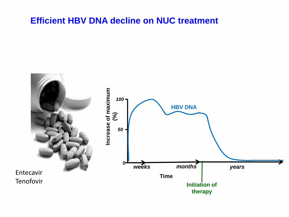
Efficient HBV DNA decline on NUC treatment
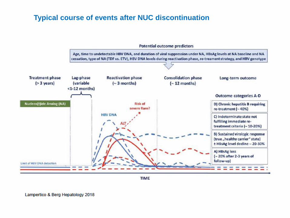
Typical course of events after NUC discontinuation
What causes or is the consequence of the virological relapse?
What causes or is the consequence of the clinical relapse?
What makes that some patients experience HBsAg loss?
Can we predict the outcome of NUC stopping?
What do we want to understand?
2016 2018
2018
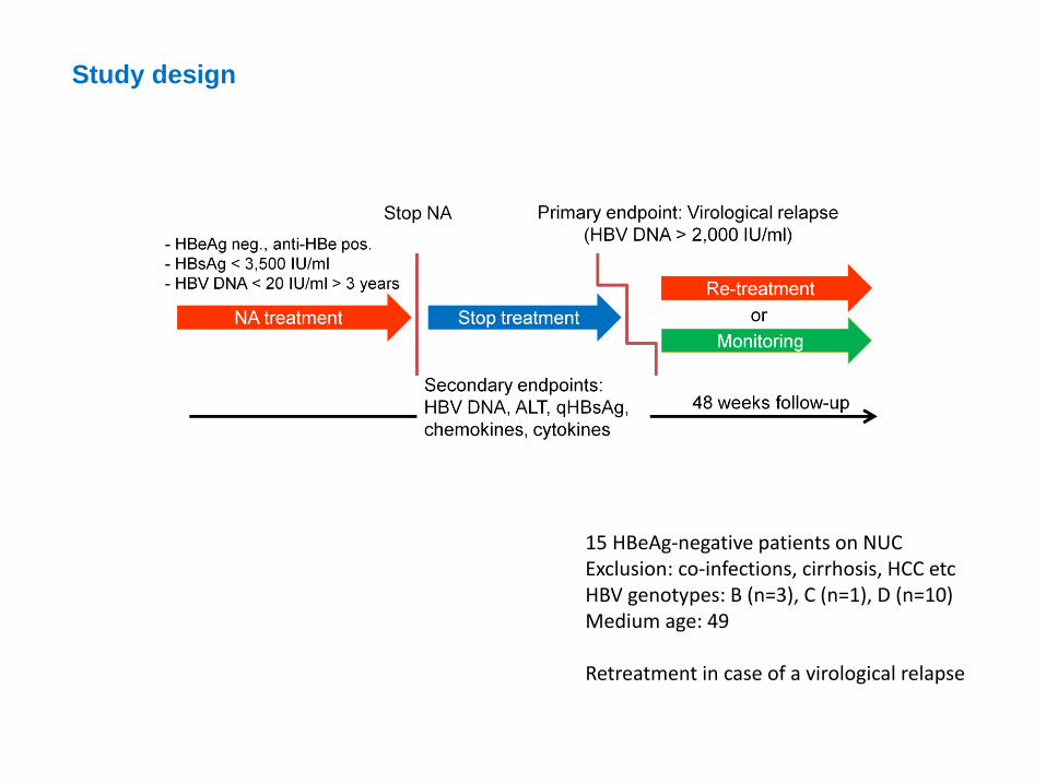
15 HBeAg-negative patients on NUCExclusion: co-infections, cirrhosis, HCC etcHBV genotypes: B (n=3), C (n=1), D (n=10) Medium age: 49
Retreatment in case of a virological relapse
Study design
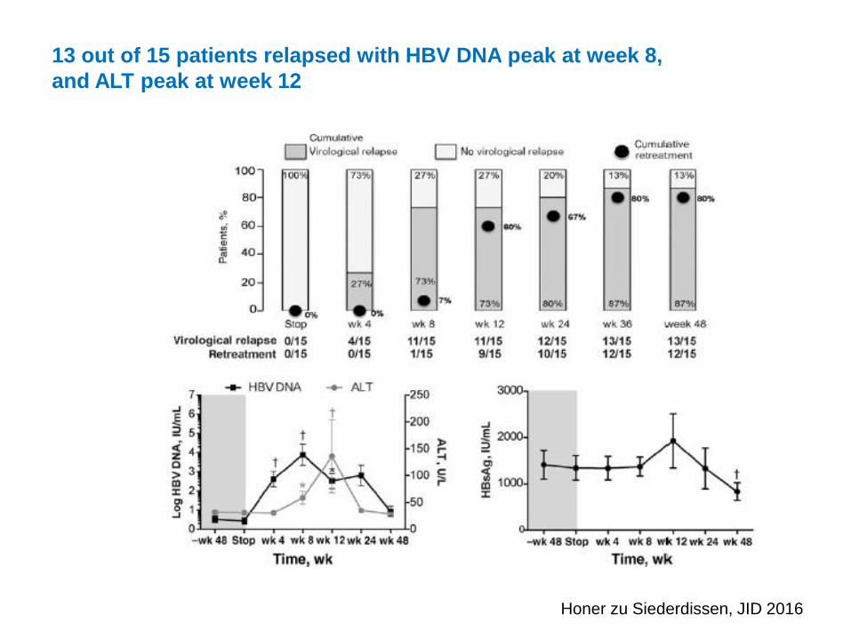
13 out of 15 patients relapsed with HBV DNA peak at week 8,
and ALT peak at week 12
Honer zu Siederdissen, JID 2016
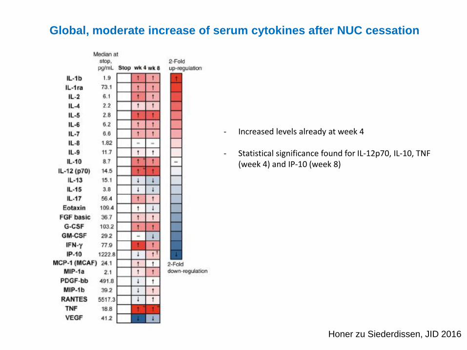
Global, moderate increase of serum cytokines after NUC cessation
Honer zu Siederdissen, JID 2016
- Increased levels already at week 4
- Statistical significance found for IL-12p70, IL-10, TNF (week 4) and IP-10 (week 8)
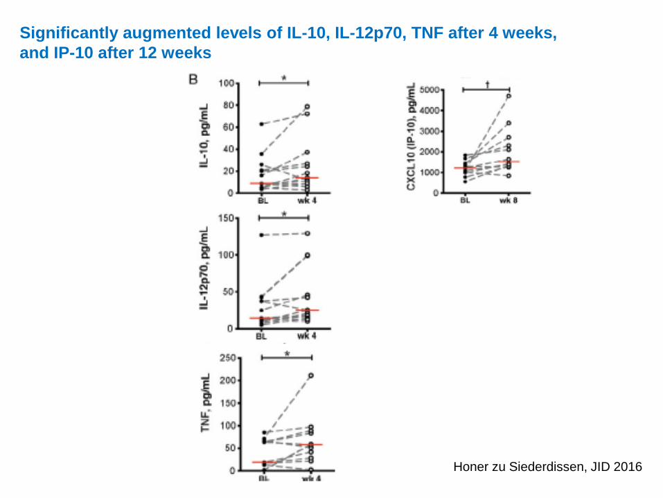
Significantly augmented levels of IL-10, IL-12p70, TNF after 4 weeks,
and IP-10 after 12 weeks
Honer zu Siederdissen, JID 2016
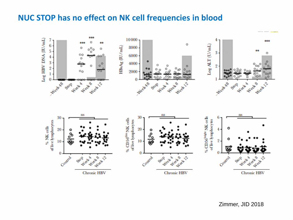
NUC STOP has no effect on NK cell frequencies in blood
Zimmer, JID 2018
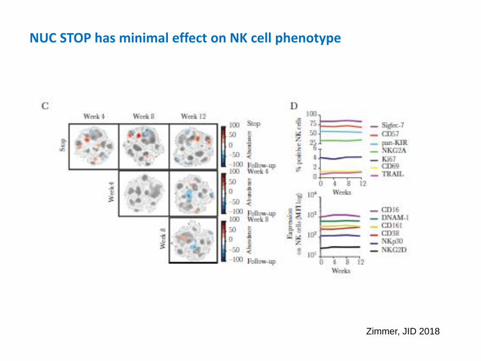
NUC STOP has minimal effect on NK cell phenotype
Zimmer, JID 2018
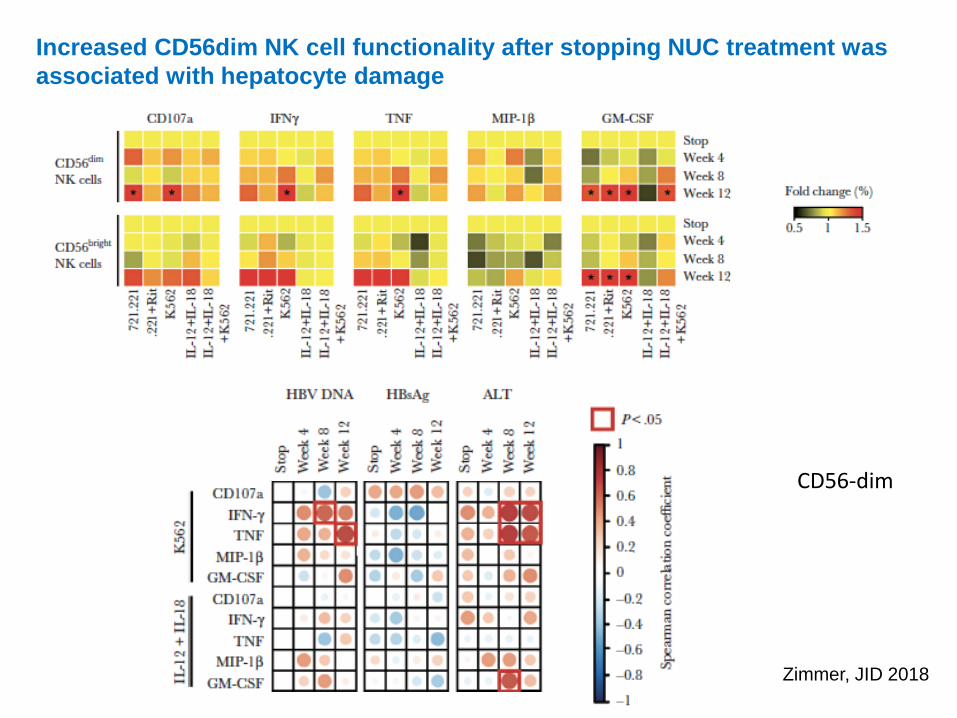
CD56-dim
Increased CD56dim NK cell functionality after stopping NUC treatment was
associated with hepatocyte damage
Zimmer, JID 2018

T cell phenotype was only slightly changed after NA discontinuation
Rinker, J Hepatol 2018
Minor, albeit significant, changes NUC STOP were observed for
PD-1 on CD4+ and CD8+ T cells,
TCF-1 on CD4+,
CD161 on CD8+ T cells.
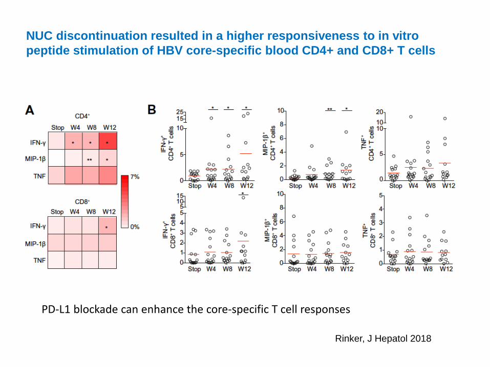
PD-L1 blockade can enhance the core-specific T cell responses
NUC discontinuation resulted in a higher responsiveness to in vitro
peptide stimulation of HBV core-specific blood CD4+ and CD8+ T cells
Rinker, J Hepatol 2018
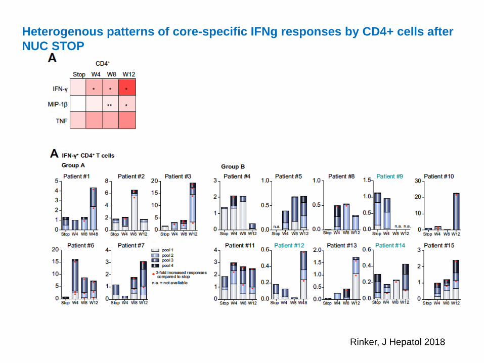
Heterogenous patterns of core-specific IFNg responses by CD4+ cells after
NUC STOP
Rinker, J Hepatol 2018
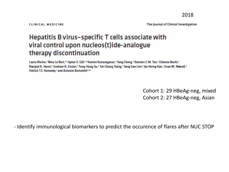
2018
Cohort 1: 29 HBeAg-neg, mixedCohort 2: 27 HBeAg-neg, Asian
- Identify immunological biomarkers to predict the occurence of flares after NUC STOP
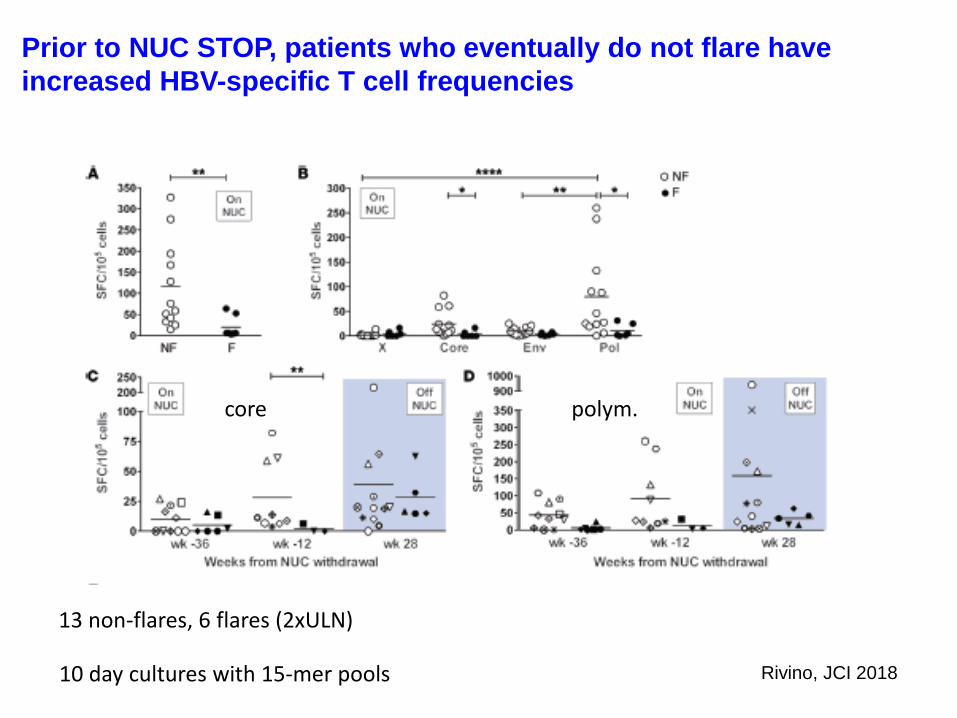
13 non-flares, 6 flares (2xULN)
Prior to NUC STOP, patients who eventually do not flare have
increased HBV-specific T cell frequencies
core polym.
10 day cultures with 15-mer pools Rivino, JCI 2018
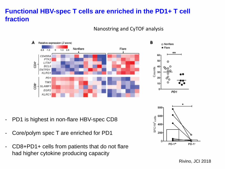
Functional HBV-spec T cells are enriched in the PD1+ T cell
fraction
Nanostring and CyTOF analysis
Rivino, JCI 2018
- PD1 is highest in non-flare HBV-spec CD8
- Core/polym spec T are enriched for PD1
- CD8+PD1+ cells from patients that do not flare
had higher cytokine producing capacity
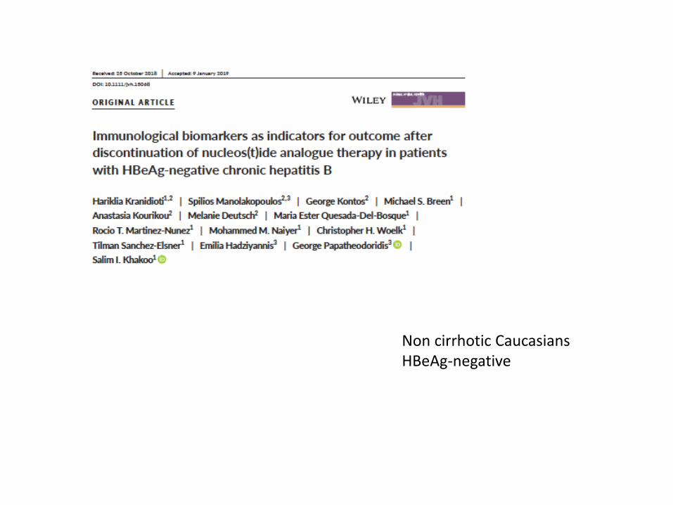
Non cirrhotic CaucasiansHBeAg-negative
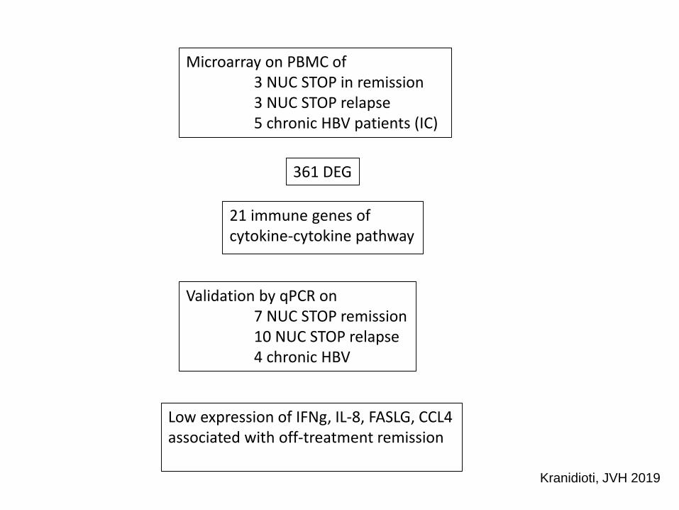
Microarray on PBMC of 3 NUC STOP in remission3 NUC STOP relapse5 chronic HBV patients (IC)
361 DEG
21 immune genes of cytokine-cytokine pathway
Validation by qPCR on 7 NUC STOP remission10 NUC STOP relapse4 chronic HBV
Low expression of IFNg, IL-8, FASLG, CCL4associated with off-treatment remission
Kranidioti, JVH 2019
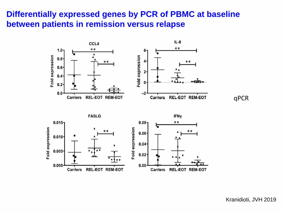
qPCR
Differentially expressed genes by PCR of PBMC at baseline
between patients in remission versus relapse
Kranidioti, JVH 2019
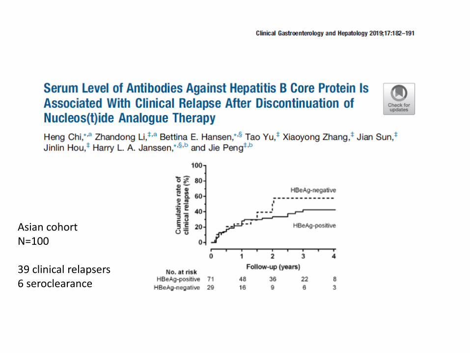
Asian cohortN=100
39 clinical relapsers6 seroclearance
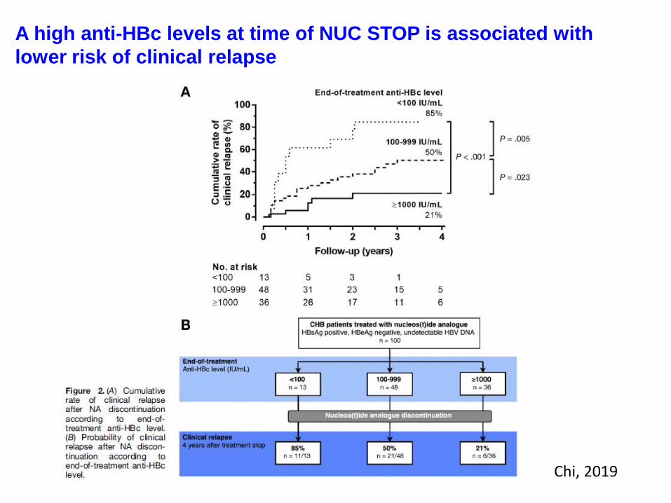
A high anti-HBc levels at time of NUC STOP is associated with
lower risk of clinical relapse
Chi, 2019
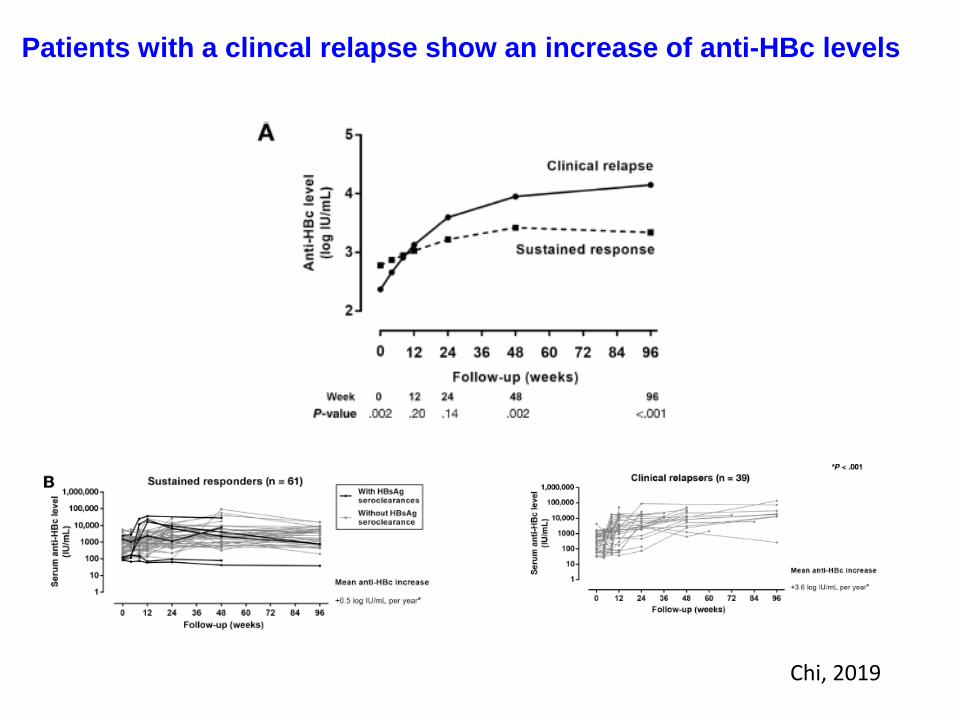
Patients with a clincal relapse show an increase of anti-HBc levels
Chi, 2019
Recap
More pronounced effects are seen with ALT flare, less pronouced with virological
relapse
All studies have been done in peripheral compartment, not liver
After NUC discontinuation the studies point towards involvement during relapse of
- increase of serum cytokines (IL-12p70, IL-10, TNF, IP-10)
- Freq of IFNg+ NK dim cells associate with ALT
- Increase of core/polym specific T cells, but lower than in non-flare patients
- Increase of anti-HBcore levels

Baseline parameters to predict outcome after NUC discontinuation
- no flare: higher baseline freq of core/polym specific T cells (PD-1+),
>40 SPC/105 cells for core/polym T cells, then no flare
- lower risk of relapse: higher baseline anti-Hbcore levels
- remission: lower ccl4, il8, faslg, ifng (full blood qPCR)
Recap
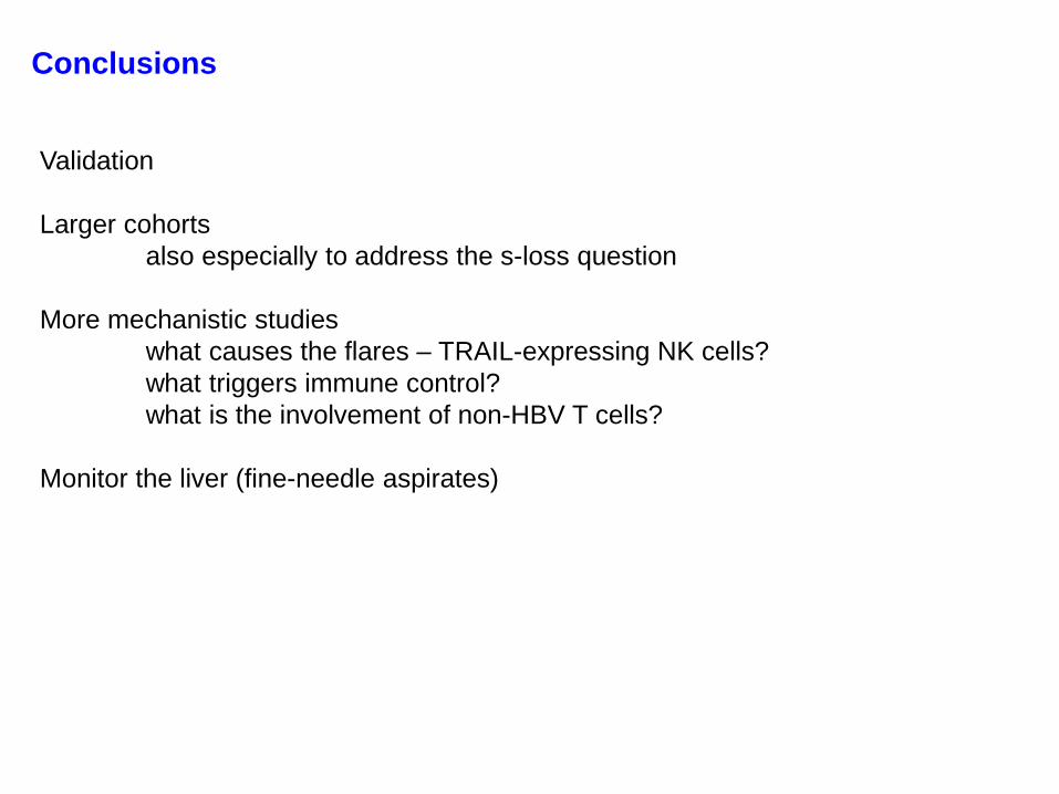
Validation
Larger cohorts
also especially to address the s-loss question
More mechanistic studies
what causes the flares – TRAIL-expressing NK cells?
what triggers immune control?
what is the involvement of non-HBV T cells?
Monitor the liver (fine-needle aspirates)
Conclusions
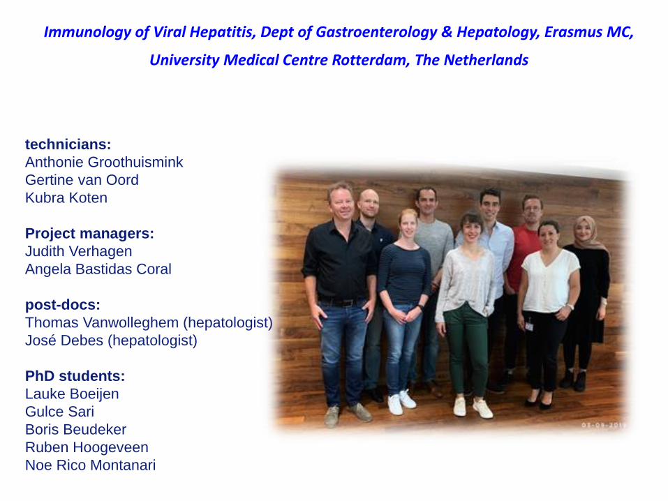
technicians:
Anthonie Groothuismink
Gertine van Oord
Kubra Koten
Project managers:
Judith Verhagen
Angela Bastidas Coral
post-docs:
Thomas Vanwolleghem (hepatologist)
José Debes (hepatologist)
PhD students:
Lauke Boeijen
Gulce Sari
Boris Beudeker
Ruben Hoogeveen
Noe Rico Montanari
Immunology of Viral Hepatitis, Dept of Gastroenterology & Hepatology, Erasmus MC,
University Medical Centre Rotterdam, The Netherlands


![[AIESEC] Welcome Week Presentation](https://static.fdocument.org/doc/165x107/55ab73551a28ab9b4b8b4589/aiesec-welcome-week-presentation.jpg)