Immunoglobulin heavy chain expression shapes the B cell receptor
8
Introduction Expression of immunoglobulin heavy chains (Igµ) in pro-B cells induces pre-B cell development by assembly of a pre-B cell receptor (pre-BCR), which is a complex of Igµ, surrogate light chains (ψL), and two signal-trans- ducing proteins, Igα and Igβ (1–6). Pre-BCR expression induces proliferative expansion and downregulation of recombinase-activating genes (RAG1 and RAG2), there- by ensuring allelic exclusion while selecting clones of cells with productive VDJ H rearrangements (7). Muta- tions in mIgµ, ψL, or Igα/Igβ genes that disrupt pre-BCR assembly impede B cell development at the pro-B cell stage in mice and humans (8–15). Pre-BCR assembly is dependent on interaction between V H variable regions and ψL, and in the mouse the IgH repertoire is selected in part on the basis of this interaction (16–18). It has been proposed that V H domains that pair well with ψL are positively selected at the pre-B cell stage, whereas V H domains that pair poorly with ψL are not. However, selection differs in mouse fetal and adult B cell development such that V H s that are counterselected in the adult are prominent in the fetal repertoire (18). In contrast to mice, there is no apparent difference in the V H repertoire between pro-B, pre-B, or fetal B cells and adult mature B cells in the human (19–30). How- ever, there is selection for IgH genes during human B cell development as determined by the length of the third complementary determining region (CDR3). Pro- and pre-B cells from adult bone marrow have longer CDR3s than mature B cells (31–33). Following successful IgH assembly and pre-BCR expression, V(D)J recombination is targeted to the light chain (IgL) genes (34–38). Those cells that produce in- frame IgL chains test their newly synthesized Igs for self- reactivity. In the mouse, B cells that produce self-reac- tive receptors are either deleted or arrested in development and undergo receptor editing (39-45). In contrast, less is known about receptor selection and the role of the BCR in regulating B cell development in humans. Here we report on the role of the BCR in Ig repertoire selection in two patients with different muta- tions in the Igµ gene that impairs BCR assembly. Methods Patient samples and cell preparation. Bone marrow samples were obtained from two Igµ-deficient patients with either a homozygous cytidine insertion in the Igµ gene (Igµ –/– ) or with a homozygous deletion of the Igµ locus (Igµ ∆ ) (see Results) (13, 46, 47) (C. Schiff, unpublished observations). Samples were obtained when the Igµ ∆ patient was 2 years old, the Igµ –/– patient was 4 years old, and her Igµ +/– brother (control) was 9 months old. The parents gave informed consent for this study. Bone marrow mononuclear cells were isolated by Ficoll gra- dients and CD34 + CD19 + pro-B cells were sorted on a The Journal of Clinical Investigation | September 2001 | Volume 108 | Number 6 879 Immunoglobulin heavy chain expression shapes the B cell receptor repertoire in human B cell development Eric Meffre, 1,2 Michèle Milili, 2 Carla Blanco-Betancourt, 2 Henedina Antunes, 3 Michel C. Nussenzweig, 1 and Claudine Schiff 2 1 Laboratory of Molecular Immunology, The Rockefeller University, Howard Hughes Medical Institute, New York, New York, USA 2 Centre d’Immunologie de Marseille-Luminy, CNRS-INSERM–Université de la Méditerranée, Campus de Luminy, Marseille, France 3 Fundacao Faculdade Federal de Ciencias, Medicas de Porto Alegre, Portugal Address correspondence to: Eric Meffre, The Rockefeller University, 1230 York Avenue, New York, New York 10021, USA. Phone: (212) 327-8098; Fax: (212) 327-8370; E-mail: [email protected]. Received for publication April 16, 2001, and accepted in revised form August 13, 2001. Developing B cells must pass a series of checkpoints that are regulated by membrane-bound Igµ through the Igα-Igβ signal transducers. To determine how Igµ expression affects B cell development and Ab selection in humans we analyzed Ig gene rearrangements in pro-B cells from two patients who are unable to produce Igµ proteins. We find that Igµ expression does not affect V H , D, or J H segment usage and is not required for human Igκ and Igλ recombination or expression. However, the heavy and light chains found in pro-B cells differed from those in peripheral B cells in that they showed unusu- ally long CDR3s. In addition, the Igκ repertoire in Igµ-deficient pro-B cells was skewed to downstream Jκs and upstream Vκs, consistent with persistent secondary V(D)J rearrangements. Thus, Igµ expres- sion is not required for secondary V(D)J recombination in pro-B cells. However, B cell receptor expres- sion shapes the Ab repertoire in humans and is essential for selection against Ab’s with long CDR3s. J. Clin. Invest. 108:879–886 (2001). DOI:10.1172/JCI200113051.
Transcript of Immunoglobulin heavy chain expression shapes the B cell receptor
IntroductionExpression of immunoglobulin heavy chains (Igµ) inpro-B cells induces pre-B cell development by assemblyof a pre-B cell receptor (pre-BCR), which is a complex ofIgµ, surrogate light chains (ψL), and two signal-trans-ducing proteins, Igα and Igβ (1–6). Pre-BCR expressioninduces proliferative expansion and downregulation ofrecombinase-activating genes (RAG1 and RAG2), there-by ensuring allelic exclusion while selecting clones ofcells with productive VDJH rearrangements (7). Muta-tions in mIgµ, ψL, or Igα/Igβ genes that disrupt pre-BCRassembly impede B cell development at the pro-B cellstage in mice and humans (8–15).
Pre-BCR assembly is dependent on interactionbetween VH variable regions and ψL, and in the mousethe IgH repertoire is selected in part on the basis of thisinteraction (16–18). It has been proposed that VH
domains that pair well with ψL are positively selectedat the pre-B cell stage, whereas VH domains that pairpoorly with ψL are not. However, selection differs inmouse fetal and adult B cell development such that VHsthat are counterselected in the adult are prominent inthe fetal repertoire (18).
In contrast to mice, there is no apparent difference inthe VH repertoire between pro-B, pre-B, or fetal B cellsand adult mature B cells in the human (19–30). How-ever, there is selection for IgH genes during human B cell development as determined by the length of the
third complementary determining region (CDR3). Pro-and pre-B cells from adult bone marrow have longerCDR3s than mature B cells (31–33).
Following successful IgH assembly and pre-BCRexpression, V(D)J recombination is targeted to the lightchain (IgL) genes (34–38). Those cells that produce in-frame IgL chains test their newly synthesized Igs for self-reactivity. In the mouse, B cells that produce self-reac-tive receptors are either deleted or arrested indevelopment and undergo receptor editing (39-45). Incontrast, less is known about receptor selection and therole of the BCR in regulating B cell development inhumans. Here we report on the role of the BCR in Igrepertoire selection in two patients with different muta-tions in the Igµ gene that impairs BCR assembly.
MethodsPatient samples and cell preparation. Bone marrow sampleswere obtained from two Igµ-deficient patients witheither a homozygous cytidine insertion in the Igµ gene(Igµ–/–) or with a homozygous deletion of the Igµ locus(Igµ∆) (see Results) (13, 46, 47) (C. Schiff, unpublishedobservations). Samples were obtained when the Igµ∆
patient was 2 years old, the Igµ–/– patient was 4 yearsold, and her Igµ+/– brother (control) was 9 months old.The parents gave informed consent for this study. Bonemarrow mononuclear cells were isolated by Ficoll gra-dients and CD34+CD19+ pro-B cells were sorted on a
The Journal of Clinical Investigation | September 2001 | Volume 108 | Number 6 879
Immunoglobulin heavy chain expression shapes the B cell receptor repertoire in human B cell development
Eric Meffre,1,2 Michèle Milili,2 Carla Blanco-Betancourt,2 Henedina Antunes,3
Michel C. Nussenzweig,1 and Claudine Schiff2
1Laboratory of Molecular Immunology, The Rockefeller University, Howard Hughes Medical Institute, New York, New York, USA
2Centre d’Immunologie de Marseille-Luminy, CNRS-INSERM–Université de la Méditerranée, Campus de Luminy, Marseille, France
3Fundacao Faculdade Federal de Ciencias, Medicas de Porto Alegre, Portugal
Address correspondence to: Eric Meffre, The Rockefeller University, 1230 York Avenue, New York, New York 10021, USA.Phone: (212) 327-8098; Fax: (212) 327-8370; E-mail: [email protected].
Received for publication April 16, 2001, and accepted in revised form August 13, 2001.
Developing B cells must pass a series of checkpoints that are regulated by membrane-bound Igµthrough the Igα-Igβ signal transducers. To determine how Igµ expression affects B cell developmentand Ab selection in humans we analyzed Ig gene rearrangements in pro-B cells from two patients whoare unable to produce Igµ proteins. We find that Igµ expression does not affect VH, D, or JH segmentusage and is not required for human Igκ and Igλ recombination or expression. However, the heavy andlight chains found in pro-B cells differed from those in peripheral B cells in that they showed unusu-ally long CDR3s. In addition, the Igκ repertoire in Igµ-deficient pro-B cells was skewed to downstreamJκs and upstream Vκs, consistent with persistent secondary V(D)J rearrangements. Thus, Igµ expres-sion is not required for secondary V(D)J recombination in pro-B cells. However, B cell receptor expres-sion shapes the Ab repertoire in humans and is essential for selection against Ab’s with long CDR3s.
J. Clin. Invest. 108:879–886 (2001). DOI:10.1172/JCI200113051.
FACSVantage after labeling with FITC anti-CD34 andphycoerythrin anti-CD19 mAb’s (Beckman Coulter,Brea, California, USA).
RNA and RT-PCR. Total RNA was extracted from104–105 purified cells using TRIzol Reagent (LifeTechnologies Inc., Rockville, Maryland, USA). RNAwas reverse transcribed with Superscript II (LifeTechnologies Inc.) according to the manufacturer’sinstructions. For RT-PCR reactions, cDNA wasamplified for 25 (actin), 35 (VH-Cµ), or 38 (Vκ-Cκ)cycles of 30 seconds at 94°C, 30 seconds at 60°C,and 30 seconds at 72°C, or for 40 cycles (Vλ-Cλ) of 30seconds at 94°C, 30 seconds at 55°C, and 30 secondsat 72°C, with a final 10-minute extension at 72°Cusing Hot Star Taq DNA polymerase (QIAGEN Inc.,Valencia, California, USA) and the followingprimers: Vλ consensus sense, 5′GGG(G/A)TC(T/C)CT-GA(C/T/G)CG(A/C/G)TTCTCTGG(C/G)TCC3′; Cλ anti-sense, 5′CACAC(T/C)AGTGTGGCCTTGTTGGCTTG3′.VH1, VH3, VH4, Cµ, Vκ consensus and Cκ primers weredescribed previously (48, 49). RT-PCR products wereanalyzed on 2% agarose gels and visualized by adding0.3 pmol of 32PdATP to the PCR reaction.
Cloning and sequencing. PCR products were gel-purified(Qiaquick; QIAGEN Inc.) and cloned into TA vectors(Invitrogen, Carlsbad, California, USA). Double-stranded DNA sequences were obtained using anti-sense Cµ, Cκ, or Cλ primers and Dye Terminator CycleSequencing (Applied Biosystems, Foster City, Califor-nia, USA). Sequences were analyzed by comparisonwith Ig basic alignment search tool (BLAST). IgHCDR3 length was determined by counting amino acidresidues between positions 94 and 102 (conserved tryp-tophan in all JH segments) and D segments were iden-tified following the criteria of Corbett et al. (50). Igκ
and Igλ CDR3 length included amino acids betweenconserved cystein 88 and the phenylalanine residueembedded in Jκ or Jλ (51). Nontemplate (N) nucleotides(52) found at Vκ-Jκ or Vλ-Jλ junctions were countedwhile template-dependent palindromic (P) nucleotides(53) were excluded. Differences in gene distributionwere analyzed with χ2 tests (Cochran-Mantel-Haenszeltest) adjusted by the Bonferroni method for multipletesting, and they were considered significant when P values were less than or equal to 0.05.
ResultsIgH and IgL transcription is independent of Igµ expression.Two patients with agammaglobulinemia and IgHmutations were studied. Igµ–/– has a cytidine insertionin the CH1 exon of the Igµ gene that leads to aframeshift and the inability to produce Igµ products(13, 46, 47). Igµ∆ has a deletion in the Ig locus from 3′of the diversity (D) region to Igγ2, with all junction (J)segments and Igµ, Igδ, Igγ3, and Igγ1 genes missing (C.Schiff, unpublished observations).
Flow cytometric analysis of bone marrow from Igµ–/–
and Igµ∆ patients revealed that in both cases B cell dif-ferentiation was arrested at the CD34+CD19+ pro-Bcell stage (Figure 1a) (12, 13, 46, 47). To characterizeIg expression in Igµ-deficient pro-B cells, transcriptsfor heavy and light chain genes were amplified bysemiquantitative RT-PCR from sorted CD34+CD19+
pro-B cells from Igµ–/–, Igµ∆, and a control sibling (Fig-ure 1). As expected, VH-Cµ mRNA was missing in Igµ∆
pro-B cells where the entire Igµ locus was deleted (Fig-ure 1b). In contrast, Igµ–/– and control pro-B cellsshowed similar levels of VH-Cµ transcripts revealingthat the absence of Igµ protein does not affect the Igµgene expression in humans (Figure 1b).
880 The Journal of Clinical Investigation | September 2001 | Volume 108 | Number 6
Figure 1Immunoglobulin rearrangements in humanpro-B cells. (a) B cell precursors in siblingcontrol (left), Igµ–/– (middle), and Igµ∆ (right)bone marrow. CD34+CD19+ pro-B cells fromcontrol and both Igµ-deficient patients weresorted as gated. (b) Heavy and light chain Iggene expression in human pro-B cells. RNAfrom FACS-sorted CD34+CD19+ pro-B cellsfrom both Igµ-deficient patients and controlwas analyzed by semiquantitative RT-PCRusing 5′ consensus VH, Vκ, or Vλ and 3′ Cµ, Cκ,or Cλ primers, respectively, and visualized by32PdATP incorporation. “Neg.” denotes anegative control without cDNA for RT-PCRreactions. Actin RT-PCR was used as mRNAloading control. Serial fivefold dilutions ofcDNA are shown.
Light chain gene transcripts were found at similarlevels in sorted control or Igµ-deficient pro-B cells(Figure 1b), but they were not amplified from Igµ–/–
total bone marrow cells that contain few pro-B cells(46, 47). We conclude that light chain genes can berecombined and expressed in the absence of Igµ inhuman pro-B cells.
Igµ-independent VH, D, and JH gene segment usage. To deter-mine whether Igµ expression is required for VH, D, or JH
segment selection, IgH genes from the three major VH
families, VH1, VH3, and VH4, were cloned and sequenced.VH, D, and JH repertoire analysis revealed no statisticallysignificant differences between Igµ–/– pro-B cells, controlpro-B cells, and peripheral B cells (19, 20, 26–30) (seesupplemental data 1, www.jci.org/cgi/content/full/-108/06/879/DC1). Of 27 D genes in humans, only theD2-2 gene segment was over-represented in Igµ–/– andcontrol pro-B cells (P = 0.02) (31, 50) (see supplementaldata 2, www.jci.org/cgi/content/full/108/06/879/DC2).
Thus, the pre-BCR is not essential for VH selection, andintrinsic genetic factors are responsible for specific VH,D, and JH gene usage in human B cells.
IgH CDR3 selection by Igµ. To determine whether Igµexpression influences CDR3 selection, CDR3 lengthand amino acid composition were analyzed in Igµ–/–
pro-B cells and compared with control pro-B andperipheral B cells. About two-thirds of IgH genes wereout of frame in Igµ–/– pro-B cells, confirming theabsence of Igµ-mediated selection in these cells (Table1) (20). In contrast, two-thirds of IgH CDR3s werefound to be in-frame in control CD34+CD19+ precur-sor cells, suggesting early Igµ-mediated positive selec-tion of a subpopulation of CD34+CD19+ cells thatexpress Igµ (Table 1). We compared IgH genesexpressed by Igµ–/– pro-B cells to the in-frame IgH genesexpressed by control CD34+CD19+ cells and found anaverage CDR3 length of 16.0 and 16.5 amino acids,respectively, whereas peripheral B cells showed an aver-age CDR3 length of 13.5 amino acids (Figure 2a). D-Dfusions that increase the length of CDR3 were found inabout 2% (4 of 198) of the IgH sequences from Igµ–/–
pro-B cells and in 2.6% (3 of 117) of those from controlCD34+CD19+ cells, but were absent in peripheral B cells(49, 50) (data not shown). We conclude that IgH CDR3length is not selected in early B cell precursors and thatlong CDR3s and D-D fusions are counterselected dur-ing late stages of B cell development.
D segments can be used in three different readingframes, but in humans, RF1 tends to encode stopcodons, RF2 usually encodes glycine residues, andhydrophilic amino acids and RF3 is biased to encodehydrophobic sequences (50). D segments in RF1 are
The Journal of Clinical Investigation | September 2001 | Volume 108 | Number 6 881
Figure 2IgH CDR3 characteristics in pro-B cells. (a) VHDJH CDR3 length in350 peripheral B cell (white bars), 117 in-frame control pro-B (graybars), and 197 Igµ–/– (black bars) individual sequences. CDR3 lengthin amino acids (aa) is indicated below. The average CDR3 length forperipheral B, control pro-B, and Igµ–/– pro-B cells was 13.5, 16.5,and 16.0 amino acids, respectively. (b) D reading frame usage in IgHCDR3s from peripheral B, control, and Igµ–/– pro-B cells. The threeRF uses reported by Corbett et al. (50) for some commonly used Dgene segments are represented. D3-3 and D3-10 encode no intra-genic stop codons whereas D6-13, D2-15, D4-17, and D3-22sequences using RF1 display stop codons.
Table 1CDR3 frame analysis in CD34+CD19+ pro-B cells
Control Igµ–/– Igµ∆
CDR3 Frame Frame Frame
Out In Stop codonA Out In Stop codonA Out In
H 69 (33%) 141 (67%) 11 (7.8%) 143 (73%) 54 (27%) 24 (44%) – –κ 10 (45%) 12 (55%) 21 (55%) 17 (45%) 25 (68%) 12 (32%)λ 11 (55%) 9 (45%) 6 (60%) 4 (40%) 15 (54%) 13 (46%)
AStop codon frequency is calculated among in-frame CDR3s.
normally under-represented in peripheral B cells andwere counterselected in control CD34+CD19+ cells (Fig-ure 2b, top and bottom row, and Table 1). In contrast,there was neither RF selection nor stop codon counter-selection in Igµ–/– pro-B cells (Figure 2b, top and bot-tom row, and Table 1). D3-3 or D3-10 genes that do notcontain intragenic stop codons in RF1 were used in allthree RFs in Igµ–/–, control CD34+CD19+, or peripheralB cells (Figure 2b, middle row). In addition, hydrophilic(RF2) and hydrophobic (RF3) Ds were used equally inIgµ–/– and control CD34+CD19+ cells whereas RF2 wasfavored in peripheral B cells (Figure 2b) (31, 50). How-ever, D3-22 RF usage was already selected in controlCD34+CD19+ cells since RF3 was clearly counterselect-ed (and/or RF2 positively selected) when functional Igµchains were generated (Figure 2b, bottom row). Thus,IgH CDR3s containing stop codons are counterselect-ed in early B cell precursors whereas CDR3s withhydrophilic or hydrophobic RF are not.
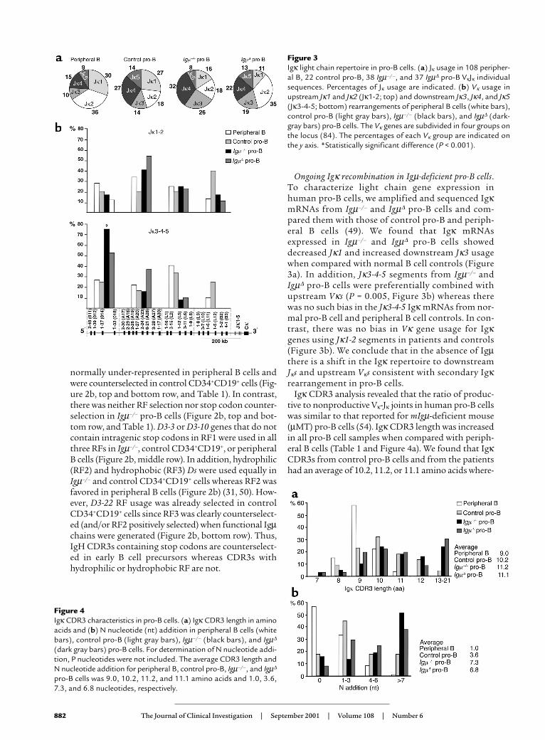
Ongoing Igκ recombination in Igµ-deficient pro-B cells.To characterize light chain gene expression inhuman pro-B cells, we amplified and sequenced IgκmRNAs from Igµ–/– and Igµ∆ pro-B cells and com-pared them with those of control pro-B and periph-eral B cells (49). We found that Igκ mRNAsexpressed in Igµ–/– and Igµ∆ pro-B cells showeddecreased Jκ1 and increased downstream Jκ3 usagewhen compared with normal B cell controls (Figure3a). In addition, Jκ3-4-5 segments from Igµ–/– andIgµ∆ pro-B cells were preferentially combined withupstream Vκs (P = 0.005, Figure 3b) whereas therewas no such bias in the Jκ3-4-5 Igκ mRNAs from nor-mal pro-B cell and peripheral B cell controls. In con-trast, there was no bias in Vκ gene usage for Igκgenes using Jκ1-2 segments in patients and controls(Figure 3b). We conclude that in the absence of Igµthere is a shift in the Igκ repertoire to downstreamJκs and upstream Vκs consistent with secondary Igκrearrangement in pro-B cells.
Igκ CDR3 analysis revealed that the ratio of produc-tive to nonproductive Vκ-Jκ joints in human pro-B cellswas similar to that reported for mIgµ-deficient mouse(µMT) pro-B cells (54). Igκ CDR3 length was increasedin all pro-B cell samples when compared with periph-eral B cells (Table 1 and Figure 4a). We found that IgκCDR3s from control pro-B cells and from the patientshad an average of 10.2, 11.2, or 11.1 amino acids where-
882 The Journal of Clinical Investigation | September 2001 | Volume 108 | Number 6
Figure 3Igκ light chain repertoire in pro-B cells. (a) Jκ usage in 108 peripher-al B, 22 control pro-B, 38 Igµ–/–, and 37 Igµ∆ pro-B VκJκ individualsequences. Percentages of Jκ usage are indicated. (b) Vκ usage inupstream Jκ1 and Jκ2 (Jκ1-2; top) and downstream Jκ3, Jκ4, and Jκ5(Jκ3-4-5; bottom) rearrangements of peripheral B cells (white bars),control pro-B (light gray bars), Igµ–/– (black bars), and Igµ∆ (dark-gray bars) pro-B cells. The Vκ genes are subdivided in four groups onthe locus (84). The percentages of each Vκ group are indicated onthe y axis. *Statistically significant difference (P < 0.001).
Figure 4Igκ CDR3 characteristics in pro-B cells. (a) Igκ CDR3 length in aminoacids and (b) N nucleotide (nt) addition in peripheral B cells (whitebars), control pro-B (light gray bars), Igµ–/– (black bars), and Igµ∆
(dark gray bars) pro-B cells. For determination of N nucleotide addi-tion, P nucleotides were not included. The average CDR3 length andN nucleotide addition for peripheral B, control pro-B, Igµ–/–, and Igµ∆
pro-B cells was 9.0, 10.2, 11.2, and 11.1 amino acids and 1.0, 3.6,7.3, and 6.8 nucleotides, respectively.
as Igκ CDR3 from peripheral B cells were only 9.0amino acids long (Figure 4a). The increased CDR3length in pro-B cells was due to terminal deoxynu-cleotidyl transferase-catalyzed (TdT-catalyzed) addi-tion of template-independent (N) nucleotides and notto template-dependent P nucleotides (Figure 4b). Onaverage, 3.6, 7.3, and 6.8 nucleotides were added by TdTto Igκ gene CDR3s in control, Igµ–/–, and Igµ∆ pro-Bcells whereas only 1 N nucleotide was found in Igκgenes expressed by peripheral B cells (Figure 4b). Weconclude that pro-B cells produce Igκ genes withunusually long CDR3s.
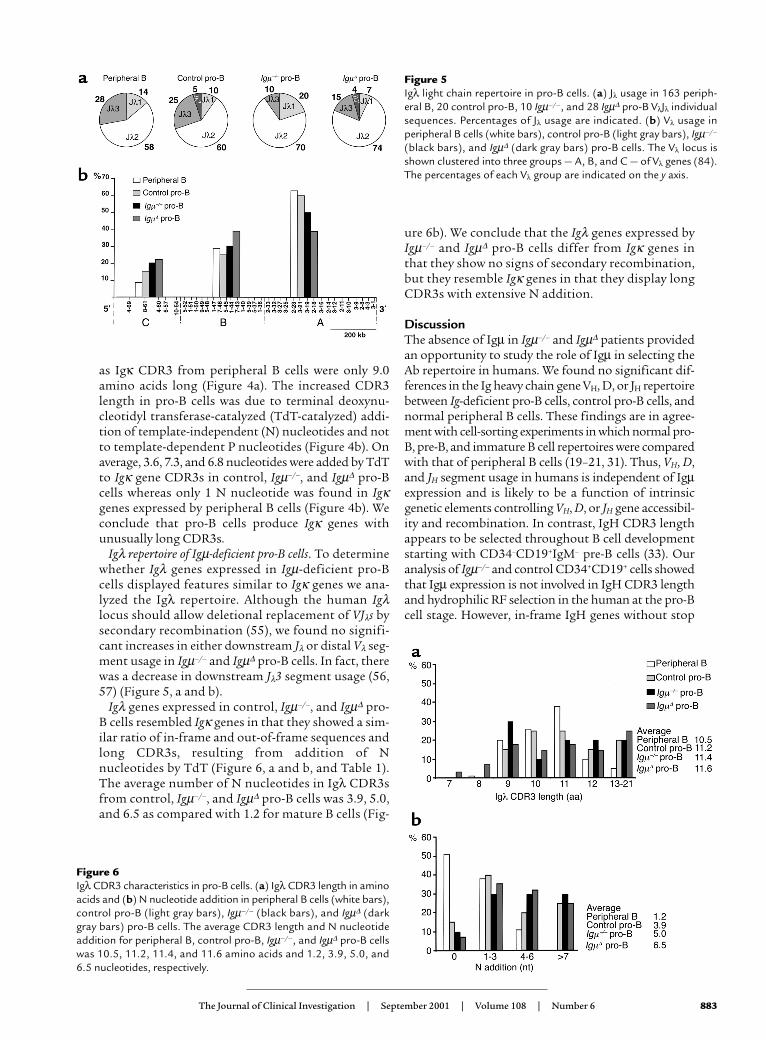
Igλ repertoire of Igµ-deficient pro-B cells. To determinewhether Igλ genes expressed in Igµ-deficient pro-Bcells displayed features similar to Igκ genes we ana-lyzed the Igλ repertoire. Although the human Igλlocus should allow deletional replacement of VJλs bysecondary recombination (55), we found no signifi-cant increases in either downstream Jλ or distal Vλ seg-ment usage in Igµ–/– and Igµ∆ pro-B cells. In fact, therewas a decrease in downstream Jλ3 segment usage (56,57) (Figure 5, a and b).
Igλ genes expressed in control, Igµ–/–, and Igµ∆ pro-B cells resembled Igκ genes in that they showed a sim-ilar ratio of in-frame and out-of-frame sequences andlong CDR3s, resulting from addition of Nnucleotides by TdT (Figure 6, a and b, and Table 1).The average number of N nucleotides in Igλ CDR3sfrom control, Igµ–/–, and Igµ∆ pro-B cells was 3.9, 5.0,and 6.5 as compared with 1.2 for mature B cells (Fig-
ure 6b). We conclude that the Igλ genes expressed byIgµ–/– and Igµ∆ pro-B cells differ from Igκ genes inthat they show no signs of secondary recombination,but they resemble Igκ genes in that they display longCDR3s with extensive N addition.
DiscussionThe absence of Igµ in Igµ–/– and Igµ∆ patients providedan opportunity to study the role of Igµ in selecting theAb repertoire in humans. We found no significant dif-ferences in the Ig heavy chain gene VH, D, or JH repertoirebetween Ig-deficient pro-B cells, control pro-B cells, andnormal peripheral B cells. These findings are in agree-ment with cell-sorting experiments in which normal pro-B, pre-B, and immature B cell repertoires were comparedwith that of peripheral B cells (19–21, 31). Thus, VH, D,and JH segment usage in humans is independent of Igµexpression and is likely to be a function of intrinsicgenetic elements controlling VH, D, or JH gene accessibil-ity and recombination. In contrast, IgH CDR3 lengthappears to be selected throughout B cell developmentstarting with CD34–CD19+IgM– pre-B cells (33). Ouranalysis of Igµ–/– and control CD34+CD19+ cells showedthat Igµ expression is not involved in IgH CDR3 lengthand hydrophilic RF selection in the human at the pro-Bcell stage. However, in-frame IgH genes without stop
The Journal of Clinical Investigation | September 2001 | Volume 108 | Number 6 883
Figure 5Igλ light chain repertoire in pro-B cells. (a) Jλ usage in 163 periph-eral B, 20 control pro-B, 10 Igµ–/–, and 28 Igµ∆ pro-B VλJλ individualsequences. Percentages of Jλ usage are indicated. (b) Vλ usage inperipheral B cells (white bars), control pro-B (light gray bars), Igµ–/–
(black bars), and Igµ∆ (dark gray bars) pro-B cells. The Vλ locus isshown clustered into three groups — A, B, and C — of Vλ genes (84).The percentages of each Vλ group are indicated on the y axis.
Figure 6Igλ CDR3 characteristics in pro-B cells. (a) Igλ CDR3 length in aminoacids and (b) N nucleotide addition in peripheral B cells (white bars),control pro-B (light gray bars), Igµ–/– (black bars), and Igµ∆ (darkgray bars) pro-B cells. The average CDR3 length and N nucleotideaddition for peripheral B, control pro-B, Igµ–/–, and Igµ∆ pro-B cellswas 10.5, 11.2, 11.4, and 11.6 amino acids and 1.2, 3.9, 5.0, and6.5 nucleotides, respectively.
codons were enriched in control CD34+CD19+ B cell pro-genitors, suggesting that an efficient selection processdriven by pre-BCRs operates in normal CD34+CD19+ Bcell precursors. These Igµ-positive B cell precursors dis-playing surface pro-B cell markers are likely to be in tran-sition to the pre-B cell stage and equivalent to the mouseC′ early pre-B cell fraction of Hardy’s classification (58).By analogy to Igµ or Igβ knockout mice, the C′ early pre-B cell fraction is missing in Igµ–/– CD34+CD19+ precur-sor B cells and results in a decrease of in-frame Igµrearrangements when compared with normalCD34+CD19+ precursor B cells (10, 59).
In the mouse, ψL has been implicated in IgH reper-toire selection by virtue of pairing with some but not allVH domains (16, 18). Analysis of the VH repertoire inλ5–/– mice showed that the normal repertoire shift seenbetween the pro-B and the pre-B cell stage was absent(16). However, differences in pairing efficiency betweenψL and IgH are not likely to influence the selectionagainst long or hydrophobic CDR3s in humans becausethese features are prevalent in control CD34+CD19+
cells, in immature B cells that have passed ψL selection,and in B cells that express ψLs in the periphery (31, 49).A more likely explanation for selection against long andhydrophobic IgH CDR3s is that these features are asso-ciated with self-reactivity and might also interferedirectly with IgH and IgL pairing (31, 49, 60, 61). Imma-ture B cells displaying such Ab’s therefore would besilenced by deletion or receptor editing, or alternatively,would fail to be positively selected in the mature B cellcompartment (39, 40, 42–44, 62–64).
In the mouse, two Igµ-mediated mechanismsaccount for selection against self-reactive or poorlypairing Ab’s during B cell development, receptor edit-ing and deletion (39–44). Editing makes a major con-tribution to the Ab repertoire in mice: up to 25% ofall Ab’s are produced by editing, but the role of dele-tion in repertoire selection is not known (65). Ourexperiments suggest that Igµ-mediated selection alsomakes a large contribution to shaping the human Abrepertoire. The selection against IgHs with long orhydrophobic CDR3s found in pro-B cells wouldrequire loss of at least 20–25% of all heavy chains.
In both mouse and human B cells, V(D)J recombina-tion is generally ordered, starting with IgH rearrange-ment in pro-B cells followed by IgL rearrangement inpre-B cells (66, 67). However, analysis of mouse Igµmutants and normal pro-B cells showed that IgL genescan recombine before IgH in pro-B cells (54, 68–71).Our experiments show that human control and Igµ-deficient pro-B cells are similar to their mouse coun-terparts in that they undergo Igκ and Igλ generearrangements. These results are in agreement withthe finding of rare IgL chain gene recombination innormal human pro-B cells and in Epstein-Barrvirus–transformed fetal B cell precursors (67, 72). Thus,IgL rearrangement in the human is similar to themouse in that it is not strictly dependent on Igµ expres-sion or pre-B cell development.
Up to 50% of human light chains in normal periph-eral B cells show N nucleotide addition, but Igκ or IgλCDR3s are never as long as 11 or 13 amino acids,respectively (49, 73–76). Igκ or Igλ genes found in con-trol and Igµ-deficient pro-B cells differ from thosefound in normal peripheral B cells in that they showextensive N nucleotide addition associated with longIgL CDR3s that can reach up to 21 amino acids. Thus,long IgL CDR3s are produced in early developing B cells but they appear to be incompatible with B celldevelopment and are deleted from the mature periph-eral B cell repertoire.
Igµ-deficient pro-B cells express Igκ genes that dis-play a bias to 3′ Jκs and 5′ Vκs consistent with persist-ent V(D)J recombination. However, the bias to 3′ Jκswas incomplete since there was no significant increasein Jκ5, the most downstream Jκ segment. In addition,there was no bias to downstream Jλs despite a genom-ic configuration that allows secondary rearrange-ments (55, 77, 78). We speculate that in the absence ofIgµ, human pro-B cells undergo several rounds ofrecombination on Igκ but do not survive long enoughto allow extensive secondary recombination. Theabsence of secondary recombination on Igλ in Igµ-deficient pro-B cells may result from a delayed recom-bination of this locus when compared with Igκ (79,80). Alternatively, secondary recombination may beless efficient for Igλ than Igκ.
Secondary Igκ recombination is prominent in Igµ-deficient pro-B cells yet not in normal control pro-Bcells. Secondary recombination in Igµ-deficient pro-Bcells therefore appears to be a default mechanism inthe absence of Igµ expression, and termination of sec-ondary recombination in developing B cells requiresBCR signaling. We speculate that the regulation of Iglight chain gene recombination during B cell develop-ment resembles that of T cell receptor-α (TCR-α)chains during T cell development in that recombina-tion is terminated by a yet to be determined positiveselection signal transduced by the BCR (81). Develop-ing B cells remain in the pre-B cell compartment for afew hours whereas developing T cells remain in theCD4+CD8+ double-positive compartment for 3–4 days(65, 82, 83). This kinetic difference may explain why Iglight chains are allelic excluded in B cells whereasTCR-α chains in T cells are not.
AcknowledgmentsWe thank M.J. Shlomchik and T.-A. Yang for commentson the manuscript and A. Deville, B. Lemmers, and J.Castro e Melo for help with bone marrow samples.
1. Pillai, S., and Baltimore, D. 1987. Formation of disulfide-linked µ2ω2tetramers in pre-B cells by the 18k ω-immunoglobulin light chain.Nature. 329:172–174.
2. Kerr, W.G., Cooper, M.D, Feng, L, Burrows, P.D, and Hendershot, L.M.1989. Mu heavy chains can associate with a pseudo-light chain complex(ΨL) in human pre-B cell lines. Int. Immunol. 1:355–361.
3. Karasuyama, H., Kudo, A., and Melchers, F. 1990. The proteins encod-ed by the VpreB and lambda 5 pre-B cell-specific genes can associatewith each other and with mu heavy chain. J. Exp. Med. 172:969–972.
4. Tsubata, T., and Reth, M. 1990. The products of pre-B cell-specific genes
884 The Journal of Clinical Investigation | September 2001 | Volume 108 | Number 6

(lambda 5 and VpreB) and the immunoglobulin mu chain form a com-plex that is transported onto the cell surface. J. Exp. Med. 172:973–976.
5. Hombach, J., Leclercq, L., Radbruch, A., Rajewsky, K., and Reth, M.1988. A novel 34-kd protein co-isolated with the IgM molecule in sur-face IgM-expressing cells. EMBO J. 7:3451–3456.
6. Hermanson, G.G., Eisenberg, D., Kincade, P.W. and Wall, R. 1988. B29:a member of the immunoglobulin gene superfamily exclusivelyexpressed on B-lineage cells. Proc. Natl. Acad. Sci. USA. 85:6890-6894.
7. Grawunder, U., et al. 1995. Down-regulation of RAG1 and RAG2 geneexpression in preB cells after functional immunoglobulin heavy chainrearrangement. Immunity. 3:601–608.
8. Kitamura, D., Roes, J., Kuhn, R., and Rajewsky, K. 1991. A B cell defi-cient mouse by targeted disruption of the membrane exons of theimmunoglobulin µ chain gene. Nature. 350:423–426.
9. Kitamura, D., et al. 1992. A critical role of λ5 protein in B cell develop-ment. Cell. 69:823–831.
10. Gong, S., and Nussenzweig, M.C.. 1996. Regulation of an early devel-opmental checkpoint in the B cell pathway by Ig beta. Science.272:411–414.
11. Mundt, C., Licence, S., Shimizu, T., Melchers, F., and Martensson, I.L.2001. Loss of Precursor B Cell Expansion but Not Allelic Exclusion inVpreB1/VpreB2 Double-deficient Mice. J. Exp. Med. 193:435–446.
12. Yel, L., et al. 1996. Mutations in the mu heavy-chain gene in patientswith agammaglobulinemia. N. Engl. J. Med. 335:1486–1493.
13. Schiff, C., Lemmers, B., Deville, A., Fougereau, M., and Meffre, E. 2000.Autosomal primary immunodeficiencies affecting human bone mar-row B cell differentiation. Immunol. Rev. 178:91–98.
14. Minegishi, Y., et al. 1998. Mutations in the human lambda5/14.1 generesult in B cell deficiency and agammaglobulinemia. J. Exp. Med.187:71–77.
15. Minegishi, Y., et al. 1999. Mutations in Igalpha (CD79a) result in a com-plete block in B-cell development. J. Clin. Invest. 104:1115–1121.
16. ten Boekel, E., Melchers, F., and Rolink, A.G. 1997. Changes in the V(H)gene repertoire of developing precursor B lymphocytes in mouse bonemarrow mediated by the pre-B cell receptor. Immunity. 7:357–368.
17. ten Boekel, E., Melchers, F., and Rolink, A.G. 1998. Precursor B cellsshowing H chain allelic inclusion display allelic exclusion at the level ofpre-B cell receptor surface expression. Immunity. 8:199–207.
18. Wasserman, R., et al. 1998. A novel mechanism for B cell repertoire mat-uration based on response by B cell precursors to pre-B receptor assem-bly. J. Exp. Med. 187:259–264.
19. Kraj, P., et al. 1997. The human heavy chain Ig V region gene repertoireis biased at all stages of B cell ontogeny, including early pre-B cells. J.Immunol. 158:5824–5832.
20. Rao, S.P., et al. 1999. Biased VH gene usage in early lineage human Bcells: evidence for preferential Ig gene rearrangement in the absence ofselection. J. Immunol. 163:2732–2740.
21. Milili, M., Schiff, C., Fougereau, M., and Tonnelle, C. 1996. The VDJrepertoire expressed in human preB cells reflects the selection of bonafide heavy chains. Eur. J. Immunol. 26:63–69.
22. Schroeder, H.W., Jr., Hillson, J.L., and Perlmutter, R.M. 1987. Earlyrestriction of the human antibody repertoire. Science. 238:791–793.
23. Schroeder, H.W., Jr., and Wang, J.Y. 1990. Preferential utilization of con-served immunoglobulin heavy chain variable gene segments duringhuman fetal life. Proc. Natl. Acad. Sci. USA. 87:6146–6150.
24. Hillson, J.L., et al. 1992. Emerging human B cell repertoire. Influence ofdevelopmental stage and interindividual variation. J. Immunol.149:3741–3752.
25. Cuisinier, A.M., Gauthier, L., Boubli, L., Fougereau, M., and Tonnelle,C. 1993. Mechanisms that generate human immunoglobulin diversityoperate from the 8th week of gestation in fetal liver. Eur. J. Immunol.23:110–118.
26. Pascual, V., and Capra, J.D. 1991. Human immunoglobulin heavy-chainvariable region genes: organization, polymorphism, and expression.Adv. Immunol. 49:1–74.
27. Huang, C., Stewart, A.K., Schwartz, R.S., and Stollar, B.D. 1992.Immunoglobulin heavy chain gene expression in peripheral blood Blymphocytes. J. Clin. Invest. 89:1331–1343.
28. Milner, E.C., Hufnagle, W.O., Glas, A.M., Suzuki, I., and Alexander, C.1995. Polymorphism and utilization of human VH Genes. Ann. N YAcad. Sci. 764:50–61.
29. Brezinschek, H.P., Brezinschek, R.I., and Lipsky, P.E. 1995. Analysis ofthe heavy chain repertoire of human peripheral B cells using single-cellpolymerase chain reaction. J. Immunol. 155:19–202.
30. Brezinschek, H.P., et al. 1997. Analysis of the human VH gene repertoire.Differential effects of selection and somatic hypermutation on humanperipheral CD5(+)/IgM+ and CD5(-)/IgM+ B cells. J. Clin. Invest.99:2488–2501.
31. Raaphorst, F.M., Raman, C.S., Tami, J., Fischbach, M., and Sanz, I. 1997.Human Ig heavy chain CDR3 regions in adult bone marrow pre-B cellsdisplay an adult phenotype of diversity: evidence for structural selec-tion of DH amino acid sequences. Int. Immunol. 9:1503–1515.
32. Dorner, T., et al. 1997. Analysis of the frequency and pattern of somat-ic mutations within nonproductively rearranged human variable heavychain genes. J. Immunol. 158:2779–2789.
33. Shiokawa, S., et al. 1999. IgM heavy chain complementarity-determin-ing region 3 diversity is constrained by genetic and somatic mechanismsuntil two months after birth. J. Immunol. 162:6060–6070.
34. Alt, F.W., et al. 1984. Ordered rearrangement of immunoglobulin heavychain variable region segments. EMBO J. 3:1209–1219.
35. Reth, M., Petrac, E., Wiese, P., Lobel, L., and Alt, F.W. 1987. Activation ofV kappa gene rearrangement in pre-B cells follows the expression ofmembrane-bound immunoglobulin heavy chains. EMBO J. 6:3299–3305.
36. Schlissel, M.S., and Baltimore, D. 1989. Activation of immunoglobulinkappa gene rearrangement correlates with induction of germline kappagene transcription. Cell. 58:1001–1007.
37. Iglesias, A., Kopf, M., Williams, G.S., Buhler, B., and Kohler, G. 1991.Molecular requirements for the mu-induced light chain gene rearrange-ment in pre-B cells. EMBO J. 10:2147–2155.
38. Tsubata, T., Tsubata, R., and Reth, M. 1992. Crosslinking of the cell sur-face immunoglobulin (mu-surrogate light chains complex) on pre-Bcells induces activation of V gene rearrangements at the immunoglob-ulin kappa locus. Int. Immunol. 4:637–641.
39. Nemazee, D.A., and Bürki, K. 1989. Clonal deletion of B lymphocytesin a transgenic mouse bearing anti-MHC class I antibody genes. Nature.337:562–566.
40. Hartley, S.B., et al. 1991. Elimination from peripheral lymphoid tissuesof self-reactive B lymphocytes recognizing membrane-bound antigens.Nature. 353:765–769.
41. Okamoto, M., et al. 1992. A transgenic model of autoimmune hemolyt-ic anemia. J. Exp. Med. 175:71–79.
42. Tiegs, S.L., Russell, D.M., and Nemazee, D. 1993. Receptor editing inself-reactive bone marrow B cells. J. Exp. Med. 177:1009–1020.
43. Gay, D., Saunders, T., Camper, S., and Weigert, M. 1993. Receptor edit-ing: an approach by autoreactive B cells to escape tolerance. J. Exp. Med.177:999–1008.
44. Radic, M.Z., Erickson, J., Litwin, S., and Weigert, M. 1993. B lympho-cytes may escape tolerance by revising their antigen receptors. J. Exp.Med. 177:1165–1173.
45. Yamagami, T., et al. 1999. Four of five RAG-expressing JCkappa–/–small pre-BII cells have no L chain gene rearrangements: detection byhigh-efficiency single cell PCR. Immunity. 11:309–316.
46. Meffre, E., et al. 1996. A human non-XLA immunodeficiency diseasecharacterized by blockage of B cell development at an early proB cellstage. J. Clin. Invest. 98:1519–1526.
47. Meffre, E., et al. 1997. A non-XLA primary deficiency causes the earliestknown defect of B cell differentiation in humans: a comparison with anXLA case. Immunol. Lett. 57:93-99.
48. Fais, F., et al. 1998. Chronic lymphocytic leukemia B cells expressrestricted sets of mutated and unmutated antigen receptors. J. Clin.Invest. 102:1515–1525.
49. Meffre, E., et al. 2000. Circulating human B cells that express surrogatelight chains and edited receptors. Nat. Immunol. 1:207–213.
50. Corbett, S.J., Tomlinson, I.M., Sonnhammer, E.L.L., Buck, D., and Win-ter, G. 1997. Sequence of the human immunoglobulin diversity (D) seg-ment locus: a systematic analysis provides no evidence for the use ofDIR segments, inverted D segments, “minor” D segments or D-Drecombination. J. Mol. Biol. 270:587–597.
51. Kabat, E.A., Wu, T.T., Perry, H.M., Gottesman, K.S., and Foeller, C. 1991.Sequences of proteins of immunological interest. US Department of Healthand Human Services. Bethesda, Maryland, USA. 2387 pp.
52. Desiderio, S.V., et al. 1984. Insertion of N regions into heavy-chaingenes is correlated with expression of terminal deoxytransferase in Bcells. Nature. 311:752–755.
53. Lafaille, J.J., DeCloux, A., Bonneville, M., Takagaki, Y., and Tonegawa,S. 1989. Junctional sequences of T cell receptor gamma delta genes:implications for gamma delta T cell lineages and for a novel intermedi-ate of V-(D)-J joining. Cell. 59:859–870.
54. Bentolila, L.A., et al. 1999. Extensive junctional diversity in Ig lightchain genes from early B cell progenitors of mu MT mice. J. Immunol.162:2123–2128.
55. Vasicek, T.J., and Leder, P. 1990. Structure and expression of the humanimmunoglobulin lambda genes. J. Exp. Med. 172:609–620.
56. Frippiat, J.P., et al. 1995. Organization of the human immunoglobulinlambda light-chain locus on chromosome 22q11.2. Hum. Mol. Genet.4:983–991.
57. Williams, S.C., et al. 1996. Sequence and evolution of the humangermline V lambda repertoire. J. Mol. Biol. 264:220–232.
58. Hardy, R.R., Carmack, C.E., Shinton, S.A., Kemp, J.D., and Hayakawa,K. 1991. Resolution and characterization of pro-B and pre-pro-B cellstages in normal mouse bone marrow. J. Exp. Med. 172:1213–1225.
59. Reichlin, A., et al. 2001. B cell development is arrested at the immatureb cell stage in mice carrying a mutation in the cytoplasmic domain ofimmunoglobulin beta. J. Exp. Med. 193:13–24.
The Journal of Clinical Investigation | September 2001 | Volume 108 | Number 6 885
60. Klonowski, K.D., Primiano, L.L., and Monestier, M. 1999. Atypical VH-D-JH rearrangements in newborn autoimmune MRL mice. J. Immunol.162:1566–1572.
61. Schroeder, H.W., and Kirkham, P.M. 2000. Marriage, divorce, andpromiscuity in human B cells. Nature Immunol. 1:187–188.
62. Sandel, P.C., and Monroe, J.G. 1999. Negative selection of immature Bcells by receptor editing or deletion is determined by site of antigenencounter. Immunity. 10:289–299.
63. Gu, H., Tarlinton, D., Muller, W., Rajewsky, K., and Forster, I. 1991.Most peripheral B cells in mice are ligand selected. J. Exp. Med.173:1357–1371.
64. Levine, M.H., et al. 2000. A B-cell receptor-specific selection step gov-erns immature to mature B cell differentiation. Proc. Natl. Acad. Sci. USA.97:2743–2748.
65. Casellas, R., et al. 2001. Contribution of receptor editing to the antibodyrepertoire. Science. 291:1541–1544.
66. Blackwell, T.K., et al. 1989. Isolation of scid pre-B cells that rearrangekappa light chain genes: formation of normal signal and abnormal cod-ing joins. EMBO J. 8:735–742.
67. Ghia, P., et al. 1996. Ordering of human bone marrow B lymphocyteprecursors by single-cell polymerase chain reaction analyses of therearrangement status of the immunoglobulin H and L chain loci. J. Exp.Med. 184:2217–2229.
68. Ehlich, A., et al. 1993. Immunoglobulin heavy and light chain genesrearrange independently at early stages of B cell development. Cell.72:695–704.
69. Chen, J., et al. 1993. Immunoglobulin gene rearrangement in B cell defi-cient mice generated by targeted deletion of the JH locus. Int. Immunol.5:647–656.
70. Grawunder, U., Haasner, D., Melchers, F., and Rolink, A. 1993.Rearrangement and expression of kappa light chain genes can occurwithout mu heavy chain expression during differentiation of pre-B cells.Int. Immunol. 5:1609–1618.
71. Novobrantseva, T.I., et al. 1999. Rearrangement and expression ofimmunoglobulin light chain genes can precede heavy chain expres-sion during normal B cell development in mice. J. Exp. Med.189:75–88.
72. Kubagawa, H., Cooper, M.D., Carroll, A.J., and Burrows, P.D. 1989.Light-chain gene expression before heavy-chain gene rearrangement in
pre-B cells transformed by Epstein-Barr virus. Proc. Natl. Acad. Sci. USA.86:2356–2360.
73. Klein, R., Jaenichen, R., and Zachau, H.G. 1993. Expressed humanimmunoglobulin kappa genes and their hypermutation. Eur. J.Immunol. 23:3248–3262.
74. Klein, U., Kuppers, R., and Rajewsky, K. 1993. Human IgM+IgD+ B cells,the major B cell subset in the peripheral blood, express V kappa geneswith no or little somatic mutation throughout life. Eur. J. Immunol.23:3272–3277.
75. Fischer, M., Klein, U., and Kuppers, R. 1997. Molecular single-cell analy-sis reveals that CD5-positive peripheral blood B cells in healthy humansare characterized by rearranged Vkappa genes lacking somatic muta-tion. J. Clin. Invest. 100:1667–1676.
76. Foster, S.J., Brezinschek, H.P., Brezinschek, R.I., and Lipsky, P.E. 1997.Molecular mechanisms and selective influences that shape the kappagene repertoire of IgM+ B cells. J. Clin. Invest. 99:1614–1627.
77. Berinstein, N., Levy, S., and Levy, R. 1989. Activation of an excludedimmunoglobulin allele in a human B lymphoma cell line. Science.244:337–339.
78. Stiernholm, N.B., and Berinstein, N.L. 1993. Up-regulated recombina-tion-activating gene expression in sIg- variants of a human mature Bcell line undergoing secondary Ig lambda rearrangements in cell cul-ture. Eur. J. Immunol. 23:1501–1507.
79. Hieter, P.A., Korsmeyer, S.J., Waldmann, T.A., and Leder, P. 1981.Human immunoglobulin kappa light-chain genes are deleted orrearranged in lambda-producing B cells. Nature. 290:368–372.
80. Korsmeyer, S.J., et al. 1981. Developmental hierarchy of immunoglob-ulin gene rearrangements in human leukemic pre-B-cells. Proc. Natl.Acad. Sci. USA. 78:7096–7100.
81. Borgulya, P., Kishi, H., Uematsu, Y., and Boehmer, H.V. 1992. Exclusionand inclusion of a and b T cell receptor alleles. Cell. 69:529–537.
82. Huesmann, M., Scott, B., Kisielow, P., and von Boehmer, H. 1991. Kinet-ics and efficacy of positive selection in the thymus of normal and T cellreceptor transgenic mice. Cell. 66:533–540.
83. Malissen, M., et al. 1988. A T cell clone expresses two T cell receptoralpha genes but uses one alpha beta heterodimer for allorecognitionand self MHC-restricted antigen recognition. Cell. 55:49–59.
84. Lefranc, M.P. 2000. Locus maps and genomic repertoire of the humanIg genes. The Immunologist. 8:80–87.
886 The Journal of Clinical Investigation | September 2001 | Volume 108 | Number 6