Immobilized metal ion a nity nanospheres for -amylase...
13
Turk J Chem (2014) 38: 28 – 40 c ⃝ T ¨ UB ˙ ITAK doi:10.3906/kim-1301-87 Turkish Journal of Chemistry http://journals.tubitak.gov.tr/chem/ Research Article Immobilized metal ion affinity nanospheres for α -amylase immobilization T¨ ulden KALBURCU 1 , M¨ unire Nalan T ¨ UZMEN 2, * , Sinan AKG ¨ OL 3 , Adil DEN ˙ IZL ˙ I 4 1 Department of Chemistry, Faculty of Science and Arts, Aksaray University, Aksaray, Turkey 2 Department of Chemistry, Faculty of Science, Dokuz Eyl¨ ul University, ˙ Izmir, Turkey 3 Department of Biochemistry, Faculty of Science, Ege University, ˙ Izmir, Turkey 4 Department of Chemistry, Faculty of Science, Hacettepe University, Ankara, Turkey Received: 31.01.2013 • Accepted: 25.04.2013 • Published Online: 16.12.2013 • Printed: 20.01.2014 Abstract: Immobilized metal chelate affinity chromatography (IMAC) support was practiced for α -amylase immo- bilization. Poly(hydroxyethylmethacrylate-methacryloylamidotryptophan)-Ni 2+ [p(HEMA-MAT)-Ni 2+ ] nanospheres, average diameter 100 nm, were produced by surfactant free emulsion polymerization. Characterizations of p(HEMA- MAT)-Ni 2+ nanospheres were carried out by Fourier transform infrared (FTIR) spectroscopy and scanning electron microscope (SEM). In addition, average particle size, size distribution, and surface charge were specified. The amount of N-methacryloylamidotryptophan (MAT) incorporated to polymer was determined as 1.95 mmol/g polymers by using nitrogen stoichiometry. The specific surface areas of poly(hydroxyethylmethacrylate) [p(HEMA)] and p(HEMA-MAT) nanospheres were calculated as 1856 m 2 /g and 1914 m 2 /g, respectively. Protein adsorption increased with increasing initial protein concentration and maximum α -amylase adsorption on p(HEMA-MAT)-Ni 2+ nanospheres was observed at pH 4.0. Both free and immobilized α -amylase showed pH optimum at pH 7.0. It was determined that the immobilized α -amylase had better thermostability than the free one. Immobilization of the enzyme did not significantly change the kinetic parameters. The storage stability of α -amylase increased upon immobilization. It was also observed that p(HEMA-MAT)-Ni 2+ nanospheres can be repeatedly used for α -amylase immobilization. Key words: α -Amylase, nanospheres, IMAC, enzyme immobilization, adsorption 1. Introduction One of the hydrolyzing enzymes widely used in different industries is α -amylase (1,4- α -d-glucan-glucanhydrolase, EC. 3.2.1.1), which is an endo-acting enzyme. While α -amylase hydrolyzes the α -1,4-glycosidic bonds, it by- passes α -1,6-glycosidic linkages in starch and related substrates. 1-4 Starch hydrolysis by α -amylase to low molecular weight products is widely utilized in the food, paper, textile, distillery, and brewing industries. 5 Amylase cannot be used repeatedly because of its poor stability. 6,7 Amylase immobilization on water insolu- ble carriers appears to be the most hopeful way to get more stable products for multiple uses. 8-15 Enzyme immobilization can be achieved by several methods. These immobilization methods consist of enveloping the enzyme into a gel matrix, encapsulation, incorporation into emulsions and membranes, binding to a support by adsorption, coordination, or covalent binding. Among these immobilization methods, adsorption has the advantages of improving the control of the reactor, permitting reuse of the enzyme, and avoiding protein con- tamination of the final product. In other respects, noncovalent adsorption techniques may be a good option * Correspondence: [email protected] 28
Transcript of Immobilized metal ion a nity nanospheres for -amylase...
Turk J Chem
(2014) 38: 28 – 40
c⃝ TUBITAK
doi:10.3906/kim-1301-87
Turkish Journal of Chemistry
http :// journa l s . tub i tak .gov . t r/chem/
Research Article
Immobilized metal ion affinity nanospheres for α-amylase immobilization
Tulden KALBURCU1, Munire Nalan TUZMEN2,∗, Sinan AKGOL3, Adil DENIZLI41Department of Chemistry, Faculty of Science and Arts, Aksaray University, Aksaray, Turkey
2Department of Chemistry, Faculty of Science, Dokuz Eylul University, Izmir, Turkey3Department of Biochemistry, Faculty of Science, Ege University, Izmir, Turkey
4Department of Chemistry, Faculty of Science, Hacettepe University, Ankara, Turkey
Received: 31.01.2013 • Accepted: 25.04.2013 • Published Online: 16.12.2013 • Printed: 20.01.2014
Abstract: Immobilized metal chelate affinity chromatography (IMAC) support was practiced for α -amylase immo-
bilization. Poly(hydroxyethylmethacrylate-methacryloylamidotryptophan)-Ni2+ [p(HEMA-MAT)-Ni2+ ] nanospheres,
average diameter 100 nm, were produced by surfactant free emulsion polymerization. Characterizations of p(HEMA-
MAT)-Ni2+ nanospheres were carried out by Fourier transform infrared (FTIR) spectroscopy and scanning electron
microscope (SEM). In addition, average particle size, size distribution, and surface charge were specified. The amount
of N-methacryloylamidotryptophan (MAT) incorporated to polymer was determined as 1.95 mmol/g polymers by using
nitrogen stoichiometry. The specific surface areas of poly(hydroxyethylmethacrylate) [p(HEMA)] and p(HEMA-MAT)
nanospheres were calculated as 1856 m2 /g and 1914 m2 /g, respectively. Protein adsorption increased with increasing
initial protein concentration and maximum α -amylase adsorption on p(HEMA-MAT)-Ni2+ nanospheres was observed at
pH 4.0. Both free and immobilized α -amylase showed pH optimum at pH 7.0. It was determined that the immobilized
α -amylase had better thermostability than the free one. Immobilization of the enzyme did not significantly change
the kinetic parameters. The storage stability of α -amylase increased upon immobilization. It was also observed that
p(HEMA-MAT)-Ni2+ nanospheres can be repeatedly used for α -amylase immobilization.
Key words: α -Amylase, nanospheres, IMAC, enzyme immobilization, adsorption
1. Introduction
One of the hydrolyzing enzymes widely used in different industries is α -amylase (1,4-α -d-glucan-glucanhydrolase,
EC. 3.2.1.1), which is an endo-acting enzyme. While α -amylase hydrolyzes the α -1,4-glycosidic bonds, it by-
passes α -1,6-glycosidic linkages in starch and related substrates.1−4 Starch hydrolysis by α -amylase to low
molecular weight products is widely utilized in the food, paper, textile, distillery, and brewing industries.5
Amylase cannot be used repeatedly because of its poor stability.6,7 Amylase immobilization on water insolu-
ble carriers appears to be the most hopeful way to get more stable products for multiple uses.8−15 Enzyme
immobilization can be achieved by several methods. These immobilization methods consist of enveloping the
enzyme into a gel matrix, encapsulation, incorporation into emulsions and membranes, binding to a support
by adsorption, coordination, or covalent binding. Among these immobilization methods, adsorption has the
advantages of improving the control of the reactor, permitting reuse of the enzyme, and avoiding protein con-
tamination of the final product. In other respects, noncovalent adsorption techniques may be a good option
∗Correspondence: [email protected]
28

KALBURCU et al./Turk J Chem
due to being simple and requiring little work and time. Otherwise, inactivated enzyme can be desorbed and
supports can be used again. This decreases the final price and amounts of residues.16 In the past few decades,
many immobilization methods and carrier materials have been investigated.17,18 Inorganic supports, synthetic
polymers, and natural macromolecules are classified as carrier materials for enzyme immobilization.19 Poly-
meric carriers have the advantageous of good mechanical properties, ease of preparation, and applicability to
introduce bio-friendly components for improving biocompatibility. Additionally, reactive functional groups on
polymeric carriers provide necessary interactions with the enzymes.20,21 Many studies about synthesis of micro-
sized polymeric matrices and their applicability in protein separation with the succor of a specific ligand coating
the surface of the particles are published.22 Unfortunately, studies on the application of nanosized particles in
the immobilization of enzymes are very limited. Nanoparticles can produce larger specific surface area due to
their nanoscopic size and hence higher protein binding capacity may be determined. Consequently, nanosized
particles with large surface areas may be suitable carriers for enzyme immobilization.23,24 While immobilizing
an enzyme into porous materials, a decrease in mass-transfer resistance is assumed for smaller porous particles.
The reason for this expectation is the shortened diffusional path of substrates when compared to large-sized
porous materials. As a result, nanoparticles provide several advantages in the optimization of immobilized
enzymes: minimum diffusional limitation, maximum surface area per unit mass, and high enzyme loading.25
Immobilized metal-affinity chromatography (IMAC), introduced by Porath et al. in 1975,26 has become
a prevalent analytical and preparative separation method. This separation method can be used for therapeutic
proteins, peptides, nucleic acids, hormones, and enzymes.27 Covalently bound chelating compounds on solid
chromatographic supports are used in IMAC to entrap metal ions. These metal ions used as affinity ligands
generate coordinative binding with some amino acid residues exposed on protein surfaces.28 IMAC is more
favorable than other biospecific affinity chromatographic techniques that have a similar order of affinity con-
stants. The benefits of IMAC can be compiled as ligand stability, high protein loading, rapid purification, mild
elution conditions, simple regeneration, and low cost. In industrial applications, these benefits are critical when
developing large-scale purification procedures.29
In the present study, poly(hydroxyethylmethacrylate-methacryloylamidotryptophan) p(HEMA-MAT)
nanospheres were synthesized by surfactant free emulsion polymerization of 2-hydroxyethyl methacrylate (HEMA)
and MAT. Then p(HEMA-MAT) nanospheres were incorporated with Ni2+ ions. This newly coined IMAC
support was characterized by FTIR spectroscopy, SEM, elemental analysis, particle size, and surface area
measurements and then used in α -amylase immobilization. α -Amylase adsorption onto these metal-chelated
nanospheres was optimized by varying different parameters such as pH, initial α -amylase concentration, temper-
ature, and ionic strength. Desorption of α -amylase and reusability of these metal-chelated affinity nanospheres
and the kinetic parameters of the enzyme (KM and Vmax) were also tested.
2. Results and discussion
2.1. Characterization of p(HEMA-MAT)-Ni2+ nanospheres
This study has the characteristic advantage of the elimination of the activation and ligand coupling steps during
the preparation of the affinity matrices. Moreover, using a known amount of ligand in the polymer preparation
mixture and the good reproducibility of the affinity matrix are further advantages over other methods.
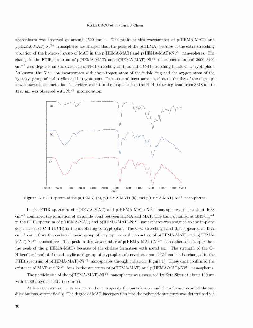
FTIR spectra of p(HEMA), p(HEMA-MAT), and p(HEMA-MAT)-Ni2+ nanospheres are shown in Figure
1. Stretching vibration of hydroxyl groups of both p(HEMA), p(HEMA-MAT), and p(HEMA-MAT)-Ni2+
29
KALBURCU et al./Turk J Chem
nanospheres was observed at around 3500 cm−1 . The peaks at this wavenumber of p(HEMA-MAT) and
p(HEMA-MAT)-Ni2+ nanospheres are sharper than the peak of the p(HEMA) because of the extra stretching
vibration of the hydroxyl group of MAT in the p(HEMA-MAT) and p(HEMA-MAT)-Ni2+ nanospheres. The
change in the FTIR spectrum of p(HEMA-MAT) and p(HEMA-MAT)-Ni2+ nanospheres around 3000–3400
cm−1 also depends on the existence of N–H stretching and aromatic C–H stretching bands of L-tryptophan.
As known, the Ni2+ ion incorporates with the nitrogen atom of the indole ring and the oxygen atom of the
hydroxyl group of carboxylic acid in tryptophan. Due to metal incorporation, electron density of these groups
moves towards the metal ion. Therefore, a shift in the frequencies of the N–H stretching band from 3378 nm to
3375 nm was observed with Ni2+ incorporation.
4000.0 3600 3200 2800 2400 2000 1800 1600 1400 1200 1000 800 650.0cm–1
a)
b)
c)
Figure 1. FTIR spectra of the p(HEMA) (a), p(HEMA-MAT) (b), and p(HEMA-MAT)-Ni2+ nanospheres.
In the FTIR spectrum of p(HEMA-MAT) and p(HEMA-MAT)-Ni2+ nanospheres, the peak at 1638
cm−1 confirmed the formation of an amide bond between HEMA and MAT. The band obtained at 1045 cm−1
in the FTIR spectrum of p(HEMA-MAT) and p(HEMA-MAT)-Ni2+ nanospheres was assigned to the in-plane
deformation of C-H (βCH) in the indole ring of tryptophan. The C–O stretching band that appeared at 1322
cm−1 came from the carboxylic acid group of tryptophan in the structure of p(HEMA-MAT) and p(HEMA-
MAT)-Ni2+ nanospheres. The peak in this wavenumber of p(HEMA-MAT)-Ni2+ nanospheres is sharper than
the peak of the p(HEMA-MAT) because of the chelate formation with metal ion. The strength of the O–
H bending band of the carboxylic acid group of tryptophan observed at around 950 cm−1 also changed in the
FTIR spectrum of p(HEMA-MAT)-Ni2+ nanospheres through chelation (Figure 1). These data confirmed the
existence of MAT and Ni2+ ions in the structures of p(HEMA-MAT) and p(HEMA-MAT)-Ni2+ nanospheres.
The particle size of the p(HEMA-MAT)-Ni2+ nanospheres was measured by Zeta Sizer at about 100 nm
with 1.189 polydispersity (Figure 2).
At least 30 measurements were carried out to specify the particle sizes and the software recorded the size
distributions automatically. The degree of MAT incorporation into the polymeric structure was determined via
30
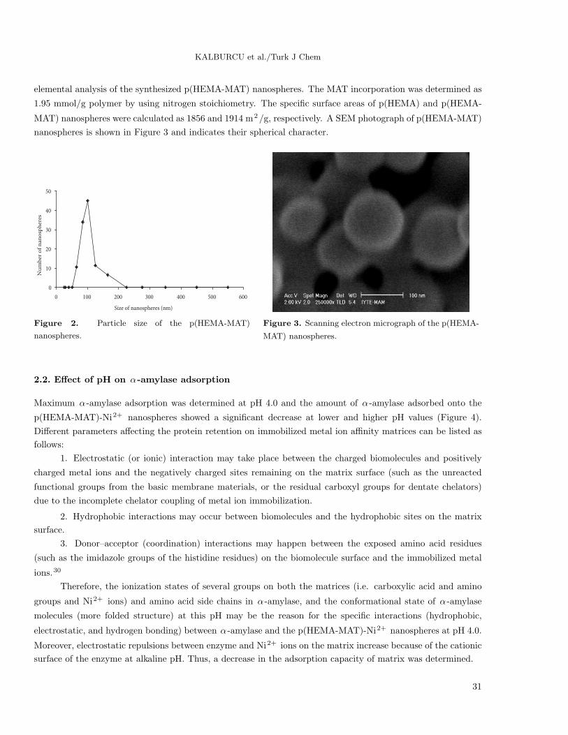
KALBURCU et al./Turk J Chem
elemental analysis of the synthesized p(HEMA-MAT) nanospheres. The MAT incorporation was determined as
1.95 mmol/g polymer by using nitrogen stoichiometry. The specific surface areas of p(HEMA) and p(HEMA-
MAT) nanospheres were calculated as 1856 and 1914 m2/g, respectively. A SEM photograph of p(HEMA-MAT)
nanospheres is shown in Figure 3 and indicates their spherical character.
0
10
20
30
40
50
0 100 200 300 400 500 600
Size of nanospheres (nm)
Nu
mb
er o
f n
an
osp
her
es
Figure 2. Particle size of the p(HEMA-MAT)
nanospheres.
Figure 3. Scanning electron micrograph of the p(HEMA-
MAT) nanospheres.
2.2. Effect of pH on α-amylase adsorption
Maximum α -amylase adsorption was determined at pH 4.0 and the amount of α -amylase adsorbed onto the
p(HEMA-MAT)-Ni2+ nanospheres showed a significant decrease at lower and higher pH values (Figure 4).
Different parameters affecting the protein retention on immobilized metal ion affinity matrices can be listed as
follows:
1. Electrostatic (or ionic) interaction may take place between the charged biomolecules and positively
charged metal ions and the negatively charged sites remaining on the matrix surface (such as the unreacted
functional groups from the basic membrane materials, or the residual carboxyl groups for dentate chelators)
due to the incomplete chelator coupling of metal ion immobilization.
2. Hydrophobic interactions may occur between biomolecules and the hydrophobic sites on the matrix
surface.
3. Donor–acceptor (coordination) interactions may happen between the exposed amino acid residues
(such as the imidazole groups of the histidine residues) on the biomolecule surface and the immobilized metal
ions.30
Therefore, the ionization states of several groups on both the matrices (i.e. carboxylic acid and amino
groups and Ni2+ ions) and amino acid side chains in α -amylase, and the conformational state of α -amylase
molecules (more folded structure) at this pH may be the reason for the specific interactions (hydrophobic,
electrostatic, and hydrogen bonding) between α -amylase and the p(HEMA-MAT)-Ni2+ nanospheres at pH 4.0.
Moreover, electrostatic repulsions between enzyme and Ni2+ ions on the matrix increase because of the cationic
surface of the enzyme at alkaline pH. Thus, a decrease in the adsorption capacity of matrix was determined.
31

KALBURCU et al./Turk J Chem
2.3. Effect of the initial concentration on α-amylase adsorption
Effect of the initial concentration of α -amylase on adsorption is shown in Figure 5. It is clearly seen from
this figure that the amount of adsorbed α -amylase increased with increasing initial α -amylase concentration.
Nonspecific α -amylase adsorption on the plain p(HEMA) nanospheres (33.0 ± 2.9 mg/g) was negligible. The
increase in the adsorption capacity of p(HEMA-MAT)-Ni2+ nanospheres can be explained by the ternary
complex formation between Ni2+ ions, MAT, and protein molecules.
0
1000
2000
3000
4000
5000
2 3 4 5 6 7 8 9 10pH
Q (
mg/
g n
ano
sph
ere)
0
1000
2000
3000
4000
5000
6000
0 0.25 0.5 0.75 1 1.25
p(HEMA-MAT)-Ni(II)
p(HEMA-MAT)
p(HEMA)
Q (
mg/
g n
ano
sph
ere)
Initial amylase concentration
Figure 4. Effect of pH on α -amylase adsorption onto
p(HEMA-MAT)-Ni2+ nanospheres (c: 1.0 mg/mL, T: 25◦C, time: 60 min).
Figure 5. Effect of the initial concentration on α -
amylase adsorption onto the p(HEMA), p(HEA-MAT),
and p(HEMA-MAT)-Ni2+ nanospheres (pH: 4.0; T:
25 ◦C; time: 60 min).
The amount of α -amylase adsorbed per unit mass of the p(HEMA-MAT) nanospheres increased first with
increasing α -amylase concentration and then reached a plateau value at 1 mg/mL. The active adsorption sites
on the nanospheres may be saturated with protein molecules at this α -amylase concentration. The maximum
amount of α -amylase adsorbed on the p(HEMA-MAT) nanospheres was determined as 2803.4 ± 107.0 mg/g at
this concentration. A nearly 2-fold increase was observed in the adsorption capacity of p(HEMA-MAT)-Ni2+
nanospheres (4468.1 ± 177.5 mg/g) at the same concentration. The electron donating side chains of proteins,
such as histidine and cysteine, are connected to metals. The chelate polymer structure and pH influence the
binding.31 The reason for this increase observed in the adsorption capacity of the Ni2+ -chelated p(HEMA-MAT)
nanospheres may be these high specific interactions.
2.4. Effect of ionic strength on α-amylase adsorption
The α -amylase adsorption capacity of p(HEMA-MAT)-Ni2+ nanospheres decreased about 40.0% with increas-
ing NaCl (Figure 6). Protein stability changes in different salt solutions because of the ion-specific effect of
salts. Stabilizing or destabilizing effects of salts may be arranged typically following the Hofmeister series:
SO2−4 > H2PO
−4 > CH3COO− > Cl− > Br− > I− > ClO−
4 > SCN− .
The physicochemical properties and interactions between proteins are affected by salts such as Na2SO4 ,
NaCl, NaBr, NaI, NaClO4 , and NaSCN. Additionally, these salts may change water structure by altering
the hydrophobic and electrostatic interactions at high concentrations.32 Binding forces within the matrices of
protein molecules, such as hydrophobic and electrostatic interactions, can be influenced by ionic strength and
32
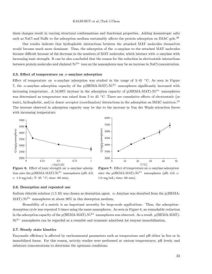
KALBURCU et al./Turk J Chem
these changes result in varying structural conformations and functional properties. Adding kosmotropic salts
such as NaCl and NaBr to the adsorption medium sustainably affects the protein adsorption on IMAC gels.30
Our results indicate that hydrophobic interactions between the attached MAT molecules themselves
would become much more dominant. Thus, the adsorption of the α -amylase to the attached MAT molecules
became difficult because of the decrease in the numbers of MAT molecules, which interact with α -amylase with
increasing ionic strength. It can be also concluded that the reason for the reduction in electrostatic interactions
between protein molecules and chelated Ni2+ ions on the nanospheres may be an increase in NaCl concentration.
2.5. Effect of temperature on α-amylase adsorption
Effect of temperature on α -amylase adsorption was studied in the range of 5–45 ◦C. As seen in Figure
7, the α -amylase adsorption capacity of the p(HEMA-MAT)-Ni2+ nanospheres significantly increased with
increasing temperature. A 54.89% increase in the adsorption capacity of p(HEMA-MAT)-Ni2+ nanospheres
was determined as temperature was raised from 5 to 45 ◦C. There are cumulative effects of electrostatic (or
ionic), hydrophobic, and/or donor–acceptor (coordination) interactions in the adsorption on IMAC matrices.33
The increase observed in adsorption capacity may be due to the increase in Van der Waals attraction forces
with increasing temperature.
2000
2500
3000
3500
4000
4500
5000
0 0.25 0.5 0.75 1
c NaCl (M)
Q (
mg/
g n
ano
sph
ere)
3000
3500
4000
4500
5000
5500
6000
0 10 20 30 40 50
T (°C)
Q (
mg/
g n
ano
sph
ere)
Figure 6. Effect of ionic strength on α -amylase adsorp-
tion onto the p(HEMA-MAT)-Ni2+ nanospheres (pH: 4.0;
c: 1.0 mg/mL; T: 25 ◦C; time: 60 min).
Figure 7. Effect of temperature on α -amylase adsorption
onto the p(HEMA-MAT)-Ni2+ nanospheres (pH: 4.0; c:
1.0 mg/mL; time: 60 min).
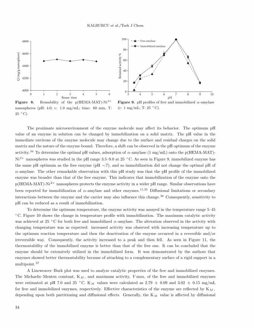
2.6. Desorption and repeated use
Sodium chloride solution (1.5 M) was chosen as desorption agent. α -Amylase was desorbed from the p(HEMA-
MAT)-Ni2+ nanospheres at about 90% in this desorption medium.
Reusability of a matrix is an important necessity for large-scale applications. Thus, the adsorption–
desorption cycle was repeated 5 times using the same nanospheres. As seen in Figure 8, no remarkable reduction
in the adsorption capacity of the p(HEMA-MAT)-Ni2+ nanospheres was observed. As a result, p(HEMA-MAT)-
Ni2+ nanospheres can be regarded as a reusable and economic adsorbent for enzyme immobilization.
2.7. Steady state kinetics
Enzymatic efficiency is affected by environmental parameters such as temperature and pH either in free or in
immobilized forms. For this reason, activity studies were performed at various temperatures, pH levels, and
substrate concentrations to determine the optimum conditions.
33
KALBURCU et al./Turk J Chem
4000
4200
4400
4600
4800
0 1 2 3 4 5 6Reuse time
Q (
mg/
g n
ano
sph
ere)
0
20
40
60
80
100
3 4 5 6 7 8 9 10
Ret
ain
ed a
ctiv
ity
%
pH
Free amylase
Immobilized amylase
Figure 8. Reusability of the p(HEMA-MAT)-Ni2+
nanospheres (pH: 4.0; c: 1.0 mg/mL; time: 60 min, T:
25 ◦C).
Figure 9. pH profiles of free and immobilized α -amylase
(c: 1 mg/mL; T: 25 ◦C).
The proximate microenvironment of the enzyme molecule may affect its behavior. The optimum pH
value of an enzyme in solution can be changed by immobilization on a solid matrix. The pH value in the
immediate environs of the enzyme molecule may change due to the surface and residual charges on the solid
matrix and the nature of the enzyme bound. Therefore, a shift can be observed in the pH optimum of the enzyme
activity.34 To determine the optimal pH values, adsorption of α -amylase (1 mg/mL) onto the p(HEMA-MAT)-
Ni2+ nanospheres was studied in the pH range 3.5–9.0 at 25 ◦C. As seen in Figure 9, immobilized enzyme has
the same pH optimum as the free enzyme (pH ∼7), and so immobilization did not change the optimal pH of
α -amylase. The other remarkable observation with this pH study was that the pH profile of the immobilized
enzyme was broader than that of the free enzyme. This indicates that immobilization of the enzyme onto the
p(HEMA-MAT)-Ni2+ nanospheres protects the enzyme activity in a wider pH range. Similar observations have
been reported for immobilization of α -amylase and other enzymes.11,35 Diffusional limitations or secondary
interactions between the enzyme and the carrier may also influence this change.36 Consequently, sensitivity to
pH can be reduced as a result of immobilization.
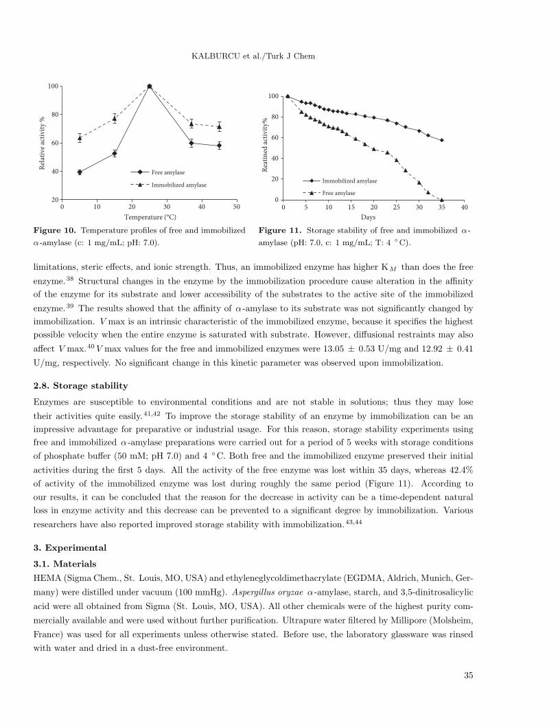
To determine the optimum temperature, the enzyme activity was assayed in the temperature range 5–45◦C. Figure 10 shows the change in temperature profile with immobilization. The maximum catalytic activity
was achieved at 25 ◦C for both free and immobilized α -amylase. The alteration observed in the activity with
changing temperature was as expected: increased activity was observed with increasing temperature up to
the optimum reaction temperature and then the deactivation of the enzyme occurred in a reversible and/or
irreversible way. Consequently, the activity increased to a peak and then fell. As seen in Figure 11, the
thermostability of the immobilized enzyme is better than that of the free one. It can be concluded that the
enzyme should be extensively utilized in the immobilized form. It was demonstrated by the authors that
enzymes showed better thermostability because of attaching to a complementary surface of a rigid support in a
multipoint.37
A Lineweaver–Burk plot was used to analyze catalytic properties of the free and immobilized enzymes.
The Michaelis–Menten constant, KM , and maximum activity, V max, of the free and immobilized enzymes
were estimated at pH 7.0 and 25 ◦C. KM values were calculated as 2.79 ± 0.09 and 3.02 ± 0.15 mg/mL
for free and immobilized enzymes, respectively. Effective characteristics of the enzyme are reflected by KM ,
depending upon both partitioning and diffusional effects. Generally, the KM value is affected by diffusional
34
KALBURCU et al./Turk J Chem
20
40
60
80
100
0 10 20 30 40 50
Rel
ativ
e ac
tivi
ty %
Temperature (°C)
Free amylase
Immobilized amylase
0
20
40
60
80
100
0 5 10 15 20 25 30 35 40
Rea
tin
ed a
ctiv
ity%
Days
Immobilized amylase
Free amylase
Figure 10. Temperature profiles of free and immobilized
α -amylase (c: 1 mg/mL; pH: 7.0).
Figure 11. Storage stability of free and immobilized α -
amylase (pH: 7.0, c: 1 mg/mL; T: 4 ◦C).
limitations, steric effects, and ionic strength. Thus, an immobilized enzyme has higher KM than does the free
enzyme.38 Structural changes in the enzyme by the immobilization procedure cause alteration in the affinity
of the enzyme for its substrate and lower accessibility of the substrates to the active site of the immobilized
enzyme.39 The results showed that the affinity of α -amylase to its substrate was not significantly changed by
immobilization. V max is an intrinsic characteristic of the immobilized enzyme, because it specifies the highest
possible velocity when the entire enzyme is saturated with substrate. However, diffusional restraints may also
affect V max.40 V max values for the free and immobilized enzymes were 13.05 ± 0.53 U/mg and 12.92 ± 0.41
U/mg, respectively. No significant change in this kinetic parameter was observed upon immobilization.
2.8. Storage stability
Enzymes are susceptible to environmental conditions and are not stable in solutions; thus they may lose
their activities quite easily.41,42 To improve the storage stability of an enzyme by immobilization can be an
impressive advantage for preparative or industrial usage. For this reason, storage stability experiments using
free and immobilized α -amylase preparations were carried out for a period of 5 weeks with storage conditions
of phosphate buffer (50 mM; pH 7.0) and 4 ◦C. Both free and the immobilized enzyme preserved their initial
activities during the first 5 days. All the activity of the free enzyme was lost within 35 days, whereas 42.4%
of activity of the immobilized enzyme was lost during roughly the same period (Figure 11). According to
our results, it can be concluded that the reason for the decrease in activity can be a time-dependent natural
loss in enzyme activity and this decrease can be prevented to a significant degree by immobilization. Various
researchers have also reported improved storage stability with immobilization.43,44
3. Experimental
3.1. Materials
HEMA (Sigma Chem., St. Louis, MO, USA) and ethyleneglycoldimethacrylate (EGDMA, Aldrich, Munich, Ger-
many) were distilled under vacuum (100 mmHg). Aspergillus oryzae α -amylase, starch, and 3,5-dinitrosalicylic
acid were all obtained from Sigma (St. Louis, MO, USA). All other chemicals were of the highest purity com-
mercially available and were used without further purification. Ultrapure water filtered by Millipore (Molsheim,
France) was used for all experiments unless otherwise stated. Before use, the laboratory glassware was rinsed
with water and dried in a dust-free environment.
35
KALBURCU et al./Turk J Chem
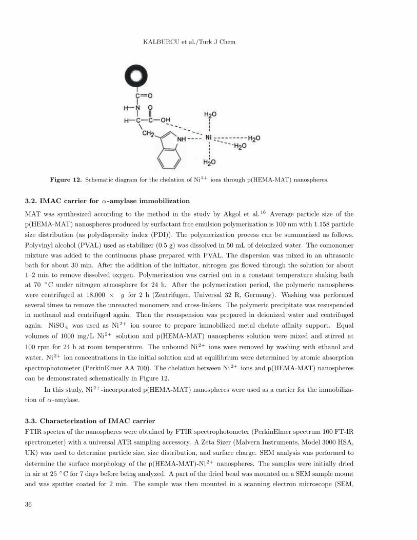
Figure 12. Schematic diagram for the chelation of Ni2+ ions through p(HEMA-MAT) nanospheres.
3.2. IMAC carrier for α-amylase immobilization
MAT was synthesized according to the method in the study by Akgol et al.16 Average particle size of the
p(HEMA-MAT) nanospheres produced by surfactant free emulsion polymerization is 100 nm with 1.158 particle
size distribution (as polydispersity index (PDI)). The polymerization process can be summarized as follows.
Polyvinyl alcohol (PVAL) used as stabilizer (0.5 g) was dissolved in 50 mL of deionized water. The comonomer
mixture was added to the continuous phase prepared with PVAL. The dispersion was mixed in an ultrasonic
bath for about 30 min. After the addition of the initiator, nitrogen gas flowed through the solution for about
1–2 min to remove dissolved oxygen. Polymerization was carried out in a constant temperature shaking bath
at 70 ◦C under nitrogen atmosphere for 24 h. After the polymerization period, the polymeric nanospheres
were centrifuged at 18,000 × g for 2 h (Zentrifugen, Universal 32 R, Germany). Washing was performed
several times to remove the unreacted monomers and cross-linkers. The polymeric precipitate was resuspended
in methanol and centrifuged again. Then the resuspension was prepared in deionized water and centrifuged
again. NiSO4 was used as Ni2+ ion source to prepare immobilized metal chelate affinity support. Equal
volumes of 1000 mg/L Ni2+ solution and p(HEMA-MAT) nanospheres solution were mixed and stirred at
100 rpm for 24 h at room temperature. The unbound Ni2+ ions were removed by washing with ethanol and
water. Ni2+ ion concentrations in the initial solution and at equilibrium were determined by atomic absorption
spectrophotometer (PerkinElmer AA 700). The chelation between Ni2+ ions and p(HEMA-MAT) nanospheres
can be demonstrated schematically in Figure 12.
In this study, Ni2+ -incorporated p(HEMA-MAT) nanospheres were used as a carrier for the immobiliza-
tion of α -amylase.
3.3. Characterization of IMAC carrier
FTIR spectra of the nanospheres were obtained by FTIR spectrophotometer (PerkinElmer spectrum 100 FT-IR
spectrometer) with a universal ATR sampling accessory. A Zeta Sizer (Malvern Instruments, Model 3000 HSA,
UK) was used to determine particle size, size distribution, and surface charge. SEM analysis was performed to
determine the surface morphology of the p(HEMA-MAT)-Ni2+ nanospheres. The samples were initially dried
in air at 25 ◦C for 7 days before being analyzed. A part of the dried bead was mounted on a SEM sample mount
and was sputter coated for 2 min. The sample was then mounted in a scanning electron microscope (SEM,
36

KALBURCU et al./Turk J Chem
Phillips, XL-30S FEG, Germany). The surface of the sample was then scanned at the desired magnification to
study the morphology of the nanospheres. The following expression was used to calculate the surface area of
p(HEMA-MAT)-Ni2+ .
N =6× 10¹⁰ × S
π × qs × d3, (1)
where N is the number of nanospheres per milliliter, S is the solids %, qs is the density of bulk polymer
(g/mL), and d is the diameter (nm). The number of nanospheres in 1 mL of suspension was determined from
the mass–volume graph of nanospheres. From all these data, the specific surface area of p(HEMA-MAT)-Ni2+
nanospheres was calculated by multiplying N and the surface area of 1 nanosphere.
An elemental analyzer (Leco, CHNS-932, USA) was used to determine the degree of MAT incorporation
into the synthesized p(HEMA-MAT) nanospheres.
3.4. Adsorption studies with α-amylase
α−Amylase adsorption on the p(HEMA-MAT)-Ni2+ nanospheres was performed in the medium of different
pHs (3.5–9.0), α -amylase concentrations (0.25–1.25 mg/mL), temperatures (5–45 ◦C), and ionic strengths (0.0–
1.0 M NaCl). α -Amylase adsorption studies were performed in a batch system for 1 h at 25 ◦C. The initial and
final concentrations of α -amylase were determined by UV/Vis spectrophotometer (Shimadzu 1601, Japan). The
amount of adsorbed α -amylase per unit mass of the on the p(HEMA-MAT)-Ni2+ nanospheres was calculated
using the following expression:
Q =(C0 − C)× V
m, (2)
where Q is the amount of α -amylase adsorbed onto unit mass of the nanospheres (mg/g), C0 and C are the
concentrations of α -amylase in the initial solution and in the supernatants obtained after centrifugation at
18,000 ×g , respectively (mg/g), V is the solution volume (mL), and m is the mass of the nanospheres used (g).
The adsorption experiments were performed in replicates of 3. For each set of data present, standard
statistical methods were used to determine the mean values and standard deviations. Confidence intervals of95% were calculated for each set of samples in order to determine the margin of error.
3.5. Desorption and repeated use
Desorption experiments on α -amylase were performed using different desorption agents. To obtain most efficient
recovery yield 1.0–1.5 M NaCl and 1 M NaSCN were used. The α -amylase immobilized p(HEMA-MAT)-Ni2+
nanospheres were incubated in desorption medium at room temperature for 2 h. The desorption ratio was
calculated from the amount of α -amylase adsorbed on the nanospheres and the final protein concentration in
the desorption medium.
Desorption ratio(%) =Amount of α-amylase desorbed
Amount of α-amylase adsorbed,×100 (3)
In order to test the repeated use of p(HEMA-MAT)-Ni2+ nanospheres, the α -amylase adsorption–desorption
cycle was repeated 5 times using the same nanospheres.
37
KALBURCU et al./Turk J Chem
3.6. Activity assays of free and immobilized α-amylase
Activities of free and immobilized α -amylase were determined by the assay suggested by Bernfeld wherein the
reducing groups liberated from starch are measured by the reduction of 3,5-dinitrosalicylic acid.30
The assay mixture includes 30% sodium-potassium tartarate (w/v), 1.6% NaOH (w/v), and 1% DNSA
(w/v). Equal volumes of enzyme and starch solutions were mixed and incubated at room temperature for 3 min
and then the assay mixture was added to the enzyme–substrate mixture as an equal volume of this mixture. This
solution was incubated in boiling water for 5 min. After cooling, the solution was diluted with distilled water
and the intensity of the red color that developed was measured in a UV/Vis spectrophotometer (Shimadzu,
Model 1601) at 540 nm.
In each set of experiments, different concentrations of starch were used to prepare a standard curve.
Nonreducing ends of starch were determined as described, and the results were converted to relative activities
(percentage of the maximum activity obtained in the series). Hydrolytic activity evaluated after enzyme
immobilization on the p(HEMA-MAT)-Ni2+ compared with activity of the same quantity of free enzyme and
residual activity was determined. One unit liberates from soluble starch 1 µmol of reducing groups (calculated
as maltose) per minute at 25 ◦C and pH 6.9 under the specified conditions.30
Units/mg =Micromoles maltose liberated
mg of enzyme in reaction mixture× 3min(4)
3.7. Steady state kinetics
Activity assays were carried out over the pH range of 3.5–9.0 and temperature range of 5–45 ◦C so as to
determine pH and temperature profiles for the free and immobilized α -amylase. Activity results dependent on
pH and temperature are presented in a standardized form with the highest value of each set being appropriated
as the value of 100% activity.
The activity assay was performed with different starch concentrations to determine the effect of substrate
concentration on both free and immobilized enzyme activities. The substrate concentrations varied from 0.1 to
1.0 g/L in 50 mM phosphate buffer at optimum pH. The Michaelis constant (KM ) and Vmax were determined
using a Lineweaver–Burk plot. All parameters were the mean of triplicate determinations from 3 independent
preparations.
4. Conclusions
To stabilize enzymes with improved intrinsic and operational stabilities may be possible with the development
of nanomaterials and nanostructured materials. Recently, studies have focused on the development of suitable
low-cost sorbents. Ni2+ -chelated p(HEMA-MAT) nanospheres used for the immobilization of α -amylase in
this study provide many advantages over conventional techniques. Coupling of a chelating ligand to the
adsorption matrix is an expensive and critical step in the preparation process of metal-chelating sorbent. In
this procedure, loading the metal ion directly on the nanospheres without further modification steps is possible,
because comonomer MAT acts as the metal-chelating ligand. Slow release of these covalently bonded chelators
from the sorbent is the other important point. A general problem encountered in any immobilized metal-chelate
affinity adsorption technique is metal-chelating ligand release, which causes a decrease in adsorption capacity.
MAT playing a role as metal-chelating ligand and/or comonomer was polymerized with HEMA and there was
no leakage of the ligand. The difficulty of multistep purification methods will be solved by this purification
method.
38
KALBURCU et al./Turk J Chem
Low cost and resistance to harsh operation conditions and high temperature are desirable features of the
p(HEMA-MAT)-Ni2+ nanospheres. p(HEMA-MAT)-Ni2+ nanospheres exhibited quite high binding capacity
(4468.1 ± 177.5 mg/g) due to their highly specific surface. Immobilization did not drastically change the
optimum pH or temperature profile of the immobilized enzyme, but the thermostability of immobilized α -
amylase was better than that of the free one. The storage stability of the immobilized enzyme was also higher
than that of the soluble enzyme under similar conditions. The reusability and the higher storage stability of
immobilized α -amylase presented in this work may provide economic advantages for large-scale biotechnological
applications.
References
1. Schenck, F. W.; Hebeda, R. E. Starch Hydrolysis Products: Worldwide Technology, Production and Applications,
VCH, New York, 1992.
2. Windish, W. W.; Mhatre, N. S. Advances in Applied Microbiology, In: Wayne, W. U. Ed., 1965.
3. Fogarty, W. M.; Kelly, C. T. In Topics in Enzyme and Fermentation Biotechnology ; Wiseman, A., Ed., 1979.
4. Vihinen, M.; Mantsala, P. Crit Rev. Biochem. Mol. Biol. 1989, 24, 329–418.
5. Lonsane, B. K.; Ramesh, M. V. Adv. Appl. Microbiol. 1990, 35, 1–56.
6. Nikolov, Z. L.; Reily, P. J. In Biocatalysts for Industry ; Dordick, J. S., Ed. Plenum Press, New York, 1991.
7. Liu, Y.; Jia, S.; Ran, J.; Wu, S. Catal. Commun. 2010, 11, 364–367.
8. Yamamoto, T. In Handbook of Amylases and Related Enzymes: Their Source, Isolation Methods, Properties and
Applications; Amylase Research Society of Japan, Ed., Pergamon Press, Oxford, 1988.
9. Bahar, T.; Celebi, S. S. Enzyme Microb. Technol. 2000, 26, 28–33.
10. Roig, M. G.; Kennedy, J. F.; Garaita, M. G. J. Biomater. Sci. Polym. Ed. 1994, 6, 661–673.
11. Ida, J.; Matsuyama, T.; Yamamoto, H. Biochem. Eng. J. 2000, 5, 179–184.
12. Yang, Y.; Chase, H. A. Biotechnol. Appl. Biochem. 1998, 28, 145–154.
13. Chen, J. P.; Sun, Y. M.; Chu, D. H. Biotechnol. Prog. 1998, 14, 473–478.
14. Bryjak, J. Bioprocess Eng. 1995, 13, 177–181.
15. Mateo, C.; Palomo, J. M.; Fernandez-Lorente, G.; Guisan, J. M.; Fernandez-Lafuente, R. Enzyme Microb. Technol.
2007, 40, 1451–1463.
16. Akgol, S.; Ozturk, N.; Denizli, A. J. Appl. Polym. Sci. 2010, 115, 1608–1615.
17. Cong, L.; Kaul, R.; Dissing, U.; Mattiasson, B. J. Biotechnol. 1995, 42, 75–84.
18. Tischer, W.; Kashe, V. Biotechnology 1999, 17, 326–335.
19. Ye, P.; Xu, Z. K.; Che, A. F.; Wu, J.; Seta, F. Biomaterials 2005, 26, 6394–6403.
20. Rebros, V.; Rosenberg, M.; Mlichova, Z.; Kristofıkova, L. Food Chem. 2007, 102, 784–787.
21. Sankalia, V.; Mashru, V.; Sankalia, J. M.; Sutariya, V. B. Eur. J. Pharm. Biopharm. 2007, 65, 215–232.
22. Peng, K.; Hidajat, K.; Udin, M. S. J. Colloid Interface Sci. 2004, 271, 277–283.
23. Choi, S. W.; Kwon, H. Y.; Kim, W. S.; Kim, J. H. Colloids Surf. A 2002, 201, 283–289.
24. Ozturk, N.; Akgol, S.; Arisoy, M.; Denizli, A. Sep. Purif. Technol. 2007, 58, 83–90.
25. Kim, J.; Grate, J. W.; Wang, P. Chem. Eng. Sci. 2006, 61, 1017–1026.
26. Tuzmen, N.; Kalburcu T.; Denizli A. Process Biochem. 2012, 47, 26–33.
27. Porath, J.; Carlson, J.; Olsson, I.; Belfrage, G. Nature 1975, 258, 598–599.
39
KALBURCU et al./Turk J Chem
28. Gaberc-Porekar, V.; Menart, V. J. Biochem. Biophys. Methods 2001, 49, 335–360.
29. Arnold, F. H. Bio. Technology 1991, 9, 150–155.
30. Gutierrez, R. E.; Martin del Vale, M.; Galan, M. A. Sep. Purif. Rev. 2007, 36, 71–111.
31. Kubota, N.; Nakagawa, Y.; Eguchi, Y. J. Appl. Polym. Sci. 1996, 62, 1153–1160.
32. Lawal, O. S. Food Chem. 2006, 95, 101–107.
33. Ueda, E. K. M.; Gout, P. W.; Morganti, L. J. Chromatogr. A 2003, 988, 1–23.
34. Mosbach, K. Sci. Am. 1971, 224, 26–33.
35. He, D.; Cai, Y.; Wei, W.; Nie, L.; Yao, S. Biochem. Engineer. J. 2000, 6, 7–11.
36. Tanyolac, D.; Yuruksoy, B. I.; Ozdural, A. R. Biochem. Eng. J. 1998, 2, 179–186.
37. O’Neill, S. P.; Dunnill, P.; Lilly, M. D. Biotechnol. Bioeng. 1971, 13, 337–352.
38. Lopez, G. P.; Ratner, B. D.; Rapoza, R. J.; Horbett, T. A. Macromol. Symp. 1993, 26, 3247–3253.
39. Arica, M. Y.; Senel, S.; Alaeddinoglu, N. G.; Patir, S.; Denizli, A. J. Appl. Polym. Sci. 2000, 75, 1685–1692.
40. Aksoy, S.; Tumturk, H.; Hasirci, N. J. Biotechnol. 1998, 60, 37–46.
41. Turunc, O.; Kahraman, M. V.; Akdemir, Z. S.; Kayaman-Apohan, N.; Gungor, A. Food Chem. 2009, 112, 992–997.
42. Kara, A.; Osman, B.; Yavuz, H.; Besirli N., Denizli A. React. Funct. Polym. 2005, 62, 61–68.
43. Chang, M. Y.; Juang, R. S. Process Biochem. 2004, 39, 1087–1090.
44. Reddy, K. R. C.; Kayastha, A. M. J. Mol. Catal. B: Enzym. 1994, 38, 104–112.
40