Identificationof Thermotogamaritima MSB8GH57 α-amylaseAmyC ... · amylase specificity of AmyC is...
11
BIOTECHNOLOGICALLY RELEVANT ENZYMES AND PROTEINS Identification of Thermotoga maritima MSB8 GH57 α-amylase AmyC as a glycogen-branching enzyme with high hydrolytic activity Xuewen Zhang 1 & Hans Leemhuis 1,2 & Štefan Janeček 3,4 & Mária Martinovičová 4 & Tjaard Pijning 5 & Marc J.E.C. van der Maarel 1 Received: 19 March 2019 /Revised: 20 May 2019 /Accepted: 21 May 2019 /Published online: 13 June 2019 # The Author(s) 2019 Abstract AmyC, a glycoside hydrolase family 57 (GH57) enzyme of Thermotoga maritima MSB8, has previously been identified as an intracellular α-amylase playing a role in either maltodextrin utilization or storage polysaccharide metabolism. However, the α- amylase specificity of AmyC is questionable as extensive phylogenetic analysis of GH57 and tertiary structural comparison suggest that AmyC could actually be a glycogen-branching enzyme (GBE), a key enzyme in the biosynthesis of glycogen. This communication presents phylogenetic and biochemical evidence that AmyC is a GBE with a relatively high hydrolytic (α- amylase) activity (up to 30% of the total activity), creating a branched α-glucan with 8.5% α-1,6-glycosidic bonds. The high hydrolytic activity is explained by the fact that AmyC has a considerably shorter catalytic loop (residues 213–220) not reaching the acceptor side. Secondly, in AmyC, the tryptophan residue (W 246) near the active site has its side chain buried in the protein interior, while the side chain is at the surface in Tk1436 and Tt1467 GBEs. The putative GBEs from three other Thermotogaceae, with very high sequence similarities to AmyC, were found to have the same structural elements as AmyC, suggesting that GH57 GBEs with relatively high hydrolytic activity may be widespread in nature. Keywords Thermotoga maritima . AmyC . Glycogen branching enzyme . Phylogeny . Crystal structure Introduction Glycoside hydrolases (GHs; EC 3.2.1.x) catalyse the hydroly- sis of O-glycosidic bonds in carbohydrates such as starch. They are ubiquitously present in all kingdoms of life. Well- known GHs are α-amylase (EC 3.2.1.1); the enzyme present in, e.g. saliva and the small intestine, responsible for the deg- radation of starch; and lactase (E.C. 3.2.1.108), which de- grades the milk sugar lactose to glucose and galactose. GHs are classified based on amino acid sequence homology in 152 different families (CAZy) (Cantarel et al. 2009; Lombard et al. 2014). Most GHs have either an inverting or a retaining reac- tion mechanism as outlined by Koshland (1953). In essence, GHs catalyse both hydrolysis and transglycosylation reactions, but the ratio varies enormously depending on the type of GH, the substrate concentration and the reaction conditions (Bissaro et al. 2015; Koshland 1953). Typical GHs with almost exclusive hydrolytic activity are isoamylases (EC 3.2.1.68), which hydrolyze the α-1,6-glyco- sidic linkage in amylopectin (Harada et al. 1972; Li et al. 2013), and α-amylases (EC 3.2.1.1), which hydrolyze the α- 1,4-glycosidic linkage in amylose, amylopectin and glycogen (van der Maarel et al. 2002). An example of a GHs with almost exclusive transglycosylating activity are 4- α- glucanotransferases (EC 2.4.1.25) that break an α-1,4-glyco- sidic linkage in amylopectin or amylose and form a new α- Electronic supplementary material The online version of this article (https://doi.org/10.1007/s00253-019-09938-1) contains supplementary material, which is available to authorized users. * Marc J.E.C. van der Maarel [email protected] 1 Department of Aquatic Biotechnology and Bioproduct Engineering, Engineering and Technology institute Groningen, University of Groningen, 9747 AG Groningen, Netherlands 2 Avebe Innovation Center, 9747 AG Groningen, Netherlands 3 Laboratory of Protein Evolution, Institute of Molecular Biology, Slovak Academy of Sciences, SK-84551 Bratislava, Slovakia 4 Department of Biology, Faculty of Natural Sciences, University of SS Cyril and Methodius, SK-91701 Trnava, Slovakia 5 Biomolecular X-ray Crystallography, Groningen Biomolecular Sciences and Biotechnology Institute, University of Groningen, 9747 AG Groningen, Netherlands Applied Microbiology and Biotechnology (2019) 103:6141–6151 https://doi.org/10.1007/s00253-019-09938-1
Transcript of Identificationof Thermotogamaritima MSB8GH57 α-amylaseAmyC ... · amylase specificity of AmyC is...

BIOTECHNOLOGICALLY RELEVANT ENZYMES AND PROTEINS
Identification of ThermotogamaritimaMSB8 GH57 α-amylase AmyCas a glycogen-branching enzyme with high hydrolytic activity
Xuewen Zhang1& Hans Leemhuis1,2 & Štefan Janeček3,4 & Mária Martinovičová4 & Tjaard Pijning5
& Marc J.E.C. vander Maarel1
Received: 19 March 2019 /Revised: 20 May 2019 /Accepted: 21 May 2019 /Published online: 13 June 2019# The Author(s) 2019
AbstractAmyC, a glycoside hydrolase family 57 (GH57) enzyme of Thermotoga maritima MSB8, has previously been identified as anintracellular α-amylase playing a role in either maltodextrin utilization or storage polysaccharide metabolism. However, the α-amylase specificity of AmyC is questionable as extensive phylogenetic analysis of GH57 and tertiary structural comparisonsuggest that AmyC could actually be a glycogen-branching enzyme (GBE), a key enzyme in the biosynthesis of glycogen. Thiscommunication presents phylogenetic and biochemical evidence that AmyC is a GBE with a relatively high hydrolytic (α-amylase) activity (up to 30% of the total activity), creating a branched α-glucan with 8.5% α-1,6-glycosidic bonds. The highhydrolytic activity is explained by the fact that AmyC has a considerably shorter catalytic loop (residues 213–220) not reachingthe acceptor side. Secondly, in AmyC, the tryptophan residue (W 246) near the active site has its side chain buried in the proteininterior, while the side chain is at the surface in Tk1436 and Tt1467 GBEs. The putative GBEs from three other Thermotogaceae,with very high sequence similarities to AmyC, were found to have the same structural elements as AmyC, suggesting that GH57GBEs with relatively high hydrolytic activity may be widespread in nature.
Keywords Thermotogamaritima . AmyC . Glycogen branching enzyme . Phylogeny . Crystal structure
Introduction
Glycoside hydrolases (GHs; EC 3.2.1.x) catalyse the hydroly-sis of O-glycosidic bonds in carbohydrates such as starch.
They are ubiquitously present in all kingdoms of life. Well-known GHs are α-amylase (EC 3.2.1.1); the enzyme presentin, e.g. saliva and the small intestine, responsible for the deg-radation of starch; and lactase (E.C. 3.2.1.108), which de-grades the milk sugar lactose to glucose and galactose. GHsare classified based on amino acid sequence homology in 152different families (CAZy) (Cantarel et al. 2009; Lombard et al.2014). Most GHs have either an inverting or a retaining reac-tion mechanism as outlined by Koshland (1953).
In essence, GHs catalyse both hydrolysis andtransglycosylation reactions, but the ratio varies enormouslydepending on the type of GH, the substrate concentration andthe reaction conditions (Bissaro et al. 2015; Koshland 1953).Typical GHs with almost exclusive hydrolytic activity areisoamylases (EC 3.2.1.68), which hydrolyze the α-1,6-glyco-sidic linkage in amylopectin (Harada et al. 1972; Li et al.2013), and α-amylases (EC 3.2.1.1), which hydrolyze the α-1,4-glycosidic linkage in amylose, amylopectin and glycogen(van der Maarel et al. 2002). An example of a GHs withalmost exclusive transglycosylating activity are 4-α-glucanotransferases (EC 2.4.1.25) that break an α-1,4-glyco-sidic linkage in amylopectin or amylose and form a new α-
Electronic supplementary material The online version of this article(https://doi.org/10.1007/s00253-019-09938-1) contains supplementarymaterial, which is available to authorized users.
* Marc J.E.C. van der [email protected]
1 Department of Aquatic Biotechnology and Bioproduct Engineering,Engineering and Technology institute Groningen, University ofGroningen, 9747 AG Groningen, Netherlands
2 Avebe Innovation Center, 9747 AG Groningen, Netherlands3 Laboratory of Protein Evolution, Institute of Molecular Biology,
Slovak Academy of Sciences, SK-84551 Bratislava, Slovakia4 Department of Biology, Faculty of Natural Sciences, University of
SS Cyril and Methodius, SK-91701 Trnava, Slovakia5 Biomolecular X-ray Crystallography, Groningen Biomolecular
Sciences and Biotechnology Institute, University of Groningen, 9747AG Groningen, Netherlands
Applied Microbiology and Biotechnology (2019) 103:6141–6151https://doi.org/10.1007/s00253-019-09938-1
1,4-glycosidic linkage when transferring a part of the donormolecule to the acceptor (Lee et al. 1970; Terada et al. 1999;van der Maarel and Leemhuis 2013).
The glycoside hydrolase family 57 (GH57) was establishedin 1996 (Henrissat and Bairoch 1996) based on the sequencesof two amylolytic enzymes fromDictyoglomus thermophilum(Fukusumi et al. 1988) and Pyrococcus furiosus (Ladermanet al. 1993) that were obviously unrelated to the members ofthe main α-amylase family GH13 (Janecek et al. 2014). Forthe family GH57 members, five conserved sequence regions(CSRs) have been established (Zona et al. 2004). Currently,GH57 holds over 2000 protein sequences (CAZy update fromFebruary 2019) comprising hydrolytic and transglycosylatingenzymes, such as α-amylase, amylopullulanase (EC3 .2 . 1 . 41 ) , du a l - s p e c i f i c i t y amy l opu l l u l an a s e /cyclomaltodextrinase (EC 3.2.1.41/54), glycogen-branchingenzyme (GBE; EC 2.4.1.18), 4-α-glucanotransferase, and α-galactosidase (EC 3.2.1.22), as well as a non-specified amy-lase (EC 3.2.1.-) and maltogenic amylase (EC 3.2.1.133)(Blesak and Janecek 2012; Blesak and Janecek 2013;Janecek et al. 2014; Martinovičová and Janeček 2018; Zonaet al. 2004). Glycogen-branching enzymes of GH57 play apivotal role in the synthesis of glycogen, cleaving an α-1,4-glycosidic linkage in the donor substrate subsequently trans-ferring the non-reducing end fragment to the C6 hydroxylposition of an internal glucosyl moiety that acts as the acceptorsubstrate (α-1,6-transglycosylation).
Ballschmiter et al. (2006) identified AmyC from the ther-mophilic bacterium Thermotoga maritimaMSB8 (TaxonomyID: 243274), an enzyme produced during the exponentialgrowth phase and showing activity towards amylose and sol-uble starch at high temperature, releasing oligosaccharides.Sequence analysis revealed that AmyC belongs to GH57and has no signal peptide. Together, the authors concludedthat AmyC is an intracellular GH57 α-amylase that may playa role in either maltodextrin utilization or storage polysaccha-ride metabolism (Ballschmiter et al. 2006).
The crystal structure of AmyC (Dickmanns et al. 2006)showed structural similarity with PDB entry 1UFA, a GH57enzyme (TT1467) with then unknown function. Santos et al.(2011) determined the crystal structure of another GH57 en-zyme, TK1436, from Thermococcus kodakaraensis KOD1,and compared its structure with that of AmyC. TK1436 wasfound to be a GBE; it features a long and flexible so-calledcatalytic loop (residues 225–245, TK1436 numbering) foldingtowards the active site with a tyrosine residue at its tip(Tyr233, TK1436 numbering); this loop was shown to beessential for branching activity and proposed to be involvedin substrate binding and/or intermediate product stabilization(Palomo et al. 2011; Santos et al. 2011). AmyC showed aconsiderably shorter catalytic loop, lacking the correspondingtyrosine residue as well as another conserved tryptophan res-idue lining the active site groove (Trp270, TK1436
numbering). While TK1436 was found to be functional as atetramer, AmyC is monomeric. The authors proposed that thedifferences in tertiary and quaternary structure relate to the factthat AmyC only showed hydrolytic activity on starch-likesubstrates. This hypothesis was further supported by the ob-servation that also TT1467 was characterized as a GBE (PDBentry 3P0B (Palomo et al. 2011)) and features the same struc-tural elements as TK1436, but differs from AmyC regardingthose.
Nevertheless, a detailed bioinformatic analysis of GH57enzymes (Blesak and Janecek 2012) clearly showed thatAmyC contains the sequence fingerprint of GBE’s; thus, itremained intriguing why the biochemical characterization ofAmyC (Ballschmiter et al. 2006) only revealed hydrolytic andnot transglycosylation (branching) activity. We therefore in-vestigated the phylogeny, activity and three-dimensionalstructure of AmyC in more detail. This communication pre-sents biochemical evidence in support of the in silico analysisthat AmyC is indeed a GBE with relatively high hydrolyticactivity (up to 30% of the total activity), and suggests whichstructural features are responsible for its specificity. Finally,three putative GH57 GBEs are identified based on structuralhomology to AmyC, suggesting that GH57 GBEs with rela-tively high hydrolytic activity are more widespread inmesophilic and thermophilic microorganisms.
Materials and methods
Materials
Amylose V was provided by Avebe (Veendam, Netherlands).Lithium bromide was obtained from Acros Organics.Isoamylase (specific activity 260 U/mg), pullulanase M1(EC 3.2.1.41, specific activity 34 U/mg) and β-amylase (EC3.2.1.2, specific activity 10,000 U/mL) were purchased fromMegazyme (Wicklow, Ireland). All other chemicals were fromSigma-Aldrich (Zwijndrecht, Netherlands)
Sequence and evolutionary comparison
All full-length protein sequences (Supplementary Table S1)were retrieved from the UniProt knowledge database (http://www.uniprot.org/) (Apweiler 2014) and/or from GenBank(https://www.ncbi.nlm.nih.gov/genbank/) (Benson et al.2014). The alignment was done for AmyC from T. maritimaand the three characterized GBEs from T. kodakaraensis, T.thermophilus and P. horikoshi, for which also their three-dimensional structures have been determined—using the pro-gram Clustal-Omega with default parameters (http://www.ebi.ac.uk/Tools/msa/clustalo/) (Sievers et al. 2011).
For all 64 GH57 enzymes and proteins (SupplementaryTable S1), their five well-established conserved sequence
6142 Appl Microbiol Biotechnol (2019) 103:6141–6151
regions (CSRs) (Zona et al. 2004) were identified according toprevious bioinformatics analyses (Blesak and Janecek 2012,2013; Janecek and Blesak 2011; Martinovičová and Janeček2018). The evolutionary tree was calculated based on thealignment of five CSRs mentioned above as a Phylip-tree typeusing the neighbour-joining clustering (Saitou and Nei 1987)and the bootstrapping procedure (Felsenstein 1985) (the num-ber of bootstrap trials used was 1000) implemented in theClustal-X package (Larkin et al. 2007). The tree was displayedwith the program iTOL (http://itol.embl.de/) (Letunic andBork 2006). Sequence logos were created using theWebLogo 3.0 server (http://weblogo.threeplusone.com/)(Crooks et al. 2004) for CSRs of all 40 GBE sequences aswell as of the single AmyC from T. maritima.
Expression and purification of AmyC
A codon-optimized gene (Genbank ID:MK704497) encodingthe GBE from T. maritima SMB8 (AmyC) was synthesized byBaseclear (Leiden, Netherlands), and cloned into pRSET B(Invitrogen) behind the His-tag sequence of the vector. Genesequence details are provided in the supplemental informa-tion. AmyC was overexpressed in Escherichia coliBL21(DE3), cultivated in Luria-Bertani (LB) medium (10 g/L of tryptone, 5 g/L yeast extract and 10 g/L NaCl) supple-mented with 100 μg/mL ampicillin. GBE expression was in-duced with 0.1 mM IPTG when the culture had an OD600 of0.8; the induction was carried out at 16 °C for 20 h. Cells wereharvested by centrifugation (5000×g, 10 min, 4 °C), washedtwice with 10 mM sodium phosphate buffer pH 7.0, and lysedusing a high-pressure homogenizer (Emulsiflex-B15; Avestin,Canada) in two cycles at 9.0 MPa and room temperature. Acell-free extract was obtained after centrifugation (20,000×g,30 min, 4 °C). AmyC was purified in two steps: first, the cell-free extract was subjected to 70 °C for 15 min, followed byremoval of the denatured proteins by centrifugation(20,000×g, 30 min, 4 °C). The His-tagged AmyCwas purifiedusing the HisPurTM Ni-NTA Resin (Thermo FisherScientific, Waltham, USA) according to the manufacturer’sprotocol. Protein concentration was quantified using theQuick Start™ Bradford Protein Assay kit (Bio-RadLaboratories, Hercules, USA). The purity and molecular massof the proteins were checked by SDS-PAGE.
Enzyme activity assays
The enzyme activity was analysed using the iodine stainingassay and monitoring the decrease of absorbance of theglucan-iodine complex (Boyer and Preiss 1978). Amylose Vwith an average DP of 700 was selected as substrate because ithas no detectable α-1,6-linkages by NMR; so, any α-1,6-link-age detected is the primed produced by the result of the actionof the GBE.
Amylose V (0.125% (w/v) dissolved in 50 mM sodiumphosphate buffer (pH 7.0) was incubated with 132.5 μg/mLAmyC at 50 °C. Ten microliters of aliquot was taken into 96-well plate, and 150 μL iodine reagent (aqueous solution of0.0127% I2 and 0.035% KI) was added, and the absorption at660 nm was determined. One unit of enzyme activity is de-fined as the amount of enzyme that gives a decrease in absor-bance of the amylose/iodine complex of 1.0 absorbance unitper minute at 660 nm (Palomo et al. 2011).
The influence of Ca2+ on AmyC activity was tested at50 °C to 80 °C in 50 mM Tris-Cl buffer, and the pH 7.5 wasadjusted at a series reaction temperatures. 0.1, 1, 3 and 5 mMCaCl2 were applied in the reaction. The activity was measuredby iodine assay described as above.
The hydrolytic and transglycosylation activity of AmyCwith amylose Vas substrate were determined by measuringthe increase in reducing ends by the bicinchoninic acid(BCA) method before and after debranching the product,respectively. Amylose V was dissolved in 1 M sodiumhydroxide and then neutralized to pH 7.0. A mixture of0.125% (w/v) amylose V in 50 mM sodium phosphate buff-er (pH 7.0) and 132.5 μg/mL AmyC was incubated at50 °C. Samples of 500 μL were taken at different timepoints and the AmyC was inactivated by incubating thesamples at 100 °C for 10 min. To debranch the product,200 μL sample was mixed with 1 μL 1 M citrate acid, 1 Uisoamylase, 0.7 U pullulanase and 1 μL 1 M CaCl2 andthen incubated at 40 °C for 16 h. The hydrolytic activitywas measured by following the increase in reducing endsduring the reaction as each product of hydrolysis bears aterminal, reducing glucose residue. Transglycosylation, orbranching activity, was measured by treating the reactionproduct with the debranching enzymes isoamylase andpullulanase, enzymes that specifically hydrolyze α-1,6-linkages; the product of the specific hydrolysis of α-1,6-linkage will also bear a terminal, reducing glucose residue.The increase in reducing ends is the amount of reducingends after debranching minus the amount of reducing endsb e f o r e d e b r a n c h i n g , a d i r e c t r e s u l t o f t h etransglycosylation/branching activity. One unit branchingactivity is defined as 1 μmol of α-1,6-linkage synthesizedper minute and one unit hydrolytic activity is defined as1 μmol of reducing end synthesized per minute.
Influence of pH and temperature on activity
The influence of pH on AmyC activity was measured at 50 °Cin 50 mM sodium phosphate buffer (pH 6.0 to 9.0) by usingthe iodine assay as described above. The influence of temper-ature on AmyC activity was determined at pH 7.0 in 50 mMsodium phosphate buffer using the activity assay mixture in-cubated at temperature ranging 40 to 90 °C.
Appl Microbiol Biotechnol (2019) 103:6141–6151 6143
High performance anion exchange chromatography
Oligosaccharide analysis was carried out by high-performanceanion exchange chromatography (HPAEC) on a Dionex ICS-3000 system (Thermo Scientific) equipped with a 4 × 250 mmCarboPac PA-1 column. A pulsed amperometric detector witha gold electrode and an Ag/AgCl pH reference electrode wereused. The system was run with a gradient of 30–600 mMNaAc in 100 mM NaOH 1 mL/min. Chromatograms wereanalysed using Chromeleon 6.8 chromatography data systemsoftware (Thermo Scientific). A mixture of glucose, maltose,maltotriose, maltotetraose, maltopentaose, maltohexaose andmaltoheptaose was used as reference. AmyC-modified prod-uct was dialyzed using dialysis tubing with a cutoff size of100 Da to 500 Da in ultrapure water. Two milligrams of drymaterial was dissolved into 1 mL 5 mM sodium acetate bufferpH 5.0 with 5 mMCaCl2. Five hundred microliters of solutionwas mixed with 2.5 U isoamylase and 1.75 U pullulanase, andincubated at 40 °C for 16 h.
1H-NMR spectroscopy
1H-NMR spectra were recorded at a probe temperature of323 K on a Varian Inova 500 spectrometer (NMR Center,University of Groningen). Before NMR analysis, sampleswere exchanged twice in D2O (99.9% D atom, Sigma-Aldrich) with intermediate lyophilization, and then dissolvedin 0.6 mL D2O. Spectra were processed using MestReNova5.3 software (Mestrelabs Research SL, Santiago deCompostella, Spain), using Whittaker Smoother baseline cor-rection and zero filling to 32 k complex points. The degree ofbranching is calculated as follows:
Degree of branching ¼ Sα−1;6Sα−1;4 þ Sα−1;6
Sα-1,6 is the peak area ofα-1,6, integrated fromNMR spec-tra; Sα-1,4 is the peak area of α-1,4, integrated from NMRspectra.
Structural homology modelling
The crystal structures of AmyC (PDB entry 2B5D),T. kodakaraensis TK1436 GBE (PDB entry 3N98; (Santoset al. 2011)), T. thermophilus TT1467 GBE (PDB entry 3P0B;(Palomo et al. 2011)) and T. litoralis 4-α-glucanotransferase incomplex with acarbose (PDB entry 1K1Y; (Imamura et al.2003)) were superimposed. Homology models of Mesotogaprima, Kosmotoga olearia and Kosmotoga pacifica putativeGBEs were generated using the Phyre server in intensive mode(Kelley et al. 2015). Structural figures were prepared usingPyMOL (The PyMOL Molecular Graphics System, Version2.0 Schrödinger, LLC).
Results
Sequence analysis of GH57 GBEs
Analysis of the GH57 GBE sequences and phylogenetic treeconstruction was performed as described in the ‘Materials andmethods’ section. Sequences of 40 GBEs (SupplementaryTable S1) were collected based on the recent exhaustive insilico analysis of the entire α-amylase family GH57(Martinovičová and Janeček 2018) that, of all 1602 GH57sequences taken from the CAZy database (Cantarel et al.2009), yielded 546 GBEs. Forty GBEs were selected in aneffort to obtain a representative sample of GBE sequenceshaving, in addition to AmyC from T. maritima (Ballschmiteret al. 2006; Dickmanns et al. 2006), the biochemically char-acterized enzymes from T. kodakaraensis (Murakami et al.2006; Santos et al. 2011), T. thermophilus (Palomo et al.2011) and P. horikoshi (Na et al. 2017), accompanied by arange of hypothetical GBEs covering various taxa from bothBacteria and Archaea including all available sequences fromthe phylum Thermotogae. In order to perform as relevant aspossible analysis and in accordance with previous in silicostudies (Blesak and Janecek 2012; Blesak and Janecek 2013;Janecek and Blesak 2011; Martinovičová and Janeček 2018),the set of sequences was completed by 23 biochemically char-acterized family GH57 members representing other enzymespecificities, accompanied by one putative representative ofthe α-amylase-like protein (Supplementary Table S1).
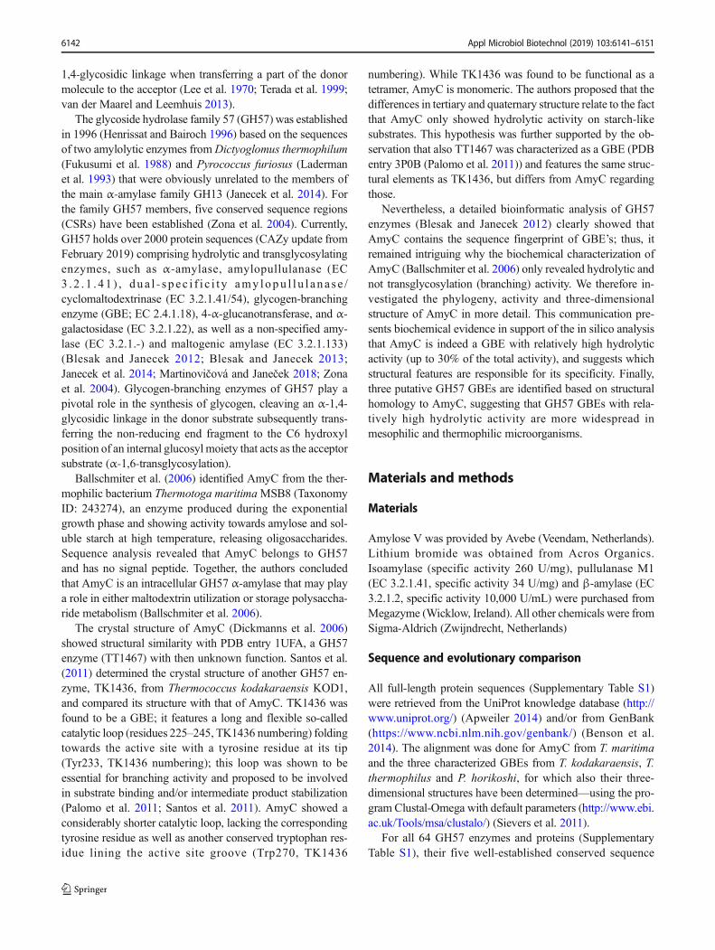
The evolutionary tree constructed of these selected se-quences shows that the (putative) GH57 GBEs cluster togeth-er (Fig. 1). Comparison of the CSRs of the 40 (putative) GH57GBEs reveals that most, but not all, of the amino acid residuesof the CSRs are conserved (Fig. 2). AmyCwas also alignmentwith the three characterized GH57 GBE, demonstrating theirsimilarity, though at the same time also revealing that someloops are distinct (Fig. S1).
Activity
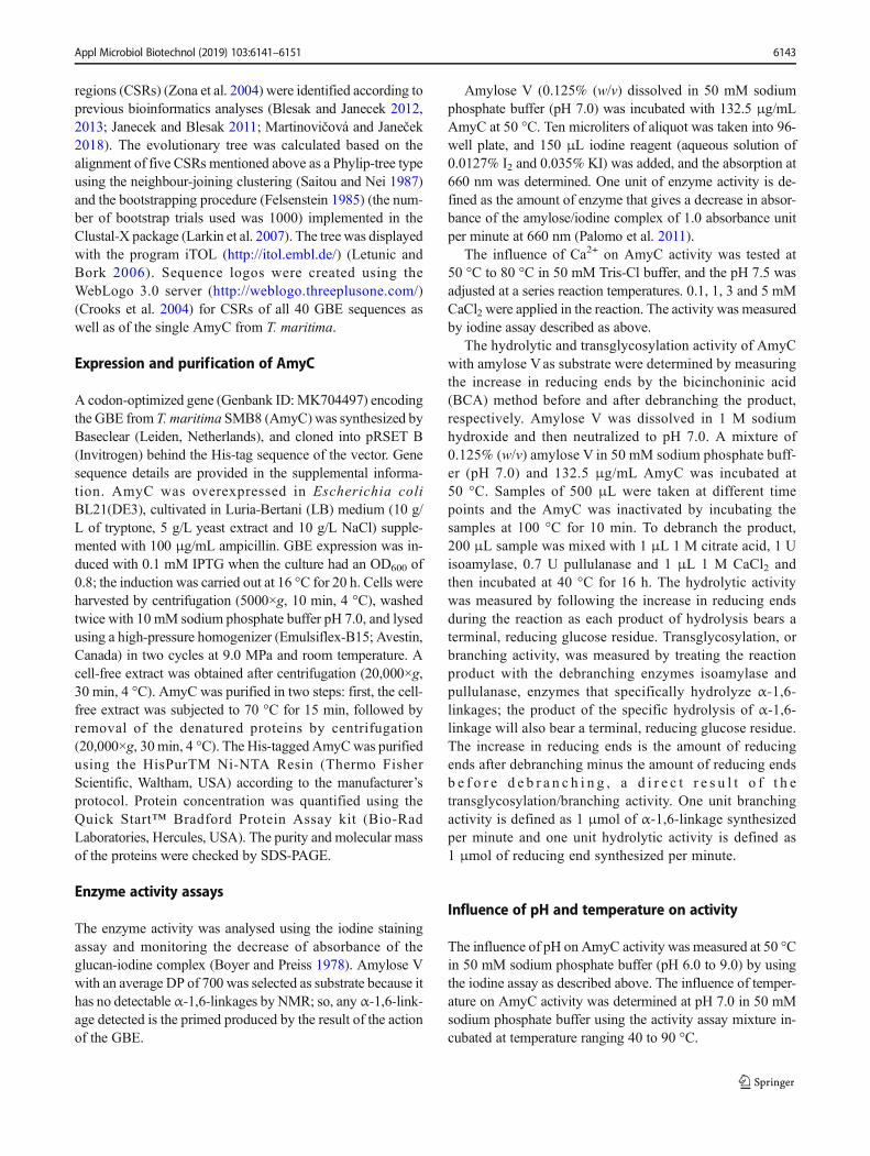
AmyC was over expressed in E. coli and purified to homoge-neity as judged by SDS-page (Fig. 3a). The previous studiesreport optimal conditions of 90 °C and pH 8.5 (Ballschmiteret al. 2006; Dickmanns et al. 2006). In a first approach, theactivity of purified recombinant enzyme was tested at 90 °Cand pH 8.5 by using the iodine staining assay and amylose Vas substrate. The absorbance of glucan-iodine complex did notchange, which showed that the recombinant AmyC was notactive at these conditions. Subsequently, the influence of tem-perature and pH on AmyC activity was investigated in detail.AmyC showed activity at temperatures of 80 °C and below.Maximum activity was found at 50 °C and pH 7.0 (Fig. 3).AmyC was not active in the presence of Ca2+ at high temper-ature, which is in agreement with Ballschmiter et al. (2006).
6144 Appl Microbiol Biotechnol (2019) 103:6141–6151
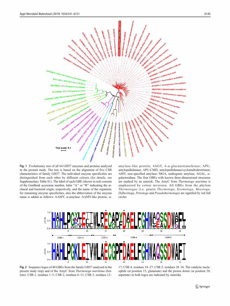
Fig. 1 Evolutionary tree of all 64 GH57 enzymes and proteins analysedin the present study. The tree is based on the alignment of five CSRcharacteristics of family GH57. The individual enzyme specificities aredistinguished from each other by different colours (for details, seeSupplementary Table S1). The label of each GBE (shown in red) consistsof the GenBank accession number, letter BA^ or BB^ indicating the ar-chaeal and bacterial origin, respectively, and the name of the organism;for remaining enzyme specificities, also the abbreviation of the enzymename is added as follows: AAMY, α-amylase; AAMY-like protein, α-
amylase-like protein; 4AGT, 4-α-glucanotransferase; APU,amylopullulanase; APU-CMD, amylopullulanase/cyclomaltodextrinase;AMY, non-specified amylase; MGA, maltogenic amylase; AGAL, α-galactosidase. The four GBEs with known three-dimensional structuresare marked by an asterisk. The AmyC from Thermotoga maritima isemphasized by colour inversion. All GBEs from the phylumThermotogae (i.e. genera Thermotoga, Kosmotoga, Mesotoga,Defluviitoga, Petrotoga and Pseudothermotoga) are signified by red fullcircles
Fig. 2 Sequence logos of 40GBEs from the family GH57 analysed in thepresent study (top) and of the AmyC from Thermotoga martitima (bot-tom). CSR-1, residues 1–5; CSR-2, residues 6–11; CSR-3, residues 12–
17; CSR-4, residues 18–27; CSR-5, residues 28–36. The catalytic nucle-ophile (at position 15, glutamate) and the proton donor (at position 20,aspartate) in both logos are indicated by asterisks
Appl Microbiol Biotechnol (2019) 103:6141–6151 6145
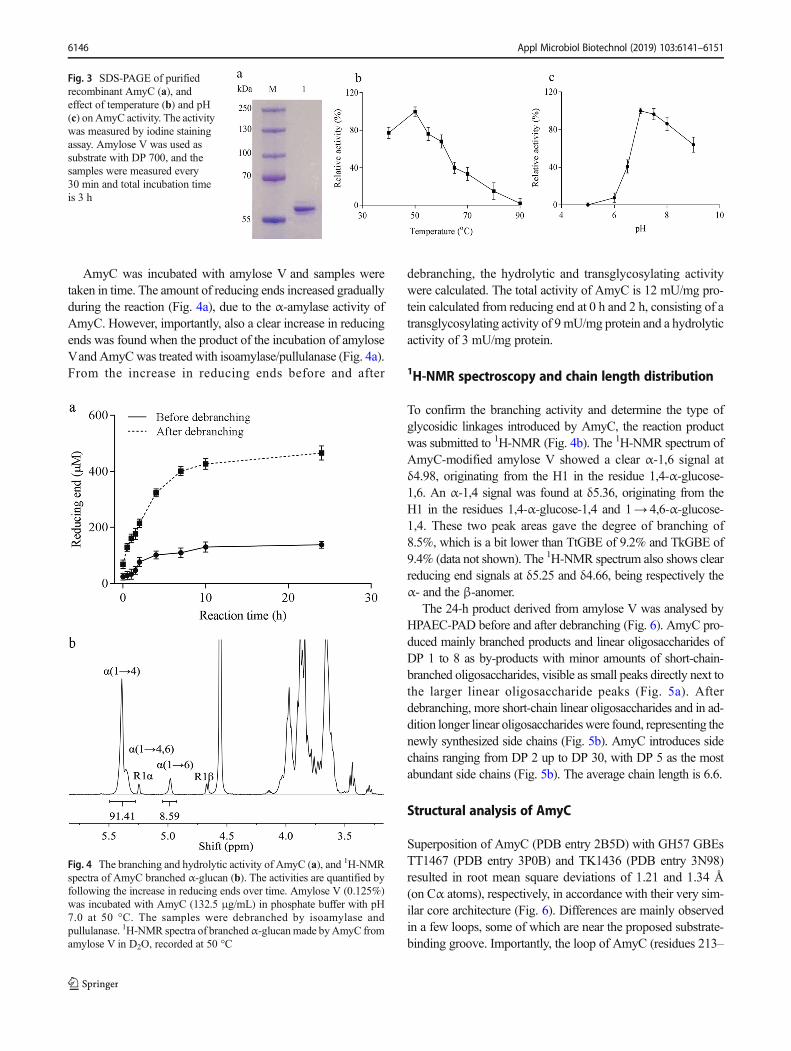
AmyC was incubated with amylose V and samples weretaken in time. The amount of reducing ends increased graduallyduring the reaction (Fig. 4a), due to the α-amylase activity ofAmyC. However, importantly, also a clear increase in reducingends was found when the product of the incubation of amyloseVand AmyCwas treated with isoamylase/pullulanase (Fig. 4a).From the increase in reducing ends before and after
debranching, the hydrolytic and transglycosylating activitywere calculated. The total activity of AmyC is 12 mU/mg pro-tein calculated from reducing end at 0 h and 2 h, consisting of atransglycosylating activity of 9mU/mg protein and a hydrolyticactivity of 3 mU/mg protein.
1H-NMR spectroscopy and chain length distribution
To confirm the branching activity and determine the type ofglycosidic linkages introduced by AmyC, the reaction productwas submitted to 1H-NMR (Fig. 4b). The 1H-NMR spectrum ofAmyC-modified amylose V showed a clear α-1,6 signal atδ4.98, originating from the H1 in the residue 1,4-α-glucose-1,6. An α-1,4 signal was found at δ5.36, originating from theH1 in the residues 1,4-α-glucose-1,4 and 1→ 4,6-α-glucose-1,4. These two peak areas gave the degree of branching of8.5%, which is a bit lower than TtGBE of 9.2% and TkGBE of9.4% (data not shown). The 1H-NMR spectrum also shows clearreducing end signals at δ5.25 and δ4.66, being respectively theα- and the β-anomer.
The 24-h product derived from amylose V was analysed byHPAEC-PAD before and after debranching (Fig. 6). AmyC pro-duced mainly branched products and linear oligosaccharides ofDP 1 to 8 as by-products with minor amounts of short-chain-branched oligosaccharides, visible as small peaks directly next tothe larger linear oligosaccharide peaks (Fig. 5a). Afterdebranching, more short-chain linear oligosaccharides and in ad-dition longer linear oligosaccharideswere found, representing thenewly synthesized side chains (Fig. 5b). AmyC introduces sidechains ranging from DP 2 up to DP 30, with DP 5 as the mostabundant side chains (Fig. 5b). The average chain length is 6.6.
Structural analysis of AmyC
Superposition of AmyC (PDB entry 2B5D) with GH57 GBEsTT1467 (PDB entry 3P0B) and TK1436 (PDB entry 3N98)resulted in root mean square deviations of 1.21 and 1.34 Å(on Cα atoms), respectively, in accordance with their very sim-ilar core architecture (Fig. 6). Differences are mainly observedin a few loops, some of which are near the proposed substrate-binding groove. Importantly, the loop of AmyC (residues 213–
Fig. 4 The branching and hydrolytic activity of AmyC (a), and 1H-NMRspectra of AmyC branched α-glucan (b). The activities are quantified byfollowing the increase in reducing ends over time. Amylose V (0.125%)was incubated with AmyC (132.5 μg/mL) in phosphate buffer with pH7.0 at 50 °C. The samples were debranched by isoamylase andpullulanase. 1H-NMR spectra of branchedα-glucanmade byAmyC fromamylose V in D2O, recorded at 50 °C
Fig. 3 SDS-PAGE of purifiedrecombinant AmyC (a), andeffect of temperature (b) and pH(c) on AmyC activity. The activitywas measured by iodine stainingassay. Amylose V was used assubstrate with DP 700, and thesamples were measured every30 min and total incubation timeis 3 h
6146 Appl Microbiol Biotechnol (2019) 103:6141–6151
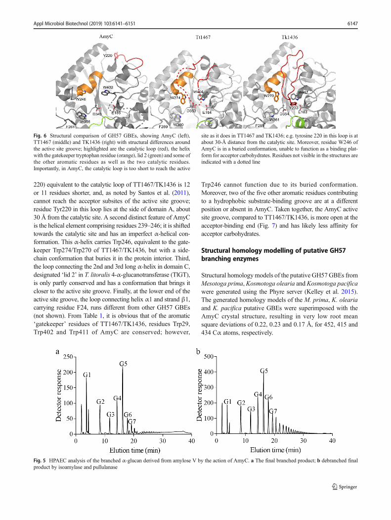
220) equivalent to the catalytic loop of TT1467/TK1436 is 12or 11 residues shorter, and, as noted by Santos et al. (2011),cannot reach the acceptor subsites of the active site groove;residue Tyr220 in this loop lies at the side of domain A, about30 Å from the catalytic site. A second distinct feature of AmyCis the helical element comprising residues 239–246; it is shiftedtowards the catalytic site and has an imperfect α-helical con-formation. This α-helix carries Trp246, equivalent to the gate-keeper Trp274/Trp270 of TT1467/TK1436, but with a side-chain conformation that buries it in the protein interior. Third,the loop connecting the 2nd and 3rd long α-helix in domain C,designated ‘lid 2’ in T. litoralis 4-α-glucanotransferase (TlGT),is only partly conserved and has a conformation that brings itcloser to the active site groove. Finally, at the lower end of theactive site groove, the loop connecting helix α1 and strand β1,carrying residue F24, runs different from other GH57 GBEs(not shown). From Table 1, it is obvious that of the aromatic‘gatekeeper’ residues of TT1467/TK1436, residues Trp29,Trp402 and Trp411 of AmyC are conserved; however,
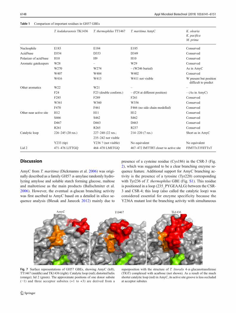
Trp246 cannot function due to its buried conformation.Moreover, two of the five other aromatic residues contributingto a hydrophobic substrate-binding groove are at a differentposition or absent in AmyC. Taken together, the AmyC activesite groove, compared to TT1467/TK1436, is more open at theacceptor-binding end (Fig. 7) and has likely less affinity foracceptor carbohydrates.
Structural homology modelling of putative GH57branching enzymes
Structural homologymodels of the putative GH57GBEs fromMesotoga prima,Kosmotoga olearia andKosmotoga pacificawere generated using the Phyre server (Kelley et al. 2015).The generated homology models of the M. prima, K. oleariaand K. pacifica putative GBEs were superimposed with theAmyC crystal structure, resulting in very low root meansquare deviations of 0.22, 0.23 and 0.17 Å, for 452, 415 and434 Cα atoms, respectively.
Fig. 6 Structural comparison of GH57 GBEs, showing AmyC (left),TT1467 (middle) and TK1436 (right) with structural differences aroundthe active site groove; highlighted are the catalytic loop (red), the helixwith the gatekeeper tryptophan residue (orange), lid 2 (green) and some ofthe other aromatic residues as well as the two catalytic residues.Importantly, in AmyC, the catalytic loop is too short to reach the active
site as it does in TT1467 and TK1436; e.g. tyrosine 220 in this loop is atabout 30-Å distance from the catalytic site. Moreover, residue W246 ofAmyC is in a buried conformation, unable to function as a binding plat-form for acceptor carbohydrates. Residues not visible in the structures areindicated with a dotted line
Fig. 5 HPAEC analysis of the branched α-glucan derived from amylose V by the action of AmyC. a The final branched product; b debranched finalproduct by isoamylase and pullulanase
Appl Microbiol Biotechnol (2019) 103:6141–6151 6147
Discussion
AmyC from T. maritima (Dickmanns et al. 2006) was origi-nally described as a family GH57α-amylase randomly hydro-lyzing amylose and soluble starch forming glucose, maltoseand maltotriose as the main products (Ballschmiter et al.2006). However, the eventual α-glucan branching activitywas first ascribed to AmyC based on a detailed in silico se-quence analysis (Blesak and Janecek 2012) mainly due to
presence of a cysteine residue (Cys186) in the CSR-3 (Fig.2), which was suggested to be a clear branching enzyme se-quence feature. Additional support for AmyC branching ac-tivity is the presence of a tyrosine (Tyr220) correspondingwith Tyr236 of T. thermophilus GBE (Fig. S1). This residueis positioned in a loop (235_PYGEAALG) between the CSR-3 and CSR-4; this loop (also called the catalytic loop) wasconsidered essential for enzyme specificity because theY236A mutant lost the branching activity with simultaneous
Table 1 Comparison of important residues in GH57 GBEs
T. kodakaraensis TK1436 T. thermophilus TT1467 T. maritima AmyC K. oleariaK. pacificaM. prima
Nucleophile E183 E184 E185 Conserved
Acid/base D354 D353 D349 Conserved
Polarizer of acid/base H10 H9 H10 Conserved
Aromatic gatekeepers W28 – W29 Conserved
W270 W274 – (W246 buried) As in AmyC
W407 W404 W402 Conserved
W416 W413 W411 not visible W present but positiondifficult to predict
Other aromatics W22 W21 – –
F24 F23 (double conform.) – (F24 at different position) – (As in AmyC)
F285 F289 F261 Conserved
W361 W360 W356 Conserved
F470 F461 F466 (no side chain modelled) Conserved
Other near active site H12 H11 H12 Conserved
S466 S462 S462 Conserved
D467 D463 D463 Conserved
R261 R265 R237 Conserved
Catalytic loop 226–245 (20 res.) 227–248 (22 res.; 214–220 (7 res.) Short as in AmyC
235–242 not visible
Y233 (tip) Y236 ? (not visible) No equivalent No equivalent
Lid 2 471–476 LITTGQ 464–470 LMETGQ 467–472 IMTTRT closer to active site FIMTTxT/FIITTxT
Fig. 7 Surface representations of GH57 GBEs, showing AmyC (left),TT1467 (middle) and TK1436 (right). Catalytic loop (red); distorted helix(orange); lid 2 (green). The approximate positions of one donor subsite(−1) and three acceptor subsites (+1 to +3) are derived from a
superposition with the structure of T. litoralis 4-α-glucanotransferase(TlGT) complexed with acarbose (not shown). As a result of the muchshorter catalytic loop (red) inAmyC, its active site groove is less occludedat acceptor subsites
6148 Appl Microbiol Biotechnol (2019) 103:6141–6151

acquiring of an increased hydrolytic ability (Palomo et al.2011). Although the sequence alignment (Fig. S1) indicatesa 12-residue deletion in the AmyC a few residues after thefunctionally important tyrosine, its presence in AmyC seemsto be conserved. It is worth mentioning that this residue is notconserved invariantly in all GBEs of the present study (datanot shown); this fact was observed previously when analysingmore than 150 hypothetical GBE sequences (Blesak andJanecek 2012).
Despite the three short regions between the CSR-3 andCSR-4, where AmyC possesses two deletions and one inser-tion in comparison with the three confirmed GBEs, AmyCunambiguously exhibits with them a high, i.e. more than47%, sequence similarity (Fig. S1). Sharing the unambiguousGBE sequence features is even more convincing within thefamily GH57 CSRs (Fig. 2). The AmyC sequence logo doeshave all the features identified as ‘sequence fingerprints’ ofGBE specificity (Blesak and Janecek 2012): (i) a cysteine inthe position 16, (ii) a leucine in the position 23 and (iii) aphenylalanine succeeded by a hydrophobic non-aromatic res-idue in positions 35 and 36, respectively (Fig. 2).
Re-classification of AmyC from T. maritima as a familyGH57 GBE is further supported by its position in the evolu-tionary tree (Fig. 1). Within the tree, the hypothetical GBErepresentatives originating from the phylum Thermotogaehave been found in three different parts: (i) generaThermotoga , Mesotoga and Kosmotoga (familiesT h e rm o t o g a c e a e a n d Ko sm o t o g a c e a e ) ; ( i i )Pseudothermotoga (family Thermotogaceae); and (iii)Defluviitoga and Petrotoga (family Petrotogaceae). In anycase, the assignment of the AmyC from T. maritima toGBEs is self-evident because the representatives of all remain-ing family GH57 enzyme specificities, such as α-amylase,amy lopu l l u l ana s e and 4 -α - g l u c ano t r an s f e r a s e(Supplementary Table S1), form their own branches and/orclusters clearly separated from all GBEs (Fig. 1).
The biochemical characteristics support the conclusionfrom the phylogenetic and sequence analyses that AmyC is afunctional branching enzyme converting amylose into abranched α-glucan with 8.5% branches. The 1H-NMR spec-trum shows clear reducing end signals at δ5.25 and δ4.66,being respectively the α- and the β-anomer, representing thehydrolysis products. The hydrolytic, i.e. α-amylase activityfound in this study (3 mU/mg), is considerably lower thanthe activity reported by Ballschmiter et al. (2006). In the pres-ent study, the BCAmethodwas used tomeasure the amount ofreducing ends, while Ballschmiter et al. (2006) used the DNSmethod, which is known to give erroneously high estimates ofglycoside hydrolase activity (Gusakov et al. 2011; McClearyand McGeough 2015), including α-amylase (Robyt andWhelan 1972). McCleary and McGeough (Robyt andWhelan 1972) concluded that DNS method should only beused to qualitatively measure glycoside hydrolase activity.
The activities reported by Ballschmiter et al. (Dickmannset al. 2006) therefore should be treated cautiously and shouldnot be compared to activities found with other methods tomeasure reducing ends.
The homology models of the M. prima, K. olearia andK. pacifica putative GBEs have an architecture highly similarto that of AmyC (Fig. S2). Only the loop connecting domainsB and C was modelled differently in the three homologues,likely due to the fact that in the AmyC structure, this loop isnot visible. The comparison in Table 1 shows that virtually allthe specific features of AmyC described above are conserved,including the shortened catalytic loop, the shifted helical ele-ment and lid 2. Therefore, it is very likely that, like AmyC, theM. prima, K. olearia and K. pacifica GBEs have a relativelyhigh hydrolytic activity among GH57 branching enzymes.The identification of putative GBEs in the mesophilic andthermophilic bacteria M. prima, K. olearia and K. pacificasuggests that AmyC is not unique, but that GH57 GBEs withrelatively high hydrolytic activity are widespread in suchorganisms.
Acknowledgements SJ thanks the Slovak Grant Agency VEGA for theGrant No. 2/0146/17. We thank Prof. Dr. Lubbert Dijkhuizen (MicrobialPhysiology, University of Groningen) for kindly allowing us to use theHPAEC-PAD equipment and Dr. Sander S. van Leeuwen (MicrobialPhysiology, University of Groningen) for the kind suggestions on theNMR measurements.
Funding information This work was financially supported by the ChinaScholarship Council (XZ) and the University of Groningen.
Compliance with ethical standards
Conflict of interest The authors declare that they have no conflict ofinterest.
Ethical approval This article does not contain any studies with humanparticipants or animals performed by any of the authors.
Open Access This article is distributed under the terms of the CreativeCommons At t r ibut ion 4 .0 In te rna t ional License (h t tp : / /creativecommons.org/licenses/by/4.0/), which permits unrestricted use,distribution, and reproduction in any medium, provided you giveappropriate credit to the original author(s) and the source, provide a linkto the Creative Commons license, and indicate if changes were made.
References
Apweiler R (2014) Activities at the universal protein resource. NucleicAcids Res 42(11):7486–7486. https://doi.org/10.1093/nar/gku469
Ballschmiter M, Futterer O, Liebl W (2006) Identification and character-ization of a novel intracellular alkaline α-amylase from the hyper-thermophilic bacterium Thermotoga maritimaMSB8. Appl EnvironMicrobiol 72(3):2206–2211. https://doi.org/10.1128/Aem.72.3.2206-2211.2006
Appl Microbiol Biotechnol (2019) 103:6141–6151 6149
Benson DA, Clark K, Karsch-Mizrachi I, Lipman DJ, Ostell J, SayersEW (2014) GenBank. Nucleic Acids Res 42(D1):D32–D37. https://doi.org/10.1093/nar/gkt1030
Bissaro B, Monsan P, Faure R, O'Donohue MJ (2015) Glycosynthesis ina waterworld: new insight into the molecular basis oftransglycosylation in retaining glycoside hydrolases. Biochem J467:17–35. https://doi.org/10.1042/Bj20141412
Blesak K, Janecek S (2012) Sequence fingerprints of enzyme specificitiesfrom the glycoside hydrolase family GH57. Extremophiles 16(3):497–506. https://doi.org/10.1007/s00792-012-0449-9
Blesak K, Janecek S (2013) Two potentially novel amylolytic enzymespecificities in the prokaryotic glycoside hydrolase α-amylase fam-ily GH57. Microbiology 159:2584–2593. https://doi.org/10.1099/mic.0.071084-0
Boyer CD, Preiss J (1978) Multiple forms of starch branching enzyme ofmaize: evidence for independent genetic control. Biochem BiophysRes Commun 80(1):169–175. https://doi.org/10.1016/0006-291X(78)91119-1
Cantarel BL, Coutinho PM, Rancurel C, Bernard T, Lombard V,Henrissat B (2009) The carbohydrate-active enzymes database(CAZy): an expert resource for glycogenomics. Nucleic Acids Res37:D233–D238. https://doi.org/10.1093/nar/gkn663
Crooks GE, Hon G, Chandonia JM, Brenner SE (2004) WebLogo: asequence logo generator. Genome Res 14(6):1188–1190. https://doi.org/10.1101/gr.849004
Dickmanns A, Ballschmiter M, Liebl W, Ficner R (2006) Structure of thenovel α-amylase AmyC from Thermotoga maritima. ActaCrystallogr D Biol Crystallogr 62:262–270. https://doi.org/10.1107/S0907444905041363
Felsenstein J (1985) Confidence limits on phylogenies - an approachusing the bootstrap. Evolution 39(4):783–791. https://doi.org/10.1111/j.1432-1033.1988.tb14056.x
Fukusumi S, Kamizono A, Horinouchi S, Beppu T (1988) Cloning andnucleotide-sequence of a heat-stable amylase gene from an anaero-bic thermophile, Dictyoglomus-Thermophilum. Eur J Biochem174(1):15–21 https://doi.org/10.1111/j.1432-1033.1988.tb14056.x
Gusakov AV, Kondratyeva EG, Sinitsyn AP (2011) Comparison of twomethods for assaying reducing sugars in the determination of carbo-hydrase activities. Int J Anal Chem 2011:1–4. https://doi.org/10.1155/2011/283658
Harada T, Misaki A, Akai H, Yokobayashi K, Sugimoto K (1972)Characterization of Pseudomonas isoamylase by its actions on am-ylopectin and glycogen: comparison with Aerobacter pullulanase.Biochim Biophys Acta 268(2):497–505. https://doi.org/10.1016/0005-2744(72)90345-2
Henrissat B, Bairoch A (1996) Updating the sequence-based classifica-tion of glycosyl hydrolases. Biochem J 316:695–696
Imamura H, Fushinobu S, Yamamoto M, Kumasaka T, Jeon BS, WakagiT, Matsuzawa H (2003) Crystal structures of 4-α-glucanotransferasefrom Thermococcus litoralis and its complex with an inhibitor. JBiol Chem 278(21):19378–19386. https://doi.org/10.1074/jbc.M213134200
Janecek S, Blesak K (2011) Sequence-structural features and evolution-ary relationships of family GH57 α-amylases and their putative α-amylase-like homologues. Protein J 30(6):429–435. https://doi.org/10.1007/s10930-011-9348-7
Janecek S, Svensson B, MacGregor EA (2014) α-Amylase: an enzymespecificity found in various families of glycoside hydrolases. CellMol Life Sci 71(7):1149–1170. https://doi.org/10.1007/s00018-013-1388-z
Kelley LA, Mezulis S, Yates CM, Wass MN, Sternberg MJE (2015) ThePhyre2 web portal for protein modeling, prediction and analysis. NatProtoc 10(6):845–858. https://doi.org/10.1038/nprot.2015.053
Koshland DE (1953) Stereochemistry and the mechanism of enzymaticreactions. Biol Rev 28(4):416–436. https://doi.org/10.1111/j.1469-185X.1953.tb01386.x
Laderman KA, Asada K, Uemori T, Mukai H, Taguchi Y, Kato I,Anfinsen CB (1993) α-Amylase from the hyperthermophilic ar-chaebacterium Pyrococcus furiosus - cloning and sequencing ofthe gene and expression in Escherichia coli. J Biol Chem 268(32):24402–24407
Larkin MA, Blackshields G, Brown NP, Chenna R, McGettigan PA,McWilliam H, Valentin F, Wallace IM, Wilm A, Lopez R,Thompson JD, Gibson TJ, Higgins DG (2007) ClustalWand clustalX version 2.0. Bioinformatics 23(21):2947–2948. https://doi.org/10.1093/bioinformatics/btm404
Lee EY, Carter J, Nielsen L, Fischer EH (1970) Purification and proper-t ies of yeas t amylo-1 ,6-g lucosidase -ol igo-1 ,4→1,4-glucantransferase. Biochemistry 9(11):2347–2355
Letunic I, Bork P (2006) Interactive tree of life (iTOL): an online tool forphylogenetic tree display and annotation. Bioinformatics 23(1):127–128
Li YR, Niu DD, Zhang L, Wang ZX, Shi GY (2013) Purification, char-acterization and cloning of a thermotolerant isoamylase producedfrom Bacillus sp CICIM 304. J Ind Microbiol Biotechnol 40(5):437–446. https://doi.org/10.1007/s10295-013-1249-7
Lombard V, Ramulu HG, Drula E, Coutinho PM, Henrissat B (2014) Thecarbohydrate-active enzymes database (CAZy) in 2013. NucleicAcids Res 42(D1):D490–D495. https://doi.org/10.1093/nar/gkt1178
Martinovičová M, Janeček Š (2018) In silico analysis of the α-amylasefamily GH57: eventual subfamilies reflecting enzyme specificities.Biotech 8(7):307. https://doi.org/10.1007/s13205-018-1325-9
McCleary BV, McGeough P (2015) A comparison of polysaccharidesubstrates and reducing sugar methods for the measurement ofendo-1, 4-β-xylanase. Appl Biochem Biotechnol 177(5):1152–1163. https://doi.org/10.1007/s12010-015-1803-z
Murakami T, Kanai T, Takata H, Kuriki T, Imanaka T (2006) A novelbranching enzyme of the GH-57 family in the hyperthermophilicarchaeon Thermococcus kodakaraensis KOD1. J Bacteriol188(16):5915–5924. https://doi.org/10.1128/Jb.00390-06
Na S, Park M, Jo I, Cha J, Ha NC (2017) Structural basis for thetransglycosylase activity of a GH57-type glycogen branching en-zyme from Pyrococcus horikoshii. Biochem Biophys ResCommun 484(4):850–856. https://doi.org/10.1016/j.bbrc.2017.02.002
Palomo M, Pijning T, Booiman T, Dobruchowska JM, van der Vlist J,Kralj S, Planas A, Loos K, Kamerling JP, Dijkstra BW, van derMaarel MJEC, Dijkhuizen L, Leemhuis H (2011) Thermusthermophilus glycoside hydrolase family 57 branching enzymecrystal structure, mechanism of action and products formed. J BiolChem 286(5):3520–3530. https://doi.org/10.1074/jbc.M110.179515
Robyt JF, Whelan WJ (1972) Reducing value methods for maltodextrins:I. chain-length dependence of alkaline 3, 5-dinitrosalicylate andchain-length independence of alkaline copper. Anal Biochem45(2):510–516. https://doi.org/10.1016/0003-2697(72)90213-8
Saitou N, Nei M (1987) The neighbor-joining method - a new method forreconstructing phylogenetic trees. Mol Biol Evol 4(4):406–425.https://doi.org/10.1093/oxfordjournals.molbev.a040454
Santos CR, Tonoli CC, Trindade DM, Betzel C, Takata H, Kuriki T,Kanai T, Imanaka T, Arni RK, Murakami MT (2011) Structuralbasis for branching-enzyme activity of glycoside hydrolase family57: structure and stability studies of a novel branching enzyme fromthe hyperthermophilic archaeon Thermococcus kodakaraensisKOD1. Proteins 79(2):547–557. https://doi.org/10.1002/prot.22902
Sievers F, Wilm A, Dineen D, Gibson TJ, Karplus K, Li W, Lopez R,McWilliam H, Remmert M, Soding J, Thompson JD, Higgins DG(2011) Fast, scalable generation of high-quality protein multiplesequence alignments using Clustal omega. Mol Syst Biol 7:539.https://doi.org/10.1038/msb.2011.75
6150 Appl Microbiol Biotechnol (2019) 103:6141–6151
Terada Y, Fujii K, Takaha T, Okada S (1999) Thermus aquaticus ATCC33923 amylomaltase gene cloning and expression and enzyme char-acterization: production of cycloamylose. Appl Environ Microbiol65(3):910–915
van der Maarel MJEC, Leemhuis H (2013) Starch modification withmicrobial α-glucanotransferase enzymes. Carbohydr Polym 93(1):116–121. https://doi.org/10.1016/j.carbpol.2012.01.065
van der Maarel MJEC, van der Veen B, Uitdehaag JCM,Leemhuis H, Dijkhuizen L (2002) Properties and applicationsof starch-converting enzymes of the α-amylase family. JBiotechnol 94(2):137–155. https://doi.org/10.1016/S0168-1656(01)00407-2
Zona R, Chang-Pi-Hin F, O’Donohue MJ, Janecek S (2004)Bioinformatics of the glycoside hydrolase family 57 and identifica-tion of catalytic residues in amylopullulanase from Thermococcushydrothermalis. Eur J Biochem 271(14):2863–2872. https://doi.org/10.1111/j.1432-1033.2004.04144.x
Publisher’s note Springer Nature remains neutral with regard tojurisdictional claims in published maps and institutional affiliations.
Appl Microbiol Biotechnol (2019) 103:6141–6151 6151