![Self-Drilling Rock Bolting System ELEBAR™-SD ELEBAR-SD-R...Rock bolting with the ELEBAR -SD system is fast and simple and requires only two (2) stages [drilling and cement grouting]](https://static.fdocument.org/doc/165x107/60da1dddb324712dfa3a7136/self-drilling-rock-bolting-system-elebara-sd-elebar-sd-r-rock-bolting-with.jpg)
Identification of Penicillium expansum causing postharvest ... · PDF file0.49 (SD) x 3...
10
257 Identification of Penicillium expansum causing postharvest blue mold decay of apple fruit Ivana Vico*, Nataša Duduk, Miljan Vasić and Milica Nikolić University of Belgrade, Faculty of Agriculture, Nemanjina 6, 11080 Zemun-Belgrade (*[email protected]) Received: December 6, 2014 Accepted: December 31, 2014 SUMMARY Penicillium expansum (Link) Thom. is one of the most important postharvest pathogens of apple fruit worldwide. It causes blue mold, a decay that can lead to significant economic losses during storage, which can also impact fruit destined for processing due to the production of carcinogenic mycotoxin patulin. Apple fruit cvs. Idared, Golden Delicious and Braeburn with blue mold symptoms were collected from five storage facilities in Serbia and nine fungal isolates were obtained. Pathogenicity of the isolates was tested and proven by artificial inoculation of healthy apples cv. Idared. In order to identify the causal agents of decay, morphological and molecular methods were used. Colony morphology and microscopic features were observed on differential media, and isolates were tested for the production of cyclopiazonic acid. Molecular analysis included PCR amplification with species specific primers for P. expansum based on polygalacturonase gene (Pepg1), universal primers for internal transcribed spacer rDNA region and primers based on β-tubulin gene. All isolates formed compact blue green colonies with characteristic earthy odor. Conidiophores were terverticillate with smooth septate stipes and conidia were smooth, globose to subglobose, born in colums. The average size of conidia was 3.38 ± 0.49 (SD) x 3 ± 0.36 (SD) μm. Using species specific primers PEF/PER the texpected amplicons of ~404 bp were obtained in all nine tested isolates and PCR conducted with the Bt-LEV- Up4/Bt-LEV-Lo1 and universal ITS1/ITS4 primer pairs generated amplicons of the expected sizes of ~800 bp and ~600 bp, respectively. MegaBlast analyses of the 2X consensus of nucleotide sequences of the isolate JP1 partial β-tubulin gene and ITS region showed 99-100% and 100% similarity with several P. expansum sequences of corresponding regions of this species deposited in GenBank. Based on morphological and molecular features, the isolates obtained from decayed apple fruit collected in several storage facilities in Serbia were identified as P. expansum. Keywords: Penicillium expansum; Postharvest decay; Apples Pestic. Phytomed. (Belgrade), 29(4), 2014, 257–266 UDC 632.4:634.11:631.576 DOI: 10.2298/PIF1404257V Original scientific paper
Transcript of Identification of Penicillium expansum causing postharvest ... · PDF file0.49 (SD) x 3...
� 257
Identification of Penicillium expansum causing postharvest blue mold decay of apple fruit
Ivana Vico*, Nataša Duduk, Miljan Vasić and Milica NikolićUniversity of Belgrade, Faculty of Agriculture, Nemanjina 6, 11080 Zemun-Belgrade(*[email protected])
Received: December 6, 2014Accepted: December 31, 2014
Summary
Penicillium expansum (Link) Thom. is one of the most important postharvest pathogens of apple fruit worldwide. It causes blue mold, a decay that can lead to significant economic losses during storage, which can also impact fruit destined for processing due to the production of carcinogenic mycotoxin patulin. Apple fruit cvs. Idared, Golden Delicious and Braeburn with blue mold symptoms were collected from five storage facilities in Serbia and nine fungal isolates were obtained. Pathogenicity of the isolates was tested and proven by artificial inoculation of healthy apples cv. Idared. In order to identify the causal agents of decay, morphological and molecular methods were used. Colony morphology and microscopic features were observed on differential media, and isolates were tested for the production of cyclopiazonic acid. Molecular analysis included PCR amplification with species specific primers for P. expansum based on polygalacturonase gene (Pepg1), universal primers for internal transcribed spacer rDNA region and primers based on β-tubulin gene. All isolates formed compact blue green colonies with characteristic earthy odor. Conidiophores were terverticillate with smooth septate stipes and conidia were smooth, globose to subglobose, born in colums. The average size of conidia was 3.38 ± 0.49 (SD) x 3 ± 0.36 (SD) μm. Using species specific primers PEF/PER the texpected amplicons of ~404 bp were obtained in all nine tested isolates and PCR conducted with the Bt-LEV-Up4/Bt-LEV-Lo1 and universal ITS1/ITS4 primer pairs generated amplicons of the expected sizes of ~800 bp and ~600 bp, respectively. MegaBlast analyses of the 2X consensus of nucleotide sequences of the isolate JP1 partial β-tubulin gene and ITS region showed 99-100% and 100% similarity with several P. expansum sequences of corresponding regions of this species deposited in GenBank. Based on morphological and molecular features, the isolates obtained from decayed apple fruit collected in several storage facilities in Serbia were identified as P. expansum.
Keywords: Penicillium expansum; Postharvest decay; Apples
Pestic. Phytomed. (Belgrade), 29(4), 2014, 257–266 UDC 632.4:634.11:631.576DOI: 10.2298/PIF1404257V Original scientific paper
258
Ivana Vico et al.
INTrODuCTION
Postharvest diseases cause losses that are greater than generally realized because the value of fresh fruits and vegetables increases several-fold with the passing from fields to consumers (Eckert & Sommer, 1967). Postharvest losses have been estimated to range from 10 to 30% per year despite the use of modern storage facilities and techniques (Harvey, 1978). Fruit crops are attacked by a wide range of microorganisms in their postharvest phase (Snowdon, 1990; Ogawa & English, 1991).
Apples are kept in storage from 4-6 months up to 1 year to ensure that fruit are available year round for consumption. While in storage, apples are highly susceptible to decay. One of the economically most important problems worldwide is blue mold decay. It is caused by Penicillium spp., among which P. expansum (Link) Thom. is prevalent. Blue mold decay leads to significant economic losses during storage that also impacts fruit destined for processing by its production of the carcinogenic mycotoxin patulin (Barkai-Golan, 2008).
P. expansum infects fruit primarily through wounds caused by stem punctures or bruises occurring at harvest or during postharvest handling. The fungus can also enter the fruit through natural openings, i.e. lenticels, stem ends and the calyx end (Rosenberger et al., 2006). Symptoms occur as light colored, soft lesions. Lesions are soft due to maceration of the tissue by polygalacturonase enzyme which plays a significant role in P. expansum virulence (Jurick et al., 2010). As a lesion expands the decayed portion can easily be separated from the surrounding sound tissue. Fungal growth on the lesion surface, at first white, becomes pale blue as sporulation occurs. Although blue mold lesions mainly start from wound infections, the fungus can cause „nesting“ in a fruit bin by growing into neighbouring healthy fruit (Sommer et al., 2002). P. expansum produces abundant conidia that are readily airborne.
Penicillium species have been studied over 200 years and the genus was first described by Link in 1809. Initially, morphological identification methods were used, but diversity within the genus has resulted in researchers seeking alternative techniques and approaches to improve accuracy. These methods have involved biochemical analyses of secondary metabolites in conjuction with morphological examination. With the emergance of more accurate and rapid molecular identification tools scientists embraced modern technology to address diversity challanges. In order to provide a more holistic approach towards the taxonomy of complex genera, morphological analysis remains the essential component
in Penicillium identification (Frisvad & Samson, 2004; Pitt & Hocking, 2009). Since several Penicillium species can cause blue mold decay and having in mind that their identification is difficult due to their morphological similarities, the aim of this study was to identify the causal agent of apple decay in several storage facilities in Serbia, using both morphological and molecular criteria.
maTErIaLS aND mETHODS
Isolation
Decayed apple fruit cvs. Idared, Golden Delicious and Braeburn with symptoms of blue mold were collected from 5 storage facilities in Svilajnac, Bela Crkva, Suvodol, Čelarevo, and Pudarci. Apples were collected during the storage season - winter 2012/2013. Isolation of the fungus from apple fruit was performed by direct plating of inner decayed tissue after surface sterilisation with 70% ethanol and aseptical removal of skin. Tissue fragments were placed on potato dextrose agar (PDA) in Petri plates and incubated at room temperature for 5 days. Following incubation, the plates were examined, and fungal cultures were transferred to sterile PDA. Pure colonies were then used to obtain monosporial cultures using the streaking method. Monosporial cultures were preserved on slants in the refregerator at 40C.
Pathogenicity test
Healthy apples cv. Idared were used to conduct Koch’s postulates. Fruit were washed and surfaced sterilized using 70% ethanol. A triangle shaped apple tissue fragment (15 mm length of each side) was taken out of the fruit. Mycelial disks with spores, cut out from 7-day old cultures grown on PDA (6 mm in diameter) were placed in the wounds and topped with apple fragments. Control fruit were treated the same way, but a sterile PDA disk was placed in each wound instead of mycelium. Four fruit were inoculated on two sides per isolate and 4 were used as control. Following inoculation, the apples were stored under moist conditions at room temperature for 7 days. The pathogen was reisolated from the lesions that developed on inoculated fruit.
morphological investigation
Colony patterns and growth on different media. Isolates were three point inoculated on the following media: PDA, Czapek yeast extract agar (CYA), malt extract
� 259
Pestic. Phytomed. (Belgrade), 29(4), 2014, 257–266
agar (MEA) and yeast extract sucrose agar (YES) in 9 cm plastic Petri plates using a dense conidium suspension (Frisvad & Samson, 2004), and incubated in the dark. Colony appearance, exudate production, pigmentation, and reverse coloration were assessed, and colony diameters were measured and recorded after a week of growth at 25oC. The isolates were also grown at 5oC and 37 oC on CYA, and colony diameter was measured after 7 days of incubation (Pitt & Hocking, 2009). Five plates were inoculated per medium per temperature.
Morphology of conidiophores, stype, and conidia. Microscopic slides were prepared from 7-day old cultures grown on PDA and MEA. Tween-treated water (5 drops of Tween 20 in 25 ml water) with or without dye (Bromphenol blue) was used as mounting media with the aid of ethanol (colony fragments were sampled with a needle and placed in a drop of 70% ethanol, after ethanol dried out the mounting media was added) (Pitt & Hocking, 2009). The morphology of conidia, type of conidiophores, stype ornamentation, and phialide shape were evaluated. Conidia measurement was performed using a compound microscope Leica DMLS equipped with digital camera Leica DC 300 and software Leica IM 1000.
Ehrlich test
The isolates were examined for production of cyclopiazonic acid and other alkaloids reacting with Ehrlich reagent (Lund, 1995). Ehrlich reagent consists of 2 g 4-dimethylaminobenzaldehide in 85 ml 96% ethanol, with 15 ml 10 N HCl added. Three agar plugs (8 mm) were cut from the center of a 7-day old culture grown on CYA at 25oC and a filter paper (~ 20 x 20 mm) wetted in Ehrlich reagent was placed on top of the mycelial side. After 2-10 minutes the reaction was scored. In a positive reaction the appearance of violet ring means that the culture contains cyclopiazonic acid or related alkaloids, and pink to red or yellow ring means that the fungus produces other alkaloids. If the reaction comes after 7-10 minutes it is regarded as weak. The experiment was repeated 5 times.
molecular analysis
DNA extraction. Fungal mycelium was obtained from isolates grown on PDA for 7 days. DNA extraction was performed following the CTAB protocol of Day and Shattock (1997). Total DNA was precipitated with isopropanol and resuspended in 30 µl of TE buffer (10
mM Tris pH 8 and 1 mM EDTA) and stored at -20˚C until use.
PCR amplification and DNA sequencing. The partial polygalacturonase gene (Pepg1) was amplified with the primer pair PEF/PER (Marek et al., 2003). PCR reaction mix (25 μL) contained 1 µl of template DNA, 1 × PCR MasterMix (Fermentas, Vilnius, Lithuania) and 1 μM of each primer. The amplification parameters for PEF/PER primers consisted of an initial denaturation at 92oC for 5 min, followed by 30 cycles at 92oC for 1 min, 55oC for 45 s and 72oC for 45 s, and final elongation at 72oC for 10 min. DNA was amplified with the primer pair Bt-LEV-Up4/Bt-LEV-Lo1, based on β-tubulin gene (de Jong et al., 2001). PCR reaction mix (25 μL) contained 1 µl of template DNA, 1 × PCR MasterMix (Fermentas, Vilnius, Lithuania) and 0.4 μM of each primer. The amplification parameters were: an initial denaturation at 96oC for 3 min, followed by 40 cycles at 96oC for 60 s, 60oC for 60 s and 72oC for 2 min, with final elongation at 72oC for 10 min. The ITS1-5.8S-ITS2 region was amplified with the universal primer pair ITS1/ITS4 (White et al., 1990). PCR reaction mix (25 μL) contained 1 µl of template DNA, 1 × PCR MasterMix (Fermentas, Vilnius, Lithuania) and 0.4 μM of each primer. Amplification parameters for the ITS region consisted of an initial denaturation at 94oC for 90 s, followed by 30 cycles at 94oC for 30 s, 55oC for 30 s and 72oC for 30 s, and final elongation at 72oC for 10 min. Samples lacking fungal DNA were employed as the negative control. PCR product (5 μL) was observed in a 1.5% agarose gel, stained in ethidium bromide and visualized with UV transilluminator. Amplified Bt-LEV-Up4/Bt-LEV-Lo1 and ITS1/ITS4 products of the isolate JP1 were directly sequenced in both directions using forward and reverse primers. The obtained sequences were assembled using Pregap4 from the Staden program package (Staden et al., 2000), and compared with sequences in GenBank NCBI using the MegaBlast algorithm (http://www.ncbi.nlm.nih.gov/).
rESuLTS
Symptoms
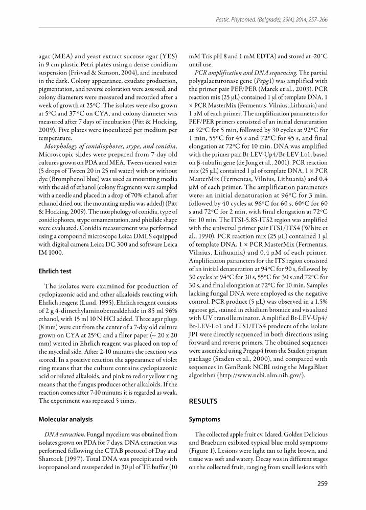
The collected apple fruit cv. Idared, Golden Delicious and Braeburn exibited typical blue mold symptoms (Figure 1). Lesions were light tan to light brown, and tissue was soft and watery. Decay was in different stages on the collected fruit, ranging from small lesions with
260
Ivana Vico et al.
no apparent sporulation to completely decayed fruit covered with blue-green spores. In most cases spores were formed on lesion surface, starting at the infection site. The decayed area on fruit had sharp margins between diseased and healthy tissue which was easily separated from the healthy part, leaving it in a shape of a “bowl”. The fruit had an earthy, musty odor.
Figure 1. Blue mold symptoms on apple fruit (natural infection)
Isolates
Nine isolates with similar compact blue green velutinous colony appearance were obtained from apple fruit of three cultivars with blue mold symptoms collected from 5 storage facilities in Serbia (Table 1)
Table 1. Fungal isolates obtained from apple fruit with blue mold symptoms
Isolate Cultivar Locality
Jpp1 Golden Delicious Pudarci
Jpp3 Golden Delicious Pudarci
JČ1 Braeburn Čelarevo
JČ2 Braeburn Čelarevo
JSuv 9 Idared Suvodol
JSuv 5 Idared Suvodol
JBC6 Golden Delicious Bela Crkva
JP1 Idared Svilajnac
JP5 Idared Svilajnac
Pathogenicity
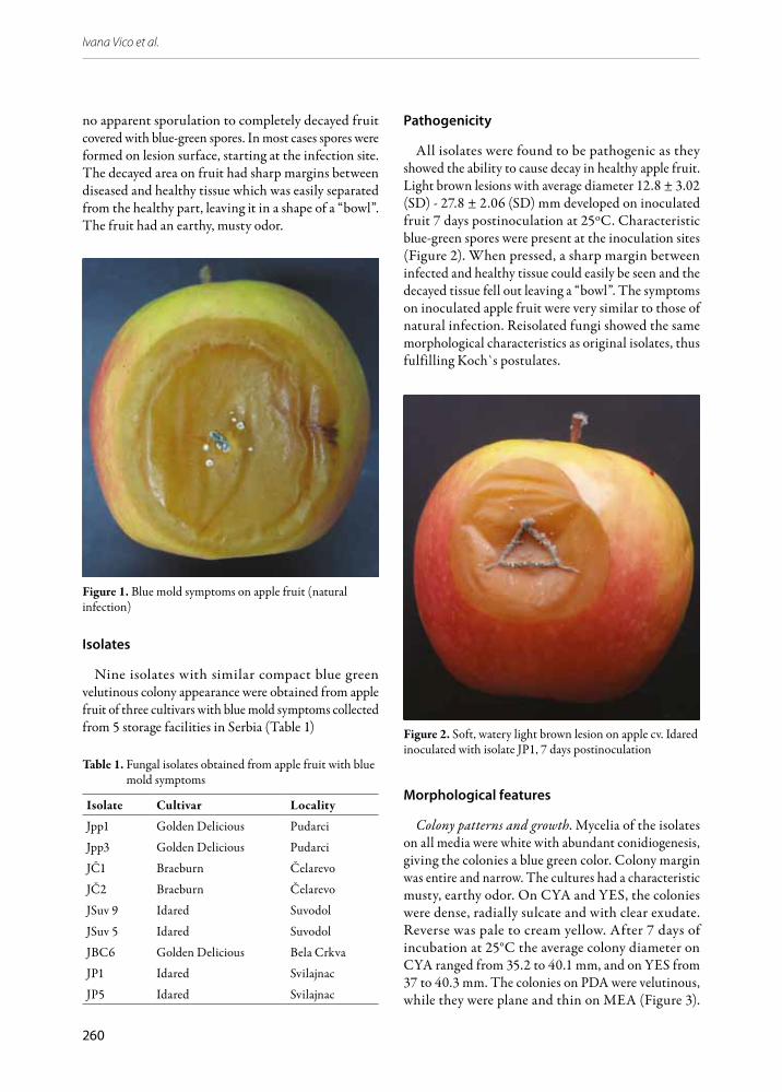
All isolates were found to be pathogenic as they showed the ability to cause decay in healthy apple fruit. Light brown lesions with average diameter 12.8 ± 3.02 (SD) - 27.8 ± 2.06 (SD) mm developed on inoculated fruit 7 days postinoculation at 25oC. Characteristic blue-green spores were present at the inoculation sites (Figure 2). When pressed, a sharp margin between infected and healthy tissue could easily be seen and the decayed tissue fell out leaving a “bowl”. The symptoms on inoculated apple fruit were very similar to those of natural infection. Reisolated fungi showed the same morphological characteristics as original isolates, thus fulfilling Koch s̀ postulates.
Figure 2. Soft, watery light brown lesion on apple cv. Idared inoculated with isolate JP1, 7 days postinoculation
morphological features
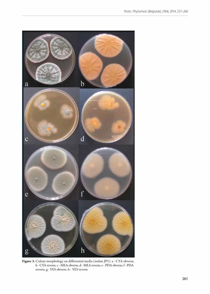
Colony patterns and growth. Mycelia of the isolates on all media were white with abundant conidiogenesis, giving the colonies a blue green color. Colony margin was entire and narrow. The cultures had a characteristic musty, earthy odor. On CYA and YES, the colonies were dense, radially sulcate and with clear exudate. Reverse was pale to cream yellow. After 7 days of incubation at 25°C the average colony diameter on CYA ranged from 35.2 to 40.1 mm, and on YES from 37 to 40.3 mm. The colonies on PDA were velutinous, while they were plane and thin on MEA (Figure 3).
� 261
Pestic. Phytomed. (Belgrade), 29(4), 2014, 257–266
Figure 3. Colony morphology on differential media (isolate JP1): a - CYA obverse, b - CYA reverse, c - MEA obverse, d - MEA reverse, e - PDA obverse, f - PDA reverse, g - YES obverse, h - YES reverse
262
Ivana Vico et al.
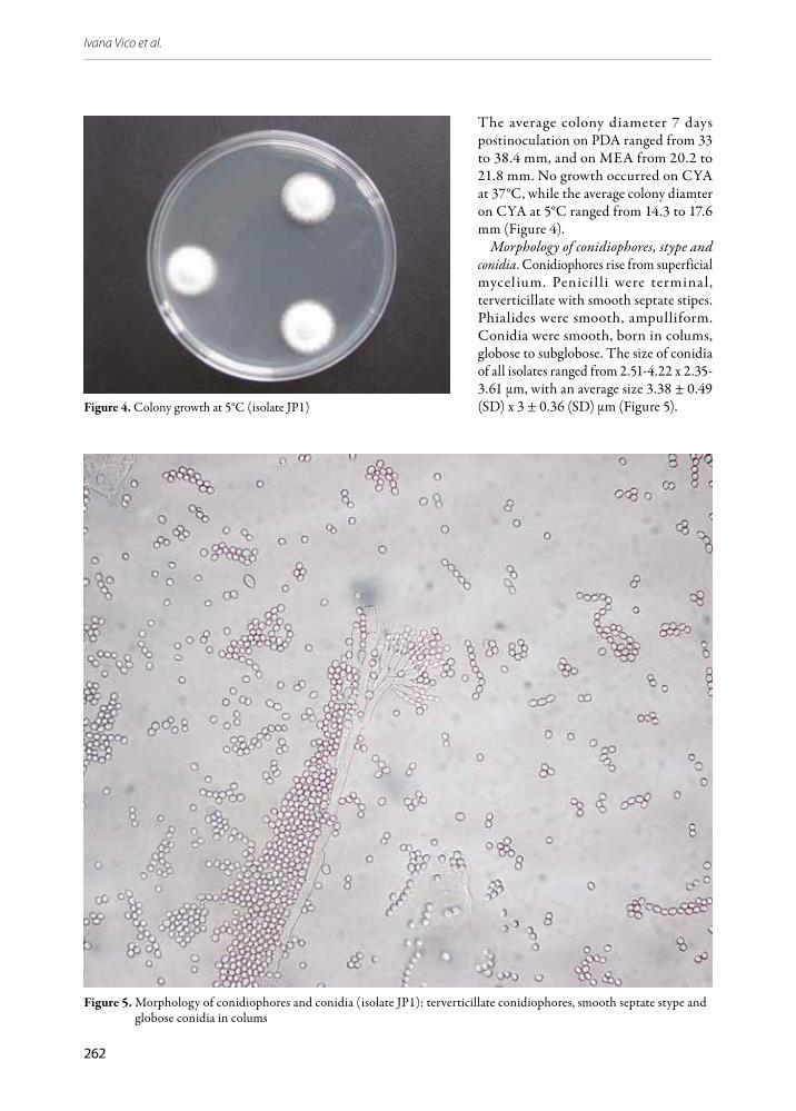
The average colony diameter 7 days postinoculation on PDA ranged from 33 to 38.4 mm, and on MEA from 20.2 to 21.8 mm. No growth occurred on CYA at 37°C, while the average colony diamter on CYA at 5°C ranged from 14.3 to 17.6 mm (Figure 4).
Morphology of conidiophores, stype and conidia. Conidiophores rise from superficial mycelium. Penicil li were terminal, terverticillate with smooth septate stipes. Phialides were smooth, ampulliform. Conidia were smooth, born in colums, globose to subglobose. The size of conidia of all isolates ranged from 2.51-4.22 x 2.35-3.61 μm, with an average size 3.38 ± 0.49 (SD) x 3 ± 0.36 (SD) μm (Figure 5).Figure 4. Colony growth at 5°C (isolate JP1)
Figure 5. Morphology of conidiophores and conidia (isolate JP1): terverticillate conidiophores, smooth septate stype and globose conidia in colums

� 263
Pestic. Phytomed. (Belgrade), 29(4), 2014, 257–266
Ehrlich test
In reaction with Ehrlich reagent the isolates formed yellow rings, which means that the isolates produced alkaloids other than cyclopiazonic acid (Figure 6).
Figure 6. Yellow ring resulting from Ehrlich test (isolate JP1)
molecular analysis
Using the specific primers PEF/PER, expected amplicons of ~404 bp were obtained in all nine tested isolates of Penicillium sp. (Figure 7). No amplification was obtained with negative control.
Figure 7. Amplification of DNA products (~404 bp) using PEF/PER primer pair (M - 100-bp ladder (Fermentas, Lithuania); 1 - isolate JPp1; 2 - isolate JPp3; 3 - isolate JČ1; 4 - isolate JČ2; 5 - isolate JSuv9; 6 - isolate JSuv5; 7 - isolate JBC6; 8 - isolate JP1; 9 - isolate JP2; - K negative control)
PCR conducted with the Bt-LEV-Up4/Bt-LEV-Lo1 and universal ITS1/ITS4 primer pairs generated amplicons of expected size of ~800 bp and ~600 bp,
respectively, with all tested isolates. No amplification was obtained with negative control.
MegaBlast analyses of the 2X consensus nucleotide sequence of partial β-tubulin gene of 780 nt of isolate JP1 showed 99-100% similarity with several P. expansum sequences of the same region deposited in GenBank. The sequence of internal transcribed spacer region of 552 nt for the same isolate was identical to sequences of the same region of P. expansum deposited in GenBank.
DISCuSSION
Decayed apple fruit collected from several storage facilities in Serbia exhibited typical symptoms of blue mold. Blue mold can be caused by different Penicillium species, among which P. expansum is usually considered to be the most important (Rosenberger, 1990). Other Penicillium species able to cause similar decay include P. solitum, P. crustosum, P. italicum, P. digitatum, P. commune, P. verrucosum, P. rugulosum, P. polonicum and P. chrysogenum (Pitt et al., 1991; Sanderson & Spotts, 1995; Amiri & Bompeix, 2005; Andersen & Thrame, 2006; Sang et al., 2010; Vico et al., 2014). Since several Penicillium species can be responsible for blue mold decay of apples, a precise characterization of the causal agent is important, especially for fruit destined for processing because of the putative presence of the mycotoxin patulin (Barkai-Golan, 2008).
Penicillium identification is not easy. It is primarily based on morphological features incorporating the use of differential media and standardised laboratory conditions. The isolates obtained from decayed apples in this study had colony morphology on differential media and microscopic features typical of those described for P. expansum giving us a tentative identification of apple decay causal agent as P. expansum (Frisvad & Samson, 2004; Pitt & Hocking, 2009). However, Penicillium is a large genus, and many species have very similar macro- and microscopic properties, and at the same time there is variability within the species, which is why further identification required other approaches such as biochemical analysis (Frisvad & Samson, 2004) and molecular identification (Skouboe et al., 1999; Samson et al., 2004; Visagie et al., 2014). In this manner all of the investigated isolates were tested for the production of cyclopiazonic acid and other alkaloids. In reaction
264
Ivana Vico et al.
with Ehrlich reagent the studied isolates of P. expansum produced yellow rings, showing that they did not produce cyclopiazonic acid but produced other alkaloids.
For further identification of Penicillium isolates in our present investigation, species specific primers (Marek et al., 2003) were used. These primers are based on a partial sequence of the polygalacturonase gene (Pepg1) and were developed for rapid and specific detection of P. expansum, and used in several studies (Oliveri et al., 2007; Elhariry et al., 2011; Sanzani et al., 2013). Amplification of the corresponding DNA of the tested isolates with P. expansum specific primers, gave a single distinct product of ~404 bp confirming that the isolated fungi from decayed apples belonged to P. expansum. Also, two more regions (ITS and β-tubulin) were amplified and sequenced for one of the isolates – isolate JP1. The internal transcribed spacer rDNA area (ITS) is used for molecular identification of Penicillium species and is useful for placing isolates into species complex or one of the 25 sections. Sometimes this region provides species identification (Visagie et al., 2014), but for Penicillium and many other genera of ascomycetes, the ITS is not variable enough for distinguishing all closely related species (Skouboe et al., 1999). That is why another region – the β-tubulin gene region can be successfully used for accurate identification of Penicillium species (Visagie et al., 2014). The obtained sequences of ITS and β-tubulin gene region of the tested isolate JP1 showed to be either identical or with 99-100% similarity to corresponding sequences for P. expansum deposited in GenBank, confirming the identity of the isolates. Based on morphological and molecular properties of nine isolates, the causal agent of blue mold apple decay in five storage facilities in Serbia was identified as P. expansum. Having in mind that this is the prevalent decay-causing agent on many commodities and that it is capable of producing mycotoxins, further insight into the spread and mechanisms of invasion might lead to a development of effective control strategies against this important postharvest pathogen.
aCkNOwLEDgEmENT
This research was supported by the project III46008 funded by the Ministry of Education, Science and Technological Development, Republic of Serbia.
rEFErENCES
Amiri, A. & Bompeix, G. (2005). Diversity and population dynamics of Penicillium spp. on apples in pre- and postharvest environments: consequences for decay development. Plant Pathology, 54, 74-81.
Andersen, B. & Thrame, U. (2006): Food-borne fungi in fruit and cereals and their production of mycotoxins. Advances in Food Mycology, 571, 137-152.
Barkai-Golan, R. (2008). Penicillium mycotoxins. In R. Barkai-Golan & Paster, N. (Eds) Mycotoxins in fruits and vegetables (pp.153-185). San Diego, USA: Elsevier.
Day, J.P. & Shattock, R.C. (1997). Aggressiveness and other factors relating to displacements of population of Phytophthora infestans in England and Wales. European Journal of Plant Pathology, 103, 379-391.
de Jong, S.N., Levesque, C.A., Verkley, G.K., Abelin, E.C.A., Rahe, J.E. & Bran, P.G. (2001). Phylogenetic relationships among Neofabraea species causing tree canker and bull’s-eye rot of apple based on DNA sequencing of ITS nuclear rDNA, mitochondrial rDNA, and the β-tubulin gene. Mycology Research, 105, 658-669.
Eckert, J.W. & N.F. Sommer (1967). Control of diseases of fruits and vegetables by postharvest treatment. Annual Review of Plant Pathology, 5, 391-432.
Elhariry, H., Bahobial, A.A. & Gherbawy, Y. (2011). Genotypic identification of Penicillium expansum and the role of processing on patulin presence in juice. Food and Chemical Toxicology, 49 (4), 941-946.
Frisvad, C. J. & Samson, A. R. (2004). Polyphrasic taxonomy of Penicillium subgenus Penicillium: A guide to identification of food and air borne terverticillate Penicillia and their mycotoxins. Studies in Mycology, 49, 1-174.
Harvey, J.M. (1978). Reduction of losses in fresh fruits and vegetables. Annual Review of Phytopathology, 16, 321-341.
Jurick, W.M. II, Vico I., Gaskins, V.L., Garrett, W.L. , Whitaker, B.D., Janisiewicz, W.J., & Conway, W. S. (2010). Purification and biochemical characterization of polygalacturonase produced by Penicillium expansum during postharvest decay of ‘Anjou’ Pear. Phytopathology, 100, 42-48.
Lund, F. (1995). Differentiating Penicillium species by detection of indole metabolites using a filter paper method. Letters in Applied Microbiology, 20, 228-231.
Marek, P., Annamalai, T., & Venkitanarayanan, K. (2003). Detection of Penicillium expansum by polymerase chain reaction. International Journal of Food Microbiology, 89, 139-144.
� 265
Pestic. Phytomed. (Belgrade), 29(4), 2014, 257–266
Ogawa, J.M., & H. English. (1991). Diseases of temperate zone tree fruit and nut crops. Davis, CA: University of California, Division of Agriculture and Natural Resources
Oliveri, C., Campisano, A., Catara A. & Cirvilleri G. (2007). Characterization and fAFLP genotyping of Penicillium strains from postharvest samples and packinghouse environments. Journal of Plant Pathology, 89(1), 29-40.
Pitt, J.I. & Hocking, A.D. (2009). Fungi and food spoilage. New York, NY: Springer.
Pitt, J.I., Spotts, R.A., Holmes, R.J., & Cruickshank, R.H., (1991). Penicillium solitum revived and its role as a pathogen of pomaceous fruit. Phytopathology, 81, 1108–1112.
Rosenberger, D. A., (1990). Blue mold. In A. L. Jones and H. S. Aldwinkle (Eds) Compendium of apple and pear diseases. St. Paul, MN: APS Press.
Rosenberger, D. A., Engle, C. A., Meyer, F. W., & Watkins, C. B. (2006). Penicillium expansum invades apples through stems during controlled atmosphere storage. Online. Plant Health Progress. doi:10.1094/PHP-2006-1213-01-RS.
Samson, R.A., Seifert, K.A., Kuijpers, A.F.A., Houbraken, J.A.M.P. and Frisvad, J.C. (2004). Phylogenetic analysis of Penicillium subgenus Penicillium using partial β-tubulin sequences. Studies in Mycology, 49: 175-200.
Sanderson, P.G. & Spotts, R.A. (1995). Postharvest decay of winter pear and apple fruit caused by species of Penicillium. Phytopathology, 85, 103–110.
Sanzani, S.M., Montemurro, C., Di Rienzo, V., Solfrizzo, M. & Ippolito, A. (2013). Genetic structure and natural variation associated with host of origin in Penicillium expansum strains causing blue mould. International Journal of Food Microbiology, 165, 111-120.
Skouboe, P., Frisvad, J. C., Taylor, J. W., Lauritsen, D., Boysen, M. & Rossen L. (1999). Phylogenetic analysis of nucleotide sequences from the ITS region of terverticillate Penicillium species. Mycology Research, 103, 837-881.
Snowdon, A.L. (1990). Color atlas of post-harvest diseases and disorders of fruits and vegetables, Vol. 1: General introduction and fruits. Boca Raton, FL: CRC Press.
Sommer, N., Fortlage, R.J., & Edwards, D.C. (2002). Postharvest diseases of selected commodities. In A. Kader (Ed.), Postharvest technology of horticultural crops (3rd ed.). Davis, CA: University of California, Agricultural and Natural Resources.
Staden R., Beal, K.F. & Bonfield J.K. (2000): The Staden package, 1998. Methods in Molecular Biology, 132, 115–130.
Sang, H., Choi, Y., & Yu S. H. (2010). Phylogenetic analysis, morphology and pathogenicity of Penicillium spp. associated with blue mold of apple in Korea. Journal of Agricultural Science, 37, 341-350.
Vico, I.,Gaskins, V., Duduk, N., Vasiċ, M., Yu, J., Peter, K. & Jurick II, W. (2014). First report of Penicillium crustosum causing blue mold on stored apple fruit in Serbia. Plant Disease, 98, 1430.
Visagie, C.M, Houbraken, J.,Frisvad, J.C., Hong, S.B., Klaassen, C.H.W., Seifert, K.A., …. & Samson, R.A. (2014). Identification and nomenclature of the genus Penicillium. Studies in Mycology, 78, 343-371.
White, T. J., Bruns, T., Lee, S. & Taylor, J. (1990). Amplification and direct sequencing of fungal ribosomal RNA genes for phylogenetics. In Innis, M. A., Gelfand, D. H., Sninski, J. J. and White, T. J. (eds), PCR protocols: A guide to methods and applications. San Diego, USA, London, UK: Academic Press.
Identifikacija Penicillium expansum prouzrokovača truleži plodova jabuke u skladišturEZImE
Penicillium expansum (Link) Thom. je jedan od najznačajnijih prouzrokovača truleži plodova jabučastog voća u toku čuvanja koji se odlikuje proizvodnjom mikotoksina patulina. U ovom radu je, na osnovu morfoloških i molekularnih osobina, identifikovana vrsta P. expansum kao prouzrokovač truleži plodova jabuke u skladištu. Plodovi jabuke sorti Ajdared, Zlatni delišes i Breburn sa simptomima truleži prikupljene su u skladištima u lokalitetima Pudarci, Suvodol,
266
Ivana Vico et al.
Čelarevo, Bela Crkva i Svilajnac. Na prikupljenim plodovima uočavale su se svetlosmeđe, meke i udubljene pege u okviru kojih je bila prisutna zelenoplava sporulacija gljive. Izolacija je obavljena standardnim postupkom na podlogu od krompir-dekstroznog agara (KDA), a patogenost izolata proverena je inokulacijom zdravih plodova jabuke. Morfološke odlike kolonija i reproduktivnih struktura izolata praćene su na dijagnostičkim hranljivim podlogama (MEA, CYA i YES). Molekularna identifikacija izolata obavljena je korišćenjem specifičnih prajmera za vrstu P. expansum zasnovanih na genu za poligalakturonazu (Pepg1), univerzalnih prajmera za ITS region i prajmera zasnovanih na genu za β-tubulin. Iz jabuke sa simptomima plave truleži dobijeno je devet izolata čija je patogenost potvrdjena veštačkim inokulacijama zdravih plodova jabuke. Ispitivani izolati su formirali kompaktne zelenoplave kolonije karakterističnog mirisa. Svi izolati su rasli na 5oC, a porasta nije bilo na 37oC. Konidije prosečne veličine 3,38 x 3 µm, bile su okruglaste do ovalne, formirane u dugim nizovima. Konidiofore su bile terverticillata, asimetričnog grananja sa glatkim stipama. Kod svih ispitivanih izolata amplikoni očekivane veličine oko 404 bp dobijeni su korišćenjem specifičnih prajmera za P. expansum PEF/PER, dok su primenom ITS1-ITS4 prajmera amplifikovani produkti veličine oko 600 bp, a primenom prajmera za deo β-tubulin gena, produkti veličine oko 800 bp. MegaBlast analizom dobijenih sekvenci ITS regiona i dela β-tubulin gena izabranog izolata JP1 utvrđena je 100%, odnosno 99-100% sličnost sa sekvencama odgovarajućih regiona vrste P. expansum deponovanim u NCBI bazi podataka. Na osnovu morfoloških i molekularnih osobina dobijenih izolata utvrđeno je da prouzrokovači truleži plodova jabuke u nekoliko skladišta u Srbiji pripadaju vrsti P. expansum.
Ključne reči: Penicillium expansum; trulež plodova; jabuka