Hyperammonemia in cirrhosis induces …Hyperammonemia in cirrhosis induces transcriptional...
6
Hyperammonemia in cirrhosis induces transcriptional regulation of myostatin by an NF-κB–mediated mechanism Jia Qiu a,1 , Samjhana Thapaliya a,1 , Ashok Runkana b , Yu Yang a , Cynthia Tsien c , Maradumane L. Mohan d , Arvind Narayanan a , Bijan Eghtesad e , Paul E. Mozdziak f , Christine McDonald a , George R. Stark g,2 , Stephen Welle h , Sathyamangla V. Naga Prasad d , and Srinivasan Dasarathy a,c,2 Departments of a Pathobiology and d Molecular Cardiology, Lerner Research Institute, c Department of Gastroenterology, Digestive Disease Institute, and Departments of e Hepatobiliary and Transplant Surgery and g Molecular Genetics, Cleveland Clinic, Cleveland, OH 44195; b Department of Internal Medicine, Fairview Hospital, Cleveland, OH 44111; f Department of Poultry Science, North Carolina State University, Raleigh, NC 27695; and h Department of Medicine, University of Rochester, New York, NY 14642 Contributed by George R. Stark, September 26, 2013 (sent for review July 9, 2013) Loss of muscle mass, or sarcopenia, is nearly universal in cirrhosis and adversely affects patient outcome. The underlying cross-talk between the liver and skeletal muscle mediating sarcopenia is not well understood. Hyperammonemia is a consistent abnormality in cirrhosis due to impaired hepatic detoxification to urea. We observed elevated levels of ammonia in both plasma samples and skeletal muscle biopsies from cirrhotic patients compared with healthy controls. Furthermore, skeletal muscle from cirrhotics had increased expression of myostatin, a known inhibitor of skeletal muscle accretion and growth. In vivo studies in mice showed that hyperammonemia reduced muscle mass and strength and in- creased myostatin expression in wild-type compared with post- developmental myostatin knockout mice. We postulated that hyperammonemia is an underlying link between hepatic dysfunc- tion in cirrhosis and skeletal muscle loss. Therefore, murine C2C12 myotubes were treated with ammonium acetate resulting in intracellular concentrations similar to those in cirrhotic muscle. In this system, we demonstrate that hyperammonemia stimulated myostatin expression in a NF-κB–dependent manner. This finding was also observed in primary murine muscle cell cultures. Hyper- ammonemia triggered activation of IκB kinase, NF-κB nuclear translocation, binding of the NF-κB p65 subunit to specific sites within the myostatin promoter, and stimulation of myostatin gene transcription. Pharmacologic inhibition or gene silencing of NF-κB abolished myostatin up-regulation under conditions of hyper- ammonemia. Our work provides unique insights into hyperammo- nemia-induced myostatin expression and suggests a mechanism by which sarcopenia develops in cirrhotic patients. signaling | portosystemic shunting S arcopenia, or loss of muscle mass, is the most common and clinically significant complication in cirrhosis, but the mech- anisms mediating this loss are not known (1). Even though the term sarcopenia has been suggested to define age-related loss of muscle mass (2), it has also been used to describe muscle loss in chronic disorders (3). Hyperammonemia is a consistent biochem- ical abnormality in cirrhotic patients due to both hepatocellular dysfunction and portosystemic shunting (4). Hyperammonemia has been shown to impair skeletal muscle protein synthesis, but its molecular mechanisms are ill-defined (5). Recently, elevated plasma concentrations of myostatin were reported in patients with cirrhosis (6). Myostatin is a critical autocrine/paracrine inhibitor of skeletal muscle growth and mass (7). The downstream targets and effects of myostatin in skeletal muscle have been extensively studied (8, 9). Myostatin inhibits skeletal muscle protein synthesis in vivo and in cell culture models using C2C12 murine myoblasts (10, 11). Other effects of myostatin include inhibition of satellite cells (12) as well as in- creased proteolysis mediated by Akt inhibition (8, 9, 13, 14). The mechanism that up-regulates skeletal muscle myostatin in cirrhosis is unknown. The goal of the present study was to determine whether hyperammonemia, a consistent metabolic abnormality in cirrhosis, is responsible for increased expression of myostatin. Hepatocellular dysfunction and portacaval shunting of cirrhosis result in systemic hyperammonemia with increased skeletal muscle uptake of ammonia (15). Studies comparing healthy subjects to cirrhotic patients have shown that the skeletal muscle metabolizes >50% of ammonia present in liver disease (15). These data suggest that the skeletal muscle detoxification of ammonia is a physiologi- cal response for metabolic support of the liver with no significant adverse pathological consequences. However, hyperammonemia transcriptionally regulates gene expression in other model systems (16–18). Our studies in the skeletal muscle demonstrate a unique, NF-κB–dependent up-regulation of myostatin transcription during hyperammonemia. Our findings indicate that hyperammonemia mediates the cross-talk between the liver and skeletal muscle and results in sarcopenia via up-regulation of myostatin expression. Results Cirrhotic Patients Have Elevated Plasma and Skeletal Muscle Ammonia Levels, Which Correlate with Decreased Muscle Mass and Increased Myostatin Expression. Because hepatic metabolism of ammonia is impaired in cirrhosis and hyperammonemia impairs Significance Loss of skeletal muscle mass, or sarcopenia, is nearly universal in cirrhosis and adversely affects the outcome of these patients. There are no established therapies to prevent or reverse sarco- penia because the mechanisms are not known. We show that the expression of myostatin, a negative regulator of skeletal muscle mass, is increased in the cirrhotic muscle and is mediated by in- creased ammonia concentration. Skeletal muscle ammonia con- centrations are significantly increased in cirrhosis, resulting in activation of the transcription factor NF-κB, which in turn in- creases the expression of myostatin. Given the high prevalence of cirrhosis, these studies are of broad general interest because ammonia-lowering strategies, NF-κB antagonists, and myostatin blocker are potential therapies to reverse sarcopenia of cirrhosis. Author contributions: J.Q., S.T., S.V.N.P., and S.D. designed research; J.Q., S.T., A.R., Y.Y., M.L.M., A.N., B.E., P.E.M., C.M., and S.D. performed research; J.Q., S.T., Y.Y., C.T., M.L.M., B.E., C.M., G.R.S., and S.W. contributed new reagents/analytic tools; J.Q., S.T., A.R., A.N., P.E.M., G.R.S., S.W., S.V.N.P., and S.D. analyzed data; and J.Q., S.T., A.R., C.T., A.N., B.E., P.E.M., C.M., G.R.S., S.W., S.V.N.P., and S.D. wrote the paper. The authors declare no conflict of interest. 1 J.Q. and S.T. contributed equally to this work. 2 To whom correspondence may be addressed. E-mail: [email protected] or [email protected]. This article contains supporting information online at www.pnas.org/lookup/suppl/doi:10. 1073/pnas.1317049110/-/DCSupplemental. 18162–18167 | PNAS | November 5, 2013 | vol. 110 | no. 45 www.pnas.org/cgi/doi/10.1073/pnas.1317049110 Downloaded by guest on April 5, 2020
Transcript of Hyperammonemia in cirrhosis induces …Hyperammonemia in cirrhosis induces transcriptional...
Hyperammonemia in cirrhosis inducestranscriptional regulation of myostatinby an NF-κB–mediated mechanismJia Qiua,1, Samjhana Thapaliyaa,1, Ashok Runkanab, Yu Yanga, Cynthia Tsienc, Maradumane L. Mohand,Arvind Narayanana, Bijan Eghtesade, Paul E. Mozdziakf, Christine McDonalda, George R. Starkg,2, Stephen Welleh,Sathyamangla V. Naga Prasadd, and Srinivasan Dasarathya,c,2
Departments of aPathobiology and dMolecular Cardiology, Lerner Research Institute, cDepartment of Gastroenterology, Digestive Disease Institute, andDepartments of eHepatobiliary and Transplant Surgery and gMolecular Genetics, Cleveland Clinic, Cleveland, OH 44195; bDepartment of Internal Medicine,Fairview Hospital, Cleveland, OH 44111; fDepartment of Poultry Science, North Carolina State University, Raleigh, NC 27695; and hDepartment of Medicine,University of Rochester, New York, NY 14642
Contributed by George R. Stark, September 26, 2013 (sent for review July 9, 2013)
Loss of muscle mass, or sarcopenia, is nearly universal in cirrhosisand adversely affects patient outcome. The underlying cross-talkbetween the liver and skeletal muscle mediating sarcopenia is notwell understood. Hyperammonemia is a consistent abnormalityin cirrhosis due to impaired hepatic detoxification to urea. Weobserved elevated levels of ammonia in both plasma samples andskeletal muscle biopsies from cirrhotic patients compared withhealthy controls. Furthermore, skeletal muscle from cirrhotics hadincreased expression of myostatin, a known inhibitor of skeletalmuscle accretion and growth. In vivo studies in mice showed thathyperammonemia reduced muscle mass and strength and in-creased myostatin expression in wild-type compared with post-developmental myostatin knockout mice. We postulated thathyperammonemia is an underlying link between hepatic dysfunc-tion in cirrhosis and skeletal muscle loss. Therefore, murine C2C12myotubes were treated with ammonium acetate resulting inintracellular concentrations similar to those in cirrhotic muscle. Inthis system, we demonstrate that hyperammonemia stimulatedmyostatin expression in a NF-κB–dependent manner. This findingwas also observed in primary murine muscle cell cultures. Hyper-ammonemia triggered activation of IκB kinase, NF-κB nucleartranslocation, binding of the NF-κB p65 subunit to specific siteswithin the myostatin promoter, and stimulation of myostatin genetranscription. Pharmacologic inhibition or gene silencing of NF-κBabolished myostatin up-regulation under conditions of hyper-ammonemia. Our work provides unique insights into hyperammo-nemia-induced myostatin expression and suggests a mechanismby which sarcopenia develops in cirrhotic patients.
signaling | portosystemic shunting
Sarcopenia, or loss of muscle mass, is the most common andclinically significant complication in cirrhosis, but the mech-
anisms mediating this loss are not known (1). Even though theterm sarcopenia has been suggested to define age-related loss ofmuscle mass (2), it has also been used to describe muscle loss inchronic disorders (3). Hyperammonemia is a consistent biochem-ical abnormality in cirrhotic patients due to both hepatocellulardysfunction and portosystemic shunting (4). Hyperammonemiahas been shown to impair skeletal muscle protein synthesis, butits molecular mechanisms are ill-defined (5).Recently, elevated plasma concentrations of myostatin were
reported in patients with cirrhosis (6). Myostatin is a criticalautocrine/paracrine inhibitor of skeletal muscle growth and mass(7). The downstream targets and effects of myostatin in skeletalmuscle have been extensively studied (8, 9). Myostatin inhibitsskeletal muscle protein synthesis in vivo and in cell culturemodels using C2C12 murine myoblasts (10, 11). Other effects ofmyostatin include inhibition of satellite cells (12) as well as in-creased proteolysis mediated by Akt inhibition (8, 9, 13, 14). The
mechanism that up-regulates skeletal muscle myostatin in cirrhosisis unknown. The goal of the present study was to determinewhether hyperammonemia, a consistent metabolic abnormality incirrhosis, is responsible for increased expression of myostatin.Hepatocellular dysfunction and portacaval shunting of cirrhosis
result in systemic hyperammonemia with increased skeletal muscleuptake of ammonia (15). Studies comparing healthy subjects tocirrhotic patients have shown that the skeletal muscle metabolizes>50% of ammonia present in liver disease (15). These data suggestthat the skeletal muscle detoxification of ammonia is a physiologi-cal response for metabolic support of the liver with no significantadverse pathological consequences. However, hyperammonemiatranscriptionally regulates gene expression in other model systems(16–18). Our studies in the skeletal muscle demonstrate a unique,NF-κB–dependent up-regulation of myostatin transcription duringhyperammonemia. Our findings indicate that hyperammonemiamediates the cross-talk between the liver and skeletal muscle andresults in sarcopenia via up-regulation of myostatin expression.
ResultsCirrhotic Patients Have Elevated Plasma and Skeletal MuscleAmmonia Levels, Which Correlate with Decreased Muscle Mass andIncreased Myostatin Expression. Because hepatic metabolism ofammonia is impaired in cirrhosis and hyperammonemia impairs
Significance
Loss of skeletal muscle mass, or sarcopenia, is nearly universal incirrhosis and adversely affects the outcome of these patients.There are no established therapies to prevent or reverse sarco-penia because themechanisms are not known.We show that theexpression of myostatin, a negative regulator of skeletal musclemass, is increased in the cirrhotic muscle and is mediated by in-creased ammonia concentration. Skeletal muscle ammonia con-centrations are significantly increased in cirrhosis, resulting inactivation of the transcription factor NF-κB, which in turn in-creases the expression of myostatin. Given the high prevalenceof cirrhosis, these studies are of broad general interest becauseammonia-lowering strategies, NF-κB antagonists, and myostatinblocker are potential therapies to reverse sarcopenia of cirrhosis.
Author contributions: J.Q., S.T., S.V.N.P., and S.D. designed research; J.Q., S.T., A.R., Y.Y.,M.L.M., A.N., B.E., P.E.M., C.M., and S.D. performed research; J.Q., S.T., Y.Y., C.T., M.L.M., B.E.,C.M., G.R.S., and S.W. contributed new reagents/analytic tools; J.Q., S.T., A.R., A.N., P.E.M.,G.R.S., S.W., S.V.N.P., and S.D. analyzed data; and J.Q., S.T., A.R., C.T., A.N., B.E., P.E.M., C.M.,G.R.S., S.W., S.V.N.P., and S.D. wrote the paper.
The authors declare no conflict of interest.1J.Q. and S.T. contributed equally to this work.2To whom correspondence may be addressed. E-mail: [email protected] or [email protected].
This article contains supporting information online at www.pnas.org/lookup/suppl/doi:10.1073/pnas.1317049110/-/DCSupplemental.
18162–18167 | PNAS | November 5, 2013 | vol. 110 | no. 45 www.pnas.org/cgi/doi/10.1073/pnas.1317049110
Dow
nloa
ded
by g
uest
on
Apr
il 5,
202
0
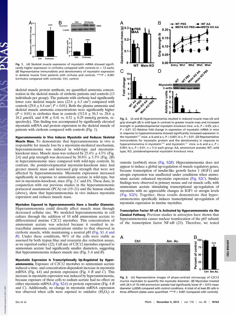
skeletal muscle protein synthesis, we quantified ammonia concen-tration in the skeletal muscle of cirrhotic patients and controls (13individuals per group). The patients with cirrhosis had significantlylower core skeletal muscle area (22.6 ± 6.3 cm2) compared withcontrols (29.8 ± 8.4 cm2; P < 0.01). Both the plasma ammonia andskeletal muscle ammonia concentrations were significantly higher(P < 0.01) in cirrhotics than in controls (312.8 ± 56.3 vs. 28.6 ±18.2 μmol/L and 4.98 ± 0.41 vs. 0.52 ± 0.29 mmol/g protein, re-spectively). This finding was accompanied by significantly elevatedmyostatin mRNA and protein expression in the skeletal muscle ofpatients with cirrhosis compared with controls (Fig. 1).
Hyperammonemia in Vivo Induces Myostatin and Reduces SkeletalMuscle Mass. To demonstrate that hyperammonemia in vivo isresponsible for muscle loss by a myostatin-mediated mechanism,hyperammonemia was induced in wild-type and myostatin-knockout mice. Muscle mass was reduced by 25.4% ± 4.5% (Fig.2A) and grip strength was decreased by 30.8% ± 5.3% (Fig. 2B)in hyperammonemic mice compared with wild-type controls. Incontrast, the postdevelopmental myostatin-knockout mice hadgreater muscle mass and increased grip strength that were notaffected by hyperammonemia. Myostatin expression increasedsignificantly in response to ammonium acetate in wild-type, butnot in myostatin-knockout, mice (Fig. 2 C and D). These data, inconjunction with our previous studies in the hyperammonemicportacaval anastamosis (PCA) rat (19–21) and the human studies(above), show that hyperammonemia in vivo induces myostatinexpression and reduces muscle mass.
Myotubes Exposed to Hyperammonemia Have a Smaller Diameter.Hyperammonemia could directly affect muscle mass throughdecreased cellular size. We modeled hyperammonemia in cellculture through the addition of 10 mM ammonium acetate todifferentiated murine C2C12 myotubes. This concentration ofammonium acetate was selected because it resulted in in-tracellular ammonia concentrations similar to that observed incirrhotic muscle, while maintaining a neutral pH (Fig. S1 A andB). Under these conditions, 96% of the cells were viable asassessed by both trypan blue and resazurin dye reduction assays,as we reported earlier (22). Cell size of C2C12 myotubes exposed toammonium acetate had significantly smaller diameters, suggestingthat hyperammonemia reduces muscle size (Fig. 3 A and B).
Myostatin Expression Is Transcriptionally Up-Regulated by Hyper-ammonemia. Exposure of C2C12 myotubes to ammonium acetateinduced a time- and concentration-dependent increase in myostatinmRNA (Fig. 4A) and protein expression (Fig. 4 B and C). Thisincrease in myostatin expression was induced by hyperammonemia,because exposure of these cells to sodium acetate had no effect oneither myostatin mRNA (Fig. S2A) or protein expression (Fig. 4 Band C). Additionally, no change in myostatin mRNA expressionwas observed when cells were exposed to oxidative (H2O2) or
osmotic (sorbitol) stress (Fig. S2B). Hyperammonemia does notappear to induce a global up-regulation of muscle regulatory genes,because transcription of insulin-like growth factor 1 (IGF1) andatrogin expression was unaffected under conditions when ammo-nium acetate enhanced myostatin expression (Fig. S2C). Similarfindings were observed in primary mouse and rat muscle cells, withammonium acetate stimulating transcriptional up-regulation ofmyostatin with no appreciable changes in IGF1 or atrogin levels(Fig. S2D). Together, these results demonstrate that hyper-ammonemia specifically induces transcriptional up-regulation ofmyostatin expression in murine myotubes.
Transcription Factor NF-κB Is Activated by Hyperammonemia via theClassical Pathway. Previous studies in astrocytes have shown thathyperammonemia causes nuclear translocation of the p65 subunitof the transcription factor NF-κB (23). Therefore, we tested
Fig. 1. (A) Skeletal muscle expression of myostatin mRNA showed signifi-cantly higher expression in cirrhotics compared with controls (n = 13 each).(B) Representative immunoblots and densitometry of myostatin expressionin skeletal muscle from patients with cirrhosis and controls. ***P < 0.001(cirrhotics compared with controls). Ctrl, control.
Fig. 2. (A and B) Hyperammonemia resulted in reduced muscle mass (A) andgrip strength (B) in wild type in contrast to greater muscle mass and increasedstrength in postdevelopmental myostatin knockout mice. a–b, P < 0.05; a,b–cP < 0.01. (C) Relative fold change in expression of myostatin mRNA in micein response to hyperammonemia showed significantly increased expression inthe myostatin+/+ mice. a–b and a–c, P < 0.001; b–c, P < 0.01. (D) Representativeimmunoblots for myostatin protein and the densitometry in response tohyperammonemia in myostatin+/+ and myostatin−/− mice. a–b and a–c, P <0.001; b–c, P < 0.01. n = 5 in each group. AA, ammonium acetate; WT, wildtype; KO, postdevelopmental myostatin knockout mice.
Fig. 3. (A) Representative images of phase-contrast microscopy of C2C12murine myotubes to quantify the myotube diameter. (B) Myotubes treatedwith 24 h of 10 mM ammonium acetate had significantly lower (P < 0.01) meandiameter (±SEM) compared with control conditions. A total of at least 85 cells inthree different plates were quantified. ***P < 0.001 (compared with controls).
Qiu et al. PNAS | November 5, 2013 | vol. 110 | no. 45 | 18163
CELL
BIOLO
GY
Dow
nloa
ded
by g
uest
on
Apr
il 5,
202
0
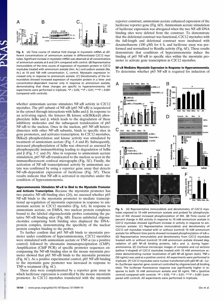
whether ammonium acetate stimulates NF-κB activity in C2C12myotubes. The p65 subunit of NF-κB (p65 NF-κB) is sequesteredin the cytosol through interactions with IκBα and β. In response toan activating signal, the kinases IK kinase α/β(IKKα/β) phos-phorylate IκBα and β, which leads to the degradation of theseinhibitory molecules and the subsequent translocation of p65NF-κB to the nucleus. Once in the nucleus, p65 NF-κB hetero-dimerizes with other NF-κB subunits, binds to specific sites ingene promoters, and activates transcription. In C2C12 myotubes,IKKα/β phosphorylation and kinase activity was stimulated bytreatment of ammonium acetate (Fig. 5 A and B). Subsequently,increased phosphorylation of IκBα was observed as assessed byphosphospecific immunoblotting leading to degradation of IκBαand β (Fig. 5 C and D). Also in response to ammonium acetatestimulation, p65 NF-κB translocated to the nucleus as seen in theimmunofluorescent confocal micrographs (Fig. 5E). Finally, theactivation of NF-κB transcriptional activity by ammonium ace-tate was confirmed by using a reporter gene assay that detectsNF-κB–dependent expression of luciferase (Fig. 5F). Theseresults indicate that NF-κB is activated in myotubes under thecondition of hyperammonemia.
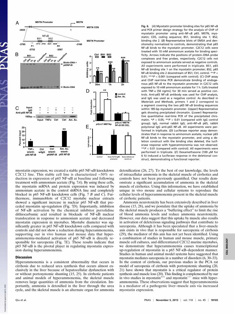
Hyperammonemia Stimulates NF-κB to Bind to the Myostatin Promoterand Activate Transcription. Because the myostatin promoter hastwo putative NF-κB binding sites (24), we assessed whether p65NF-κB binds to the myostatin promoter to mediate transcrip-tional up-regulation of myostatin expression in response to am-monium acetate in C2C12 myotubes (Fig. 6A). In response toammonium acetate, on EMSA, two nuclear protein complexesbound to the labeled oligonucleotide probes containing the pu-tative NF-κB binding sites (Fig. 6B). Excess unlabeled oligonu-cleotides comprising both binding sites displaced the DNA–
protein complex, demonstrating the specificity of the nuclearprotein complex binding to the probes.To further confirm that p65 NF-κB binds to myostatin pro-
moter under conditions of hyperammonemia, C2C12 myotubeswere stimulated with ammonium acetate or TNF-α (as a positivecontrol) followed by chromatin immunoprecipitation (ChIP).Amplification (ChIP PCR) of specific promoter sequences en-compassing the NF-ĸB binding sites in the mouse myostatin pro-moter showed that p65 NF-κB binds to the myostatin promoter(Fig. 6C). As a positive experimental control, p65 NF-κB bindingto the myostatin gene promoter was increased in response toTNF-α treatment (Fig. 6C).These data were complemented by a reporter gene assay in
which luciferase expression is controlled by the mouse myostatinpromoter. In C2C12 myotubes transfected with the myostatin
reporter construct, ammonium acetate enhanced expression of theluciferase reporter gene (Fig. 6D). Ammonium acetate stimulationof luciferase expression was abrogated when the two NF-κB DNAbinding sites were deleted from the construct. To demonstratethat the deletional construct was functional, C2C12 myotubes withthe full-length and deletional construct were incubated withdexamethasone (100 μM) for 6 h, and luciferase assay was per-formed and normalized to Renilla activity (Fig. 6E). These resultsdemonstrate that conditions of hyperammonemia induce thebinding of p65 NF-κB to specific sites within the myostatin pro-moter to activate gene transcription in C2C12 myotubes.
NF-κB Mediates Myostatin Expression in Response to Hyperammonemia.To determine whether p65 NF-κB is required for induction of
Fig. 4. (A) Time course of relative fold change in myostatin mRNA at dif-ferent concentrations of ammonium acetate in differentiated C2C12 myo-tubes. Significant increase in myostatin mRNAwas observed at all concentrationsof ammonium acetate at 6 and 24 h compared with control. (B) Representativeimmunoblots of the time course of expression of myostatin protein in C2C12myotubes treated with ammonium acetate (Am. Ac.) and sodium acetate (Na.Ac.) at 10 and 100 mM concentration. C, control. Myostatin expression in-creased only in response to ammonium acetate. (C) Densitometry of the im-munoblots showed increased expression of myostatin protein in a time- andconcentration-dependent manner only in response to ammonium acetate,demonstrating that these changes are specific to hyperammonemia. Allexperiments were performed in triplicate. *P < 0.05; **P < 0.01; ***P < 0.001(compared with controls).
Fig. 5. (A) Representative immunoblots and densitometry of C2C12 myo-tubes exposed to 10 mM ammonium acetate and probed for phosphoryla-tion of IKK showed increased phosphorylation of IKK. (B) Time course ofpercent change in IKK activity in response to 10 mM ammonium acetate inC2C12 myotubes showed significant increase in activity in response to am-monium acetate. (C) Representative immunoblots and densitometry ofC2C12 cell myotubes treated with or without (control) 10 mM ammoniumacetate for different time points showed increased phosphorylation of IκB-α.(D) Representative immunoblots and densitometry from C2C12 myotubestreated with or without (control) 10 mM ammonium acetate showed deg-radation of p65 NF-κB binding proteins, IκB-α and -β, during hyper-ammonemia. (E) Confocal microscopic images of complete and cut sections(yellow V-shaped) of C2C12 myotubes treated with 10 mM ammonium ac-etate demonstrating nuclear localization of p65 NF-κB (green stain). TNF-α(50 ng/mL) was used as a positive control. All experiments were performed intriplicate. (F) C2C12 myotubes were nuclear transfected with p65 NF-κB −luc-6× (luciferase reporter gene construct controlled by oligomerized κB bindingsites). The luciferase fluorescence response was significantly higher in re-sponse to both 10 mM ammonium acetate and 50 ng/mL TNF-α (positivecontrol) compared with controls. *P < 0.05; **P < 0.01; ***P < 0.001 (com-pared with control). All experiments were performed in triplicate.
18164 | www.pnas.org/cgi/doi/10.1073/pnas.1317049110 Qiu et al.
Dow
nloa
ded
by g
uest
on
Apr
il 5,
202
0
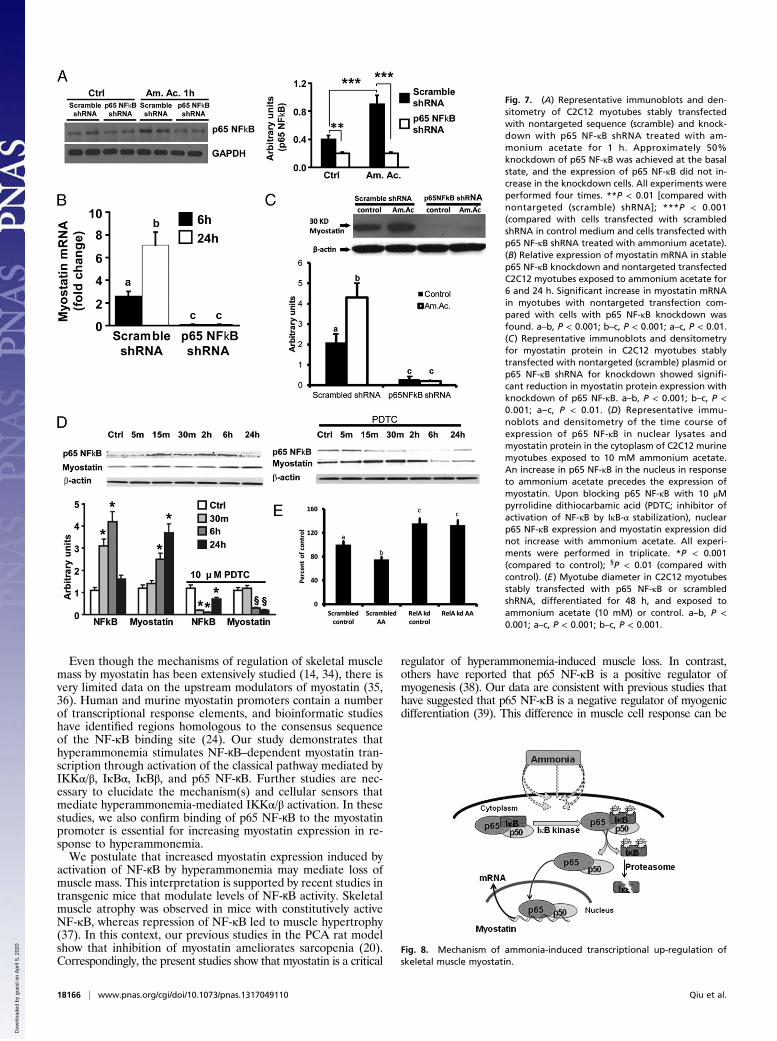
myostatin expression, we created a stable p65 NF-κB knockdownC2C12 line. This stable cell line is characterized ∼50% re-duction in expression of p65 NF-κB at baseline and followingtreatment with ammonium acetate (Fig. 7A). By using these cells,the myostatin mRNA and protein expression was induced byammonium acetate in the control shRNA line and completelyblocked in p65 NF-κB knockdown cells (Fig. 7 B and C). Fur-thermore, immunoblots of C2C12 myotube nuclear extractsshowed a significant increase in nuclear p65 NF-κB that pre-ceded myostatin up-regulation (Fig. 7D). Importantly, inhibitionof NF-κB activation by the chemical inhibitor pyrrolidinedithiocarbamic acid resulted in blockade of NF-κB nucleartranslocation in response to ammonium acetate and decreasedmyostatin expression in myotubes. Myotube diameter was sig-nificantly greater in p65 NF-κB knockdown cells compared withcontrols and did not show a reduction during hyperammonemia,supporting our in vivo human and mouse data that hyper-ammonemia-mediated activation of p65 NF-κB is directly re-sponsible for sarcopenia (Fig. 7E). These results indicate thatp65 NF-κB is the pivotal player in regulating myostatin expres-sion during hyperammonemia.
DiscussionHyperammonemia is a consistent abnormality that occurs incirrhosis due to reduced urea synthesis that occurs almost ex-clusively in the liver because of hepatocellular dysfunction withor without portosystemic shunting (15, 25). In cirrhotic patientsand animal models of hyperammonemia, the skeletal muscleremoves large quantities of ammonia from the circulation. Im-portantly, ammonia is detoxified in the liver through the ureacycle, and the skeletal muscle is an alternate organ for ammonia
detoxification (26, 27). To the best of our knowledge, the levelsof intracellular ammonia in the skeletal muscle of cirrhotics andcontrols have not been previously quantified. Our results dem-onstrate a significant accumulation of ammonia in the skeletalmuscle of cirrhotics. Using this information, we have establishedunique in vivo mouse and cellular systems to reproduce thecellular levels of hyperammonemia present in the skeletal muscleof cirrhotic patients.Ammonia neurotoxicity has been extensively described in liver
disease (15, 28), and we postulate that the uptake of ammonia bythe skeletal muscle produces a “buffering effect” to limit the riseof blood ammonia levels and reduce ammonia neurotoxicity.However, our data suggest that this uptake by muscle also resultsin activation of deleterious signaling pathways that contribute tosarcopenia. Although it has been speculated that a liver–muscleaxis exists in vivo that is responsible for sarcopenia of cirrhosis(29), the mediator of this axis has not yet been identified. Usinga combination of studies in human and mouse muscle, primarymuscle cell cultures, and differentiated C2C12 murine myotubes,we demonstrate that hyperammonemia causes transcriptionalup-regulation of myostatin in a p65 NF-κB–dependent manner.Studies in human and animal model systems have suggested thatmyostatin mediates sarcopenia in a number of disorders (6, 30–33).In the context of cirrhosis, our previous studies in the PCA ratmodel of sarcopenia of cirrhosis with portosystemic shunting (20,21) have shown that myostatin is a critical regulator of proteinsynthesis and muscle loss (20). This finding is complemented by ourin vivo studies in myostatin+/+ and myostatin−/− mice with hyper-ammonemia. These observations suggest that hyperammonemiais a mediator of a pathogenic liver–muscle axis via increasedmyostatin expression.
Fig. 6. (A) Myostatin promoter binding sites for p65 NF-κBand PCR primer design strategy for the analysis of ChIP ofmyostatin promoter using anti–NF-κB p65. MSTN, myo-statin; CDS, coding sequence; BS1, binding site 1; BS2,binding site 2. (B) Representative blots of EMSA and den-sitometry normalized to controls, demonstrating that p65NF-κB binds to the myostatin promoter. C2C12 cells weretreated with 10 mM ammonium acetate for binding speci-ficity. Arrows indicate the positions of protein–DNA probecomplexes and free probes, respectively. C2C12 cells notexposed to ammonium acetate served as negative controls.All experiments were performed in triplicate. BS1, p65NF-κB binding site 1 on the myostatin promoter; BS2, p65NF-κB binding site 2 downstream of BS1; Ctrl, control. **P <0.01; ***P < 0.001 (compared with control). (C) ChIP assayand ChIP real-time PCR demonstrate binding of endoge-nous p65 NF-κB to the myostatin promoter in C2C12 cellsexposed to 10 mM ammonium acetate for 1 h. Cells treatedwith TNF-α (50 ng/mL) for 30 min served as positive con-trols. Anti-p65 NF-κB antibody was used for ChIP analysis,and IgG was used as a negative control. As described inMaterials and Methods, primers 1 and 2 correspond toa segment covering the two p65 NF-κB binding sequenceswithin 185-bp myostatin promoter. (Upper) Representativegels showing precipitated chromatin. (Lower) Representa-tive quantitative real-time PCR of the precipitated chro-matin. *P < 0.05; **P < 0.01 (compared with IgG controlgroup). IgG, normal rabbit IgG; anti–NF-κB p65, rabbitpolyclonal IgG anti-p65 NF-κB. All experiments were per-formed in triplicate. (D) Luciferase reporter assay demon-strates that in response to ammonium acetate, nuclear p65NF-κB binds to the myostatin promoter, and using a de-letion construct with the binding sites deleted, the lucif-erase response with hyperammonemia was not observed.**P < 0.01 (compared with control). All experiments wereperformed in triplicate. (E ) Dexamethasone (100 μM for6 h) induced a luciferase response in the deletional con-struct, demonstrating a functional reporter.
Qiu et al. PNAS | November 5, 2013 | vol. 110 | no. 45 | 18165
CELL
BIOLO
GY
Dow
nloa
ded
by g
uest
on
Apr
il 5,
202
0
Even though the mechanisms of regulation of skeletal musclemass by myostatin has been extensively studied (14, 34), there isvery limited data on the upstream modulators of myostatin (35,36). Human and murine myostatin promoters contain a numberof transcriptional response elements, and bioinformatic studieshave identified regions homologous to the consensus sequenceof the NF-κB binding site (24). Our study demonstrates thathyperammonemia stimulates NF-ĸB–dependent myostatin tran-scription through activation of the classical pathway mediated byIKKα/β, IκBα, IκBβ, and p65 NF-ĸB. Further studies are nec-essary to elucidate the mechanism(s) and cellular sensors thatmediate hyperammonemia-mediated IKKα/β activation. In thesestudies, we also confirm binding of p65 NF-ĸB to the myostatinpromoter is essential for increasing myostatin expression in re-sponse to hyperammonemia.We postulate that increased myostatin expression induced by
activation of NF-ĸB by hyperammonemia may mediate loss ofmuscle mass. This interpretation is supported by recent studies intransgenic mice that modulate levels of NF-ĸB activity. Skeletalmuscle atrophy was observed in mice with constitutively activeNF-κB, whereas repression of NF-κB led to muscle hypertrophy(37). In this context, our previous studies in the PCA rat modelshow that inhibition of myostatin ameliorates sarcopenia (20).Correspondingly, the present studies show that myostatin is a critical
regulator of hyperammonemia-induced muscle loss. In contrast,others have reported that p65 NF-κB is a positive regulator ofmyogenesis (38). Our data are consistent with previous studies thathave suggested that p65 NF-κB is a negative regulator of myogenicdifferentiation (39). This difference in muscle cell response can be
Fig. 7. (A) Representative immunoblots and den-sitometry of C2C12 myotubes stably transfectedwith nontargeted sequence (scramble) and knock-down with p65 NF-κB shRNA treated with am-monium acetate for 1 h. Approximately 50%knockdown of p65 NF-κB was achieved at the basalstate, and the expression of p65 NF-κB did not in-crease in the knockdown cells. All experiments wereperformed four times. **P < 0.01 [compared withnontargeted (scramble) shRNA]; ***P < 0.001(compared with cells transfected with scrambledshRNA in control medium and cells transfected withp65 NF-κB shRNA treated with ammonium acetate).(B) Relative expression of myostatin mRNA in stablep65 NF-κB knockdown and nontargeted transfectedC2C12 myotubes exposed to ammonium acetate for6 and 24 h. Significant increase in myostatin mRNAin myotubes with nontargeted transfection com-pared with cells with p65 NF-κB knockdown wasfound. a–b, P < 0.001; b–c, P < 0.001; a–c, P < 0.01.(C) Representative immunoblots and densitometryfor myostatin protein in C2C12 myotubes stablytransfected with nontargeted (scramble) plasmid orp65 NF-κB shRNA for knockdown showed signifi-cant reduction in myostatin protein expression withknockdown of p65 NF-κB. a–b, P < 0.001; b–c, P <0.001; a–c, P < 0.01. (D) Representative immu-noblots and densitometry of the time course ofexpression of p65 NF-κB in nuclear lysates andmyostatin protein in the cytoplasm of C2C12 murinemyotubes exposed to 10 mM ammonium acetate.An increase in p65 NF-κB in the nucleus in responseto ammonium acetate precedes the expression ofmyostatin. Upon blocking p65 NF-κB with 10 μMpyrrolidine dithiocarbamic acid (PDTC; inhibitor ofactivation of NF-κB by IκB-α stabilization), nuclearp65 NF-κB expression and myostatin expression didnot increase with ammonium acetate. All experi-ments were performed in triplicate. *P < 0.001(compared to control); §P < 0.01 (compared withcontrol). (E) Myotube diameter in C2C12 myotubesstably transfected with p65 NF-κB or scrambledshRNA, differentiated for 48 h, and exposed toammonium acetate (10 mM) or control. a–b, P <0.001; a–c, P < 0.001; b–c, P < 0.001.
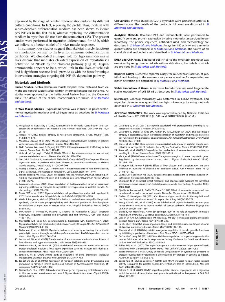
Fig. 8. Mechanism of ammonia-induced transcriptional up-regulation ofskeletal muscle myostatin.
18166 | www.pnas.org/cgi/doi/10.1073/pnas.1317049110 Qiu et al.
Dow
nloa
ded
by g
uest
on
Apr
il 5,
202
0
explained by the stage of cellular differentiation induced by differentculture conditions. In fact, replacing the proliferating medium withserum-deprived differentiation medium has been shown to inducep65 NF-κB in the first 24 h, whereas replacing the differentiationmedium in myotubes did not have the same effect (38). The presentstudies were performed in myotubes differentiated for 48 h, whichwe believe is a better model of in vivo muscle responses.In summary, our studies suggest that skeletal muscle functions
as a metabolic partner to the liver for ammonia detoxification incirrhotics. We elucidated a unique role for hyperammonemia inliver disease that mediates elevated expression of myostatin viaactivation of NF-κB by the classical pathway (Fig. 8). Hyper-ammonemia appears to be a critical link in the liver–muscle axisand is significant because it will provide us with the basis for uniqueintervention strategies targeting this NF-ĸB–dependent pathway.
Materials and MethodsHuman Studies. Rectus abdominis muscle biopsies were obtained from cir-rhotic and control subjects after written informed consent was obtained. Allstudies were approved by the Institutional Review Board at the ClevelandClinic. The details of the clinical characteristics are shown in SI Materialsand Methods.
In Vivo Mouse Studies. Hyperammonemia was induced in postdevelop-mental myostatin knockout and wild-type mice as described in SI Materialsand Methods.
Cell Cultures. In vitro studies in C2C12 myotubes were performed after 48-hdifferentiation. The details of the protocols followed are discussed in SIMaterials and Methods.
Analytical Methods. Real-time PCR and immunoblots were performed toquantify gene and protein expression by using methods standardized in ourlaboratory. The primer sequences, antibodies used, and methodology aredescribed in SI Materials and Methods. Assays for IKK activity and ammoniaquantification are described in SI Materials and Methods. The source of allchemicals and antibodies is also described in SI Materials and Methods.
EMSA and ChIP Assay. Binding of p65 NF-κB to the myostatin promoter wasexamined by using commercial kits with modifications, the details of whichare provided in SI Materials and Methods.
Reporter Assays. Luciferase reporter assays for nuclear translocation of p65NF-κB and binding to the consensus sequence as well as for myostatin pro-moter activation are described in SI Materials and Methods.
Stable Knockdown of Genes. A lentivirus transduction was used to generatestable knockdown of p65 NF-κB as described in SI Materials and Methods.
Microscopy. Confocal microscopy was performed in C2C12 myotubes, andmyotube diameter was quantified on light microscopy by using methodsdescribed in SI Materials and Methods.
ACKNOWLEDGMENTS. This work was supported in part by National Institutesof Health Grants R01 DK83413 (to S.D.) and R01DK082437 (to C.M.).
1. Periyalwar P, Dasarathy S (2012) Malnutrition in cirrhosis: Contribution and con-sequences of sarcopenia on metabolic and clinical responses. Clin Liver Dis 16(1):95–131.
2. Hepple RT (2012) Muscle atrophy is not always sarcopenia. J Appl Physiol (1985)113(4):677–679.
3. Montano-Loza AJ, et al. (2012) Muscle wasting is associated with mortality in patientswith cirrhosis. Clin Gastroenterol Hepatol 10(2):166–173.
4. Olde Damink SW, Jalan R, Dejong CH (2009) Interorgan ammonia trafficking in liverdisease. Metab Brain Dis 24(1):169–181.
5. Holecek M, Sprongl L, Tichý M (2000) Effect of hyperammonemia on leucine andprotein metabolism in rats. Metabolism 49(10):1330–1334.
6. García PS, Cabbabe A, Kambadur R, Nicholas G, Csete M (2010) Brief-reports: Elevatedmyostatin levels in patients with liver disease: A potential contributor to skeletalmuscle wasting. Anesth Analg 111(3):707–709.
7. Huang Z, Chen X, Chen D (2011) Myostatin: A novel insight into its role in metabolism,signal pathways, and expression regulation. Cell Signal 23(9):1441–1446.
8. Trendelenburg AU, et al. (2009) Myostatin reduces Akt/TORC1/p70S6K signaling, in-hibiting myoblast differentiation and myotube size. Am J Physiol Cell Physiol 296(6):C1258–C1270.
9. Amirouche A, et al. (2009) Down-regulation of Akt/mammalian target of rapamycinsignaling pathway in response to myostatin overexpression in skeletal muscle. En-docrinology 150(1):286–294.
10. Taylor WE, et al. (2001) Myostatin inhibits cell proliferation and protein synthesis inC2C12 muscle cells. Am J Physiol Endocrinol Metab 280(2):E221–E228.
11. Welle S, Burgess K, Mehta S (2009) Stimulation of skeletal muscle myofibrillar proteinsynthesis, p70 S6 kinase phosphorylation, and ribosomal protein S6 phosphorylationby inhibition of myostatin in mature mice. Am J Physiol Endocrinol Metab 296(3):E567–E572.
12. McCroskery S, Thomas M, Maxwell L, Sharma M, Kambadur R (2003) Myostatinnegatively regulates satellite cell activation and self-renewal. J Cell Biol 162(6):1135–1147.
13. Morissette MR, Cook SA, Buranasombati C, Rosenberg MA, Rosenzweig A (2009)Myostatin inhibits IGF-I-induced myotube hypertrophy through Akt. Am J Physiol CellPhysiol 297(5):C1124–C1132.
14. McFarlane C, et al. (2006) Myostatin induces cachexia by activating the ubiquitinproteolytic system through an NF-kappaB-independent, FoxO1-dependent mecha-nism. J Cell Physiol 209(2):501–514.
15. Lockwood AH, et al. (1979) The dynamics of ammonia metabolism in man. Effects ofliver disease and hyperammonemia. J Clin Invest 63(3):449–460.
16. Jiménez-Martí E, del Olmo ML (2008) Addition of ammonia or amino acids to a ni-trogen-depleted medium affects gene expression patterns in yeast cells during al-coholic fermentation. FEMS Yeast Res 8(2):245–256.
17. Jousse C, et al. (2004) Amino acids as regulators of gene expression: Molecularmechanisms. Biochem Biophys Res Commun 313(2):447–452.
18. ter Schure EG, et al. (1998) Repression of nitrogen catabolic genes by ammonia andglutamine in nitrogen-limited continuous cultures of Saccharomyces cerevisiae. Mi-crobiology 144(Pt 5):1451–1462.
19. Dasarathy S, et al. (2007) Altered expression of genes regulating skeletal muscle massin the portacaval anastomosis rat. Am J Physiol Gastrointest Liver Physiol 292(4):G1105–G1113.
20. Dasarathy S, et al. (2011) Sarcopenia associated with portosystemic shunting is re-versed by follistatin. J Hepatol 54(5):915–921.
21. Dasarathy S, Dodig M, Muc SM, Kalhan SC, McCullough AJ (2004) Skeletal muscleatrophy is associated with an increased expression of myostatin and impaired satellitecell function in the portacaval anastamosis rat. Am J Physiol Gastrointest Liver Physiol287(6):G1124–G1130.
22. Qiu J, et al. (2012) Hyperammonemia-mediated autophagy in skeletal muscle con-tributes to sarcopenia of cirrhosis. Am J Physiol Endocrinol Metab 303(8):E983–E993.
23. Sinke AP, et al. (2008) NFkappaB in the mechanism of ammonia-induced astrocyteswelling in culture. J Neurochem 106(6):2302–2311.
24. Ma K, et al. (2001) Characterization of 5′-regulatory region of human myostatin gene:Regulation by dexamethasone in vitro. Am J Physiol Endocrinol Metab 281(6):E1128–E1136.
25. Shangraw RE, Jahoor F (1999) Effect of liver disease and transplantation on ureasynthesis in humans: Relationship to acid-base status. Am J Physiol 276(5 Pt 1):G1145–G1152.
26. Ganda OP, Ruderman NB (1976) Muscle nitrogen metabolism in chronic hepatic in-sufficiency. Metabolism 25(4):427–435.
27. Chatauret N, et al. (2006) Direct molecular and spectroscopic evidence for increasedammonia removal capacity of skeletal muscle in acute liver failure. J Hepatol 44(6):1083–1088.
28. Gjedde A, Lockwood A, Duffy TE, Plum F (1976) Effect of ammonia on cerebral me-tabolism of rats with portocaval shunts. Trans Am Neurol Assoc 101:180–181.
29. Fischer JE, Hasselgren PO (1991) Cytokines and glucocorticoids in the regulation ofthe “hepato-skeletal muscle axis” in sepsis. Am J Surg 161(2):266–271.
30. Benny Klimek ME, et al. (2010) Acute inhibition of myostatin-family proteins pre-serves skeletal muscle in mouse models of cancer cachexia. Biochem Biophys ResCommun 391(3):1548–1554.
31. Elkina Y, von Haehling S, Anker SD, Springer J (2011) The role of myostatin in musclewasting: An overview. J Cachexia Sarcopenia Muscle 2(3):143–151.
32. Gruson D, Ahn SA, Ketelslegers JM, Rousseau MF (2011) Increased plasma myostatinin heart failure. Eur J Heart Fail 13(7):734–736.
33. Ju CR, Chen RC (2012) Serum myostatin levels and skeletal muscle wasting in chronicobstructive pulmonary disease. Respir Med 106(1):102–108.
34. Thomas M, et al. (2000) Myostatin, a negative regulator of muscle growth, functionsby inhibiting myoblast proliferation. J Biol Chem 275(51):40235–40243.
35. De Santis C, Jerry DR (2011) Differential tissue-regulation of myostatin genes in theteleost fish Lates calcarifer in response to fasting. Evidence for functional differen-tiation. Mol Cell Endocrinol 335(2):158–165.
36. Spiller MP, et al. (2002) The myostatin gene is a downstream target gene of basichelix-loop-helix transcription factor MyoD. Mol Cell Biol 22(20):7066–7082.
37. Balasubramanian S, et al. (2006) Enhanced ubiquitination of cytoskeletal proteins inpressure overloaded myocardium is accompanied by changes in specific E3 ligases.J Mol Cell Cardiol 41(4):669–679.
38. Baeza-Raja B, Muñoz-Cánoves P (2004) p38 MAPK-induced nuclear factor-kappaBactivity is required for skeletal muscle differentiation: Role of interleukin-6. Mol BiolCell 15(4):2013–2026.
39. Bakkar N, et al. (2008) IKK/NF-kappaB regulates skeletal myogenesis via a signalingswitch to inhibit differentiation and promote mitochondrial biogenesis. J Cell Biol180(4):787–802.
Qiu et al. PNAS | November 5, 2013 | vol. 110 | no. 45 | 18167
CELL
BIOLO
GY
Dow
nloa
ded
by g
uest
on
Apr
il 5,
202
0