HUMAN MOTOR END PLATE: SOME ULTRASTRUCTURAL … · HUMAN MOTOR END PLATE: SOME ULTRASTRUCTURAL...
9
HUMAN MOTOR END PLATE: SOME ULTRASTRUCTURAL ASPECTS OF ITS DEVELOPMENT GUILBERTO MINGUETTI, PhD * W, G. P. ΜAIR, MD, FRCPth ** Material from human foetuses of nine weeks to nine months development was examined by electron microscopy to obtain some information about the various stages in the development of skeletal muscle and structures intimately related to it such as blood vessels, nerves, neuromuscular spindles, motor end plates and fibroblasts. Regarding the motor end plates, the earliest evidence of the formation of such structures was observed in the muscle of foetuses of nine and ten weeks development. At this stage the developing motor end plate consits of several small axons covered on their outer aspect by Schwann cell cytoplasm, while on the inner aspect they are separated by a layer of basement membrane of the myotube with which they make contact. At this stage there is no evidence of secondary synaptic clefts but by the twenty eight weeks the motor end plate is similar to that seen in the adult. MATERIAL AND METHODS The material used in this investigation is derived from twenty seven human foetuses ranging from nine weeks to nine months development. They have been studied in detail in longitudinal and transverse sections by electron microscopy. The age of the foetuses was. established by the obstetricians who suplied the specimend and was calculated according to the crown-rump lenght. The ages of the foetuses of 5,7,8 and 9 months development were based on the menstrual age. The muscle was usually taken from the thigh, even that from the minute foetuses and laid immediately on a piece of card and kept slightly streched by means of pins applied to either end of the specimen. The specimens were then immersed in cold 3% glutaraldehyJe in Sorensen's phosphate buffer at pH 7.4. After tw,o hours of fixation the muscle held by the pins was released and cut in small pieces of about 1-2 mm thick. These were washed in two changes of Sorensen's buffer at pH 7.4 Trabalho realizado no Instituto de Neurologia da Universidade de Londres (Queen Square): *Professor Assistente do Departamento de Ciências Fisiológicas e Disciplina de Neurologia, Universidade Federal do Paraná (Curitiba — Paraná — Brasil); **Consultant neuropathologist, Institute of Neurology, Queen Square, London WC1N 3BG — England. Acknowledgements — We would like to thank Mr. B. C. Young for his valuable technical assistance and the obstetricians for providing most of the specimens examined in this investigation. Our thanks are also due to the Muscular Dystrophy Group of Great Britain for providing facilities and equipment essential for this work.
Transcript of HUMAN MOTOR END PLATE: SOME ULTRASTRUCTURAL … · HUMAN MOTOR END PLATE: SOME ULTRASTRUCTURAL...
HUMAN MOTOR END PLATE: SOME ULTRASTRUCTURAL ASPECTS OF ITS DEVELOPMENT
GUILBERTO MINGUETTI, PhD * W, G. P. ΜAIR, MD, FRCPth **
Material from human foetuses of nine weeks to nine months development was examined by electron microscopy to obtain some information about the various stages in the development of skeletal muscle and structures intimately related to it such as blood vessels, nerves, neuromuscular spindles, motor end plates and fibroblasts.
Regarding the motor end plates, the earliest evidence of the formation of such structures was observed in the muscle of foetuses of nine and ten weeks development. At this stage the developing motor end plate consits of several small axons covered on their outer aspect by Schwann cell cytoplasm, while on the inner aspect they are separated by a layer of basement membrane of the myotube with which they make contact. At this stage there is no evidence of secondary synaptic clefts but by the twenty eight weeks the motor end plate is similar to that seen in the adult.
MATERIAL AND METHODS
The material used in this investigation is derived from twenty seven human foetuses ranging from nine weeks to nine months development. They have been studied in detail in longitudinal and transverse sections by electron microscopy.
The age of the foetuses was. established by the obstetricians who suplied the specimend and was calculated according to the crown-rump lenght. The ages of the foetuses of 5,7,8 and 9 months development were based on the menstrual age. The muscle was usually taken from the thigh, even that from the minute foetuses and laid immediately on a piece of card and kept slightly streched by means of pins applied to either end of the specimen. The specimens were then immersed in cold 3% glutaraldehyJe in Sorensen's phosphate buffer at pH 7.4. After tw,o hours of fixation the muscle held by the pins was released and cut in small pieces of about 1-2 mm thick. These were washed in two changes of Sorensen's buffer at pH 7.4
Trabalho realizado no Instituto de Neurologia da Universidade de Londres (Queen Square): *Professor Assistente do Departamento de Ciências Fisiológicas e Disciplina de Neurologia, Universidade Federal do Paraná (Curitiba — Paraná — Brasil); **Consultant neuropathologist, Institute of Neurology, Queen Square, London WC1N 3BG — England.
Acknowledgements — We would like to thank Mr. B. C. Young for his valuable technical assistance and the obstetricians for providing most of the specimens examined in this investigation. Our thanks are also due to the Muscular Dystrophy Group of Great Britain for providing facilities and equipment essential for this work.
for 15 minutes each. Post fixation was carried out for two hours at room temperature in cold 1% osmium tetroxide in Michaelia* veronal-acetate buffer at pH 7.4. After being "washed in distilled water and dehydrated in ascending grades of alcohols, they were placed successively in propylene oxide and in a mixture of equal parts of propylene oxide and Bp on 812. Finally they were embeded in fresh Epon mixture. Sections 1-2^ thick were cut on an LKB Ultratome and stained with toluidine blue by the method of Trump, Smuckler and Bennditt (1961) and examined by light microscopy. Thin sections of appropriate regions were collected on cooper grids, stained by uranyl acetate and lead citrate (Reynolds, 1968) and examined in a Siemens Elmiskop I.
RESULTS
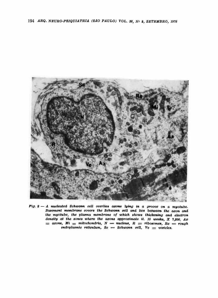
The muscle of foetuses of nine and ten weeks development show evidence of the early formation of motor end plates (Fig. 1 and 2). At this stage Schwann cells containing several small axons lie close to a myotube which at the zone of contact has on its surface a shallow depression. The nucleus of these Schwann cells at this stage is rounded and lies at the periphery of the cell, and presents a granular appearance with occasional more electron dense bands at the nuclear margin. The cytoplasm is abundant and contains many tubules of rough endoplasmic reticulum attached to which are ribosomes. Mitochondria are frequent. This cytoplasm lies external to the nerve terminals which have a very distinct limiting membrane, the axolemma. Schwann cell cytoplasm does not intervene between the terminals and the basement membrane of the adjacent myotube. The plasma membrane of the myotube along the zone of contact with the nerve terminals is more electron dense than the rest of the plasma membrane. The basement membrane of the myotube is continuous with that covering the Schwann cell overlying the terminals: they contain mitochondria, filaments and neurotubules and in addition many vesicles similar to synaptic vesicles.
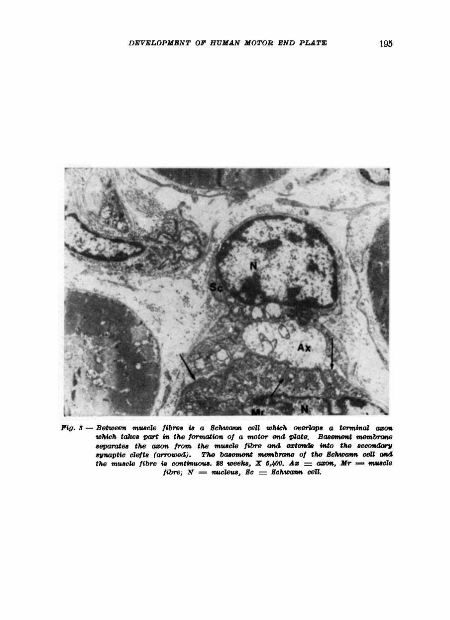
In the many sections of foetal muscle were examined relatively few motor end plates were encoutered and unfortunately the formation of the motor end plate could not be traced in very great detail. However by twenty eight weeks development the motor end plate has reached a fairly high degree of structural perfection and is similar to the motor end plate of the adult (Fig. 3 and 4). At this stage the Schwann cell enclosing the terminal part of the axon contains less cytoplasm than in the motor end plate of nine and ten weeks development
The nucleus of the Schwann cell has a very dense rim of chromatin and some small masses of similar dense chromatin throughout the nucleus. The basement membrane of the Schwann cell overlying the terminal axon is continuous with that of the muscle fibre and no Schwann cell cytoplasm occurs on the aspect of the terminal axon in contact with the basement membrane of the muscle fibre. The Schwann cell cytoplasm contains numerous mitochondria and many ribosomes but much less rough endoplasmic reticulum than in earlier stages of development The terminal axon has a distinct axolemma and contains tubules and vesicles. A groove of varying depth occurs on the muscle fibre and this is the primary synaptic cleft from which there occur numerous extensions which indent the muscle fibre to form the secondary synaptic clefts.
Basement membrane occurs within the primary and secondary synaptic clefts. A distinct narrow space is seen between the basement membrane of the synaptic clefts and very electron dense plasma membrane of the muscle fibre in relation to the secondary synaptic clefts.
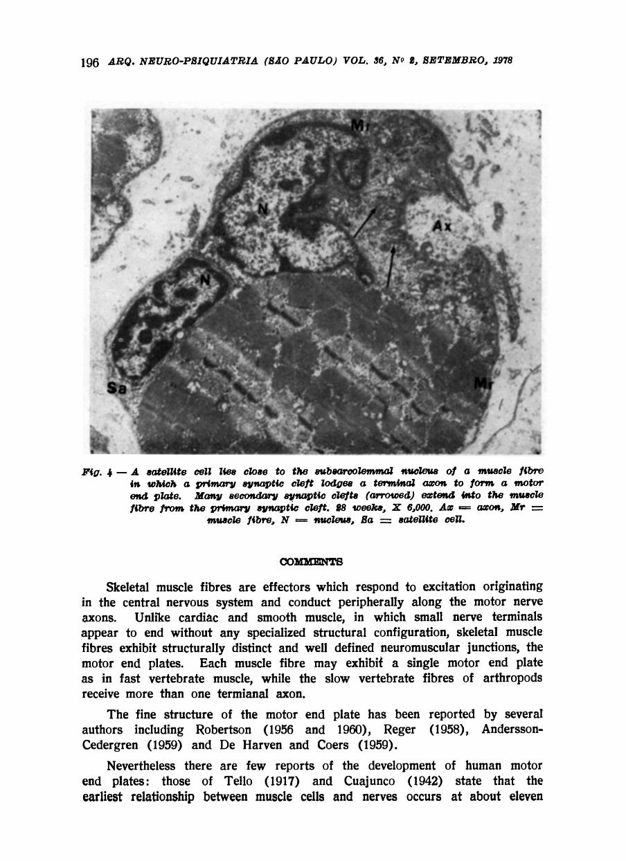
A subsarcolemmal nucleus of the muscle fibre generally lies in the proximity of the motor end plate and sometimes a cell like a satellite cell is lodged nearby within the basement membrane of the muscle fibre (Fig. 4).
COMMENTS
Skeletal muscle fibres are effectors which respond to excitation originating in the central nervous system and conduct peripherally along the motor nerve axons. Unlike cardiac and smooth muscle, in which small nerve terminals appear to end without any specialized structural configuration, skeletal muscle fibres exhibit structurally distinct and well defined neuromuscular junctions, the motor end plates. Each muscle fibre may exhibit a single motor end plate as in fast vertebrate muscle, while the slow vertebrate fibres of arthropods receive more than one termianal axon.
The fine structure of the motor end plate has been reported by several authors including Robertson (1956 and 1960), Reger (1958), Andersson-Cedergren (1959) and De Harven and Coers (1959).
Nevertheless there are few reports of the development of human motor end plates: those of Tello (1917) and Cuajunco (1942) state that the earliest relationship between muscle cells and nerves occurs at about eleven
weeks but the formation of the neuromuscular junction takes place only when the muscle cells are fully differentiated into muscle fibres. Later, Couteaux (1960) reported that two main phases in- the evolution of neuromuscular relationship can ben identified in mammals. In the first phase, "the muscle cells are still in myotube form and few motor nerve fibres proceed through the muscle. These are the "exploring" nerve fibres whose extremities have as yet developed no lasting connections with the muscle cell even when they are apposed to the myotubes". In the second phase, "when the muscle nuclei have completed their migration to the surface of the muscle fibre, the nerve fibres send out either terminal or lateral sprouts, generally accompanied by Schwann cell nuclei, and each of these sprouts enter into close relations with a muscle fibre at the level of nuclei, now peripheral".
Couteaux also described that "at the contact of the nerve sprout the muscle nucleus divides repeatedly. These divisions of muscle nuclei are generally preceded by nucleolar bipartition and appear in all respects comparable to amitosis". The result is "an accumulation of muscle nuclei and sarcoplasm at the junctional zone". Couteaux saw the narrow infoldings of the sarcolemma — the secondary synaptic clefts — only in the second phase when the muscle fibres were fully differentiated.
All these reports are based on observations by light microscopy. However, while there are many reports on the ultrastructure of the embryogenesis of the motor end plate in experimental animals, there are few on this topic in man. They are those of Fidzianska (1971) and Zacks (1973) who agree that "junctional foldings of the post synaptic membrane" may be found as early as ten weeks development. This presumably refers to the formation of the secondary synaptic clefts.
In the present study the early motor end plate is identified in the foetus of nine weeks and consists of small axons covered on their outer aspect by Schwann cell cytoplasm while on the inner aspect they are separated by a layer of basement membrane of the myotube with which they make contact. Cytoplasm of the Schwann cell does not intervene between the axons and the myotube. A groove which is the primary synaptic cleft indents the surface of the myotube at the region of contact and at this region the plasma membrane of the muscle cell is more electron dense than elsewhere. At this stage there is no evidence of secondary synaptic clefts.
The next stage at which the motor end plate was examinned was at twenty eight weeks by which time the structure is similar to that seen in the adult. Now there are multiple secondary synaptic clefts which estend from the primary synaptic cleft to indent the surface of the muscle fibre. They contain, like the primary cleft, a layer of basement membrane.
In the foetus of twenty eight weeks satellite cells occur in the neighbourhood of the motor end plate and near the subsarcolemmal nucleus of the muscle fibre. It seems likely that these satellite cells are source for the proliferation of cells at the junctional zone of the axon and muscle fibre and if observed by light microscopy would give the impression of an increase of subsarcolemmal nuclei
at this region as was reported by Couteaux (1960) who was under the impression that the muscle nuclei in this zone divided amitoticaly.
The axons forming the motor end plate contain mitochondria, neurotubules, neurofilaments and numerous .synaptic vesicles. These vesicles occur even at nine weeks development and their presence, along with the presence of well formed myofibrils in the respective myotube, may suggest a primary physiological relationship between the structures.
RESUMO Placa motora humana: alguns aspectos ultraestruturais do seu desenvolvi
mento. Fetos humanos de nove semanas a nove meses de desenvolvimento foram
examinados por ultramicroscopia afim de se obter informações sobre os vários estadios de desenvolvimento do músculo esquelético e algumas estruturas intimamente relacionadas a ele.
Com relação às placas motoras, as mesmas já são vistas em fetos de nove a dez semanas de vida intra-embrionária e consistem de grupos de pequenos axônios cobertos na parte externa pela célula de Schwann, enquanto na parte interna os axônios são separados do miotubo por uma camada de membrana basal. O citoplasma da célula de Schwann não intervem entre os axônios e o miotubo. Na região de contacto dos axônios com a superfície do miotubo ocorre uma invaginação que é a fenda sináptica primária e nessa área a membrana plasmática é mais eletrodensa que em outras áreas. Nesse estadio (nove e dez semanas) não há evidências de fenda sináptica secundária, mas os axônios já apresentam neurofilamentos, neurotubulos e uma quantidade apreciável de vesículas sinápticas.
O próximo estadio no qual as placas motoras foram examinadas em detalhes foi na vigégima oitava semana quando então suas estruturas são semelhantes àquelas do adulto: a membrana basal da célula de Schwann que cobre os axônios terminais é contínua com a membrana basal da fibra muscular. Os axônios terminais contêm numerosos neurofilamentos, neurotubulos e vesículas sinápticas. Da fenda sináptica primária ocorrem numerosas pequenas invagi¬ nações que formam as fendas sinápticas secundárias e é claramente visível uma camada de membrana basal dentro das fendas sinápticas primárias e secundárias. A fibra muscular possue geralmente um núcleo sub-sarcolêmico nas proximidades da placa motora e algumas vezes células semelhantes à "célula satélite" se alojam perto da placa motora entre a membrana basal e a membrana plasmática da fibra muscular].
REFERENCES 1. ANDERSON-CEDERGREN, E. — Ultrastructure of motor end plate and sarcoplas
mic components of mouse skeletal muscle fibre. J. Ultrastruc. Res. Suppl. 1:1-191, 1959.
2. COUTEAUX, R. — Motor end plate structure. In The Structure and Function of Muscle — Ed. Bourne, G. H. Vol. 1, pp 337-380. Academic Press, New York, 1960.
3. CUAJUNCO, F. — Development of the human motor end plate. Contrib. Embryol. Carneg. Inst. 30:127-152, 1942.
4. DE HARVEN, E. & COERS, C. — Electron microscope study of the human neuromuscular junction. J. biophys. biochem. Cytol. 6:7-10, 1959.
5. FIDZIANSKA, ANNA. — Electron microscopic study of the development of foetal muscle, motor end plate and nerve. Acta neuropath. (Berlin). 17:234-247, 1971.
6. REGER, J. F. — The fine structure of neuromuscular synapses of gastrocnemii from mouse and frog. Anat. Rec. 130:7-23, 1958.
7. REYNOLDS, E. S. — The use of lead citrate at high pH as an electron-opaque stain in electron microscopy. J . Cell. Biol. 17:208-212, 1963.
8. ROBERTSON, J. D. — Ultrastructure of reptilian myoneural junction. J. biophys. biochem. Cytol. 2:381-394, 1956.
9. ROBERTSON, J. D. — Electron microscopy of the motor end plate and neuromuscular spindle. Am. J. Phys. Med. 39:1-43, 1960.
10. TELLO, J. F. — Genesis de las terminaciones nerviosas motrices y sensitivas: I. En el sistema locomotor de los vertebrados superiores. Histogenesis muscular. Trab. Lab. Invest. Biol. Univ. Madrid. 15:101-199, 1917.
11. TRUMP, B. F . ; SMUCKLER, E. A. & BENNDITT, E. P. — A method for staining epoxy sections for light microscopy. J . Ultrastruc. Res. 5:343-348, 1961.
12. ZACXS, S. I. — The Motor End Plate. Robert E. Kreger Publishing. New York. 1973.
Current address of Dr. Guilberto Minguetti: Rua Brigadeiro Franco 122 (Mercês) — 80000 Curitiba, PR. — Brasil

![JIG CYLINDERS WITH GUIDES 12~φ63 · 3/3/2019 · Sensor Switches 733 ... adhesive. Always confirm that the rod end plate and hexagon socket head bolts ... [41°F] could freeze,](https://static.fdocument.org/doc/165x107/60b34d35e55a32784a7e37c5/jig-cylinders-with-guides-12i63-332019-sensor-switches-733-adhesive.jpg)