Human gd Thymocytes Are Functionally Immature and ... gd Thymocytes Are Functionally Immature and...
8
of July 18, 2018. This information is current as Signaling Type 1 Effector T Cells upon IL-2/IL-15 Immature and Differentiate into Cytotoxic Thymocytes Are Functionally δ γ Human Sousa and Bruno Silva-Santos Julie C. Ribot, Sérgio T. Ribeiro, Daniel V. Correia, Ana E. http://www.jimmunol.org/content/192/5/2237 doi: 10.4049/jimmunol.1303119 January 2014; 2014; 192:2237-2243; Prepublished online 31 J Immunol Material Supplementary 9.DCSupplemental http://www.jimmunol.org/content/suppl/2014/01/31/jimmunol.130311 References http://www.jimmunol.org/content/192/5/2237.full#ref-list-1 , 23 of which you can access for free at: cites 46 articles This article average * 4 weeks from acceptance to publication Fast Publication! • Every submission reviewed by practicing scientists No Triage! • from submission to initial decision Rapid Reviews! 30 days* • Submit online. ? The JI Why Subscription http://jimmunol.org/subscription is online at: The Journal of Immunology Information about subscribing to Permissions http://www.aai.org/About/Publications/JI/copyright.html Submit copyright permission requests at: Email Alerts http://jimmunol.org/alerts Receive free email-alerts when new articles cite this article. Sign up at: Print ISSN: 0022-1767 Online ISSN: 1550-6606. Immunologists, Inc. All rights reserved. Copyright © 2014 by The American Association of 1451 Rockville Pike, Suite 650, Rockville, MD 20852 The American Association of Immunologists, Inc., is published twice each month by The Journal of Immunology by guest on July 18, 2018 http://www.jimmunol.org/ Downloaded from by guest on July 18, 2018 http://www.jimmunol.org/ Downloaded from
Transcript of Human gd Thymocytes Are Functionally Immature and ... gd Thymocytes Are Functionally Immature and...
of July 18, 2018.This information is current as
SignalingType 1 Effector T Cells upon IL-2/IL-15Immature and Differentiate into Cytotoxic
Thymocytes Are FunctionallyδγHuman
Sousa and Bruno Silva-SantosJulie C. Ribot, Sérgio T. Ribeiro, Daniel V. Correia, Ana E.
http://www.jimmunol.org/content/192/5/2237doi: 10.4049/jimmunol.1303119January 2014;
2014; 192:2237-2243; Prepublished online 31J Immunol
MaterialSupplementary
9.DCSupplementalhttp://www.jimmunol.org/content/suppl/2014/01/31/jimmunol.130311
Referenceshttp://www.jimmunol.org/content/192/5/2237.full#ref-list-1
, 23 of which you can access for free at: cites 46 articlesThis article
average*
4 weeks from acceptance to publicationFast Publication! •
Every submission reviewed by practicing scientistsNo Triage! •
from submission to initial decisionRapid Reviews! 30 days* •
Submit online. ?The JIWhy
Subscriptionhttp://jimmunol.org/subscription
is online at: The Journal of ImmunologyInformation about subscribing to
Permissionshttp://www.aai.org/About/Publications/JI/copyright.htmlSubmit copyright permission requests at:
Email Alertshttp://jimmunol.org/alertsReceive free email-alerts when new articles cite this article. Sign up at:
Print ISSN: 0022-1767 Online ISSN: 1550-6606. Immunologists, Inc. All rights reserved.Copyright © 2014 by The American Association of1451 Rockville Pike, Suite 650, Rockville, MD 20852The American Association of Immunologists, Inc.,
is published twice each month byThe Journal of Immunology
by guest on July 18, 2018http://w
ww
.jimm
unol.org/D
ownloaded from
by guest on July 18, 2018
http://ww
w.jim
munol.org/
Dow
nloaded from
The Journal of Immunology
Human gd Thymocytes Are Functionally Immature andDifferentiate into Cytotoxic Type 1 Effector T Cells uponIL-2/IL-15 Signaling
Julie C. Ribot, Sergio T. Ribeiro, Daniel V. Correia, Ana E. Sousa, and Bruno Silva-Santos
Cytotoxicity and IFN-g production by human gd T cells underlie their potent antitumor functions. However, it remains unclear
where and how human gd T cells acquire these key effector properties. Given the recent disclosure of a major contribution of the
thymus to murine gd T cell functional differentiation, in this study we have analyzed a series of human pediatric thymuses. We
found that ex vivo–isolated gd thymocytes produced negligible IFN-g and lacked cytolytic activity against leukemia cells. How-
ever, these properties were selectively acquired upon stimulation with IL-2 or IL-15, but not IL-4 or IL-7. Unexpectedly, TCR
activation was dispensable for these stages of functional differentiation. The effects of IL-2/IL-15 depended on MAPK/ERK
signaling and induced de novo expression of the transcription factors T-bet and eomesodermin, as well as the cytolytic enzyme
perforin, required for the cytotoxic type 1 program. These findings have implications for the manipulation of gd T cells in cancer
immunotherapy. The Journal of Immunology, 2014, 192: 2237–2243.
All jawed and jawless vertebrates have evolved three lin-eages of lymphocytes based on somatic gene diversifi-cation of Ag receptors (1). Within the lymphocyte trilogy,
gd T cells clearly remain the most poorly understood lineage, both interms of Ag recognition and differentiation into effector cell subsets(2). This notwithstanding, gd T cells are already being targeted incancer immunotherapy (3, 4) based on multiple promising preclinicalstudies in both mice (5–8) and humans (3, 9–11).Human gd PBLs are endowed with potent cytotoxicity against
hematological (3, 9, 10) and epithelial (11, 12) malignant cells.Moreover, gd PBLs are highly polarized toward IFN-g productionsince early life, as preterm babies harbor significant proportions ofIFN-g+ gd (but not ab) T cells in the blood (13), and CMV in-fection in utero promotes the differentiation of IFN-g+ and perforin+
gd T cells (14). gd T cells thus constitute the first functional pop-ulation of circulating T cells (13).Given an individual’s history of infections, circulating gd PBLs
can display very heterogeneous phenotypes ranging from naive toeffector/memory and terminally differentiated effector cells (15). It istherefore difficult to inquire where and how human gd T cells acquiretheir effector functions. In healthy individuals, these are tightly linkedto IFN-g production, as alternative functional states, such as IL-17 orIL-22 secretion, are very rare (16–18).
Interestingly, we (19) and others (20, 21) have shown that mu-rine gd T cells acquire their effector properties during thymicdevelopment, in a process regulated by TCRgd (and coreceptor)signaling (22). For example, IFN-g–producing gd T cells requireTCR and CD27 signals for differentiation in the mouse thymus(19–21). This raises the question whether human gd thymocytescan also complete their functional differentiation before beingexported to the periphery. Although thymic commitment to thegd T cell lineage is controlled by Notch signaling (23, 24), muchless is known about the subsequent steps of functional differ-entiation of human gd T cells (16, 25, 26). This will likely havemajor implications for their manipulation in cancer immuno-therapy.Building on these considerations, in this study we have used
pediatric thymic tissue to address the molecular mechanisms ofhuman gd T cell differentiation toward antitumor lymphocytes.Our results reveal an NK-like mode of differentiation that is de-pendent on IL-2/IL-15 signals but surprisingly not on TCR acti-vation. Interestingly, this process must take place in the periphery,because, unlike their murine counterparts, human gd thymocytesare devoid of cytotoxic type 1 effector functions. Finally, our datadisclose an MAPK/ERK-mediated differentiation pathway that mayconstitute an important target for future modulation of gd T cellactivity in the clinic.
Materials and MethodsEthics
Thymic specimens were routinely obtained during pediatric correctivecardiac surgery, after parent’s written informed consent. The study wasapproved by the Ethics Board of the Faculdade de Medicina da Uni-versidade de Lisboa.
Lymphocyte preparations
Thymic samples (from newborn to 9-y-old children) were processed bytissue dispersion and Histopaque-1077 (Sigma-Aldrich) density gradient.Peripheral blood was collected from anonymous healthy volunteers, dilutedwith 1 volume of PBS (Invitrogen Life Technologies), and separated on aHistopaque-1077 density gradient. gd T cells were isolated (to.95% purity)by magnetic cell sorting by positive selection (Miltenyi Biotec). Alternatively,CD1a+ gd T cells or CD32CD42CD82TCRgd2 thymic progenitors wereelectronically sorted on a FACSAria cell sorter (BD Biosciences).
Instituto de Medicina Molecular, Faculdade de Medicina, Universidade de Lisboa,1649-028 Lisbon, Portugal
Received for publication November 19, 2013. Accepted for publication January 3,2014.
This work was supported by Fundacao para a Ciencia e Tecnologia Grant EXPL/BIM-ONC/0490/2012 and by the Young Investigator Program of the European Mo-lecular Biology Organization.
J.C.R. participated in the execution of all experiments, designed the study, and wrotethe paper; S.T.R. performed the experiment in Fig. 4A; D.V.C. performed the exper-iment in Fig. 2G; A.E.S. contributed to the design of the study; and B.S.-S. designedand supervised the study and wrote the paper.
Address correspondence and reprint requests to Prof. Bruno Silva-Santos and Dr.Julie C. Ribot, Instituto de Medicina Molecular, Avenida Prof. Egas Moniz, 1649-028Lisbon, Portugal. E-mail addresses: [email protected] (B.S.-S.) and [email protected] (J.C.R.)
The online version of this article contains supplemental material.
Copyright� 2014 by The American Association of Immunologists, Inc. 0022-1767/14/$16.00
www.jimmunol.org/cgi/doi/10.4049/jimmunol.1303119
by guest on July 18, 2018http://w
ww
.jimm
unol.org/D
ownloaded from
Cell culture
Isolated gd T cells were cultured at 106 cells/ml at 37˚C, 5% CO2 in round-bottom 96-well plates with RPMI 1640 and 2 mM L-glutamine (InvitrogenLife Technologies) supplemented with 10% FBS (Invitrogen Life Tech-nologies), 1 mM sodium pyruvate (Invitrogen Life Technologies), and 50mg/ml penicillin and streptomycin (Invitrogen Life Technologies). Indi-cated cytokines were added when mentioned (all from PreproTech, 10 ng/ml).To study the effects of chemical inhibitors of signal transduction, the MEKinhibitor UO126, the PI3K inhibitor LY294002, and the STAT5 inhib-itor Ssi (all from Calbiochem) were added at 20 mM after 4 d in culture.CD32CD42CD82TCRgd2 thymic progenitors were seeded at 2 3 105
cells/well into 48-well tissue culture plates (BD Biosciences) containinga subconfluent monolayer of OP9-DL1 cells. Cocultures were performed inculture medium consisting of DMEM (Invitrogen Life Technologies) sup-plemented with 20% FCS, 100 IU/ml streptomycin, penicillin, and L-glu-tamine (Invitrogen Life Technologies) in the presence of 10 ng/ml IL-7(PreproTech). Where mentioned, IL-2 (10 ng/ml; PreproTech) was added.Every 4–5 d, cells were harvested by forceful pipetting and transferred toa fresh confluent monolayer of OP9-DL1 cells.
In vitro tumor-killing assays
The MOLT-4 leukemia cell line was stained with CellTrace Far Red7-hydroxy-9H-(1,3-dichloro-9,9-dimethylacridin-2-one–succinimidyl ester(1 mM; Molecular Probes/Invitrogen) and each batch of 3 3 104 tumorcells was incubated with 3 3 105 gd T cells in RPMI 1640 for 3 h in around-bottom plate with 96 wells. Cells were stained with annexin V–FITC(BD Biosciences) and analyzed by FACS.
Flow cytometry (FACS) analysis
Surface and intracellular stainings were performed as previously described(19). Cells were labeled with the following fluorescent mAbs: anti–TCRgd-FITC (SA6.E9) from Invitrogen; anti–Vd1-FITC (TS8.2) from Thermo Scien-tific; anti–Vd2-PE (B6), anti–CD3-PerCP-Cy5.5 (UCHT1), anti–CD69-PE-Cy7(FN50), anti–TNF-a-PE-Cy7 (MAb11), anti–CD107a-Pacific Blue (H4A3),and anti–CD45RA-allophycocyanin (HI100) from BioLegend; and anti–CD8a-PE (HIT8a), anti–IL-2-PE (MQ1-17H12), anti–CD27-PE-Cy7 (LG.7F9),anti–IFN-g-allophycocyanin (4S.B3), anti–CD4-eFluor 450 (RPA-T4), andanti–CD1a-eFluor 450 (HI49) from eBioscience. Cell proliferation wasmeasured by following a standard CFSE staining protocol (19) (CellTraceCFSE cell proliferation kit from Invitrogen; final concentration, 0.5 mM),whereas apoptosis was assessed by annexin V–FITC (BD Pharmingen)staining. Cells were analyzed on a FACSFortessa (BD Biosciences) and usingFlowJo software (Tree Star).
Immunoblotting
Cells were lysed in lysis buffer (50 mM Tris [pH 7.6], 150 mM EDTA,1% Nonidet P-40 in PBS) enriched with a protease inhibitor mixture(Roche) and a phosphatase inhibitor mixture (Roche). The total proteinswere quantified using a Bradford assay (Bio-Rad), following the manu-facturer’s instructions. For following analysis, 30 mg total protein wasdenatured in Laemmli buffer (Bio-Rad), boiled for 5 min, and loadedin a 10% SDS-PAGE. After electrophoretic separation, the proteinswere transferred to nitrocellulose blotting paper (Amersham Biosciences).The membranes were blocked with 5% BSA and 0.5% Tween 20(Sigma-Aldrich) in PBS and probed with the following primary Abs:p-ERK1/2 (Thr202/Try204), ERK1, p-AKT (Ser473) (Cell SignalingTechnology), p-STAT5 (Tyr694/699) (Merck/Millipore), STAT5a (SantaCruz Biotechnology), and connexin (Sicgen). These were detectedby the appropriate HRP-conjugated secondary Abs and developed bychemiluminescence.
Real-time quantitative PCR
Total RNAwas extracted using the RNeasy Mini Kit (Qiagen) according tothe manufacturer’s protocol. Concentration and purity were determined byspectrophotometry, and integrity was confirmed using an Agilent 2100bioanalyzer with an RNA 6000 Nano assay (Agilent Technologies). TotalRNA was reverse-transcribed into cDNA using random hexamers and Super-Script II first-strand synthesis reagents (Invitrogen). Real-time quantitativePCR was performed on an ABI Prism 7500 Fast sequence detection systemusing SYBR Green detection system (both from Applied Biosystems). Foreach transcript, quantification was done using the calibration curve method.The following primers were used: B2m, forward, 59-CTATCCAGCGT-ACTCCAAAGATTC-39, reverse, 59-CTTGCTGAAAGACAAGTCTGA-ATG-39; Tbx21, forward, 59-CACCTGTTGTGGTCCAAGTTT-39, reverse,
59-AACATCCTGTAGTGGCTGGTG-39; Pfn, forward, 59-GCAATGTG-CATGTGTCTGTG-39, reverse, 59-GGGAGTGTGTACCACATGGA-39.
Statistical analysis
Differences between populations were assessed using the Student t test andare indicated in the figures when significant.
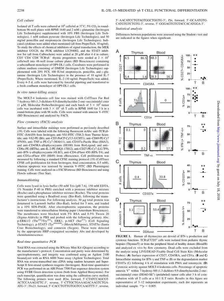
FIGURE 1. Human gd thymocytes are devoid of IFN-g production and
cytotoxic functions. TCRgd+CD3+ cells were isolated from pediatric thymic
biopsies (Thymus/T) or from the peripheral blood of healthy donors (Blood/B)
and analyzed ex vivo by flow cytometry. Dead cells were excluded from
the analysis using LIVE/DEAD Fixable Dead Cell Stain Kits (Molecular
Probes). (A) Surface expression of CD27, CD45RA, and CD1a. (B and C)
Intracellular staining for IFN-g and TNF-a (B) or the degranulation marker
CD107a (C) following 4 h of stimulation with PMA and ionomycin. (D)
Cytotoxic activity against MOLT-4 leukemia cells. Percentage of apoptotic
annexin V+ within 7-hydroxy-9H-(1,3-dichloro-9,9-dimethylacridin-2-one–
succinimidyl ester (DDAO-SE+) (prelabeled) tumor cells after 3 h of coin-
cubation with gd T cells at a 10:1 E:T ratio. Results in this figure are
representative of 3–12 independent experiments; each dot represents an
individual sample. **p , 0.005.
2238 IL-2/IL-15–MEDIATED gd T CELL FUNCTIONAL DIFFERENTIATION
by guest on July 18, 2018http://w
ww
.jimm
unol.org/D
ownloaded from
ResultsHuman gd thymocytes are devoid of cytotoxicity and IFN-gproduction
Inspired by the recent identification of fully differentiated effectorgd T cell subsets in the murine thymus (19–21, 26), we started this
study by analyzing the surface phenotype and functional potential
of gd T cells isolated from human pediatric thymic samples. Based
on the differentiation markers CD1a (27), CD27, and CD45RA (15),
the vast majority of gd thymocytes showed an immature and naive
phenotype, which contrasted with the dominant effector/memory
phenotype (15) of gd PBLs (Fig. 1A). Also unlike these, gd thymo-
cytes produced negligible proinflammatory cytokines, particularly
IFN-g and TNF-a (Fig. 1B). Moreover, gd thymocytes lackedcytolytic activity (Fig. 1C), namely against leukemia target cells,which were promptly killed by their PBL counterparts (Fig. 1D).These data clearly demonstrate that, unlike murine gd thymocytes(19–21, 26), human gd T cells do not complete their functionaldifferentiation in the thymus, and they thus lack the cytotoxic type1 characteristics of gd PBLs.
IL-2 and IL-15 signals drive human gd cytotoxic type 1 celldifferentiation
The functional immaturity of human gd thymocytes made them anideal system to investigate the molecular cues required for ac-quisition of antitumor effector properties. Focusing first on IFN-g
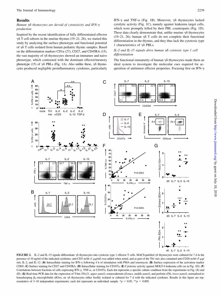
FIGURE 2. IL-2 and IL-15 signals differentiate gd thymocytes into cytotoxic type 1 effector T cells. MACS-purified gd thymocytes were cultured for 7 d in the
presence of 10 ng/ml of the indicated cytokines; anti-CD3 mAb (1 mg/ml) was added when noted, and as part of the Th1 mix also contained anti-CD28 mAb (5 mg/
ml), IL-2, and IL-12. (A) Intracellular staining for IFN-g following 4 h of stimulation with PMA and ionomycin. (B) Surface expression of the activation marker
CD69. (C) Surface staining for CD27 and CD45RA. (D) Intracellular staining for CD107a. (E) Cytotoxic activity against MOLT-4 leukemia cells (as in Fig. 1D). (F)
Correlations between fractions of cells expressing IFN-g, TNF-a, or CD107a. Each dot represents a specific culture condition from the experiments in Fig. (A) and
(D). (G) Real-time PCR data for the expression of T-bet (Tbx21, upper panel), eomesodermin (Eomes, middle panel), and perforin (Pfn, lower panel), normalized to
housekeeping b2-microglobulin (B2m), on gd thymocytes either freshly isolated or cultured for 7 d with the indicated cytokines. Results in this figure are rep-
resentative of 3–18 independent experiments; each dot represents an individual sample. *p , 0.05, **p , 0.005.
The Journal of Immunology 2239
by guest on July 18, 2018http://w
ww
.jimm
unol.org/D
ownloaded from
production, we considered that naive CD4+ T cells typically requireTCR/CD3 and CD28 ligation in the presence of IL-2 and IL-12 fordifferentiation along the “T helper 1” pathway (28). Although sucha “Th1 mix” was indeed capable of generating IFN-g+ gd T cells,we surprisingly found that IL-2 alone was also sufficient (Fig. 2A,upper panel). This effect was potentiated by IL-12, whereas TGF-babrogated the process. Unexpectedly, the addition of TCR stim-ulation via anti-CD3 mAb did not enhance, but rather reduced, theIL-2–mediated differentiation of human IFN-g+ gd T cells (Fig. 2A,upper panel).We next tested a large panel of individual cytokines and ob-
served that besides IL-2, only IL-15 (but notably not IL-4 or IL-7)
was able to induce IFN-g production in gd thymocyte cultures
(Fig. 2A, middle panel). Both IL-2 and IL-15 treatments also pro-
moted TNF-a expression (Fig. 2A, lower panel), upregulated the
activation marker CD69 (Fig. 2B), and drove thymocytes along
the effector/memory differentiation pathway, toward a CD45RA+
CD27– T effector memory stage (Fig. 2C).Concerning gd T cell cytotoxicity, IL-2 and IL-15 (but not IL-7)
induced the expression of the degranulation marker CD107a on gd
thymocytes (Fig. 2D) and endowed them with potent killing ca-
pacity against leukemia target cells (Fig. 2E). Of note, exogenous
IL-2 and IL-15 also enhanced the effector functions of gd PBLs,
especially their degranulation/cytotoxic potential (Supplemental
Fig. 1).The acquisition of IFN-g/TNF-a production and cytolytic ca-
pacity by gd thymocytes were positively correlated, suggesting
a common pathway of cytotoxic type 1 differentiation (Fig. 2F).
Moreover, IL-2/IL-15 signals induced de novo expression of the
type 1 master transcription factors T-bet and eomesodermin, as well
as the cytolytic molecule perforin (Fig. 2G). These data firmly
demonstrate that IL-2 and IL-15 are key functional differentiation
factors for human gd T cells. Importantly, they also show that IL-2
and IL-15 signals are sufficient, in the absence of TCR activation,
to generate fully functional gd T cells from immature thymocytes.
Vd1 and Vd2 T cell subsets follow similar rules of functionaldifferentiation
Given that human gd T cells comprise two major subsets, Vd1+
cells (5–30% of gd PBLs but more abundant in tissues) and Vd2+
cells (60–95% of gd PBLs), both strongly biased toward cytotoxic
type 1 functions (3, 29), we next assessed whether they followed
similar rules of differentiation. Consistent with the literature (25),
the thymic gd repertoire was largely biased for Vd1+ thymocytes,
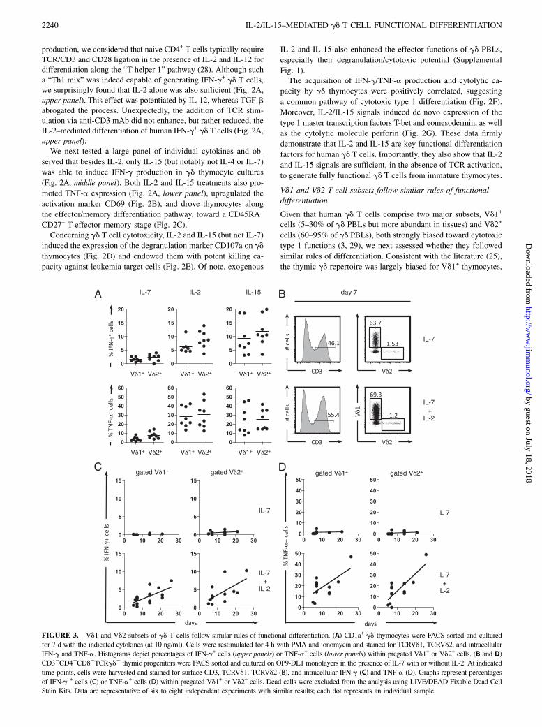
FIGURE 3. Vd1 and Vd2 subsets of gd T cells follow similar rules of functional differentiation. (A) CD1a+ gd thymocytes were FACS sorted and cultured
for 7 d with the indicated cytokines (at 10 ng/ml). Cells were restimulated for 4 h with PMA and ionomycin and stained for TCRVd1, TCRVd2, and intracellular
IFN-g and TNF-a. Histograms depict percentages of IFN-g+ cells (upper panels) or TNF-a+ cells (lower panels) within pregated Vd1+ or Vd2+ cells. (B and D)
CD32CD42CD82TCRgd2 thymic progenitors were FACS sorted and cultured on OP9-DL1 monolayers in the presence of IL-7 with or without IL-2. At indicated
time points, cells were harvested and stained for surface CD3, TCRVd1, TCRVd2 (B), and intracellular IFN-g (C) and TNF-a (D). Graphs represent percentages
of IFN-g + cells (C) or TNF-a+ cells (D) within pregated Vd1+ or Vd2+ cells. Dead cells were excluded from the analysis using LIVE/DEAD Fixable Dead Cell
Stain Kits. Data are representative of six to eight independent experiments with similar results; each dot represents an individual sample.
2240 IL-2/IL-15–MEDIATED gd T CELL FUNCTIONAL DIFFERENTIATION
by guest on July 18, 2018http://w
ww
.jimm
unol.org/D
ownloaded from
with an average Vd1/Vd2 ratio of 25, which was maintainedin vitro upon IL-7, IL-2, or IL-15 treatment (Supplemental Fig.
2A). In all thymic samples analyzed, Vd1+ T cells behaved asexpected: they were functionally immature ex vivo and differen-
tiated into type 1 effectors in response to IL-2 or IL-15 stimuli(Supplemental Fig. 2B). However, the results obtained by gating
on the Vd2+ population were affected by an important intersamplevariation (Supplemental Fig. 2B). We considered that this could be
due to blood contamination or recirculation (back to the thymus)of mature Vg9Vd2 cells, which are much more abundant in the
blood than in the thymus (,3% of total gd thymocytes). To over-come these problems, we purified CD1a+ gd T cells, which are
exclusive to the thymus (Fig. 1A, lower panel) and cultured themfor 7 d with IL-7, IL-2, or IL-15. We found that both Vd1+ and Vd2+
cells similarly acquired type 1 effector properties in response to IL-2and IL-15, but not IL-7 (Fig. 3A).As an alternative developmental strategy, we differentiated gd
T cells from sorted CD32TCRgd2CD42CD82 thymic precursors
cultured on OP9-DL1 monolayers, as previously described (24). Inthe presence of IL-7 alone, Vd1 and Vd2 T cells developed normally
(Fig. 3B) to a Vd1/Vd2 ratio similar to that observed ex vivo(Supplemental Fig. 2A). We therefore think this is an elegant model
to characterize gd T cell differentiation from very early devel-opmental stages. Most importantly, the further addition of IL-2
was necessary to generate IFN-g– (Fig. 3C) and TNF-a– (Fig. 3D)producing gd T cells, and this occurred similarly for Vd1 and Vd2
T cell subsets (Fig. 3C, 3D). These data demonstrate that IL-2 (orIL-15) signals drive the functional differentiation of both major
subsets of human gd T cells.
IL-2/IL-15 signals induce gd type 1 cell differentiation via theMAPK/ERK pathway
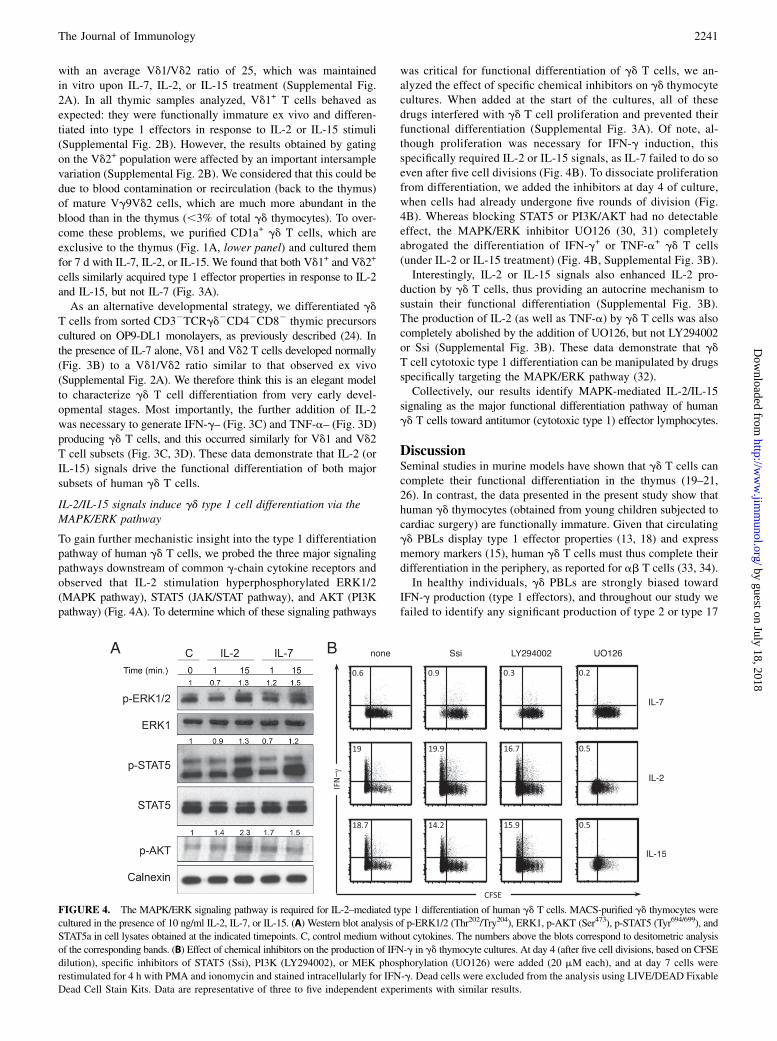
To gain further mechanistic insight into the type 1 differentiationpathway of human gd T cells, we probed the three major signalingpathways downstream of common g-chain cytokine receptors andobserved that IL-2 stimulation hyperphosphorylated ERK1/2(MAPK pathway), STAT5 (JAK/STAT pathway), and AKT (PI3Kpathway) (Fig. 4A). To determine which of these signaling pathways
was critical for functional differentiation of gd T cells, we an-alyzed the effect of specific chemical inhibitors on gd thymocytecultures. When added at the start of the cultures, all of thesedrugs interfered with gd T cell proliferation and prevented theirfunctional differentiation (Supplemental Fig. 3A). Of note, al-though proliferation was necessary for IFN-g induction, thisspecifically required IL-2 or IL-15 signals, as IL-7 failed to do soeven after five cell divisions (Fig. 4B). To dissociate proliferationfrom differentiation, we added the inhibitors at day 4 of culture,when cells had already undergone five rounds of division (Fig.4B). Whereas blocking STAT5 or PI3K/AKT had no detectableeffect, the MAPK/ERK inhibitor UO126 (30, 31) completelyabrogated the differentiation of IFN-g+ or TNF-a+ gd T cells(under IL-2 or IL-15 treatment) (Fig. 4B, Supplemental Fig. 3B).Interestingly, IL-2 or IL-15 signals also enhanced IL-2 pro-
duction by gd T cells, thus providing an autocrine mechanism tosustain their functional differentiation (Supplemental Fig. 3B).The production of IL-2 (as well as TNF-a) by gd T cells was alsocompletely abolished by the addition of UO126, but not LY294002or Ssi (Supplemental Fig. 3B). These data demonstrate that gdT cell cytotoxic type 1 differentiation can be manipulated by drugsspecifically targeting the MAPK/ERK pathway (32).Collectively, our results identify MAPK-mediated IL-2/IL-15
signaling as the major functional differentiation pathway of humangd T cells toward antitumor (cytotoxic type 1) effector lymphocytes.
DiscussionSeminal studies in murine models have shown that gd T cells cancomplete their functional differentiation in the thymus (19–21,26). In contrast, the data presented in the present study show thathuman gd thymocytes (obtained from young children subjected tocardiac surgery) are functionally immature. Given that circulatinggd PBLs display type 1 effector properties (13, 18) and expressmemory markers (15), human gd T cells must thus complete theirdifferentiation in the periphery, as reported for ab T cells (33, 34).In healthy individuals, gd PBLs are strongly biased toward
IFN-g production (type 1 effectors), and throughout our study wefailed to identify any significant production of type 2 or type 17
FIGURE 4. The MAPK/ERK signaling pathway is required for IL-2–mediated type 1 differentiation of human gd T cells. MACS-purified gd thymocytes were
cultured in the presence of 10 ng/ml IL-2, IL-7, or IL-15. (A) Western blot analysis of p-ERK1/2 (Thr202/Try204), ERK1, p-AKT (Ser473), p-STAT5 (Tyr694/699), and
STAT5a in cell lysates obtained at the indicated timepoints. C, control medium without cytokines. The numbers above the blots correspond to desitometric analysis
of the corresponding bands. (B) Effect of chemical inhibitors on the production of IFN-g in gd thymocyte cultures. At day 4 (after five cell divisions, based on CFSE
dilution), specific inhibitors of STAT5 (Ssi), PI3K (LY294002), or MEK phosphorylation (UO126) were added (20 mM each), and at day 7 cells were
restimulated for 4 h with PMA and ionomycin and stained intracellularly for IFN-g. Dead cells were excluded from the analysis using LIVE/DEAD Fixable
Dead Cell Stain Kits. Data are representative of three to five independent experiments with similar results.
The Journal of Immunology 2241
by guest on July 18, 2018http://w
ww
.jimm
unol.org/D
ownloaded from
cytokines. Namely, IL-17, which is constitutively expressed bya subset of murine gd T cells (19, 26), is rarely expressed in hu-man gd T cells from healthy donors (16–18). In contrast, IL-17–producing gd T cells seem to accumulate to high numbers inclinical cases of bacterial meningitis, and their rules of differen-tiation have been previously dissected (17, 35).In our study, the naive and immature phenotype of human gd
thymocytes provided an ideal system to investigate the molecularcues required for acquisition of type 1 effector properties. Our workdemonstrated that IL-2 or IL-15 signals are sufficient to drive thedifferentiation of human gd T cells into IFN-g/TNF-a producersalso endowed with potent cytotoxicity against tumor targets.The redundant functions of IL-2 and IL-15 can be explained by
the structure of their respective receptors, which share not only thecommon g-chain but also their second signaling subunit, IL-2Rb.A third subunit, IL-2Ra or IL-15Ra, is cytokine specific andstabilizes binding but apparently lacks signaling activity. Struc-tural comparisons of IL-2/IL2-Ra and IL-15/IL15-Ra interactionshave emphasized their similarities (36, 37), and it has been re-cently demonstrated that they induce similar downstream tran-scriptional effects (38).On the other hand, IL-7 clearly failed to trigger differentiation of
cytotoxic type 1 gd T cells. This is in stark contrast with the majorrole described for IL-7 in the functional differentiation of humanNKT cells (39) and IL-17–producing gd T cells (40). These lines ofevidence establish an interesting cytokine dichotomy for human gd
T cells: whereas IL-7 promotes type 17 effector functions, IL-2 andIL-15 are the main drivers of the type 1 program. Of note, becauseIL-15 stimulation induces gd T cells to produce IL-2 (SupplementalFig. 3B), this cytokine could also be the direct (autocrine) mediatorof the type 1 program downstream of IL-15 signals.Unexpectedly, TCR activation was not required for the func-
tional differentiation of human gd thymocytes. Recent work fromHayday and colleagues (41) on murine gd T cells suggests thatinnate-like gd T cell subsets may lack TCR responsiveness in theperiphery following strong TCR signaling during thymic devel-opment. Naturally, we cannot assess whether human gd thymo-cytes (the starting point of our in vitro experiments) have alreadyreceived TCR signals in vivo. Were this to be the case, IL-2 andIL-15 would act as terminal differentiation factors in cells that hadbeen previously selected via their TCR. This is also reminiscent ofthe two-step model proposed by Lio and Hsieh (42) and Farrar andcolleagues (43) for murine Foxp3+ regulatory T cell development.Importantly, whereas for regulatory T cells this two-step process iscompleted in the thymus, human gd T cell functional differenti-ation seemingly involves a second step that takes place in theperiphery. Physiologically, this likely depends on the provision ofIL-2 by activated T cells (either gd T cells themselves or their abcounterparts) or IL-15 by myeloid and epithelial cells. Concerninga potential autocrine IL-2 loop based on gd T cells, we havepreviously shown that IL-2 production requires TCR activation inthe presence of CD28 costimulation (44). Therefore, CD80/CD86,as expressed by APCs, including dendritic cells but also Vg9Vd2T cells, are likely additional players in the functional differentiationof gd T cells. For therapeutic purposes (in cancer immunother-apy), our results strongly suggest that the provision of exogenousIL-2 or IL-15 may be critical, not only for gd PBL activation andexpansion, but also particularly for extensive differentiation ofantitumor effectors from recent thymic emigrants and circulatingnaive gd T cells.Downstream of IL-2/IL-15 receptors, our data provide the im-
portant insight that gd T cell cytotoxic type 1 differentiation canbe manipulated by drugs specifically targeting the MAPK/ERKpathway (32). In this context, dual-specificity phosphatases seem
particularly promising targets for modulating MAPK-dependentimmune processes (32).In conclusion, our results detach human gd T cells from para-
digms of T cell differentiation: their differentiation program is notcompleted in the thymus, unlike murine gd T cells (19, 20, 26),and it does not require TCR/co-receptor activation in the periph-ery, in contrast with naive CD4+ T cells (28). Instead, IL-2/IL-15signals are sufficient for functional differentiation of human gdT cells, which clearly aligns them with NK cells (45) and somenaive CD8+ T cell populations (46). Thus, the three main cyto-toxic type 1 lymphocyte subsets share a common, IL-2/IL-15–dependent differentiation program with key implications in cancerimmunotherapy.
AcknowledgmentsWe thank Miguel Abecasis and Rui Anjos at Hospital of Santa Cruz for
provision of the pediatric thymic samples; Joao Barata, Vanda Povoa,
and Daniel Ribeiro for reagents and expert advice; Helena Nunes Cabaco,
Iris Caramalho, Catarina Mota, Vania Silva, and Francisca Matos for tech-
nical assistance; and Marc Bonneville, Emmanuel Scotet, Antonio Lopes,
Daniel Pennington, and Henrique Veiga-Fernandes for helpful discussions.
DisclosuresThe authors have no financial conflicts of interest.
References1. Hirano, M., P. Guo, N. McCurley, M. Schorpp, S. Das, T. Boehm, and
M. D. Cooper. 2013. Evolutionary implications of a third lymphocyte lineage inlampreys. Nature 501: 435–438.
2. Hayday, A. C. 2009. gd T cells and the lymphoid stress-surveillance response.Immunity 31: 184–196.
3. Gomes, A. Q., D. S. Martins, and B. Silva-Santos. 2010. Targeting gdT lymphocytes for cancer immunotherapy: from novel mechanistic insight toclinical application. Cancer Res. 70: 10024–10027.
4. Hannani, D., Y. Ma, T. Yamazaki, J. Dechanet-Merville, G. Kroemer, andL. Zitvogel. 2012. Harnessing gd T cells in anticancer immunotherapy. TrendsImmunol. 33: 199–206.
5. Street, S. E., Y. Hayakawa, Y. Zhan, A. M. Lew, D. MacGregor, A. M. Jamieson,A. Diefenbach, H. Yagita, D. I. Godfrey, and M. J. Smyth. 2004. Innate immunesurveillance of spontaneous B cell lymphomas by natural killer cells and gdT cells. J. Exp. Med. 199: 879–884.
6. Girardi, M., D. E. Oppenheim, C. R. Steele, J. M. Lewis, E. Glusac, R. Filler,P. Hobby, B. Sutton, R. E. Tigelaar, and A. C. Hayday. 2001. Regulation ofcutaneous malignancy by gd T cells. Science 294: 605–609.
7. Liu, Z., I. E. Eltoum, B. Guo, B. H. Beck, G. A. Cloud, and R. D. Lopez. 2008.Protective immunosurveillance and therapeutic antitumor activity of gd T cellsdemonstrated in a mouse model of prostate cancer. J. Immunol. 180: 6044–6053.
8. Gao, Y., W. Yang, M. Pan, E. Scully, M. Girardi, L. H. Augenlicht, J. Craft, andZ. Yin. 2003. gd T cells provide an early source of interferon g in tumor im-munity. J. Exp. Med. 198: 433–442.
9. Lanca, T., D. V. Correia, C. F. Moita, H. Raquel, A. Neves-Costa, C. Ferreira,J. S. Ramalho, J. T. Barata, L. F. Moita, A. Q. Gomes, and B. Silva-Santos. 2010.The MHC class Ib protein ULBP1 is a nonredundant determinant of leukemia/lymphoma susceptibility to gd T-cell cytotoxicity. Blood 115: 2407–2411.
10. Correia, D. V., M. Fogli, K. Hudspeth, M. G. da Silva, D. Mavilio, and B. Silva-Santos. 2011. Differentiation of human peripheral blood Vd1+ T cells expressingthe natural cytotoxicity receptor NKp30 for recognition of lymphoid leukemiacells. Blood 118: 992–1001.
11. Corvaisier, M., A. Moreau-Aubry, E. Diez, J. Bennouna, J. F. Mosnier, E. Scotet,M. Bonneville, and F. Jotereau. 2005. Vg9Vd2 T cell response to colon carci-noma cells. J. Immunol. 175: 5481–5488.
12. Wrobel, P., H. Shojaei, B. Schittek, F. Gieseler, B. Wollenberg, H. Kalthoff,D. Kabelitz, and D. Wesch. 2007. Lysis of a broad range of epithelial tumourcells by human gd T cells: involvement of NKG2D ligands and T-cell receptor-versus NKG2D-dependent recognition. Scand. J. Immunol. 66: 320–328.
13. Gibbons, D. L., S. F. Haque, T. Silberzahn, K. Hamilton, C. Langford, P. Ellis,R. Carr, and A. C. Hayday. 2009. Neonates harbour highly active gd T cells withselective impairments in preterm infants. Eur. J. Immunol. 39: 1794–1806.
14. Vermijlen, D., M. Brouwer, C. Donner, C. Liesnard, M. Tackoen, M. VanRysselberge, N. Twite, M. Goldman, A. Marchant, and F. Willems. 2010. Humancytomegalovirus elicits fetal gd T cell responses in utero. J. Exp. Med. 207: 807–821.
15. Dieli, F., F. Poccia, M. Lipp, G. Sireci, N. Caccamo, C. Di Sano, and A. Salerno.2003. Differentiation of effector/memory Vd2 T cells and migratory routes inlymph nodes or inflammatory sites. J. Exp. Med. 198: 391–397.
16. Caccamo, N., M. Todaro, G. Sireci, S. Meraviglia, G. Stassi, and F. Dieli. 2013.Mechanisms underlying lineage commitment and plasticity of human gd T cells.Cell. Mol. Immunol. 10: 30–34.
2242 IL-2/IL-15–MEDIATED gd T CELL FUNCTIONAL DIFFERENTIATION
by guest on July 18, 2018http://w
ww
.jimm
unol.org/D
ownloaded from
17. Ness-Schwickerath, K. J., C. Jin, and C. T. Morita. 2010. Cytokine requirementsfor the differentiation and expansion of IL-17A- and IL-22-producing humanVg2Vd2 T cells. J. Immunol. 184: 7268–7280.
18. DeBarros, A., M. Chaves-Ferreira, F. d’Orey, J. C. Ribot, and B. Silva-Santos.2011. CD70-CD27 interactions provide survival and proliferative signals thatregulate T cell receptor-driven activation of human gd peripheral bloodlymphocytes. Eur. J. Immunol. 41: 195–201.
19. Ribot, J. C., A. deBarros, D. J. Pang, J. F. Neves, V. Peperzak, S. J. Roberts,M. Girardi, J. Borst, A. C. Hayday, D. J. Pennington, and B. Silva-Santos. 2009.CD27 is a thymic determinant of the balance between interferon-g- and inter-leukin 17-producing gd T cell subsets. Nat. Immunol. 10: 427–436.
20. Jensen, K. D., X. Su, S. Shin, L. Li, S. Youssef, S. Yamasaki, L. Steinman,T. Saito, R. M. Locksley, M. M. Davis, et al. 2008. Thymic selection determinesgd T cell effector fate: antigen-naive cells make interleukin-17 and antigen-experienced cells make interferon g. Immunity 29: 90–100.
21. Turchinovich, G., and A. C. Hayday. 2011. Skint-1 identifies a common mo-lecular mechanism for the development of interferon-g-secreting versusinterleukin-17-secreting gd T cells. Immunity 35: 59–68.
22. Turchinovich, G., and D. J. Pennington. 2011. T cell receptor signalling in gdcell development: strength isn’t everything. Trends Immunol. 32: 567–573.
23. Garcıa-Peydro, M., V. G. de Yebenes, and M. L. Toribio. 2003. Sustained Notch1signaling instructs the earliest human intrathymic precursors to adopt a gd T-cellfate in fetal thymus organ culture. Blood 102: 2444–2451.
24. Van de Walle, I., E. Waegemans, J. De Medts, G. De Smet, M. De Smedt,S. Snauwaert, B. Vandekerckhove, T. Kerre, G. Leclercq, J. Plum, et al. 2013. SpecificNotch receptor-ligand interactions control human TCR-ab/gd development by in-ducing differential Notch signal strength. J. Exp. Med. 210: 683–697.
25. Bonneville, M., R. L. O’Brien, and W. K. Born. 2010. gd T cell effector functions:a blend of innate programming and acquired plasticity. Nat. Rev. Immunol. 10:467–478.
26. Prinz, I., B. Silva-Santos, and D. J. Pennington. 2013. Functional development ofgd T cells. Eur. J. Immunol. 43: 1988–1994.
27. Blom, B., M. C. Verschuren, M. H. Heemskerk, A. Q. Bakker, E. J. van Gastel-Mol, I. L. Wolvers-Tettero, J. J. van Dongen, and H. Spits. 1999. TCR generearrangements and expression of the pre-T cell receptor complex during humanT-cell differentiation. Blood 93: 3033–3043.
28. Messi, M., I. Giacchetto, K. Nagata, A. Lanzavecchia, G. Natoli, and F. Sallusto.2003. Memory and flexibility of cytokine gene expression as separable propertiesof human TH1 and TH2 lymphocytes. Nat. Immunol. 4: 78–86.
29. Behr, C., L. Couzi, J. Taupin, and J. Dechanet-Merville. 2009. Vd2-negative gdT-cells, a multi-reactive tissue subset: from innate to adaptive altered-self sur-veillance. Open Immunol. J. 2: 106–118.
30. Favata, M. F., K. Y. Horiuchi, E. J. Manos, A. J. Daulerio, D. A. Stradley,W. S. Feeser, D. E. Van Dyk, W. J. Pitts, R. A. Earl, F. Hobbs, et al. 1998.Identification of a novel inhibitor of mitogen-activated protein kinase kinase.J. Biol. Chem. 273: 18623–18632.
31. DeSilva, D. R., E. A. Jones, M. F. Favata, B. D. Jaffee, R. L. Magolda,J. M. Trzaskos, and P. A. Scherle. 1998. Inhibition of mitogen-activated proteinkinase kinase blocks T cell proliferation but does not induce or prevent anergy.J. Immunol. 160: 4175–4181.
32. Jeffrey, K. L., M. Camps, C. Rommel, and C. R. Mackay. 2007. Targeting dual-specificity phosphatases: manipulating MAP kinase signalling and immuneresponses. Nat. Rev. Drug Discov. 6: 391–403.
33. Boursalian, T. E., J. Golob, D. M. Soper, C. J. Cooper, and P. J. Fink. 2004.Continued maturation of thymic emigrants in the periphery. Nat. Immunol. 5:418–425.
34. Houston, E. G., Jr., R. Nechanitzky, and P. J. Fink. 2008. Cutting edge: contactwith secondary lymphoid organs drives postthymic T cell maturation. J. Immunol.181: 5213–5217.
35. Caccamo, N., C. La Mendola, V. Orlando, S. Meraviglia, M. Todaro, G. Stassi,G. Sireci, J. J. Fournie, and F. Dieli. 2011. Differentiation, phenotype, and functionof interleukin-17-producing human Vg9Vd2 T cells. Blood 118: 129–138.
36. Olsen, S. K., N. Ota, S. Kishishita, M. Kukimoto-Niino, K. Murayama,H. Uchiyama, M. Toyama, T. Terada, M. Shirouzu, O. Kanagawa, andS. Yokoyama. 2007. Crystal structure of the interleukin-15.interleukin-15 re-ceptor a complex: insights into trans and cis presentation. J. Biol. Chem. 282:37191–37204.
37. Chirifu, M., C. Hayashi, T. Nakamura, S. Toma, T. Shuto, H. Kai, Y. Yamagata,S. J. Davis, and S. Ikemizu. 2007. Crystal structure of the IL-15-IL-15Racomplex, a cytokine-receptor unit presented in trans. Nat. Immunol. 8: 1001–1007.
38. Ring, A. M., J. X. Lin, D. Feng, S. Mitra, M. Rickert, G. R. Bowman,V. S. Pande, P. Li, I. Moraga, R. Spolski, et al. 2012. Mechanistic and structuralinsight into the functional dichotomy between IL-2 and IL-15. Nat. Immunol. 13:1187–1195.
39. de Lalla, C., N. Festuccia, I. Albrecht, H. D. Chang, G. Andolfi, U. Benninghoff,F. Bombelli, G. Borsellino, A. Aiuti, A. Radbruch, et al. 2008. Innate-like ef-fector differentiation of human invariant NKT cells driven by IL-7. J. Immunol.180: 4415–4424.
40. Michel, M. L., D. J. Pang, S. F. Haque, A. J. Potocnik, D. J. Pennington, andA. C. Hayday. 2012. Interleukin 7 (IL-7) selectively promotes mouse and humanIL-17-producing gd cells. Proc. Natl. Acad. Sci. USA 109: 17549–17554.
41. Wencker, M., G. Turchinovich, R. Di Marco Barros, L. Deban, A. Jandke,A. Cope, and A. C. Hayday. 2014. Innate-like T cells straddle innate and adaptiveimmunity by altering antigen-receptor responsiveness. Nat. Immunol. 15: 80–87.
42. Lio, C. W., and C. S. Hsieh. 2008. A two-step process for thymic regulatoryT cell development. Immunity 28: 100–111.
43. Burchill, M. A., J. Yang, K. B. Vang, J. J. Moon, H. H. Chu, C. W. Lio,A. L. Vegoe, C. S. Hsieh, M. K. Jenkins, and M. A. Farrar. 2008. Linked T cellreceptor and cytokine signaling govern the development of the regulatory T cellrepertoire. Immunity 28: 112–121.
44. Ribot, J. C., A. Debarros, L. Mancio-Silva, A. Pamplona, and B. Silva-Santos.2012. B7-CD28 costimulatory signals control the survival and proliferation ofmurine and human gd T cells via IL-2 production. J. Immunol. 189: 1202–1208.
45. Meazza, R., B. Azzarone, A. M. Orengo, and S. Ferrini. 2011. Role of common-g chain cytokines in NK cell development and function: perspectives for im-munotherapy. J. Biomed. Biotechnol. 2011: 861920.
46. Alves, N. L., B. Hooibrink, F. A. Arosa, and R. A. van Lier. 2003. IL-15 inducesantigen-independent expansion and differentiation of human naive CD8+ T cellsin vitro. Blood 102: 2541–2546.
The Journal of Immunology 2243
by guest on July 18, 2018http://w
ww
.jimm
unol.org/D
ownloaded from