How many conserved sequence regions ... - Biologia,...
13
Biologia, Bratislava, 57/Suppl. 11: 29—41, 2002 REVIEW How many conserved sequence regions are there in the α-amylase family? Štefan Janeček* Institute of Molecular Biology, Slovak Academy of Sciences, Dúbravská cesta 21, SK-84251 Bratislava, Slovakia; tel.: ++ 421 2 5930 7420, fax: ++ 421 2 5930 7416, e-mail: [email protected] JANEČEK, Š., How many conserved sequence regions are there in the α- amylase family? Biologia, Bratislava, 57/Suppl. 11: 29—41, 2002; ISSN 0006-3088. The α-amylase family, i.e. the clan GH-H of glycoside hydrolases, is the largest family of glycoside hydrolases, transferases and isomerases comprising nearly 30 different enzyme specificities. One of the most interesting features of this family is that its members contain a few very well-conserved sequence regions despite the overall low sequence similarity. It seems that only 4 amino acid residues may be totally invariantly conserved throughout the family (Arg204 plus the three catalytic residues: Asp206, Glu230 and Asp297; Taka-amylase A numbering). Four conserved sequence regions covering the strands β3, β4, β5, and β7 of the catalytic (β/α) 8 -barrel domain were identified and used for defining the α-amylase family. The present review is therefore focused on the three additional conserved sequence regions proposed after the basic characteristics of the family have been established. Two of these three regions cover roughly the strands β2 and β8 of the catalytic (β/α) 8 -barrel and one is located near the C-terminus of domain B (in the β3→ α3 connection of the catalytic (β/α) 8 -barrel). While the four original conserved sequence regions contain the catalytic and substrate-binding residues of the individual mem- bers of the family, the three conserved sequence regions later identified are shown to contain amino acid residues connected to a given enzyme specificity. The problems that may arise with correctly identifying the β5-strand catalytic glutamate, positioned in the conserved sequence region III, are discussed and a way that may lead to a correct solution is indicated. In conclusion, it is proposed that the α-amylase enzyme family should be characterised by as many conserved sequence regions as possible. Key words: alpha-amylase family, conserved sequence regions, sequence simi- larities, specificity features, evolutionary relationships. Abbreviations: CGTase, cyclodextrin glucanotransferase. Introduction The α-amylase enzyme family was originally recognised as a group of starch hydrolases and related enzymes that exhibit clear sequence sim- ilarities and a predicted common supersecondary fold, a parallel (β/α)8-barrel (MACGREGOR, 1988; SVENSSON, 1988; MACGREGOR & SVENSSON, 1989; JESPERSEN et al., 1991; 1993). From the be- ginning it covered several different enzyme speci- * Corresponding author 29
Transcript of How many conserved sequence regions ... - Biologia,...
Biologia, Bratislava, 57/Suppl. 11: 29—41, 2002 REVIEW
How many conserved sequence regionsare there in the α-amylase family?
Štefan Janeček*
Institute of Molecular Biology, Slovak Academy of Sciences, Dúbravská cesta 21, SK-84251 Bratislava,Slovakia; tel.: ++ 421 2 5930 7420, fax: ++ 421 2 5930 7416, e-mail: [email protected]
JANEČEK, Š., How many conserved sequence regions are there in the α-amylase family? Biologia, Bratislava, 57/Suppl. 11: 29—41, 2002; ISSN0006-3088.
The α-amylase family, i.e. the clan GH-H of glycoside hydrolases, is the largestfamily of glycoside hydrolases, transferases and isomerases comprising nearly30 different enzyme specificities. One of the most interesting features of thisfamily is that its members contain a few very well-conserved sequence regionsdespite the overall low sequence similarity. It seems that only 4 amino acidresidues may be totally invariantly conserved throughout the family (Arg204plus the three catalytic residues: Asp206, Glu230 and Asp297; Taka-amylaseA numbering). Four conserved sequence regions covering the strands β3, β4,β5, and β7 of the catalytic (β/α)8-barrel domain were identified and usedfor defining the α-amylase family. The present review is therefore focusedon the three additional conserved sequence regions proposed after the basiccharacteristics of the family have been established. Two of these three regionscover roughly the strands β2 and β8 of the catalytic (β/α)8-barrel and one islocated near the C-terminus of domain B (in the β3→ α3 connection of thecatalytic (β/α)8-barrel). While the four original conserved sequence regionscontain the catalytic and substrate-binding residues of the individual mem-bers of the family, the three conserved sequence regions later identified areshown to contain amino acid residues connected to a given enzyme specificity.The problems that may arise with correctly identifying the β5-strand catalyticglutamate, positioned in the conserved sequence region III, are discussed anda way that may lead to a correct solution is indicated. In conclusion, it isproposed that the α-amylase enzyme family should be characterised by asmany conserved sequence regions as possible.
Key words: alpha-amylase family, conserved sequence regions, sequence simi-larities, specificity features, evolutionary relationships.
Abbreviations: CGTase, cyclodextrin glucanotransferase.
Introduction
The α-amylase enzyme family was originallyrecognised as a group of starch hydrolases andrelated enzymes that exhibit clear sequence sim-
ilarities and a predicted common supersecondaryfold, a parallel (β/α)8-barrel (MACGREGOR, 1988;SVENSSON, 1988; MACGREGOR & SVENSSON,1989; JESPERSEN et al., 1991; 1993). From the be-ginning it covered several different enzyme speci-
* Corresponding author
29
ficities, including cyclodextrin glucanotransferase(CGTase), isoamylase, branching enzyme, neop-ullulanase and oligo-1,6-glucosidase in additionto α-amylase (BINDER et al., 1986; AMEMURA
et al., 1988; ROMEO et al., 1988; KURIKI &IMANAKA, 1989; WATANABE et al., 1990). Theα-amylase family has been known as the family13 of glycoside hydrolases from 1991 when thesequence-based classification of all glycoside hy-drolases, transferases, and isomerases was estab-lished (HENRISSAT, 1991).
The concept of this group of enzymes asthe α-amylase family was proposed a year later(TAKATA et al., 1992). According to that defini-tion the members of the α-amylase family are en-zymes that satisfy the four requirements: (i) theyact on α-glucosidic linkages; (ii) they hydrolyse orform by transglycosylation α-glucosidic linkages;(iii) their amino-acid sequence contains four con-served regions; and (iv) they contain Asp, Glu andAsp residues corresponding to the Asp206, Glu230and Asp297 of Taka-amylase A.
The current situation in the α-amylase fam-ily is as follows: (i) the family contains almost30 different enzyme specificities covering hydro-lases, transferases, and isomerases (SVENSSON etal., 2002); (ii) the family constitutes a clan GH-H of glycoside hydrolases comprising the families13, 70, and 77 (COUTINHO & HENRISSAT, 1999);(iii) the enzymes from the family can operate onα-1,1-, α-1,2-, α-1,3- and α-1,5-glucosidic linkagesin addition to the α-1,4- and α-1,6-bonds origi-nally considered (MACGREGOR et al., 2001); (iv)there may be at most only 4 invariant amino acidresidues, i.e. 3 catalytic ones plus the Arg in posi-tion i−2 with respect to the catalytic nucleophile,the aspartate in the β4-strand (JANEČEK, 2000a);(v) the relationship of the main α-amylase fam-ily (clan GH-H) to the “extremophilic” α-amylasefamily, family 57 of glycoside hydrolases, has stillbeen neither confirmed nor disproved (JANEČEK,1998; IMAMURA et al., 2001a); and (vi) from thepoints of view of sequence and evolution, somemammalian transport proteins and antigens areperhaps related to the enzymatic members of thefamily (JANEČEK, 2000b).
Many specialised reports are available focus-ing on protein engineering of the individual en-zymes and certain groups from the α-amylasefamily, as well as on the possibilities of tailor-ing their properties to meet specific needs (e.g.,KURIKI et al., 1996; MATSUI & SVENSSON, 1997;BINDERUP & PREISS, 1998; IBUKA et al., 1998;WIND et al., 1998; SAAB-RINCÓN et al., 1999;BEIER et al., 2000; YAMAMOTO et al., 2000;
GOTTSCHALK et al., 2001; MORI et al., 2001; KIM
et al., 2001; LEEMHUIS et al., 2002). The find-ings have been thoroughly discussed in various re-views (e.g., MACGREGOR, 1993; SVENSSON, 1994;JANEČEK, 1997a; NIELSEN & BORCHERT, 2000;PARK et al., 2000; VAN DER VEEN et al., 2000;UITDEHAAG et al., 2002; VAN DER MAAREL etal., 2002). In general, however, the residues playingthe most important roles are located in the shortsequence stretches conserved throughout the fam-ily and known as the conserved sequence regions.This review therefore aims to present and sum-marise the short sequence regions that are bestconserved either throughout the α-amylase familyor in several closely-related enzyme groups fromthe family, and to show the relationships betweenthese conserved sequence regions and the individ-ual enzyme specificities.
From family to clan and from clan to a . . .suprafamily ?
When the number of sequences recognised as be-longing to the α-amylase family was low, i.e. sev-eral tens at the beginning of the 1990s (MAC-GREGOR & SVENSSON, 1989; HENRISSAT, 1991;TAKATA et al., 1992), it was clear that, de-spite the existence of more enzyme specifici-ties, all the sequences constitute a classical en-zyme family with members showing divergentevolution (JESPERSEN et al., 1993; JANEČEK,1994a). As one of the main criteria for decidingwhether or not a new sequence belongs to theα-amylase family, the presence of the four con-served sequence regions (e.g., for Taka-amylaseA – I: 117 DVVANH, II: 202 GLRIDTVKH, III:230 EVLD, IV: 292 FVENHD) was used as a goodcriterion (NAKAJIMA et al., 1986).
This basic trend has, in fact, been main-tained up to the present day (MACGREGOR etal., 2001) although the number of sequences hasreached almost one thousand (COUTINHO & HEN-RISSAT, 1999). According to the HENRISSAT clas-sification, however, the original α-amylase fam-ily, family 13 of glycoside hydrolases (HENRIS-SAT, 1991), has been changed to a clan, glyco-side hydrolases clan GH-H, covering the families13, 70, and 77 (BOURNE & HENRISSAT, 2001).While the original family 13 and the family 77seem to be very closely related to each other(PRZYLAS et al., 2000), i.e. they roughly fulfilthe attributes of a single family of similar re-action mechanism, fold, and catalytic machineryseen as identical catalytic residues on identicalelements of secondary structure, family 70 con-
30

tains enzymes with a version of the (β/α)8-barrelstructure that is a circularly permuted form ofthe classical α-amylase-type (β/α)8-barrel (MAC-GREGOR et al., 1996). Although an alternativeprediction without circular permutation was pre-sented for the family 70 glucosyltransferases (DE-VULAPALLE et al., 1997), the predictions tak-ing into account the possibility of circular per-mutations seem more reasonable and have beensupported by mutagenesis (MACGREGOR et al.,1996; MONCHOIS et al., 1999; 2000; TSAI et al.,2000).
The existence of a single α-amylase fam-ily, the family 13 of glycoside hydrolases, hasappeared more “complicated” since 1988 and1993 when two “unusual” amino acid sequencesof α-amylases were reported: the AmyA fromDictyoglomus thermophilum (FUKUSUMI et al.,1988) and one from Pyrococcus furiosus (LADER-MAN et al., 1993). These and also some othermostly extremophilic amylolytic enzymes (DONG
et al., 1997b; JEON et al., 1997; ERRA-PUJADA
et al., 1999) were found to lack the character-istic conserved sequence regions; this resultedin a new family, family 57 of glycoside hydro-lases being established (HENRISSAT & BAIROCH,1996). Later, however, a distant evolutionary rela-tionship between the major family 13 and fam-ily 57 was indicated by using HCA (hydropho-bic cluster analysis) on the amino-acid sequenceof archaeal Methanococcus jannaschii α-amylase(JANEČEK, 1998). At present family 57 containsmembers mostly from extremophiles (COUTINHO
& HENRISSAT, 1999) having α-amylase, 4-α-glucanotransferase, amylopullulanase and even α-galactosidase (VAN LIESHOUT et al., 2001) speci-ficities in addition to numerous hypothetical pro-teins coming from various complete-genome se-quencing projects. Recent determination of thethree-dimensional structure of the 4-α-glucano-transferase from Thermococcus litoralis, with site-directed mutated glutamate residues, indicated(IMAMURA et al., 2001a,b) that the HCA-basedpredictions (JANEČEK, 1998) may not match thecatalytic amino acids and thus may invalidate thepostulated close relationship of glycoside hydro-lase family 57 to clan GH-H. Despite this re-cent finding, the common retaining mechanismand a common (β/α)8-barrel fold used by boththe clan GH-H and the family 57 preserve thepossibility of classifying them into an even largergroup than the clan, e.g. a suprafamily represent-ing relatedness at a higher hierarchal level than aclan.
Conserved sequence regions
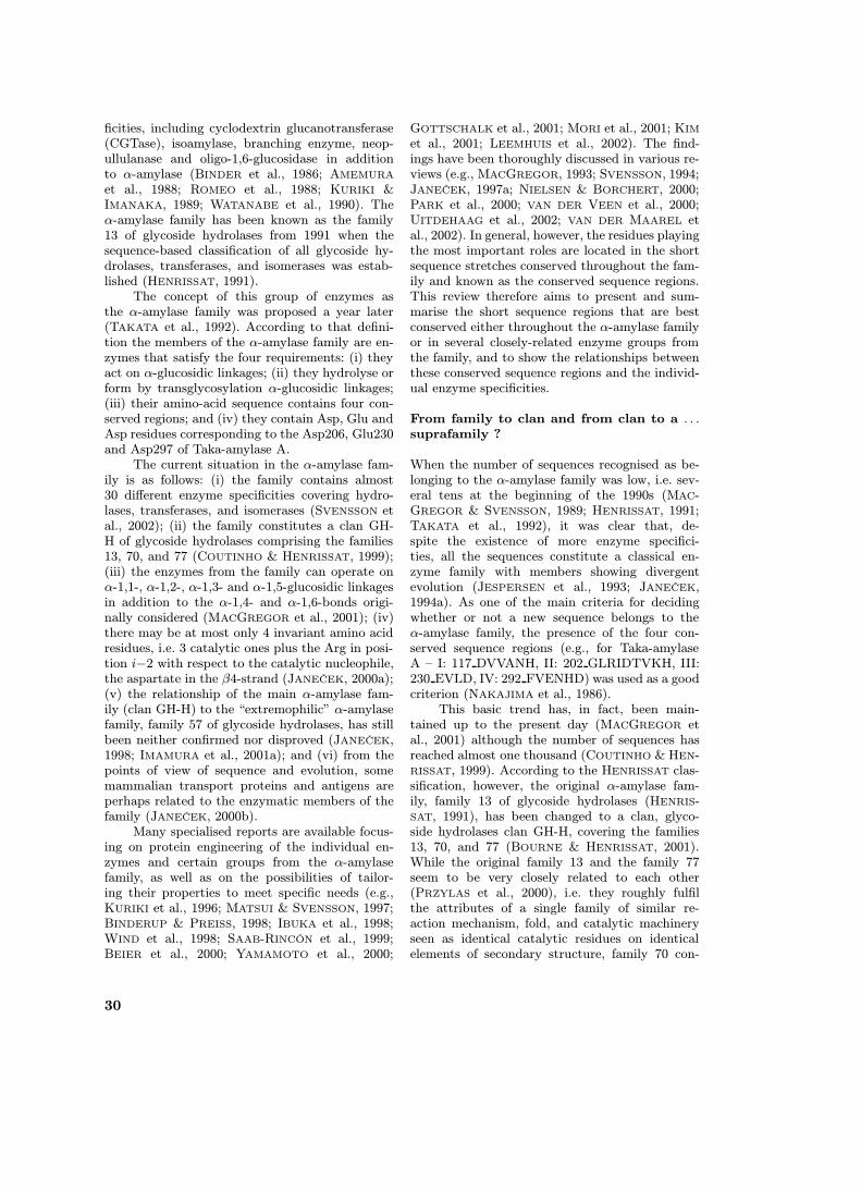
Study of the α-amylase family is practically im-possible without taking into account the ques-tion of the conserved sequence regions. For a con-siderable time, the sequence similarity has beenknown to be extremely low (about 10%) evenfor the α-amylases alone (i.e. for EC 3.2.1.1) asdescribed from different microorganisms, plants,and animals (NAKAJIMA et al., 1986). Later whenthe family grew, i.e. when many sequences fromvarious sources and with different enzyme speci-ficities became available, the number of identicalresidues among the α-amylase family enzymes haddecreased to 8-10 amino acids in 1994 (JANEČEK,1994a; SVENSSON, 1994). At present there mightbe at most only 4 residues that are totally invari-antly conserved throughout the α-amylase fam-ily, i.e. the 3 catalytic residues corresponding toAsp206, Glu230 and Asp297 in Taka-amylase Aplus the β4 arginine equivalent to the Arg204 inTaka-amylase A. The β3 aspartate (Asp117 inTaka-amylase A) is also extremely well conserved(Fig. 1), however, it has been found substituted atleast one time (Š. JANEČEK, unpublished results):by glutamate in trehalose synthase from Ther-mus aquaticus (TSUSAKI et al., 1997). Despite thisfact, amino-acid sequences of enzymes from theα-amylase family contain several short conservedstretches, the so-called conserved sequence regions(Fig. 1). Of the four above-mentioned invariantly-conserved residues Asp206 (the catalytic nucle-ophile) and Glu230 (the proton donor) togetherwith the second conserved aspartate, Asp 297, andtwo histidine residues, His122 and His296, werepostulated as being crucial for Taka-amylase A(MATSUURA et al., 1984). They form the basisof the four well-known conserved sequence regionsin the α-amylase family enzymes (the regions I,II, III and IV in Fig. 1) and are positioned nearthe C-termini of strands β3, β4, β5, and β7 ofthe catalytic (β/α)8-barrel domain (NAKAJIMA etal., 1986; MACGREGOR et al., 2001). It is clear,however, that the two histidines are not conservedthroughout the family (Fig. 1).
Although NAKAJIMA et al. (1986) establishedthe four conserved sequence regions of α-amylases,the authors paid less attention to the remainingpart of their alignment of 11 different α-amylasesequences. This fact was mentioned already byMACGREGOR (1988), who indicated that therecould be additional sequence similarities in α-amylases that were found later in other enzymesbelonging to the α-amylase family by SVENSSON
(1988), MACGREGOR & SVENSSON (1989) and
31
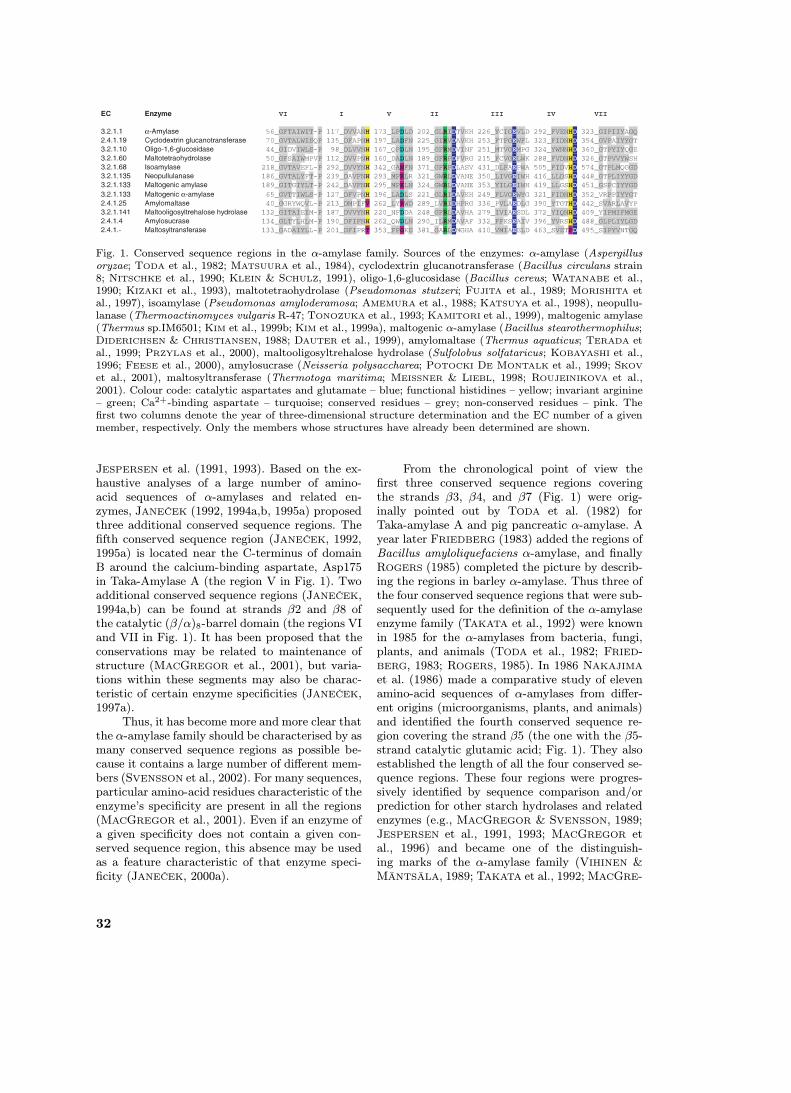
Fig. 1. Conserved sequence regions in the α-amylase family. Sources of the enzymes: α-amylase (Aspergillusoryzae; TODA et al., 1982; MATSUURA et al., 1984), cyclodextrin glucanotransferase (Bacillus circulans strain8; NITSCHKE et al., 1990; KLEIN & SCHULZ, 1991), oligo-1,6-glucosidase (Bacillus cereus; WATANABE et al.,1990; KIZAKI et al., 1993), maltotetraohydrolase (Pseudomonas stutzeri; FUJITA et al., 1989; MORISHITA etal., 1997), isoamylase (Pseudomonas amyloderamosa; AMEMURA et al., 1988; KATSUYA et al., 1998), neopullu-lanase (Thermoactinomyces vulgaris R-47; TONOZUKA et al., 1993; KAMITORI et al., 1999), maltogenic amylase(Thermus sp.IM6501; KIM et al., 1999b; KIM et al., 1999a), maltogenic α-amylase (Bacillus stearothermophilus;DIDERICHSEN & CHRISTIANSEN, 1988; DAUTER et al., 1999), amylomaltase (Thermus aquaticus; TERADA etal., 1999; PRZYLAS et al., 2000), maltooligosyltrehalose hydrolase (Sulfolobus solfataricus; KOBAYASHI et al.,1996; FEESE et al., 2000), amylosucrase (Neisseria polysaccharea; POTOCKI DE MONTALK et al., 1999; SKOV
et al., 2001), maltosyltransferase (Thermotoga maritima; MEISSNER & LIEBL, 1998; ROUJEINIKOVA et al.,2001). Colour code: catalytic aspartates and glutamate – blue; functional histidines – yellow; invariant arginine– green; Ca2+-binding aspartate – turquoise; conserved residues – grey; non-conserved residues – pink. Thefirst two columns denote the year of three-dimensional structure determination and the EC number of a givenmember, respectively. Only the members whose structures have already been determined are shown.
JESPERSEN et al. (1991, 1993). Based on the ex-haustive analyses of a large number of amino-acid sequences of α-amylases and related en-zymes, JANEČEK (1992, 1994a,b, 1995a) proposedthree additional conserved sequence regions. Thefifth conserved sequence region (JANEČEK, 1992,1995a) is located near the C-terminus of domainB around the calcium-binding aspartate, Asp175in Taka-Amylase A (the region V in Fig. 1). Twoadditional conserved sequence regions (JANEČEK,1994a,b) can be found at strands β2 and β8 ofthe catalytic (β/α)8-barrel domain (the regions VIand VII in Fig. 1). It has been proposed that theconservations may be related to maintenance ofstructure (MACGREGOR et al., 2001), but varia-tions within these segments may also be charac-teristic of certain enzyme specificities (JANEČEK,1997a).
Thus, it has become more and more clear thatthe α-amylase family should be characterised by asmany conserved sequence regions as possible be-cause it contains a large number of different mem-bers (SVENSSON et al., 2002). For many sequences,particular amino-acid residues characteristic of theenzyme’s specificity are present in all the regions(MACGREGOR et al., 2001). Even if an enzyme ofa given specificity does not contain a given con-served sequence region, this absence may be usedas a feature characteristic of that enzyme speci-ficity (JANEČEK, 2000a).
From the chronological point of view thefirst three conserved sequence regions coveringthe strands β3, β4, and β7 (Fig. 1) were orig-inally pointed out by TODA et al. (1982) forTaka-amylase A and pig pancreatic α-amylase. Ayear later FRIEDBERG (1983) added the regions ofBacillus amyloliquefaciens α-amylase, and finallyROGERS (1985) completed the picture by describ-ing the regions in barley α-amylase. Thus three ofthe four conserved sequence regions that were sub-sequently used for the definition of the α-amylaseenzyme family (TAKATA et al., 1992) were knownin 1985 for the α-amylases from bacteria, fungi,plants, and animals (TODA et al., 1982; FRIED-BERG, 1983; ROGERS, 1985). In 1986 NAKAJIMA
et al. (1986) made a comparative study of elevenamino-acid sequences of α-amylases from differ-ent origins (microorganisms, plants, and animals)and identified the fourth conserved sequence re-gion covering the strand β5 (the one with the β5-strand catalytic glutamic acid; Fig. 1). They alsoestablished the length of all the four conserved se-quence regions. These four regions were progres-sively identified by sequence comparison and/orprediction for other starch hydrolases and relatedenzymes (e.g., MACGREGOR & SVENSSON, 1989;JESPERSEN et al., 1991, 1993; MACGREGOR etal., 1996) and became one of the distinguish-ing marks of the α-amylase family (VIHINEN &MÄNTSÄLA, 1989; TAKATA et al., 1992; MACGRE-
32
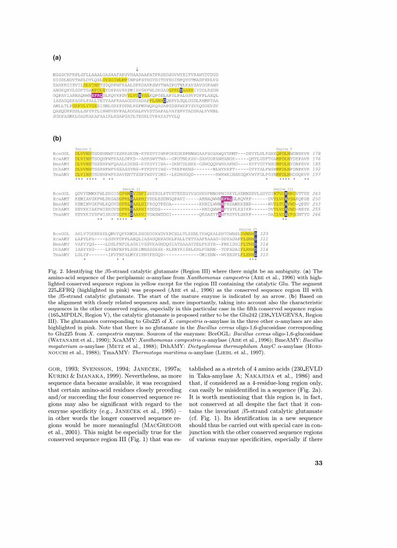
Fig. 2. Identifying the β5-strand catalytic glutamate (Region III) where there might be an ambiguity. (a) Theamino-acid sequence of the periplasmic α-amylase from Xanthomonas campestris (ABE et al., 1996) with high-lighted conserved sequence regions in yellow except for the region III containing the catalytic Glu. The segment225 EFRQ (highlighted in pink) was proposed (ABE et al., 1996) as the conserved sequence region III withthe β5-strand catalytic glutamate. The start of the mature enzyme is indicated by an arrow. (b) Based onthe alignment with closely related sequences and, more importantly, taking into account also the characteristicsequences in the other conserved regions, especially in this particular case in the fifth conserved sequence region(165 MPDLN, Region V), the catalytic glutamate is proposed rather to be the Glu242 (238 YLVGEVSA, RegionIII). The glutamates corresponding to Glu225 of X. campestris α-amylase in the three other α-amylases are alsohighlighted in pink. Note that there is no glutamate in the Bacillus cereus oligo-1,6-glucosidase correspondingto Glu225 from X. campestris enzyme. Sources of the enzymes: BceOGL: Bacillus cereus oligo-1,6-glucosidase(WATANABE et al., 1990); XcaAMY: Xanthomonas campestris α-amylase (ABE et al., 1996); BmeAMY: Bacillusmegaterium α-amylase (METZ et al., 1988); DthAMY: Dictyoglomus thermophilum AmyC α-amylase (HORI-NOUCHI et al., 1988); TmaAMY: Thermotoga maritima α-amylase (LIEBL et al., 1997).
GOR, 1993; SVENSSON, 1994; JANEČEK, 1997a;KURIKI & IMANAKA, 1999). Nevertheless, as moresequence data became available, it was recognisedthat certain amino-acid residues closely precedingand/or succeeding the four conserved sequence re-gions may also be significant with regard to theenzyme specificity (e.g., JANEČEK et al., 1995) –in other words the longer conserved sequence re-gions would be more meaningful (MACGREGOR
et al., 2001). This might be especially true for theconserved sequence region III (Fig. 1) that was es-
tablished as a stretch of 4 amino acids (230 EVLDin Taka-amylase A; NAKAJIMA et al., 1986) andthat, if considered as a 4-residue-long region only,can easily be misidentified in a sequence (Fig. 2a).It is worth mentioning that this region is, in fact,not conserved at all despite the fact that it con-tains the invariant β5-strand catalytic glutamate(cf. Fig. 1). Its identification in a new sequenceshould thus be carried out with special care in con-junction with the other conserved sequence regionsof various enzyme specificities, especially if there
33
is limited experimental support (Fig. 2b). ABE etal. (1999) solved the problem by site-directed mu-tagenesis on isoamylase, which identified the cor-rect glutamic acid. A similar strategy was appliedby, e.g., DEVULAPALLE et al. (1997) to glucosyl-transferase, SARCABAL et al. (2000) to amylosu-crase and NAKAYAMA et al. (2001) to glycogendebranching enzyme.
Importance of the additional conserved se-quence regions
The fifth conserved sequence region – in domain BThe fifth conserved sequence region (173 LPDLDin Taka-amylase A; Fig. 1) was proposed first forα-amylases (JANEČEK, 1992) and later was iden-tified also in the other members of the α-amylasefamily (JANEČEK, 1995a). It is positioned near theC-terminus of domain B around the aspartate in-volved in binding the calcium ion, Asp175 in Taka-amylase A (e.g. MATSUURA et al., 1984; QIAN etal., 1993; KADZIOLA et al., 1994; MACHIUS et al.,1995). In most cases there are 28–30 residues be-tween this Ca2+-binding aspartate and the β4-strand catalytic aspartate, i.e. Asp206 in Taka-amylase A (JANEČEK, 1992, 1995a).
Although this region seems to be not fullyconserved or not easily identifiable in some fam-ily members (JANEČEK, 1997a), in several casesthis stretch is very characteristic for a special en-zyme specificity (Fig. 3). The most striking ex-ample is the oligo-1,6-glucosidase with the tem-plate sequence QPDLN. This region in the formof QxDLN (x = P, A, I, W) is found alsoin α-glucosidase, dextran-glucosidase, trehalose-6-phosphate hydrolase, amylosucrase, sucrose phos-phorylase, isomaltulose synthase and trehalosesynthase (e.g., JANEČEK, 1997a, 2000a). There isa characteristic change from QPDLN (the oligo-1,6-glucosidase group) to MPKLN (the neopul-lulanase group) via the MPDLN belonging to agroup of odd members of the α-amylase fam-ily (Fig. 3). It has already been pointed out(JANEČEK et al., 1997) that structurally the do-main B of these members should resemble veryclosely the domain B of oligo-1,6-glucosidase fromBacillus cereus (KIZAKI et al., 1993). Remarkably,the mammalian proteins associated with transportof dibasic and neutral amino acids across cell mem-branes also unambiguously contain this conservedsequence region (Fig. 3) as well as an entire seg-ment that corresponds perfectly (almost 50% iden-tity) with the whole domain B of the enzymes fromthe α-amylase family (JANEČEK, 2000b). The se-quence QxDLN as the fifth conserved sequence
Fig. 3. The fifth conserved sequence region of the α-amylase family enzymes. The figure focuses on the so-called oligo-1,6-glucosidase and neopullulanase groupsof the family. Sources of the enzymes: Taka-amylaseA (TODA et al., 1982); oligo-1,6-glucosidase (WATAN-ABE et al., 1990); α-glucosidase (JANDA et al., 2000);dextran glucosidase (RUSSELL & FERRETTI, 1990);trehalose-6-phosphate hydrolase (RIMMELE & BOOS,1994); amylosucrase (POTOCKI DE MONTALK et al.,1999); sucrose phosphorylase (FERRETTI et al., 1988);sucrose isomerase (ZHANG et al., 2002); trehalose syn-thase (TSUSAKI et al., 1996); cyclomaltodextrinase(PODKOVYROV & ZEIKUS, 1992); maltogenic amylase(KIM et al., 1999b); neopullulanase (TONOZUKA et al.,1993); Bacillus megaterium (METZ et al., 1988); Ther-motoga maritima (LIEBL et al., 1997); Xanthomonascampestris (ABE et al., 1996); Dictyoglomus ther-mophilum AmyC (HORINOUCHI et al., 1988); Dic-tyoglomus thermophilum AmyB (HORINOUCHI et al.,1988). For illustration also the corresponding segmentis shown of the human protein responsible for thetransport of dibasic and neutral amino acids across cellmembranes (amino-acid transporter; BERTRAN et al.,1993) containing the oligo-1,6-glucosidase-type of do-main B (JANEČEK et al., 1997). Colour code: Ca2+-binding aspartate – blue; lysine playing the role ofa calcium – red; oligo-1,6-glucosidase-like glutamine –yellow; neopullulanase-like methionine – green.
region could thus be used as a fingerprint thatdefines the proposed “oligo-1,6-glucosidase group”within the frame of the α-amylase family (OSLAN-COVÁ & JANEČEK, 2002).
34
The members of the family with characteris-tic sequences in this region MPKLN (or MPKINand MPKLR; Figs 1,3), such as cyclomaltodextri-nases, maltogenic amylases and neopullulanases,have a shorter domain B than the majority andpresumably do not bind a calcium ion due to theAsp→Lys substitution (JANEČEK et al., 1997).Enzymes with these specificities could also form agroup of closely-related members of the α-amylasefamily (MATZKE et al., 2000; PARK et al., 2000;MACGREGOR et al., 2001; OH et al., 2001), theneopullulanase group (OSLANCOVÁ & JANEČEK,2002) (Fig. 3). Indeed, it has recently been shownthat the longer side-chain of lysine, correspondingin neopullulanase to the Ca2+-binding aspartateof Taka-amylase A, may play the stabilising roleof a Ca2+ ion by occupying the equivalent positionand fixing the relative orientation and location ofthe catalytic (β/α)8-barrel and domain B (KAMI-TORI et al., 1999). In isoamylase, for instance, thisregion is 342 GANFN (Fig. 1) with the side-chainof Asn344 occupying the position of the Ca2+ ionconserved in other members of the α-amylase fam-ily, while isoamylase does not contain this Ca2+,but has Ca2+ at a different position (KATSUYA etal., 1998).
Finally, the members of the α-amylase fam-ily containing the intermediary sequence MPDLN,especially some particular α-amylases (Fig. 3),may exhibit mixed substrate specificity of α-amylases, cyclomaltodextrinases, and neopullu-lanases (BRUMM et al. 1991; ABE et al., 1996;JANEČEK, 1997a). In this respect, the two α-amylases from Dictyoglomus thermophilum, des-ignated as AmyB and AmyC (HORINOUCHI etal. 1988), containing MPKIN and MPDLN, re-spectively (Fig. 3), should be of great interest(JANEČEK, 2000a).
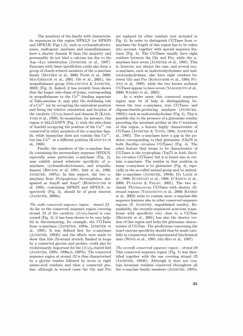
The sixth conserved sequence region – strand β2As far as the conserved sequence region coveringstrand β2 of the catalytic (β/α)8-barrel is con-cerned (Fig. 4), it has been shown to be very help-ful in discriminating, for example, the CGTasesfrom α-amylases (JANEČEK, 1994a, JANEČEK etal., 1995). It was defined first for α-amylases(JANEČEK, 1994b) and the efforts were made toshow that this β2-strand stretch, flanked in loopsby a conserved glycine and proline, could also beevolutionarily important for the (β/α)8-barrel fold(JANEČEK, 1995c, 1996a,b, 1997b). The conservedsequence region at strand β2 is thus characterisedby a glycine residue followed by seven or eightamino-acid residues and then a conserved pro-line, although in several cases the Gly and Pro
are replaced by other residues (not included inFig. 4). In order to distinguish CGTases from α-amylases the length of this region has to be takeninto account, together with special sequence fea-tures (Fig. 4). The CGTases usually have eightresidues between the Gly and Pro, while the α-amylases have seven (JANEČEK et al., 1995). Thisis, however, not always the case, and several exo-α-amylases, such as maltotriohydrolases and mal-totetraohydrolases, also have eight residues be-tween Gly and Pro (KOBAYASHI et al., 1994; FU-JITA et al., 1989), while the two known archaealCGTases appear to have seven (YAMAMOTO et al.,2000; RASHID et al., 2002).
In a wider sense this conserved sequenceregion may be of help in distinguishing be-tween the true α-amylases, true CGTases, andoligosaccharide-producing amylases (JANEČEK,1995b), such as maltotriohydrolase (Fig. 4). This ispossible due to the presence of a glutamine residuepreceding the invariant proline at the C-terminusof this region, a feature highly characteristic ofCGTases (JANEČEK & TÓTH, 1994; JANEČEK etal., 1995). The α-amylases have a gap in the po-sition corresponding to that glutamine (Gln78 inboth Bacillus circulans CGTases) (Fig. 4). Theother feature that seems to be characteristic ofCGTases is the tryptophan (Trp75 in both Bacil-lus circulans CGTases) but it is found also in cer-tain α-amylases. The residue in that position inmany α-amylases is be glutamine (Fig. 4), espe-cially in the so-called animal group and/or animal-like α-amylases (JANEČEK, 1994b; DA LAGE etal., 1998; SUMITANI et al., 1998; D’AMICO et al.,2000; PUJADAS & PALAU, 2001). The two ar-chaeal Thermococcus CGTases with shorter β2-strand regions (YAMAMOTO et al., 2000; RASHID
et al., 2002) seem to contain more α-amylase-likesequence features also in other conserved sequenceregions (Š. JANEČEK, unpublished results). Re-markably, the recently-sequenced acarviose trans-ferase with specificity very close to a CGTase(HEMKER et al., 2001) has also the shorter ver-sion of this region and lacks the glutamine charac-teristic of CGTase. The predictions concerning theexact enzyme specificity should thus be made care-fully in conjunction with experimental biochemicaldata (WIND et al., 1995; DEL-RIO et al., 1997).
The seventh conserved sequence region – strand β8This conserved sequence region (Fig. 5) was iden-tified together with the one covering strand β2(JANEČEK, 1994b). Although it does not con-tain invariant residues conserved throughout allthe α-amylase family members (JANEČEK, 1997a;
35
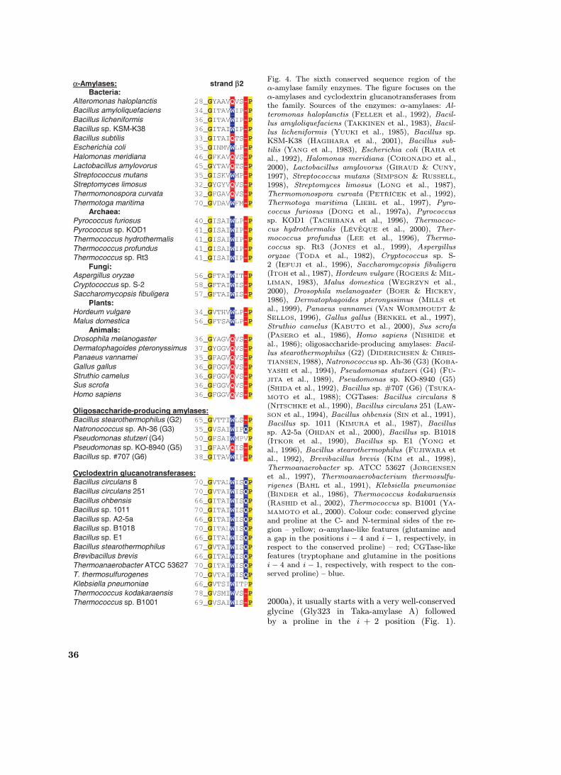
Fig. 4. The sixth conserved sequence region of theα-amylase family enzymes. The figure focuses on theα-amylases and cyclodextrin glucanotransferases fromthe family. Sources of the enzymes: α-amylases: Al-teromonas haloplanctis (FELLER et al., 1992), Bacil-lus amyloliquefaciens (TAKKINEN et al., 1983), Bacil-lus licheniformis (YUUKI et al., 1985), Bacillus sp.KSM-K38 (HAGIHARA et al., 2001), Bacillus sub-tilis (YANG et al., 1983), Escherichia coli (RAHA etal., 1992), Halomonas meridiana (CORONADO et al.,2000), Lactobacillus amylovorus (GIRAUD & CUNY,1997), Streptococcus mutans (SIMPSON & RUSSELL,1998), Streptomyces limosus (LONG et al., 1987),Thermomonospora curvata (PETŘÍČEK et al., 1992),Thermotoga maritima (LIEBL et al., 1997), Pyro-coccus furiosus (DONG et al., 1997a), Pyrococcussp. KOD1 (TACHIBANA et al., 1996), Thermococ-cus hydrothermalis (LÉVEQUE et al., 2000), Ther-mococcus profundus (LEE et al., 1996), Thermo-coccus sp. Rt3 (JONES et al., 1999), Aspergillusoryzae (TODA et al., 1982), Cryptococcus sp. S-2 (IEFUJI et al., 1996), Saccharomycopsis fibuligera(ITOH et al., 1987), Hordeum vulgare (ROGERS & MIL-LIMAN, 1983), Malus domestica (WEGRZYN et al.,2000), Drosophila melanogaster (BOER & HICKEY,1986), Dermatophagoides pteronyssimus (MILLS etal., 1999), Panaeus vannamei (VAN WORMHOUDT &SELLOS, 1996), Gallus gallus (BENKEL et al., 1997),Struthio camelus (KABUTO et al., 2000), Sus scrofa(PASERO et al., 1986), Homo sapiens (NISHIDE etal., 1986); oligosaccharide-producing amylases: Bacil-lus stearothermophilus (G2) (DIDERICHSEN & CHRIS-TIANSEN, 1988), Natronococcus sp. Ah-36 (G3) (KOBA-YASHI et al., 1994), Pseudomonas stutzeri (G4) (FU-JITA et al., 1989), Pseudomonas sp. KO-8940 (G5)(SHIDA et al., 1992), Bacillus sp. #707 (G6) (TSUKA-MOTO et al., 1988); CGTases: Bacillus circulans 8(NITSCHKE et al., 1990), Bacillus circulans 251 (LAW-SON et al., 1994), Bacillus ohbensis (SIN et al., 1991),Bacillus sp. 1011 (KIMURA et al., 1987), Bacillussp. A2-5a (OHDAN et al., 2000), Bacillus sp. B1018(ITKOR et al., 1990), Bacillus sp. E1 (YONG etal., 1996), Bacillus stearothermophilus (FUJIWARA etal., 1992), Brevibacillus brevis (KIM et al., 1998),Thermoanaerobacter sp. ATCC 53627 (JØRGENSEN
et al., 1997), Thermoanaerobacterium thermosulfu-rigenes (BAHL et al., 1991), Klebsiella pneumoniae(BINDER et al., 1986), Thermococcus kodakaraensis(RASHID et al., 2002), Thermococcus sp. B1001 (YA-MAMOTO et al., 2000). Colour code: conserved glycineand proline at the C- and N-terminal sides of the re-gion – yellow; α-amylase-like features (glutamine anda gap in the positions i − 4 and i − 1, respectively, inrespect to the conserved proline) – red; CGTase-likefeatures (tryptophane and glutamine in the positionsi − 4 and i − 1, respectively, with respect to the con-served proline) – blue.
2000a), it usually starts with a very well-conservedglycine (Gly323 in Taka-amylase A) followedby a proline in the i + 2 position (Fig. 1).
36
For some specificities it is not easy to identifythis region without information derived from thethree-dimensional structure (cf. JANEČEK, 2000a;PRZYLAS et al., 2000).
Nevertheless, it has already been demon-strated that this region may contain the featurescharacteristic of archaeal α-amylases (JANEČEK
et al., 1999). Another example is shown in Fig-ure 5 highlighting the features typical for ei-ther the “oligo-1,6-glucosidase” or the “neopul-lulanase” groups that were also discussed abovewith regard to the fifth conserved sequence re-gion (Fig. 3). These features are mainly the tyro-sine and aspartate (Tyr454 and Asp456 in neop-ullulanase in Fig. 5) at the C-terminal side ofthe region. They could be taken as characteris-tic residues of the neopullulanase group, while theoligo-1,6-glucosidase-like enzymes should containglutamine and glutamate, respectively, in the cor-responding positions (Fig. 5). Concerning the β8-strand region, amylosucrase, sucrose phosphory-lase, and trehalose synthase seem to be rathermore like neopullulanase members, although theyseem to be clear oligo-1,6-glucosidase-like enzymeswith regard to the fifth conserved sequence re-gion (cf. Figs 3,5). This is not surprising at allif the parts of their sequences between strandsβ7 and β8 are taken into account; these are dif-ferent from, and longer than, the equivalent seg-ments of oligo-1,6-glucosidase, α-glucosidase, dex-tran glucosidase and trehalose-6-phosphate hydro-lase (OSLANCOVÁ & JANEČEK, 2002). This obser-vation may also be related to the fact that the for-mer members are transferases while the later onesare hydrolases (MACGREGOR et al., 2001). Finallyone can imagine that many possible combinationsof the various characteristic motifs will actually bepossible and may occur in nature.
The non-catalytic proteins, related to the α-amylase family from the structural and evolution-ary points of view (JANEČEK et al., 1997; BRÖER
& WAGNER, 2002), have the segment coveringstrand β8 of the enzymatic members of the familyperfectly conserved in the corresponding positions(Fig. 5). This fact should be of interest becausethree of the four best-conserved sequence regionswith catalytic residues (strands β4, β5 and β7) areless well conserved (JANEČEK, 2000b).
Conclusion
According to our present knowledge, the α-amylase family consisting of families 13, 70 and77 of glycoside hydrolases (i.e. clan GH-H) cov-ers hydrolases, transferases, and isomerases (i.e.
Fig. 5. The seventh conserved sequence region ofthe α-amylase family enzymes. The figure focuses onthe so-called oligo-1,6-glucosidase and neopullulanasegroups of the family. Sources of the enzymes are thesame as in Figure 3, the one for the 4F2 heavy-chain antigen (GOTTESDIENER et al., 1988) beingadded. Colour code: conserved residues – yellow; oligo-1,6-glucosidase-like glutamine and glutamate – red;neopullulanase-like tyrosine and aspartate – blue.
the enzyme classes 3, 2 and 5). The amino acidtransporters and 4F2 heavy-chain antigens (i.e.non-enzymes) are also recognised as relatives ofthe family. It is possible that the main α-amylasefamily (clan GH-H) is related to the amylolyticfamily 57 of glycoside hydrolases, although the hi-erarchy will very probably be at a higher level (i.e.less strict common criteria) than a clan in termsof its definition. The members of the α-amylaseenzyme family act mainly on α-1,4- and α-1,6-glucosidic linkages; however, they also operate onα-1,1-, α-1,2-, α-1,3- and α-1,5-glucosidic bonds.Despite the presence of several conserved sequenceregions the invariant amino-acid residues are ex-tremely few, i.e. three catalytic residues in strandsβ4, β5 and β7 plus the arginine in position i − 2with regard to the β4-strand catalytic aspartate.
Concerning the number of conserved se-quence regions it could be postulated that: (i) each
37
enzyme specificity and/or even a distinct taxo-nomic group from the α-amylase family may haveits own conserved sequence regions and/or char-acteristic sequence similarities located in the cat-alytic (β/α)8-barrel domain and/or outside thisdomain; (ii) these regions may discriminate be-tween the α-amylase family members from thepoint of view of their specificity, structure andevolution; and (iii) all conserved sequence regionsmay not be common for the entire α-amylase fam-ily and they may indicate closer evolutionary re-latedness with functionally different enzymes andproteins.
If conserved sequence regions are considered,it can be concluded that the α-amylase family en-zymes should be characterised by 4, 5 and in somecases 7 conserved sequence regions, i.e. by as manyas possible. So if we go back to the question raisedin the title of this review: “How many conservedsequence regions are there in the α-amylase fam-ily?”, the exact answer is less important than thepractical use of the information characteristic ofspecific regions.
Acknowledgements
This work was supported in part by the VEGA GrantNo. 2/2057/22 from the Slovak Grant Agency.
References
ABE, J. I., SHIBATA, Y., FUJISUE, M. & HIZUKURI, S.1996. Microbiology 142: 1505–1512.
ABE, J. I., USHIJIMA, C. & HIZUKURI, S. 1999. Appl.Environ. Microbiol. 65: 4163–4170.
AMEMURA, A., CHAKRABORTY, R., FUJITA, M.,NOUMI, T. & FUTAI, M. 1988. J. Biol. Chem. 263:9271–9275.
BAHL, H., BURCHHARDT, G., SPREINAT, A., HAEC-KEL, K., WIENECKE, A., SCHMIDT, B. & ANTRA-NIKIAN, G. 1991. Appl. Environ. Microbiol. 57:1554–1559.
BEIER, L., SVENDSEN, A., ANDERSEN, C., FRANDSEN,T. P., BORCHERT, T. V. & CHERRY, J. R. 2000.Protein Eng. 13: 509–513.
BENKEL, B. F., NGUYEN, T., AHLUWALIA, N., BEN-KEL, I. & HICKEY, D. A. 1997. Gene 192: 261–270.
BERTRAN, J., WERNER, A., CHILLARON, J., NUNES,V., BIBER, J., TESTAR, X., ZORZANO, A., ES-TIVILL, X., MURER, H. & PALACIN, M. 1993. J.Biol. Chem. 268: 14842–14849.
BINDER, F., HUBER, O. & BÖCK, A. 1986. Gene 47:269–277.
BINDERUP, K. & PREISS, J. 1998. Biochemistry 37:9033–9037.
BOER, P. H. & HICKEY, D. A. 1986. Nucleic Acids Res.14: 8399–8411.
BOURNE, Y. & HENRISSAT, B. 2001. Curr. Opin.Struct. Biol. 11: 593–600.
BRÖER, S. & WAGNER, C. A. 2002. Cell Biochem. Bio-phys. 36: 155–168.
BRUMM, P. J., HEBEDA, R. E. & TEAGUE, W. M. 1991.Starch 43: 315–319.
CANDUSSIO, A., SCHMID, G. & BOCK, A. 1990. Eur.J. Biochem. 191: 177–185.
CORONADO, M. J., VARGAS, C., MELLADO, E., TE-GOS, G., DRAINAS, C., NIETO, J. J. & VENTOSA,A. 2000. Microbiology 146: 861–868.
COUTINHO, P. M. & HENRISSAT, B. 1999. Carbohy-drate-Active Enzymes server at URL: http://afmb.cnrs-mrs.fr/∼cazy/CAZY/index.html; accessedMarch 22, 2002.
DA LAGE, J. L., RENARD, E., CHARTOIS, F., LEMEU-NIER, F. & CARIOU, M. L. 1998. Proc. Natl. Acad.Sci. USA 95: 6848–6853.
D’AMICO, S., GERDAY, C. & FELLER, G. 2000. Gene253: 95–105.
DAUTER, Z., DAUTER, M., BRZOZOWSKI, A. M.,CHRISTENSEN, S., BORCHERT, T. V., BEIER, L.,WILSON, K. S. & DAVIES, G. J. 1999. Biochem-istry 38: 8385–8392.
DEL-RIO, G., MORETT, E. & SOBERON, X. 1997.FEBS Lett. 416: 221–224.
DEVULAPALLE, K. S., GOODMAN, S. D., GAO, Q.HEMSLEY, A. & MOOSER, G. 1997. Protein Sci.6: 2489–2493.
DIDERICHSEN, B. & CHRISTIANSEN, L. 1988. FEMSMicrobiol. Lett. 56: 53–60.
DONG, G., VIEILLE, C., SAVCHENKO, A. & ZEIKUS,J. G. 1997a. Appl. Environ. Microbiol. 63: 3569–3576.
DONG, G., VIEILLE, C. & ZEIKUS, J. G. 1997b. Appl.Environ. Microbiol. 63: 3577–3584.
ERRA-PUJADA, M., DEBEIRE, P., DUCHIRON, F. &O’DONOHUE, M. J. 1999. J. Bacteriol. 181: 3284–3287.
FELLER, G., LONHIENNE, T., DEROANNE, C., LIBI-OULLE, C., VAN BEEUMEN, J. & GERDAY, C. 1992.J. Biol. Chem. 267: 5217–5221.
FERRETTI, J. J., HUANG, T. T. & RUSSELL, R. R. B.1988. Infect. Immun. 56: 1585–1588.
FRIEDBERG, F. 1983. FEBS Lett. 152: 139–140.FRILLINGOS, S., LINDEN, A., NIEHAUS, F., VARGAS,
C., NIETO, J. J., VENTOSA, A., ANTRANIKIAN, G.& DRAINAS, C. 2000. J. Appl. Microbiol. 88: 495–503.
FUJITA, M., TORIGOE, K., NAKADA, T., TSUSAKI, K.,KUBOTA, M., SAKAI, S. & TSUJISAKA, Y. 1989. J.Bacteriol. 171: 1333–1339.
FUJIWARA, S., KAKIHARA, H., WOO, K. B., LEJEUNE,A., KANEMOTO, M., SAKAGUCHI, K. & IMANAKA,T. 1992. Appl. Environ. Microbiol. 58: 4016–4025.
FUKUSUMI, S., KAMIZONO, A., HORINOUCHI, S., &BEPPU, T. 1988. Eur. J. Biochem. 174: 15–21.
GIRAUD, E. & CUNY, G. 1997. Gene 198: 149–157.GOTTESDIENER, K. M., KARPINSKI, B. A., LIND-
STEN, T., STROMINGER, J. L., JONES, N. H.,
38

THOMPSON, C. B. & LEIDEN, J. M. 1988. Mol.Cell. Biol. 8: 3809–3819.
GOTTSCHALK, T. E., TULL, D., AGHAJARI, N.,HASER, R. & SVENSSON, B. 2001. Biochemistry40: 12844–12854
HAGIHARA, H., HAZASHI, Y., ENDO, K., IGARASHI,K., OZAWA, T., KAWAI, S., OZAKI, K. & ITO, S.2001. Eur. J. Biochem. 268: 3974–3982.
HEMKER, M., STRATMANN, A., GOEKE, K., SCHRÖ-DER, W., LENZ, J., PIEPERSBERG, W. & PAPE, H.2001. J. Bacteriol. 183: 4484–4492.
HENRISSAT, B. 1991. Biochem. J. 280: 309–316.HENRISSAT, B. & BAIROCH, A. 1996. Biochem. J. 316:
695–696.HONG, S. H. & MARMUR, J. 1986. Gene 41: 75–84.HORINOUCHI, S., FUKUSUMI, S., OHSHIMA, T. &
BEPPU, T. 1988. Eur. J. Biochem. 176: 243–253.IBUKA, A., TONOZUKA, T., MATSUZAWA, H. & SAKAI,
H. 1998. J. Biochem. 123: 275–282.IEFUJI, H., CHINO, M., KATO, M. & IIMURA, Y. 1996.
Biochem. J. 318: 989–996.IMAMURA, H., FUSHINOBU, S., JEON, B. S., WAK-
AGI, T. & MATSUZAWA, H. 2001a. Biochemistry40: 12400–12406.
IMAMURA, H., FUSHINOBU, S., YAMAMOTO, M., KU-MASAKA, T., WAKAGI, T. & MATSUZAWA, H.2001b. J. Appl. Glycosci. 48: 171–175.
ITKOR, P., TSUKAGOSHI, N. & UDAKA, S. 1990.Biochem. Biophys. Res. Commun. 166: 630–636.
ITOH, T., YAMASHITA, Y. & FUKUI, S. 1987. FEBSLett. 219: 339–342.
JANDA, L., DAMBORSKÝ, J., PETŘÍČEK, M., SPÍŽEK,J. & TICHÝ, P. 2000. J. Appl. Microbiol. 88: 773–783.
JANEČEK, Š. 1992. Biochem. J. 288: 1069–1070.JANEČEK, Š. 1994a. FEBS Lett. 353: 119–123.JANEČEK, Š. 1994b. Eur. J. Biochem. 224: 519–524.JANEČEK, Š. 1995a. FEBS Lett. 377: 6–8.JANEČEK, Š. 1995b. Biologia, Bratislava 50: 515–522.JANEČEK, Š. 1995c. Protein Sci. 4: 1239–1242.JANEČEK, Š. 1996a. Protein Sci. 5: 1136–1143.JANEČEK, Š. 1996b. Biochem. J. 319: 1005–1006.JANEČEK, Š. 1997a. Prog. Biophys. Mol. Biol. 67: 67–
97.JANEČEK, Š. 1997b. Int. J. Biol. Macromol. 21: 277–
280.JANEČEK, Š. 1998. Folia Microbiol. 43: 123–128.JANEČEK, Š. 2000a. In: OHNISHI, M., HAYASHI, T.,
ISHIJIMA, S. & KURIKI, T. (eds) Glycoenzymes,Japan Academic Societies Center, Tokyo, pp. 19–54.
JANEČEK, Š. 2000b. Trends Glycosci. Glycotechnol.12: 363–371.
JANEČEK, Š., LÉVEQUE, E., BELARBI, A. & HAYE, B.1999. J. Mol. Evol. 48: 421–426.
JANEČEK, Š., MACGREGOR, E. A. & SVENSSON, B.1995. Biochem. J. 305: 685–686.
JANEČEK, Š., SVENSSON, B. & HENRISSAT, B. 1997.J. Mol. Evol. 45: 322–331.
JANEČEK, Š. & TÓTH, D. 1994. Biologia, Bratislava49: 301–306.
JANSE, B. J. H., STEYN, A. J. C. & PRETORIUS, I. S.1993. Curr. Genet. 24: 400–407.
JEON, B. S., TAGUCHI, H., SAKAI, H., OHSHIMA,T., WAKAGI, T. & MATSUZAWA,H. 1997. Eur. J.Biochem. 248: 171–178.
JESPERSEN, H. M., MACGREGOR, E. A., HENRISSAT,B., SIERKS, M. R. & SVENSSON, B. 1993. J. Pro-tein Chem. 12: 791–805.
JESPERSEN, H. M., MACGREGOR, E. A., SIERKS, M.R. & SVENSSON, B. 1991. Biochem. J. 80: 51–55.
JONES, R. A., JERMIIN, L. S., EASTEAL, S., PATEL, B.K. & BEACHMAN, I. R. 1999. J. Appl. Microbiol.86: 93–107.
JØRGENSEN, S. T., TANGNEY, M., STARNES, R. L.,AMEMIYA, K. & JØRGENSEN, P. L. 1997. Biotech-nol. Lett. 19: 1027–1031.
KABUTO, S., OGAWA, T., MURAMOTO, K., OOST-HUIZEN ,V. & NAUDE, R. J. 2000. Comp. Biochem.Physiol. 127B: 481–490.
KADZIOLA, A., ABE, J. I., SVENSSON, B. & HASER,R. 1994. J. Mol. Biol. 239: 104–121.
KAMITORI, S., KONDO, S., OKUYAMA, K., YOKOTA,T., SHIMURA, Y., TONOZUKA, T. & SAKANO, Y.1999. J. Mol. Biol. 287: 907–921.
KATSUYA, Y., MEZAKI, Y., KUBOTA, M. & MAT-SUURA, Y. 1998. J. Mol. Biol. 281: 885–897.
KIM, J. S., CHA, S. S., KIM, H. J., KIM, T. J., HA, N.C., OH, S. T., CHO, H. S., CHO, M. J., KIM, M.J., LEE, H. S., KIM, J. W., CHOI, K. Y., PARK, K.H. & OH, B. H. 1999a. J. Biol. Chem. 274: 26279–26286.
KIM, M. H., SOHN, C. B. & OH, T. K. 1998. FEMSMicrobiol. Lett. 164: 411–418.
KIM, T. J., KIM, M. J., KIM, B. C., KIM, J. C.,CHEONG, T. K., KIM, J. W. & PARK, K. H. 1999b.Appl. Environ. Microbiol. 65: 1644–1951.
KIM, T. J., NGUYEN, V. D., LEE, H. S., KIM, M. J.,CHO, H. Y., KIM, Y. W., MOON, T. W., PARK, C.S., KIM, J. W., OH, B. H., LEE, S. B., SVENSSON,B. & PARK, K. H. 2001. Biochemistry 40: 14182–14190.
KIMURA, K., KATAOKA, S., ISHII, Y., TAKANO, T. &YAMANE, K. 1987. J. Bacteriol. 169: 4399–4402.
KIZAKI, H., HATA, Y., WATANABE, K., KATSUBE, Y.& SUZUKI, Y. 1993. J. Biochem. 113: 646–649.
KLEIN, C. & SCHULZ, G. E. 1991. J. Mol. Biol. 217:737–750.
KOBAYASHI, K., KATO, M., MIURA, Y., KETTOKU,M., KOMEDA, T. & IWAMATSU, A. 1996. Biosci.Biotech. Biochem. 60: 1882–1885.
KOBAYASHI, T., KANAI, H., AONO, R., HOROKOSHI,K. & KUDO, T. 1994. J. Bacteriol. 176: 5131–5134.
KURIKI, T. 1992. Trends Glycosci. Glycotechnol. 4:567–572.
KURIKI, T. & IMANAKA, T. 1989. J. Gen. Microbiol.135: 1521–1528.
KURIKI, T. & IMANAKA, T. 1999. J. Biosci. Bioeng.87: 557–565.
KURIKI, T., KANEKO, H., YANASE, M., TAKATA, H.,SHIMADA, J., HANDA, S., TAKADA, T., UMEYAMA,
39
H. & OKADA, S. 1996. J. Biol. Chem. 271: 17321–17329.
LADERMAN, K. A., ASADA, K., UEMORI, T., MUKAI,H., TAGUCHI, Y., KATO, I., & ANFINSEN, C. B..1993. J. Biol. Chem. 268: 24402–24407.
LAWSON, C. L., VAN MONTFORT, R., STROKOPYTOV,B., ROZEBOOM, H. J., KALK, K. H., DE VRIES, G.E., PENNINGA, D., DIJKHUIZEN, L. & DIJKSTRA,B. W. 1994. J. Mol. Biol. 236: 590–600.
LEE, J. T., KANAI, H., KOBAYASHI, T., AKIBA, T. &KUDO, T. 1996. J. Ferment. Bioeng. 82: 432–438.
LEEMHUIS, H., DIJKSTRA, B. W. & DIJKHUIZEN, L.2002. FEBS Lett. 514: 189–192.
LÉVEQUE, E., HAYE, B. & BELARBI, A. 2000. FEMSMicrobiol. Lett. 186: 67–71.
LIEBL, W., STEMPLINGER, I. & RUILE, P. 1997. J.Bacteriol. 179: 941–948.
LONG, C. M., VIROLLE, M. J., CHANG, S. Y., CHANG,S. & BIBB, M. J. 1987. J. Bacteriol. 169: 5745–5754.
MACGREGOR, E. A. 1988. J. Protein Chem. 7: 399–415.
MACGREGOR, E. A. 1993. Starch 7: 232–237.MACGREGOR, E. A., JANEČEK, Š. & SVENSSON, B.
2001. Biochim. Biophys. Acta 1546: 1–20.MACGREGOR, E. A., JESPERSEN, H. M. & SVENSSON,
B. 1996. FEBS Lett. 378: 263–266.MACGREGOR, E. A. & SVENSSON, B. 1989. Biochem.
J. 259: 145–152.MACHIUS, M., WIEGAND, G. & HUBER, R. 1995. J.
Mol. Biol. 246: 545–559.MATSUI, I. & SVENSSON, B. 1997. J. Biol. Chem. 272:
22456–22463.MATSUURA, Y., KUSUNOKI, M., HARADA, W. &
KAKUDO, M. 1984. J. Biochem. 95: 697–702.MATZKE, J., HERRMANN, A., SCHNEIDER, E. &
BAKKER, E. P. 2000. FEMS Microbiol. Lett. 183:55–61.
MEISSNER, H. & LIEBL, W. 1998. Eur. J. Biochem.250: 1050–1058.
METZ, R. J., ALLEN, L. N., CAO, T. M. & ZEMAN, N.W. 1988. Nucelic Acids Res. 16: 5203.
MILLS, K. L., HART, B. J., LYNCH, N. R., THOMAS, W.R. & SMITH, W. 1999. Int. Arch. Allergy Immunol.120: 100–107.
MONCHOIS, V., LAKEY, J. H. & RUSSELL, R. R. B.1999. FEMS Microbiol. Lett. 177: 243–248.
MONCHOIS, V., VIGNON, M., ESCALIER, P. C., SVENS-SON, B. & RUSSELL, R. R. B. 2000. Eur. J.Biochem. 267: 4127–4136.
MORI, H., BAK-JENSEN, K. S., GOTTSCHALK, T. E.,MOTAWIA, M. S., DAMAGER, I., MOLLER, B. L. &SVENSSON, B. 2001. Eur. J. Biochem. 268: 6545–6558.
MORISHITA, Y., HASEGAWA, K., MATSUURA, Y.,KATSUBE, Y., KUBOTA, M. & SAKAI, S. 1997. J.Mol. Biol. 267: 661–672.
NAKAJIMA, R., IMANAKA, T. & AIBA, S. 1986. Appl.Microbiol. Biotechnol. 23: 355–360.
NAKAYAMA, A., YAMAMOTO, K. & TABATA, S. 2001.J. Biol. Chem. 276: 28824–28828.
NIELSEN, J. E. & BORCHERT, T. V. 2000. Biochim.Biophys. Acta 1543: 253–274.
NISHIDE, T., NAKAMURA, Y., EMI, M., YAMAMOTO,T., OGAWA, M., MORI, T. & MATSUBARA, K.1986. Gene 41: 299–304.
NITSCHKE, L., HEEGER, K., BENDER, H. & SCHULZ,G. E. 1990. Appl. Microbiol. Biotechnol. 33: 542–546.
OH, B. H., KIM, J. S., CHO, H. S., KIM, T. J., LEE, H.S. & PARK, K. H. 2001. In: The First Symposiumon the Alpha-Amylase Family, Smolenice Castle,Slovakia, Sep 30 – Oct 4, 2001, p. 45.
OHDAN, K., KURIKI, T., TAKATA, H. & OKADA, S.2000. Appl. Microbiol. Biotechnol. 53: 430–434.
OSLANCOVÁ, A. & JANEČEK, Š. 2002. Cell. Mol. LifeSci. (in press).
PARK, K. H., KIM, T. J., CHEONG, T. K., KIM, J.W., OH, B. H. & SVENSSON, B. 2000. Biochim.Biophys. Acta 1478: 165–185.
PASERO, L., MAZZÉI-PIERRON, Y., ABADIE, B., CHI-CHEPORTICHE, Y. & MARCHIS-MOUREN, G. 1986.Biochim. Biophys. Acta 869: 147–157.
PETŘÍČEK, M., TICHÝ, P. & KUNCOVÁ, M. 1992. Gene112: 77–83.
PODKOVYROV, S. M. & ZEIKUS, J. G. 1992. J. Bacte-riol. 174: 5400–5405.
POTOCKI DE MONTALK, G., REMAUD-SIMEON, M.,WILLEMOT, R.M., PLANCHOT, V. & MONSAN, P.1999. J. Bacteriol. 181: 375–381.
PRZYLAS, I., TOMOO, K., TERADA, Y., TAKAHA, T.,FUJII, K., SAENGER, W. & STRÄTER, N. 2000. J.Mol. Biol. 296: 873–886.
PUJADAS, G. & PALAU, J. 2001. Mol. Biol. Evol. 18:38–54.
QIAN, M., HASER, R. & PAYAN, F. 1993. J. Mol. Biol.231: 785–799.
RAHA, M., KAWAGISHI, I., MUELLER, V., KIHARA,M., MACNAB, R. M. 1992. J. Bacteriol. 174: 6644–6652.
RASHID, N., CORNISTA, J., EZAKI, S., FUKUI, T.,ATOMI, H. & IMANAKA, T. 2002. J. Bacteriol. 184:777–784.
RIMMELE, M. & BOOS, W. 1994. J. Bacteriol. 176:5654–5664.
ROGERS, J. C. 1985. Biochem. Biophys. Res. Commun.128: 470–476.
ROGERS, J. C. & MILLIMAN, C. 1983. J. Biol. Chem.258: 8169–8174.
ROMEO, T., KUMAR, A. & PREISS, J. 1988. Gene 70:363–376.
ROUJEINIKOVA, A., RAASCH, C., BURKE, J., BAKER,P. J., LIEBL, W & RICE, D. W. 2001. J. Mol. Biol.312: 119–131.
RUSSELL, R. R. B. & FERRETTI, J. J. 1990. J. Gen.Microbiol. 136: 803–810.
SAAB-RINCÓN, G., DEL-RÍO, G., SANTAMARÍA, R. I.,LÓPEZ-MUNGUÍA, A. & SOBERÓN, X. 1999. FEBSLett. 453: 100–106.
SARCABAL, P., REMAUD-SIMEON, M., WILLEMOT, R.,POTOCKI DE MONTALK, G., SVENSSON, B. &MONSAN, P. 2000. FEBS Lett. 474: 33–37.
40
SHIDA, O., TAKANO, T., TAKAGI, H., KADOWAKI, K.& KOBAYASHI, S. 1992. Biosci. Biotech. Biochem.56: 76–80.
SHIROKIZAWA, O., AKIBA, T. & HORIKOSHI, K. 1990.FEMS Microbiol. Lett. 70: 131–136.
SIMPSON, C. L. & RUSSELL, R. R. B. 1998. J. Bacte-riol. 180: 4711–4717.
SIN, K. A., NAKAMURA, A., KOBAYASHI, K., MASAKI,H. & UOZUMI, T. 1991. Appl. Microbiol. Biotech-nol. 35: 600–605.
SKOV, L. K., MIRZA, O., HENRIKSEN, A., DE MON-TALK, G. P., REMAUD-SIMEON, M., SARCABAL,P., WILLEMOT, R. M., MONSAN, P. & GAJHEDE,M. 2001. J. Biol. Chem. 276: 25273–25278.
SUMITANI, J. I., NAGAE, H., KAWAGUCHI, T. & ARAI,M. 1998. J. Ferment. Bioeng. 85: 428–432.
SVENSSON, B. 1988. FEBS Lett. 230: 72–76.SVENSSON, B. 1994. Plant Mol. Biol. 25: 141–157.SVENSSON, B., TOVBORG JENSEN, M., MORI, H.,
SASS BAK-JENSEN, K., BØNSAGER, B., NIELSEN,P. K., KRAMHØFT, B., PRÆTORIUS-IBBA, M.,NØHR, J., JUGE, N., GREFFE, L., WILLIAMSON,G. & DRIGUEZ, H. 2002. Biologia, Bratislava 57(Suppl. 11): 5–19.
TACHIBANA, Y., MENDEZ LECLERE, M., FUJIWARA,S., TAKAGI, M. & IMANAKA, T. 1996. J. Ferment.Bioeng. 82: 224–232.
TAKATA, H., KURIKI, T., OKADA, S., TAKESADA, Y.,IIZUKA, M., MINAMIURA, N. & IMANAKA, T. 1992.J. Biol. Chem. 267: 18447–18452.
TAKKINEN, K., PETTERSSON, R. F., KALKKINEN, N.,PALVA, I., SÖDERLUND, H. & KÄÄRIÄINEN, L.1983. J. Biol. Chem. 258: 1007–1013.
TERADA, Y., FUJII, K., TAKAHA, T. & OKADA, S.1999. Appl. Environ. Microbiol. 65: 910–915.
TODA, H., KONDO, K., & NARITA, K. 1982. Proc.Japan Acad. B58: 208–212.
TONOZUKA, T., OHTSUKA, M., MOGI, S. I., SAKAI,H., OHTA, T. & SAKANO, Y. 1993. Biosci. Biotech.Biochem. 57: 395–401.
TSAI, Y.-W., CHIA, J.-S., SHIAU, Y.-Y., CHOU, H.-C.,LIAW, Y.-C., LOU, K.-L. 2000. FEMS Microbiol.Lett. 188: 75–79.
TSUKAMOTO, A., KIMURA, K., ISHII, Y., TAKANO, T.& YAMANE, Y. 1988. Biochem. Biophys. Res. Com-mun. 151: 25–31.
TSUSAKI, K., NISHIMOTO, T., NAKADA, T., KUBOTA,M., CHAEN, H., FUKUDA, S., SUGIMOTO, T. & KU-RIMOTO, M. 1997. Biochim. Biophys. Acta 1334:28–32.
TSUSAKI, K., NISHIMOTO, T., NAKADA, T., KUBOTA,M., CHAEN, H., SUGIMOTO, T. & KURIMOTO, M.1996. Biochim. Biophys. Acta 1290: 1–3.
UITDEHAAG, J. C. M., VAN DER VEEN, B. A., DI-JKHUIZEN, L. & DIJKSTRA, B. W. 2002. EnzymeMicrob. Technol. 30: 295–304.
VAN DER MAAREL, M. J., VAN DER VEEN, B., UIT-DEHAAG, J. C., LEEMHUIS, H. & DIJKHUIZEN, L.2002. J. Biotechnol. 94: 137–155.
VAN DER VEEN, B. A., UITDEHAAG, J. C., DIJKSTRA,B. W. & DIJKHUIZEN, L. 2000. Biochim. Biophys.Acta 1543: 336–360.
VAN LIESHOUT, J. F. T., VERHEES, C. H., VAN
DER OOST, J. & DE VOS, W. M. 2001. In: TheFirst Symposium on the Alpha-Amylase Family,Smolenice Castle, Slovakia, Sep 30 – Oct 4, 2001,p. 26.
VAN WORMHOUDT, A., & SELLOS, D. 1996. J. Mol.Evol. 42: 543–551.
VIHINEN, M. & MÄNTSÄLÄ, P. 1989. Crit. Rev.Biochem. Mol. Biol. 24: 329–418.
WATANABE, K., KITAMURA, K., IHA, H. & SUZUKI,Y. 1990. Eur. J. Biochem. 192: 609–620.
WEGRZYN, T., REILLY, K., CIPRIANI, G., MURPHY,P., NEWCOMB, R., GARDNER, R. & MACRAE, E.2000. Eur. J. Biochem. 267: 1313–1322.
WIND, R. D., BUITELAAR, R. M. & DIJKHUIZEN, L.1998. Eur. J. Biochem. 253: 598–605.
WIND, R, D., LIEBL, W., BUITELAAR, R. M., PEN-NINGA, D., SPREINAT, A., DIJKHUIZEN, L., BAHL,H. 1995. Appl. Environ. Microbiol. 61: 1257–1265.
YAMAMOTO, T., FUJIWARA, S., TACHIBANA, Y., TAK-AGI, M., FUKUI, K. & IMANAKA, T. 2000. J. Biosci.Bioeng. 89: 206–209.
YANG, M., GALIZZI, A. & HENNER, D. 1983. Nucl.Acids Res. 11: 237–249.
YONG, J., CHOI, J. N., PARK, S. S., PARK, C. S., PARK,K. H. & CHOI, Y. D. 1996. Biotechnol. Lett. 18:1223–1228.
YUUKI, T., NOMURA, T., TEZUKA, H., TSUBOI, A.,YAMAGATA, H., TSUKAGOSHI, N. & UDAKA, S.1985. J. Biochem. 98: 1147–1156.
ZHANG, D., LI, X & ZHANG, L. H. 2002. Appl. Environ.Microbiol. 68: 2676–2682.
ZHOU, J., BABA, T., TAKANO, T., KOBAYASHI, S. &ARAI, Y. 1989. FEBS Lett. 255: 37–41.
Received May 7, 2002Accepted May 31, 2002
41