
Horseradish Peroxidase: Modulation of Properties by ...
13
Peroxidases (EC 1.11.1.X) are enzymes of the oxi doreductase class that catalyze the oxidation of a wide range of substrates using hydrogen peroxide. Their molecular weight ranges from 17 to 84 kDa and the polypeptide chain from 153 to 753 amino acids. The pro teins usually include 1011 αhelices, while βsheets are absent or rare. Most peroxidases consist of two structural domains with a heme, ferriprotoporphyrin IX prosthetic group, in a hydrophobic pocket (Fig. 1) [1]. The generally accepted classification proposed in 1992 [2] divides peroxidases into two large superfamilies, animal peroxidases and plant peroxidases. The superfam ily of plant peroxidases is further divided into three class es. This system initially based on amino acid sequence comparison was later confirmed by Xray data obtained for representatives of different peroxidase classes. The first class of plant peroxidases includes intracel lular enzymes such as ascorbate peroxidase, yeast cytochrome c, and bacterial catalaseperoxidase. These proteins are not glycosylated and contain neither disulfide bonds nor calcium ions. The second class includes secret ed fungal peroxidases. The beststudied representatives of this class are lignin peroxidase and manganese peroxidase involved in lignin oxidation as well as peroxidase from Coprinus cinereus (previously known as Arthromyces ramosus). These proteins are monomeric glycoproteins with four disulfide bonds and two conserved calcium binding domains. The third class is represented by secret ed plant peroxidases. These enzymes are also monomeric glycoproteins with four disulfide bonds and two calcium binding sites; however, the arrangement of the disulfide bonds and the protein chain folding differs between per oxidases of the second and third classes. Horseradish peroxidase isozyme C (HRP) is clearly the most popular plant peroxidase. Currently, it is the model enzyme of the third class of plant peroxidases. Although other peroxidases can outperform it in many parameters, HRP finds the widest application in labora tory work. Data obtained over dozens of years allow this enzyme to be used in the development of immune tests [3, 4], various biosensors [57], and histological markers [8] as well as in gene therapy [9, 10] and organic synthesis [11, 12]. The studies of the mechanism of HRP action target ed to the improvement or modification of its properties are ISSN 00062979, Biochemistry (Moscow), 2011, Vol. 76, No. 13, pp. 13911401. © Pleiades Publishing, Ltd., 2011. Published in Russian in Uspekhi Biologicheskoi Khimii, 2011, Vol. 51, pp. 3764. REVIEW 1391 Abbreviations: ABTS, 2,2′azinobis(3ethylbenzothiazoline6 sulfonate); BFR, bifunctional reagent; EH, ethylhydrazine; HRP, horseradish peroxidase isozyme C; MFR, monofunc tional reagent; MH, methylhydrazine; nHRP, native HRP; PEH, phenylethylhydrazine; PHZ, phenylhydrazine; rsHRP, reconstructed HRP; SASA, solvent accessible surface area; SHD, synthetic heme derivative. * To whom correspondence should be addressed. Horseradish Peroxidase: Modulation of Properties by Chemical Modification of Protein and Heme G. S. Zakharova 1,2 , I. V. Uporov 3 , and V. I. Tishkov 1,2,3 * 1 Bach Institute of Biochemistry, Russian Academy of Sciences, Leninsky pr. 33, 119071 Moscow, Russia; Email: [email protected] 2 Innovations and High Technologies MSU Ltd., 109559 Moscow, Russia 3 Chemical Faculty, Lomonosov Moscow State University, 119992 Moscow, Russia Received June 24, 2011 Revision received July 7, 2011 Abstract—Horseradish peroxidase (HRP) is one of the most studied enzymes of the plant peroxidase superfamily. HRP is also widely used in different bioanalytical applications and diagnostic kits. The methods of genetic engineering and protein design are now widely used to study the catalytic mechanism and to improve properties of the enzyme. Here we review the results of another approach to HRP modification—through the chemical modification of amino acids or prosthetic group of the enzyme. Computer models of HRPs with modified hemes are in good agreement with the experimental data. DOI: 10.1134/S0006297911130037 Key words: horseradish peroxidase, HRP, heme, chemical modification, catalytic properties, stability, computer modeling
Transcript of Horseradish Peroxidase: Modulation of Properties by ...
Peroxidases (EC 1.11.1.X) are enzymes of the oxi�
doreductase class that catalyze the oxidation of a wide
range of substrates using hydrogen peroxide. Their
molecular weight ranges from 17 to 84 kDa and the
polypeptide chain from 153 to 753 amino acids. The pro�
teins usually include 10�11 α�helices, while β�sheets are
absent or rare. Most peroxidases consist of two structural
domains with a heme, ferriprotoporphyrin IX prosthetic
group, in a hydrophobic pocket (Fig. 1) [1].
The generally accepted classification proposed in
1992 [2] divides peroxidases into two large superfamilies,
animal peroxidases and plant peroxidases. The superfam�
ily of plant peroxidases is further divided into three class�
es. This system initially based on amino acid sequence
comparison was later confirmed by X�ray data obtained
for representatives of different peroxidase classes.
The first class of plant peroxidases includes intracel�
lular enzymes such as ascorbate peroxidase, yeast
cytochrome c, and bacterial catalase�peroxidase. These
proteins are not glycosylated and contain neither disulfide
bonds nor calcium ions. The second class includes secret�
ed fungal peroxidases. The best�studied representatives of
this class are lignin peroxidase and manganese peroxidase
involved in lignin oxidation as well as peroxidase from
Coprinus cinereus (previously known as Arthromyces
ramosus). These proteins are monomeric glycoproteins
with four disulfide bonds and two conserved calcium�
binding domains. The third class is represented by secret�
ed plant peroxidases. These enzymes are also monomeric
glycoproteins with four disulfide bonds and two calcium�
binding sites; however, the arrangement of the disulfide
bonds and the protein chain folding differs between per�
oxidases of the second and third classes.
Horseradish peroxidase isozyme C (HRP) is clearly
the most popular plant peroxidase. Currently, it is the
model enzyme of the third class of plant peroxidases.
Although other peroxidases can outperform it in many
parameters, HRP finds the widest application in labora�
tory work. Data obtained over dozens of years allow this
enzyme to be used in the development of immune tests [3,
4], various biosensors [5�7], and histological markers [8]
as well as in gene therapy [9, 10] and organic synthesis
[11, 12].
The studies of the mechanism of HRP action target�
ed to the improvement or modification of its properties are
ISSN 0006�2979, Biochemistry (Moscow), 2011, Vol. 76, No. 13, pp. 1391�1401. © Pleiades Publishing, Ltd., 2011.
Published in Russian in Uspekhi Biologicheskoi Khimii, 2011, Vol. 51, pp. 37�64.
REVIEW
1391
Abbreviations: ABTS, 2,2′�azino�bis(3�ethylbenzothiazoline�6�
sulfonate); BFR, bifunctional reagent; EH, ethylhydrazine;
HRP, horseradish peroxidase isozyme C; MFR, monofunc�
tional reagent; MH, methylhydrazine; n�HRP, native HRP;
PEH, phenylethylhydrazine; PHZ, phenylhydrazine; rs�HRP,
reconstructed HRP; SASA, solvent accessible surface area;
SHD, synthetic heme derivative.
* To whom correspondence should be addressed.
Horseradish Peroxidase: Modulation of Propertiesby Chemical Modification of Protein and Heme
G. S. Zakharova1,2, I. V. Uporov3, and V. I. Tishkov1,2,3*
1Bach Institute of Biochemistry, Russian Academy of Sciences, Leninsky pr. 33,
119071 Moscow, Russia; E�mail: [email protected] and High Technologies MSU Ltd., 109559 Moscow, Russia
3Chemical Faculty, Lomonosov Moscow State University, 119992 Moscow, Russia
Received June 24, 2011
Revision received July 7, 2011
Abstract—Horseradish peroxidase (HRP) is one of the most studied enzymes of the plant peroxidase superfamily. HRP is
also widely used in different bioanalytical applications and diagnostic kits. The methods of genetic engineering and protein
design are now widely used to study the catalytic mechanism and to improve properties of the enzyme. Here we review the
results of another approach to HRP modification—through the chemical modification of amino acids or prosthetic group
of the enzyme. Computer models of HRPs with modified hemes are in good agreement with the experimental data.
DOI: 10.1134/S0006297911130037
Key words: horseradish peroxidase, HRP, heme, chemical modification, catalytic properties, stability, computer modeling

1392 ZAKHAROVA et al.
BIOCHEMISTRY (Moscow) Vol. 76 No. 13 2011
Fig. 1. Structure of heme and its chemically modified analogs. Compounds 1�4 are products of the catalytic oxidation of different substrates:
1, phenylhydrazine [87]; 2a, phenylethylhydrazine; 2b, ethylhydrazine; 2c, methylhydrazine [88]; 3, sodium azide [89]; 4, potassium cyanide
[90]. Compounds 5a and 5b are products of the incubation in acetate buffer (pH 4.4) or other acids (n�caproic and phenylacetic acids) at sim�
ilar pH [91]. Compounds 6�9 are chemically modified protoporphyrin analogs [97, 98, 101, 102].
heme

MODULATION OF PROPERTIES OF HORSERADISH PEROXIDASE 1393
BIOCHEMISTRY (Moscow) Vol. 76 No. 13 2011
still actively performed largely based on directed mutage�
nesis. This review presents and analyzes the data relying
on a different approach, the chemical modification of the
enzyme amino acids or the heme porphyrin ring.
BRIEF DESCRIPTION OF HORSERADISH
PEROXIDASE
The roots of horseradish (Armoracia rusticana) con�
tain many different peroxidases, but isozyme C has the
greatest share among them. The isoelectric point (pI) of
HRP is 9, and its activity peaks at pH 6�8. The HRP
molecular weight is about 44 kDa, 18�22% of which cor�
responds to the carbohydrate moiety of the protein.
Commonly, eight out of nine asparagine carbohydrate
linkage sites (Asn13, 57, 158, 186, 198, 214, 255, and
268) are glycosylated [13]. The carbohydrate moiety
composition varies depending on many factors such as
growth stage and conditions. Oligosaccharides have a sta�
bilizing effect on the enzyme. They protect the polypep�
tide chain from proteolysis and modification by free rad�
icals generated in the reaction [14�17].
The three�dimensional structure of HRP remained
unknown for long, until a recombinant enzyme was pro�
duced [18], since all attempts to crystallize the native
HRP failed due to its heterogeneous glycosylation. The
recombinant unglycosylated enzyme was isolated from E.
coli cells expressing an HRP gene synthesized on the basis
of the amino acid sequence determined by Karen
Welinder [19]. To date, the three�dimensional structures
of six other plant peroxidases from peanut [20], barley
[21], Arabidopsis thaliana [22], soybean [23], tobacco (I.
G. Gazaryan, personal communication), and royal palm
tree [24] have been determined.
The X�ray data demonstrated that the HRP polypep�
tide chain of 308 amino acids folds into 13 α�helices and
three β�sheets [25]. The proper protein conformation is
stabilized by four disulfide bonds (Cys11–Cys91,
Cys44–Cys49, Cys97–Cys301, and Cys177–Cys209).
The protein consists of two domains, distal and proximal,
each containing a calcium�binding site where the cations
are held by seven coordinate bonds. The structural signif�
icance of Ca2+ has been confirmed by numerous experi�
mental data [26�32] and computer simulations [33].
Since the calcium�binding sites are linked to the active
site by a hydrogen�bonding system, Ca2+ depletion
induces significant changes in the enzyme properties [31].
The distal cation has a weaker bond than the proximal
one [1]. Its removal roughly halves the catalytic activity
and decreases HRP thermostability, stability at extreme
pH, and storage stability. The distal Ca2+ removal induces
minor changes on the enzyme three�dimensional struc�
ture [28], while complete calcium depletion inactivates
the enzyme due to significant conformational changes in
the active site [29�31].
The HRP active site contains heme, a prosthetic
group that consists of a ferric iron complex with proto�
porphyrin IX (Fig. 1). The Fe3+ ion has four coordinate
bonds within the heme and one with the nitrogen atom of
the imidazole ring in the proximal His170. The sixth
coordinate position remains vacant in the relaxed state of
the enzyme, but during catalysis, hydrogen peroxide
binds to the iron atom to form a six�coordinate complex
[34].
HRP can oxidize a wide range of substrates; typical
substrates include phenols, indoles, aromatic amines, and
sulfonates. The oxidation of aromatic substrates leads to
their polymerization, which can be used to purify water
from aromatic pollutants [35, 36].
The HRP catalytic cycle includes the following
stages:
HRP + H2O2 → Compound I + H2O (1)
Compound I + AH2 → Compound II + AH• (2)
Compound II + AH2 → HRP + AH•, (3)
where AH2 is an electron donor substrate and AH• is the
radical reaction product.
In the first stage, two electrons are removed from the
enzyme (from the iron atom and from the porphyrin ring)
as a result of its interaction with hydrogen peroxide. This
yields a cation radical called Compound I. Reduction of
Compound I by an electron donor to the initial ferri�
enzyme proceeds in two stages through the formation of
Compound II. Thus, the substrate conversion follows a
ping�pong mechanism (Compound I reacts with the first
substrate molecule to yield Compound II, which conse�
quently reacts with the second substrate molecule). The
catalytic mechanism is considered in more detail in
reviews [15, 37�39].
MUTAGENIC STUDIES OF HORSERADISH
PEROXIDASE
This highly efficient approach allows us to identify
the amino acids critical for the catalysis, structure main�
tenance, or stability of the enzyme. It can be used to gen�
erate enzymes with improved stability [40, 41] or altered
substrate specificity [42]. Such studies underlie practical
generation of enzymes with desired properties. In our lab,
this approach has been applied to generate biocatalysts
with unique properties from formate dehydrogenase and
D�amino acid oxidase [43�47].
Amino acid substitutions can be introduced into an
enzyme by random (directed evolution, genetic mosaic,
etc.) and directed mutagenesis. The latter is often referred
to as rational design of proteins considering that point
substitutions are introduced after the analysis of enzyme

1394 ZAKHAROVA et al.
BIOCHEMISTRY (Moscow) Vol. 76 No. 13 2011
amino acid sequences or structures. In the case of HRP,
rational design is largely used since it is expressed in
insoluble form in E. coli cells and a refolding from the
inclusion bodies is requires to make the enzyme active
[18]. Frances Arnold and colleagues tried directed evolu�
tion to introduce point substitutions into HRP that allow
active enzyme expression in E. coli cells [48] and methy�
lotrophic [49] and baker’s yeast [50]. However, the exper�
iments were not successful in E. coli, while hyperglycosy�
lation took place in the yeasts: the molecular weight of the
carbohydrate moiety could reach 60 kDa, while it is as
low as 10 kDa in the natural enzyme.
HRP directed mutagenesis has been applied to elu�
cidate the role of amino acids in heme binding in the dis�
tal part of the active site, to study the mechanisms of
interaction with H2O2 and catalytic substrate oxidation,
to identify potential sites for binding different substrate
types, and to evaluate the role of calcium cations in the
folding and catalytic activity of HRP. The major conclu�
sions of these experiments include: (1) distal His42 and
Arg38 play the key role in H2O2 binding and heterolytic
cleavage of the O–O bond [51�53]; (2) Phe41 is involved
in the heme coordination, controls the ferryl oxygen
accessibility in Compound I, and modulates the sub�
strate specificity [51, 54�57]; (3) hydrogen bonds
between His42, Asn70, and Glu64 connect the distal cal�
cium�binding site and the active site of the enzyme.
Apparently, this is required for the proper orientation of
the His42 imidazole ring relative to porphyrin [58�60];
(4) the strength of heme binding in the active site
depends on the coordinate bond between the heme iron
and proximal His170 [61]; (5) the role of Asp247 can
consist of making His170 more basic, which maintains
the five�coordinate state of heme [25]; (6) Phe179 and
Phe142 are critical for binding aromatic substrates [62�
64]; (7) Trp117 mediates the energy migration in the
molecule and has an effect on the enzyme folding and
stability [65, 66]; (8) Thr171 is a factor of the enzyme
proper conformation since it connects heme and proxi�
mal calcium�binding site [67]; (9) unstructured regions
of the polypeptide chain are an important factor of the
enzyme stability. Amino acid substitutions in these
regions (Thr102Ala, Gln106Arg, Gln107Asp,
Thr110Val, and Ile180Phe) decreased the enzyme ther�
mostability [68]; (10) substitutions of Asn13 and Asn268
(glycosylated in native HRP) with Asp increases the
enzyme thermostability and resistance to inactivation by
hydrogen peroxide [17].
A detailed description of the introduced point muta�
tions and their effects can be found in reviews [15, 37�39].
Thus, genetic engineering tools open up many
opportunities to produce an enzyme with new properties;
however, mutations are not the only way to modify
enzyme properties. Chemical modifications can be
equally effective. This approach can be exemplified by the
modification of solvent�exposed amino acids.
CHEMICAL MODIFICATION
OF PROTEIN MOIETY
Chemical modification experiments were conducted
on both native horseradish peroxidase (n�HRP) with the
oligosaccharide moiety and recombinant unglycosylated
enzyme. The n�HRP was used in most cases since it
allowed the enzyme to be immobilized and conjugated to
other proteins (antibodies in particular) through the car�
bohydrate moiety. In addition, the glycosylated enzyme is
more stable and accessible.
Chemical modification of His [69�71], Tyr, Arg,
Asp, and Glu [72] either had no effect or deteriorated the
enzyme properties. Only two out of three His residues
(40, 42, and 170) proved accessible for the modification,
since His170 participates in the heme coordination [71].
The modification of His42 rendered the enzyme inactive;
however, it could bind aromatic substrates and form
Compound I [70, 71]. These results support the key role
of His42 in the energy transfer from the substrate to the
prosthetic group.
Positive results were only obtained after lysine
residues were modified; accordingly, lysines became the
most common target [73�84]. Six lysines in the HRP
molecule (Lys65, 84, 149, 174, 232, and 241 [19])
demonstrated different susceptibility to modification.
Lys232 proved the most reactive; Lys241 and Lys174 were
less reactive; while Lys65, Lys84, and Lys149 are hardly
modified [77]. These data agree with other publications
demonstrating that there are three modified lysines per
HRP molecule [75, 77, 78, 80�84].
Both bifunctional and monofunctional reagents
(BFRs and MFRs) were used to modify Lys residues (Table
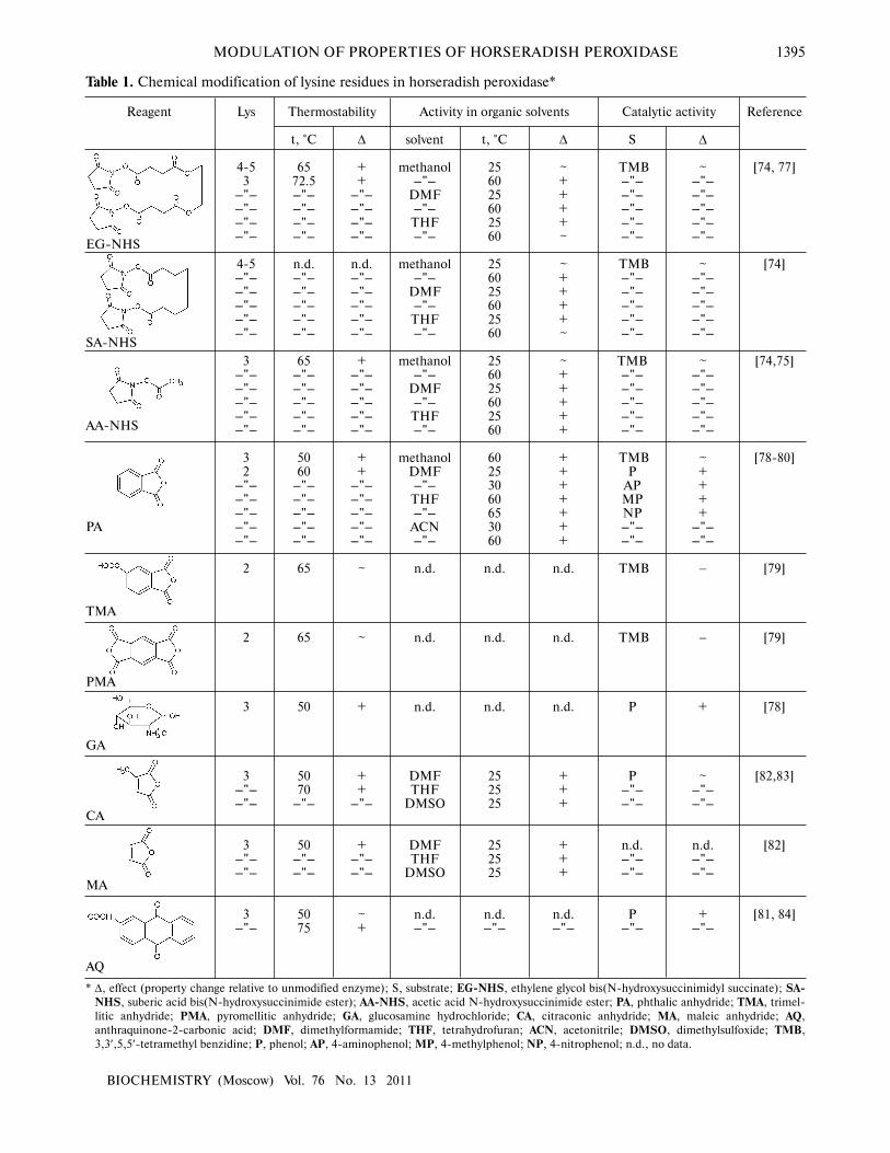
1). Experimental data indicate that the exposure to BFRs
had a stabilizing effect. For instance, the inactivation rate
of EG�NHS�HRP (hereafter, the left part of the enzyme
abbreviation refers to the modification agent; Table 1) at
65°C was four times less than that of the native enzyme
[77]. EG�NHS�HRP and SA�NHS�HRP were also more
stable in dimethylformamide (DMF), methanol, and
tetrahydrofuran (THF), while the catalytic activity of the
modified and native enzymes remained similar.
The modification of HRP by MFRs was also suc�
cessful. AA�NHS�HRP was five times more stable at 65°C
and much more active in organic solvents (methanol,
DMF, and THF) than n�HRP [75]. The exposure to
phthalic anhydride (PA) [78�80], glucosamine
hydrochloride (GA) [78], and anthraquinone�2�carbonic
acid (AQ) [81, 84] generated not only more stable but also
more catalytically active (with high kcat/Km) enzymes in
oxidation reactions with phenol and its derivatives (4�
amino�, 4�methyl�, and 4�nitrophenol). The AQ modifi�
cation improved both the catalytic (kcat) and Michaelis
(Km) constants of the enzyme reaction with phenol. In
addition, AQ proved to be an effective mediator of elec�
tron transfer between peroxidase and electrode, which
MODULATION OF PROPERTIES OF HORSERADISH PEROXIDASE 1395
BIOCHEMISTRY (Moscow) Vol. 76 No. 13 2011
Reagent
EG�NHS
SA�NHS
AA�NHS
PA
TMA
PMA
GA
CA
MA
AQ
Lys
4�53
��"����"����"����"��
4�5��"����"����"����"����"��
3��"����"����"����"����"��
32
��"����"����"����"����"��
2
2
3
3��"����"��
3��"����"��
3��"��
t, °C
6572.5��"����"����"����"��
n.d.��"����"����"����"����"��
65��"����"����"����"����"��
5060
��"����"����"����"����"��
65
65
50
5070
��"��
50��"����"��
5075
∆
++
��"����"����"����"��
n.d.��"����"����"����"����"��
+��"����"����"����"����"��
++
��"����"����"����"����"��
~
~
+
++
��"��
+��"����"��
~+
solvent
methanol��"��
DMF��"��THF��"��
methanol��"��
DMF��"��THF��"��
methanol��"��
DMF��"��THF��"��
methanolDMF��"��THF��"��ACN��"��
n.d.
n.d.
n.d.
DMFTHF
DMSO
DMFTHF
DMSO
n.d.��"��
t, °C
256025602560
256025602560
256025602560
60253060653060
n.d.
n.d.
n.d.
252525
252525
n.d.��"��
∆
~++++~
~++++~
~+++++
+++++++
n.d.
n.d.
n.d.
+++
+++
n.d.��"��
S
TMB��"����"����"����"����"��
TMB��"����"����"����"����"��
TMB��"����"����"����"����"��
TMBP
APMPNP��"����"��
TMB
TMB
P
P��"����"��
n.d.��"����"��
P��"��
∆
~��"����"����"����"����"��
~��"����"����"����"����"��
~��"����"����"����"����"��
~++++
��"����"��
−
−
+
~��"����"��
n.d.��"����"��
+��"��
Reference
[74, 77]
[74]
[74,75]
[78�80]
[79]
[79]
[78]
[82,83]
[82]
[81, 84]
* ∆, effect (property change relative to unmodified enzyme); S, substrate; EG�NHS, ethylene glycol bis(N�hydroxysuccinimidyl succinate); SA�NHS, suberic acid bis(N�hydroxysuccinimide ester); AA�NHS, acetic acid N�hydroxysuccinimide ester; PA, phthalic anhydride; TMA, trimel�
litic anhydride; PMA, pyromellitic anhydride; GA, glucosamine hydrochloride; CA, citraconic anhydride; MA, maleic anhydride; AQ,
anthraquinone�2�carbonic acid; DMF, dimethylformamide; THF, tetrahydrofuran; ACN, acetonitrile; DMSO, dimethylsulfoxide; TMB,
3,3′,5,5′�tetramethyl benzidine; P, phenol; AP, 4�aminophenol; MP, 4�methylphenol; NP, 4�nitrophenol; n.d., no data.
Thermostability
Table 1. Chemical modification of lysine residues in horseradish peroxidase*
Activity in organic solvents Catalytic activity

1396 ZAKHAROVA et al.
BIOCHEMISTRY (Moscow) Vol. 76 No. 13 2011
increased the overall efficiency of the process. The cat�
alytic activity of PA�HRP was studied both in aquatic
solution and in the presence of DMF. In aqueous solu�
tion, the enzyme demonstrated a higher catalytic activity
(kcat) towards all above�mentioned substrates and a lower
Km compared to n�HRP. In DMF solution, PA�HRP
1/Km was higher (except for 4�nitrophenol), while the
catalytic constant was slightly lower. Nevertheless, the
efficiency of PA�HRP (kcat/Km) was higher than that of n�
HRP. The thermostability of PA�HRP was also substan�
tially higher: about 80% activity was observed after 90�
min incubation at 50°C. The half�life time (τ1/2) of the
modified enzyme at 50°C was 10 times that of n�HRP
(four times at 65°C). The resistance to organic solvents
(methanol, DMF, ACN, and THF) also increased.
A similar stability increase was observed in the
enzymes modified by citraconic and maleic anhydrides
(CA�HRP and MA�HRP, respectively) [82, 83]. Their
stability at room temperature increased in dimethylsulf�
oxide (DMSO), THF, and DMF. The half�life time of the
generated enzymes at 70°C increased roughly three�fold,
and their activity after 3�h incubation at 50°C was 82, 74,
and 30% of baseline for MA�HRP, CA�HRP, and n�HRP,
respectively. Since no significant changes in the ther�
mostability of TMA�HRP and PMA�HRP were observed,
their further analysis was not pursued [79].
Thus, two different approaches to surface chemical
modification of HRP based on BFRs and MFRs can be
recognized. In the first case, intramolecular cross�links
are formed. It was found that there is a single cross�link
per HRP molecule in its proximal domain (between
Lys232 and Lys241); at the same time, Lys174 is also sub�
ject to modification. These results agree with previous
data [77] on the accessibility of lysine amino groups for
the modification and on their mutual arrangement that
delimits cross�links between them by bifunctional agents
of definite lengths. In this case, the stabilizing effect can
be attributed to an increased rigidity of the molecule
around the proximal calcium�binding domain, which can
interfere with the dissociation of calcium ion, an impor�
tant structural factor [77].
The following consequences of HRP modification
by MFRs can be recognized: (1) neutralization of the
positive lysine residues (e.g. the enzyme acetylation by
AA�NHS [74]), and (2) Lys charge reversion to negative
(e.g. phthalic anhydride modification [78�80]). The
reagents mediating electron transfer (such as
anthraquinone�2�carbonic acid [81, 84]) should be men�
tioned specifically, since the main modification target in
this case is to increase the efficiency of electron transfer
between HRP and electrode rather than to increase the
enzyme stability. In terms of enzyme stability improve�
ment, the highest effect was observed when a substituent
introduced a single negative charge per modified lysine
residue. In the case of the enzyme modification by TMAand PMA, the number of introduced negative charges per
lysine residue increased to two and three, respectively;
however, no stabilizing effect was observed. Some authors
believe that changes in the electrostatic interaction pat�
tern of the enzyme can improve its stability [77, 79, 83].
Thus, chemical modification of lysine residues can
substantially improve the properties of horseradish perox�
idase. However, both mutagenesis and chemical modifi�
cation of amino acids affect only the protein moiety of the
enzyme. An alternative approach is to introduce changes
to the heme prosthetic group.
CHEMICAL MODIFICATION
OF PROSTHETIC GROUP
Heme is bound non�covalently in peroxidase, and
there are routine methods for heme removal for produc�
tion of the apoenzyme as well as for reconstruction of the
holoenzyme from the apoenzyme and heme [85, 86]. The
latter method is actively used in the refolding of recombi�
nant HRP from inclusion bodies [57]. The same tech�
niques were applied to study the impact of porphyrin
modification on the properties of the peroxidase.
Two trends can be recognized in the experiments on
heme chemical modification: (1) studies on the mecha�
nism of protoporphyrin modifications resulting from its
interaction with radical reaction products (autocatalytic
modifications) and the impact of these modifications on
the enzyme properties; and (2) directed chemical modifi�
cation of heme to study the properties of HRP with mod�
ified protoporphyrin. Figure 1 shows the heme structure
and its modification variants studied by different
researchers. Hereafter, the numbers of the presented pro�
toporphyrin derivatives are used to abbreviate their com�
plexes with the apoenzyme.
Compounds 1�5 in Fig. 1 result from the autocat�
alytic heme modification [87�91], and most of them are
δ�meso�heme derivatives (the δ�meso�carbon in the
Fischer nomenclature corresponds to C20 in the IUPAC
system). Introducing substituents into this position usual�
ly irreversibly inactivates the enzyme. The reaction with
methylhydrazine (MH) is the exception: the enzyme with
δ�meso�methylheme as the prosthetic group (compound
2c; Fig. 1) purified from the substrates and reaction prod�
ucts by gel filtration gradually restored the catalytic activ�
ity, although the rate of the enzyme reactivation was not
specified [88].
The heme�modifying radicals formed in the catalyt�
ic reaction should also interact with the protein moiety,
which can also be a factor of HRP inactivation. Chemical
modification of the polypeptide chain has been experi�
mentally confirmed by introducing radioactive labels to
the corresponding substrates. The proportion between the
modifications in the protoporphyrin and protein moiety
proved to substantially depend on the substrate type. For
instance, the radicals largely attack the protein moiety

MODULATION OF PROPERTIES OF HORSERADISH PEROXIDASE 1397
BIOCHEMISTRY (Moscow) Vol. 76 No. 13 2011
rather than the prosthetic group when phenylhydrazine
(PHZ) was used as the substrate [87]. An opposite pattern
was observed when phenylethylhydrazine (PEH), ethyl�
hydrazine (EH), methylhydrazine (MH) [88], or azide
ion [89] was used as the substrate. In order to separate the
effects of protein moiety and prosthetic group modifica�
tions on the enzyme inactivation, heme was isolated from
modified HRPs and incorporated into unmodified
apoenzyme. Such reconstructed enzymes proved to have
low or no activity [90, 91]. Hence, the heme modification
is one of the main factors of HRP inactivation. On the
other hand, these data indicate the heme accessibility in
the C18�C20 region and the significance of this region for
the enzyme activity.
It is of interest to note the following typical features
of the autocatalytic modification. In all cases, the rate of
enzyme inactivation directly increased with the substrate
concentration; however, the highest activity loss was
reached at different enzyme/substrate ratios: 11�13 PHZ
molecules, 11 PEH molecules, 32 EH molecules, or 80
MH molecules per enzyme molecule. At the
azide/enzyme ratio of 60 : 1, the residual activity of the
enzyme was 20%. A further increase in the ratio demon�
strated a minor improvement. A decrease of the residual
activity to 5% of baseline required a 600 molar excess of
azide over the enzyme. A complete inactivation of the
enzyme by azide (as well as by PHZ) was observed only
after its purification by gel chromatography and repeated
incubation with azide and hydrogen peroxide. The
authors attributed this effect to an unidentified reaction
product that binds the enzyme and protects it from the
radical attack [87, 89].
Acetate ion is also capable of modifying HRP in the
presence of hydrogen peroxide. For instance, the residual
activity of HRP was 5.6 ± 3.4% after incubating the
enzyme in 50 mM acetate buffer (pH 4.4) with a 150
molar excess of H2O2 for 20 min [91]. A more thorough
investigation of the process demonstrated that the heme
modification was accompanied by its degradation. Direct
degradation of intact heme was observed as well.
Accordingly, high acetate concentrations and a short�
term incubation were used to maximize the yield of the
modified enzyme. Similar results were obtained for n�
caproic or phenylacetic acids instead of acetate, while
incubation in citrate buffer yielded no modified proto�
porphyrin [91]. Thus, heme modification was observed
only with alkyl carbonic acids.
It is of importance that heme�containing proteins
feature a band in the absorption spectrum around 403 nm
(Soret band). The changes in the intensity and position of
the Soret band peak reflect the changes in the enzyme
structure in the active site region [92]. In all cases of heme
autocatalytic modification mentioned above (except
acetylation), the activity loss was accompanied by a
notable decrease in the Soret band intensity and its shift
to the red.
Similar experiments were performed with other
heme�containing enzymes and proteins. For instance,
reaction with phenylhydrazine inactivated cytochrome P�
450 [93], myoglobin [94], and catalase [95, 96]. Catalase
was also inactivated by azide ion. However, no heme chem�
ical modification took place in these cases. Instead, the
iron atom reacted with the radicals, and the enzyme inhi�
bition resulted from the formation of a stable six�coordi�
nate complex, which prevented its interaction with H2O2.
Evaluation of the solvent accessible surface area
(SASA) demonstrates that nearly all heme in HRP is
screened from solvent molecules by the protein moiety
except for the C18�C20 region (SASA equals 3.2 and
~2.4 Å2 for C18 and C20, respectively). This indicates that
the HRP active site is closed for relatively large molecules,
which prevents their interaction with the iron atom.
Thus, these data on autocatalytic modification sug�
gest the following conclusions: (1) HRP heme is embed�
ded in a narrow crevice, which prevents even relatively
small molecules to enter in the crevice; (2) the only rela�
tively open protoporphyrin region (δ�edge of heme) is
subject to radical attack and, hence, modifications; (3)
this process inactivates the enzyme.
Let us now consider the second group of heme�mod�
ified enzymes generated by the incorporation of synthet�
ic heme derivatives (SHDs) into the apoenzyme. Their
structures are shown in Fig. 1 (compounds 6�9). All these
SHDs were produced by chemical modification of heme
propionic groups and differ only by the nature of the sub�
stituent. Reconstructed HRPs (rs�HRPs) were obtained
using the standard method (with minor modifications,
e.g. increased incubation time) [97, 102]. SHD incuba�
tion with the apoenzyme led to its incorporation into the
protein and restored the HRP catalytic activity. The addi�
tion of unmodified heme to the rs�HRP solution did not
increase the enzyme activity any more, which confirms
the complete SHD incorporation into the enzyme
crevice, while similar absorption spectra of rs�HRP and
n�HRP confirms the process specificity.
Introduction of a substituent into heme changed
both the protein structure and the enzyme properties
compared to n�HRP. The molecule was compacted in
most cases (excluding 6�rs�HRP where no notable
changes were observed, Table 2; hereafter, the number in
the designations of HRPs with modified heme corre�
sponds to the number of heme structure in Fig. 1). The
compaction can explain the higher thermal stability of 8�and 9�rs�HRP than that of n�HRP: the half�life time τ1/2
increased 2�4 times [97, 98]. In addition to the higher
thermostability, the stability of the enzyme in organic sol�
vents increased in some cases. The stability of 8�rs�HRP
in acetonitrile, dimethylsulfoxide, and methanol was
much higher than that of n�HRP (this pattern was less
pronounced for 8b�rs�HRP). 9a�, 9b�, and 9c�rs�HRP
were more stable in dimethylformamide (DMF) solution
at 60°C [98].
1398 ZAKHAROVA et al.
BIOCHEMISTRY (Moscow) Vol. 76 No. 13 2011
Introduction of a substituent into the heme also
modified the spectral properties of the enzymes. The flu�
orescence of the protein is due to Trp, Tyr, and Phe
residues. The HRP molecule contains a single tryptophan
(Trp117), five tyrosines, and 20 phenylalanines [19]. At
the excitation wavelength of 295 nm, tryptophan fluores�
cence predominates in the fluorescence spectrum of the
enzyme [99]. Tryptophan fluorescence in n�HRP peaks at
328 nm. The fluorescence spectrum of HRP depends on
excitation energy migration from Trp117 to heme.
Hence, changes in the enzyme structure in the heme�
binding region that affect the Trp117 distance to or orien�
tation relative to the heme plane as well as changes in the
tryptophan environment modify its emission spectrum
[100]. Reconstructed enzymes with SHDs demonstrate
increased fluorescence intensity and peak shift to the red.
This further confirms the modified structure of rs�HRPs,
which is most likely mediated by an increased heme–Trp
distance and increased polarity of the tryptophan envi�
ronment [97, 98].
In all cases, the catalytic activity of rs�HRPs differed
from that of n�HRP. For instance, the activity towards
guaiacol, thiocyanate, and iodide substantially decreased
in the case of 6�rs�HRP. At the same time, the capacity to
bind guaiacol remained similar to that of n�HRP, while
the dissociation constant of the complex with thiocyanate
decreased 20 times [101]. 7a�rs�HRP demonstrated no
catalytic activity towards 2,2′�azino�bis(3�ethylbenzo�
thiazoline�6�sulfonate) (ABTS) and ferrocenes. At the
same time, 7b�rs�HRP activity increased towards fer�
rocenes but decreased towards ABTS threefold compared
to n�HRP, while their ability to bind ABTS remained
unaltered. The authors attribute the changed substrate
specificity to the emergence of a new binding site includ�
ing ferrocene [102]. Decreased Km was observed for 8a�and 8b�rs�HRP in the oxidation of phenol and its para�
derivatives (p�hydroxybenzoic acid and p�aminophenol)
[97], which can be due to the enhanced hydrophobic
interactions between aromatic substrates and benzene
ring of the substituent in the R2 position (Fig. 1).
SHDs resulting from the esterification of para�deriv�
atives of phenol with carboxyl groups of heme had differ�
ent impacts on the enzyme catalytic properties. 9a�rs�
HRP activity and affinity to phenol increased, while the
catalytic properties of 9b� and 9c�rs�HRP degraded.
These changes are attributed to different donor–acceptor
properties of the substituents: electron acceptors (p�
nitrophenol) decrease the electron density of the por�
phyrin ring, thus accelerating reduction of Compound II
to the native enzyme [98].
Thus, experiments on the production of reconstruct�
ed horseradish peroxidase with modified heme suggest
the following conclusions: (1) incorporation of SHD into
the apoenzyme efficiently proceeds even when bulky sub�
stituents are used for the modification of the propionic
groups; (2) the produced reconstructed enzymes retain
more or less catalytic activity (which can substantially
outperform that of n�HRP towards certain substrates);
(3) the catalytic properties of HRP can be modulated by
variation of substituents introduced into the heme.
COMPUTER SIMULATION OF HRP STRUCTURE
WITH MODIFIED HEME DERIVATIVES
To understand the mechanism and effects of changes
in the properties of peroxidase with modified heme deriv�
atives, we performed computer simulations of the three�
dimensional structures of the apoenzyme–modified
heme complexes. Below is what we consider the most
interesting results. The simulations were generated using
the Builder module of the Insight II software package
(Accelrys, USA). The starting HRP structure was
obtained from PDB:1ATJ. Builder tools were used to add
extra chemical groups. The simulation was visually ana�
lyzed to identify too close contacts between atoms of
introduced substituents and neighboring amino acids. If
any, torsion angles of side chains of amino acids were var�
ied to move the atoms apart to an acceptable distance
(this procedure was applied to Pro141 in 1a� and 2a�HRP
and Arg31 in 9a�, 9b�, and 9c�rs�HRP). Then the struc�
ture was subjected to 500 steps of energy minimization
using the Discover�3 module of Insight II to bring it to
equilibrium. Energy minimization is justified when the
Table 2. Content of secondary structures in wild�type HRP and HRPs reconstructed from apoenzyme and modified
heme
Enzyme
α�Helices
β�Sheets
Loops
WT
37
13
49
6* [101]
32.4
17.6
50
a
���
���
���
b
���
���
���
a
55
9
36
b
44
12
44
a
50
10
40
b
41
13
45
c
61
7
32
7* [102] 8* [97] 9* [98]
* The numbers of synthetic heme derivatives correspond to Fig. 1.
MODULATION OF PROPERTIES OF HORSERADISH PEROXIDASE 1399
BIOCHEMISTRY (Moscow) Vol. 76 No. 13 2011
initial structural disturbance can be minimized by minor
atomic shifts in a local area of the protein. Sometimes the
structural disturbance was too high to be eliminated by
energy minimization. For instance, the benzene ring in
R1 intersected with Pro141 in 1a�HRP, and this defect
could not be removed in this way. In such cases, stepwise
simulation was performed: a methyl group was introduced
into R1; the structure was subjected to the minimization;
the methyl group was replaced with a benzene ring (in the
resulting structure, the close contacts between atoms of
benzene ring and amino acids allowed energy minimiza�
tion); and the structure was subjected to the minimiza�
tion. Molecular dynamics was used at the next step to
bring the protein structure to further equilibrium. The
standard molecular dynamics protocol was applied at
constant temperature (T = 300 K) in vacuum (two calci�
um cations and bound water molecules were added to the
system). The integration time step was 1 fsec and the total
simulation time was 20 psec. The CVFF force field was
used in all calculations [103]. In all cases, the most signif�
icant structural changes took place within the first 5 psec,
which indicates the stability of the resulting structures.
Figure 2 (see color insert) presents the results of the
1a�HRP simulation. The initial and simulated structures
are shown in light and dark colors, respectively. One can
see the most pronounced structural changes in the region
of amino acids 140 to 143. The introduction of the ben�
zene substituent into the heme brings this group to the
place occupied by Pro141 in the native enzyme. The steric
stress in the modified enzyme structure was released by a
small porphyrin ring turning relative to the Fe–His170
axis and heme sliding deeper into the crevice. Phe68 and
Phe142 involved in aromatic substrate binding as well as
catalytic His42 and Arg38 critical for hydrogen peroxide
degradation were also affected. Experimental data indi�
cate that 1a�HRP reacted with H2O2 to form Compound
I, but it was not reduced to Compound II after an elec�
tron�donor substrate was added. The computer simulation
agrees well with the previous suggestion [87] that the
absence of 1a�HRP catalytic activity towards guaiacol is
due to steric restrictions for the heme–substrate contact.
In the case of 2a�HRP (Fig. 3; see color insert)
where the benzene ring is not so rigidly connected to pro�
toporphyrin, the shift of amino acids 140�143 is less pro�
nounced. In addition, the close contact between the
introduced substituents and Phe141 can be avoided with�
out heme shift. Otherwise the pattern is similar to the pre�
vious case, and the inactivation of the enzyme is also like�
ly due to the substrate block of the active site by the δ�
meso�substituent.
The structure of the complex between the dimethyl
ester of protoporphyrin and apoenzyme (6�rs�HRP) gen�
erated by computer simulation is nearly identical to that
of the recombinant enzyme (data not shown). However,
spectral analysis indicates structural changes in the heme�
binding region: the Soret band in the reconstructed per�
oxidase shifted 6 nm to the long wavelength side and its
intensity decreased [101]. In addition, the enzyme
demonstrated low activity towards both aromatic sub�
strates (e.g. guaiacol) and small two�electron donors (e.g.
iodide ion) [101]. The authors attribute the observed
decrease in the catalytic activity to the changed heme
position in the enzyme crevice as a result of disturbed
hydrogen bonding network between the protein moiety
and esterified carboxyl groups. In addition, the neutral�
ization of their negative charges by the modification can
decrease the efficiency of hydrogen peroxide reduction,
thus, decelerating the formation of Compound I. The
kinetic constants were not determined for either catalytic
stage in [101], hence further studies are needed to explain
the low activity of 6�rs�HRP.
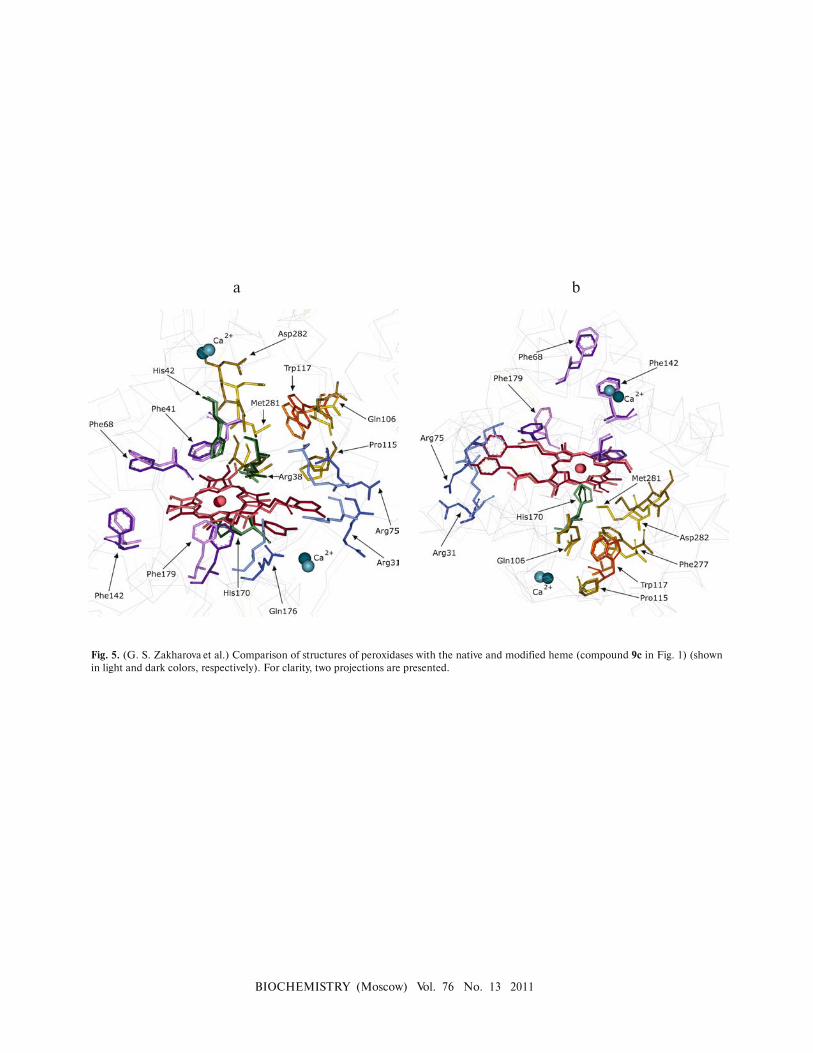
The introduction of bulky substituents into R2 and R3
leads to notable conformational changes in the molecule.
The position of amino acids adjacent to the modified pro�
pionic groups of heme (such as Arg31, Arg75, Asp174,
Gln176, and Leu218) significantly changes. The sub�
stituents “push apart” these amino acids to free enough
space in the resulting crevice. At the same time, the por�
phyrin ring position does not substantially change (in the
case of 9a�rs�HRP, heme slides slightly deeper into the
globule (Fig. 4a; see color insert); in the case of 9c�rs�
HRP, the porphyrin ring slightly turns around the
Fe–His170 axis (Fig. 5; see color insert)). The changes in
the position of Phe68 and Phe142 are noticeable; however,
these amino acids have high B�factor values, which explain
their high mobility in molecular dynamics simulations.
The active site region (Arg38, His42, and Phe41) remained
largely unchanged. Experimental data on enhanced fluo�
rescence of the tryptophan relative to n�HRP indicate
more pronounced structural changes in 9c�rs�HRP than
in 9a�rs�HRP [98]. Indeed, Figs. 4 and 5 demonstrate that
the difference in the position of amino acids around
Trp117 between the modified and unmodified enzymes is
more pronounced in 9c�rs�HRP in the simulations.
As mentioned above, the methods of genetic engi�
neering and protein design prevail in studying the mech�
anism of enzyme action and production of biocatalysts
with desired properties. However, well�known methods of
chemical modification based on the structural data still
remain relevant. Sometimes these methods make it possi�
ble to produce enzymes with properties that cannot be
obtained using genetic engineering. This can be clearly
demonstrated by HRP derivatives modified by
anthraquinone�2�carbonic acid, which mediates electron
transport from the active site to the electrode. In practice,
such products can be used to generate reagentless third�
generation biosensors. The computer simulation results
for the structure of HRP with chemically modified heme
are in a good agreement with the experimental data.
Thus, computer simulations can be used not only to
explain data but also to design experiments for heme
modification.

BIOCHEMISTRY (Moscow) Vol. 76 No. 13 2011
Fig. 2. (G. S. Zakharova et al.) Comparison of structures of per�
oxidases with the native and modified heme (compound 1a in
Fig. 1) (shown in light and dark colors, respectively).
Fig. 3. (G. S. Zakharova et al.) Comparison of structures of per�
oxidases with the native and modified heme (compound 2a in Fig.
1) (shown in light and dark colors, respectively).
Fig. 4. (G. S. Zakharova et al.) Comparison of structures of peroxidases with the native and modified heme (compound 9a in Fig. 1) (shown
in light and dark colors, respectively). For clarity, two projections are presented.
a b
BIOCHEMISTRY (Moscow) Vol. 76 No. 13 2011
Fig. 5. (G. S. Zakharova et al.) Comparison of structures of peroxidases with the native and modified heme (compound 9c in Fig. 1) (shown
in light and dark colors, respectively). For clarity, two projections are presented.
a b

1400 ZAKHAROVA et al.
BIOCHEMISTRY (Moscow) Vol. 76 No. 13 2011
This work was supported by the Russian Foundation
for Basic Research (project No. 10�04�00887a).
REFERENCES
1. Banci, L. (1997) J. Biotechnol., 53, 253�263.
2. Welinder, K. G. (1992) Curr. Opin. Struct. Biol., 2, 388�
393.
3. Zhang, S., Zou, J., and Yu, F. (2008) Talanta, 76, 122�127.
4. Song, Z., Yuan, R., Chai, Y., Zhuo, Y., Jiang, W., Su, H.,
Che, X., and Li, J. (2010) Chem. Commun. (Camb.), 46,
6750�6752.
5. Yao, H., and Hu, N. (2010) J. Phys. Chem. B, 114, 3380�
3386.
6. Shan, D., Li, Q. B., Ding, S. N., Xu, J. Q., Cosnier, S., and
Xue, H. G. (2010) Biosens. Bioelectron., 26, 536�541.
7. Korkut, S., Keskinler, B., and Erhan, E. (2008) Talanta,
76, 1147�1152.
8. Li, J., Wang, Y., Chiu, S. L., and Cline, H. T. (2010) Front.
Neural Circuits, 4, 6.
9. Greco, O., Rossiter, S., Kanthou, C., Folkes, L. K.,
Wardman, P., Tozer, G. M., and Dachs, G. U. (2001) Mol.
Cancer Ther., 1, 151�160.
10. Tupper, J., Stratford, M. R., Hill, S., Tozer, G. M., and
Dachs, G. U. (2010) Cancer Gene Ther., 17, 420�428.
11. Shogren, R. L., Willett, J. L., and Biswas, A. (2009)
Carbohydr. Polym., 75, 189�191.
12. Cruz�Silva, R., Amaro, E., Escamilla, A., Nicho, M. E.,
Sepulveda�Guzman, S., Arizmendi, L., Romero�Garcia,
J., Castillon�Barraza, F. F., and Farias, M. H. (2008) J.
Colloid Interface Sci., 328, 263�269.
13. Wuhrer, M., Balog, C. I. A., Koeleman, C. A. M., Deelder,
A. M., and Hokke, C. H. (2005) Biochim. Biophys. Acta,
1723, 229�239.
14. Clarke, J., and Shannon, L. M. (1976) Biochim. Biophys.
Acta, 427, 428�442.
15. Veitch, N. C. (2004) Phytochemistry, 65, 249�259.
16. Tams, J. W., and Welinder, K. G. (1998) FEBS Lett., 421,
234�236.
17. Asad, S., Khajeh, K., and Ghaemi, N. (2010) Appl.
Biochem. Biotechnol., 164, 454�463.
18. Smith, A. T., Santama, N., Dacey, S., Edwards, M., Bray,
R. C., Thorneley, R. N. F., and Burke, J. F. (1990) J. Biol.
Chem., 265, 13335�13343.
19. Welinder, K. G. (1979) Eur. J. Biochem., 96, 483�502.
20. Schuller, D. J., Ban, N., Huystee, R. B., McPherson, A.,
and Poulos, T. L. (1996) Structure, 4, 311�321.
21. Henriksen, A., Welinder, K. G., and Gajhede, M. (1998) J.
Biol. Chem., 273, 2241�2248.
22. Ostergaard, L., Teilum, K., Mirza, O., Mattsson, O.,
Petersen, M., Welinder, K. G., Mundy, J., Gajhede, M.,
and Henriksen, A. (2000) Plant. Mol. Biol., 44, 231�243.
23. Henriksen, A., Mirza, O., Indiani, C., Teilum, K.,
Smulevich, G., Welinder, K. G., and Gajhede, M. (2001)
Protein Sci., 10, 108�115.
24. Watanabe, L., de Moura, P. R., Bleicher, L., Nascimento,
A. S., Zamorano, L. S., Calvete, J. J., Sanz, L., Perez, A.,
Bursakov, S., Roig, M. G., Shnyrov, V. L., and Polikarpov,
I. (2010) J. Struct. Biol., 169, 226�242.
25. Gajhede, M., Schuller, D. J., Henriksen, A., Smith, A. T.,
and Poulos, T. L. (1997) Nat. Struct. Biol., 4, 1032�1038.
26. Haschke, R. H., and Friedhoff, J. M. (1978) Biochem.
Biophys. Res. Commun., 80, 1039�1042.
27. Ogawa, S., Shiro, Y., and Morishima, I. (1979) Biochem.
Biophys. Res. Commun., 90, 674�678.
28. Howes, B. D., Feis, A., Raimondi, L., Indiani, C., and
Smulevich, G. (2001) J. Biol. Chem., 276, 40704�40711.
29. Laberge, M., Huang, Q., Schweitzer�Stenner, R., and
Fidy, J. (2003) Biophys. J., 84, 2542�2552.
30. Laberge, M., Szigeti, K., and Fidy, J. (2004) Biopolymers,
74, 41�45.
31. Szigeti, K., Smeller, L., Osvath, S., Majer, Z., and Fidy, J.
(2008) Biochim. Biophys. Acta, 1784, 1965�1974.
32. Huang, Q., Laberge, M., Szigeti, K., Fidy, J., and
Schweitzer�Stenner, R. (2003) Biopolymers, 72, 241�248.
33. Banci, L., Carloni, P., Diaz, A., and Savellini, G. G. (1996)
J. Biol. Inorg. Chem., 1, 264�272.
34. Berglund, G. I., Carlsson, G. H., Smith, A. T., Szoke, H.,
Henriksen, A., and Hajdu, J. (2002) Nature, 417, 463�
468.
35. Singh, N., and Singh, J. (2002) Prep. Biochem. Biotechnol.,
32, 127�133.
36. Ibrahim, M. S., Ali, H. I., Taylor, K. E., Biswas, N., and
Bewtra, J. K. (2001) Water Environ. Res., 73, 165�172.
37. Veitch, N. C., and Smith, A. T. (2001) Adv. Inorg. Chem.,
51, 107�162.
38. Ryan, B. J., Carolan, N., and O’Fagain, C. (2006) Trends
Biotechnol., 24, 355�363.
39. Gazaryan, I. G., Khushpul’yan, D. M., and Tishkov, V. I.
(2006) Uspekhi Biol. Khim., 46, 303�322.
40. Rojkova, A. M., Galkin, A. G., Kulakova, L. B., Serov, A.
E., Savitsky, P. A., Fedorchuk, V. V., and Tishkov, V. I.
(1999) FEBS Lett., 445, 183�188.
41. Tishkov, V. I., Galkin, A. G., Marchenko, G. N., Egorova,
O. A., Sheluho, D. V., Kulakova, L. B., Dementieva, L. A.,
and Egorov, A. M. (1993) Biochem. Biophys. Res. Commun.,
192, 976�981.
42. Serov, A. E., Popova, A. S., Fedorchuk, V. V., and Tishkov,
V. I. (2002) Biochem. J., 367, 841�847.
43. Tishkov, V. I., Galkin, A. G., Fedorchuk, V. V., Savitsky, P.
A., Rojkova, A. M., Gieren, H., and Kula, M. R. (1999)
Biotechnol. Bioeng., 64, 187�193.
44. Tishkov, V. I., and Popov, V. O. (2004) Biochemistry
(Moscow), 69, 1252�1267.
45. Tishkov, V. I., and Khoronenkova, S. V. (2005) Biochemistry
(Moscow), 70, 40�54.
46. Tishkov, V. I., and Popov, V. O. (2006) Biomol. Eng., 23, 89�
110.
47. Tishkov, V. I., Savin, S. S., and Khoronenkova, S. V. (2008)
Russ. Chem. Bull., 57, 1033�1041.
48. Lin, Z., Thorsen, T., and Arnold, F. H. (1999) Biotechnol.
Prog., 15, 467�471.
49. Morawski, B., Lin, Z., Cirino, P., Joo, H., Bandara, G.,
and Arnold, F. H. (2000) Protein Eng., 13, 377�384.
50. Morawski, B., Quan, S., and Arnold, F. H. (2001)
Biotechnol. Bioeng., 76, 99�107.
51. Smulevich, G., Paoli, M., Burke, J. F., Sanders, S. A.,
Thorneley, R. N. F., and Smith, A. T. (1994) Biochemistry,
33, 7398�7407.
52. Rodriguez�Lopez, J. N., Smith, A. T., and Thorneley, R. N.
F. (1996) J. Biol. Chem., 271, 4023�4030.
53. Newmyer, S. L., and deMontellano, P. R. O. (1996) J. Biol.
Chem., 271, 14891�14896.

MODULATION OF PROPERTIES OF HORSERADISH PEROXIDASE 1401
BIOCHEMISTRY (Moscow) Vol. 76 No. 13 2011
54. Smith, A. T., Sanders, S. A., Thorneley, R. N., Burke, J. F.,
and Bray, R. R. (1992) Eur. J. Biochem., 207, 507�519.
55. Veitch, N. C., Williams, R. J., Bray, R. C., Burke, J. F.,
Sanders, S. A., Thorneley, R. N., and Smith, A. T. (1992)
Eur. J. Biochem., 207, 521�531.
56. Heering, H. A., Smith, A. T., and Smulevich, G. (2002)
Biochem. J., 363, 571�579.
57. Gazaryan, I. G., Doseeva, V. V., Galkin, A. G., and
Tishkov, V. I. (1994) FEBS Lett., 354, 248�250.
58. Tanaka, M., Nagano, S., Ishimori, K., and Morishima, I.
(1997) Biochemistry, 36, 9791�9798.
59. Nagano, S., Tanaka, M., Watanabe, Y., and Morishima, I.
(1995) Biochem. Biophys. Res. Commun., 207, 417�423.
60. Nagano, S., Tanaka, M., Ishimori, K., Watanabe, Y., and
Morishima, I. (1996) Biochemistry, 35, 14251�14258.
61. Newmyer, S. L., Sun, J., Loehr, T. M., and deMontellano,
P. R. O. (1996) Biochemistry, 35, 12788�12795.
62. Veitch, N. C., Gao, Y., Smith, A. T., and White, C. G.
(1997) Biochemistry, 36, 14751�14761.
63. Veitch, N. C., Williams, R. J. P., Bone, N. M., Burke, J. F.,
and Smith, A. T. (1995) Eur. J. Biochem., 233, 650�658.
64. Howes, B. D., Heering, H. A., Roberts, T. O., Schneider�
Belhadadd, F., Smith, A. T., and Smulevich, G. (2001)
Biopolymers, 62, 261�267.
65. Gazaryan, I. G., Chubar, T. A., Ignatenko, O. V., Mareeva,
E. A., Orlova, M. A., Kapeliuch, Y. L., Savitsky, P. A.,
Rojkova, A. M., and Tishkov, V. I. (1999) Biochem. Biophys.
Res. Commun., 262, 297�301.
66. Ignatenko, O. V., Gazaryan, I. G., Mareeva, E. A., Chubar,
T. A., Fechina, V. A., Savitsky, P. A., Rojkova, A. M., and
Tishkov, V. I. (2000) Biochemistry (Moscow), 65, 583�587.
67. Howes, B. D., Brissett, N. C., Doyle, W. A., Smith, A. T.,
and Smulevich, G. (2005) FEBS J., 272, 5514�5521.
68. Ryan, B. J., O’Connell, M. J., and O’Fagain, C. (2008)
Biochimie, 90, 1389�1396.
69. Urrutigoity, M., Baboulene, M., and Lattes, A. (1991)
Bioorg. Chem., 19, 66�76.
70. Urrutigoity, M., Baboulene, M., Lattes, A., Souppe, J., and
Seris, J. L. (1991) Biochim. Biophys. Acta, 1079, 209�213.
71. Bhattacharyya, D. K., Bandyopadhyay, U., and Banerjee,
R. K. (1993) J. Biol. Chem., 268, 22292�22298.
72. O’Brien, A. M. (1997) Chemical Modification and
Characterization of Horseradish Peroxidase and Its
Derivatives for Environmental Applications: PhD thesis,
Dublin City University, Ireland.
73. Ryan, O., Smyth, M. R., and O’Fagain, C. O. (1994)
Enzym. Microb. Technol., 16, 501�505.
74. Miland, E., Smyth, M. R., and O’Fagain, C. (1996) Enzym.
Microb. Technol., 19, 63�67.
75. Miland, E., Smyth, M. R., and O’Fagain, C. (1996) Enzym.
Microb. Technol., 19, 242�249.
76. Garcia, D., Ortega, F., and Marty, J. L. (1998) Biotechnol.
Appl. Biochem., 27, 49�54.
77. O’Brien, A. M., O’Fagain, C., Nielsen, P. F., and Welinder,
K. G. (2001) Biotechnol. Bioeng., 76, 277�284.
78. Liu, J. Z., Song, H. Y., Weng, L. P., and Ji, L. N. (2002) J.
Mol. Catal. B: Enzym., 18, 225�232.
79. O’Brien, A. M., Smith, A. T., and O’Fagain, C. (2003)
Biotechnol. Bioeng., 81, 233�240.
80. Song, H. Y., Yao, J. H., Liu, J. Z., Zhou, S. J., Xiong, Y.
H., and Ji, L. N. (2005) Enzym. Microb. Technol., 36, 605�
611.
81. Mogharrab, N., and Ghourchian, H. (2005) Electrochem.
Commun., 7, 466�471.
82. Liu, J. Z., Wang, T. L., Huang, M. T., Song, H. Y., Weng,
L. P., and Ji, L. N. (2006) Protein Eng. Des. Sel., 19, 169�
173.
83. Hassani, L., Ranjbar, B., Khajeh, K., Naderi�Manesh, H.,
Naderi�Manesh, M., and Sadeghi, M. (2006) Enzym.
Microb. Technol., 38, 118�125.
84. Mogharrab, N., Ghourchian, H., and Amininasab, M.
(2007) Biophys. J., 92, 1192�1203.
85. Teale, F. W. J. (1959) Biochim. Biophys. Acta, 35, 543.
86. Torres, E., Baeza, A., and Vazquez�Duhalt, B. (2002) J.
Mol. Catal. B: Enzym., 19/20, 437�441.
87. Ator, M. A., and deMontellano, P. R. O. (1987) J. Biol.
Chem., 262, 1542�1551.
88. Ator, M. A., David, S. K., and deMontellano, P. R. O.
(1987) J. Biol. Chem., 262, 14954�14960.
89. DeMontellano, P. R. O., David, S. K., Ator, M. A., and
Tew, D. (1988) Biochemistry, 27, 5470�5476.
90. Chen, Y. R., Deterding, L. J., Tomer, K. B., and Mason,
R. P. (2000) Biochemistry, 39, 4415�4422.
91. Huang, L. S., Colas, C., and deMontellano, P. R. O.
(2004) JACS, 126, 12865�12873.
92. Strickla, E. H. (1968) Biochim. Biophys. Acta, 151, 70.
93. Jonen, H. G., Werringloer, J., Prough, R. A., and
Estabrook, R. W. (1982) J. Biol. Chem., 257, 4404�
4411.
94. Ringe, D., Petsko, G. A., Kerr, D. E., and deMontellano,
P. R. O. (1984) Biochemistry, 23, 2�4.
95. Deissero, A., and Dounce, A. L. (1970) Physiol. Rev., 50,
319.
96. Demontellano, P. R. O., and Kerr, D. E. (1983) J. Biol.
Chem., 258, 558�563.
97. Feng, J. Y., Liu, J. Z., and Ji, L. N. (2008) Biochimie, 90,
1337�1346.
98. Song, H. Y., Liu, J. Z., Weng, L. P., and Ji, L. N. (2009) J.
Mol. Catal. B: Enzym., 57, 48�54.
99. Ugarova, N. N., Savitski, A. P., and Berezin, I. V. (1981)
Biochim. Biophys. Acta, 662, 210�219.
100. Chattopadhyay, K., and Mazumdar, S. (2000)
Biochemistry, 39, 263�270.
101. Adak, S., and Banerjee, R. K. (1998) Biochem. J., 334, 51�
56.
102. Ryabov, A. D., Goral, V. N., Gorton, L., and Csoregi, E.
(1999) Chem. Eur. J., 5, 961�967.
103. Dauber�Osguthorpe, P., Roberts, V. A., Osguthorpe, D. J.,
Wolff, J., Genest, M., and Hagler, A. T. (1988) Proteins:
Struct. Func. Gen., 4, 31�47.