Highly efficient cyclosarin degradation mediated by a β ...
12
1543 Highly efficient cyclosarin degradation mediated by a β-cyclodextrin derivative containing an oxime-derived substituent Michael Zengerle 1 , Florian Brandhuber 2 , Christian Schneider 1 , Franz Worek 2 , Georg Reiter 2 and Stefan Kubik *1 Full Research Paper Open Access Address: 1 Fachbereich Chemie - Organische Chemie, Technische Universität Kaiserslautern, Erwin-Schrödinger-Straße, D-67663 Kaiserslautern, Germany, Fax: +49-631-205-3921 and 2 Institut für Pharmakologie und Toxikologie der Bundeswehr, Neuherbergstraße 11, D-80937 München, Germany Email: Stefan Kubik * - [email protected] * Corresponding author Keywords: acetylcholinesterase; cyclodextrins; cyclosarin; neurotoxic organophosphonates; oximes Beilstein J. Org. Chem. 2011, 7, 1543–1554. doi:10.3762/bjoc.7.182 Received: 28 September 2011 Accepted: 08 November 2011 Published: 22 November 2011 This article is part of the Thematic Series "Supramolecular chemistry II". Guest Editor: C. A. Schalley © 2011 Zengerle et al; licensee Beilstein-Institut. License and terms: see end of document. Abstract The potential of appropriately substituted cyclodextrins to act as scavengers for neurotoxic organophosphonates under physiolog- ical conditions was evaluated. To this end, a series of derivatives containing substituents with an aldoxime or a ketoxime moiety along the narrow opening of the β-cyclodextrin cavity was synthesized, and the ability of these compounds to reduce the inhibitory effect of the neurotoxic organophosphonate cyclosarin on its key target, acetylcholinesterase, was assessed in vitro. All compounds exhibited a larger effect than native β-cyclodextrin, and characteristic differences were noted. These differences in activity were correlated with the structural and electronic parameters of the substituents. In addition, the relatively strong effect of the cyclodex- trin derivatives on cyclosarin degradation and, in particular, the observed enantioselectivity are good indications that noncovalent interactions between the cyclodextrin ring and the substrate, presumably involving the inclusion of the cyclohexyl moiety of cyclosarin into the cyclodextrin cavity, contribute to the mode of action. Among the nine compounds investigated, one exhibited remarkable activity, completely preventing acetylcholinesterase inhibition by the (−)-enantiomer of cyclosarin within seconds under the conditions of the assay. Thus, these investigations demonstrate that decoration of cyclodextrins with appropriate substituents represents a promising approach for the development of scavengers able to detoxify highly toxic nerve agents. 1543 Introduction Cyclodextrins, cyclic oligosaccharides composed of α-1,4- linked D-glucose units, represent one of the most important classes of host systems in supramolecular chemistry [1]. Their easy availability, their ability to include organic nonpolar mole- cules into the cavity made up by the cyclically arranged glucose units in aqueous solution, their predictable and controllable
Transcript of Highly efficient cyclosarin degradation mediated by a β ...
1543
Highly efficient cyclosarin degradationmediated by a β-cyclodextrin derivative
containing an oxime-derived substituentMichael Zengerle1, Florian Brandhuber2, Christian Schneider1,
Franz Worek2, Georg Reiter2 and Stefan Kubik*1
Full Research Paper Open Access
Address:1Fachbereich Chemie - Organische Chemie, Technische UniversitätKaiserslautern, Erwin-Schrödinger-Straße, D-67663 Kaiserslautern,Germany, Fax: +49-631-205-3921 and 2Institut für Pharmakologieund Toxikologie der Bundeswehr, Neuherbergstraße 11, D-80937München, Germany
Email:Stefan Kubik* - [email protected]
* Corresponding author
Keywords:acetylcholinesterase; cyclodextrins; cyclosarin; neurotoxicorganophosphonates; oximes
Beilstein J. Org. Chem. 2011, 7, 1543–1554.doi:10.3762/bjoc.7.182
Received: 28 September 2011Accepted: 08 November 2011Published: 22 November 2011
This article is part of the Thematic Series "Supramolecular chemistry II".
Guest Editor: C. A. Schalley
© 2011 Zengerle et al; licensee Beilstein-Institut.License and terms: see end of document.
AbstractThe potential of appropriately substituted cyclodextrins to act as scavengers for neurotoxic organophosphonates under physiolog-
ical conditions was evaluated. To this end, a series of derivatives containing substituents with an aldoxime or a ketoxime moiety
along the narrow opening of the β-cyclodextrin cavity was synthesized, and the ability of these compounds to reduce the inhibitory
effect of the neurotoxic organophosphonate cyclosarin on its key target, acetylcholinesterase, was assessed in vitro. All compounds
exhibited a larger effect than native β-cyclodextrin, and characteristic differences were noted. These differences in activity were
correlated with the structural and electronic parameters of the substituents. In addition, the relatively strong effect of the cyclodex-
trin derivatives on cyclosarin degradation and, in particular, the observed enantioselectivity are good indications that noncovalent
interactions between the cyclodextrin ring and the substrate, presumably involving the inclusion of the cyclohexyl moiety of
cyclosarin into the cyclodextrin cavity, contribute to the mode of action. Among the nine compounds investigated, one exhibited
remarkable activity, completely preventing acetylcholinesterase inhibition by the (−)-enantiomer of cyclosarin within seconds under
the conditions of the assay. Thus, these investigations demonstrate that decoration of cyclodextrins with appropriate substituents
represents a promising approach for the development of scavengers able to detoxify highly toxic nerve agents.
1543
IntroductionCyclodextrins, cyclic oligosaccharides composed of α-1,4-
linked D-glucose units, represent one of the most important
classes of host systems in supramolecular chemistry [1]. Their
easy availability, their ability to include organic nonpolar mole-
cules into the cavity made up by the cyclically arranged glucose
units in aqueous solution, their predictable and controllable
Beilstein J. Org. Chem. 2011, 7, 1543–1554.
1544
binding properties, and their relatively straightforward chem-
ical modification have made cyclodextrins indispensable tools
in applications such as sensing [2], nanotechnology [3,4],
polymer chemistry [5-8], medicinal chemistry [9,10], food
chemistry [11], and others. Importantly, the scope of cyclodex-
trins goes beyond molecular recognition since the recognition
event can in some cases be coupled with the chemical transfor-
mation of a substrate. This property was already realized in
1959 when it was shown that native cyclodextrins accelerate the
cleavage of some acetic acid esters [12]. Subsequent work then
established cyclodextrins containing appropriate substituents or
dimeric cyclodextrins as a potent class of enzyme mimics
[13,14]. Interestingly, it was also demonstrated relatively early
in the field of cyclodextrin chemistry that native cyclodextrins
are able to accelerate the cleavage of phosphates and phospho-
nates [15-18], including the highly neurotoxic organophospho-
nates (OP) sarin and soman [19-21]. While α-cyclodextrin, the
cyclodextrin containing six anhydroglucose units along the ring,
was shown to be most effective for sarin [17,22], the larger
β-cyclodextrin with the seven-membered ring was demon-
strated to also mediate soman degradation [19,20]. Moreover,
the cyclodextrins were shown to act enantioselectively, being
more effective for the more toxic (R)-(−)-enantiomer of sarin,
for example [17,22]. Surprisingly, this work has largely been
overlooked despite the fact that it provided strong indications
for the potential applicability of cyclodextrins for the detoxifi-
cation of chemical warfare agents. Only very recently has the
use of cyclodextrins to induce degradation of neurotoxic OPs
been addressed again. These investigations showed that
β-cyclodextrin derivatives with a substituent along the wider
rim of the cavity, bearing a nucleophilic group in the form of an
iodosylbenzoate [23-25] or an oxime [26], efficiently react with
paraoxon, cyclosarin (GF), and tabun thus reducing the
inhibitory effects of these OPs on the key target of OP toxicity,
acetylcholinesterase (AChE). Moreover, the results indicate that
the mode of action of these cyclodextrin derivatives involves
the formation of an inclusion complex with the OP.
The question thus arises as to whether suitable cyclodextrin
derivatives could also be used in vivo as antidotes against OP
poisonings. Such compounds should be able to act as scav-
engers by rapidly decomposing the OP into nontoxic byprod-
ucts before inhibition of AChE occurs. Catalytic action of the
scavenger is desirable, reducing the necessary dose of the drug,
but is not required if the toxicity is low. Since the amount
of data currently available is too low to assess whether this
approach has a realistic prospect of success, we initiated a
research program aimed at the synthesis of a large number of
structurally diverse cyclodextrin derivatives and the evaluation
of their effect on OP degradation. In terms of structure, these
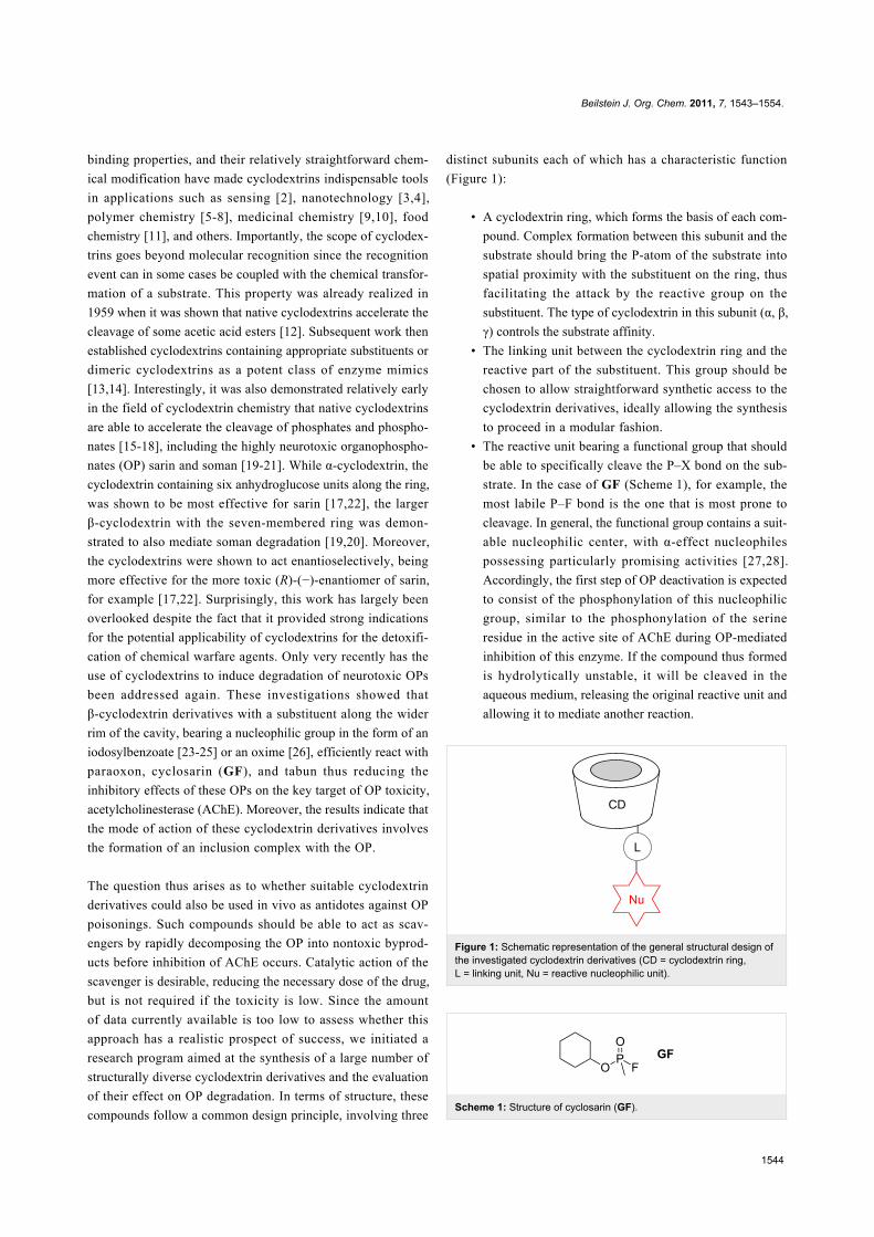
compounds follow a common design principle, involving three
distinct subunits each of which has a characteristic function
(Figure 1):
• A cyclodextrin ring, which forms the basis of each com-
pound. Complex formation between this subunit and the
substrate should bring the P-atom of the substrate into
spatial proximity with the substituent on the ring, thus
facilitating the attack by the reactive group on the
substituent. The type of cyclodextrin in this subunit (α, β,
γ) controls the substrate affinity.
• The linking unit between the cyclodextrin ring and the
reactive part of the substituent. This group should be
chosen to allow straightforward synthetic access to the
cyclodextrin derivatives, ideally allowing the synthesis
to proceed in a modular fashion.
• The reactive unit bearing a functional group that should
be able to specifically cleave the P–X bond on the sub-
strate. In the case of GF (Scheme 1), for example, the
most labile P–F bond is the one that is most prone to
cleavage. In general, the functional group contains a suit-
able nucleophilic center, with α-effect nucleophiles
possessing particularly promising activities [27,28].
Accordingly, the first step of OP deactivation is expected
to consist of the phosphonylation of this nucleophilic
group, similar to the phosphonylation of the serine
residue in the active site of AChE during OP-mediated
inhibition of this enzyme. If the compound thus formed
is hydrolytically unstable, it will be cleaved in the
aqueous medium, releasing the original reactive unit and
allowing it to mediate another reaction.
Figure 1: Schematic representation of the general structural design ofthe investigated cyclodextrin derivatives (CD = cyclodextrin ring,L = linking unit, Nu = reactive nucleophilic unit).
Scheme 1: Structure of cyclosarin (GF).
Beilstein J. Org. Chem. 2011, 7, 1543–1554.
1545
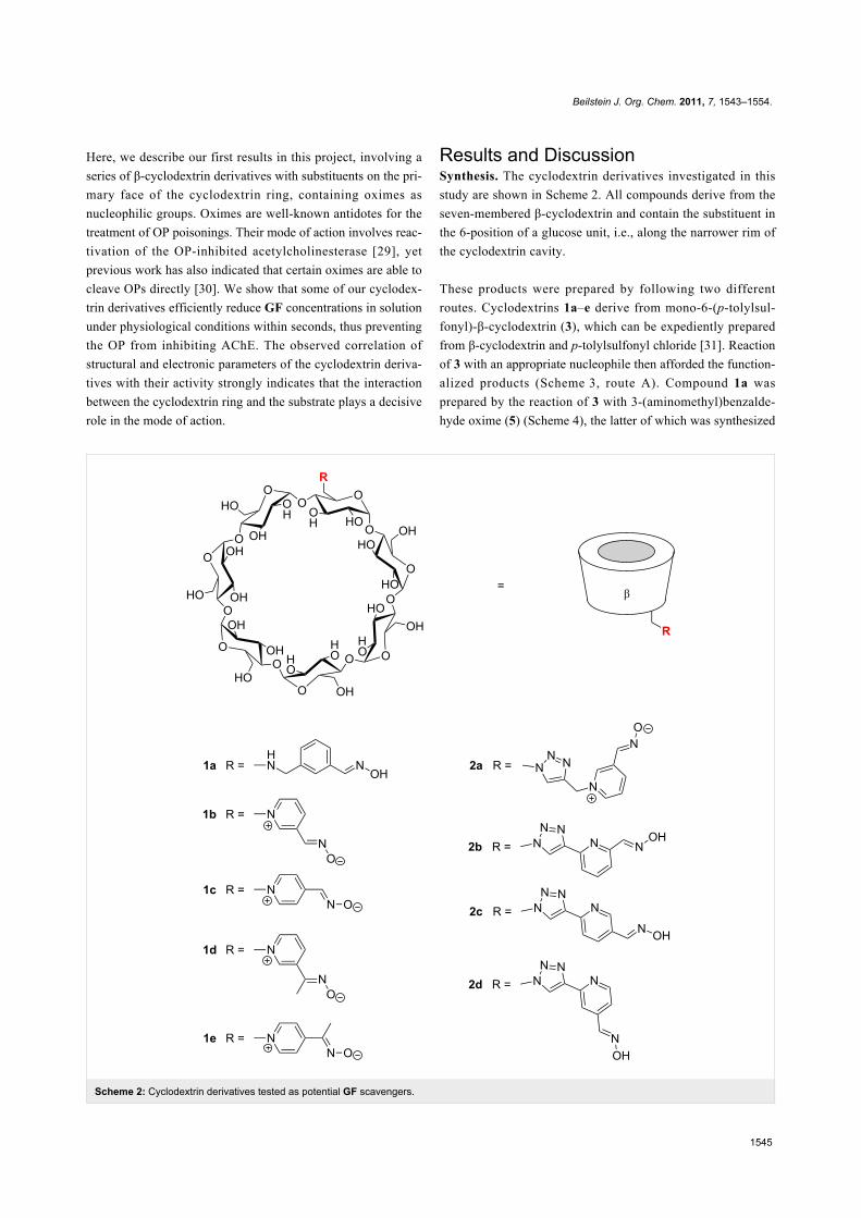
Scheme 2: Cyclodextrin derivatives tested as potential GF scavengers.
Here, we describe our first results in this project, involving a
series of β-cyclodextrin derivatives with substituents on the pri-
mary face of the cyclodextrin ring, containing oximes as
nucleophilic groups. Oximes are well-known antidotes for the
treatment of OP poisonings. Their mode of action involves reac-
tivation of the OP-inhibited acetylcholinesterase [29], yet
previous work has also indicated that certain oximes are able to
cleave OPs directly [30]. We show that some of our cyclodex-
trin derivatives efficiently reduce GF concentrations in solution
under physiological conditions within seconds, thus preventing
the OP from inhibiting AChE. The observed correlation of
structural and electronic parameters of the cyclodextrin deriva-
tives with their activity strongly indicates that the interaction
between the cyclodextrin ring and the substrate plays a decisive
role in the mode of action.
Results and DiscussionSynthesis. The cyclodextrin derivatives investigated in this
study are shown in Scheme 2. All compounds derive from the
seven-membered β-cyclodextrin and contain the substituent in
the 6-position of a glucose unit, i.e., along the narrower rim of
the cyclodextrin cavity.
These products were prepared by following two different
routes. Cyclodextrins 1a–e derive from mono-6-(p-tolylsul-
fonyl)-β-cyclodextrin (3), which can be expediently prepared
from β-cyclodextrin and p-tolylsulfonyl chloride [31]. Reaction
of 3 with an appropriate nucleophile then afforded the function-
alized products (Scheme 3, route A). Compound 1a was
prepared by the reaction of 3 with 3-(aminomethyl)benzalde-
hyde oxime (5) (Scheme 4), the latter of which was synthesized
Beilstein J. Org. Chem. 2011, 7, 1543–1554.
1546
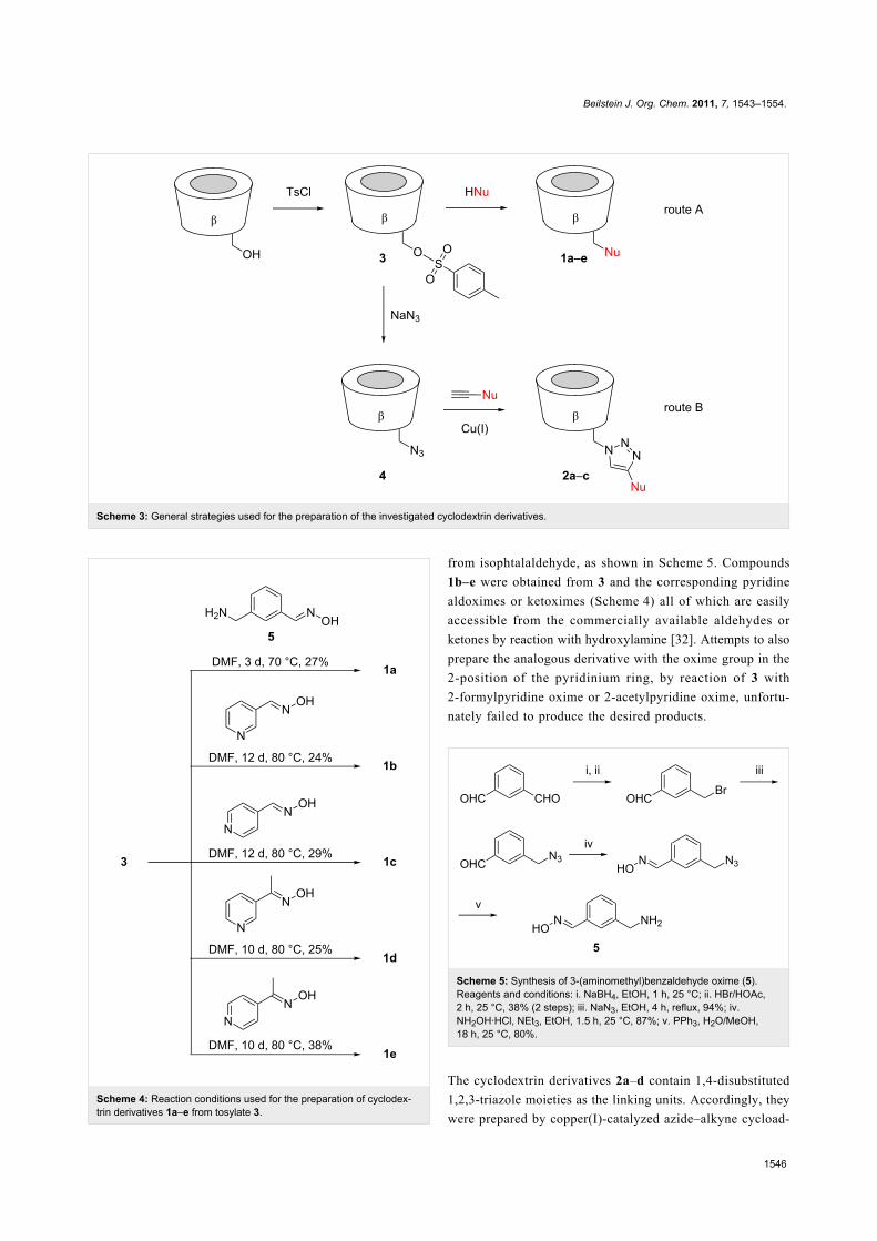
Scheme 3: General strategies used for the preparation of the investigated cyclodextrin derivatives.
Scheme 4: Reaction conditions used for the preparation of cyclodex-trin derivatives 1a–e from tosylate 3.
from isophtalaldehyde, as shown in Scheme 5. Compounds
1b–e were obtained from 3 and the corresponding pyridine
aldoximes or ketoximes (Scheme 4) all of which are easily
accessible from the commercially available aldehydes or
ketones by reaction with hydroxylamine [32]. Attempts to also
prepare the analogous derivative with the oxime group in the
2-position of the pyridinium ring, by reaction of 3 with
2-formylpyridine oxime or 2-acetylpyridine oxime, unfortu-
nately failed to produce the desired products.
Scheme 5: Synthesis of 3-(aminomethyl)benzaldehyde oxime (5).Reagents and conditions: i. NaBH4, EtOH, 1 h, 25 °C; ii. HBr/HOAc,2 h, 25 °C, 38% (2 steps); iii. NaN3, EtOH, 4 h, reflux, 94%; iv.NH2OH·HCl, NEt3, EtOH, 1.5 h, 25 °C, 87%; v. PPh3, H2O/MeOH,18 h, 25 °C, 80%.
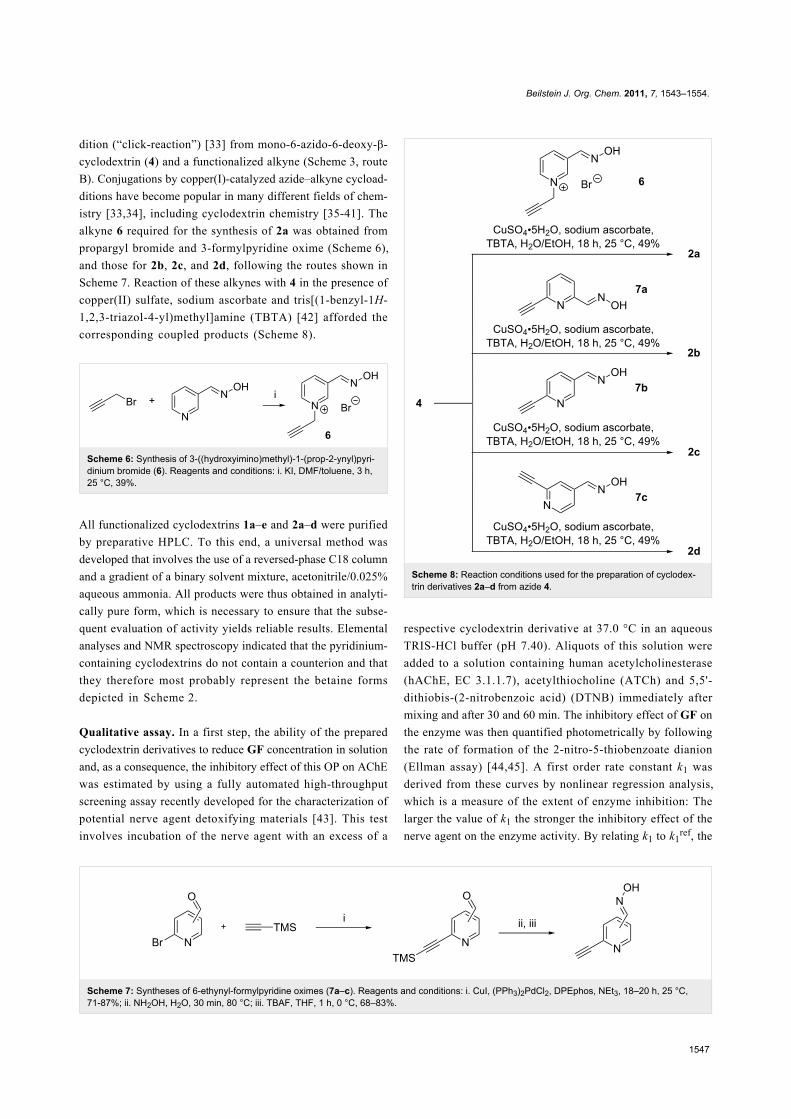
The cyclodextrin derivatives 2a–d contain 1,4-disubstituted
1,2,3-triazole moieties as the linking units. Accordingly, they
were prepared by copper(I)-catalyzed azide–alkyne cycload-
Beilstein J. Org. Chem. 2011, 7, 1543–1554.
1547
Scheme 7: Syntheses of 6-ethynyl-formylpyridine oximes (7a–c). Reagents and conditions: i. CuI, (PPh3)2PdCl2, DPEphos, NEt3, 18–20 h, 25 °C,71-87%; ii. NH2OH, H2O, 30 min, 80 °C; iii. TBAF, THF, 1 h, 0 °C, 68–83%.
dition (“click-reaction”) [33] from mono-6-azido-6-deoxy-β-
cyclodextrin (4) and a functionalized alkyne (Scheme 3, route
B). Conjugations by copper(I)-catalyzed azide–alkyne cycload-
ditions have become popular in many different fields of chem-
istry [33,34], including cyclodextrin chemistry [35-41]. The
alkyne 6 required for the synthesis of 2a was obtained from
propargyl bromide and 3-formylpyridine oxime (Scheme 6),
and those for 2b, 2c, and 2d, following the routes shown in
Scheme 7. Reaction of these alkynes with 4 in the presence of
copper(II) sulfate, sodium ascorbate and tris[(1-benzyl-1H-
1,2,3-triazol-4-yl)methyl]amine (TBTA) [42] afforded the
corresponding coupled products (Scheme 8).
Scheme 6: Synthesis of 3-((hydroxyimino)methyl)-1-(prop-2-ynyl)pyri-dinium bromide (6). Reagents and conditions: i. KI, DMF/toluene, 3 h,25 °C, 39%.
All functionalized cyclodextrins 1a–e and 2a–d were purified
by preparative HPLC. To this end, a universal method was
developed that involves the use of a reversed-phase C18 column
and a gradient of a binary solvent mixture, acetonitrile/0.025%
aqueous ammonia. All products were thus obtained in analyti-
cally pure form, which is necessary to ensure that the subse-
quent evaluation of activity yields reliable results. Elemental
analyses and NMR spectroscopy indicated that the pyridinium-
containing cyclodextrins do not contain a counterion and that
they therefore most probably represent the betaine forms
depicted in Scheme 2.
Qualitative assay. In a first step, the ability of the prepared
cyclodextrin derivatives to reduce GF concentration in solution
and, as a consequence, the inhibitory effect of this OP on AChE
was estimated by using a fully automated high-throughput
screening assay recently developed for the characterization of
potential nerve agent detoxifying materials [43]. This test
involves incubation of the nerve agent with an excess of a
Scheme 8: Reaction conditions used for the preparation of cyclodex-trin derivatives 2a–d from azide 4.
respective cyclodextrin derivative at 37.0 °C in an aqueous
TRIS-HCl buffer (pH 7.40). Aliquots of this solution were
added to a solution containing human acetylcholinesterase
(hAChE, EC 3.1.1.7), acetylthiocholine (ATCh) and 5,5'-
dithiobis-(2-nitrobenzoic acid) (DTNB) immediately after
mixing and after 30 and 60 min. The inhibitory effect of GF on
the enzyme was then quantified photometrically by following
the rate of formation of the 2-nitro-5-thiobenzoate dianion
(Ellman assay) [44,45]. A first order rate constant k1 was
derived from these curves by nonlinear regression analysis,
which is a measure of the extent of enzyme inhibition: The
larger the value of k1 the stronger the inhibitory effect of the
nerve agent on the enzyme activity. By relating k1 to k1ref, the
Beilstein J. Org. Chem. 2011, 7, 1543–1554.
1548
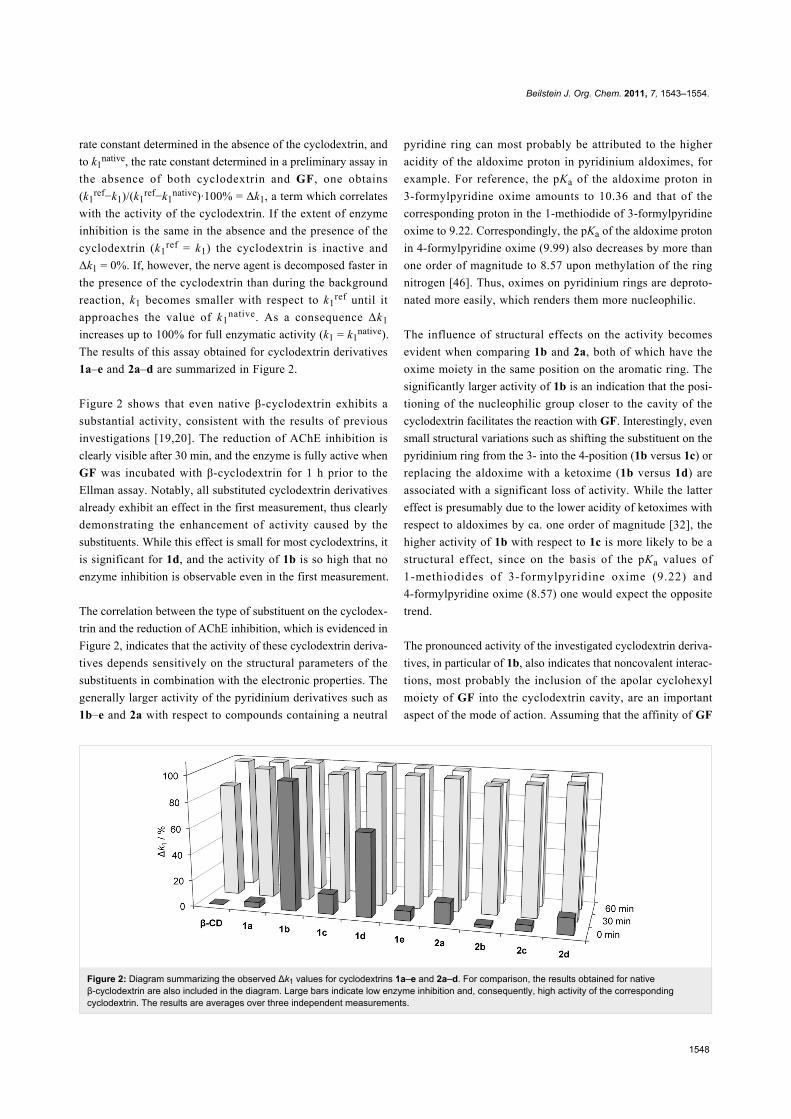
Figure 2: Diagram summarizing the observed Δk1 values for cyclodextrins 1a–e and 2a–d. For comparison, the results obtained for nativeβ-cyclodextrin are also included in the diagram. Large bars indicate low enzyme inhibition and, consequently, high activity of the correspondingcyclodextrin. The results are averages over three independent measurements.
rate constant determined in the absence of the cyclodextrin, and
to k1native, the rate constant determined in a preliminary assay in
the absence of both cyclodextrin and GF, one obtains
(k1ref−k1)/(k1
ref−k1native)·100% = Δk1, a term which correlates
with the activity of the cyclodextrin. If the extent of enzyme
inhibition is the same in the absence and the presence of the
cyclodextrin (k1ref = k1) the cyclodextrin is inactive and
Δk1 = 0%. If, however, the nerve agent is decomposed faster in
the presence of the cyclodextrin than during the background
reaction, k1 becomes smaller with respect to k1ref until it
approaches the value of k1native. As a consequence Δk1
increases up to 100% for full enzymatic activity (k1 = k1native).
The results of this assay obtained for cyclodextrin derivatives
1a–e and 2a–d are summarized in Figure 2.
Figure 2 shows that even native β-cyclodextrin exhibits a
substantial activity, consistent with the results of previous
investigations [19,20]. The reduction of AChE inhibition is
clearly visible after 30 min, and the enzyme is fully active when
GF was incubated with β-cyclodextrin for 1 h prior to the
Ellman assay. Notably, all substituted cyclodextrin derivatives
already exhibit an effect in the first measurement, thus clearly
demonstrating the enhancement of activity caused by the
substituents. While this effect is small for most cyclodextrins, it
is significant for 1d, and the activity of 1b is so high that no
enzyme inhibition is observable even in the first measurement.
The correlation between the type of substituent on the cyclodex-
trin and the reduction of AChE inhibition, which is evidenced in
Figure 2, indicates that the activity of these cyclodextrin deriva-
tives depends sensitively on the structural parameters of the
substituents in combination with the electronic properties. The
generally larger activity of the pyridinium derivatives such as
1b–e and 2a with respect to compounds containing a neutral
pyridine ring can most probably be attributed to the higher
acidity of the aldoxime proton in pyridinium aldoximes, for
example. For reference, the pKa of the aldoxime proton in
3-formylpyridine oxime amounts to 10.36 and that of the
corresponding proton in the 1-methiodide of 3-formylpyridine
oxime to 9.22. Correspondingly, the pKa of the aldoxime proton
in 4-formylpyridine oxime (9.99) also decreases by more than
one order of magnitude to 8.57 upon methylation of the ring
nitrogen [46]. Thus, oximes on pyridinium rings are deproto-
nated more easily, which renders them more nucleophilic.
The influence of structural effects on the activity becomes
evident when comparing 1b and 2a, both of which have the
oxime moiety in the same position on the aromatic ring. The
significantly larger activity of 1b is an indication that the posi-
tioning of the nucleophilic group closer to the cavity of the
cyclodextrin facilitates the reaction with GF. Interestingly, even
small structural variations such as shifting the substituent on the
pyridinium ring from the 3- into the 4-position (1b versus 1c) or
replacing the aldoxime with a ketoxime (1b versus 1d) are
associated with a significant loss of activity. While the latter
effect is presumably due to the lower acidity of ketoximes with
respect to aldoximes by ca. one order of magnitude [32], the
higher activity of 1b with respect to 1c is more likely to be a
structural effect, since on the basis of the pKa values of
1-methiodides of 3-formylpyridine oxime (9.22) and
4-formylpyridine oxime (8.57) one would expect the opposite
trend.
The pronounced activity of the investigated cyclodextrin deriva-
tives, in particular of 1b, also indicates that noncovalent interac-
tions, most probably the inclusion of the apolar cyclohexyl
moiety of GF into the cyclodextrin cavity, are an important
aspect of the mode of action. Assuming that the affinity of GF
Beilstein J. Org. Chem. 2011, 7, 1543–1554.
1549
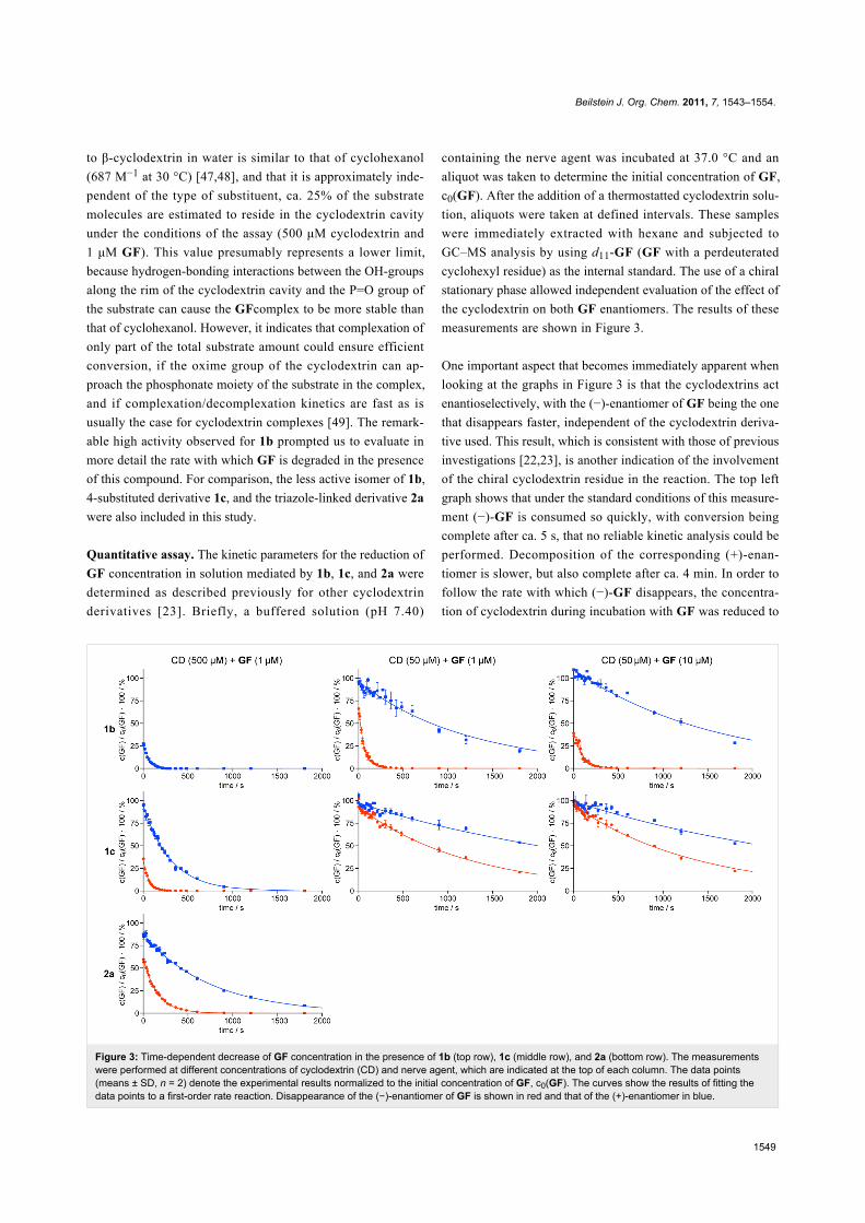
Figure 3: Time-dependent decrease of GF concentration in the presence of 1b (top row), 1c (middle row), and 2a (bottom row). The measurementswere performed at different concentrations of cyclodextrin (CD) and nerve agent, which are indicated at the top of each column. The data points(means ± SD, n = 2) denote the experimental results normalized to the initial concentration of GF, c0(GF). The curves show the results of fitting thedata points to a first-order rate reaction. Disappearance of the (−)-enantiomer of GF is shown in red and that of the (+)-enantiomer in blue.
to β-cyclodextrin in water is similar to that of cyclohexanol
(687 M−1 at 30 °C) [47,48], and that it is approximately inde-
pendent of the type of substituent, ca. 25% of the substrate
molecules are estimated to reside in the cyclodextrin cavity
under the conditions of the assay (500 μM cyclodextrin and
1 μM GF). This value presumably represents a lower limit,
because hydrogen-bonding interactions between the OH-groups
along the rim of the cyclodextrin cavity and the P=O group of
the substrate can cause the GFcomplex to be more stable than
that of cyclohexanol. However, it indicates that complexation of
only part of the total substrate amount could ensure efficient
conversion, if the oxime group of the cyclodextrin can ap-
proach the phosphonate moiety of the substrate in the complex,
and if complexation/decomplexation kinetics are fast as is
usually the case for cyclodextrin complexes [49]. The remark-
able high activity observed for 1b prompted us to evaluate in
more detail the rate with which GF is degraded in the presence
of this compound. For comparison, the less active isomer of 1b,
4-substituted derivative 1c, and the triazole-linked derivative 2a
were also included in this study.
Quantitative assay. The kinetic parameters for the reduction of
GF concentration in solution mediated by 1b, 1c, and 2a were
determined as described previously for other cyclodextrin
derivatives [23]. Briefly, a buffered solution (pH 7.40)
containing the nerve agent was incubated at 37.0 °C and an
aliquot was taken to determine the initial concentration of GF,
c0(GF). After the addition of a thermostatted cyclodextrin solu-
tion, aliquots were taken at defined intervals. These samples
were immediately extracted with hexane and subjected to
GC–MS analysis by using d11-GF (GF with a perdeuterated
cyclohexyl residue) as the internal standard. The use of a chiral
stationary phase allowed independent evaluation of the effect of
the cyclodextrin on both GF enantiomers. The results of these
measurements are shown in Figure 3.
One important aspect that becomes immediately apparent when
looking at the graphs in Figure 3 is that the cyclodextrins act
enantioselectively, with the (−)-enantiomer of GF being the one
that disappears faster, independent of the cyclodextrin deriva-
tive used. This result, which is consistent with those of previous
investigations [22,23], is another indication of the involvement
of the chiral cyclodextrin residue in the reaction. The top left
graph shows that under the standard conditions of this measure-
ment (−)-GF is consumed so quickly, with conversion being
complete after ca. 5 s, that no reliable kinetic analysis could be
performed. Decomposition of the corresponding (+)-enan-
tiomer is slower, but also complete after ca. 4 min. In order to
follow the rate with which (−)-GF disappears, the concentra-
tion of cyclodextrin during incubation with GF was reduced to
Beilstein J. Org. Chem. 2011, 7, 1543–1554.
1550
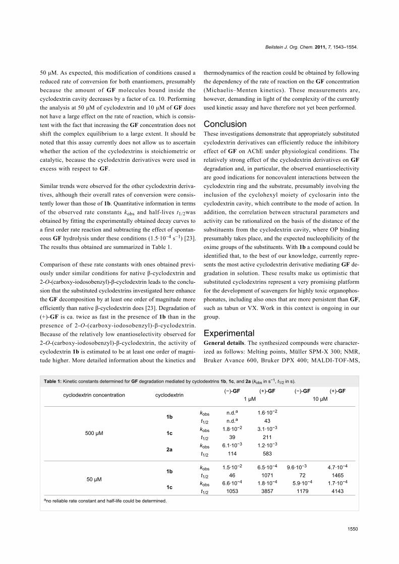
Table 1: Kinetic constants determined for GF degradation mediated by cyclodextrins 1b, 1c, and 2a (kobs in s−1, t1/2 in s).
cyclodextrin concentration cyclodextrin(−)-GF (+)-GF (−)-GF (+)-GF
1 μM 10 μM
500 μM
1bkobs n.d.a 1.6·10−2
t1/2 n.d.a 43
1ckobs 1.8·10−2 3.1·10−3
t1/2 39 211
2akobs 6.1·10−3 1.2·10−3
t1/2 114 583
50 μM1b
kobs 1.5·10−2 6.5·10−4 9.6·10−3 4.7·10−4
t1/2 46 1071 72 1465
1ckobs 6.6·10−4 1.8·10−4 5.9·10−4 1.7·10−4
t1/2 1053 3857 1179 4143ano reliable rate constant and half-life could be determined.
50 μM. As expected, this modification of conditions caused a
reduced rate of conversion for both enantiomers, presumably
because the amount of GF molecules bound inside the
cyclodextrin cavity decreases by a factor of ca. 10. Performing
the analysis at 50 μM of cyclodextrin and 10 μM of GF does
not have a large effect on the rate of reaction, which is consis-
tent with the fact that increasing the GF concentration does not
shift the complex equilibrium to a large extent. It should be
noted that this assay currently does not allow us to ascertain
whether the action of the cyclodextrins is stoichiometric or
catalytic, because the cyclodextrin derivatives were used in
excess with respect to GF.
Similar trends were observed for the other cyclodextrin deriva-
tives, although their overall rates of conversion were consis-
tently lower than those of 1b. Quantitative information in terms
of the observed rate constants kobs and half-lives t1/2was
obtained by fitting the experimentally obtained decay curves to
a first order rate reaction and subtracting the effect of spontan-
eous GF hydrolysis under these conditions (1.5·10−4 s−1) [23].
The results thus obtained are summarized in Table 1.
Comparison of these rate constants with ones obtained previ-
ously under similar conditions for native β-cyclodextrin and
2-O-(carboxy-iodosobenzyl)-β-cyclodextrin leads to the conclu-
sion that the substituted cyclodextrins investigated here enhance
the GF decomposition by at least one order of magnitude more
efficiently than native β-cyclodextrin does [23]. Degradation of
(+)-GF is ca. twice as fast in the presence of 1b than in the
presence of 2-O-(carboxy-iodosobenzyl)-β-cyclodextrin.
Because of the relatively low enantioselectivity observed for
2-O-(carboxy-iodosobenzyl)-β-cyclodextrin, the activity of
cyclodextrin 1b is estimated to be at least one order of magni-
tude higher. More detailed information about the kinetics and
thermodynamics of the reaction could be obtained by following
the dependency of the rate of reaction on the GF concentration
(Michaelis–Menten kinetics). These measurements are,
however, demanding in light of the complexity of the currently
used kinetic assay and have therefore not yet been performed.
ConclusionThese investigations demonstrate that appropriately substituted
cyclodextrin derivatives can efficiently reduce the inhibitory
effect of GF on AChE under physiological conditions. The
relatively strong effect of the cyclodextrin derivatives on GF
degradation and, in particular, the observed enantioselectivity
are good indications for noncovalent interactions between the
cyclodextrin ring and the substrate, presumably involving the
inclusion of the cyclohexyl moiety of cyclosarin into the
cyclodextrin cavity, which contribute to the mode of action. In
addition, the correlation between structural parameters and
activity can be rationalized on the basis of the distance of the
substituents from the cyclodextrin cavity, where OP binding
presumably takes place, and the expected nucleophilicity of the
oxime groups of the substituents. With 1b a compound could be
identified that, to the best of our knowledge, currently repre-
sents the most active cyclodextrin derivative mediating GF de-
gradation in solution. These results make us optimistic that
substituted cyclodextrins represent a very promising platform
for the development of scavengers for highly toxic organophos-
phonates, including also ones that are more persistent than GF,
such as tabun or VX. Work in this context is ongoing in our
group.
ExperimentalGeneral details. The synthesized compounds were character-
ized as follows: Melting points, Müller SPM-X 300; NMR,
Bruker Avance 600, Bruker DPX 400; MALDI-TOF-MS,
Beilstein J. Org. Chem. 2011, 7, 1543–1554.
1551
Scheme 9: Schematic protocol for the qualitative assay.
Bruker Ultraflex TOF/TOF; ESI–MS, Bruker Esquire 3000; IR,
FT-IR System Spectrum BX, Perkin-Elmer; elemental analysis,
Elementar vario Micro cube. All chemicals, unless other stated,
are commercially available and were used without further
purification. Cyclosarin and deuterated cyclosarin (d11-
cyclosarin, d11-GF) (>98% by GC–MS, 1H NMR, and 31P
NMR) were made available by the German Ministry of Defense
(All experiments with cyclosarin were performed at the Institut
für Pharmakologie und Toxikologie der Bundeswehr). Hemo-
globin-free erythrocyte ghosts as a source for human erythro-
cyte acetylcholinesterase (AChE, EC 3.1.1.7) were prepared
according to the procedure of Dodge et al. [50] with minor
modifications [51]. AChE activity was adjusted to 4000 U/l by
dilution with phosphate buffer (0.1 M, pH 7.40). Aliquots were
stored at a temperature of −80 °C. Prior to use, ghosts were
homogenized with a Sonoplus HD 2070 ultrasonic homogenator
(Bandelin electronic, Berlin, Germany) twice for 5 s with a 20 s
interval to achieve a homogeneous matrix. For the preparative
HPLC the following conditions were used: HPLC, Dionex
UltiMate 3000; column, Thermo Fisher, BetaBasic-18,
250 × 21.2 mm, 5 μm particle size; flow, 12 mL/min; eluent,
aqueous: 0.025% aqueous ammonia, organic: acetonitrile; for
the separation of neutral compounds (1a, 2b–2d) the following
gradient was used: 0–6 min, 0% organic; 6–27 min, linear
increase of organic to 40%; 27–33 min, 40% organic;
33–39 min, linear decrease to 0% organic; 39–45 min, 0%
organic; charged compounds (1b–1e, 2a) were purified by
employing a slightly different gradient: 0–6 min, 0% organic;
6–22 min, linear increase to 15% organic; 22–25 min, linear
increase to 50% organic; 25–30 min, 50% organic; 30–37 min,
linear decrease to 0% organic; 37–45 min, 0% organic.
Qualitative assay. The qualitative test was performed by using
a Tecan Freedom EVO liquid handling system (Männedorf,
Switzerland) [43]. A solution of cyclodextrin (500 µM) and GF
(1 µM) was prepared and incubated in TRIS-HCl buffer (0.1 M,
pH 7.40) at 37.0 °C (Scheme 9). Immediately after mixing, a
sample (25 µL) was transferred to a measuring plate prefilled
with buffer (2400 µL, 0.1 M TRIS-HCl, pH 7.40 and 0.3 mM
DTNB) and human AChE (10 µL) and preheated to 37.0 °C.
ATCh (50 µL, 49.7 mM) was added immediately, the
microplate was transferred to a photometer and the absorption
was measured at 436 nm for 30 min while the temperature was
maintained at 37.0 °C. Further aliquots of the test mixture were
taken after 30 min and 60 min and treated analogously. The rate
constants k1 were determined by nonlinear regression analysis
of the resulting curves by using Prism 5.0 (GraphPad Software,
San Diego, CA, USA) as described previously [43]. All results
shown are mean values of n = 3 experiments. The value for
k1native was determined in independent experiments to be
8.56·10−5 s−1.
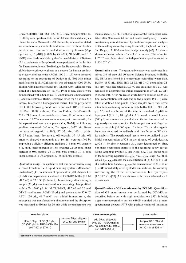
Quantitative assay. The quantitative assay was performed in a
stirred 2.0 ml cryo vial (Wheaton Science Products, Millville,
NJ, USA) positioned in a temperature controlled water bath.
Buffer (1850 µL, TRIS-HCl 0.1 M, pH 7.40) containing GF
(1.1 µM) was incubated at 37.0 °C and an aliquot (50 µL) was
removed to determine the initial concentration of GF, c0(GF)
(Scheme 10). After preheated cyclodextrin solution (200 µL,
final concentration 500 µM) was added, aliquots (50 µL) were
taken at defined time points. These samples were transferred
into a tube containing sodium formate buffer (20 µL, 300 µM,
pH 3.5) and a solution of the internal standard d11-GF in
2-propanol (2.5 µL, 10 µg/mL). Afterward, ice-cold hexane
(500 µL) was immediately added, and the mixture was shaken
vigorously and stored on ice. Each sample was centrifuged as
soon as possible (10.000 rpm, 10 min, 4 °C), and the organic
layer was removed immediately and transferred to GC vials
for analysis. The experimental results were normalized to the
initial concentration of GF in the absence of cyclodextrin,
c0(GF). The kinetic constants kobs were determined by, first,
nonlinear regression analysis of the resulting decay curves
(using GraphPad Prism 5.0, San Diego, CA, USA) on the basis
of the following equation: c(+/-)GF,t = c(+/-)GF,0*·exp(−kobs·t), in
which c(+/-)GF,t denotes the concentration of (+)-GF or (−)-GF
at a certain time t and c(+/-)GF,0* the concentration of (+)-GF or
(−)-GFimmediately after cyclodextrin addition, followed by
subtracting the effect of spontaneous GF hydrolysis
(1.5·10−4 s−1) [23]. All data shown are the mean values of n = 2
experiments.
Quantification of GF enantiomers by PCI MS. Quantifica-
tion of GF enantiomers was performed by GC–MS, as
described before but with slight modifications [52]. In brief,
a gas chromatographic system 6890N coupled with a mass
spectrometer detector 5973 with positive chemical ionization
Beilstein J. Org. Chem. 2011, 7, 1543–1554.
1552
Scheme 10: Schematic protocol of the quantitative assay.
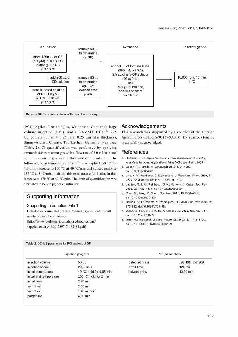
Table 2: GC–MS parameters for PCI analysis of GF.
injection program MS parameters
injection volume 50 µL detected mass m/z 198, m/z 209injection speed 20 µL/min dwell time 125 msinitial temperature 40 °C, hold for 0.05 min solvent delay 13.00 mininitial end temperature 260 °C, hold for 2 mininitial time 2.70 minvent time 2.60 minvent flow 10.0 mL/minpurge time 4.60 min
(PCI) (Agilent Technologies, Waldbronn, Germany), large
volume injection (LVI), and a GAMMA DEXTM 225
GC column (30 m × 0.25 mm, 0.25 µm film thickness,
Sigma–Aldrich Chemie, Taufkirchen, Germany) was used
(Table 2). CI quantification was performed by applying
ammonia 6.0 as reactant gas with a flow rate of 2.0 mL/min and
helium as carrier gas with a flow rate of 1.3 mL/min. The
following oven temperature program was applied: 50 °C for
4.5 min, increase to 100 °C at 40 °C/min and subsequently to
135 °C at 3 °C/min, maintain this temperature for 2 min, further
increase to 170 °C at 40 °C/min. The limit of quantification was
estimated to be 2.5 pg per enantiomer.
Supporting InformationSupporting Information File 1Detailed experimental procedures and physical data for all
newly prepared compounds.
[http://www.beilstein-journals.org/bjoc/content/
supplementary/1860-5397-7-182-S1.pdf]
AcknowledgementsThis research was supported by a contract of the German
Armed Forces (E/UR3G/9G127/9A803). The generous funding
is gratefully acknowledged.
References1. Dodziuk, H., Ed. Cyclodextrins and Their Complexes: Chemistry,
Analytical Methods, Applications; Wiley-VCH: Weinheim, 2006.2. Ogoshi, T.; Harada, A. Sensors 2008, 8, 4961–4982.
doi:10.3390/s80849613. Ling, X. Y.; Reinhoudt, D. N.; Huskens, J. Pure Appl. Chem. 2009, 81,
2225–2233. doi:10.1351/PAC-CON-09-07-044. Ludden, M. J. W.; Reinhoudt, D. N.; Huskens, J. Chem. Soc. Rev.
2006, 35, 1122–1134. doi:10.1039/b600093m5. Chen, G.; Jiang, M. Chem. Soc. Rev. 2011, 40, 2254–2266.
doi:10.1039/c0cs00153h6. Harada, A.; Takashima, Y.; Yamaguchi, H. Chem. Soc. Rev. 2009, 38,
875–882. doi:10.1039/b705458k7. Wenz, G.; Han, B.-H.; Müller, A. Chem. Rev. 2006, 106, 782–817.
doi:10.1021/cr970027+8. Ritter, H.; Tabatabai, M. Prog. Polym. Sci. 2002, 27, 1713–1720.
doi:10.1016/S0079-6700(02)00022-9
Beilstein J. Org. Chem. 2011, 7, 1543–1554.
1553
9. Aniskevich, S.; Leone, B. J.; Brull, S. J. Expert Rev. Neurother. 2011,11, 185–198. doi:10.1586/ern.11.2
10. Loftsson, T.; Brewster, M. E. J. Pharm. Pharmacol. 2010, 62,1607–1621. doi:10.1111/j.2042-7158.2010.01030.x
11. Szente, L.; Szejtli, J. Trends Food Sci. Technol. 2004, 15, 137–142.doi:10.1016/j.tifs.2003.09.019
12. Cramer, F.; Dietsche, W. Chem. Ber. 1959, 92, 1739–1747.doi:10.1002/cber.19590920804
13. Marinescu, L.; Bols, M. Trends Glycosci. Glycotechnol. 2009, 21,309–323. doi:10.4052/tigg.21.309
14. Breslow, R.; Dong, S. D. Chem. Rev. 1998, 98, 1997–2012.doi:10.1021/cr970011j
15. Mochida, K.; Matsui, Y.; Ota, Y.; Arakawa, K.; Date, Y.Bull. Chem. Soc. Jpn. 1976, 49, 3119–3123. doi:10.1246/bcsj.49.3119
16. Brass, H. J.; Bender, M. L. J. Am. Chem. Soc. 1973, 95, 5391–5399.doi:10.1021/ja00797a046
17. van Hooidonk, C.; Groos, C. C. Recl. Trav. Chim. Pays-Bas 1970, 89,845–856. doi:10.1002/recl.19700890810Interestingly, this work contains an early application of an indicatordisplacement assay, a sensing technique whose usefulness insupramolecular chemistry was demonstrated in recent yearsparticularly by the Anslyn group, see: Wiskur, S. L; Aït-Haddou, H.;Lavigne, J. J.; Anslyn, E. V. Acc. Chem. Res. 2001, 34, 963–972.doi:10.1021/ar9600796
18. Hennrich, N.; Cramer, F. J. Am. Chem. Soc. 1965, 87, 1121–1126.doi:10.1021/ja01083a032
19. Désiré, B.; Saint-André, S. Experientia 1987, 43, 395–397.doi:10.1007/BF01940424
20. Désiré, B.; Saint-André, S. Fundam. Appl. Toxicol. 1986, 7, 646–657.doi:10.1016/0272-0590(86)90114-4
21. van Hooidonk, C. Recl. Trav. Chim. Pays-Bas 1972, 91, 1103–1109.doi:10.1002/recl.19720910910
22. van Hooidonk, C.; Breebaart-Hansen, J. C. A. E.Recl. Trav. Chim. Pays-Bas 1970, 89, 289–299.doi:10.1002/recl.19700890309
23. Müller, S.; Koller, M.; Le Provost, R.; Lafont, O.; Estour, F.; Wille, T.;Thiermann, H.; Worek, F.; Reiter, G. Toxicol. Lett. 2011, 200, 53–58.doi:10.1016/j.toxlet.2010.10.014
24. Wille, T.; Tenberken, O.; Reiter, G.; Müller, S.; Le Provost, R.;Lafont, O.; Estour, F.; Thiermann, H.; Worek, F. Toxicology 2009, 265,96–100. doi:10.1016/j.tox.2009.09.018
25. Masurier, N.; Estour, F.; Froment, M.-T.; Lefèvre, B.; Debouzy, J.-C.;Brasme, B.; Masson, P.; Lafont, O. Eur. J. Med. Chem. 2005, 40,615–623. doi:10.1016/j.ejmech.2005.02.008
26. Le Provost, R.; Wille, T.; Louise, L.; Masurier, N.; Müller, S.; Reiter, G.;Renard, P.-Y.; Lafont, O.; Worek, F.; Estour, F. Org. Biomol. Chem.2011, 9, 3026–3032. doi:10.1039/c0ob00931h
27. Smith, B. M. Chem. Soc. Rev. 2008, 37, 470–478.doi:10.1039/b705025a
28. Morales-Rojas, H.; Moss, R. A. Chem. Rev. 2002, 102, 2497–2522.doi:10.1021/cr9405462
29. Marrs, T. C.; Rice, P.; Vale, J. A. Toxicol. Rev. 2006, 25, 297–323.30. Louise-Leriche, L.; Pǎunescu, E.; Saint-André, G.; Baati, R.;
Romieu, A.; Wagner, A.; Renard, P.-Y. Chem.–Eur. J. 2010, 16,3510–3523. doi:10.1002/chem.200902986
31. McNaughton, M.; Engman, L.; Birmingham, A.; Powis, G.;Cotgreave, I. A. J. Med. Chem. 2004, 47, 233–239.doi:10.1021/jm030916r
32. Ginsburg, S.; Wilson, I. B. J. Am. Chem. Soc. 1957, 79, 481–485.doi:10.1021/ja01559a067
33. Meldal, M.; Tornøe, C. W. Chem. Rev. 2008, 108, 2952–3015.doi:10.1021/cr0783479
34. Lallana, E.; Riguera, R.; Fernandez-Megia, E. Angew. Chem., Int. Ed.2011, 50, 8794–8804. doi:10.1002/anie.201101019Angew. Chem. 2011, 123, 8956–8966. doi:10.1002/ange.201101019
35. Ward, S.; Ling, C.-C. Eur. J. Org. Chem. 2011, 4853–4861.doi:10.1002/ejoc.201100445
36. Böhm, I.; Ritter, H. Macromol. Chem. Phys. 2011, 212, 1080–1085.doi:10.1002/macp.201100006
37. Christensen, H. S.; Sigurskjold, B. W.; Frihed, T. G.; Marinescu, L. G.;Pedersen, C. M.; Bols, M. Eur. J. Org. Chem. 2011, 5279–5290.doi:10.1002/ejoc.201100671
38. Jazkewitsch, O.; Ritter, H. Macromolecules 2011, 44, 375–382.doi:10.1021/ma102456n
39. Méndez-Ardoy, A.; Guilloteau, N.; Di Giorgio, C.; Vierling, P.;Santoyo-González, F.; Ortiz Mellet, C.; García Fernández, J. M.J. Org. Chem.í 2011, 76, 5882–5894. doi:10.1021/jo2007785
40. Tran, D. N.; Blaszkiewicz, C.; Menuel, S.; Roucoux, A.; Philippot, K.;Hapiot, F.; Monflier, E. Carbohydr. Res. 2011, 346, 210–218.doi:10.1016/j.carres.2010.11.024
41. Trellenkamp, T.; Ritter, H. Macromolecules 2010, 43, 5538–5543.doi:10.1021/ma100812q
42. Chan, T. R.; Hilgraf, R.; Sharpless, K. B.; Fokin, V. V. Org. Lett. 2004,6, 2853–2855. doi:10.1021/ol0493094
43. Wille, T.; Thiermann, H.; Worek, F. Toxicol. in Vitro 2010, 24,1026–1031. doi:10.1016/j.tiv.2009.11.023
44. Eyer, P.; Worek, F.; Kiderlen, D.; Sinko, G.; Stuglin, A.;Simeon-Rudolf, V.; Reiner, E. Anal. Biochem. 2003, 312, 224–227.doi:10.1016/S0003-2697(02)00506-7
45. Worek, F.; Mast, U.; Kiderlen, D.; Diepold, C.; Eyer, P. Clin. Chim. Acta1999, 288, 73–90. doi:10.1016/S0009-8981(99)00144-8
46. Mason, S. F. J. Chem. Soc. 1960, 22–26. doi:10.1039/JR960000002247. Rekharsky, M. V.; Inoue, Y. Chem. Rev. 1998, 98, 1875–1918.
doi:10.1021/cr970015o48. Rekharsky, M. V.; Schwarz, F. P.; Tewari, Y. B.; Goldberg, R. N.;
Tanaka, M.; Yamashoji, Y. J. Phys. Chem. 1994, 98, 4098–4103.doi:10.1021/j100066a032
49. Connors, K. A. Chem. Rev. 1997, 97, 1325–1358.doi:10.1021/cr960371r
50. Dodge, J. T.; Mitchell, C.; Hanahan, D. J. Arch. Biochem. Biophys.1963, 100, 119–130. doi:10.1016/0003-9861(63)90042-0
51. Worek, F.; Reiter, G.; Eyer, P.; Szinicz, L. Arch. Toxicol. 2002, 76,523–529. doi:10.1007/s00204-002-0375-1
52. Reiter, G.; Koller, M.; Thiermann, H.; Dorandeu, F.; Mikler, J.;Worek, F. J. Chromatogr., B 2007, 859, 9–15.doi:10.1016/j.jchromb.2007.08.040
Beilstein J. Org. Chem. 2011, 7, 1543–1554.
1554
License and TermsThis is an Open Access article under the terms of the
Creative Commons Attribution License
(http://creativecommons.org/licenses/by/2.0), which
permits unrestricted use, distribution, and reproduction in
any medium, provided the original work is properly cited.
The license is subject to the Beilstein Journal of Organic
Chemistry terms and conditions:
(http://www.beilstein-journals.org/bjoc)
The definitive version of this article is the electronic one
which can be found at:
doi:10.3762/bjoc.7.182


![Efficient construction of highly functionalizedS1 Efficient construction of highly functionalized spiro[γ-butyrolactone-pyrrolidin-3,3′-oxindole] tricyclic skeletons via an organocatalytic](https://static.fdocument.org/doc/165x107/60fac77bcf8dba3437692a22/efficient-construction-of-highly-s1-efficient-construction-of-highly-functionalized.jpg)