HaloTag Technology: Focus on Imaging Technical Manual #TM260 · Promega Corporation ·2800 Woods...
43
Technical Manual HaloTag ® Technology: Focus on Imaging INSTRUCTIONS FOR USE OF PRODUCTS G2991, G3221, G1001, G1002, G2801, G2802, G8251, G8252, G8272, G8273, G8581, G8582, G8471, G8472, G9281, G9211, P1691, P6751, P6711, P6741, P1681, P6771, AND P6761. PRINTED IN USA. Revised 3/13 Part# TM260 7529TA U2OS cells stably expressing truncated β1-integrin-HaloTag ® fusion protein sequentially labeled with HaloTag ® Alexa Fluor ® 488 Ligand and HaloTag ® TMR Ligand.
Transcript of HaloTag Technology: Focus on Imaging Technical Manual #TM260 · Promega Corporation ·2800 Woods...

T e c h n i c a l M a n u a l
HaloTag® Technology:Focus on ImagingINSTRUCTIONS FOR USE OF PRODUCTS G2991, G3221, G1001, G1002,G2801, G2802, G8251, G8252, G8272, G8273, G8581, G8582, G8471,G8472, G9281, G9211, P1691, P6751, P6711, P6741, P1681, P6771, ANDP6761.
PRINTED IN USA.Revised 3/13 Part# TM260
7529
TA
U2OS cells stably expressing truncated β1-integrin-HaloTag® fusionprotein sequentially labeled with HaloTag® Alexa Fluor® 488 Ligand andHaloTag® TMR Ligand.
tm260.0313:EIVD_TM.qxd 2/28/2013 2:58 PM Page a
Promega Corporation · 2800 Woods Hollow Road · Madison, WI 53711-5399 USA Toll Free in USA 800-356-9526 · Phone 608-274-4330 · Fax 608-277-2516 · www.promega.comPrinted in USA. Part# TM260Revised 3/13 Page 1
1. Introduction to HaloTag® Technology and Cell Imaging .........................2
2. Product Components and Storage Conditions ............................................4A. Fluorescent Ligands .............................................................................................4B. Customizable Reactive Ligands .........................................................................7
3. Generating a HaloTag® Fusion Protein.........................................................8A. Beginning with an Expression-Validated HaloTag® ORF Clone..................8B. Creating Your Own HaloTag® Fusion Protein ................................................8
4. Mammalian Live-Cell Labeling, Cell Fixation, ICC, SDS-PAGE Flow Cytometry and Imaging .........................................................................9A. Rapid Labeling (15–60 minutes).........................................................................9B. No-Wash Labeling (overnight).........................................................................12C. Pulse-Chase Labeling.........................................................................................13D. Fixing Cells after Labeling ................................................................................16E. Multiplexing with HaloTag® Using Immunocytochemistry (ICC) ............17F. Quantitation of HaloTag® Protein Fusions, SDS-PAGE Analysis ..............20G. Flow Cytometry Analysis .................................................................................22
5. Protein Detection Using HaloTag® Monoclonoal Antibody (for Western Blotting) .....................................................................................24
6. How to Generate HaloTag® Ligands from Building Blocks ...................25A. General Protocol for Reporter Group Labeling .............................................25B. Protein Labeling with HaloTag® Amine Ligand ...........................................26C. Antibody Labeling with HaloTag® Succinimidyl Ester Ligand..................26D. Specific Example Protocol for Generating an Alexa Fluor® 488 HaloTag®
Ligand ..................................................................................................................27
7. Labeling Proteins in Mammalian Cell Lysates or Proteins Expressed in Cell-Free Systems........................................................................................27
8. Labeling E. coli Lysate to Analyze Protein Expression Level ................29A. Expressing HaloTag® Fusions in E. coli ..........................................................29B. Labeling E. coli Lysate to Analyze Protein Expression Level......................29C. Labeling Intact E. coli for Imaging Analysis...................................................30
9. General Information, Tips and Troubleshooting .....................................32A. General Information...........................................................................................32B. Composition of Buffers and Solutions ............................................................34C. Troubleshooting..................................................................................................34D. Literature Cited and Additional References ..................................................37
10. Related Products ..............................................................................................38
HaloTag® Technology:Focus on Imaging
All technical literature is available on the Internet at: www.promega.com/protocols/ Please visit the web site to verify that you are using the most current version of this
Technical Manual. Please contact Promega Technical Services if you have questions on useof this system. E-mail: [email protected].
tm260.0313:EIVD_TM.qxd 2/28/2013 2:58 PM Page 1
1. Introduction to HaloTag® Technology and Cell Imaging
Understanding the functional role of proteins and how they interact within thecell is increasingly important. Often multiple recombinant protein fusions areneeded to accomplish this research, which at times can be slow andcumbersome because of inefficiencies from moving between differentapplications. As a result, there is a need for a recombinant protein tag thataffords flexibility between expression and localization, protein purification,protein interaction discovery, screening and further functional analyses.
The HaloTag® platform(a–e) addresses the need for flexibility in functionalprotein analysis. This modular technology is based on the formation of acovalent bond between a fusion tag and synthetic ligands, and is designed toenable complete characterization of protein function in cellular andbiochemical environments. There are a variety of HaloTag® Ligands(a–d) andkits available for fluorescent cell imaging and protein capture/purification, aswell as reactive customizable Ligand Building blocks(a,e,f).
The HaloTag® reporter protein is an engineered, catalytically inactive derivativeof a hydrolase, which forms a covalent bond with HaloTag® ligands (Figure 1).Under physiological conditions this covalent bond forms rapidly and is highlyspecific and essentially irreversible, yielding a complex that is stable evenunder stringent conditions (1–3). The HaloTag® protein is a 33kDa monomericprotein not endogenous to mammalian, plant or E. coli cells; therefore, there areno levels of nonspecific activity, only high labeling specificity. This technologyhas been used successfully in many systems, including: mammalian cells (4),bacteria (5), yeast (6), plants (7) and animal models (8).
Advantages of Cell Imaging Using HaloTag® Fluorescent Ligands
Gain Precision with Temporal and Spatial Control of Labeling: The modularnature of HaloTag® technology and the rapid binding kinetics with its ligandsallow direct imaging of both protein trafficking and turnover. The user hasspatial and temporal control of labeling because available ligands have bothvaried fluorescent spectra and also cell-permeant and impermeant properties.For example, a researcher can label only the surface pool of a membrane-bound protein with a cell-impermeant ligand and follow its biology,internalization or other fate. Or a protein can first be labeled with a ligand ofone “color”, and after a period of time labeled again with a ligand containing adye of a different color. Such pulse-chase labeling allows direct observation ofprotein trafficking and/or turnover over time.
Promega Corporation · 2800 Woods Hollow Road · Madison, WI 53711-5399 USA Toll Free in USA 800-356-9526 · Phone 608-274-4330 · Fax 608-277-2516 · www.promega.comPart# TM260 Printed in USA.Page 2 Revised 3/13
tm260.0313:EIVD_TM.qxd 2/28/2013 2:58 PM Page 2
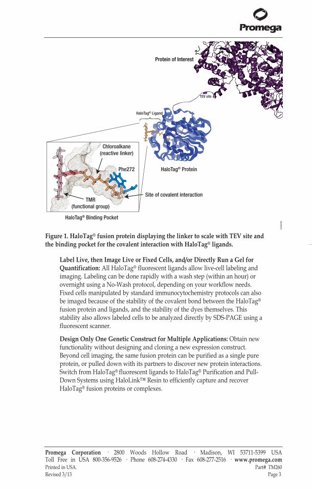
Figure 1. HaloTag® fusion protein displaying the linker to scale with TEV site andthe binding pocket for the covalent interaction with HaloTag® ligands.
Label Live, then Image Live or Fixed Cells, and/or Directly Run a Gel forQuantification: All HaloTag® fluorescent ligands allow live-cell labeling andimaging. Labeling can be done rapidly with a wash step (within an hour) orovernight using a No-Wash protocol, depending on your workflow needs.Fixed cells manipulated by standard immunocytochemistry protocols can alsobe imaged because of the stability of the covalent bond between the HaloTag®
fusion protein and ligands, and the stability of the dyes themselves. Thisstability also allows labeled cells to be analyzed directly by SDS-PAGE using afluorescent scanner.
Design Only One Genetic Construct for Multiple Applications: Obtain newfunctionality without designing and cloning a new expression construct.Beyond cell imaging, the same fusion protein can be purified as a single pureprotein, or pulled down with its partners to discover new protein interactions.Switch from HaloTag® fluorescent ligands to HaloTag® Purification and Pull-Down Systems using HaloLink™ Resin to efficiently capture and recoverHaloTag® fusion proteins or complexes.
Promega Corporation · 2800 Woods Hollow Road · Madison, WI 53711-5399 USA Toll Free in USA 800-356-9526 · Phone 608-274-4330 · Fax 608-277-2516 · www.promega.comPrinted in USA. Part# TM260Revised 3/13 Page 3
1045
0TA
HaloTag® Ligand
TMR(functional group)
Chloroalkane(reactive linker)
Phe272
Site of covalent interaction
Protein of Interest
HaloTag® Binding Pocket
HaloTag® Protein
TEV site
tm260.0313:EIVD_TM.qxd 2/28/2013 2:58 PM Page 3
Promega Corporation · 2800 Woods Hollow Road · Madison, WI 53711-5399 USA Toll Free in USA 800-356-9526 · Phone 608-274-4330 · Fax 608-277-2516 · www.promega.comPart# TM260 Printed in USA.Page 4 Revised 3/13
2. Product Components and Storage Conditions
2.A. Fluorescent Ligands
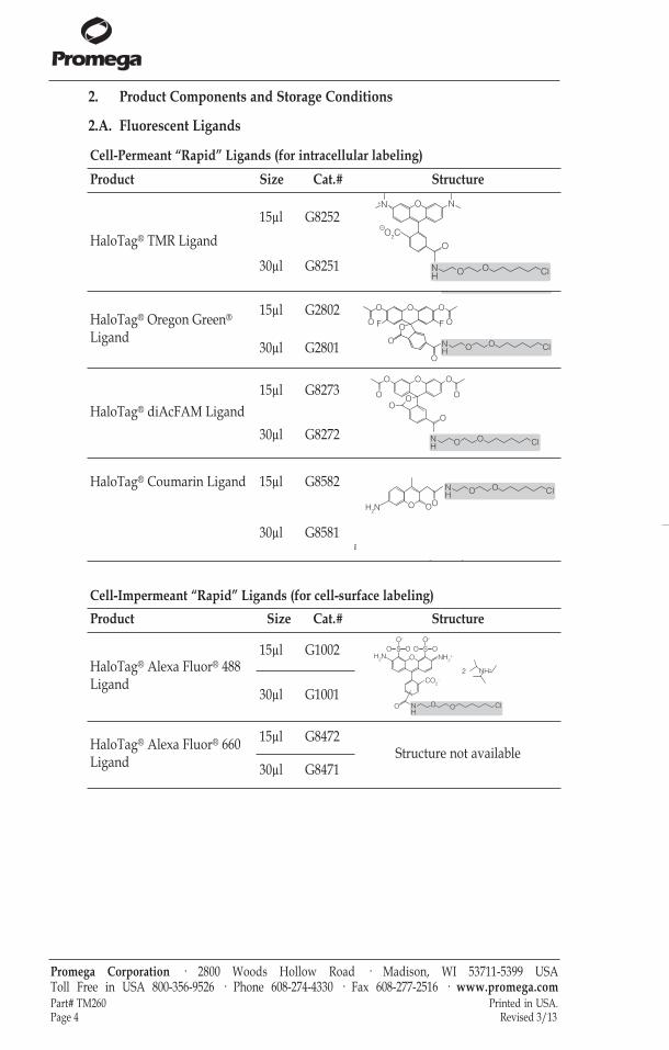
Cell-Permeant “Rapid” Ligands (for intracellular labeling)Product Size Cat.# Structure
HaloTag® TMR Ligand15µl G8252
NH
OO Cl
O
NO
O2C
+N
–
30µl G8251
HaloTag® Oregon Green®
Ligand
15µl G2802H
NH
OO Cl
O
F
O
OO
O
O
O
O F
30µl G2801
HaloTag® diAcFAM Ligand15µl G8273
OO
O
OO
OOO
NH
OO Cl30µl G8272
HaloTag® Coumarin Ligand 15µl G8582O O
NH
OO Cl
H2NO
O O
abeling), rapid labeling30µl G8581
Cell-Impermeant “Rapid” Ligands (for cell-surface labeling)Product Size Cat.# Structure
HaloTag® Alexa Fluor® 488Ligand
15µl G1002
NH
OO Cl
O
CO2–
NH2+H2N
SO OO–
SO OO–
O
NH+2
30µl G1001
HaloTag® Alexa Fluor® 660Ligand
15µl G8472Structure not available
30µl G8471
tm260.0313:EIVD_TM.qxd 2/28/2013 2:58 PM Page 4
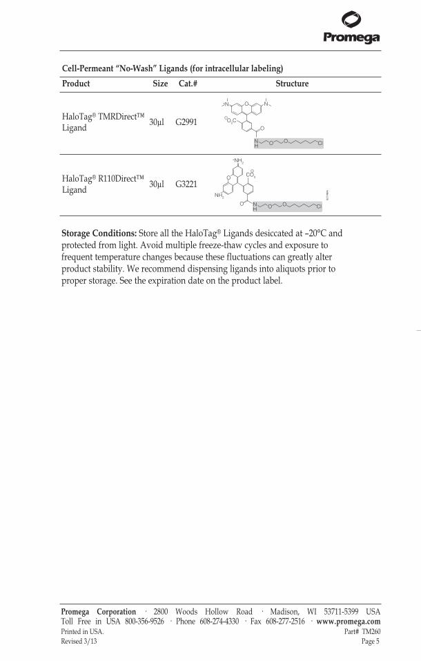
Storage Conditions: Store all the HaloTag® Ligands desiccated at –20°C andprotected from light. Avoid multiple freeze-thaw cycles and exposure tofrequent temperature changes because these fluctuations can greatly alterproduct stability. We recommend dispensing ligands into aliquots prior toproper storage. See the expiration date on the product label.
Promega Corporation · 2800 Woods Hollow Road · Madison, WI 53711-5399 USA Toll Free in USA 800-356-9526 · Phone 608-274-4330 · Fax 608-277-2516 · www.promega.comPrinted in USA. Part# TM260Revised 3/13 Page 5
Cell-Permeant “No- Wash” Ligands (for intracellular labeling)Product Size Cat.# Structure
HaloTag® TMRDirect™Ligand 30µl G2991
NH
OO Cl
O
NO
O2C
+N
–
HaloTag® R110Direct™Ligand 30µl G3221
6274
MA
NH
OO ClO
CO2
–
NH2
O
+NH2
tm260.0313:EIVD_TM.qxd 2/28/2013 2:58 PM Page 5
2. Product Components and Storage Conditions (continued)
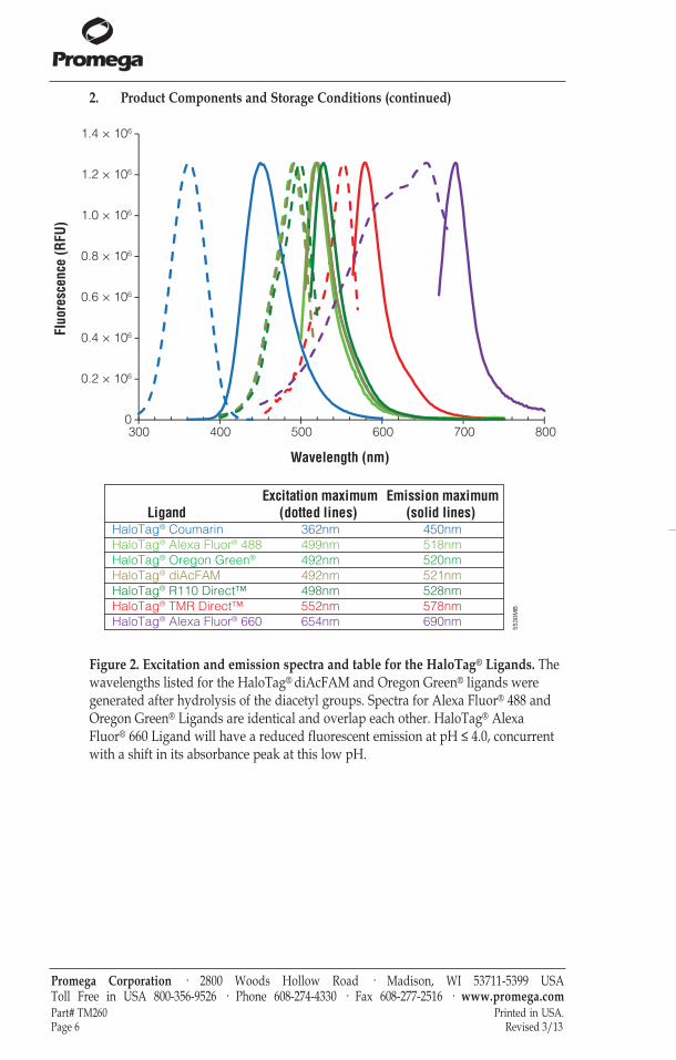
Figure 2. Excitation and emission spectra and table for the HaloTag® Ligands. Thewavelengths listed for the HaloTag® diAcFAM and Oregon Green® ligands weregenerated after hydrolysis of the diacetyl groups. Spectra for Alexa Fluor® 488 andOregon Green® Ligands are identical and overlap each other. HaloTag® AlexaFluor® 660 Ligand will have a reduced fluorescent emission at pH ≤ 4.0, concurrentwith a shift in its absorbance peak at this low pH.
Promega Corporation · 2800 Woods Hollow Road · Madison, WI 53711-5399 USA Toll Free in USA 800-356-9526 · Phone 608-274-4330 · Fax 608-277-2516 · www.promega.comPart# TM260 Printed in USA.Page 6 Revised 3/13
5530
MB
Wavelength (nm)
Fluo
resc
ence
(RFU
)
300 400 500 600 700 8000
0.2 × 10⁶
0.4 × 10⁶
0.6 × 10⁶
0.8 × 10⁶
1.0 × 10⁶
1.2 × 10⁶
1.4 × 10⁶
LigandExcitation maximum
(dotted lines) Emission maximum
(solid lines) HaloTag® CoumarinHaloTag® Alexa Fluor® 488
HaloTag® Alexa Fluor® 660
HaloTag® Oregon Green®
HaloTag® diAcFAMHaloTag® R110 Direct™HaloTag® TMR Direct™
362nm 450nm499nm492nm492nm498nm552nm654nm
518nm520nm521nm528nm578nm690nm
tm260.0313:EIVD_TM.qxd 2/28/2013 2:58 PM Page 6
2.B. Customizable Reactive Ligands
Storage Conditions: Store Cat.# P1691 and P6751 at or below –70°C under inertatmosphere. Store Cat.# P6711 and P6741 at or below –20°C in an air-tightcontainer in the absence of light. Store Cat.# P1681, P6771 and P6761 at orbelow –20°C under inert atmosphere in the absence of light.
HaloTag® Antibodies
Product Size Conc. Cat.#Anti-HaloTag® pAb(a) 200µg 1mg/ml G9281Anti-HaloTag® Monoclonal Antibody(a)
(for Westerns) 200µg 1mg/ml G9211
Note: A complete listing of HaloTag® Purification and Pull-Down Systems andReagents, including biotin ligands, is provided in Section 10, Related Products.
Promega Corporation · 2800 Woods Hollow Road · Madison, WI 53711-5399 USA Toll Free in USA 800-356-9526 · Phone 608-274-4330 · Fax 608-277-2516 · www.promega.comPrinted in USA. Part# TM260Revised 3/13 Page 7
HaloTag® Ligand Building Blocks
Product Size Cat.# Structure
HaloTag® SuccinimidylEster (O2) Ligand 5mg P1691 H
NO
OCl
O
O
ON
O
O
HaloTag® SuccinimidylEster (O4) Ligand 5mg P6751 H
NO
OO
O +
O
O
ON
O
OCl
HaloTag® Amine (O2)Ligand 5mg P6711
O
-Cl+H3N OCl
HaloTag® Amine (O4)Ligand 5mg P6741
-Cl+H3N
OO
ClOO
HaloTag® Iodoacetamide(O2) Ligand 5mg P1681
HN
OO Cl
O
I + R -SH R
HaloTag® Iodoacetamide(O4) Ligand 5mg P6771
HN
O
O
O
O
O
I +Cl
HaloTag® Thiol (O4)Ligand 5mg P6761 HS
O
O
O
O
Cl
tm260.0313:EIVD_TM.qxd 2/28/2013 2:58 PM Page 7
Promega Corporation · 2800 Woods Hollow Road · Madison, WI 53711-5399 USA Toll Free in USA 800-356-9526 · Phone 608-274-4330 · Fax 608-277-2516 · www.promega.comPart# TM260 Printed in USA.Page 8 Revised 3/13
3. Generating a HaloTag® Fusion Protein
3.A. Beginning with an Expression-Validated HaloTag® ORF Clone
Promega offers expression-validated HaloTag® ORF clones in partnership withKazusa DNA Research Institute. Cloning and validating the protein-codingregion of a gene into a vector requires a significant amount of time, and often,there is a high failure rate prior to validating a clone. To circumvent this timesink for our customers, Promega, in partnership with Kazusa, has producedpremade, bench-validated ORF clones. This content is compatible with andoptimized for HaloTag® protein analysis and TNT® T7 cell-free expressiontechnologies. In addition, these validated ORF clones can be easily transferredinto alternate vectors using the Flexi® Cloning System.
Find your protein precloned into a HaloTag® Vector and validated at:www.promega.com/FindMyGene
3.B. Creating Your Own HaloTag® Fusion Protein
Conventional Cloning into a Multiple Cloning Region
The pHTN HaloTag® CMV-neo Vector (Cat.# G7721, for N-terminal HaloTag®
fusions) and the pHTC HaloTag® CMV-neo Vector (Cat.# G7711, for C-terminal HaloTag® fusions) are non-Flexi HaloTag® vectors with a commonmultiple cloning region designed to express an N-terminally or C-terminallytagged HaloTag® fusion protein in mammalian cells from a CMV promoter. Inaddition these vectors contain a neomycin (G418) selection cassette that allowsantibiotic selection of stable cell lines expressing the fusion protein. Themultiple cloning region allows the user to transfer the HaloTag® protein pluslinker sequence into any vector of choice.
Flexi® Cloning Method
The Flexi® Vector System is a directional cloning method for protein-codingsequence. It is based on rare-cutting restriction enzymes, SgfI and PmeI, andprovides a rapid, and high-fidelity way to transfer protein-coding regionswithout the need to resequence. All Flexi® Vectors carry the lethal barnasegene, which is replaced by the DNA fragment of interest, providing positiveselection for the successful ligation of the insert. We recommend cloning intoan N-terminal vector (first as pFN28A HaloTag® CMV-neo Flexi® Vector, Cat.#G8441) with later transfer into a C-terminal vector (such as pFC27K HaloTag®
CMV-neo Flexi® Vector, Cat.# G8431) if needed.
tm260.0313:EIVD_TM.qxd 2/28/2013 2:58 PM Page 8
Promega Corporation · 2800 Woods Hollow Road · Madison, WI 53711-5399 USA Toll Free in USA 800-356-9526 · Phone 608-274-4330 · Fax 608-277-2516 · www.promega.comPrinted in USA. Part# TM260Revised 3/13 Page 9
4. Mammalian Live-Cell Labeling, Cell Fixation, ICC, SDS-PAGE, Flow Cytometry and Imaging
4.A. Rapid Labeling (15–60 minutes)
This protocol is intended for labeling live cells with HaloTag® TMR, diAcFAM,Oregon Green®, Coumarin, Alexa Fluor® 488 or Alexa Fluor® 660 Ligands.
Representative data are shown in Figures 3 and 4.
Materials to Be Supplied by the User(Solution compositions are provided in Section 9.B.)• chambered cover glass with cells expressing HaloTag® fusion protein• complete culture medium appropriate for your cells at 37°C• culture medium, lacking phenol red at 37°C (optional)• 1X PBS (pH 7.5, optional for washes)• confocal microscope or wide-field fluorescent microscope equipped with
appropriate filter sets and/or lasers (see Section 9.A and Figure 2)• 37°C + CO2 cell culture incubator
1. Prepare a 1:200 dilution of HaloTag® Ligand in warm culture medium justprior to addition to cells. This is a 5X working stock solution.
2. Label cells by replacing one-fifth of the existing volume of medium withthe 5X HaloTag® Ligand working stock solution, and mix gently.This results in the recommended final labeling concentrations of 5μM TMR;3.5µM Alexa Fluor® 660; 1μM diAcFAM, Oregon Green® or Alexa Fluor®
488; and 10μM Coumarin Ligand.
3. Incubate for 15 minutes in a 37°C + CO2 cell culture incubator.
4. For cell-permeant ligands, gently replace the ligand-containing mediumwith an equal (or greater) volume of warm fresh medium (or 1X PBS[pH 7.5]). Repeat this two times, ending with warm complete medium, fora total of three complete rinses, and proceed to Step 5.For cell-impermeant ligands (Alexa Fluor®-containing ligands) replace theligand-containing medium with an equal (or greater) volume of warm freshmedium twice, and proceed to Step 7. Because they are cell-impermeant,these ligands do not require washing out of unbound ligand.
5. Incubate cells in complete culture medium at 37°C + CO2 in a cell cultureincubator for 30 minutes to wash unbound ligand.
6. Replace the medium with an equal volume of fresh warm culture medium.Use of medium lacking phenol red may improve imaging.
7. Transfer to a microscope, and capture images.
tm260.0313:EIVD_TM.qxd 2/28/2013 2:58 PM Page 9
Promega Corporation · 2800 Woods Hollow Road · Madison, WI 53711-5399 USA Toll Free in USA 800-356-9526 · Phone 608-274-4330 · Fax 608-277-2516 · www.promega.comPart# TM260 Printed in USA.Page 10 Revised 3/13
4.A. Rapid Labeling (15–60 minutes) (continued)
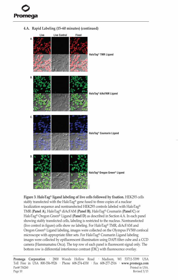
Figure 3. HaloTag® ligand labeling of live cells followed by fixation. HEK293 cellsstably transfected with the HaloTag® gene fused to three copies of a nuclearlocalization sequence and nontransfected HEK293 controls labeled with HaloTag®
TMR (Panel A), HaloTag® diAcFAM (Panel B), HaloTag® Coumarin (Panel C) orHaloTag® Oregon Green® Ligand (Panel D) as described in Section 4.A. In each panelshowing stably transfected cells, labeling is restricted to the nucleus. Nontransfected(live control in figure) cells show no labeling. For HaloTag® TMR, diAcFAM andOregon Green® Ligand labeling, images were collected on the Olympus FV500 confocalmicroscope with appropriate filter sets. For HaloTag® Coumarin Ligand labelingimages were collected by epifluorescent illumination using DAPI filter cube and a CCDcamera (Hammamatsu Orca). The top row of each panel is fluorescent signal only. Thebottom row is differential interference contrast (DIC) with fluorescence overlay.
6300
TA
A.
B.
C.
D.
Live Live Control Fixed
HaloTag® TMR Ligand
HaloTag® diAcFAM Ligand
HaloTag® Coumarin Ligand
HaloTag® Oregon Green® Ligand
tm260.0313:EIVD_TM.qxd 2/28/2013 2:58 PM Page 10
Promega Corporation · 2800 Woods Hollow Road · Madison, WI 53711-5399 USA Toll Free in USA 800-356-9526 · Phone 608-274-4330 · Fax 608-277-2516 · www.promega.comPrinted in USA. Part# TM260Revised 3/13 Page 11
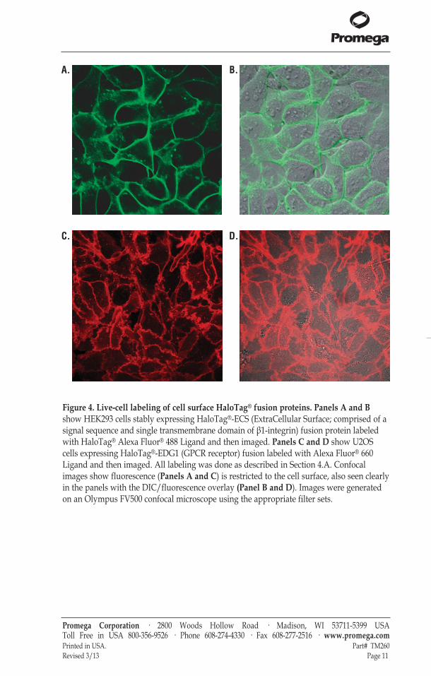
Figure 4. Live-cell labeling of cell surface HaloTag® fusion proteins. Panels A and Bshow HEK293 cells stably expressing HaloTag®-ECS (ExtraCellular Surface; comprised of asignal sequence and single transmembrane domain of β1-integrin) fusion protein labeledwith HaloTag® Alexa Fluor® 488 Ligand and then imaged. Panels C and D show U2OScells expressing HaloTag®-EDG1 (GPCR receptor) fusion labeled with Alexa Fluor® 660Ligand and then imaged. All labeling was done as described in Section 4.A. Confocalimages show fluorescence (Panels A and C) is restricted to the cell surface, also seen clearlyin the panels with the DIC/fluorescence overlay (Panel B and D). Images were generatedon an Olympus FV500 confocal microscope using the appropriate filter sets.
A. B.
C. D.
tm260.0313:EIVD_TM.qxd 2/28/2013 2:58 PM Page 11
4.B. No-Wash Labeling (overnight)
This protocol is intended for labeling of live cells with the cell-permeantHaloTag® TMRDirect™ or R110Direct™ Ligand. This protocol can be used tolabel cells that are adherent or nonadherent, and adherent cells can already beplated or still be in suspension at time of labeling. Representative data areshown in Figure 5.Note: We do not recommend using the HaloTag® TMR Ligand (Cat.# G8251,G8252) for this protocol.
Materials to Be Supplied by the User(Solution compositions are provided in Section 9.B.)• cells expressing HaloTag® fusion protein (in suspension or plated)• chambered cover glass or other cell culture device• complete culture medium appropriate for your cells at 37°C• culture medium lacking phenol red (optional) at 37°C• confocal microscope or wide-field fluorescent microscope equipped
with appropriate filter sets and/or lasers (Section 9.A and Figure 2)• 37 °C + CO2 cell culture incubator
1. Prepare a 1:200 dilution of HaloTag® TMRDirect™ or R110Direct™ Ligandin warm culture medium just prior to addition to cells. This is a 5Xworking stock solution.
2. For adherent cells: Replace one-fifth of the existing volume of medium withthe 5X HaloTag® ligand working stock solution, and mix gently.
For cell suspensions: Add 5X ligand working stock to existing cellsuspension, resulting in a 1X final concentration.
Step 2 results in the recommended final labeling concentration of 100nMHaloTag® TMRDirect™ or R110Direct™ Ligand.
3. Plate cells (if necessary), and incubate overnight in a 37°C + CO2 cellculture incubator.
4. Gently replace the ligand-containing medium with an equal (or greater)volume of warm fresh medium, or fix cells (for end-point assays; seeSection 4.D).
5. Transfer to an imaging device, and capture images.
Promega Corporation · 2800 Woods Hollow Road · Madison, WI 53711-5399 USA Toll Free in USA 800-356-9526 · Phone 608-274-4330 · Fax 608-277-2516 · www.promega.comPart# TM260 Printed in USA.Page 12 Revised 3/13
tm260.0313:EIVD_TM.qxd 2/28/2013 2:58 PM Page 12
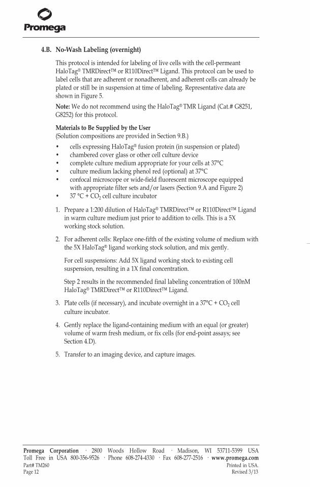
Figure 5. No-wash, live-cell labeling results in high signal-to-noise ratio andspecificity. U2OS cells stably expressing HaloTag® protein fused to three copies of anuclear localization sequence were labeled with the HaloTag® R110Direct™ Ligandas described in Section 4.B. HaloTag® labeling shows a high signal-to-noise ratioand specificity: the label is restricted to the nucleus of all cells. Panel A showsfluorescence image of cells labeled with R110Direct™ Ligand; Panel B is an overlayof the fluorescent and DIC image. Images were generated on an Olympus FV500confocal microscope using the appropriate filter sets.
4.C. Pulse-Chase Labeling
This protocol is intended to serve as a guide for pulse-chase labeling cellsexpressing HaloTag® fusions in order to directly observe protein turnover orprotein trafficking. This protocol is intended for labeling live cells with any ofthe HaloTag® Ligands. Representative data are shown in Figure 6.
Materials to Be Supplied by the User(Solution compositions are provided in Section 9.B.)• chambered cover glass with cells expressing HaloTag® fusion protein• complete culture medium appropriate for your cells at 37°C• culture medium lacking phenol red at 37°C (optional)• 1X PBS (pH 7.5, optional for washes)• confocal microscope or wide-field fluorescent microscope equipped with
appropriate filter sets and lasers (Section 9.A and Figure 2)• 37°C + CO2 cell culture incubator
1. Prepare a 1:200 dilution of pulse labeling ligand in warm culture mediumjust prior to addition to cells. This is a 5X working stock solution.
2. Label cells by replacing one-fifth of the existing volume of medium withthe 5X HaloTag® ligand working stock solution, and mix gently. Thisresults in the recommended final labeling concentrations of 5μM TMR,3.5µM Alexa Fluor® 660; 1μM diAcFAM, Oregon Green® or Alexa Fluor®
488; 10μM Coumarin; and 100nM TMRDirect™ or R110Direct™ Ligands.Note: If protein fusion is membrane-bound with HaloTag® protein on thesurface, pulse labeling should be done with an impermeable ligand (AlexaFluor® 488 or Alexa Fluor® 660 Ligand) to label the surface (plasmamembrane) pool first.
Promega Corporation · 2800 Woods Hollow Road · Madison, WI 53711-5399 USA Toll Free in USA 800-356-9526 · Phone 608-274-4330 · Fax 608-277-2516 · www.promega.comPrinted in USA. Part# TM260Revised 3/13 Page 13
7530
TA
tm260.0313:EIVD_TM.qxd 2/28/2013 2:58 PM Page 13
4.C. Pulse-Chase Labeling (continued)
3. For Rapid-Labeling ligands (TMR, Oregon Green®, diAcFAM, Coumarinand Alexa Fluor® Ligands), incubate for 15 minutes in a 37°C + CO2 cellculture incubator.For No-Wash-Labeling ligands (TMRDirect™ and R110Direct™ Ligands),incubate overnight in a 37°C + CO2 cell culture incubator.
4. If chase labeling is to take place immediately following the pulse, thenproceed immediately to Step 5.If chase labeling is not to take place immediately, simply replace theligand-containing medium with an equal volume of warm fresh mediumand either allow time to pass prior to chase and/or execute intendedbiology as per your specific needs prior to Step 5.
5. Prepare a 1:1,000 dilution of chase ligand in warm culture medium justprior to addition to cells, and gently replace the medium currently on yourcells with an equal volume of this 1X chase labeling ligand.
6. For Rapid-Labeling ligands (TMR, Oregon Green®, diAcFAM, Coumarinand Alexa Fluor® Ligands), incubate for 15 minutes in a 37°C + CO2 cellculture incubator.For No-Wash-Labeling ligands (TMRDirect™ and R110Direct™ Ligands),incubate overnight in a 37°C + CO2 cell culture incubator.
7. For the cell-permeant Rapid Labeling ligands (TMR, Oregon Green®,diAcFAM and Coumarin Ligands) gently replace the ligand-containingmedium with an equal (or greater) volume of warm fresh medium (or 1XPBS [pH 7.5]). Repeat this two times, ending with warm complete medium,for a total of three complete rinses and proceed to Step 8.For the cell-impermeant Rapid Labeling ligands (Alexa Flour® 488 or 660Ligand), gently replace the ligand-containing medium with an equal (orgreater) volume of warm fresh medium twice and proceed to Step 10.
For No-Wash-Labeling ligands (TMRDirect™ and R110Direct™ ), gentlyreplace the ligand-containing medium with an equal (or greater) volume ofwarm fresh medium, and proceed to Step 10.
8. Incubate cells in complete culture medium at 37°C + CO2 in a cell cultureincubator for 30 minutes to wash out unbound ligand.
9. Replace the medium with an equal volume of fresh warm culture medium(use of medium lacking phenol red may improve imaging).
10. Transfer to a microscope, and capture images.
Promega Corporation · 2800 Woods Hollow Road · Madison, WI 53711-5399 USA Toll Free in USA 800-356-9526 · Phone 608-274-4330 · Fax 608-277-2516 · www.promega.comPart# TM260 Printed in USA.Page 14 Revised 3/13
tm260.0313:EIVD_TM.qxd 2/28/2013 2:58 PM Page 14
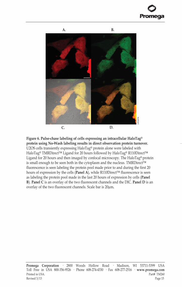
Figure 6. Pulse-chase labeling of cells expressing an intracellular HaloTag®
protein using No-Wash labeling results in direct observation protein turnover.U2OS cells transiently expressing HaloTag® protein alone were labeled withHaloTag® TMRDirect™ Ligand for 20 hours followed by HaloTag® R110Direct™Ligand for 20 hours and then imaged by confocal microscopy. The HaloTag® proteinis small enough to be seen both in the cytoplasm and the nucleus. TMRDirect™fluorescence is seen labeling the protein pool made prior to and during the first 20hours of expression by the cells (Panel A), while R110Direct™ fluorescence is seenas labeling the protein pool made in the last 20 hours of expression by cells (PanelB). Panel C is an overlay of the two fluorescent channels and the DIC. Panel D is anoverlay of the two fluorescent channels. Scale bar is 20µm.
Promega Corporation · 2800 Woods Hollow Road · Madison, WI 53711-5399 USA Toll Free in USA 800-356-9526 · Phone 608-274-4330 · Fax 608-277-2516 · www.promega.comPrinted in USA. Part# TM260Revised 3/13 Page 15
1051
8TA
A.
C. D.
B.
tm260.0313:EIVD_TM.qxd 2/28/2013 2:58 PM Page 15
4.D. Fixing Cells after Labeling
This protocol is intended to serve as a guide for fixation of cells expressing aHaloTag® fusion protein. The covalent bond that forms between the ligandand HaloTag® protein during live-cell labeling and the stability of the dyesallows you to subsequently fix, permeabilize and wash the cells usingstandard conditions without significant loss of the specific fluorescent signal.We recommend the use of paraformaldehyde (PFA) as a fixative because itcrosslinks proteins in cells and at the membrane and has the added benefit ofreducing cell loss from the growth surface.
Fixed cells can be treated with detergents, such as Triton® X-100, topermeabilize cells for downstream immunocytochemical applications. Theconditions here are sufficient to permeabilize the plasma membrane.Alternative or additional detergents might be necessary to permeabilize otherstructures. Representative data are shown in Figures 3, 7 and 8.
Materials to Be Supplied by the User(Solution compositions are provided in Section 9.B.)All solutions should be at room temperature unless otherwise specified.• 4% paraformaldehyde/0.2M sucrose/1X PBS (pH 7.5), warmed to 37°C• 1X PBS buffer (pH 7.5)• 1X PBS + 0.1% Triton® X-100• confocal microscope or wide-field fluorescent microscope equipped with
appropriate filter sets and lasers (Section 9.A and Figure 2).
1. Follow Steps 1–3 of Section 4.A (Rapid Labeling) or 4.B (No-WashLabeling), or Steps 1–6 of Section 4.C (Pulse-Chase Labeling) to label cellswith a HaloTag® ligand, if desired.
2. Replace the medium with an equal volume of warm 4% paraformaldehyde/0.2M sucrose/1X PBS (pH 7.5), and incubate for 10 minutes at room temperature.
3. Replace fixative with an equal volume of 1X PBS + 0.1% Triton® X-100, andincubate for 10 minutes at room temperature.
4. Replace the detergent-containing solution with an equal volume of 1X PBS,and then either transfer to a microscope and capture images, store at 4°C,or proceed to Section 4.E for immunocytochemistry protocol.
Promega Corporation · 2800 Woods Hollow Road · Madison, WI 53711-5399 USA Toll Free in USA 800-356-9526 · Phone 608-274-4330 · Fax 608-277-2516 · www.promega.comPart# TM260 Printed in USA.Page 16 Revised 3/13
tm260.0313:EIVD_TM.qxd 2/28/2013 2:58 PM Page 16
4.E. Multiplexing with HaloTag® Using Immunocytochemistry (ICC)
This protocol is intended to serve as a guide for multiplexing with HaloTag®
technology in fixed cells by ICC using any antibody of interest. The particularantibody used in this protocol is a purified rabbit polyclonal antibody raisedagainst the HaloTag® protein (9). The antibody was purified using Protein Gaffinity resin and has been shown to detect HaloTag® fusion proteins in bothimmunocytochemistry and Western blot applications with high sensitivity andspecificity. Further, this antibody labels HaloTag® fusion proteins independentlyof HaloTag® ligands. Because these labels do not interfere with one another, it ispossible to colabel HaloTag® fusion proteins with a fluorescent ligand and theAnti-HaloTag® pAb in conjunction with an anti-rabbit secondary antibodybearing a spectrally distinct fluorescent tag. Representative data are shown inFigures 7 and 8.
Materials to Be Supplied by the User(Solution compositions are provided in Section 9.B.)All solutions should be at room temperature. • cells fixed following the steps in Section 4.D• fluorophore-conjugated anti-rabbit secondary antibody (Ab) of choice• PBS + 2% serum from same host as secondary Ab + 0.01% Triton® X-100• PBS + 1% serum from same host as secondary Ab• PBS + 0.1% sodium azide (optional, for storage)• confocal microscope or wide-field fluorescent microscope equipped with
appropriate filter sets and lasers (Section 9.A and Figure 2)
1. Replace the 1X PBS with an equal volume of PBS + 2% serum from samehost as secondary Ab + 0.01% Triton® X-100, and block for 1 hour at roomtemperature.
2. Dilute the Anti-HaloTag® pAb 1:500 in PBS + 1% serum from same host assecondary Ab to a final labeling concentration of 2μg/ml.
3. Replace the blocking solution with the antibody solution, and incubate for1 hour at room temperature.
4. Wash cells twice with PBS + 1% serum from same host as secondary Ab for10 minutes at room temperature.
5. Dilute the secondary antibody according to the manufacturer'srecommendations in PBS + 1% s serum from same host as secondary Ab.
6. Replace the wash solution with the secondary antibody solution, andincubate for 30 minutes at room temperature.
7. Wash cells twice with PBS + 1% serum from same host as secondary Ab for10 minutes each wash at room temperature.
8. Replace wash solution with 1X PBS.
9. Transfer to a microscope, and capture images. Store cells at 4°C in 1X PBSor PBS + 0.1% sodium azide.
Promega Corporation · 2800 Woods Hollow Road · Madison, WI 53711-5399 USA Toll Free in USA 800-356-9526 · Phone 608-274-4330 · Fax 608-277-2516 · www.promega.comPrinted in USA. Part# TM260Revised 3/13 Page 17
tm260.0313:EIVD_TM.qxd 2/28/2013 2:58 PM Page 17
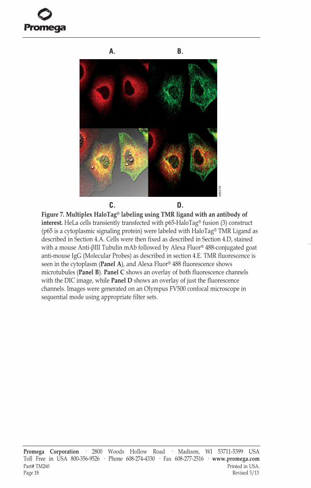
Figure 7. Multiplex HaloTag® labeling using TMR ligand with an antibody ofinterest. HeLa cells transiently transfected with p65-HaloTag® fusion (3) construct(p65 is a cytoplasmic signaling protein) were labeled with HaloTag® TMR Ligand asdescribed in Section 4.A. Cells were then fixed as described in Section 4.D, stainedwith a mouse Anti-βIII Tubulin mAb followed by Alexa Fluor® 488-conjugated goatanti-mouse IgG (Molecular Probes) as described in section 4.E. TMR fluorescence isseen in the cytoplasm (Panel A), and Alexa Fluor® 488 fluorescence showsmicrotubules (Panel B). Panel C shows an overlay of both fluorescence channelswith the DIC image, while Panel D shows an overlay of just the fluorescencechannels. Images were generated on an Olympus FV500 confocal microscope insequential mode using appropriate filter sets.
Promega Corporation · 2800 Woods Hollow Road · Madison, WI 53711-5399 USA Toll Free in USA 800-356-9526 · Phone 608-274-4330 · Fax 608-277-2516 · www.promega.comPart# TM260 Printed in USA.Page 18 Revised 3/13
4883
TA
A. B.
C. D.
tm260.0313:EIVD_TM.qxd 2/28/2013 2:58 PM Page 18
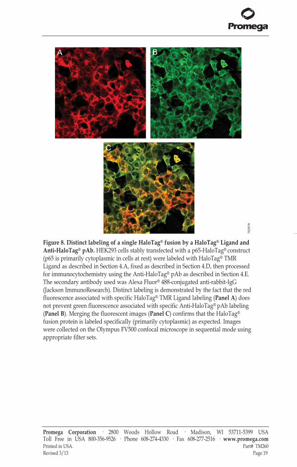
Figure 8. Distinct labeling of a single HaloTag® fusion by a HaloTag® Ligand andAnti-HaloTag® pAb. HEK293 cells stably transfected with a p65-HaloTag® construct(p65 is primarily cytoplasmic in cells at rest) were labeled with HaloTag® TMRLigand as described in Section 4.A, fixed as described in Section 4.D, then processedfor immunocytochemistry using the Anti-HaloTag® pAb as described in Section 4.E.The secondary antibody used was Alexa Fluor® 488-conjugated anti-rabbit-IgG(Jackson ImmunoResearch). Distinct labeling is demonstrated by the fact that the redfluorescence associated with specific HaloTag® TMR Ligand labeling (Panel A) doesnot prevent green fluorescence associated with specific Anti-HaloTag® pAb labeling(Panel B). Merging the fluorescent images (Panel C) confirms that the HaloTag®
fusion protein is labeled specifically (primarily cytoplasmic) as expected. Imageswere collected on the Olympus FV500 confocal microscope in sequential mode usingappropriate filter sets.
Promega Corporation · 2800 Woods Hollow Road · Madison, WI 53711-5399 USA Toll Free in USA 800-356-9526 · Phone 608-274-4330 · Fax 608-277-2516 · www.promega.comPrinted in USA. Part# TM260Revised 3/13 Page 19
7528
TA
tm260.0313:EIVD_TM.qxd 2/28/2013 2:58 PM Page 19
4.F. Quantitation of HaloTag® Protein Fusions, SDS-PAGE Analysis
The following protocol is intended to serve as a guide for SDS-PAGE-basedHaloTag® applications. Because the covalent bond between the HaloTag®
protein and HaloTag® ligand withstands denaturation and the fluorescentdyes in the ligands are stable, you can directly quantify labeled fusion proteinfollowing SDS-PAGE using a fluorescent scanner (i.e., cell-to-gel analysis).
Such gels also can be subsequently used for Western blot analysis using theAnti-HaloTag® mAb at a final labeling concentration of 1μg/ml (1:1,000dilution; see Section 5 below for protocol).Note: Although all of the HaloTag® ligands perform in this function, theHaloTag® TMR, TMRDirect™ or R110Direct™ Ligands are recommended fordirect SDS-PAGE fluorescence scanning of an intracellularly expressed proteinfusion. Either of the AlexaFluor®-containing ligands is recommended forextracellularly expressed proteins that place HaloTag® on the surface of cells.Representative data are shown in Figure 9.
Materials to be Supplied by the User(Solution compositions are provided in Section 9.B.)• cells expressing HaloTag® fusion protein• 1X PBS (pH 7.5)• 1X SDS sample buffer• heat block or water bath at 95°C• equipment and running buffer necessary for SDS-PAGE• fluorescent scanner
1. To label cells, follow one of the following protocols:Steps 1–3 of Section 4.A or 4.B (Rapid or No Wash Labeling)Steps 1-6 of Section 4.C (Pulse-Chase Labeling).Note: There is no need to go through the 30-minute wash step for anintracellularly expressed protein fusion labeled by Rapid Labeling protocolas all unbound ligand is very small and will migrate on the gel much fasterthan the labeled fusion protein (with sample dye front).
2. Replace ligand containing media with 1X PBS (pH 7.5) in order to avoid aprominent band on the gel stemming from complete media.
3. Lyse cells by replacing the 1X PBS from the wells with approximately100–150μl of 1X SDS sample buffer per cm2 of cell growth area.
4. Collect cell lysate, and incubate for 5 minutes at 95°C.
5. Perform SDS-PAGE by loading approximately 10μl (5–10μg total protein)of each sample per well of the gel, or store samples at –20°C for later use.
Promega Corporation · 2800 Woods Hollow Road · Madison, WI 53711-5399 USA Toll Free in USA 800-356-9526 · Phone 608-274-4330 · Fax 608-277-2516 · www.promega.comPart# TM260 Printed in USA.Page 20 Revised 3/13
tm260.0313:EIVD_TM.qxd 2/28/2013 2:58 PM Page 20
6. Analyze the gel on a fluorescence scanner.Note: The dye front might contain fluorescent material that can complicatedetection (unbound ligand and/or tracking dyes used in sample buffers).To eliminate these sources of nonspecific fluorescence, simply run the geluntil the dye front migrates off of the gel or cut the dye front off of thebottom of the gel before scanning.
7. After scanning, the proteins can be transferred to nitrocellulose for Westernblot analysis if used promptly (i.e., gel is not fixed, remains moist and isnot left in buffer or deionized water).
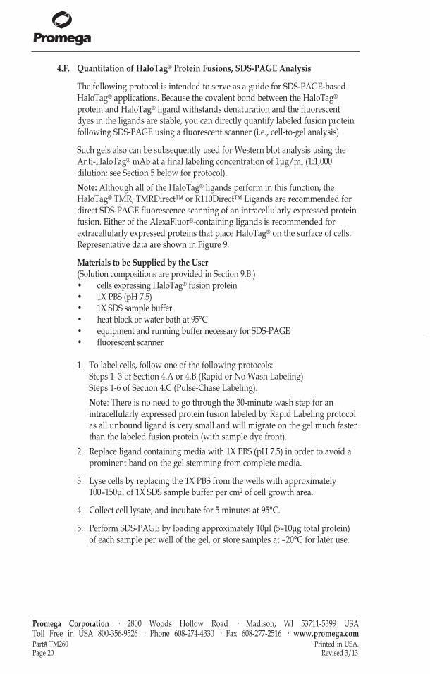
Figure 9. SDS-PAGE analysis shows fast, efficient and highly specific labeling ofthe HaloTag® protein in live cells. CHO-K1 untransfected control cells (lanes 1–6)or cells transiently transfected with a vector containing HaloTag® alone (lanes 7–12)were labeled with 5μM HaloTag® TMR Ligand for different periods of time at 37°C(0.5, 1, 2, 5, 15 or 30 minutes) and treated as described in Section 4. Following SDS-PAGE, the gel was analyzed on a Hitachi FMBIO® fluorescence imaging system.
Promega Corporation · 2800 Woods Hollow Road · Madison, WI 53711-5399 USA Toll Free in USA 800-356-9526 · Phone 608-274-4330 · Fax 608-277-2516 · www.promega.comPrinted in USA. Part# TM260Revised 3/13 Page 21
4781
TA
121110987654321
– ~33kDa
tm260.0313:EIVD_TM.qxd 2/28/2013 2:58 PM Page 21
4.G. Flow Cytometry Analysis
Representative data are shown in Figure 10.
Materials to Be Supplied by the User(Solution compositions are provided in Section 9.B.)• large-well format plate of HaloTag®-expressing cells and negative control
cells that do not express the HaloTag® fusion protein• complete culture medium appropriate for your cells at 37°C• for adherent cells: 1X PBS (pH 7.5) and 1X PBS containing 3mM EDTA
(nonenzymatic) or trypsin solution (see Step 2 below)• flow cytometer with appropriate filter sets and lasers for ligand used
(Section 9.A and Figure 2)• 37°C + CO2 cell culture incubator
1. Label cells expressing HaloTag® protein with ligand of choice (Section 4.A,Rapid Labeling or 4.B, No-Wash Labeling).Recommended controls:a. Label cells that are not expressing HaloTag® protein to assess
background with chosen ligand.b. Prepare unlabeled cells expressing HaloTag® protein to
assess background from endogenous cell fluorescence or morphologicalchanges due to expression.
2. For adherent cells with HaloTag® fusion protein expressed on the cellsurface: Rinse cells twice with an equal volume of 1X PBS, incubate in 1XPBS with 3mM EDTA or use other nonenzymatic solution at 37°C for 5–15minutes (depending on cell line) in order to gently get the cells off of theplate.For adherent cells with HaloTag® fusion protein expressed inside the cell:Suspend cells using trypsin-containing solution in a manner appropriatefor the cell line.
3. Collect, pellet, count and resuspend cells in medium at 37°C to aconcentration of 0.5 to 1 × 10⁶ cells/ml.
4. Analyze cells by flow cytometry.
Promega Corporation · 2800 Woods Hollow Road · Madison, WI 53711-5399 USA Toll Free in USA 800-356-9526 · Phone 608-274-4330 · Fax 608-277-2516 · www.promega.comPart# TM260 Printed in USA.Page 22 Revised 3/13
tm260.0313:EIVD_TM.qxd 2/28/2013 2:58 PM Page 22
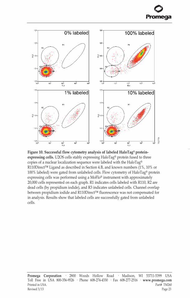
Figure 10. Successful flow cytometry analysis of labeled HaloTag® protein-expressing cells. U2OS cells stably expressing HaloTag® protein fused to threecopies of a nuclear localization sequence were labeled with the HaloTag®
R110Direct™ Ligand as described in Section 4.B, and known numbers (1%, 10% or100% labeled) were gated from unlabeled cells. Flow cytometry of HaloTag® proteinexpressing cells was performed using a MoFlo® instrument with approximately20,000 cells represented on each graph. R1 indicates cells labeled with R110, R2 aredead cells (by propidium iodide), and R3 indicates unlabeled cells. Channel overlapbetween propidium iodide and R110Direct™ fluorescence was not compensated forin analysis. Results show that labeled cells are successfully gated from unlabeledcells.
Promega Corporation · 2800 Woods Hollow Road · Madison, WI 53711-5399 USA Toll Free in USA 800-356-9526 · Phone 608-274-4330 · Fax 608-277-2516 · www.promega.comPrinted in USA. Part# TM260Revised 3/13 Page 23
7527
TA
tm260.0313:EIVD_TM.qxd 2/28/2013 2:58 PM Page 23
5. Protein Detection Using HaloTag® Monoclonal Antibody (for Western blotting)
The following protocol is meant to serve as a general guide for the use of Anti-HaloTag® Monoclonal Antibody (Cat.# G9211) in Western blotting. The Anti-HaloTag® pAb (Cat.# G9281) is recommended for all other immunologicaldetection methods. Representative data are shown in Figure 11.
Materials to Be Supplied by the User• mammalian cells expressing HaloTag® fusion• appropriate mammalian lysis buffer (e.g., Mammalian Lysis Buffer,
Cat.# G9381)• SDS-PAGE gel and gel running apparatus• nitrocellulose or PVDF membrane and Western blotting apparatus• TBST (1X TBS with 0.1% Tween®)• 5% BSA in TBST• anti-mouse IgG secondary antibody appropriately conjugated for Western
blotting analysis and corresponding detection substrate (e.g., Anti-Mouse IgG (H+L), AP Conjugate Cat.# S3721) andWestern Blue® Stabilized Substrate for Alkaline Phosphatase, Cat.# S3841)
• rotating platform for gentle agitation
1. Lyse mammalian cells expressing HaloTag® fusion using an appropriate lysisbuffer of choice. Recommended control: Cells that are not expressing HaloTag® (untransfected).
2. Run lysates on SDS-PAGE, and transfer to nitrocellulose or PDVF membraneas per manufacturer protocol.
3. Place resulting nitrocellulose or PVDF membranes in TBST containing 5% BSAto block for an hour at room temperature or overnight at 4°C, with gentleagitation.
4. Replace blocking buffer with a solution of anti-HaloTag® mAb at 1:1,000dilution in TBST, and leave at room temperature for 1 hour with gentleagitation.
5. Replace primary antibody-containing solution with TBST. Wash 3 times for 15 minutes at room temperature with gentle agitation.
6. Replace wash with anti-mouse IgG conjugated secondary antibody atmanufacturer recommended concentration (e.g., for anti-mouse IgG-APconjugated use at 1:7,500 dilution) in TBST and leave for 30 minutes at roomtemperature with gentle agitation.
7. Replace secondary solution with TBST. Wash 3 times for 15 minutes at roomtemperature with gentle agitation.
8. Detect HaloTag® protein bands using appropriate substrate (e.g., APsubstrate).
Promega Corporation · 2800 Woods Hollow Road · Madison, WI 53711-5399 USA Toll Free in USA 800-356-9526 · Phone 608-274-4330 · Fax 608-277-2516 · www.promega.comPart# TM260 Printed in USA.Page 24 Revised 3/13
tm260.0313:EIVD_TM.qxd 2/28/2013 2:58 PM Page 24
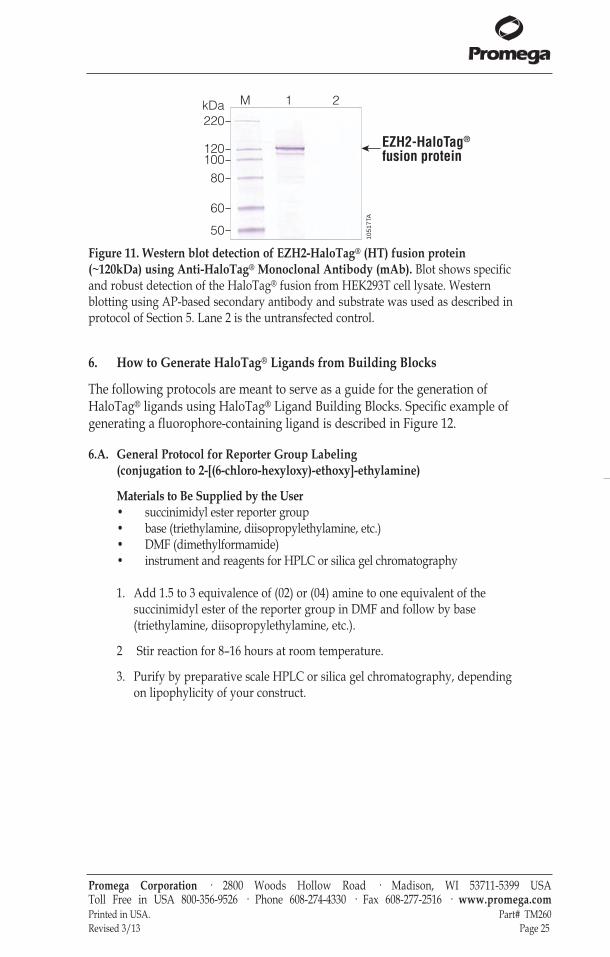
Figure 11. Western blot detection of EZH2-HaloTag® (HT) fusion protein(~120kDa) using Anti-HaloTag® Monoclonal Antibody (mAb). Blot shows specificand robust detection of the HaloTag® fusion from HEK293T cell lysate. Westernblotting using AP-based secondary antibody and substrate was used as described inprotocol of Section 5. Lane 2 is the untransfected control.
6. How to Generate HaloTag® Ligands from Building Blocks
The following protocols are meant to serve as a guide for the generation ofHaloTag® ligands using HaloTag® Ligand Building Blocks. Specific example ofgenerating a fluorophore-containing ligand is described in Figure 12.
6.A. General Protocol for Reporter Group Labeling (conjugation to 2-[(6-chloro-hexyloxy)-ethoxy]-ethylamine)
Materials to Be Supplied by the User• succinimidyl ester reporter group• base (triethylamine, diisopropylethylamine, etc.)• DMF (dimethylformamide)• instrument and reagents for HPLC or silica gel chromatography
1. Add 1.5 to 3 equivalence of (02) or (04) amine to one equivalent of thesuccinimidyl ester of the reporter group in DMF and follow by base(triethylamine, diisopropylethylamine, etc.).
2 Stir reaction for 8–16 hours at room temperature.
3. Purify by preparative scale HPLC or silica gel chromatography, dependingon lipophylicity of your construct.
Promega Corporation · 2800 Woods Hollow Road · Madison, WI 53711-5399 USA Toll Free in USA 800-356-9526 · Phone 608-274-4330 · Fax 608-277-2516 · www.promega.comPrinted in USA. Part# TM260Revised 3/13 Page 25
1051
7TA
M 1 2
220
120100
80
60
50
kDa
EZH2-HaloTag®
fusion protein
tm260.0313:EIVD_TM.qxd 2/28/2013 2:58 PM Page 25
6.B. Protein Labeling with HaloTag® Amine Ligand
Materials to Be Supplied by the User• protein of interest for conjugation• water, 0.1M MES (pH 4.7–6.0) or 0.1 M sodium phosphate (pH 7.3)• DMSO or DMF• EDC (1-ethyl-3-(3-dimethylaminopropyl) carbodiimide hydrochloride)• Sephadex® G-25 (for gel filtration separation of ligated protein)
1. Dissolve the protein to be modified at a concentration of 10mg/ml in eitherwater, 0.1M MES (pH 4.7–6.0), or 0.1M sodium phosphate (pH 7.3).
2. Prepare HaloTag® Amine (02 or 04) Ligand stock solution at 5mg/ml inDMSO or DMF.
3. Take a 10-fold-molar-excess-to-protein aliquot of this HaloTag® ligandstock solution and dilute it in buffer (same as what is used in Step 1).
4. Add this aqueous HaloTag® ligand stock solution to the protein solution;the final protein concentration will be 5 mg/ml or greater.
5. Add EDC (1-ethyl-3-(3-dimethylaminopropyl) carbodiimide hydrochloride)in ten-fold molar excess to protein as a 0.1 to 0.5M aqueous solution andreact for 2 hours at room temperature.
6. Purify the ligated protein by gel filtration separation using Sephadex® G-25in the buffer of choice.
6.C. Antibody Labeling with HaloTag® Succinimidyl Ester Ligand
Materials to Be Supplied by the User• antibody of interest for conjugation• 0.1M sodium phosphate, 0.15M sodium chloride (pH 7.2) [pH 7.0 to 7.6 can
be used]• DMSO or DMF• Sephadex® G-25 (for gel filtration separation of ligated antibody) or means of
dialysis against 0.1M sodium phosphate, 0.15M sodium chloride, 10mMEDTA (pH 7.2)
1. Dissolve antibody in 0.1M sodium phosphate, 0.15M sodium chloride (pH7.2) at 1–5 mg/ml.
2. Prepare HaloTag® Succinimidyl Ester (02 or 04) Ligand stock solution in5mg/ml DMSO or DMF.
3. Add 10–40µl of HaloTag® Ligand stock solution per millilter of 1mg/mlantibody solution and react at room temperature for 30 minutes.Note: A 12-fold molar excess works well to ensure at least one label perantibody. Most protocols for general antibody labeling uses 20-fold excess,but higher concentrations of ligand to antibody will label more amines.Further, excess labels may or may not hinder the antibody-epitopeinteraction.
Promega Corporation · 2800 Woods Hollow Road · Madison, WI 53711-5399 USA Toll Free in USA 800-356-9526 · Phone 608-274-4330 · Fax 608-277-2516 · www.promega.comPart# TM260 Printed in USA.Page 26 Revised 3/13
tm260.0313:EIVD_TM.qxd 2/28/2013 2:58 PM Page 26
4. Purify the ligated antibody by gel filtration using Sephadex® G-25 or bydialysis against 0.1M sodium phosphate, 0.15M sodium chloride, 10mMEDTA (pH 7.2).
6.D. Specific Example Protocol for Generating an Alexa Fluor® 488 HaloTag®
Ligand
1. Begin with 5mg of Alexa Fluor® 488 NHS ester (7.8 × 10⁻⁶ mol) from LifeTechnologies.
2. In purchased vial, add a small magnetic stir bar and dissolve the dye in0.5ml DMF (stored over molecular sieves to remove excess water).
3. With continuous stirring, add a 3-fold excess of amino ligand (2.3 × 10⁻⁵
mol) followed by one drop of diisoproplyethylamine (excess). Note: Add the amine ligand as a stock solution in either DMF or methylenechloride. It stores well at –78°C in solution. If using a dye-SE, you can use 1.5 to 3 equivalence of ligand to dye-SE.
4. Stir for 12 hours and monitor by analytical HPLC (C12 or C18) using 0.1%TFA, aqueous phase and acetonitrile, organic phase.
5. Once the reaction is determined to be complete, dilute the reaction mixturewith 1ml of water and inject on a preparative HPLC column (VarianMicrosorb 60-8 C18; 250 x 21.4 mm). Purify the compound using the samemobile phase as in the analytical HPLC in Step 4.
7. Labeling Proteins in Mammalian Cell Lysates or Proteins Expressed in Cell-Free Systems
Fluorescent labeling of HaloTag® fusion protein with the HaloTag®
TMRDirect™ Ligand provides a rapid and convenient method to monitorprotein expression and the purification efficiency. HaloTag® TMRDirect™Ligand (555Ex/585Em) is used in the example below because it is provided withthe HaloTag® Mammalian Protein Detection and Purification System (Cat.#G6795). If desired, HaloTag® R110Direct™ Ligand (502Ex/527Em) can easily besubstituted for this ligand.
For further information regarding HaloTag® labeling after expression refer toTechnical Manual TM348 HaloTag® Mammalian Protein Detection and PurificationSystems at: www.promega.com/protocols
Promega Corporation · 2800 Woods Hollow Road · Madison, WI 53711-5399 USA Toll Free in USA 800-356-9526 · Phone 608-274-4330 · Fax 608-277-2516 · www.promega.comPrinted in USA. Part# TM260Revised 3/13 Page 27
tm260.0313:EIVD_TM.qxd 2/28/2013 2:58 PM Page 27
7. Labeling Proteins in Mammalian Cell Lysates or Proteins Expressed in Cell-Free Systems (continued)
Materials to be Supplied by the User(Solution compositions are provided in Section 9.B.)• lysate containing HaloTag® fusion protein• DMSO or 1X PBS (pH 7.5), optional• 4X SDS sample buffer• heat block or water bath at 70°C• equipment and running buffer necessary for SDS-PAGE• fluorescent scanner
1. Dilute the HaloTag® TMRDirect™ Ligand stock solution (100μM) twofold inDMSO to make a 50μM working solution that can be stored, protected fromlight, at –20°C; alternatively, the stock solution can be prepared in 1X PBS butcannot be stored.
2. Combine 10μl of lysate containing the HaloTag® fusion protein or theequivalent amount of unbound fraction with 19μl of HaloTag® ProteinPurification Buffer and 1μl of 50μM HaloTag® TMRDirect™ Ligand.
3. Incubate at room temperature for 15 minutes protected from light.
4. Add 10μl of 4X SDS sample buffer (Section 9.B), and heat at 70°C for 3minutes.
5. Load 10μl onto an SDS-polyacrylamide gel.
6. Scan the gel on a fluorescent scanner and quantitate band intensities.Note: The dye front might contain fluorescent material that can complicatedetection. It might be necessary to run the gel until the dye front migrates offof the gel or cut the dye front off of the bottom of the gel before scanning it.
Promega Corporation · 2800 Woods Hollow Road · Madison, WI 53711-5399 USA Toll Free in USA 800-356-9526 · Phone 608-274-4330 · Fax 608-277-2516 · www.promega.comPart# TM260 Printed in USA.Page 28 Revised 3/13
tm260.0313:EIVD_TM.qxd 2/28/2013 2:58 PM Page 28
8. Labeling E. coli Lysate to Analyze Protein Expression Level
These protocols are intended to serve as a guide for the labeling of E. colilysates and intact bacteria.
8.A. Expressing HaloTag® Fusions in E. coli
Materials to be Supplied by the User(Solution compositions are provided in Section 9.B.)• transformed E. coli KRX colonies expressing HaloTag® fusion protein
(Single Step (KRX) Competent Cells [Cat.# L3002])• LB containing appropriate antibiotics• glucose• L-Rhamnose Monohydrate (Cat.# L5701 or L5702)• bacterial growth incubator at 25°C and 37°C
Note: These expression conditions are unique to E. coli KRX cells.
1. Inoculate a colony into 2ml LB + appropriate antibiotics, and growovernight at 37°C.Note: For toxic proteins add 0.4% glucose to the medium.
2. Dilute the overnight culture 1:100 into 2ml auto induction medium.
8.B. Labeling E. coli Lysate to Analyze Protein Expression Level
Materials to be Supplied by the User• 50mM HEPES (pH 7.5)• 10X Fast Break™ Cell Lysis Reagent (Cat.# V8571, V8572 or V8573)• lysozyme• RQ1 RNase-Free DNase (Cat.# M6101)• 4X SDS sample buffer• freezer at –70°C• centrifuge capable of 14,000 × g at 4°C• heat block or water bath at 70°C• equipment and running buffer necessary for SDS-PAGE• fluorescent scanner
Promega Corporation · 2800 Woods Hollow Road · Madison, WI 53711-5399 USA Toll Free in USA 800-356-9526 · Phone 608-274-4330 · Fax 608-277-2516 · www.promega.comPrinted in USA. Part# TM260Revised 3/13 Page 29
Mode of Induction Approximate O.D.at Induction
GrowthTemperature Growth Time
Late: 0.15% glucose +0.2% rhamnose A600 = 1–1.2 25°C 24 hours
Early: 0.05% glucose +0.05% rhamnose A600 = 0.6–0.8 25°C 16–18 hours
tm260.0313:EIVD_TM.qxd 2/28/2013 2:58 PM Page 29
8.B. Labeling E. coli Lysate to Analyze Protein Expression Level (continued)
1. Pellet 1.5ml culture and resuspend in 0.5ml of 50mM HEPES (pH 7.5).Freeze for 20 minutes at –70°C.
2. Thaw the cells at room temperature and lyse by adding 0.5ml lysis buffer(1X FastBreak™ Cell Lysis Reagent + 2mg/ml + lysozyme + 0.02 units RQ1RNase-free DNase) for 30 minutes with constant rotation.
3. Keep 0.5ml for analysis of crude lysate and prepare from the other 1mlsoluble lysate by centrifugation at 14,000 × g for 30 minutes at 4°C.
4. Label both crude and soluble lysates with the HaloTag® fluorescent ligand.Dilute the ligand into a 50µM working solution (below). Incubate for 20 minutes at room temperature.
5. Add 10µl of 4X SDS loading buffer, and incubate for 2 minutes at 70°C.
6. Analyze 10–20µl by SDS-PAGE.
7. First detect by a fluorescent scanner, and then stain using Coomassie® blue.Note: The dye front might contain fluorescent material that can complicatedetection. It might be necessary to run the gel until the dye front migratesoff of the gel or cut the dye front off of the bottom of the gel prior to scan.
8.C. Labeling E. coli for Imaging Analysis
Representative data are shown in Figure 12.
Materials to Be Supplied by the User• E. coli culture expressing HaloTag® fusion protein (Section 8.A)• bacterial growth incubator at 25°C• centrifuge capable of 2,000 × g• 1X PBS• chambered coverglass• 0.1% gelatin• confocal microscope equipped with appropriate filter sets and
lasers (Section 9.A and Figure 2)
Promega Corporation · 2800 Woods Hollow Road · Madison, WI 53711-5399 USA Toll Free in USA 800-356-9526 · Phone 608-274-4330 · Fax 608-277-2516 · www.promega.comPart# TM260 Printed in USA.Page 30 Revised 3/13
Component Volume
Lysate 20µl
Ligand (50µM) 1µl
1X 50mM HEPES (pH 7.5) 9µl
Total Volume 30µl
tm260.0313:EIVD_TM.qxd 2/28/2013 2:58 PM Page 30
1. To label bacteria, dilute overnight culture 1:10 in LB, add HaloTag®
TMRDirect™ Ligand or HaloTag® TMR Ligand to a final concentration of1µM (1:100 dilution for TMRDirect™ or 1:5,000 for TMR) and label at 25°Cwithout mixing for 1 hour.
2. Prepare the chambered coverglass by precoating the glass chambers with0.1% gelatin (200μl /chamber of 8-well coverglass) for 1 hour at roomtemperature.
3. In order to wash unbound ligand, follow Steps a–e below, 3 times using 1X PBS.
a. Pellet the cells at 2000 × g for 5 minutes.b. Resuspend in 1ml of 1X PBS.c. Incubate without mixing for 30 minutes at room temperature.d. Pellet the cells at 2000 × g for 5 minutes.e. Discard the 1X PBS.
4. For imaging purposes, after the third wash, resuspend the cells in 200µl of1X PBS, and proceed to Step 5.
For SDS-PAGE analysis, you can collect the cells, lyse and analyze forlabeling efficiency on a gel as described above in Section 8.B.
5. To plate the labeled E. coli, first aspirate the gelatin from the chamberedcover glass and then immediately add the cells; plating these at undiluted,1:2.5, 1:5 and 1:10 dilutions. Allow 2–3 hours for the cells to adhere beforeimaging.
6. Transfer to a confocal microscope, and capture images.
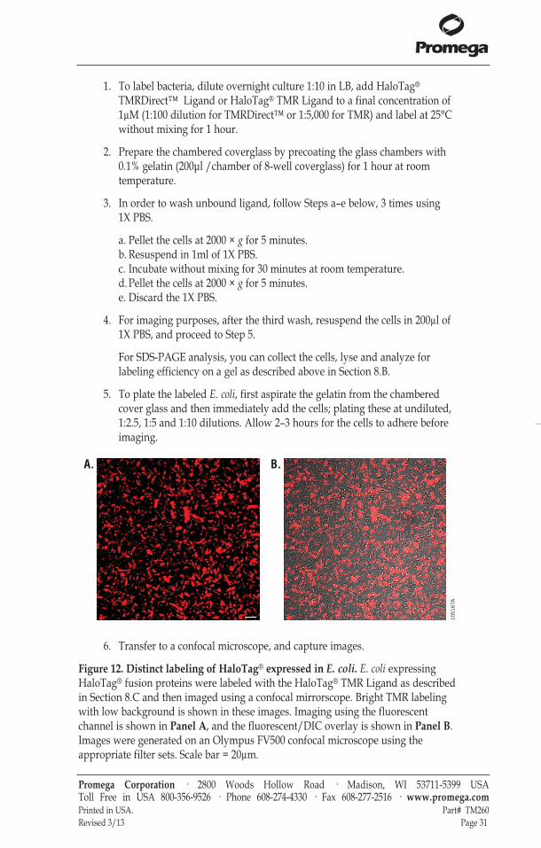
Figure 12. Distinct labeling of HaloTag® expressed in E. coli. E. coli expressingHaloTag® fusion proteins were labeled with the HaloTag® TMR Ligand as describedin Section 8.C and then imaged using a confocal mirrorscope. Bright TMR labelingwith low background is shown in these images. Imaging using the fluorescentchannel is shown in Panel A, and the fluorescent/DIC overlay is shown in Panel B.Images were generated on an Olympus FV500 confocal microscope using theappropriate filter sets. Scale bar = 20µm.
Promega Corporation · 2800 Woods Hollow Road · Madison, WI 53711-5399 USA Toll Free in USA 800-356-9526 · Phone 608-274-4330 · Fax 608-277-2516 · www.promega.comPrinted in USA. Part# TM260Revised 3/13 Page 31
1051
6TA
A. B.
tm260.0313:EIVD_TM.qxd 2/28/2013 2:58 PM Page 31
9. General Information, Tips and Troubleshooting
9.A. General Information
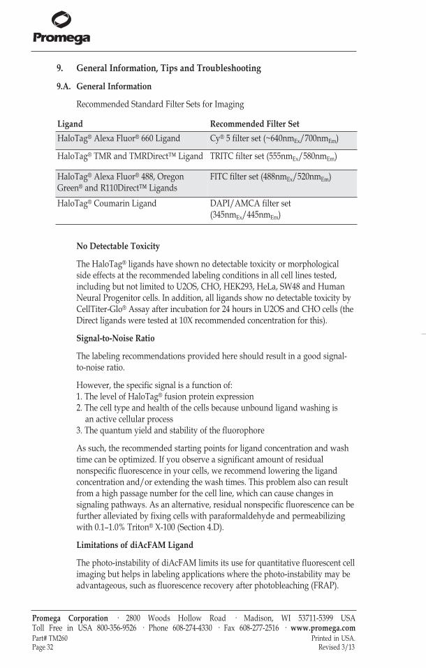
Recommended Standard Filter Sets for Imaging
No Detectable Toxicity
The HaloTag® ligands have shown no detectable toxicity or morphologicalside effects at the recommended labeling conditions in all cell lines tested,including but not limited to U2OS, CHO, HEK293, HeLa, SW48 and HumanNeural Progenitor cells. In addition, all ligands show no detectable toxicity byCellTiter-Glo® Assay after incubation for 24 hours in U2OS and CHO cells (theDirect ligands were tested at 10X recommended concentration for this).
Signal-to-Noise Ratio
The labeling recommendations provided here should result in a good signal-to-noise ratio.
However, the specific signal is a function of:1. The level of HaloTag® fusion protein expression2. The cell type and health of the cells because unbound ligand washing is
an active cellular process3. The quantum yield and stability of the fluorophore
As such, the recommended starting points for ligand concentration and washtime can be optimized. If you observe a significant amount of residualnonspecific fluorescence in your cells, we recommend lowering the ligandconcentration and/or extending the wash times. This problem also can resultfrom a high passage number for the cell line, which can cause changes insignaling pathways. As an alternative, residual nonspecific fluorescence can befurther alleviated by fixing cells with paraformaldehyde and permeabilizingwith 0.1–1.0% Triton® X-100 (Section 4.D).
Limitations of diAcFAM Ligand
The photo-instability of diAcFAM limits its use for quantitative fluorescent cellimaging but helps in labeling applications where the photo-instability may beadvantageous, such as fluorescence recovery after photobleaching (FRAP).
Promega Corporation · 2800 Woods Hollow Road · Madison, WI 53711-5399 USA Toll Free in USA 800-356-9526 · Phone 608-274-4330 · Fax 608-277-2516 · www.promega.comPart# TM260 Printed in USA.Page 32 Revised 3/13
Ligand Recommended Filter SetHaloTag® Alexa Fluor® 660 Ligand Cy® 5 filter set (~640nmEx/700nmEm)
HaloTag® TMR and TMRDirect™ Ligand TRITC filter set (555nmEx/580nmEm)
HaloTag® Alexa Fluor® 488, OregonGreen® and R110Direct™ Ligands
FITC filter set (488nmEx/520nmEm)
HaloTag® Coumarin Ligand DAPI/AMCA filter set(345nmEx/445nmEm)
tm260.0313:EIVD_TM.qxd 2/28/2013 2:58 PM Page 32
Both the HaloTag® Oregon Green® and R110Direct™ Ligands exhibitsignificantly increased photostability compared with diAcFAM and are usefultools in cell-based imaging applications that require quantitation offluorescence or the use of high excitation energies with a green ligand.
Use of HaloTag® Ligands in Serum-Containing Medium
All HaloTag® ligands may be directly diluted into serum-containing medium.However, the HaloTag® diAcFAM and Oregon Green® Ligands must be addedto the cells immediately following this dilution because the diacetyl groupscan be hydrolyzed by serum esterases. This deacetylation converts theHaloTag® diAcFAM and Oregon Green® Ligands into cell-impermeantfluorescent ligand derivatives. Once inside the cell, these ligands are modified,and, as will all of the other cell-permeant ligands, excess unbound ligand iseliminated from the cell in a time- and temperature dependent manner by anactive cellular mechanism.
While all cell types tested allow entry of the cell-permeant ligands and accessto all major subcellular compartments, there are differences in rates of efflux.In general, efflux of the HaloTag® diAcFAM and Oregon Green® Ligands issignificantly slower than that of other ligands, and the rate of efflux can varysignificantly between cell types. Cells that have delayed efflux of unboundmodified ligand might have limiting amounts of this active mechanism orsuffer from poor health. Thus, while the use of serum results in a strict timingrequirement for the addition of ligand to the cell culture, the presence ofserum minimizes stress to cells and maintains their ability to actively eliminateunbound ligand, which results in lower background.
Streamlining the Protocol
The cell-impermeant Alexa Fluor® 488 and Alexa Fluor® 660 Ligands do notrequire a 30-minute wash step as unbound ligand does not enter cells. Amongthe cell-permeant ligand options, HaloTag® TMR and Coumarin Ligands areeasily washed from cells. Therefore, you may be able to streamline theprotocol in Section 4.A, Steps 4–6, without affecting the signal-to-noise ratio.
Promega Corporation · 2800 Woods Hollow Road · Madison, WI 53711-5399 USA Toll Free in USA 800-356-9526 · Phone 608-274-4330 · Fax 608-277-2516 · www.promega.comPrinted in USA. Part# TM260Revised 3/13 Page 33
tm260.0313:EIVD_TM.qxd 2/28/2013 2:58 PM Page 33
9.B. Composition of Buffers and Solutions
9.C. Troubleshooting
For questions not addressed here, please contact your local Promega Branch Office or Distributor.Contact information available at: www.promega.com. E-mail: [email protected]
Symptoms Possible Causes and Comments
Fusion protein not expressed Poor transfection efficiency. Optimizeor expressed only at low levels transfection conditions and use high-quality
endotoxin-free DNA.
Consider a construct in which the order ofthe HaloTag® protein and the protein of interest are reversed; N- or C-terminal fusionsmay perform differently.
Check reading frame of construct by sequencingor performing a rapid cell-free translation(such as TNT® T7 Quick Coupled TranscriptionTranslation System, Cat.# L1170) followed bylabeling, SDS-PAGE and fluorescence scanning of gel.
Weak or inappropriate regulatory elements; consider changing promoters and/orother regulatory elements.
Promega Corporation · 2800 Woods Hollow Road · Madison, WI 53711-5399 USA Toll Free in USA 800-356-9526 · Phone 608-274-4330 · Fax 608-277-2516 · www.promega.comPart# TM260 Printed in USA.Page 34 Revised 3/13
4% paraformaldehyde/0.2Msucrose/1X PBS (pH 7.5)Prepare fresh for each use.
LB Broth (1X)1% casein peptone Type M
0.5% NaCl0.5% yeast extract
1X PBS buffer (pH 7.5)137mM NaCl
2.68mM KCl1.47mM KH2PO4
8.1mM Na2HPO4
4X SDS-sample buffer0.24M Tris
2% SDS50.4% glycerol0.4M DTT3mM bromophenol blue
Titrate to pH 6.8 using HCl.
1X SDS-sample buffer1:4 dilution of 4X SDS Sample Buffer infiltered water.
Tris-buffered saline (TBS)20mM Tris-HCl (pH 7.5)
150mM NaCl
tm260.0313:EIVD_TM.qxd 2/28/2013 2:58 PM Page 34
Symptoms Possible Causes and Comments
Weak or absent fluorescent signal Avoid proteolysis if lysing cells in PBS;in labeled cells add protease inhibitors during lysis, and
perform lysis at 4°C.
Culture cells for a longer period of time beforelabeling to ensure that you have adequate protein expression and cell density.
Optimize cell health. Expose cells only tocomplete culture medium at 37°C underproper CO2 conditions throughout labelingand imaging.
Optimize cell-labeling protocols. Increaselabeling time.
Store the HaloTag® ligands desiccated at –20°C,and protect them from light. Dispense theHaloTag® ligand into aliquots, avoidingmultiple freeze-thaw cycles.
Use freshly diluted HaloTag® Ligand, and addimmediately to label cells; avoid deacetylation.See Section 9.A.
Ensure that you are using the appropriate filer set for imaging. See Section 9.A andFigure 2.
Adjust the settings on your fluorescence detection instrument (e.g., laser power, PMTgain and aperture for a confocal microscope).
High background fluorescence Increase time of wash and perform washesin live cells using complete medium (i.e., with serum).
Image cells in complete medium lacking phenol red.
Strictly follow temperature recommendationsduring labeling.
Adjust instrumentation settings (e.g., reducelaser power, reduce gain on PMT or aperturefor a confocal microscope).
Promega Corporation · 2800 Woods Hollow Road · Madison, WI 53711-5399 USA Toll Free in USA 800-356-9526 · Phone 608-274-4330 · Fax 608-277-2516 · www.promega.comPrinted in USA. Part# TM260Revised 3/13 Page 35
tm260.0313:EIVD_TM.qxd 2/28/2013 2:58 PM Page 35
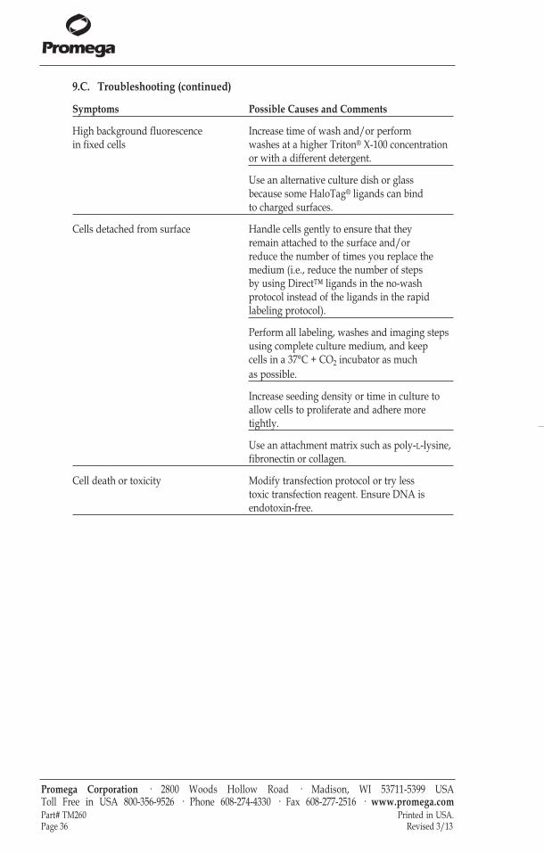
9.C. Troubleshooting (continued)
Symptoms Possible Causes and Comments
High background fluorescence Increase time of wash and/or performin fixed cells washes at a higher Triton® X-100 concentration
or with a different detergent.
Use an alternative culture dish or glass because some HaloTag® ligands can bindto charged surfaces.
Cells detached from surface Handle cells gently to ensure that they remain attached to the surface and/orreduce the number of times you replace themedium (i.e., reduce the number of stepsby using Direct™ ligands in the no-washprotocol instead of the ligands in the rapidlabeling protocol).
Perform all labeling, washes and imaging stepsusing complete culture medium, and keepcells in a 37°C + CO2 incubator as muchas possible.
Increase seeding density or time in culture toallow cells to proliferate and adhere moretightly.
Use an attachment matrix such as poly-L-lysine,fibronectin or collagen.
Cell death or toxicity Modify transfection protocol or try lesstoxic transfection reagent. Ensure DNA isendotoxin-free.
Promega Corporation · 2800 Woods Hollow Road · Madison, WI 53711-5399 USA Toll Free in USA 800-356-9526 · Phone 608-274-4330 · Fax 608-277-2516 · www.promega.comPart# TM260 Printed in USA.Page 36 Revised 3/13
tm260.0313:EIVD_TM.qxd 2/28/2013 2:58 PM Page 36
9.D. Literature Cited and Additional References
Literature Cited
1. Los, G. et al. (2005) HaloTag® interchangeable labeling technology for cell imagingand protein capture. Cell Notes 11, 2–6.
2. Los, G. and Wood, K. (2007) The HaloTag: A novel technology for cell imaging andprotein analysis. Methods. Mol. Biol. 356, 195–208.
3. Los, G. et al. (2008) HaloTag: A novel protein labeling technology for cell imaging andprotein analysis. ACS Chem. Biol. 3, 383–82.
4. Huybrechts, S. J. et al. (2009) Peroxisome dynamics in cultured mammalian cells.Traffic 10, 1722–33.
5. Lee, H.L. et al. (2010) Superresolution imaging of targeted proteins in fixed and livingcells using photoactivatable organic fluorophores. J. Am. Chem. Soc. 132, 15099–101.
6. Reek-Peterson, S.L. and Vale, R.D. (2009) Regulation of the processivity andintracellular localization of Saccharomyces cerevisiae dynein by dynactin. Proc. Natl.Acad. Sci. USA 106, 5669–74.
7. Lang, C. et al. (2006) HaloTag™ : A new versatile reporter gene system in plant cells.J. Exp. Bot. 57, 2985–92.
8. Kosaka, N. et al. (2009) In vivo stable tumor-specific painting in various colors usingdehalogenase-based protein-tag fluorescent ligands. Bioconjugate Chem. 20, 1367–74.
9. Learish, R.D. (2007) A polyclonal HaloTag® antibody for Western blotting andimmunocytochemistry. Promega Notes 95, 23–4.
Additional References
Learn more about the HaloTag® technology, visit www.promega.com/halotag
Learn more about cell imaging, visit www.promega.com/imaging
He,Y. et al. (2011) Identification of a lysosomal pathway that modulatesglucocorticoid signaling and the inflammatory response. Sci. Signal. 4 (180).
Strauch, R.C. et al. (2011) Reporter protein-targeted probes for magnetic resonanceimaging. J Amer Chem Soc. 133, 16346–9.
Hong, H. et al. (2011) HaloTag: a novel reporter gene for positron emissiontomography. Am. J. Transl. Res. 3, 392–403.
Takemoto, K. et al. (2011) Chromophore-assisted light inactivation of HaloTag®
fusion proteins labeled with eosin in living cells. ACS Chem. Biol. 6, 401–6.
Camarda, G. et al. (2010) Regulated oligomerization and molecular interactions ofthe early gametocyte protein Pfg27 in Plasmodium falciparum sexualdifferentiation. Int. J. Parasitology 40, 663-673.
Benink, H.A. et al. (2009) Direct pH measurement by using subcellular targeting of5(and 6-) carboxyseminaphthorhodafluor in mammalian cells. BioTechniques 47,769–74.
Promega Corporation · 2800 Woods Hollow Road · Madison, WI 53711-5399 USA Toll Free in USA 800-356-9526 · Phone 608-274-4330 · Fax 608-277-2516 · www.promega.comPrinted in USA. Part# TM260Revised 3/13 Page 37
tm260.0313:EIVD_TM.qxd 2/28/2013 2:58 PM Page 37
Schröder, J. et al. (2009) In vivo labeling method using a genetic construct fornanoscale resolution microscopy. Biophys J. 96, L1–3.
Yamaguchi, K. et al. (2009) Pulse-chase experiment for the analysis of proteinstability in cultured mammalian cells by covalent fluorescent labeling of fusionproteins. Methods Mol. Biol. 577, 121–31.
Ogawa, M. et al. (2008) Arabidopsis CLV3 peptide directly binds CLV1 ectodomain.Science 319, 294.
Reck-Peterson, S.L. et al. (2006) Single-molecule analysis of dynein processivity andstepping behavior. Cell 126, 335–48.
Eisenstein, M. (2006) Helping cells to tell a colorful tale. Nat. Meth. 3, 647–55.
Zhang, Y. et al. (2006) HaloTag® protein-mediated site-specific conjugation ofbioluminescent proteins to quantum dots. Angew. Chem. Int. Ed. Engl. 45, 4936–40.
Shinohara, H. and Matsubayahshi, Y. (2007) Functional immobilization of plantreceptor-like kinase onto microbeads towards receptor array construction andreceptor-based ligand fishing. Plant J. 52, 175–84.
Hata, T. and Nakayama, M. (2007) Rapid single-tube method for small-scale affinitypurification of polyclonal antibodies using HaloTag® Technology. J. Biochem.Biophys. Meth. 70, 679–82.
10. Related Products
Biotin Ligands
Promega Corporation · 2800 Woods Hollow Road · Madison, WI 53711-5399 USA Toll Free in USA 800-356-9526 · Phone 608-274-4330 · Fax 608-277-2516 · www.promega.comPart# TM260 Printed in USA.Page 38 Revised 3/13
Biotin-ContainingLigands
HaloTag® BiotinLigand
30µl G8282
5529
MA
NH
OO Cl
S
HNNH
OO Reactive Linker
Functional Reporter
HaloTag® PEG-BiotinLigand 30µl G8591
5527
MA
NH
OO
S
HNNH
OO O
NH
OO ClOO
Spacer
Reactive Linker
Functional Reporter
tm260.0313:EIVD_TM.qxd 2/28/2013 2:58 PM Page 38
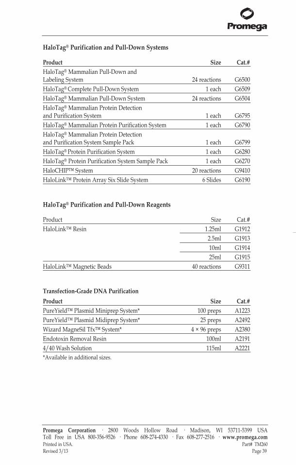
HaloTag® Purification and Pull-Down Systems
Product Size Cat.#HaloTag® Mammalian Pull-Down andLabeling System 24 reactions G6500HaloTag® Complete Pull-Down System 1 each G6509HaloTag® Mammalian Pull-Down System 24 reactions G6504HaloTag® Mammalian Protein Detectionand Purification System 1 each G6795HaloTag® Mammalian Protein Purification System 1 each G6790HaloTag® Mammalian Protein Detectionand Purification System Sample Pack 1 each G6799HaloTag® Protein Purification System 1 each G6280HaloTag® Protein Purification System Sample Pack 1 each G6270HaloCHIP™ System 20 reactions G9410HaloLink™ Protein Array Six Slide System 6 Slides G6190
HaloTag® Purification and Pull-Down Reagents
Product Size Cat.#HaloLink™ Resin 1.25ml G1912
2.5ml G191310ml G191425ml G1915
HaloLink™ Magnetic Beads 40 reactions G9311
Transfection-Grade DNA PurificationProduct Size Cat.#PureYield™ Plasmid Miniprep System* 100 preps A1223PureYield™ Plasmid Midiprep System* 25 preps A2492Wizard MagneSil Tfx™ System* 4 × 96 preps A2380Endotoxin Removal Resin 100ml A21914/40 Wash Solution 115ml A2221*Available in additional sizes.
Promega Corporation · 2800 Woods Hollow Road · Madison, WI 53711-5399 USA Toll Free in USA 800-356-9526 · Phone 608-274-4330 · Fax 608-277-2516 · www.promega.comPrinted in USA. Part# TM260Revised 3/13 Page 39
tm260.0313:EIVD_TM.qxd 2/28/2013 2:58 PM Page 39
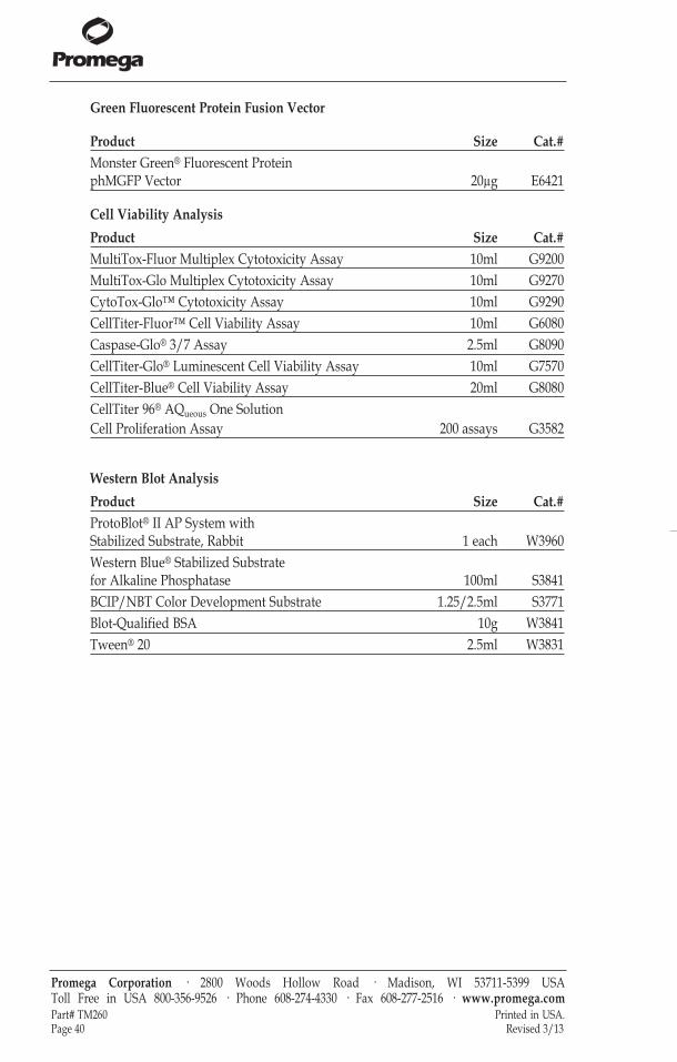
Green Fluorescent Protein Fusion Vector
Product Size Cat.#Monster Green® Fluorescent Protein phMGFP Vector 20µg E6421
Cell Viability AnalysisProduct Size Cat.#MultiTox-Fluor Multiplex Cytotoxicity Assay 10ml G9200MultiTox-Glo Multiplex Cytotoxicity Assay 10ml G9270CytoTox-Glo™ Cytotoxicity Assay 10ml G9290CellTiter-Fluor™ Cell Viability Assay 10ml G6080Caspase-Glo® 3/7 Assay 2.5ml G8090CellTiter-Glo® Luminescent Cell Viability Assay 10ml G7570CellTiter-Blue® Cell Viability Assay 20ml G8080CellTiter 96® AQueous One Solution Cell Proliferation Assay 200 assays G3582
Western Blot AnalysisProduct Size Cat.#ProtoBlot® II AP System withStabilized Substrate, Rabbit 1 each W3960Western Blue® Stabilized Substratefor Alkaline Phosphatase 100ml S3841BCIP/NBT Color Development Substrate 1.25/2.5ml S3771Blot-Qualified BSA 10g W3841Tween® 20 2.5ml W3831
Promega Corporation · 2800 Woods Hollow Road · Madison, WI 53711-5399 USA Toll Free in USA 800-356-9526 · Phone 608-274-4330 · Fax 608-277-2516 · www.promega.comPart# TM260 Printed in USA.Page 40 Revised 3/13
tm260.0313:EIVD_TM.qxd 2/28/2013 2:58 PM Page 40

Cloning Vectors
Conventional
Product Size Cat.#pHTN HaloTag® CMV-neo Vector 20µg G7721pHTC HaloTag® CMV-neo Vector 20µg G7711
Flexi® Cloning Vectors
Product Size Cat.#pFN28A HaloTag® CMV-neo Flexi® Vector 20µg G8441pFC27K HaloTag® CMV-neo Flexi® Vector 20µg G8431
Many additional Flexi® vectors are available for your convenience. You can see allHaloTag® Flexi® Vectors at www.promega.com by searching for “halotag flexivectors”. Additionally, information about the full line of reagents and tools availablefor the HaloTag® technology is available at www.promega.com/halotag
Promega Corporation · 2800 Woods Hollow Road · Madison, WI 53711-5399 USA Toll Free in USA 800-356-9526 · Phone 608-274-4330 · Fax 608-277-2516 · www.promega.comPrinted in USA. Part# TM260Revised 3/13 Page 41
tm260.0313:EIVD_TM.qxd 2/28/2013 2:58 PM Page 41
Promega Corporation · 2800 Woods Hollow Road · Madison, WI 53711-5399 USA Toll Free in USA 800-356-9526 · Phone 608-274-4330 · Fax 608-277-2516 · www.promega.comPart# TM260 Printed in USA.Page 42 Revised 3/13
(a)BY USE OF THIS PRODUCT, RESEARCHER AGREES TO BE BOUND BY THE TERMS OF THIS LIMITED USESTATEMENT. If the researcher is not willing to accept the conditions of this limited use statement, and the product is unused,Promega will accept return of the unused product and provide the researcher with a full refund.Researchers may only use this product in their own research and may modify or link Promega HaloTag® ligands to Promega orcustomer-supplied moieties. No other use or transfer of this product is authorized without the express, written consent ofPromega including, without limitation, commercial use. With respect to any uses outside this label license, including anydiagnostic, therapeutic or prophylactic uses, please contact Promega for supply and licensing information. PROMEGA MAKESNO REPRESENTATIONS OR WARRANTIES OF ANY KIND, EITHER EXPRESSED OR IMPLIED, INCLUDING FORMERCHANTABILITY OR FITNESS FOR A PARTICULAR PURPOSE WITH REGARDS TO THE PRODUCT. The terms of thisagreement shall be governed under the laws of the State of Wisconsin, USA. (b)U.S. Pat. No. 7,867,726, Japanese Pat. No. 4748685 and other patents pending(c)U.S. Pat. No. RE42931, Japanese Pat. No. 4748685 and other patents pending. (d)The fluorescent dye (the "Dye") of this product (the "Product") is provided under an agreement between Molecular Probes,Inc., and Promega Corporation, and the manufacture, use, sale or import of this Product is subject to: (i) one or more of USPatents and pending US applications and corresponding international equivalents owned by Molecular Probes, Inc. (a whollyowned subsidiary of Invitrogen Corporation); and (ii) one or more of US Patents and pending US applications andcorresponding international equivalents owned by Promega Corporation. The purchase of this Product conveys to the buyer thenon-transferable right to use the purchased amount of the Product in research conducted by the buyer (whether the buyer is anacademic or for-profit entity). The use of this Product for providing medical or diagnostic results or services, including withoutlimitation testing, analysis, or screening, or for providing clinical information or clinical analysis in return for compensation on aper-test basis, is expressly excluded. The buyer cannot sell or otherwise transfer (a) this Product, (b) its components or (c)materials made using this Product or its components to a third party or otherwise use this Product or its components ormaterials made using this Product or its components for Commercial Purposes. Commercial Purposes means any activity by aparty for consideration and may include but is not limited to: (1) use of the Product or its components in manufacturing; (2) useof the Product or its components to provide a service, information, or data; (3) use of the Product or its components fortherapeutic, diagnostic or prophylactic purposes; or (4) resale of the Product or its components, whether or not such Product orits components are resold for use in research. For information on purchasing a license to the Dye for purposes other than as setforth above, contact Molecular Probes, Inc., Business Development, 29851 Willow Creek Road, Eugene, OR 97402-9132. Tel: (541)465-8300. Fax: (541) 335-0504. For information on purchasing a license to this Product for purposes other than as set forth above,contact Promega Corporation. (e)Patent Pending.(f)Certain applications of this product may require licenses from others. © 2004–2013 Promega Corporation. All Rights Reserved.Caspase-Glo, CellTiter 96, CellTiter-Blue, CellTiter-Glo, Flexi, HaloTag, MagneSil, Monster Green, ProtoBlot, TNT, WesternBlue, and Wizard are registered trademarks of Promega Corporation. CellTiter-Fluor, CytoTox-Glo, FastBreak, HaloCHIP,HaloLink, PureYield, R110Direct, Tfx and TMRDirect are trademarks of Promega Corporation.Alexa Fluor and Oregon Green are registered trademarks of Molecular Probes, Inc. Coomassie is a registered trademark ofImperical Chemical Industries, Ltd. Cy and Sephadex are registered trademarks of GE Healthcare Biosciences. FMBIO is aregistered trademark of Hitachi Software Engineering Company, Ltd. MoFlo is a registered trademark of Dako Denmark A/S.Triton is a registered trademark of Union Carbide Chemicals & Plastics Technology Corporation. Tween is a registeredtrademark of ICI Americas, Inc.Products may be covered by pending or issued patents or may have certain limitations. Please visit our Web site for moreinformation.All prices and specifications are subject to change without prior notice.Product claims are subject to change. Please contact Promega Technical Services or access the Promega online catalog for themost up-to-date information on Promega products.
tm260.0313:EIVD_TM.qxd 2/28/2013 2:58 PM Page 42