Half-sandwich complexes of rhodium containing cysteine...
36
1 Half-sandwich complexes of rhodium containing cysteine-derived ligands† María Carmona, a Ricardo Rodríguez,* a Fernando J. Lahoz, a Pilar García-Orduña, a Iñaki Osante, b Carlos Cativiela, b José A. López, a and Daniel Carmona* a Received …… DOI: …… The modified cysteine S-benzyl-α-methyl-L-cysteine (HL2) has been prepared starting from L-cysteine chlorhydrate. The reaction of commercial S-benzyl-L-cysteine (HL1) or HL2 with the dimer [{(η 5 -C 5 Me 5 )RhCl} 2 (μ-Cl) 2 ] gives rise to the cationic complexes [(η 5 -C 5 Me 5 )RhCl(HL)]Cl (HL = HL1 (1), HL2 (2)) in which the cysteine ligand exhibits a κ 2 N,S coordination mode. In basic medium, HL1 or HL2 react with [{(η 5 - C 5 Me 5 )RhCl} 2 (μ-Cl) 2 ] affording mixtures of the two epimers at the metal centre of the neutral complexes [(η 5 -C 5 Me 5 )RhCl(κ 2 N,O-L)] (HL = HL1 (3), HL2 (4)), in which the amino carboxylate adopts a κ 2 N,O mode of coordination, along with variable amounts of the cationic compounds [(η 5 -C 5 Me 5 )Rh(κ 3 N,O,S-L)]Cl (HL = HL1 (6Cl), HL2 (7Cl)), which contain κ 3 N,O,S coordinated cysteine-derived ligands. However, in basic medium, the N-Boc substituted cysteine S-benzyl-N-Boc-L-cysteine (HL3) only yields the κ 2 O,S coordinated derivative [(η 5 -C 5 Me 5 )RhCl(κ 2 O,S-L3)] (5) as a mixture of two diastereomers depending on the configuration at the metal centre. The bidentate chelate complexes 3-5 react with AgSbF 6 rendering the hexafluoroantimonates [(η 5 - C 5 Me 5 )Rh(κ 3 N,O,S-L)][SbF 6 ] (HL = HL1 (6Sb), HL2 (7Sb), HL3 (8Sb)) with tridentate coordination. Compound 8Sb reacts with NaHCO 3 giving the neutral complex [(η 5 -C 5 Me 5 )Rh(κ 3 N,O,S-L3 −H )] (9) which can be also prepared by reacting the dimer [{(η 5 -C 5 Me 5 )RhCl} 2 (μ-Cl) 2 ] with HL3 in the presence of two equivalents of NaHCO 3 .
Transcript of Half-sandwich complexes of rhodium containing cysteine...
1
Half-sandwich complexes of rhodium containing cysteine-derived
ligands†
María Carmona,a Ricardo Rodríguez,*a Fernando J. Lahoz,a Pilar García-Orduña,a Iñaki Osante,b Carlos Cativiela,b José A. López,a and Daniel Carmona*a
Received ……
DOI: ……
The modified cysteine S-benzyl-α-methyl-L-cysteine (HL2) has been prepared starting
from L-cysteine chlorhydrate. The reaction of commercial S-benzyl-L-cysteine (HL1) or
HL2 with the dimer [{(η5-C5Me5)RhCl}2(μ-Cl)2] gives rise to the cationic complexes
[(η5-C5Me5)RhCl(HL)]Cl (HL = HL1 (1), HL2 (2)) in which the cysteine ligand
exhibits a κ2N,S coordination mode. In basic medium, HL1 or HL2 react with [{(η5-
C5Me5)RhCl}2(μ-Cl)2] affording mixtures of the two epimers at the metal centre of the
neutral complexes [(η5-C5Me5)RhCl(κ2N,O-L)] (HL = HL1 (3), HL2 (4)), in which the
amino carboxylate adopts a κ2N,O mode of coordination, along with variable amounts
of the cationic compounds [(η5-C5Me5)Rh(κ3N,O,S-L)]Cl (HL = HL1 (6Cl), HL2
(7Cl)), which contain κ3N,O,S coordinated cysteine-derived ligands. However, in basic
medium, the N-Boc substituted cysteine S-benzyl-N-Boc-L-cysteine (HL3) only yields
the κ2O,S coordinated derivative [(η5-C5Me5)RhCl(κ2O,S-L3)] (5) as a mixture of two
diastereomers depending on the configuration at the metal centre. The bidentate chelate
complexes 3-5 react with AgSbF6 rendering the hexafluoroantimonates [(η5-
C5Me5)Rh(κ3N,O,S-L)][SbF6] (HL = HL1 (6Sb), HL2 (7Sb), HL3 (8Sb)) with
tridentate coordination. Compound 8Sb reacts with NaHCO3 giving the neutral complex
[(η5-C5Me5)Rh(κ3N,O,S-L3−H)] (9) which can be also prepared by reacting the dimer
[{(η5-C5Me5)RhCl}2(μ-Cl)2] with HL3 in the presence of two equivalents of NaHCO3.
2
The new compounds contain up to four stereogenic centres, namely, Rh, S, N, and C.
The absolute configuration of the complexes has been established by spectroscopic and
diffractometric means including the crystal structure determination of [(η5-
C5Me5)RhCl(κ2O,S-L3)] (5), [(η5-C5Me5)Rh(κ3N,O,S-L1)][SbF6] (6Sb), [(η5-
C5Me5)Rh(κ3N,O,S-L2)][SbF6] (7Sb), and [(η5-C5Me5)Rh(κ3N,O,S-L3−H)] (9). Variable
temperature 1H NMR studies reveal the existence of epimerization processes and
theoretical calculations discriminate their nature.
a Instituto de Síntesis Química y Catálisis Homogénea (ISQCH), CSIC - Universidad de Zaragoza, Departamento de Química Inorgánica, Pedro Cerbuna 12, 50009 Zaragoza, Spain, E-mail: [email protected], [email protected] b Instituto de Síntesis Química y Catálisis Homogénea (ISQCH), CSIC - Universidad de Zaragoza, Departamento de Química Orgánica, Pedro Cerbuna 12, 50009 Zaragoza, Spain †Electronic supplementary information (ESI) available: Kinetic data. Files containing full details of the structural analysis of complexes 6Sb, 7Sb, and 9 (CIF format). CCDC 1485794-1485796. Metallacycles characterization for complexes 6Sb, 7Sb and 9. Hydrogen bond interactions in 6Sb and 7Sb. CD spectra. Preparation of HL2 by S-benzylation of IV. Frequency list of all ground and transition states. Cartesian coordinates for all DFT-calculated structures (XYZ). For ESI and crystallographic data in CIF or other electronic format see DOI: ………..
3
Introduction
Amino acids are biologically relevant molecules whose coordination chemistry toward
transition-metal ions has been largely investigated owing to their easy availability,
versatile bonding modes and potential applications in a variety of chemical fields.1
Complexation of α-amino acids to metallic systems gives rise to a variety of
coordination modes. Thus, although for no functionalized α-amino acids monodentate
and bidentate chelate coordination are known, the latter being the prevalent one, for
functionalized α-amino acids tridentate chelate coordination modes have also been
described.1d,2 Moreover, amino carboxylate derivatives of transition metals have found
application in fields as different as asymmetric synthesis,1a,3 bioinorganic,4
supramolecular5 or solid state chemistry.6
Sulphur-containing amino acids have drawn a great deal of attention due to their
crucial and significant functions in biological systems.7 In particular, it has been shown
that silver complexes containing cysteine or glutathione play a key role in bacterial
inactivation.8 In the same line, the active sites of metalloproteins like acetyl coenzyme
A synthase9 or [NiFe] hydrogenase10 contain nickel ions that are coordinated
exclusively by donors derived from cysteine. Very recently, Buglyó et al. have shown
that the thioether containing amino acids D- and L-methionine and S-methyl-L-cysteine
coordinate to half-sandwich solvated cations of formula [(ηn-ring)M(H2O)3]2+ ((ηn-
ring)M = (η5-C5Me5)Rh, (η5-C5Me5)Ir, (η6-p-MeC6H4iPr)Ru or (η6-p-MeC6H4iPr)Os)
mostly in a κ3N,O,S manner.11
From a coordination point of view, cysteine behaves as a multifunctional organic
ligand able to bind to a variety of metal ions through amine, carboxylate and thiolate
groups to form chiral mononuclear and multinuclear complexes.5b In some cases,
4
mixtures of products differing in coordination modes and nuclearity are obtained and
the factors that govern their distribution and interconversion remain unclear.4b
On the other hand, it is well known that the incorporation of quaternary amino acids
in peptide sequences provides very useful information on their bioactive conformation
and results in beneficial physiological effects.12
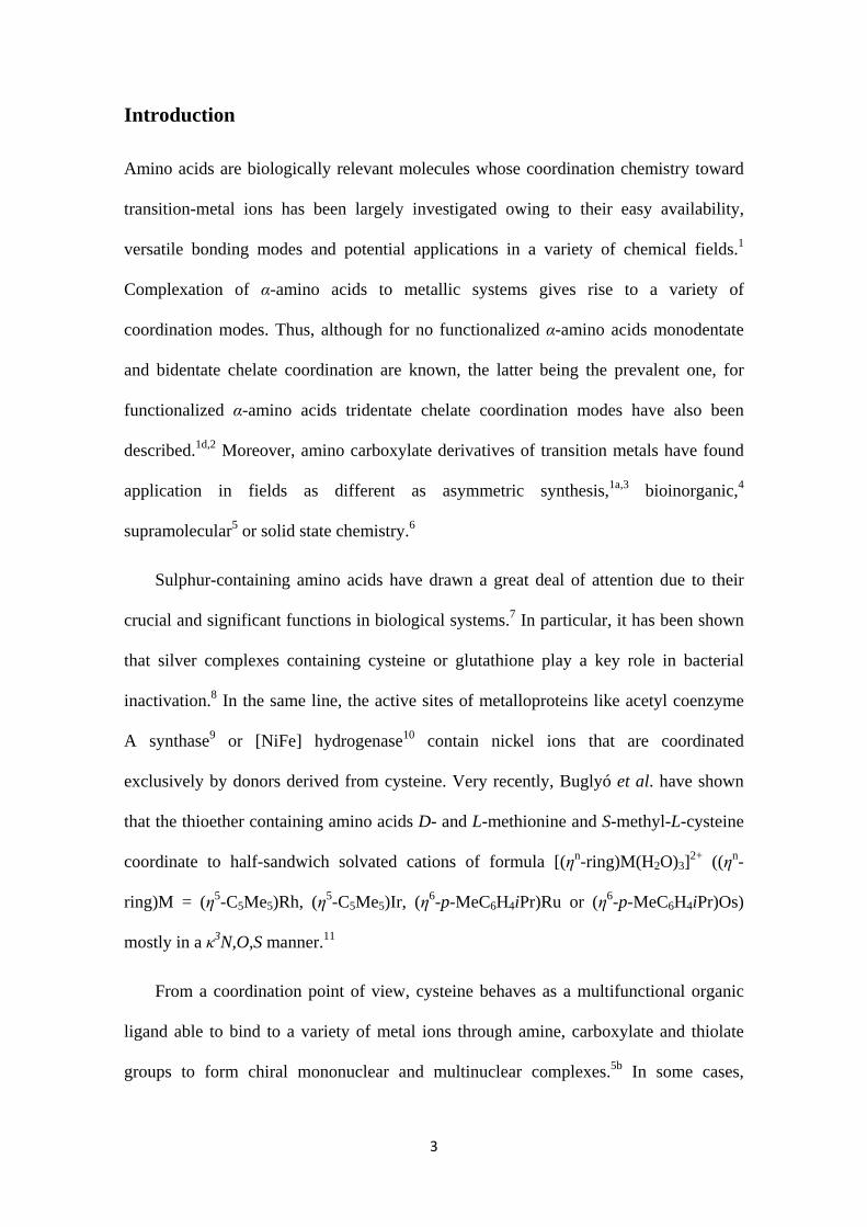
With all these concerns in mind, in the present paper we report on the controlled
preparation of new half sandwich rhodium (III) complexes containing modified
cysteines such as S-benzyl-L-cysteine (HL1), the quaternary S-benzyl-α-methyl-L-
cysteine (HL2) and the N-protected S-benzyl-N-Boc-L-cysteine (HL3) (Chart 1).
Special attention will be paid to the coordination features of the employed amino acids.
The chirality of the compounds is assessed and the epimerization processes that take
place are studied experimentally and theoretically.
BnS NH2
COOH
S-Benzyl-L-cysteine
HL1
BnS NH2
COOH
S-Benzyl--methyl-L-cysteine
HL2
Me
BnS NHBoc
COOH
S-Benzyl-N-Boc-L-cysteine
HL3
Chart 1. Modified cysteines
5
Results and discussion
Cysteine-derived ligands
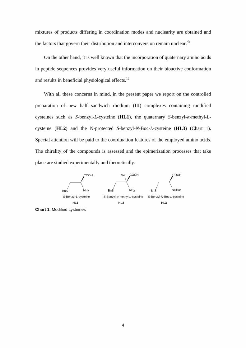
Modified cysteines HL1 and HL3 (Chart 1) are commercially available. Cysteine HL2
was prepared starting from L-cysteine hydrochloride methyl ester according to the
sequence of reactions depicted in Scheme 1. Reaction of the L-cysteine hydrochloride
methyl ester with pivalaldehyde affords tiazolidine I. N-formylation of I with sodium
formate renders diastereoselectively the cis methyl ester II in 83 % isolated yield.
Subsequent reaction with lithium diisopropyl amide (LDA), using N,N’-
dimethylpropylurea (DMPU) as co-solvent, forms the corresponding enolate which
trapped with methyl iodide gives rise, again diastereoselectively, to the trans tert-butyl
to methyl isomer of the 4-methyl thiazolidine III. Hydrolysis of III generates α-methyl-
L-cysteine hydrochloride IV.13 S-benzylation of IV, that renders HL2, was carried out
by reacting IV with benzylbromide in basic medium, following a modification of the
procedure described for the S-benzylation of L-cysteine by Wang et al.14 (see Electronic
Supplementary Information).
HS NH2.HCl
CO2Me H
tBu
O
NHS
CO2Me
tBu
NCHOS
CO2Me
tBu
NCHOS
CO2Me
tBu
HS NH2.HCl
CO2H
BnS NH2
CO2H
1) LDA2) MeITHF/DMPU
HCl 5MBrBn
NaOH/EtOH
Me
MeMe
(CH3CO)2O
HCO2H/HCO2Nahexane+
IVHL2
I II
III
NEt3
Scheme 1. Synthesis of HL2
6
Rhodium complexes
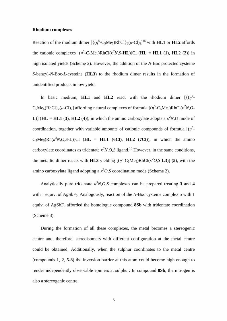
Reaction of the rhodium dimer [{(η5-C5Me5)RhCl}2(μ-Cl)2]15 with HL1 or HL2 affords
the cationic complexes [(η5-C5Me5)RhCl(κ2N,S-HL)]Cl (HL = HL1 (1), HL2 (2)) in
high isolated yields (Scheme 2). However, the addition of the N-Boc protected cysteine
S-benzyl-N-Boc-L-cysteine (HL3) to the rhodium dimer results in the formation of
unidentified products in low yield.
In basic medium, HL1 and HL2 react with the rhodium dimer [{(η5-
C5Me5)RhCl}2(μ-Cl)2] affording neutral complexes of formula [(η5-C5Me5)RhCl(κ2N,O-
L)] (HL = HL1 (3), HL2 (4)), in which the amino carboxylate adopts a κ2N,O mode of
coordination, together with variable amounts of cationic compounds of formula [(η5-
C5Me5)Rh(κ3N,O,S-L)]Cl (HL = HL1 (6Cl), HL2 (7Cl)), in which the amino
carboxylate coordinates as tridentate κ3N,O,S ligand.16 However, in the same conditions,
the metallic dimer reacts with HL3 yielding [(η5-C5Me5)RhCl(κ2O,S-L3)] (5), with the
amino carboxylate ligand adopting a κ2O,S coordination mode (Scheme 2).
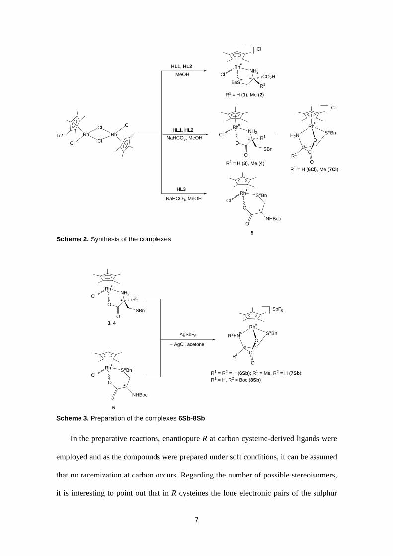
Analytically pure tridentate κ3N,O,S complexes can be prepared treating 3 and 4
with 1 equiv. of AgSbF6. Analogously, reaction of the N-Boc cysteine complex 5 with 1
equiv. of AgSbF6 afforded the homologue compound 8Sb with tridentate coordination
(Scheme 3).
During the formation of all these complexes, the metal becomes a stereogenic
centre and, therefore, stereoisomers with different configuration at the metal centre
could be obtained. Additionally, when the sulphur coordinates to the metal centre
(compounds 1, 2, 5-8) the inversion barrier at this atom could become high enough to
render independently observable epimers at sulphur. In compound 8Sb, the nitrogen is
also a stereogenic centre.
7
Rh
ClNH2
O
O
R1
SBn
Rh
ClS*Bn
O
ONHBoc
Rh
Cl
Cl
ClRh
ClHL1, HL2
HL3
NaHCO3, MeOH
NaHCO3, MeOH
R1 = H (3), Me (4)
5
1/2
Rh
ClNH2
BnSCO2H
HL1, HL2
MeOH
+
*
*
*
*
*
*
*
Rh
H2N S*Bn
O
C
OR1
Cl
*
*
R1
R1 = H (1), Me (2)
R1 = H (6Cl), Me (7Cl)
Cl
Scheme 2. Synthesis of the complexes
Rh
ClNH2
O
O
R1
SBn
Rh
ClS*Bn
O
ONHBoc
3, 4
5
AgSbF6
AgCl, acetone
R1 = R2 = H (6Sb); R1 = Me, R2 = H (7Sb);
R1 = H, R2 = Boc (8Sb)
*
*
*
*
Rh
R2HN S*Bn
O
C
OR1
SbF6
*
*
*
Scheme 3. Preparation of the complexes 6Sb-8Sb
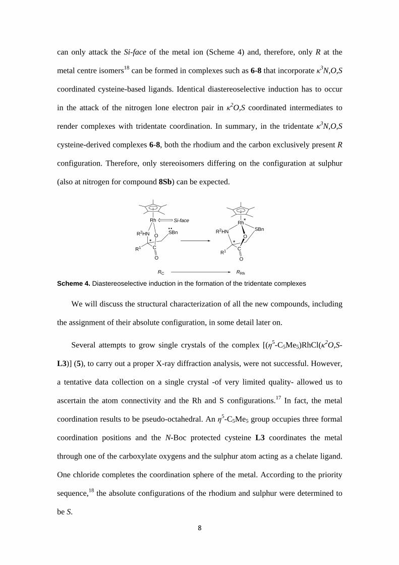
In the preparative reactions, enantiopure R at carbon cysteine-derived ligands were
employed and as the compounds were prepared under soft conditions, it can be assumed
that no racemization at carbon occurs. Regarding the number of possible stereoisomers,
it is interesting to point out that in R cysteines the lone electronic pairs of the sulphur
8
can only attack the Si-face of the metal ion (Scheme 4) and, therefore, only R at the
metal centre isomers18 can be formed in complexes such as 6-8 that incorporate κ3N,O,S
coordinated cysteine-based ligands. Identical diastereoselective induction has to occur
in the attack of the nitrogen lone electron pair in κ2O,S coordinated intermediates to
render complexes with tridentate coordination. In summary, in the tridentate κ3N,O,S
cysteine-derived complexes 6-8, both the rhodium and the carbon exclusively present R
configuration. Therefore, only stereoisomers differing on the configuration at sulphur
(also at nitrogen for compound 8Sb) can be expected.
Rh
R2HN SBn
O
C
OR1
*
*
Rh
R2HN SBnO
C
O
R1
*
Si-face
RC RRh
Scheme 4. Diastereoselective induction in the formation of the tridentate complexes
We will discuss the structural characterization of all the new compounds, including
the assignment of their absolute configuration, in some detail later on.
Several attempts to grow single crystals of the complex [(η5-C5Me5)RhCl(κ2O,S-
L3)] (5), to carry out a proper X-ray diffraction analysis, were not successful. However,
a tentative data collection on a single crystal -of very limited quality- allowed us to
ascertain the atom connectivity and the Rh and S configurations.17 In fact, the metal
coordination results to be pseudo-octahedral. An η5-C5Me5 group occupies three formal
coordination positions and the N-Boc protected cysteine L3 coordinates the metal
through one of the carboxylate oxygens and the sulphur atom acting as a chelate ligand.
One chloride completes the coordination sphere of the metal. According to the priority
sequence,18 the absolute configurations of the rhodium and sulphur were determined to
be S.
9
Molecular structure of the complexes (RRh,SS,RC)-[(η5-C5Me5)Rh(κ3N,O,S-
L1)][SbF6] (6Sb) and (RRh,SS,RC)-[(η5-C5Me5)Rh(κ3N,O,S-L2)][SbF6] (7Sb)
Single crystals of the complexes 6Sb and 7Sb were grown by slow diffusion of diethyl
ether into methanolic solutions of diastereomeric mixtures of these compounds and the
solid state molecular structures were determined by X-ray diffraction methods.
Interestingly, complex 7Sb crystallizes with two different conformational isomers in the
unit cell 7Sb-A and 7Sb-B, differing in the disposition of the benzyl substituent around
the S-C(14) bond. In spite of this differentiation, both independent molecules of 7Sb
exhibit similar bond lengths, and stereochemical features.
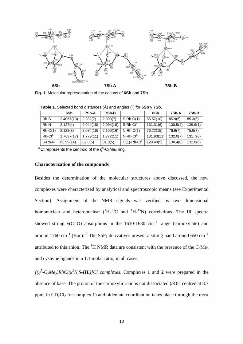
Figure 1 shows selected views of the cations of the molecules and representative
geometrical parameters are listed in Table 1. Both complexes exhibit the so-called
“three-legged piano-stool” geometry. An η5-C5Me5 group occupies three fac positions
and the corresponding cysteine, S-benzyl-L-cysteine (6Sb) or S-benzyl-α-methyl-L-
cysteine (7Sb), occupies the three remaining coordination sites adopting a κ3N,O,S
coordination mode. According to the ligand priority sequence,18 the absolute
configuration is RRh,SS,RC for the two compounds.
The tridentate coordination of S-benzyl-L-cysteine (6Sb) or S-benzyl-α-methyl-L-
cysteine (7Sb) leads to the formation of two five-membered, Rh-O-C-C-N and Rh-S-C-
C-N, and one six-membered Rh-S-C-C-C-O metallacycles with 5E/T1, E5 and B4,1
conformations, respectively. Similar deviations from planarity and puckering phase
values have been found in both complexes (see Electronic Supplementary Information).
The solid state structure of 6Sb and 7Sb are stabilized by strong hydrogen bond
networks established between N-H protons and carboxylate oxygen atoms together with
weak C-H···O hydrogen bonds (see Electronic Supplementary Information).
10
6Sb 7Sb-A 7Sb-B
Fig. 1. Molecular representation of the cations of 6Sb and 7Sb.
Table 1. Selected bond distances (Å) and angles (º) for 6Sb y 7Sb.
6Sb 7Sb-A 7Sb-B 6Sb 7Sb-A 7Sb-B
Rh-S 2.4067(13) 2.382(7) 2.393(7) S-Rh-O(1) 85.57(10) 85.9(5) 85.3(5)
Rh-N 2.127(4) 2.044(18) 2.094(18) S-Rh-Cta 131.31(6) 130.5(4) 129.6(1)
Rh-O(1) 2.128(3) 2.084(16) 2.100(16) N-Rh-O(1) 76.22(15) 76.9(7) 75.9(7)
Rh-Cta 1.7637(17) 1.779(11) 1.772(11) N-Rh-Cta 131.93(11) 132.0(7) 131.7(6)
S-Rh-N 82.99(14) 82.0(5) 81.8(5) O(1)-Rh-Cta 129.49(9) 130.4(6) 132.8(6)
a Ct represents the centroid of the η5-C5Me5 ring.
Characterization of the compounds
Besides the determination of the molecular structures above discussed, the new
complexes were characterized by analytical and spectroscopic means (see Experimental
Section). Assignment of the NMR signals was verified by two dimensional
homonuclear and heteronuclear (1H-13C and 1H-15N) correlations. The IR spectra
showed strong ν(C=O) absorptions in the 1610-1630 cm1 range (carboxylate) and
around 1760 cm1 (Boc).19 The SbF6 derivatives present a strong band around 650 cm1
attributed to this anion. The 1H NMR data are consistent with the presence of the C5Me5
and cysteine ligands in a 1:1 molar ratio, in all cases.
[(η5-C5Me5)RhCl(κ2N,S-HL)]Cl complexes. Complexes 1 and 2 were prepared in the
absence of base. The proton of the carboxylic acid is not dissociated (δOH centred at 8.7
ppm, in CD2Cl2 for complex 1) and bidentate coordination takes place through the most
11
nucleophilic nitrogen and sulphur atoms of the cysteine rendering a Rh-N-C-C-S five-
membered metallacycle (Scheme 2).
In the solid state, the two epimers at the metal centre were isolated in 85/15 molar
ratio in both cases.20 While, in the major isomer, the pro-S proton of the NH2 group
shows a NOE relationship with the C5Me5 protons, in the minor isomer a NOE
relationship occurs between the pro-R proton and the protons of the cyclic ligand. These
data indicate that the metal centre has S configuration in the major isomer and R in the
minor one.
Only one set of signals for each epimer was observed in the 1H NMR spectra from
298 to 193 K. As the coordinated sulphur is a stereogenic centre, either it adopts only
one configuration or both epimers at sulphur quickly exchange even at 193 K.
[(η5-C5Me5)Rh(κ3N,O,S-L)]+ complexes. In the cationic complexes of formula [(η5-
C5Me5)Rh(κ3N,O,S-L)]+ (6-8) the cysteine adopts a κ3N,O,S coordination mode
(Schemes 2 and 3) as revealed by the molecular structures of the complexes 6Sb and
7Sb determined by diffractometric means (Figure 1).
As stated above, the configuration at rhodium as well as that at the amino acid
carbon is R. For compounds 6Sb and 7Sb, although at room temperature, the NMR
spectra consist of only one set of signals, below 253 K a new set of signals was
observed in a relative intensity lower than 2 %. We tentatively assign these isomers to
the two epimers at the sulphur and, according to the solid state molecular structures, the
major component would correspond to the S at sulphur diastereomer. However, for
compound 8Sb, only one set of signals was observed in the 1H NMR spectra from 298
to 193 K. We assume that compound 8Sb is diastereopure.
12
As expected, nitrogen coordination strongly affects 15N NMR chemical shift. Thus,
while this nucleus resonates at 86.71 ppm with respect to 15NH3 in free S-benzyl-N-Boc-
L-cysteine HL3, it appears at 68.28 ppm in the metallic derivative 8Sb, in which the
cysteine nitrogen is coordinated to the metal centre.21 The chemical shift of the
asymmetric carbon C* is also strongly affected by the coordination of the nitrogen.
Thus, for example, this carbon resonates at 53.06 ppm in free HL3 but it appears at
62.76 ppm in 8Sb. This strong deshielding contrasts with the shielding of about 6.3 ppm
found for the methylene carbon C*CH2S, adjacent to the sulphur which is also
coordinated to the metal centre in 8Sb. So, δ values of the asymmetric carbon and that
of the methylene C*CH2S are useful criteria for diagnostic the coordination of the
nitrogen and sulphur atoms of the cysteine ligand, respectively.
When the cysteine-derived ligand displays a κ3 coordination mode (complexes 6-8),
the C5Me5 protons give a singlet at around 1.90 ppm, independently from which
cysteine-derived ligand was involved.
At room temperature, the most significant absorption in the circular dichroism
spectra of these compounds is a negative maximum in the 370-377 nm range. As free α-
amino acids do not show Cotton effects above 230 nm,22 these absorptions were
tentatively assigned to transitions associated to the R configuration at the metal centre.
In summary, compounds 6-8 are obtained as chlorides or hexafluoroantimonates of
the epimers at sulphur of cations of formula [(η5-C5Me5)Rh(κ3N,O,S-L)]+ in which the
amino carboxylates act as tridentate ligands. The configuration of the major isomer of
the hexafluoroantimonates 6Sb-8Sb (≥ 98 %) is RRh,SS,RC.
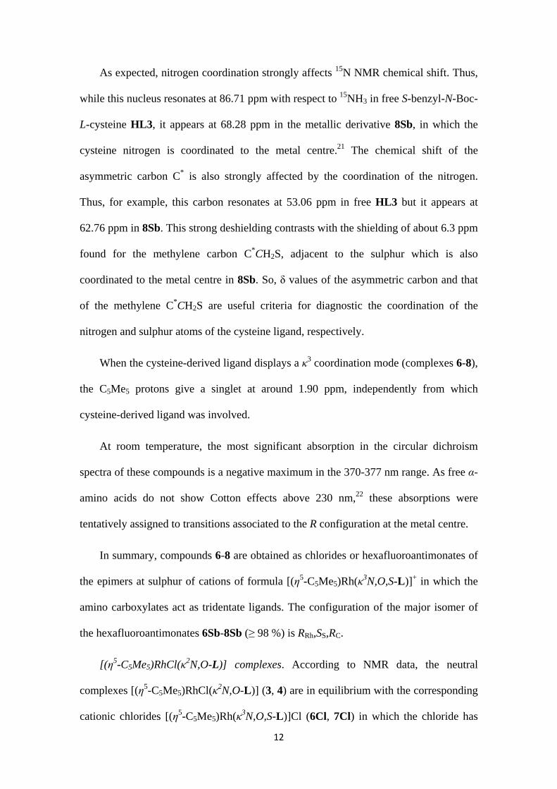
[(η5-C5Me5)RhCl(κ2N,O-L)] complexes. According to NMR data, the neutral
complexes [(η5-C5Me5)RhCl(κ2N,O-L)] (3, 4) are in equilibrium with the corresponding
cationic chlorides [(η5-C5Me5)Rh(κ3N,O,S-L)]Cl (6Cl, 7Cl) in which the chloride has
13
been replaced from the metal coordination sphere by the sulphur atom. Changing the
tertiary L1 by the quaternary L2 cysteine-based ligand, the amount of the compound
with tridentate coordination increases from 11 to 42 %, in chloroform, and from 56 to
85 %, in the more polar solvent methanol. In solution, the two epimers at the metal atom
of the compounds 3 and 4 were detected. The concentration of the complex with
tridentate coordination also increases as temperature increases but the molar ratio
between the two epimers at the metal centre remains constant. As the tridentate
coordination can only be achieved from the RRh,RC epimer (Scheme 4), the equilibria
depicted in Scheme 5 have to operate, i. e., to increase the concentration of the complex
with tridentate coordination, the S at the metal centre isomer has to epimerize to the R at
the metal centre isomer, to maintain constant the molar ratio of epimers at the metal. In
fact, ROESY experiments clearly establish the existence of exchange between the two
epimers at the metal centre as well as between the R at the metal centre isomers and the
corresponding complexes [(η5-C5Me5)Rh(κ3N,O,S-L)]+.
In good agreement with their neutral nature, the C5Me5 protons of the bidentate
cysteine complexes 3 and 4 give a singlet at around 1.60 ppm, about 0.3 ppm shielded
with respect to the cationic complexes 6-8 with tridentate coordination.
Rh
R2HN SBnO
C
OR1
*
*
Rh
R2HNSBnO
C
OR1
*
*
ClRh
NHR2
O
CR1
O
*
*
Cl
SBnSRh,RC RRh,RC RRh,RC
Cl
3, 4 6Cl, 7Cl
Scheme 5. Equilibria between 3 and 4 and the cationic tridentate chlorides 6Cl and 7Cl
14
In the major bidentate epimer, irradiation of the proton or Me bonded to the
asymmetric carbon originates enhancement for the C5Me5 protons. This NOE effect
indicates that the configuration of the metal centre in the major epimers is R.
In summary, the reaction of the dimer [{(η5-C5Me5)RhCl}2(μ-Cl)2] with cysteines
HL1 and HL2, in the presence of NaHCO3, renders equilibrium mixtures of the two
epimers at the metal centre of the κ2N,O-L complexes (RRh,RC and SRh,RC)-[(η5-
C5Me5)RhCl(κ2N,O-L)] (3, 4) with the corresponding κ3N,O,S compounds [(η5-
C5Me5)Rh(κ3N,O,S-L)]Cl (6Cl, 7Cl), mostly as RRh,SS,RC diastereomers (Scheme 2).
The [(η5-C5Me5)RhCl(κ2O,S-L3)] complex. When the dimer [{(η5-C5Me5)RhCl}2(μ-
Cl)2] reacts with the cysteine HL3, in the presence of NaHCO3, the complex [(η5-
C5Me5)RhCl(κ2O,S-L3)] (5) was isolated. At room temperature, the NMR spectrum of
the isolated solid shows the presence of the two epimers at the metal centre in 97/3
molar ratio.20 The molecular structure, determined by X-ray diffraction methods (see
above), establishes that the configuration at the rhodium in the major epimer is S.
According to this structural determination, the chemical shift of the nitrogen (89.85
ppm, major isomer) and asymmetric carbon C* (50.65, 50.59 ppm, major and minor
isomers, respectively) indicate that the nitrogen is not coordinated to the metal centre.
The chemical shift of the C5Me5 protons, around 1.60 ppm, corresponds to a chelate
bidentate coordination for the cysteine.
At low temperature (below 233 K) traces of a third isomer were detected that were
tentatively assigned to the epimer at sulphur of the S at Rh isomer.
Deprotonation of the complex (RRh,SS,RC)-[(η5-C5Me5)Rh(κ3N,O,S-L3)][SbF6] (8Sb)
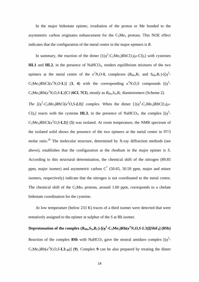
Reaction of the complex 8Sb with NaHCO3 gave the neutral amiduro complex [(η5-
C5Me5)Rh(κ3N,O,S-L3H)] (9). Complex 9 can be also prepared by treating the dimer
15
[{(η5-C5Me5)RhCl}2(μ-Cl)2] with the cysteine HL3 in the presence of 2 equivalents of
NaHCO3 (Scheme 6).
8Sb
Rh
BocHN S*Bn
O
C
O
SbF6
*
*
MeOH
2 NaCl
9
Rh
BocN S*Bn
O
C
O
*
*
NaSbF6
2 NaHCO3, HL3
NaHCO3
Rh
Cl
Cl
ClRh
Cl
1/2
*
*
Scheme 6. Preparation of complex 9
In complex 9, the chemical shift of the asymmetric carbon (67.72 ppm) and that of
the C5Me5 protons (1.90 ppm) suggest that the nitrogen is coordinated to the metal
centre and that the cysteine-derived ligand retains its κ3N,O,S coordination mode. A
negative maximum centred at 342 nm in the circular dichroism spectrum is attributed to
the R at the metal centre configuration (see above). All these data were corroborated by
the determination of its crystal structure by X-ray diffractometry.
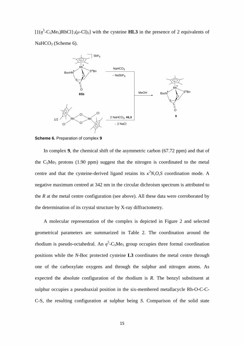
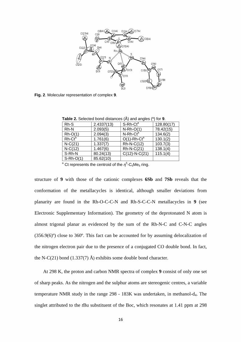
A molecular representation of the complex is depicted in Figure 2 and selected
geometrical parameters are summarized in Table 2. The coordination around the
rhodium is pseudo-octahedral. An η5-C5Me5 group occupies three formal coordination
positions while the N-Boc protected cysteine L3 coordinates the metal centre through
one of the carboxylate oxygens and through the sulphur and nitrogen atoms. As
expected the absolute configuration of the rhodium is R. The benzyl substituent at
sulphur occupies a pseudoaxial position in the six-membered metallacycle Rh-O-C-C-
C-S, the resulting configuration at sulphur being S. Comparison of the solid state
16
Fig. 2. Molecular representation of complex 9.
Table 2. Selected bond distances (Å) and angles (º) for 9. Rh-S 2.4337(13) S-Rh-Cta 128.80(17) Rh-N 2.093(5) N-Rh-O(1) 78.42(15) Rh-O(1) 2.094(3) N-Rh-Cta 134.6(2) Rh-Cta 1.761(6) O(1)-Rh-Cta 130.1(2) N-C(21) 1.337(7) Rh-N-C(12) 103.7(3) N-C(12) 1.467(6) Rh-N-C(21) 138.1(4) S-Rh-N 80.24(13) C(12)-N-C(21) 115.1(4) S-Rh-O(1) 85.62(10)
a Ct represents the centroid of the η5-C5Me5 ring.
structure of 9 with those of the cationic complexes 6Sb and 7Sb reveals that the
conformation of the metallacycles is identical, although smaller deviations from
planarity are found in the Rh-O-C-C-N and Rh-S-C-C-N metallacycles in 9 (see
Electronic Supplementary Information). The geometry of the deprotonated N atom is
almost trigonal planar as evidenced by the sum of the Rh-N-C and C-N-C angles
(356.9(6)º) close to 360º. This fact can be accounted for by assuming delocalization of
the nitrogen electron pair due to the presence of a conjugated CO double bond. In fact,
the N-C(21) bond (1.337(7) Å) exhibits some double bond character.
At 298 K, the proton and carbon NMR spectra of complex 9 consist of only one set
of sharp peaks. As the nitrogen and the sulphur atoms are stereogenic centres, a variable
temperature NMR study in the range 298 - 183K was undertaken, in methanol-d4. The
singlet attributed to the tBu substituent of the Boc, which resonates at 1.41 ppm at 298
17
K, was taken as reference. On cooling, this singlet broadens out, coalesces at about 243
K and splits into two differently populated signals (60/40 ratio) below 235 K. The low
temperature limiting spectrum was achieved at 193 K. The process obeys a first-order
rate law, with derived activation parameters at 293 K of ΔH = 0.56 0.03 kcal·mol1,
ΔS = 44.3 5.6 cal·mol1·K1 and ΔG = 12.43 1.62 kcal·mol1 (see Electronic
Supplementary Information).
The experimentally observed NMR features strongly indicate that an epimerization
process, at nitrogen or at sulphur, is taking place. Epimerization at one of the two
stereocentres is frozen at low temperature but the other quickly epimerizes even at 183
K. Trying to discriminate the behaviour of these two stereocentres, theoretical
calculations were performed by DFT methods.
The geometry of complex 9 was optimized on the basis of the determined solid
structure (see above). According to the almost planar environment around the nitrogen,
DFT calculations showed a very low transition state for the epimerization process at this
atom. Therefore, the kinetic data measured for the fluxional rearrangement observed
must correspond to the epimerization process at sulphur. Moreover, the negative value,
experimentally found, for the activation entropy strongly suggested an associative
process. Taking into account the experimental NMR conditions, the most plausible
pathway would involve the coordination of a methanol molecule. Besides, the whole
process must comprise the rotation of the benzyl group around the C(13)-S bound,
facilitated by decoordination of the sulphur atom. So, the modelled process consists of:
decoordination of sulphur, methanol coordination at the subsequently generated vacant,
rotation of the benzyl group, methanol decoordination and, finally, sulphur
recoordination with opposite configuration.
18
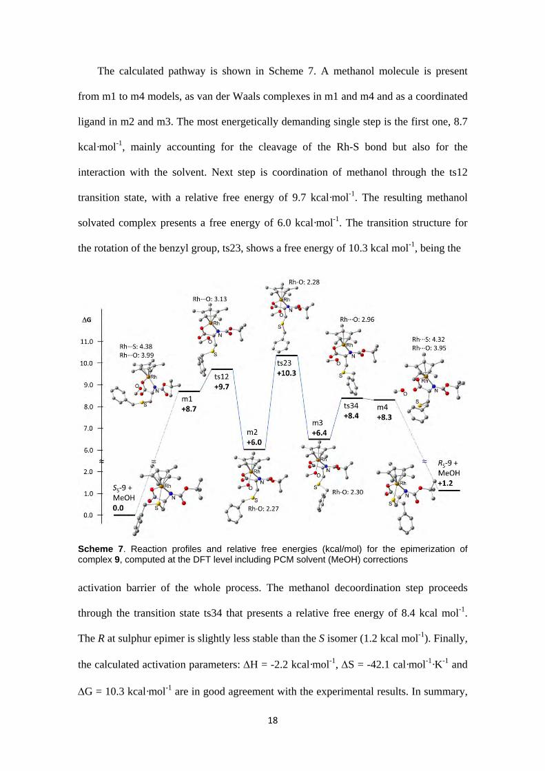
The calculated pathway is shown in Scheme 7. A methanol molecule is present
from m1 to m4 models, as van der Waals complexes in m1 and m4 and as a coordinated
ligand in m2 and m3. The most energetically demanding single step is the first one, 8.7
kcal·mol-1, mainly accounting for the cleavage of the Rh-S bond but also for the
interaction with the solvent. Next step is coordination of methanol through the ts12
transition state, with a relative free energy of 9.7 kcal·mol-1. The resulting methanol
solvated complex presents a free energy of 6.0 kcal·mol-1. The transition structure for
the rotation of the benzyl group, ts23, shows a free energy of 10.3 kcal mol-1, being the
Scheme 7. Reaction profiles and relative free energies (kcal/mol) for the epimerization of complex 9, computed at the DFT level including PCM solvent (MeOH) corrections
activation barrier of the whole process. The methanol decoordination step proceeds
through the transition state ts34 that presents a relative free energy of 8.4 kcal mol-1.
The R at sulphur epimer is slightly less stable than the S isomer (1.2 kcal mol-1). Finally,
the calculated activation parameters: H = -2.2 kcal·mol-1, S = -42.1 cal·mol-1·K-1 and
G = 10.3 kcal·mol-1 are in good agreement with the experimental results. In summary,
19
DFT calculations support that the observed fluxional process in 9 is the epimerization at
sulphur.
Conclusions
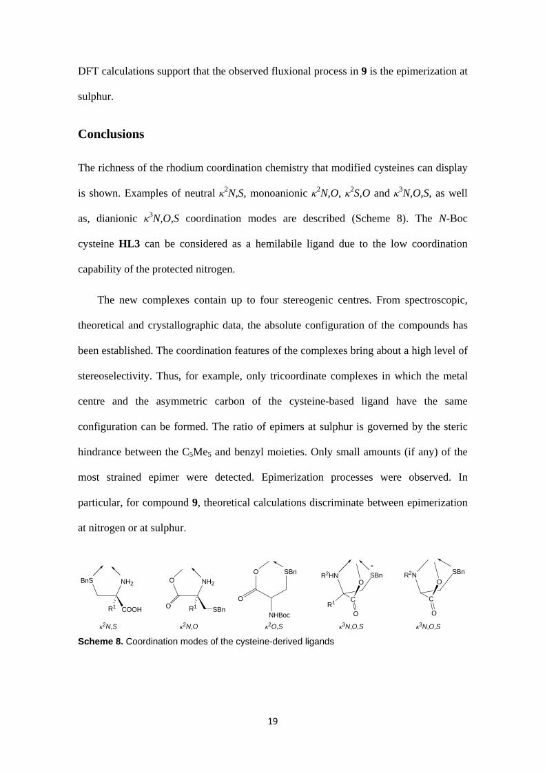
The richness of the rhodium coordination chemistry that modified cysteines can display
is shown. Examples of neutral κ2N,S, monoanionic κ2N,O, κ2S,O and κ3N,O,S, as well
as, dianionic κ3N,O,S coordination modes are described (Scheme 8). The N-Boc
cysteine HL3 can be considered as a hemilabile ligand due to the low coordination
capability of the protected nitrogen.
The new complexes contain up to four stereogenic centres. From spectroscopic,
theoretical and crystallographic data, the absolute configuration of the compounds has
been established. The coordination features of the complexes bring about a high level of
stereoselectivity. Thus, for example, only tricoordinate complexes in which the metal
centre and the asymmetric carbon of the cysteine-based ligand have the same
configuration can be formed. The ratio of epimers at sulphur is governed by the steric
hindrance between the C5Me5 and benzyl moieties. Only small amounts (if any) of the
most strained epimer were detected. Epimerization processes were observed. In
particular, for compound 9, theoretical calculations discriminate between epimerization
at nitrogen or at sulphur.
3N,O,S
BnS NH2
COOHR1
O NH2
R1O SBn
O SBn
O
NHBoc
R2N SBn
O
C
O
R2HN SBnO
C
OR1
3N,O,S2O,S2N,O2N,S
Scheme 8. Coordination modes of the cysteine-derived ligands
20
Experimental Section
General information
All preparations have been carried out under argon. All solvents were treated in a PS-
400-6 Innovative Technologies Solvent Purification System (SPS) and degassed prior to
use. Infrared spectra were recorded on Perkin-Elmer Spectrum-100 (ATR mode) FT–IR
spectrometer. Carbon, hydrogen, nitrogen and sulphur analyses were performed using a
Perkin-Elmer 240 B microanalyzer. 1H and 13C spectra were recorded on a Bruker AV–
300 (300.13 MHz), a Bruker AV–400 (400.16 MHz) or a Bruker AV–500 (500.13
MHz) spectrometers. In both, 1H NMR and 13C NMR measurements the chemical shifts
are expressed in ppm downfield from SiMe4. J values are given in Hz. COSY, NOESY,
HSQC, HMQC, and HMBC 1H–X (X = 1H, 13C) correlation spectra were obtained using
standard procedures. CD spectra were determined in acetone (ca. 4 × 10⁻⁴ mol L-1
solutions) in a 1 cm path length cell by using a JASCO J–810 spectropolarimeter. Mass
spectra were obtained with a Micro Tof-Q Bruker Daltonics spectrometer. Rhodium
trichloride was purchased from Johnson Matthey. Cysteines HL1 and HL3 are
commercially available from Acros and Aldrich, respectively.
Preparation of the complexes [(η5-C5Me5)RhCl(2N,S-HL)]Cl, (HL = HL1 (1), HL2
(2))
At room temperature, to a suspension of the dimer [{(η5-C5Me5)RhCl}2(μ-Cl)2] (100.0
mg, 0.16 mmol), in 10 mL of MeOH, 0.32 mmol of the corresponding cysteine-derived
ligand were added. The resulting orange solution was stirred for 1 h and then was
filtered to remove any insoluble material. The solution was concentrated under reduced
pressure to ca. 1 mL. The slow addition of Et2O led to the precipitation of an orange
solid which was washed with Et2O (3 × 5 mL) and vacuum-dried.
21
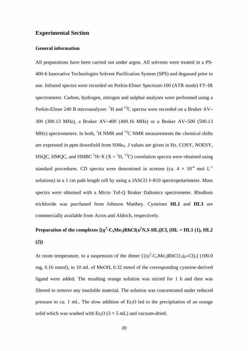
[(η5-C5Me5)RhCl(2N,S-HL1)]Cl (1). Yield: 95 %. Diastereomeric ratio: 85/15. Anal.
calcd for C20H28Cl2NO2RhS·2H2O,23 %: C, 44.62; H, 5.62; N, 2.60; S, 5.96. Found, %:
C, 44.58; H, 6.00; N, 2.56; S, 6.11. IR (solid, cm1): ν(OH) 2909 (vbr), ν(C=O) 1719
(s).
SRh,RC diastereomer (85 %). 1H NMR (500.13 MHz, CD2Cl2, 298 K, ppm): δ 9.7 - 7.75
(br, 1H, OH), 7.65 - 7.20 (2 × m, 5H, HAr), 6.16 (pt, J = 10.0 Hz, 1H, NH), 4.58 (AB
system, J = 11.4 Hz, 2H, CH2Ph), 4.22 (brs, 1H, NH), 4.04 (m, 1H,
C*H), 3.54 (ABXX’ system, JAB = 13.9, JAX = 10.7, JAX’ = 3.6 Hz, 2H,
CH2C*), 1.62 (s, 15H, C5Me5).
13C{1H} NMR (125.8 MHz, CD2Cl2, 298
K, ppm): δ 170.35 (C=O), 133.77, 131.05, 129.02, 128.81 (6C, CAr), 98.84 (d, JRhC = 7.7
Hz, 5C, C5Me5), 58.82 (C*), 40.60 (CH2Ph), 38.62 (CH2C*), 9.12 (5C, C5Me5).
RRh,RC diastereomer (15 %). 1H NMR (500.13 MHz, CD2Cl2, 298 K, ppm): δ 9.70 - 7.75
(m, 1H, OH), 7.65 - 7.20 (2 × m, 5H, HAr), 6.36 (brs, 1H, NH), 4.33 (AB system, J =
12.2 Hz, 2H, CH2Ph), 3.80 (m, 2H, C*H, NH), 3.27 (ABXX’ system, JAB = 12.9, JAX =
12.0, JAX’ = 3.6 Hz, 2H, CH2C*), 1.69 (s, 15H, C5Me5).
13C{1H} NMR (125.8 MHz,
CD2Cl2, 298 K, ppm): δ 170.65 (C=O), 133.53, 130.60, 129.24, 128.81 (6C, CAr), 99.15
(d, JRhC = 7.7 Hz, 5C, C5Me5), 58.64 (C*), 39.55 (CH2Ph), 38.06 (CH2C*), 9.41 (5C,
C5Me5).
[(η5-C5Me5)RhCl(2N,S-HL2)]Cl (2). Yield: 85 %. Diastereomeric ratio: 85/15. Anal.
calcd for C21H30Cl2NO2RhS·3H2O, %: C, 42.87; H, 6.17; N, 2.38; S, 5.45. Found, %: C,
42.66; H, 5.93; N, 2.28; S, 5.27. IR (solid, cm1): ν(OH) 2969 (vbr), ν(C=O) 1734 (s).
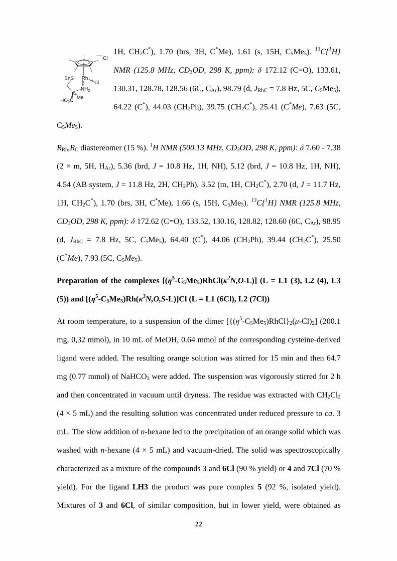
SRh,RC diastereomer (85 %). 1H NMR (500.13 MHz, CD3OD, 298 K, ppm): δ 7.60 - 7.38
(2 × m, 5H, HAr), 5.22 (brd, J = 10.6 Hz, 1H, NH), 4.42 (AB system, J = 11.7 Hz, 2H,
CH2Ph), 4.06 (brd, J = 10.6 Hz, 1H, NH), 3.52 (m, 1H, CH2C*), 2.60 (d, J = 11.7 Hz,
Cl
RhCl
BnS
NH2
HO2CH
22
1H, CH2C*), 1.70 (brs, 3H, C*Me), 1.61 (s, 15H, C5Me5).
13C{1H}
NMR (125.8 MHz, CD3OD, 298 K, ppm): δ 172.12 (C=O), 133.61,
130.31, 128.78, 128.56 (6C, CAr), 98.79 (d, JRhC = 7.8 Hz, 5C, C5Me5),
64.22 (C*), 44.03 (CH2Ph), 39.75 (CH2C*), 25.41 (C*Me), 7.63 (5C,
C5Me5).
RRh,RC diastereomer (15 %). 1H NMR (500.13 MHz, CD3OD, 298 K, ppm): δ 7.60 - 7.38
(2 × m, 5H, HAr), 5.36 (brd, J = 10.8 Hz, 1H, NH), 5.12 (brd, J = 10.8 Hz, 1H, NH),
4.54 (AB system, J = 11.8 Hz, 2H, CH2Ph), 3.52 (m, 1H, CH2C*), 2.70 (d, J = 11.7 Hz,
1H, CH2C*), 1.70 (brs, 3H, C*Me), 1.66 (s, 15H, C5Me5).
13C{1H} NMR (125.8 MHz,
CD3OD, 298 K, ppm): δ 172.62 (C=O), 133.52, 130.16, 128.82, 128.60 (6C, CAr), 98.95
(d, JRhC = 7.8 Hz, 5C, C5Me5), 64.40 (C*), 44.06 (CH2Ph), 39.44 (CH2C*), 25.50
(C*Me), 7.93 (5C, C5Me5).
Preparation of the complexes [(η5-C5Me5)RhCl(κ2N,O-L)] (L = L1 (3), L2 (4), L3
(5)) and [(η5-C5Me5)Rh(κ3N,O,S-L)]Cl (L = L1 (6Cl), L2 (7Cl))
At room temperature, to a suspension of the dimer [{(η5-C5Me5)RhCl}2(μ-Cl)2] (200.1
mg, 0,32 mmol), in 10 mL of MeOH, 0.64 mmol of the corresponding cysteine-derived
ligand were added. The resulting orange solution was stirred for 15 min and then 64.7
mg (0.77 mmol) of NaHCO3 were added. The suspension was vigorously stirred for 2 h
and then concentrated in vacuum until dryness. The residue was extracted with CH2Cl2
(4 × 5 mL) and the resulting solution was concentrated under reduced pressure to ca. 3
mL. The slow addition of n-hexane led to the precipitation of an orange solid which was
washed with n-hexane (4 × 5 mL) and vacuum-dried. The solid was spectroscopically
characterized as a mixture of the compounds 3 and 6Cl (90 % yield) or 4 and 7Cl (70 %
yield). For the ligand LH3 the product was pure complex 5 (92 %, isolated yield).
Mixtures of 3 and 6Cl, of similar composition, but in lower yield, were obtained as
Cl
RhCl
BnS
NH2
HO2CMe
23
follows: to a suspension of [(η5-C5Me5)RhCl(acac)] (200.0 mg, 0.54 mmol), in 10 mL
of MeOH, 114.3 mg (0.54 mmol) of HL1 were added. The suspension was vigorously
stirred for 24 h and then was concentrated under reduced pressure to ca. 3 mL. The slow
addition of n-hexane led to the precipitation of an orange solid which was washed with
n-hexane (3 × 10 mL) and vacuum-dried.
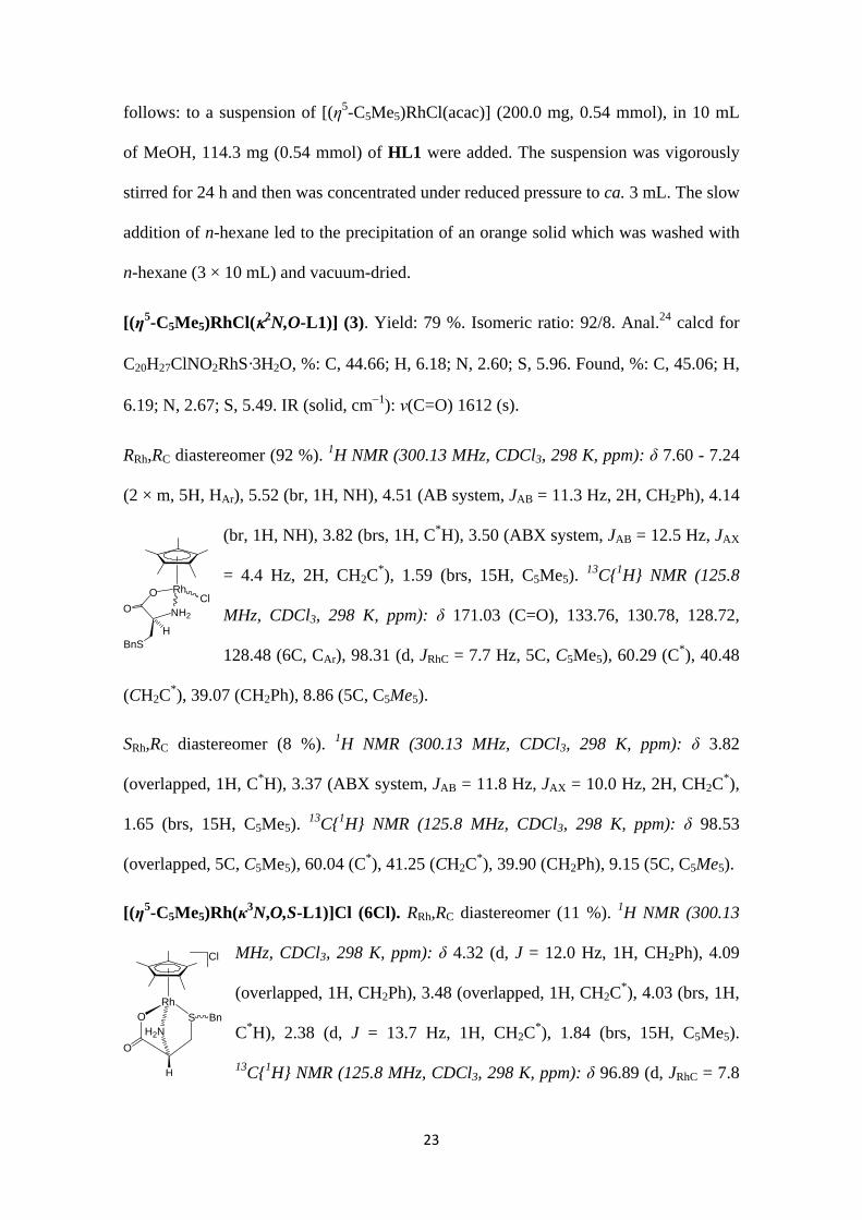
[(η5-C5Me5)RhCl(2N,O-L1)] (3). Yield: 79 %. Isomeric ratio: 92/8. Anal.24 calcd for
C20H27ClNO2RhS·3H2O, %: C, 44.66; H, 6.18; N, 2.60; S, 5.96. Found, %: C, 45.06; H,
6.19; N, 2.67; S, 5.49. IR (solid, cm1): ν(C=O) 1612 (s).
RRh,RC diastereomer (92 %). 1H NMR (300.13 MHz, CDCl3, 298 K, ppm): δ 7.60 - 7.24
(2 × m, 5H, HAr), 5.52 (br, 1H, NH), 4.51 (AB system, JAB = 11.3 Hz, 2H, CH2Ph), 4.14
(br, 1H, NH), 3.82 (brs, 1H, C*H), 3.50 (ABX system, JAB = 12.5 Hz, JAX
= 4.4 Hz, 2H, CH2C*), 1.59 (brs, 15H, C5Me5).
13C{1H} NMR (125.8
MHz, CDCl3, 298 K, ppm): δ 171.03 (C=O), 133.76, 130.78, 128.72,
128.48 (6C, CAr), 98.31 (d, JRhC = 7.7 Hz, 5C, C5Me5), 60.29 (C*), 40.48
(CH2C*), 39.07 (CH2Ph), 8.86 (5C, C5Me5).
SRh,RC diastereomer (8 %). 1H NMR (300.13 MHz, CDCl3, 298 K, ppm): δ 3.82
(overlapped, 1H, C*H), 3.37 (ABX system, JAB = 11.8 Hz, JAX = 10.0 Hz, 2H, CH2C*),
1.65 (brs, 15H, C5Me5). 13C{1H} NMR (125.8 MHz, CDCl3, 298 K, ppm): δ 98.53
(overlapped, 5C, C5Me5), 60.04 (C*), 41.25 (CH2C*), 39.90 (CH2Ph), 9.15 (5C, C5Me5).
[(η5-C5Me5)Rh(κ3N,O,S-L1)]Cl (6Cl). RRh,RC diastereomer (11 %). 1H NMR (300.13
MHz, CDCl3, 298 K, ppm): δ 4.32 (d, J = 12.0 Hz, 1H, CH2Ph), 4.09
(overlapped, 1H, CH2Ph), 3.48 (overlapped, 1H, CH2C*), 4.03 (brs, 1H,
C*H), 2.38 (d, J = 13.7 Hz, 1H, CH2C*), 1.84 (brs, 15H, C5Me5).
13C{1H} NMR (125.8 MHz, CDCl3, 298 K, ppm): δ 96.89 (d, JRhC = 7.8
RhClO
NH2O
HBnS
OH2N
S Bn
Cl
Rh
H
O
24
Hz, 5C, C5Me5), 60.21 (C*), 39.68 (CH2Ph), 32.42 (CH2C*), 9.43 (5C, C5Me5).
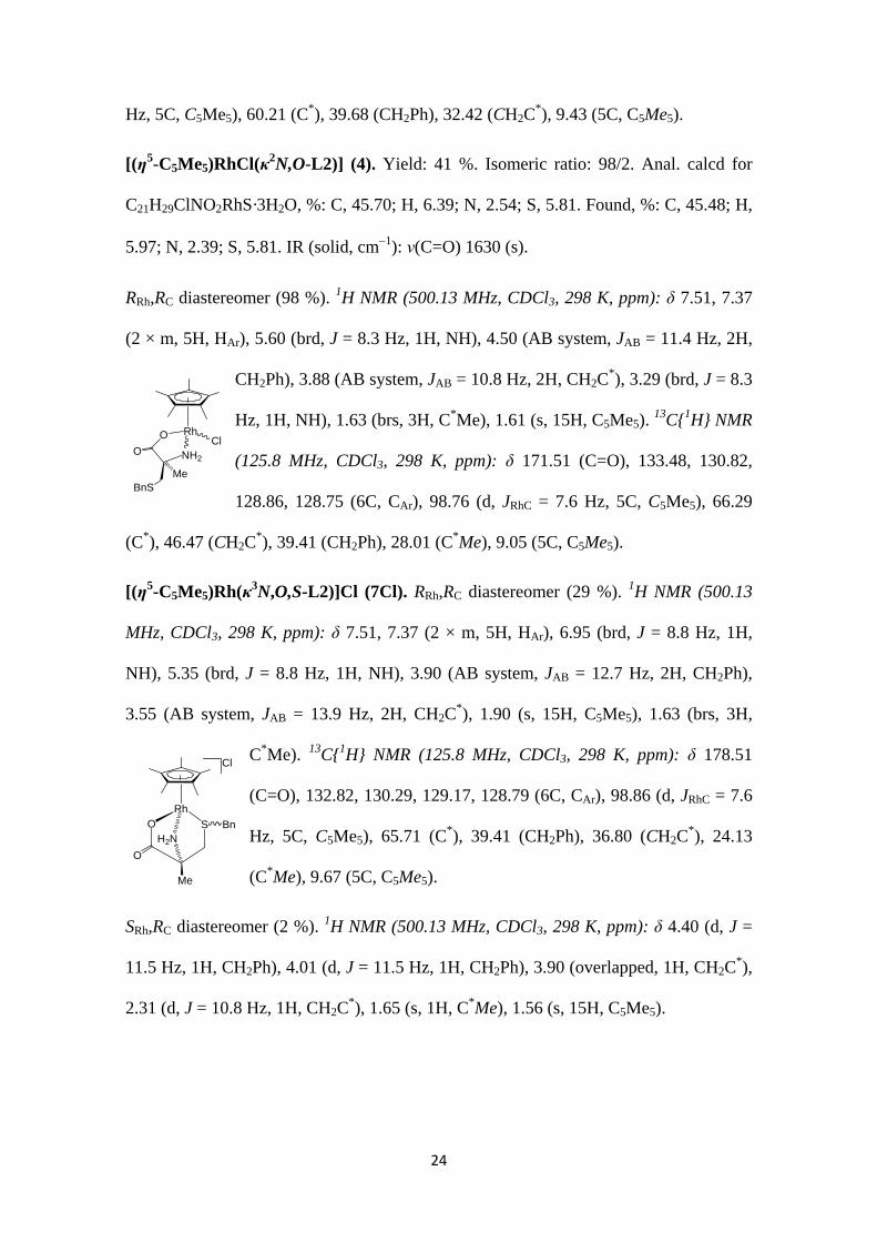
[(η5-C5Me5)RhCl(κ2N,O-L2)] (4). Yield: 41 %. Isomeric ratio: 98/2. Anal. calcd for
C21H29ClNO2RhS·3H2O, %: C, 45.70; H, 6.39; N, 2.54; S, 5.81. Found, %: C, 45.48; H,
5.97; N, 2.39; S, 5.81. IR (solid, cm1): ν(C=O) 1630 (s).
RRh,RC diastereomer (98 %). 1H NMR (500.13 MHz, CDCl3, 298 K, ppm): δ 7.51, 7.37
(2 × m, 5H, HAr), 5.60 (brd, J = 8.3 Hz, 1H, NH), 4.50 (AB system, JAB = 11.4 Hz, 2H,
CH2Ph), 3.88 (AB system, JAB = 10.8 Hz, 2H, CH2C*), 3.29 (brd, J = 8.3
Hz, 1H, NH), 1.63 (brs, 3H, C*Me), 1.61 (s, 15H, C5Me5). 13C{1H} NMR
(125.8 MHz, CDCl3, 298 K, ppm): δ 171.51 (C=O), 133.48, 130.82,
128.86, 128.75 (6C, CAr), 98.76 (d, JRhC = 7.6 Hz, 5C, C5Me5), 66.29
(C*), 46.47 (CH2C*), 39.41 (CH2Ph), 28.01 (C*Me), 9.05 (5C, C5Me5).
[(η5-C5Me5)Rh(κ3N,O,S-L2)]Cl (7Cl). RRh,RC diastereomer (29 %). 1H NMR (500.13
MHz, CDCl3, 298 K, ppm): δ 7.51, 7.37 (2 × m, 5H, HAr), 6.95 (brd, J = 8.8 Hz, 1H,
NH), 5.35 (brd, J = 8.8 Hz, 1H, NH), 3.90 (AB system, JAB = 12.7 Hz, 2H, CH2Ph),
3.55 (AB system, JAB = 13.9 Hz, 2H, CH2C*), 1.90 (s, 15H, C5Me5), 1.63 (brs, 3H,
C*Me). 13C{1H} NMR (125.8 MHz, CDCl3, 298 K, ppm): δ 178.51
(C=O), 132.82, 130.29, 129.17, 128.79 (6C, CAr), 98.86 (d, JRhC = 7.6
Hz, 5C, C5Me5), 65.71 (C*), 39.41 (CH2Ph), 36.80 (CH2C*), 24.13
(C*Me), 9.67 (5C, C5Me5).
SRh,RC diastereomer (2 %). 1H NMR (500.13 MHz, CDCl3, 298 K, ppm): δ 4.40 (d, J =
11.5 Hz, 1H, CH2Ph), 4.01 (d, J = 11.5 Hz, 1H, CH2Ph), 3.90 (overlapped, 1H, CH2C*),
2.31 (d, J = 10.8 Hz, 1H, CH2C*), 1.65 (s, 1H, C*Me), 1.56 (s, 15H, C5Me5).
RhClO
NH2O
MeBnS
OH2N
S Bn
Cl
Rh
Me
O
25
[(η5-C5Me5)RhCl(κ2O,S-L3)] (5). Yield: 92 %. Isomeric ratio: 97/3. Anal. calcd for
C25H35ClNO4RhS, %: C, 51.40; H, 5.99; N, 2.40; S, 5.48. Found, %: C, 50.91; H, 6.43;
N, 2.46; S, 5.63. IR (solid, cm1): ν(C=OBoc) 1757 (s), ν(C=O) 1620 (s).
SRh,RC diastereomer (97 %). 1H NMR (300.13 MHz, CDCl3, 298 K, ppm): δ 7.61, 7.32
(2 × m, 5H, HAr), 6.08 (br, 1H, NH), 4.83 (AB system, JAB = 12.2 Hz, 2H, CH2Ph), 4.29
(br, 1H, C*H), 3.21 (AB part, ABX system, JAB = 12.1 Hz, JAX = 1.8 Hz,
2H, CH2C*), 1.44 (s, 15H, C5Me5), 1.41 (s, 9H, MetBu).
13C{1H} NMR
(75.47 MHz, CDCl3, 298 K, ppm): δ 175.12 (C=O), 155.09 (C=OBoc),
134.34, 131.13, 128.58, 128.19 (6C, CAr), 96.98 (d, JRhC = 8.2 Hz, 5C, C5Me5), 79.05
(CtBu), 50.65 (C*), 38.83 (CH2Ph), 34.70 (CH2C*), 28.42 (3C, MetBu), 8.52 (5C, C5Me5).
RRh,RC diastereomer (3 %). 1H NMR (300.13 MHz, CDCl3, 298 K, ppm): δ 4.64 (br, 1H,
CH2Ph), 4.38 (br, 1H, C*H), 3.87 (br, 1H, CH2Ph), 2.66 (br, 1H, CH2C*), 2.24
(overlapped, 1H, CH2C*), 1.61 (s, 15H, C5Me5), 1.38 (s, 9H, MetBu).
13C{1H} NMR
(75.47 MHz, CDCl3, 298 K, ppm): δ 50.59 (C*), 39.53 (brs, CH2Ph), 36.20 (brs, CH2C*),
28.79 (3C, MetBu), 8.37 (5C, C5Me5).
Preparation of the complexes [(η5-C5Me5)Rh(κ3N,O,S-L)][SbF6] (L = L1 (6Sb), L2
(7Sb), L3 (8Sb))
To a solution of mixtures of 3 and 6Cl or 4 and 7Cl or pure 5 (0.25 mmol) in 10 mL of
acetone, 85.9 mg (0.25 mmol) of AgSbF6 were added. After stirring for 1 h, the AgCl
formed was filtered off and the solution was concentrated under reduced pressure to ca.
3 mL. The slow addition of Et2O led to the precipitation of a yellow-orange solid which
was washed with Et2O (3 × 5 mL) and vacuum-dried.
[(η5-C5Me5)Rh(κ2N,O,S-L1)][SbF6] (6Sb). Yield: 85 %. Anal. calcd for
C20H27F6NO2RhSSb·2H2O, %: C, 33.36; H, 4.34; N, 1.94; S, 4.45. Found, %: C, 33.60;
RhClO
SBn
H
O
NHBoc
26
H, 3.90; N, 1.71; S, 4.60. IR (solid, cm1): ν(C=O) 1642 (s), ν(SbF6) 653 (s). CD
(acetone, 5.0 × 10⁻⁴ M, RT): λ, nm, (Δε): 370 (6.42).
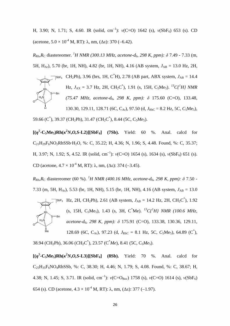
RRh,RC diastereomer. 1H NMR (300.13 MHz, acetone-d6, 298 K, ppm): δ 7.49 - 7.33 (m,
5H, HAr), 5.70 (br, 1H, NH), 4.82 (br, 1H, NH), 4.16 (AB system, JAB = 13.0 Hz, 2H,
CH2Ph), 3.96 (brs, 1H, C*H), 2.78 (AB part, ABX system, JAB = 14.4
Hz, JAX = 3.7 Hz, 2H, CH2C*), 1.91 (s, 15H, C5Me5).
13C{1H} NMR
(75.47 MHz, acetone-d6, 298 K, ppm): δ 175.60 (C=O), 133.48,
130.30, 129.11, 128.71 (6C, CAr), 97.50 (d, JRhC = 8.2 Hz, 5C, C5Me5),
59.66 (C*), 39.37 (CH2Ph), 31.47 (CH2C*), 8.44 (5C, C5Me5).
[(η5-C5Me5)Rh(κ2N,O,S-L2)][SbF6] (7Sb). Yield: 60 %. Anal. calcd for
C21H29F6NO2RhSSb·H2O, %: C, 35.22; H, 4.36; N, 1.96; S, 4.48. Found, %: C, 35.37;
H, 3.97; N, 1.92; S, 4.52. IR (solid, cm1): ν(C=O) 1654 (s), 1634 (s), ν(SbF6) 651 (s).
CD (acetone, 4.7 × 10⁻⁴ M, RT): λ, nm, (Δε): 374 (3.45).
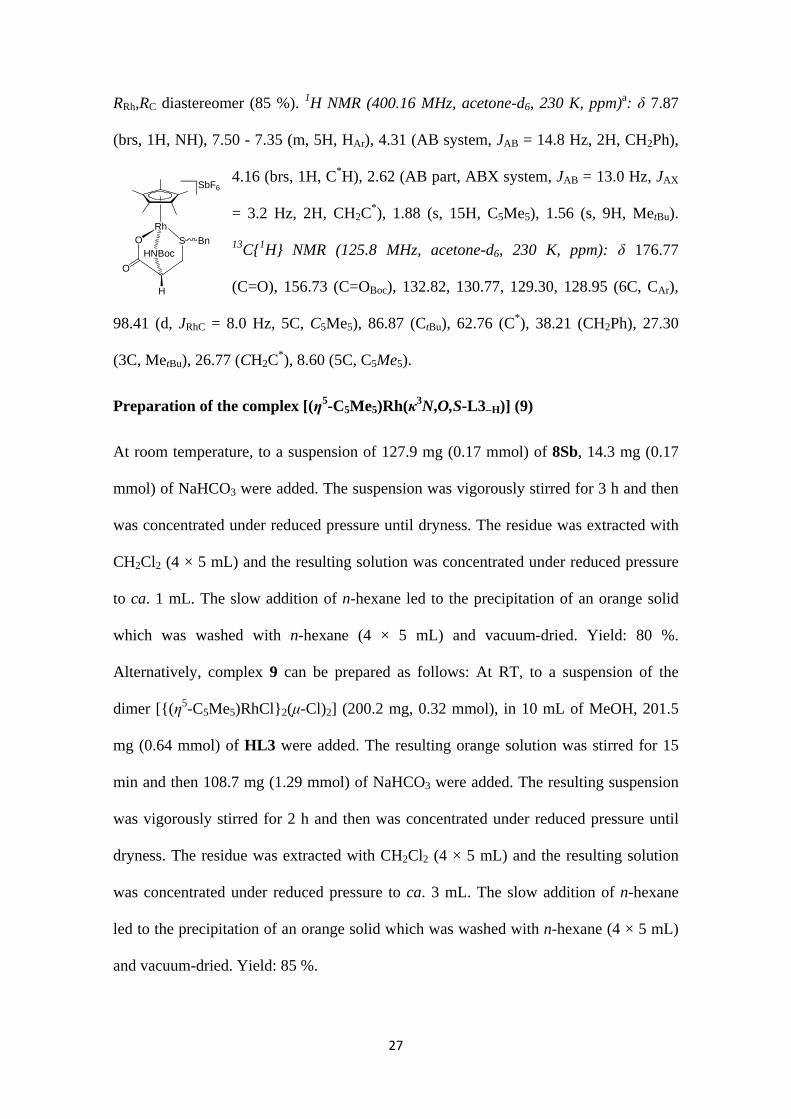
RRh,RC diastereomer (60 %). 1H NMR (400.16 MHz, acetone-d6, 298 K, ppm): δ 7.50 -
7.33 (m, 5H, HAr), 5.53 (br, 1H, NH), 5.15 (br, 1H, NH), 4.16 (AB system, JAB = 13.0
Hz, 2H, CH2Ph), 2.61 (AB system, JAB = 14.2 Hz, 2H, CH2C*), 1.92
(s, 15H, C5Me5), 1.43 (s, 3H, C*Me). 13C{1H} NMR (100.6 MHz,
acetone-d6, 298 K, ppm): δ 175.91 (C=O), 133.38, 130.36, 129.11,
128.69 (6C, CAr), 97.23 (d, JRhC = 8.1 Hz, 5C, C5Me5), 64.89 (C*),
38.94 (CH2Ph), 36.06 (CH2C*), 23.57 (C*Me), 8.41 (5C, C5Me5).
[(η5-C5Me5)Rh(κ2N,O,S-L3)][SbF6] (8Sb). Yield: 70 %. Anal. calcd for
C25H35F6NO4RhSSb, %: C, 38.30; H, 4.46; N, 1.79; S, 4.08. Found, %: C, 38.67; H,
4.38; N, 1.45; S, 3.71. IR (solid, cm1): ν(C=OBoc) 1758 (s), ν(C=O) 1614 (s), ν(SbF6)
654 (s). CD (acetone, 4.3 × 10⁻⁴ M, RT): λ, nm, (Δε): 377 (1.97).
OH2N
S Bn
SbF6
Rh
H
O
OH2N
S Bn
SbF6
Rh
Me
O
27
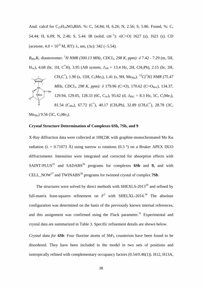
RRh,RC diastereomer (85 %). 1H NMR (400.16 MHz, acetone-d6, 230 K, ppm)a: δ 7.87
(brs, 1H, NH), 7.50 - 7.35 (m, 5H, HAr), 4.31 (AB system, JAB = 14.8 Hz, 2H, CH2Ph),
4.16 (brs, 1H, C*H), 2.62 (AB part, ABX system, JAB = 13.0 Hz, JAX
= 3.2 Hz, 2H, CH2C*), 1.88 (s, 15H, C5Me5), 1.56 (s, 9H, MetBu).
13C{1H} NMR (125.8 MHz, acetone-d6, 230 K, ppm): δ 176.77
(C=O), 156.73 (C=OBoc), 132.82, 130.77, 129.30, 128.95 (6C, CAr),
98.41 (d, JRhC = 8.0 Hz, 5C, C5Me5), 86.87 (CtBu), 62.76 (C*), 38.21 (CH2Ph), 27.30
(3C, MetBu), 26.77 (CH2C*), 8.60 (5C, C5Me5).
Preparation of the complex [(η5-C5Me5)Rh(κ3N,O,S-L3H)] (9)
At room temperature, to a suspension of 127.9 mg (0.17 mmol) of 8Sb, 14.3 mg (0.17
mmol) of NaHCO3 were added. The suspension was vigorously stirred for 3 h and then
was concentrated under reduced pressure until dryness. The residue was extracted with
CH2Cl2 (4 × 5 mL) and the resulting solution was concentrated under reduced pressure
to ca. 1 mL. The slow addition of n-hexane led to the precipitation of an orange solid
which was washed with n-hexane (4 × 5 mL) and vacuum-dried. Yield: 80 %.
Alternatively, complex 9 can be prepared as follows: At RT, to a suspension of the
dimer [{(η5-C5Me5)RhCl}2(μ-Cl)2] (200.2 mg, 0.32 mmol), in 10 mL of MeOH, 201.5
mg (0.64 mmol) of HL3 were added. The resulting orange solution was stirred for 15
min and then 108.7 mg (1.29 mmol) of NaHCO3 were added. The resulting suspension
was vigorously stirred for 2 h and then was concentrated under reduced pressure until
dryness. The residue was extracted with CH2Cl2 (4 × 5 mL) and the resulting solution
was concentrated under reduced pressure to ca. 3 mL. The slow addition of n-hexane
led to the precipitation of an orange solid which was washed with n-hexane (4 × 5 mL)
and vacuum-dried. Yield: 85 %.
OHN
S Bn
SbF6
Rh
H
O
Boc
28
Anal. calcd for C25H34NO4RhS, %: C, 54.84; H, 6.26; N, 2.56; S, 5.86. Found, %: C,
54.44; H, 6.09; N, 2.46; S, 5.44. IR (solid, cm1): ν(C=O) 1627 (s), 1621 (s). CD
(acetone, 4.0 × 10⁻⁴ M, RT): λ, nm, (Δε): 342 (5.54).
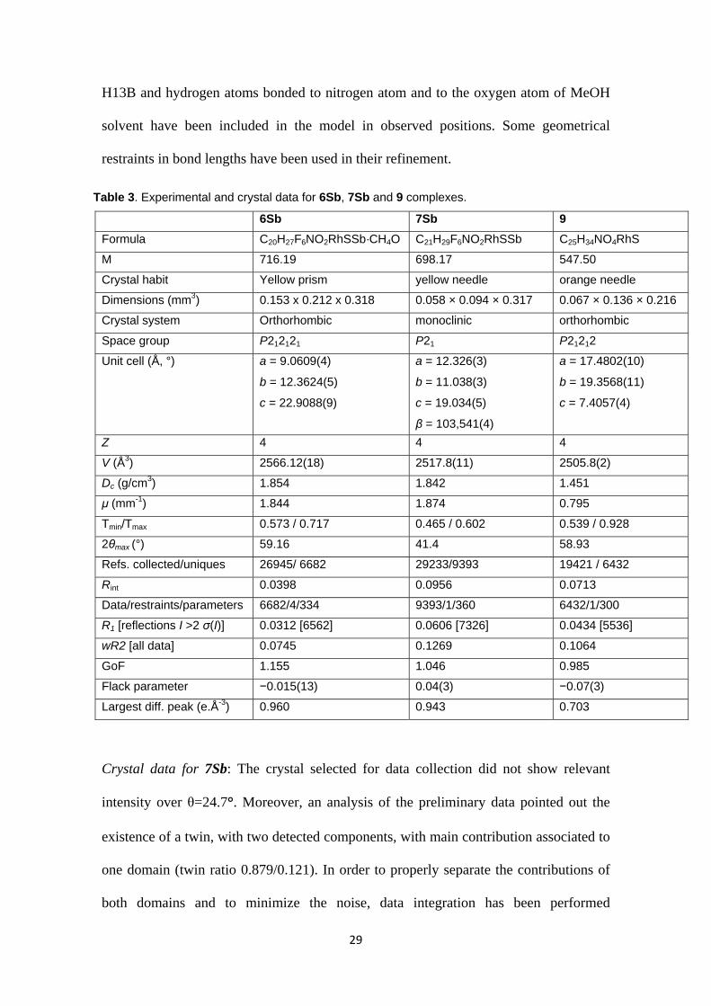
RRh,RC diastereomer. 1H NMR (300.13 MHz, CDCl3, 298 K, ppm): δ 7.42 - 7.29 (m, 5H,
HAr), 4.68 (br, 1H, C*H), 3.95 (AB system, JAB = 13.4 Hz, 2H, CH2Ph), 2.15 (br, 2H,
CH2C*), 1.90 (s, 15H, C5Me5), 1.41 (s, 9H, MetBu).
13C{1H} NMR (75.47
MHz, CDCl3, 298 K, ppm): δ 179.96 (C=O), 170.62 (C=OBoc), 134.37,
129.94, 129.05, 128.33 (6C, CAr), 95.62 (d, JRhC = 8.3 Hz, 5C, C5Me5),
81.54 (CtBu), 67.72 (C*), 40.17 (CH2Ph), 32.89 (CH2C*), 28.78 (3C,
MetBu) 9.56 (5C, C5Me5).
Crystal Structure Determination of Complexes 6Sb, 7Sb, and 9
X-Ray diffraction data were collected at 100(2)K with graphite-monochromated Mo Kα
radiation (λ = 0.71073 Å) using narrow ω rotations (0.3 º) on a Bruker APEX DUO
diffractometer. Intensities were integrated and corrected for absorption effects with
SAINT-PLUS25 and SADABS26 programs for complexes 6Sb and 9, and with
CELL_NOW27 and TWINABS28 programs for twinned crystal of complex 7Sb.
The structures were solved by direct methods with SHEXLS-201329 and refined by
full-matrix least-squares refinement on F2 with SHELXL-2014.30 The absolute
configuration was determined on the basis of the previously known internal references,
and this assignment was confirmed using the Flack parameter.31 Experimental and
crystal data are summarized in Table 3. Specific refinement details are shown below.
Crystal data for 6Sb: Four fluorine atoms of SbF6 counterion have been found to be
disordered. They have been included in the model in two sets of positions and
isotropically refined with complementary occupancy factors (0.54/0.46(1)). H12, H13A,
ON
S Bn
Rh
H
O
Boc
29
H13B and hydrogen atoms bonded to nitrogen atom and to the oxygen atom of MeOH
solvent have been included in the model in observed positions. Some geometrical
restraints in bond lengths have been used in their refinement.
Table 3. Experimental and crystal data for 6Sb, 7Sb and 9 complexes.
6Sb 7Sb 9
Formula C20H27F6NO2RhSSb·CH4O C21H29F6NO2RhSSb C25H34NO4RhS
M 716.19 698.17 547.50
Crystal habit Yellow prism yellow needle orange needle
Dimensions (mm3) 0.153 x 0.212 x 0.318 0.058 × 0.094 × 0.317 0.067 × 0.136 × 0.216
Crystal system Orthorhombic monoclinic orthorhombic
Space group P212121 P21 P21212
Unit cell (Å, °) a = 9.0609(4)
b = 12.3624(5)
c = 22.9088(9)
a = 12.326(3)
b = 11.038(3)
c = 19.034(5)
β = 103,541(4)
a = 17.4802(10)
b = 19.3568(11)
c = 7.4057(4)
Z 4 4 4
V (Å3) 2566.12(18) 2517.8(11) 2505.8(2)
Dc (g/cm3) 1.854 1.842 1.451
μ (mm-1) 1.844 1.874 0.795
Tmin/Tmax 0.573 / 0.717 0.465 / 0.602 0.539 / 0.928
2θmax (°) 59.16 41.4 58.93
Refs. collected/uniques 26945/ 6682 29233/9393 19421 / 6432
Rint 0.0398 0.0956 0.0713
Data/restraints/parameters 6682/4/334 9393/1/360 6432/1/300
R1 [reflections I >2 σ(I)] 0.0312 [6562] 0.0606 [7326] 0.0434 [5536]
wR2 [all data] 0.0745 0.1269 0.1064
GoF 1.155 1.046 0.985
Flack parameter −0.015(13) 0.04(3) −0.07(3)
Largest diff. peak (e.Å-3) 0.960 0.943 0.703
Crystal data for 7Sb: The crystal selected for data collection did not show relevant
intensity over θ=24.7°. Moreover, an analysis of the preliminary data pointed out the
existence of a twin, with two detected components, with main contribution associated to
one domain (twin ratio 0.879/0.121). In order to properly separate the contributions of
both domains and to minimize the noise, data integration has been performed
30
considering both components, to a maximal resolution of θ=21°. This restriction
prevents an adequate refinement of the anisotropic displacement parameters for the
lighter atoms, unless a large number of bonding restrains were included. Eventually, we
decided to consider only Rh, Sb, S and non-disordered F atoms to have anisotropic
atomic displacement parameters.
Fluorine atoms of a SbF6 counterion have been found to be disordered. When
convergence is achieved, five residual density peaks are found in intermolecular
regions. Attempts to interpret them as water solvent molecules do not improve the
model, as they are too close to methylic hydrogen atoms of η5-C5Me5 fragments.
Crystal data for 9: The (η5-C5Me5) group and a methyl fragment have been found to be
disordered. Hydrogen H12 has been included in the model in observed position and
freely refined.
Computational details
The DFT calculations were carried out with the Gaussian 09 software package,32
optimizing the structures using the b3-lyp hybrid functional and the basis set LanL2DZ
for rhodium atoms and 6-31G(d,p) for the remaining ones.33 All minima (no imaginary
frequencies) and transition states (one imaginary frequency) were characterized by
calculating the Hessian matrix. The transition state search was performed with a relaxed
PES scan of the key geometrical parameter and then the highest energy structure was
optimized as a transition state by the default Gaussian 09 algorithms. The chemical
correctness of the transition states found were confirmed by visual inspection of the
normal mode having a negative vibrational frequency, followed by moving the TS
geometries along the reaction path using the GaussView program utilities and
reoptimizing to verify the nature of the products.
31
The energetics of the epimerization mechanism is discussed based on the relative
Gibbs free energy with respect to the infinitely separated reactants. All reported free
energies involve ZPE and gas-phase thermal corrections (entropy and enthalpy, 298.15
K, 1 atm) and the conductor-like polarizable continuum model approach (CPCM)
implemented in the Gaussian 09 software was used in the final single point calculations
on the above gas-phase optimized geometries to incorporate the solvent effect of
methanol in the thermodynamic data.
Acknowledgments
We thank the Ministerio de Economía y Competitividad of Spain (CTQ2012-32095,
CTQ2014-53033-P, CTQ2015-67366-P and CTQ2013-408555R) and Gobierno de
Aragón and European Social Fund (Grupos Consolidados: Catalizadores
Organometálicos Enantioselectivos, Aminoácidos y Péptidos and Inorganic Molecular
Architecture) for financial support. This work was supported by the CONSOLIDER
INGENIO 2010 program under the project “Factoría de Cristalización” (CSD2006-
0015). M. C. acknowledges Diputación General de Aragón, CSIC and European Social
Fund for a grant. R. R. and P. G. O. acknowledge CSIC, European Social Fund and
Ministerio de Economía y Competitividad of Spain for a JAE and a Ramón y Cajal
(RYC‐2013‐13800) grants and for a PTA contract, respectively. We thank the Centro de
Supercomputación de Galicia (CESGA) for generous allocation of computational
resources.
32
Notes and references
1 (a) J. Paradowska, M. Stodulski and J. Mlynarski, Angew. Chem. Int. Ed. 2009,
48, 4288-4297. (b) Comprehensive Coordination Chemistry II, J. A. McCleverty
and T. J. Meyer, Eds.; Elsevier Science, 2003. (c) J. Chin, S.-S. Lee, K.-J. Lee, S.
Park and D. H. Kim, Nature 1999, 401, 254-257. (d) K. Severin, R. Bergs and W.
Beck, Angew. Chem. Int. Ed. 1998, 37, 1635-1654. (e) T. G. Appleton, Coord.
Chem. Rev. 1997, 166, 313-359. (f) N. Paulic and N. Raos, J. Coord. Chem. 1994,
31, 181-190. (g) A. Iakovidis and N. Hadjiliadis, Coord. Chem. Rev. 1994,
135/136, 17-63. (h) H. Kozlowski and L. D. Pettit, In Chemistry of the Platinum
Group Metals; Hartley, F. R., Ed.; Elsevier: New York, 1991; Chapter 15, p 530.
(i) S. H. Laurie, In Comprehensive Coordination Chemistry; Wilkinson, G., Ed.;
Pergamon: Oxford, 1987; Vol. 2, Chapter 20.2, p 739-776. (j) A. A. Ioganson,
Russ. Chem. Rev. 1985, 54, 277-292. (k) L. D. Pettit and M. Bezer, Coord. Chem.
Rev. 1985, 61, 97-114.
2 F. A. Egbewande, L. E. H. Paul, B. Therrien and J. Furrer, Eur. J. Inorg. Chem.
2014, 1174-1184.
3 K. Micskei, T. Patonay, L. Caglioti and G. Palyi, Chem. Biodiv. 2010, 7, 1660-
1669.
4 See for example: (a) L. E. H. Paul, B. Therrien and J. Furrer, Inorg. Chem. 2012,
51, 1957-1067. (b) F. Wang, H. Chen, J. A. Parkinson, P. del S. Murdoch and P. J.
Sadler, Inorg. Chem. 2002, 41, 4509-4523.
5 (a) A. Igashira-Kamiyama and T. Konno, Dalton Trans. 2011, 40, 7249-7263. (b)
T. Aridomi, K. Takamura, A. Igashira-Kamiyama, T. Kawamoto and T. Konno,
Chem. Eur. J. 2008, 14, 7752-7755.
33
6 S. Niu, W. Lv, G. Zhou, Y. He, B. Li, Q.-H. Yang and F. Kang, Chem. Commun.
2015, 51, 17720-17723.
7 X. Chen, Y. Zhou, X. Peng and J. Yoon, Chem. Soc. Rev. 2010, 39, 2120-2135.
8 (a) O. Gordon, T. V. Slenters, P. S. Brunetto, A. E. Villaruz, D. E. Sturdevant, M.
Otto, R. Landmann and K. M. Fromm, Antimicrob. Agents Chemother. 2010, 54,
4208−4218. (b) S. Y. Liau, D. C. Read, W. J. Pugh, J. R. Furr and A. D. Russell,
Lett. Appl. Microbiol. 1997, 25, 279-283.
9 C. Darnault, A. Volbeda, E. J. Kim, P. Legrand, X. Vernède, P. A. Lindahl and J.
C. Fontecilla-Camps, Nat. Struct. Biol. 2003, 10, 271-279.
10 A. Volbeda, M. H. Charon, C. Piras, E. C. Hatchikian, M. Frey and J. C.
Fontecilla-Camps, Nature 1995, 373, 580-587.
11 J. Patalenszki, L. Biró, A. C. Bényei, T. R. Muchova, J. Kasparkova and P.
Bublyó, RSC Adv. 2015, 5, 8094-8107.
12 C. Cativiela and M. Ordoñez, Tetrahedron Asymmetry 2009, 20, 1-63, and
references therein.
13 D. Pattenden, S. M. Thom and M. F. Jones, Tetrahedron 1993, 49, 2131-2138.
14 Y. Jia, X. Dong, P. Zhou, X. Liu, L. Pan, H. Xin, Y. Z. Zhu and Y. Wang, Eur. J.
Med. Chem. 2012, 55, 176-187.
15 J. W. Kang, K. Moseley and P. M. Maitlis, J. Am. Chem. Soc. 1969, 91, 5970-
5977.
16 Alternatively, complex 3 can be prepared by treating with HL1 the
acetylacetonate [(η5-C5Me5)RhCl(acac)] (W. Rigby, H.-B. Lee, P. M. Bailey, J. A.
McCleverty and P. M. Maitlis, J. Chem. Soc., Dalton Trans. 1979, 387-394).
34
17 All the trials give rise to twin and weakly diffracting crystals. Eventually a set of
data was tentatively measured. F. J. Lahoz, P. García-Orduña,. CSD Communication
to the Cambridge Structural Database, CCDC: Cambridge, UK, 2016. deposition number
CCDC 1485793.
18 (a) R. S. Cahn, C. Ingold and V. Prelog, Angew. Chem., Int. Ed. Engl. 1966, 5,
385-415. (b) V. Prelog and G. Helmchen, Angew. Chem., Int. Ed. Engl. 1982, 21,
567-583. (c) C. Lecomte, Y. Dusausoy, J. Protas, J. Tirouflet and A. Dormond, J.
Organomet. Chem. 1974, 73, 67-76.
19 K. Nakamoto, Infrared and Raman Spectra of Inorganic and Coordination
Compounds, 4th ed.; Wiley-Interscience: New York, 1986.
20 NMR analyses of aliquots show that the diastereomeric composition does not
change importantly over the reaction time. These measured compositions matches
up with the one that was determined for the isolated solid.
21 The 15N NMR data were obtained through 1H-15N HMQC experiments.
22 D. Carmona, F. J. Lahoz, R. Atencio, L. A. Oro, M. P. Lamata, F. Viguri, E. San
José, C. Vega, J. Reyes, F. Joó and A. Kathó, Chem. Eur. J. 1999, 5, 1544-1564.
23 Most of the compounds crystallize with variable amounts of water which are
detected by 1H NMR spectroscopy.
24 The microanalyses quoted for complexes 3 and 4 correspond to mixtures of 3 and
6Cl and of 4 and 7Cl, respectively.
25 SAINT+, version 6.01: Area-Detector Integration Software, Bruker AXS,
Madison, WI, 2001.
26 (a) R. H. Blessing, Acta Crystallogr. 1995, A51, 33-38. (b) SADABS, Area
Detector Absorption Correction Program, Bruker AXS, Madison, WI, 1996.
35
27 G. M. Sheldrick, CELL_NOW, University of Göttingen, Germany, 2008.
28 G. M. Sheldrick, TWINABS, University of Göttingen, Germany, 2008.
29 (a) G. M. Sheldrick, Acta Crystallogr. 1990, A46, 467-473. (b) G. M. Sheldrick,
Acta Crystallogr. 2008, A64, 112-122.
30 G. M. Sheldrick, Acta Crystallogr. 2015, C71, 3-8.
31 H. D. Flack, Acta Crystallogr. 1983, A39, 876-881.
32 M. J. Frisch, et al. Gaussian 09 (Revision D.01); Gaussian, Inc., Wallingford, CT,
2009.
33 (a) C. Lee, W. Yang and R. G. Parr, Phys. Rev. B 1988, 37, 785-789. (b) A. D.
Becke, J. Chem. Phys. 1993, 98, 1372-1377. (c) A. D. Becke, J. Chem. Phys.
1993, 98, 5648-5652. (d) N. E. Schultz, Y. Zhao and D. G. Truhlar, J. Phys.
Chem. A 2005, 109, 11127-11143.
36
For table of contents use only
Half-sandwich complexes of rhodium containing cysteine-derived
ligands
María Carmona, Ricardo Rodríguez, Fernando J. Lahoz, Pilar García-Orduña, Iñaki Osante, Carlos Cativiela, José A. López, and Daniel Carmona
a Instituto de Síntesis Química y Catálisis Homogénea (ISQCH), CSIC - Universidad de Zaragoza, Departamento de Química Inorgánica, Pedro Cerbuna 12, 50009 Zaragoza, Spain, E-mail: [email protected], [email protected] b Instituto de Síntesis Química y Catálisis Homogénea (ISQCH), CSIC - Universidad de Zaragoza, Departamento de Química Orgánica, Pedro Cerbuna 12, 50009 Zaragoza, Spain
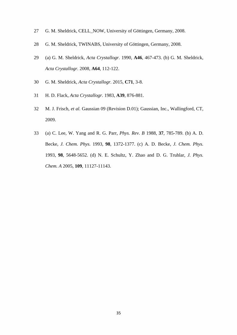
Five distinct coordination modes have been disclosed for rhodium complexes
containing modified cysteines. From spectroscopic, theoretical and crystallographic
data, the absolute configuration of the compounds has been established.
3N,O,S
S N O N
O
O S
O
Rh Rh
N SO
O
N SO
O
RhRh
3N,O,S
2O,S2N,S
Rh
2N,O
![ON THE ZEROES OF HALF-INTEGRAL WEIGHT EISENSTEIN …...the zeroes of integral weight modular forms, including [5] and [7]. On the other hand, there have been studies of half integral](https://static.fdocument.org/doc/165x107/5f1055f47e708231d4489a78/on-the-zeroes-of-half-integral-weight-eisenstein-the-zeroes-of-integral-weight.jpg)