Gut Feelings About α‐Synuclein in Gastrointestinal...
10
Gut Feelings About a-Synuclein in Gastrointestinal Biopsies: Biomarker in the Making? Claudio Ruffmann, MD 1,2 and Laura Parkkinen, PhD 1,2 * 1 Oxford Parkinson’s Disease Centre, University of Oxford, Oxford, United Kingdom 2 Nuffield Department of Clinical Neurosciences, Academic Unit of Neuropathology, University of Oxford, Oxford, United Kingdom ABSTRACT: In recent years, several studies have investigated the potential of immunohistochemical detec- tion of a-synuclein in the gastrointestinal tract to diag- nose Parkinson’s disease (PD). Although methodological heterogeneity has hindered comparability between stud- ies, it has become increasingly apparent that the high sensitivity and specificity reported in preliminary studies has not been sustained in subsequent large-scale stud- ies. What constitutes pathological a-synuclein in the ali- mentary canal that could distinguish between PD patients and controls and how this can be reliably detected represent key outstanding questions in the field. In this review, we will comment on and compare the variable technical aspects from previous studies, and by highlighting some advantages and shortcomings we hope to delineate a standardized approach to facilitate the consensus criteria urgently needed in the field. Fur- thermore, we will describe alternative detection techni- ques to conventional immunohistochemistry that have recently emerged and may facilitate ease of interpreta- tion and reliability of gastrointestinal a-synuclein detec- tion. Such techniques have the potential to detect the presence of pathological a-synuclein and include the paraffin-embedded tissue blot, the proximity ligation assay, the protein misfolding cyclic amplification tech- nique, and the real-time quaking-induced conversion assay. Finally, we will review 2 nonsynonymous theories that have driven enteric a-synuclein research, namely, (1) that a-synuclein propagates in a prion-like fashion from the peripheral nervous system to the brain via vagal con- nections and (2) that gastrointestinal a-synuclein deposi- tion may be used as a clinically useful biomarker in PD. V C 2016 The Authors. Movement Disorders published by Wiley Periodicals, Inc. on behalf of International Parkin- son and Movement Disorder Society. Key Words: biomarker; biopsy; enteric nervous sys- tem; Parkinson’s disease; a-synuclein Idiopathic Parkinson’s disease (PD) is a neurodegener- ative condition characterized by typical motor symp- toms of tremor, bradykinesia, and rigidity, which enable its diagnosis in the majority of cases. Nonetheless, mis- diagnosis can occur in up to 20% of cases, particularly in the early phases of disease. 1 Thus, the postmortem detection by immunohistochemistry (IHC) of aggrega- tion of a-synuclein (ASN) in the brain coupled with neuronal loss in the substantia nigra remains the gold standard for the definite diagnosis of PD. The progressive pathological changes, however, start years before the clinical onset of symptoms, and by the time of diagnosis, the disease is too advanced for any neuroprotective therapies to have full impact. In recent years, several studies have shown that ASN aggregation can also be detected outside the central nervous system (CNS), particularly in the enteric nervous system (ENS) of the gastrointestinal (GI) tract of PD patients. 2-5 The contrasting results and varying methodologies applied have recently been reviewed in detail by Visanji and col- leagues. 6 However, reflecting the fast development in this field, a number of studies on ENS ASN detection as a ------------------------------------------------------------ This is an open access article under the terms of the Creative Commons Attribution License, which permits use, distribution and reproduction in any medium, provided the original work is properly cited. *Correspondence to: Dr. Laura Parkkinen, Nuffield Department of Clinical Neurosciences, Academic Unit of Neuropathology, University of Oxford, West Wing, Level 1, OX3 9DU Oxford, UK, E-mail: [email protected] Funding agencies: This study was funded by the Monument Trust Discovery Award from Parkinson’s UK. Dr. Parkkinen receives research support from Parkinson’s UK, the Michael J. Fox Foundation, and Alzheimer’s Research UK. Relevant conflicts of interest/funding disclosures: The authors report no financial disclosures or conflict of interests. Received: 13 August 2015; Revised: 12 October 2015; Accepted: 15 October 2015 Published online 22 January 2016 in Wiley Online Library (wileyonlinelibrary.com). DOI: 10.1002/mds.26480 REVIEW Movement Disorders, Vol. 31, No. 2, 2016 193
-
Upload
truongquynh -
Category
Documents
-
view
214 -
download
0
Transcript of Gut Feelings About α‐Synuclein in Gastrointestinal...
Gut Feelings About a-Synuclein in Gastrointestinal Biopsies:Biomarker in the Making?
Claudio Ruffmann, MD1,2 and Laura Parkkinen, PhD1,2*
1Oxford Parkinson’s Disease Centre, University of Oxford, Oxford, United Kingdom2Nuffield Department of Clinical Neurosciences, Academic Unit of Neuropathology, University of Oxford, Oxford, United Kingdom
ABSTRACT: In recent years, several studies haveinvestigated the potential of immunohistochemical detec-tion of a-synuclein in the gastrointestinal tract to diag-nose Parkinson’s disease (PD). Although methodologicalheterogeneity has hindered comparability between stud-ies, it has become increasingly apparent that the highsensitivity and specificity reported in preliminary studieshas not been sustained in subsequent large-scale stud-ies. What constitutes pathological a-synuclein in the ali-mentary canal that could distinguish between PDpatients and controls and how this can be reliablydetected represent key outstanding questions in thefield. In this review, we will comment on and comparethe variable technical aspects from previous studies, andby highlighting some advantages and shortcomings wehope to delineate a standardized approach to facilitatethe consensus criteria urgently needed in the field. Fur-thermore, we will describe alternative detection techni-ques to conventional immunohistochemistry that have
recently emerged and may facilitate ease of interpreta-tion and reliability of gastrointestinal a-synuclein detec-tion. Such techniques have the potential to detect thepresence of pathological a-synuclein and include theparaffin-embedded tissue blot, the proximity ligationassay, the protein misfolding cyclic amplification tech-nique, and the real-time quaking-induced conversionassay. Finally, we will review 2 nonsynonymous theoriesthat have driven enteric a-synuclein research, namely, (1)that a-synuclein propagates in a prion-like fashion fromthe peripheral nervous system to the brain via vagal con-nections and (2) that gastrointestinal a-synuclein deposi-tion may be used as a clinically useful biomarker in PD.VC 2016 The Authors. Movement Disorders published byWiley Periodicals, Inc. on behalf of International Parkin-son and Movement Disorder Society.
Key Words: biomarker; biopsy; enteric nervous sys-tem; Parkinson’s disease; a-synuclein
Idiopathic Parkinson’s disease (PD) is a neurodegener-ative condition characterized by typical motor symp-toms of tremor, bradykinesia, and rigidity, which enable
its diagnosis in the majority of cases. Nonetheless, mis-diagnosis can occur in up to 20% of cases, particularlyin the early phases of disease.1 Thus, the postmortemdetection by immunohistochemistry (IHC) of aggrega-tion of a-synuclein (ASN) in the brain coupled withneuronal loss in the substantia nigra remains the goldstandard for the definite diagnosis of PD.
The progressive pathological changes, however, startyears before the clinical onset of symptoms, and by thetime of diagnosis, the disease is too advanced for anyneuroprotective therapies to have full impact. In recentyears, several studies have shown that ASN aggregationcan also be detected outside the central nervous system(CNS), particularly in the enteric nervous system (ENS)of the gastrointestinal (GI) tract of PD patients.2-5 Thecontrasting results and varying methodologies appliedhave recently been reviewed in detail by Visanji and col-leagues.6 However, reflecting the fast development in thisfield, a number of studies on ENS ASN detection as a
------------------------------------------------------------This is an open access article under the terms of the Creative CommonsAttribution License, which permits use, distribution and reproduction inany medium, provided the original work is properly cited.
*Correspondence to: Dr. Laura Parkkinen, Nuffield Department ofClinical Neurosciences, Academic Unit of Neuropathology, University ofOxford, West Wing, Level 1, OX3 9DU Oxford, UK,E-mail: [email protected]
Funding agencies: This study was funded by the Monument TrustDiscovery Award from Parkinson’s UK. Dr. Parkkinen receives researchsupport from Parkinson’s UK, the Michael J. Fox Foundation, andAlzheimer’s Research UK.
Relevant conflicts of interest/funding disclosures: The authors reportno financial disclosures or conflict of interests.
Received: 13 August 2015; Revised: 12 October 2015; Accepted: 15October 2015
Published online 22 January 2016 in Wiley Online Library(wileyonlinelibrary.com). DOI: 10.1002/mds.26480
R E V I E W
Movement Disorders, Vol. 31, No. 2, 2016 193
PD biomarker have since then added to the growingbody of literature.7-10 These have provided new evidencethat GI ASN detection may facilitate a presymptomaticdiagnosis of PD, with evidence of ASN accumulation incolonic biopsies up to 8 years prior to the developmentof motor symptoms,7,11 which also suggests that ASNaccumulation may start in the ENS long before it affectsthe brain. Recent studies have also supported the use ofin vivo gastric detection of ASN to confirm a diagnosisof PD in clinically symptomatic subjects,8 whereas othershave identified the appendiceal mucosa as a potentiallyuseful source of tissue for detection of ASN.9 If con-firmed, these findings could meet the current urgent needfor a biomarker in PD, which could complement or per-haps even be superior to directly imaging changes in thenigrostriatal system to predict, confirm, track progres-sion, and phenotypically stratify PD patients.12
However, a critical step in this process will be todevelop a method that is sufficiently sensitive and specific.To date, both the use of different types of antibodies andmorphological assessment of pathology have fallen shortof accurately distinguishing between the physiologicaland pathological forms of the ASN protein. Given thatASN accumulation has repeatedly been detected in the GItract of neurologically unimpaired individuals,9,13,14 thequestion of what constitutes the pathological species ofASN has increasingly emerged as the key outstandingquestion in the field and will be addressed throughout thisreview. For the purposes of clarity, we will use the termASN immunoreactive staining (ASN-IRS) to describe anykind of ASN-positive staining described in the literature(ie, aggregates, inclusions, fibers, etc.). Based on the mor-phology of staining, we will subdivide this into somatic(ie, round, Lewy body–like inclusions or diffuse stainingin the perikaryon of ganglionic cells), neuritic (ie, thread-like staining in dendrites and axons), or synaptic (ie, dot-like staining in the neuropil).
Sensitivity and Specificity of EntericNeuronal ASN-IRS in PD
Although initial studies with small cohorts showedpromisingly high sensitivity and specificity rates,3,5 thiswas not sustained in subsequent large-scale studies(Table 1; also see review by Visanji et al).7,15 Sensitivityas low as 10% was reported in the largest cohort todate, in which ASN-IRS was detected in biopsies fromvarious levels of the GI tract in only 7 of 62 PDpatients.7 However, case selection was potentially lessrigorous in this study, which relied entirely on retro-spective review of medical records, and analyzed tissuewas derived from biopsies done for reasons unrelated toPD. On the other hand, studies examining “ad hoc”fresh biopsies from patients included after clinicalexamination by a neurologist collectively obtained asensitivity of 86%.2,3,5,8,14 There is also significant dis-
crepancy in specificity, with some studies reporting anoptimal 100% specificity,2-5,7 whereas others detectedASN-IRS in more than 50%15 or as many as 82%14 ofcontrol subjects. Visanji and colleagues found 9 of 11control individuals to have ASN-IRS in both colonicand rectal biopsies, which could not be distinguishedfrom PD patients by any specific feature such as loca-tion, area, or intensity.14
To our knowledge, two studies have reported in vivoASN-IRS exclusively in neurologically unimpaired sub-jects.9,13 In the most recent of these, Gray and colleaguesreported somatic, diffuse ASN-IRS in the appendices of20 of 20 neurologically unimpaired subjects but alsoabundant neuritic and synaptic ASN-IRS in the laminapropria with an apicobasal gradient of increasing fiberdensity.9 It is unlikely that prodromal PD underlies theASN-IRS in all neurologically unimpaired subjectsdescribed in these studies; thus, these findings raise con-cerns about the use of colonic biopsies to diagnose PD.
Biopsy Sampling
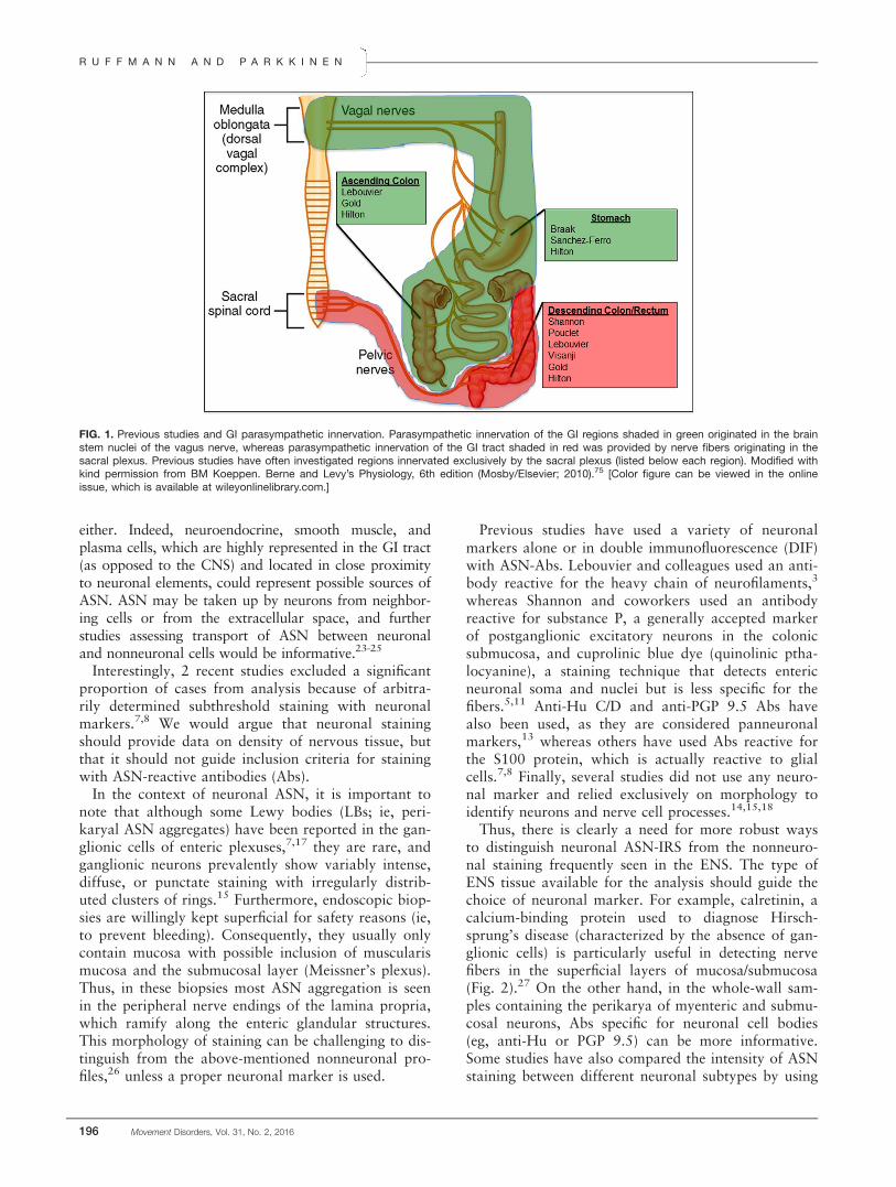
One highly variable parameter between studies is thesite of the GI tract biopsy (Fig. 1). Several differentregions of the GI tract have been harvested includingsubmandibular glands,16 stomach,8 appendix,9 ascend-ing and descending colon,3 sigmoid colon, and rec-tum.5,14 Both postmortem17-19 and in vivo3,4 studieshave reported a higher density of ASN-IRS in the rostralregions of the GI tract compared with the more distalones. However, findings from more recent studieswould seem to question this hypothesis. Hilton and col-leagues reported the absence of ASN-IRS in the esopha-gus, whereas 8% of biopsies from the stomach and13% of both small and large intestine showed ASN-IRstaining,7 suggesting a caudo-rostral gradient, if any.Furthermore, Visanji and colleagues found equally prev-alent ASN-IRS when comparing biopsies taken 5 cm(rectum) and 20 cm (sigmoid colon) from the analverge.14 Finally, Gray and colleagues showed that neu-ritic ASN-IRS was much more abundant in the mucosaof the appendix than in stomach, ileum, or colon, high-lighting appendix as the optimal anatomical locus.9
Several reports have also demonstrated the impact ofthe amount of tissue analyzed on the likelihood of cap-turing ASN-IRS.8,18,19 Of note, routine 4- to 6-lm-thickparaffin sections of the GI biopsies do not enable visual-ization of neuritic ASN-IRS such as those seen on 150-lm-thick cryosections.18 Most recently, in a study exam-ining gastric biopsies Sanchez-Ferro and coworkersreported a minimum cutoff of 4 slides per case to reliablyfind ASN-IRS in PD patients.8 Thus, increasing the num-ber of consecutive sections analyzed could considerablyfacilitate depicting the scattered ASN-IRS. These consid-erations should be borne in mind when interpretingreported results. For example, the 100% specificity
R U F F M A N N A N D P A R K K I N E N
194 Movement Disorders, Vol. 31, No. 2, 2016
reported by Hilton and coworkers could be at least partlydue to the assessment of a single section for each controlcase as opposed to 2 sections for each PD case.7 Con-versely, access to larger amounts of tissue, including thedeeper myenteric plexus in the postmortem studies, maypartly underlie the higher incidence of ASN-IRS in neuro-logically unimpaired individuals reported by Gold andcolleagues.15
To date, studies have mostly used qualitative assess-ment of sections to define ASN-IRS, and only 2 studiesdeveloped semiquantitative grading scales to measurethe severity of staining (also see the review by Visanjiet al).3,15 Virtual microscopy and whole-slide imagingcould allow a more robust quantitative analysis ofenteric ASN-IRS and potentially facilitate definition ofreliable cutoff scores to distinguish PD patients fromcontrols. To date, 1 study has used computerized scan-ning of stained sections, but the authors did not
attempt software-based quantification of the density ofASN-IRS on single sections.14 Such software-basedimage analysis could also allow adjustment for signifi-cant variability between biopsies in surface area, den-sity of nervous tissue, and representation of differentorgan layers (mucosa, submucosa, muscle, etc.), all ofwhich may affect the yield of ASN-IRS.
Localization of ASN-IRS toNeurons in the ENS
In addition to neurons, ASN is physiologicallyexpressed in hematopoietic,20 endothelial, and neuroen-docrine cells21 and in muscle fibers.22 Therefore, thedetection of nonneuronal ASN does not necessarily indi-cate a disease state. Conversely, we cannot currently ruleout the potential pathogenic role of nonneuronal ASN
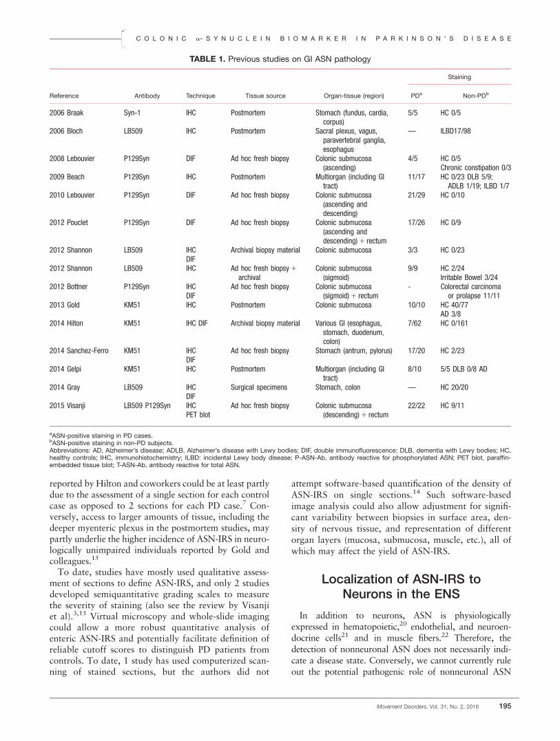
TABLE 1. Previous studies on GI ASN pathology
Reference Antibody Technique Tissue source Organ-tissue (region)
Staining
PDa Non-PDb
2006 Braak Syn-1 IHC Postmortem Stomach (fundus, cardia,corpus)
5/5 HC 0/5
2006 Bloch LB509 IHC Postmortem Sacral plexus, vagus,paravertebral ganglia,esophagus
— ILBD17/98
2008 Lebouvier P129Syn DIF Ad hoc fresh biopsy Colonic submucosa(ascending)
4/5 HC 0/5Chronic constipation 0/3
2009 Beach P129Syn IHC Postmortem Multiorgan (including GItract)
11/17 HC 0/23 DLB 5/9;ADLB 1/19; ILBD 1/7
2010 Lebouvier P129Syn DIF Ad hoc fresh biopsy Colonic submucosa(ascending anddescending)
21/29 HC 0/10
2012 Pouclet P129Syn DIF Ad hoc fresh biopsy Colonic submucosa(ascending anddescending)1 rectum
17/26 HC 0/9
2012 Shannon LB509 IHCDIF
Archival biopsy material Colonic submucosa 3/3 HC 0/23
2012 Shannon LB509 IHC Ad hoc fresh biopsy1
archivalColonic submucosa(sigmoid)
9/9 HC 2/24Irritable Bowel 3/24
2012 Bottner P129Syn IHCDIF
Ad hoc fresh biopsy Colonic submucosa(sigmoid)1 rectum
- Colorectal carcinomaor prolapse 11/11
2013 Gold KM51 IHC Postmortem Colonic submucosa 10/10 HC 40/77AD 3/8
2014 Hilton KM51 IHC DIF Archival biopsy material Various GI (esophagus,stomach, duodenum,colon)
7/62 HC 0/161
2014 Sanchez-Ferro KM51 IHCDIF
Ad hoc fresh biopsy Stomach (antrum, pylorus) 17/20 HC 2/23
2014 Gelpi KM51 IHC Postmortem Multiorgan (including GItract)
8/10 5/5 DLB 0/8 AD
2014 Gray LB509 IHCDIF
Surgical specimens Stomach, colon — HC 20/20
2015 Visanji LB509 P129Syn IHCPET blot
Ad hoc fresh biopsy Colonic submucosa(descending)1 rectum
22/22 HC 9/11
aASN-positive staining in PD cases.bASN-positive staining in non-PD subjects.Abbreviations: AD, Alzheimer’s disease; ADLB, Alzheimer’s disease with Lewy bodies; DIF, double immunofluorescence; DLB, dementia with Lewy bodies; HC,healthy controls; IHC, immunohistochemistry; ILBD: incidental Lewy body disease; P-ASN-Ab, antibody reactive for phosphorylated ASN; PET blot, paraffin-embedded tissue blot; T-ASN-Ab, antibody reactive for total ASN.
C O L O N I C a- S Y N U C L E I N B I O M A R K E R I N P A R K I N S O N ’ S D I S E A S E
Movement Disorders, Vol. 31, No. 2, 2016 195
either. Indeed, neuroendocrine, smooth muscle, andplasma cells, which are highly represented in the GI tract(as opposed to the CNS) and located in close proximityto neuronal elements, could represent possible sources ofASN. ASN may be taken up by neurons from neighbor-ing cells or from the extracellular space, and furtherstudies assessing transport of ASN between neuronaland nonneuronal cells would be informative.23-25
Interestingly, 2 recent studies excluded a significantproportion of cases from analysis because of arbitra-rily determined subthreshold staining with neuronalmarkers.7,8 We would argue that neuronal stainingshould provide data on density of nervous tissue, butthat it should not guide inclusion criteria for stainingwith ASN-reactive antibodies (Abs).
In the context of neuronal ASN, it is important tonote that although some Lewy bodies (LBs; ie, peri-karyal ASN aggregates) have been reported in the gan-glionic cells of enteric plexuses,7,17 they are rare, andganglionic neurons prevalently show variably intense,diffuse, or punctate staining with irregularly distrib-uted clusters of rings.15 Furthermore, endoscopic biop-sies are willingly kept superficial for safety reasons (ie,to prevent bleeding). Consequently, they usually onlycontain mucosa with possible inclusion of muscularismucosa and the submucosal layer (Meissner’s plexus).Thus, in these biopsies most ASN aggregation is seenin the peripheral nerve endings of the lamina propria,which ramify along the enteric glandular structures.This morphology of staining can be challenging to dis-tinguish from the above-mentioned nonneuronal pro-files,26 unless a proper neuronal marker is used.
Previous studies have used a variety of neuronalmarkers alone or in double immunofluorescence (DIF)with ASN-Abs. Lebouvier and colleagues used an anti-body reactive for the heavy chain of neurofilaments,3
whereas Shannon and coworkers used an antibodyreactive for substance P, a generally accepted markerof postganglionic excitatory neurons in the colonicsubmucosa, and cuprolinic blue dye (quinolinic ptha-locyanine), a staining technique that detects entericneuronal soma and nuclei but is less specific for thefibers.5,11 Anti-Hu C/D and anti-PGP 9.5 Abs havealso been used, as they are considered panneuronalmarkers,13 whereas others have used Abs reactive forthe S100 protein, which is actually reactive to glialcells.7,8 Finally, several studies did not use any neuro-nal marker and relied exclusively on morphology toidentify neurons and nerve cell processes.14,15,18
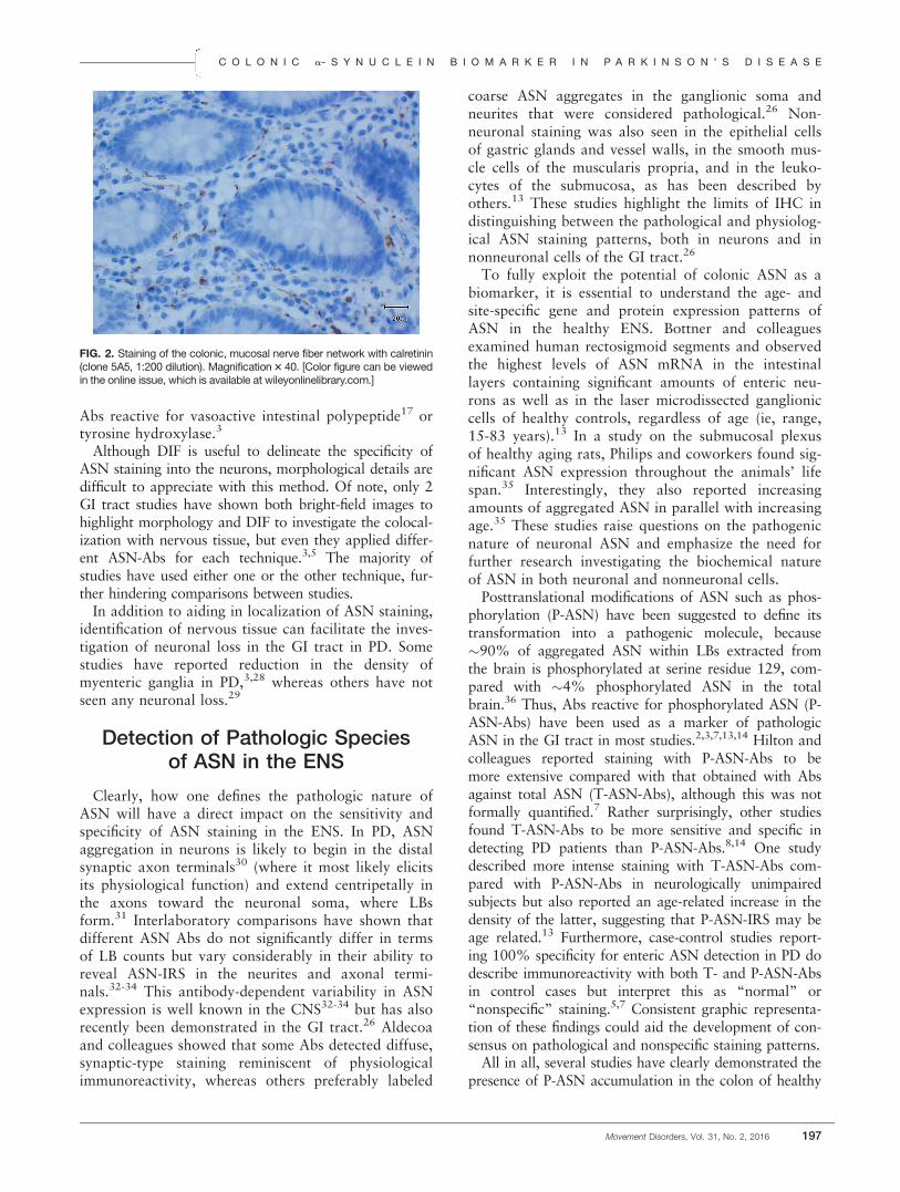
Thus, there is clearly a need for more robust waysto distinguish neuronal ASN-IRS from the nonneuro-nal staining frequently seen in the ENS. The type ofENS tissue available for the analysis should guide thechoice of neuronal marker. For example, calretinin, acalcium-binding protein used to diagnose Hirsch-sprung’s disease (characterized by the absence of gan-glionic cells) is particularly useful in detecting nervefibers in the superficial layers of mucosa/submucosa(Fig. 2).27 On the other hand, in the whole-wall sam-ples containing the perikarya of myenteric and submu-cosal neurons, Abs specific for neuronal cell bodies(eg, anti-Hu or PGP 9.5) can be more informative.Some studies have also compared the intensity of ASNstaining between different neuronal subtypes by using
FIG. 1. Previous studies and GI parasympathetic innervation. Parasympathetic innervation of the GI regions shaded in green originated in the brainstem nuclei of the vagus nerve, whereas parasympathetic innervation of the GI tract shaded in red was provided by nerve fibers originating in thesacral plexus. Previous studies have often investigated regions innervated exclusively by the sacral plexus (listed below each region). Modified withkind permission from BM Koeppen. Berne and Levy’s Physiology, 6th edition (Mosby/Elsevier; 2010).75 [Color figure can be viewed in the onlineissue, which is available at wileyonlinelibrary.com.]
R U F F M A N N A N D P A R K K I N E N
196 Movement Disorders, Vol. 31, No. 2, 2016
Abs reactive for vasoactive intestinal polypeptide17 ortyrosine hydroxylase.3
Although DIF is useful to delineate the specificity ofASN staining into the neurons, morphological details aredifficult to appreciate with this method. Of note, only 2GI tract studies have shown both bright-field images tohighlight morphology and DIF to investigate the colocal-ization with nervous tissue, but even they applied differ-ent ASN-Abs for each technique.3,5 The majority ofstudies have used either one or the other technique, fur-ther hindering comparisons between studies.
In addition to aiding in localization of ASN staining,identification of nervous tissue can facilitate the inves-tigation of neuronal loss in the GI tract in PD. Somestudies have reported reduction in the density ofmyenteric ganglia in PD,3,28 whereas others have notseen any neuronal loss.29
Detection of Pathologic Speciesof ASN in the ENS
Clearly, how one defines the pathologic nature ofASN will have a direct impact on the sensitivity andspecificity of ASN staining in the ENS. In PD, ASNaggregation in neurons is likely to begin in the distalsynaptic axon terminals30 (where it most likely elicitsits physiological function) and extend centripetally inthe axons toward the neuronal soma, where LBsform.31 Interlaboratory comparisons have shown thatdifferent ASN Abs do not significantly differ in termsof LB counts but vary considerably in their ability toreveal ASN-IRS in the neurites and axonal termi-nals.32-34 This antibody-dependent variability in ASNexpression is well known in the CNS32-34 but has alsorecently been demonstrated in the GI tract.26 Aldecoaand colleagues showed that some Abs detected diffuse,synaptic-type staining reminiscent of physiologicalimmunoreactivity, whereas others preferably labeled
coarse ASN aggregates in the ganglionic soma andneurites that were considered pathological.26 Non-neuronal staining was also seen in the epithelial cellsof gastric glands and vessel walls, in the smooth mus-cle cells of the muscularis propria, and in the leuko-cytes of the submucosa, as has been described byothers.13 These studies highlight the limits of IHC indistinguishing between the pathological and physiolog-ical ASN staining patterns, both in neurons and innonneuronal cells of the GI tract.26
To fully exploit the potential of colonic ASN as abiomarker, it is essential to understand the age- andsite-specific gene and protein expression patterns ofASN in the healthy ENS. Bottner and colleaguesexamined human rectosigmoid segments and observedthe highest levels of ASN mRNA in the intestinallayers containing significant amounts of enteric neu-rons as well as in the laser microdissected ganglioniccells of healthy controls, regardless of age (ie, range,15-83 years).13 In a study on the submucosal plexusof healthy aging rats, Philips and coworkers found sig-nificant ASN expression throughout the animals’ lifespan.35 Interestingly, they also reported increasingamounts of aggregated ASN in parallel with increasingage.35 These studies raise questions on the pathogenicnature of neuronal ASN and emphasize the need forfurther research investigating the biochemical natureof ASN in both neuronal and nonneuronal cells.
Posttranslational modifications of ASN such as phos-phorylation (P-ASN) have been suggested to define itstransformation into a pathogenic molecule, because�90% of aggregated ASN within LBs extracted fromthe brain is phosphorylated at serine residue 129, com-pared with �4% phosphorylated ASN in the totalbrain.36 Thus, Abs reactive for phosphorylated ASN (P-ASN-Abs) have been used as a marker of pathologicASN in the GI tract in most studies.2,3,7,13,14 Hilton andcolleagues reported staining with P-ASN-Abs to bemore extensive compared with that obtained with Absagainst total ASN (T-ASN-Abs), although this was notformally quantified.7 Rather surprisingly, other studiesfound T-ASN-Abs to be more sensitive and specific indetecting PD patients than P-ASN-Abs.8,14 One studydescribed more intense staining with T-ASN-Abs com-pared with P-ASN-Abs in neurologically unimpairedsubjects but also reported an age-related increase in thedensity of the latter, suggesting that P-ASN-IRS may beage related.13 Furthermore, case-control studies report-ing 100% specificity for enteric ASN detection in PD dodescribe immunoreactivity with both T- and P-ASN-Absin control cases but interpret this as “normal” or“nonspecific” staining.5,7 Consistent graphic representa-tion of these findings could aid the development of con-sensus on pathological and nonspecific staining patterns.
All in all, several studies have clearly demonstrated thepresence of P-ASN accumulation in the colon of healthy
FIG. 2. Staining of the colonic, mucosal nerve fiber network with calretinin(clone 5A5, 1:200 dilution). Magnification 3 40. [Color figure can be viewedin the online issue, which is available at wileyonlinelibrary.com.]
C O L O N I C a- S Y N U C L E I N B I O M A R K E R I N P A R K I N S O N ’ S D I S E A S E
Movement Disorders, Vol. 31, No. 2, 2016 197
controls, which suggests that this may not be the“pathologic” form of the protein, but rather that ASNphosphorylation is a physiologically occurring process.37
There is also emerging evidence that the earlierintermediate species of the ASN aggregation process,that is, soluble, oligomeric aggregates of ASN, mayultimately be the pathogenic species underlying neuro-nal death in PD.38,39 Thus, Abs reactive for oligomericforms of ASN40 could improve specificity and sensitiv-ity for pathological staining in the GI tract.
Alternative Techniques
To date, conventional IHC has been the techniqueof choice to explore ASN pathology in the GI tract.By virtue of detecting also physiologic ASN, however,IHC has low specificity. There is thus a need for moresensitive and specific techniques to detect the accumu-lation of the earliest, pathological ASN species in thesynaptic axonal terminals and neurites.
Paraffin-Embedded Tissue Blot
One such potential in situ technique is the paraffin-embedded tissue (PET) blot, which was originallyapplied to detect the pathological prion protein in spo-radic Creutzfeldt-Jakob disease (sCJD).41 In PD andDLB brain, the ASN-PET blot has revealed numeroussynaptic ASN microaggregates in cortical and subcort-ical gray matter in addition to a relatively sparse num-ber of LBs seen with conventional IHC.30 It is basedon prolonged high-concentration treatment with pro-teinase K (PK), an enzyme that digests any physio-logic, monomeric ASN (enhancing specificity) but alsoreveals epitopes (increasing sensitivity).42 To date, 1study has used this technique to detect synaptic ASNaggregates in the sigmoid colons and rectums of PDpatients and neurologically unimpaired controls. Withthe ASN-PET blot, sensitivity was 86% in the overallPD sample, whereas specificity was disappointinglydismal, with 100% of controls showing staining.14
The presence of PK-resistant ASN in the PET blot ofcolonic tissue in all 11 neurologically unimpaired sub-jects certainly merits further investigation, as this tech-nique was hypothesized to detect the pathologicalform of ASN alone. It is possible that medical condi-tions unrelated to PD could cause protein aggregationin the GI microenvironment. CNS tumors of variouslineages have been linked to altered expression of 1 ormore members of the synuclein family,43,44 and g-synuclein is a well-established marker of breast can-cer.45 Considering that controls for this study wererecruited from cancer screening programs, it is possi-ble that mechanisms related to colonic neoplasmscould have affected the state of aggregation of ASN inthese subjects. Also, although fibrillar ASN fromhuman Lewy body disease patients is more than 10
times more resistant to PK than its physiological, solu-ble counterpart, the PK treatment conditions appliedby Visanji and coworkers may not have eliminated allthe soluble ASN protein in the GI tract. Thus, experi-ments showing total elimination of physiological ASNwith higher PK treatment or other denaturing condi-tions could shed more light on this method. Further-more, although Visanji and coworkers used blockingpeptides to demonstrate that their PET blot signal wasspecific for ASN, neuronal localization of the ASNstaining was not immunohistochemically ascer-tained.14 Visanji and colleagues interestingly hypothe-sized that the reduction in the PK-resistant ASNaggregation in PD compared with control colon couldbe a result of its disaggregation and release in theform of cytotoxic fibrillar oligomers, shown to occurin vitro.46 Thus, techniques that would allow detec-tion of such ASN oligomers in situ in the colonic biop-sies would be very useful.
Proximity Ligation Assay
The proximity ligation assay (PLA) was originallydeveloped for cancer research and has subsequentlybeen used in cardiology, immunology, and microbiol-ogy.47-49 This technique is based on detection ofsimultaneous and proximal binding of target mole-cules (such as proteins) by 2 probes tagged with com-plementary strands of DNA. Consequent ligation andformation of circular DNA can then be amplified andeasily visualized.47 In contrast to conventional IHC,PLA greatly increases sensitivity detecting concentra-tions of the target molecule in the zeptomolar range(10-21 mol), whereas the required pairwise ligationincreases specificity. The first PLA-based assay todetect ASN oligomers in human PD brain tissue(ASN-PLA) was recently introduced together withextensive data showing specificity of the assay withdifferent in vitro ASN oligomerization systems.50
ASN-PLA was shown to specifically label very earlyperikaryal aggregates (ie, pale bodies) but not late-stage inclusions such as LBs. Moreover, AS-PLArevealed previously unrecognized pathology in theform of extensive diffuse deposition of ASN, whichwas not detected by IHC or observed in controls.
Protein Misfolding Cyclic Amplification andReal-Time Quaking-Induced Conversion
Assays
None of the GI studies so far have examined thebiochemical composition of ASN using standard West-ern blotting (WB), which is probably because of diffi-culty in obtaining in vivo frozen material from theENS. However, WB would be more sensitive thanIHC and would allow the quantitative assessment ofpathological aggregated ASN. Availability of frozentissue would also allow the application of other
R U F F M A N N A N D P A R K K I N E N
198 Movement Disorders, Vol. 31, No. 2, 2016
ultrasensitive techniques such as protein misfoldingcyclic amplification (PMCA) and real-time quaking-induced conversion (RT-QuIC) assays. Although boththese techniques were originally developed to detectthe pathological prion protein51-53 and serve as diag-nostic tests for sCJD, they show the potential to beapplied to other abnormally misfolded proteins suchas ASN.54,55 Importantly, rather than simply meas-uring the concentration of ASN, these techniquesinvestigate the seeding capacity of ASN, that is, theability of ASN to aggregate and induce further aggre-gation of nearby normal ASN. PMCA combines cyclesof incubation at 37 8C (which promotes fibril forma-tion) and sonication (which causes fragmentation offibrils into smaller seeds), and the end product isdetected by WB. Although PMCA provides fasterkinetics of aggregation than RT-QuIC, the sonicationsteps introduce variability hindering reproducibility. InRT-QuIC, a recombinant protein is mixed or “seeded”with small amounts of aggregated protein from thepatient’s sample (eg, frozen GI biopsy). Interactionbetween recombinant and pathologic, patient-derivedmisfolded protein is promoted by intermittent shaking(“quaking”), which leads to formation of furtheraggregates. These form bonds with thioflavin T gener-ating a fluorescent signal that can be easily detectedand quantified. It remains to be seen whether theassessment of functional properties of ASN on GI tis-sue will prove to have superior sensitivity and specific-ity for in vivo diagnosis of PD.
ASN Detection in the ENS:Gateway to the Brain?
Already prior to the era of ASN IHC, characteristicinclusions histologically and ultrastructurally identicalto LBs were found in various parts of the GI tract inpatients with PD.17,56,57 However, this did not sparkmuch scientific interest until the theory of prion-likepropagation was introduced in the field of PDresearch. According to this hypothesis, the accumula-tion of ASN in the dorsal motor nucleus of the vagus(DMV), generally considered to be the first vulnerableregion of the CNS to become affected in PD, mayactually originate in the ENS, where vagal fibers syn-apse with parasympathetic ganglionic neurons.18,58 Aputative pathogen capable of passing the mucosal lin-ing of the GI tract might induce ASN misfolding inthe terminal axons of postganglionic enteric neuronsfollowed by retrograde transport to the cholinergicpreganglionic neurons of the DMV.
Several studies report how intracerebral injection ofexogenous, preformed fibrillar ASN in the brain ofvarious mouse models of PD induces a progressiveASN-IRS pattern that suggests propagation of ASNpathology via a prion-like, conformational self-
templating mechanism.59,60 Experimental evidence forthe spread of ASN pathology from the PNS to theCNS via peripheral nerves comes from studies inwhich preformed amyloidogenic ASN filaments wereinjected into the intestinal wall or the hind leg muscleof transgenic and wild-type mice.61,62 ASN was trans-ported via the vagus nerve and reached the DMV in atime-dependent manner.62 Mice with intramuscularinjection also developed a relatively rapid, widespreadinduction of ASN inclusion pathology in the CNS,which was significantly delayed or even prevented bysciatic nerve transection.61 However, the soluble nona-myloidogenic form of ASN (D71-82) also inducedCNS ASN-IR inclusions, suggesting that other mecha-nisms apart from conformational self-templating couldbe involved.61 Further mechanistic insight comes fromanimal models showing that chronic oral ingestion oflow-dose rotenone can lead to accumulation of ASNin the ENS ganglia. This was followed by ASN accu-mulation in the DMV and SN, progress that was pre-vented by vagotomy.63,64
Although the vagal connection from the ENS to theDMV is often invoked to account for the transmissionof ASN pathology, it should be noted that a number ofstudies have examined the distal GI tract, more readilyaccessible to routine biopsies (Fig. 1).5,14 This region,however, is not reached by vagal projections accordingto previous anatomical studies in humans and ani-mals.65,66 There is a general consensus on the distribu-tion of vagal innervation to the GI tract, which fallsprogressively following a rostrocaudal gradient: 50% ofENS ganglia receive vagal input at the level of theascending colon, whereas there is no vagal input in thedescending colon or rectum, which receive parasympa-thetic innervation from the sacral plexus and pelvicnerves. Thus, propagation of misfolded ASN from thedistal colon and rectum would potentially require anextra passage in the rostral direction, possibly throughenteric interneurons, to reach the vagal nerve endings.Alternatively, misfolded ASN in the distal colon andrectum could travel retrogradely to the preganglionicparasympathetic neurons in the sacral plexus. However,according to anatomical studies by Braak and col-leagues,67 this region is not involved by ASN pathologyuntil later stages of disease and would thus not seem tobe linked to early ASN accumulation in the distal GItract. Furthermore, the neurons that do receive vagalfibers are almost exclusively found in the myentericplexus,65 whereas reported staining in PD is most oftenconfined to mucosal nerve fibers or to the submucosalganglionic cells where the fibers originate. Thus, onewould need to account for this further step in the PNS-CNS pathway linking the mucosal nerve fibers emanat-ing from submucosal neurons to the myenteric plexus.68
To validate whether the peripheral ASN pathologyprecedes or occurs concomitantly to that occurring in
C O L O N I C a- S Y N U C L E I N B I O M A R K E R I N P A R K I N S O N ’ S D I S E A S E
Movement Disorders, Vol. 31, No. 2, 2016 199
the CNS and whether it progresses in a predictableway as has been proposed,69 we need further confirm-atory, human postmortem studies assessing both thebrain and the ENS. Although there are case reports onASN-IRS in the PNS without concomitant Lewypathology in the CNS,70 the majority of postmortemstudies investigating both CNS and PNS fail to pro-vide evidence of centripetal spread of ASN.10,18,19,71
Taken together, these findings suggest that CNS ASNdeposition may in fact precede PNS involvement.Interestingly, to our knowledge no experimental mod-els have yet investigated the spread of ASN pathologyin the other direction (ie, CNS to PNS), as has beenreported to occur with prion transmission.72 It is alsoplausible that ASN deposition in the PNS could serveas a focal biomarker, not necessarily spreading any-where but occurring simultaneously with the brainpathology and thus serving as a surrogate marker.
What Kind of Biomarker CouldEnteric ASN Be?
A prerequisite for the development of preventivetreatment for PD is the ability to diagnose the diseasebefore a significant loss of nigral neurons hasoccurred. A recent consensus paper by the MovementDisorder Society task force emphasized the need todefine “preclinical” and “prodromal” PD and developbiomarkers for PD.73 Assuming it will become possi-ble to reliably define the presence of pathological ASNin the ENS that distinguishes patients with PD from
controls, such a biomarker could have several poten-tial applications.
Detection of ASN-IRS in colonic biopsies taken upto 8 years prior to the onset of PD motor symptomssupports the potential of GI ASN as a presymptomaticdiagnostic biomarker. To date, 6 subjects with pre-symptomatic GI ASN-IRS have been described in 2separate studies.7,11 Of note, review of medical chartsin 3 of these individuals failed to reveal constipationin the premotor phases. To understand how early inthe disease process we see the enteric ASN accumula-tion and how it correlates with the disease progres-sion, systematic screening should be performed onasymptomatic subjects with established risk factors forPD (eg, REM sleep behavior disease order and geneticmutation carriers). Results from longitudinally fol-lowed, uniformly assessed cohorts could more reliablyconfirm or refute enteric ASN detection as a predictivemarker and its role in premotor gastrointestinalimpairment.
Another possibility is that ASN may be consistentlydetectable only after the onset of motor symptoms, inwhich case it could be used to confirm the diagnosismade on clinical grounds. To increase specificity in thiscontext, further studies are needed assessing GI ASNstaining in other, “atypical” parkinsonian syndromes.Although the postganglionic neurons are generallyspared in multiple system atrophy, 1 study reportedcolonic ASN-IRS in 1 of 6 patients.74 Progressive supra-nuclear palsy is a tauopathy and would thus not beexpected to disclose significant ASN pathology.
To date, only 1 study has attempted clinicopatho-logical correlation, reporting an association betweenmore severe colonic ASN staining and levodopa-unresponsive symptoms such as dysarthria and pos-tural instability.3 Thus, phenotypic stratification inrelation to enteric ASN accumulation is a rather unex-plored area but is expected to evolve once more stand-ardized criteria for the definition and quantification ofpathology are established.
Finally, ASN staining in the GI tract may character-ize only a proportion of cases of “idiopathic” PD. Forexample, one could hypothesize that a certain percent-age of individuals will be affected by ASN pathologyoriginating from the nasal mucosa, whereas in othersthe possible spread of pathology might commence inthe ENS.
Conclusions and Future Work
We have discussed the several sources of heterogene-ity that need to be addressed prior to validation of GIASN detection as a tissue-based diagnostic tool forPD. Ideally, these should be addressed in the contextof a cooperative, multicenter effort to establish con-sensus on the best method to use for a robust and
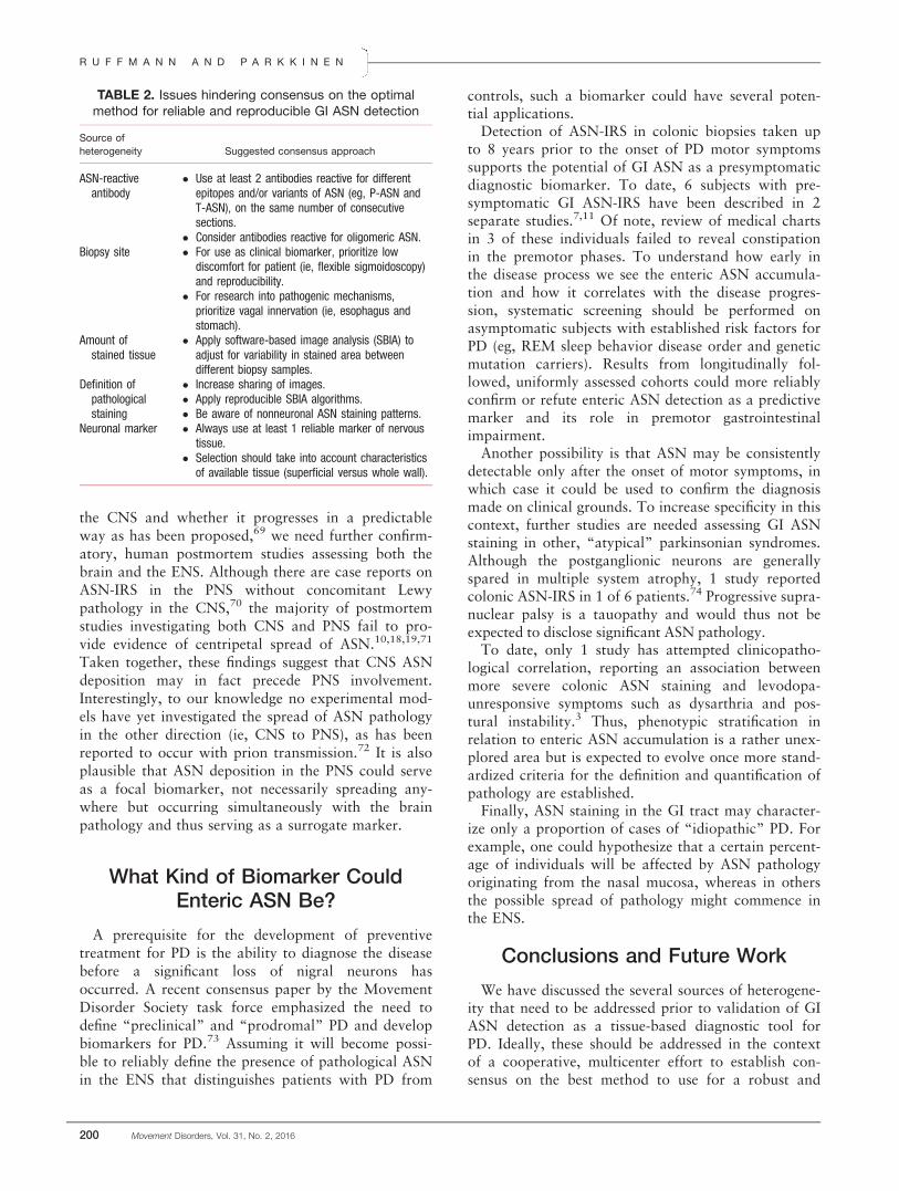
TABLE 2. Issues hindering consensus on the optimalmethod for reliable and reproducible GI ASN detection
Source of
heterogeneity Suggested consensus approach
ASN-reactiveantibody
� Use at least 2 antibodies reactive for differentepitopes and/or variants of ASN (eg, P-ASN andT-ASN), on the same number of consecutivesections.
� Consider antibodies reactive for oligomeric ASN.Biopsy site � For use as clinical biomarker, prioritize low
discomfort for patient (ie, flexible sigmoidoscopy)and reproducibility.
� For research into pathogenic mechanisms,prioritize vagal innervation (ie, esophagus andstomach).
Amount ofstained tissue
� Apply software-based image analysis (SBIA) toadjust for variability in stained area betweendifferent biopsy samples.
Definition ofpathologicalstaining
� Increase sharing of images.� Apply reproducible SBIA algorithms.� Be aware of nonneuronal ASN staining patterns.
Neuronal marker � Always use at least 1 reliable marker of nervoustissue.
� Selection should take into account characteristicsof available tissue (superficial versus whole wall).
R U F F M A N N A N D P A R K K I N E N
200 Movement Disorders, Vol. 31, No. 2, 2016
informative ASN detection in the GI tract (Table 2).Although there is consistent evidence of differentspecies of ASN, currently available techniques cannotdistinguish reliably between physiological and patho-logical forms of ASN. Alternative detection methodssuch as PET blot, PLA, PMCA, and RT-QuIC mayprove to be more sensitive and specific tools for thedetection of early, disease-associated ASN aggregationin the axonal terminals and neurites of the GI tract,where the pathological process most likely starts.Future studies should also apply software-based imageanalysis for a more accurate and reproducible quantifi-cation of pathological changes. For the detection of GIASN to be clinically useful, these studies should bebased on longitudinally assessed, population-derivedcase-control cohorts with extensive clinical characteri-zation.
Acknowledgments: The authors thank Dr. Olaf Ansorge from theUniversity of Oxford for his helpful suggestions.
References1. Hughes AJ, Daniel SE, Kilford L, Lees AJ. Accuracy of clinical
diagnosis of idiopathic Parkinson’s disease: a clinico-pathologicalstudy of 100 cases. J Neurol Neurosurg Psychiatry 1992;55:181-184.
2. Lebouvier T, Chaumette T, Damier P, et al. Pathological lesions incolonic biopsies during Parkinson’s disease. Gut 2008;57:1741-1743.
3. Lebouvier T, Neunlist M, Bruley des Varannes S, et al. Colonicbiopsies to assess the neuropathology of Parkinson’s disease and itsrelationship with symptoms. PloS One 2010;5:e12728.
4. Pouclet H, Lebouvier T, Coron E, et al. A comparison betweenrectal and colonic biopsies to detect Lewy pathology in Parkinson’sdisease. Neurobiol Dis 2012;45:305-309.
5. Shannon KM, Keshavarzian A, Mutlu E, et al. Alpha-synuclein incolonic submucosa in early untreated Parkinson’s disease. MovDisord 2012;27:709-715.
6. Visanji NP, Marras C, Hazrati LN, Liu LW, Lang AE. Alimentary,my dear Watson?. The challenges of enteric alpha-synuclein as aParkinson’s disease biomarker. Mov Disord 2014;29:444-450.
7. Hilton D, Stephens M, Kirk L, et al. Accumulation of alpha-synuclein in the bowel of patients in the pre-clinical phase of Par-kinson’s disease. Acta Neuropathol 2014;127:235-241.
8. Sanchez-Ferro A, Rabano A, Catalan MJ, et al. In vivo gastricdetection of alpha-synuclein inclusions in Parkinson’s disease. MovDisord 2015;30:517-524.
9. Gray MT, Munoz DG, Gray DA, Schlossmacher MG, Woulfe JM.Alpha-synuclein in the appendiceal mucosa of neurologically intactsubjects. Mov Disord 2014;29:991-998.
10. Gelpi E, Navarro-Otano J, Tolosa E, et al. Multiple organ involve-ment by alpha-synuclein pathology in Lewy body disorders. MovDisord 2014;29:1010-1018.
11. Shannon KM, Keshavarzian A, Dodiya HB, Jakate S, KordowerJH. Is alpha-synuclein in the colon a biomarker for premotor Par-kinson’s disease?. Evidence from 3 cases. Mov Disord 2012;27:716-719.
12. Marek K, Jennings D. Can we image premotor Parkinson disease?Neurology 2009;72:S21-S26.
13. Bottner M, Zorenkov D, Hellwig I, et al. Expression pattern andlocalization of alpha-synuclein in the human enteric nervous sys-tem. Neurobiol Dis 2012;48:474-480.
14. Visanji NP, Marras C, Kern DS, et al. Colonic mucosal a-synucleinlacks specificity as a biomarker for Parkinson disease. Neurology2015;84:609-616.
15. Gold A, Turkalp ZT, Munoz DG. Enteric alpha-synuclein expres-sion is increased in Parkinson’s disease but not Alzheimer’s disease.Mov Disord 2013;28:237-240.
16. Beach TG, Adler CH, Dugger BN, et al. Submandibular glandbiopsy for the diagnosis of Parkinson disease. J Neuropathol ExpNeurol 2013;72:130-136.
17. Wakabayashi K, Takahashi H, Takeda S, Ohama E, Ikuta F. Par-kinson’s disease: the presence of Lewy bodies in Auerbach’s andMeissner’s plexuses. Acta Neuropathol 1988;76:217-221.
18. Braak H, de Vos RA, Bohl J, Del Tredici K. Gastric alpha-synuclein immunoreactive inclusions in Meissner’s and Auerbach’splexuses in cases staged for Parkinson’s disease-related brainpathology. Neurosci Lett 2006;396:67-72.
19. Beach TG, Adler CH, Sue LI, et al. Multi-organ distribution ofphosphorylated alpha-synuclein histopathology in subjects withLewy body disorders. Acta Neuropathol 2010;119:689-702.
20. Nakai M, Fujita M, Waragai M, et al. Expression of alpha-synuclein, a presynaptic protein implicated in Parkinson’s disease,in erythropoietic lineage. Biochem Biophys Res Comm 2007;358:104-110.
21. Tamo W, Imaizumi T, Tanji K, et al. Expression of alpha-synuclein, the precursor of non-amyloid beta component of Alzhei-mer’s disease amyloid, in human cerebral blood vessels. NeurosciLett 2002;326:5-8.
22. Askanas V, Engel WK, Alvarez RB, McFerrin J, Broccolini A.Novel immunolocalization of alpha-synuclein in human muscle ofinclusion-body myositis, regenerating and necrotic muscle fibers,and at neuromuscular junctions. J Neuropathol Exp Neurol 2000;59:592-598.
23. Luk KC, Song C, O’Brien P, et al. Exogenous alpha-synucleinfibrils seed the formation of Lewy body-like intracellular inclusionsin cultured cells. Proc Natl Acad Sci U S A 2009;106:20051-20056.
24. Desplats P, Lee HJ, Bae EJ, et al. Inclusion formation and neuronalcell death through neuron-to-neuron transmission of alpha-synu-clein. Proc Natl Acad Sci U S A 2009;106:13010-130105.
25. Volpicelli-Daley LA, Luk KC, Patel TP, et al. Exogenous alpha-synuclein fibrils induce Lewy body pathology leading to synapticdysfunction and neuron death. Neuron 2011;72:57-71.
26. Aldecoa I, Navarro-Otano J, Stefanova N, et al. Alpha-synucleinimmunoreactivity patterns in the enteric nervous system. NeurosciLett 2015;602:145-149.
27. Morris MI, Soglio DB, Ouimet A, Aspirot A, Patey N. A study ofcalretinin in Hirschsprung pathology, particularly in total colonicaganglionosis. J Ped Surg 2013;48:1037-1043.
28. Singaram C, Ashraf W, Gaumnitz EA, et al. Dopaminergic defectof enteric nervous system in Parkinson’s disease patients withchronic constipation. Lancet 1995;346:861-864.
29. Annerino DM, Arshad S, Taylor GM, Adler CH, Beach TG,Greene JG. Parkinson’s disease is not associated with gastrointesti-nal myenteric ganglion neuron loss. Acta Neuropathol 2012;124:665-680.
30. Kramer ML, Schulz-Schaeffer WJ. Presynaptic alpha-synucleinaggregates, not Lewy bodies, cause neurodegeneration in dementiawith Lewy bodies. J Neurosci 2007;27:1405-1410.
31. Kanazawa T, Adachi E, Orimo S, Nakamura A, Mizusawa H,Uchihara T. Pale neurites, premature alpha-synuclein aggregateswith centripetal extension from axon collaterals. Brain Pathol2012;22:67-78.
32. Croisier E, DE MR, Deprez M, et al. Comparative study of com-mercially available anti-alpha-synuclein antibodies. NeuropatholAppl Neurobiol 2006;32:351-356.
33. Beach TG, White CL, Hamilton RL, et al. Evaluation of alpha-synuclein immunohistochemical methods used by invited experts.Acta Neuropathol 2008;116:277-288.
34. Alafuzoff I, Parkkinen L, Al-Sarraj S, et al. Assessment of alpha-synuclein pathology: a study of the BrainNet Europe Consortium.J Neuropathol Exp Neurol 2008;67:125-143.
35. Phillips RJ, Martin FN, Billingsley CN, Powley TL. Alpha-synu-clein expression patterns in the colonic submucosal plexus of theaging Fischer 344 rat: implications for biopsies in aging and neuro-degenerative disorders? Neurogastroenterol Motil 2013;25:e621-e633.
C O L O N I C a- S Y N U C L E I N B I O M A R K E R I N P A R K I N S O N ’ S D I S E A S E
Movement Disorders, Vol. 31, No. 2, 2016 201
36. Fujiwara H, Hasegawa M, Dohmae N, et al. alpha-Synuclein isphosphorylated in synucleinopathy lesions. Nature Cell Biol 2002;4:160-164.
37. Muntane G, Ferrer I, Martinez-Vicente M. alpha-synuclein phos-phorylation and truncation are normal events in the adult humanbrain. Neuroscience 2012;200:106-119.
38. Caughey B, Lansbury PT. Protofibrils, pores, fibrils, and neurode-generation: separating the responsible protein aggregates from theinnocent bystanders. Annu Rev Neurosci 2003;26:267-298.
39. Rochet JC, Outeiro TF, Conway KA, et al. Interactions amongalpha-synuclein, dopamine, and biomembranes: some clues forunderstanding neurodegeneration in Parkinson’s disease. J Neurosci2004;23:23-34.
40. Vaikath NN, Majbour NK, Paleologou KE, et al. Generation andcharacterization of novel conformation-specific monoclonal antibod-ies for alpha-synuclein pathology. Neurobiol Dis 2015;79:81-99.
41. Schulz-Schaeffer WJ, Tschoke S, Kranefuss N, et al. The paraffin-embedded tissue blot detects PrP(Sc) early in the incubation timein prion diseases. Am J Pathol 2000;156:51-56.
42. Miake H, Mizusawa H, Iwatsubo T, Hasegawa M. Biochemicalcharacterization of the core structure of alpha-synuclein filaments.J Biol Chem 2002;277:19213-19.
43. Fung KM, Rorke LB, Giasson B, Lee VM, Trojanowski JQ.Expression of alpha-, beta-, and gamma-synuclein in glial tumorsand medulloblastomas. Acta Neuropathol 2003;106:167-175.
44. Kawashima M, Suzuki SO, Doh-ura K, Iwaki T. alpha-Synuclein isexpressed in a variety of brain tumors showing neuronal differen-tiation. Acta Neuropathol 2000;99:154-160.
45. Wu K, Quan Z, Weng Z, et al. Expression of neuronal proteinsynuclein gamma gene as a novel marker for breast cancer progno-sis. Breast Cancer Res Treat 2007;101:259-267.
46. Cremades N, Cohen SI, Deas E, et al. Direct observation of theinterconversion of normal and toxic forms of alpha-synuclein. Cell2012;149:1048-1059.
47. Fredriksson S, Gullberg M, Jarvius J, et al. Protein detection usingproximity-dependent DNA ligation assays. Nat Biotechnol 2002;20:473-477.
48. Gullberg M, Gustafsdottir SM, Schallmeiner E, et al. Cytokinedetection by antibody-based proximity ligation. Proc Natl Acad SciU S A 2004;101:8420-8424.
49. Soderberg O, Gullberg M, Jarvius M, et al. Direct observation ofindividual endogenous protein complexes in situ by proximity liga-tion. Nat Methods 2006;3:995-1000.
50. Roberts RF, Wade-Martins R, Alegre-Abarrategui J. Direct visual-ization of alpha-synuclein oligomers reveals previously undetectedpathology in Parkinson’s disease brain. Brain 2015;138:1642-1657.
51. Saborio GP, Permanne B, Soto C. Sensitive detection of pathologi-cal prion protein by cyclic amplification of protein misfolding.Nature 2001;411:810-813.
52. Atarashi R, Satoh K, Sano K, et al. Ultrasensitive human priondetection in cerebrospinal fluid by real-time quaking-induced con-version. Nat Med 2011;17:175-178.
53. McGuire LI, Peden AH, Orru CD, et al. Real time quaking-induced conversion analysis of cerebrospinal fluid in sporadicCreutzfeldt-Jakob disease. Ann Neurol 2012;72:278-285.
54. Roostaee A, Beaudoin S, Staskevicius A, Roucou X. Aggregationand neurotoxicity of recombinant alpha-synuclein aggregates initi-ated by dimerization. Mol Neurodegener 2013;8:5.
55. Herva ME, Zibaee S, Fraser G, Barker RA, Goedert M, SpillantiniMG. Anti-amyloid compounds inhibit alpha-synuclein aggregationinduced by protein misfolding cyclic amplification (PMCA). J BiolChem 2014;289:11897-1905.
56. Qualman SJ, Haupt HM, Yang P, Hamilton SR. Esophageal Lewybodies associated with ganglion cell loss in achalasia. Similarity toParkinson’s disease. Gastroenterology 1984;87:848-856.
57. Kupsky WJ, Grimes MM, Sweeting J, Bertsch R, Cote LJ. Parkin-son’s disease and megacolon: concentric hyaline inclusions (Lewybodies) in enteric ganglion cells. Neurology 1987;37:1253-1255.
58. Hawkes CH, Del Tredici K, Braak H. Parkinson’s disease: a dual-hit hypothesis. Neuropathol Appl Neurobiol 2007;33:599-614.
59. Hardy J. Expression of normal sequence pathogenic proteins forneurodegenerative disease contributes to disease risk: ‘permissivetemplating’ as a general mechanism underlying neurodegeneration.Biochem Soc Trans 2005;33:578-581.
60. Jucker M, Walker LC. Self-propagation of pathogenic proteinaggregates in neurodegenerative diseases. Nature 2013;501:45-51.
61. Sacino AN, Brooks M, Thomas MA, et al. Intramuscular injectionof alpha-synuclein induces CNS alpha-synuclein pathology and arapid-onset motor phenotype in transgenic mice. Proc Natl AcadSci U S A 2014;111:10732-10737.
62. Holmqvist S, Chutna O, Bousset L, et al. Direct evidence of Par-kinson pathology spread from the gastrointestinal tract to thebrain in rats. Acta Neuropath 2014;128:805-820.
63. Pan-Montojo F, Anichtchik O, Dening Y, et al. Progression of Par-kinson’s disease pathology is reproduced by intragastric adminis-tration of rotenone in mice. PloS One 2010;5:e8762.
64. Pan-Montojo F, Schwarz M, Winkler C, et al. Environmental tox-ins trigger PD-like progression via increased alpha-synuclein releasefrom enteric neurons in mice. Sci Rep 2012;2:898.
65. Berthoud HR, Jedrzejewska A, Powley TL. Simultaneous labelingof vagal innervation of the gut and afferent projections from thevisceral forebrain with dil injected into the dorsal vagal complex inthe rat. J Comp Neurol 1990;301:65-79.
66. Chang HY, Mashimo H, Goyal RK. Musings on the wanderer:what’s new in our understanding of vago-vagal reflex? IV. Currentconcepts of vagal efferent projections to the gut. Am J Phys Gas-troint Liver Phys 2003;284:G357-G366.
67. Del Tredici K, Braak H. Spinal cord lesions in sporadic Parkinson’sdisease. Acta Neuropath 2012;124:643-664.
68. Furness JB. The enteric nervous system and neurogastroenterology.Nature Rev Gastroent Hepat 2012;9:286-294.
69. Braak H, Rub U, Gai WP, Del Tredici K. Idiopathic Parkinson’sdisease: possible routes by which vulnerable neuronal types may besubject to neuroinvasion by an unknown pathogen. J NeuralTransm 2003;110:517-536.
70. Miki Y, Mori F, Wakabayashi K, Kuroda N, Orimo S. IncidentalLewy body disease restricted to the heart and stellate ganglia. MovDisord 2009;24:2299-2301.
71. Bloch A, Probst A, Bissig H, Adams H, Tolnay M. Alpha-synucleinpathology of the spinal and peripheral autonomic nervous systemin neurologically unimpaired elderly subjects. Neuropath App Neu-robiol 2006;32:284-295.
72. Lawson VA, Furness JB, Klemm HM, et al. The brain to gut path-way: a possible route of prion transmission. Gut 2010;59:1643-651.
73. Berg D, Postuma RB, Bloem B, et al. Time to redefine PD?. Intro-ductory statement of the MDS Task Force on the definition of Par-kinson’s disease. Mov Disord 2014;29:454-462.
74. Pouclet H, Lebouvier T, Coron E, et al. Analysis of colonic alpha-synuclein pathology in multiple system atrophy. ParkinsonismRelat Disord 2012;18:893-895.
75. Koeppen BM SB, Berne RM. Berne & Levy Physiology. 6th ed,updated ed. Philadelphia: Mosby/Elsevier; 2010.
R U F F M A N N A N D P A R K K I N E N
202 Movement Disorders, Vol. 31, No. 2, 2016