Genome sequences characterizing five mutations in RNA ... · peptidolytic activities from a...
13
Genome sequences characterizing five mutations in RNA polymerase and major capsid of phages ϕA318 and ϕAs51 of Vibrio alginolyticus with different burst efficiencies Liu et al. Liu et al. BMC Genomics 2014, 15:505 http://www.biomedcentral.com/1471-2164/15/505
Transcript of Genome sequences characterizing five mutations in RNA ... · peptidolytic activities from a...
Genome sequences characterizing five mutationsin RNA polymerase and major capsid of phagesϕA318 and ϕAs51 of Vibrio alginolyticus withdifferent burst efficienciesLiu et al.
Liu et al. BMC Genomics 2014, 15:505http://www.biomedcentral.com/1471-2164/15/505

Liu et al. BMC Genomics 2014, 15:505http://www.biomedcentral.com/1471-2164/15/505
RESEARCH ARTICLE Open Access
Genome sequences characterizing five mutationsin RNA polymerase and major capsid of phagesϕA318 and ϕAs51 of Vibrio alginolyticus withdifferent burst efficienciesWangta Liu2, Ying-Rong Lin1, Ming-Wei Lu3, Ping-Jyun Sung4, Wei-Hsien Wang1,4 and Chan-Shing Lin1*
Abstract
Background: The burst size of a phage is important prior to phage therapy and probiotic usage. The efficiency fora phage to burst its host bacterium can result from molecular domino effects of the phage gene expressions whichdominate to control host machinery after infection. We found two Podoviridae phages, ϕA318 and ϕAs51, burst acommon host V. alginolyticus with different efficiencies of 72 and 10 PFU/bacterium, respectively. Presumably, thegenome sequences can be compared to explain their differences in burst sizes.
Results: Among genes in 42.5 kb genomes with a GC content of 43.5%, 16 out of 47 open-reading frames (ORFs)were annotated to known functions, including RNA polymerase (RNAP) and phage structure proteins. 11 strongphage promoters and three terminators were found. The consensus sequence for the new vibriophage promotersis AATAAAGTTGCCCTATA, where the AGTTG bases of −8 through −12 are important for the vibriophage specificity,especially a consensus T at −9 position eliminating RNAP of K1E, T7 and SP6 phages to transcribe the genes. ϕA318and ϕAs51 RNAP shared their own specific promoters. In comparing ϕAs51 with ϕA318 genomes, only twonucleotides were deleted in the RNAP gene and three mutating nucleotides were found in the major capsid genes.
Conclusion: Subtle analyses on the residue alterations uncovered the effects of five nucleotide mutations on thefunctions of the RNAP and capsid proteins, which account for the host-bursting efficiency. The deletion of twonucleotides in RNAP gene truncates the primary translation due to early stop codon, while a second translationalpeptide starting from GTG just at deletion point can remediate the polymerase activity. Out of three nucleotidemutations in major capsid gene, H53N mutation weakens the subunit assembly between capsomeres for the phagehead; E313K reduces the fold binding between β-sheet and Spine Helix inside the peptide.
Keywords: RNA polymerase truncation, Spine Helix, Mutation in F-loop
BackgroundVibrio strains are reported to be fatal pathogens not onlyto humans but also to aquaculture fish and shellfish.Several virulence factors that they produce—includinghemolysin, caseinase, gelatinase, lipase, and phospholip-ase—are detrimental to animal health [1-4]. For instance,an extracellular protease with endopeptidolytic and exo-peptidolytic activities from a pathogenic V. pelagius strainis responsible for the lethal effect and vibriosis in turbot.
* Correspondence: [email protected] of Marine Biotechnology and Resources, Asia-Pacific OceanResearch Center, National Sun Yat-sen University, Kaohsiung 80424, TaiwanFull list of author information is available at the end of the article
© 2014 Liu et al.; licensee BioMed Central Ltd.Commons Attribution License (http://creativecreproduction in any medium, provided the orDedication waiver (http://creativecommons.orunless otherwise stated.
The different virulence factors are produced at differ-ent stages of infection. Hemolysin-producing bacteriainclude V. parahaemolyticus, V. alginolyticus, V. cholerae,V. hollisae, Aeromonas veronii, and V. anguillarum (Listo-nella anguillarum).Vibrio alginolyticus, a Gram-negative halophilic bacter-
ium that frequently occurs in the normal microbiota ofmarine environments, is a pathogen of epizootic out-breaks, which causes serious mortality of fish and shellfishand also can transmit to humans. The disease caused byV. alginolyticus often has lethal consequences for fish lar-vae throughout the world, and it has become one of the
This is an Open Access article distributed under the terms of the Creativeommons.org/licenses/by/2.0), which permits unrestricted use, distribution, andiginal work is properly credited. The Creative Commons Public Domaing/publicdomain/zero/1.0/) applies to the data made available in this article,
Liu et al. BMC Genomics 2014, 15:505 Page 2 of 12http://www.biomedcentral.com/1471-2164/15/505
major limiting factors in aquaculture in developingcountries. For example, it was a causal agent of vibriosisoutbreaks in grouper sea bream Sparus aurata [2]. V. algi-nolyticus causes several symptoms in sea bream: septi-cemia, hemorrhaging, dark skin, and ulcers on the skinsurface. Some fish accumulate fluid in the peritonealcavity or have hemorrhagic livers. In several cases ofhigh-mortality outbreaks, Vibrio alginolyticus and Vibriosplendidus biovar II were the primary organisms found inmoribund clam larvae (Ruditapes decussatus), and thesetwo species processed extracellular products collabora-tively at high concentrations to kill the hemocytes afterfour hours of incubation. Neurotoxic effects caused by thesupernates from diseased strains of Vibrio alginolyticusand Vibrio anguillarum caused several symptoms in trout,including convulsions, wriggling, contortive swimmingand respiratory arrest [2].Chemotherapeutic agents are effective to control
pathogenic bacteria in aquaculture. Nevertheless, emer-gence of antibiotic-resistant bacteria has become a crit-ical issue [3]. As a result of management practices inproduction cycles, the use of probiotics to preventpathogenic bacterium growth in aquaculture is a practiceto reduce both issues on antibiotic resistance and drugresidues in food. One alternative is to use bacteriophagesto control bacterial growth, which is similar to phagetherapy in medical care. This idea has been proposed asa cure for coral disease [5]. A variety of bacteriophagesthat can quickly lyse the pathogens have high potentialfor future applications in aquaculture.The burst size of a phage represents an overall outcome
that is so important for phage therapy and probiotic usage.The molecular interactions between phage and its hostunder certain environmental conditions are complicatedto resolve. Sequences of bacteriophage genomes provide amagnificent tool to investigate interactions with hosts. Inaddition, vibriophages can be used as a new typing schemefor pathogen identification and as candidate agents forphage therapy and probiotics. For example, the rolesof lytic bacteriophages in cholera epidemics were in-vestigated, such as filamentous CTXϕ, myovirus ICP1,and podovirus ICP3 of Vibrio cholerae, causing infantand adult deaths for centuries [6]. Though quite alarge number of Vibrio phages have been described,only a few of complete genome sequences are known.Genome sequences of lytic vibriophages are new keysfor molecular typing, pathogenesis, and even assess-ment of therapeutic efficacy. Complete genomes ofsome phages of V. cholerae, V. parahaemolyticus, and V.vulnificus were documented, yet no genome for V. algino-lyticus podovirus has been reported. In this study, wecharacterized the genome sequences of two newly-isolatedphages that can lyse V. alginolyticus ATCC 17749 withdifferent efficiencies.
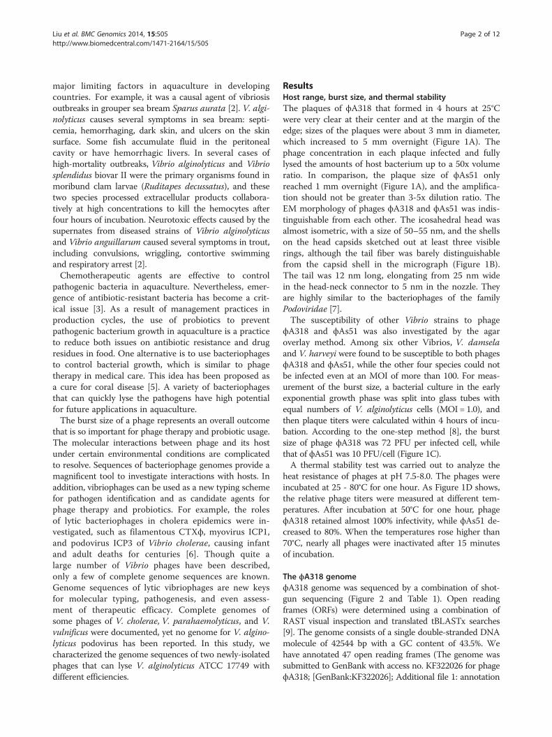
ResultsHost range, burst size, and thermal stabilityThe plaques of ϕA318 that formed in 4 hours at 25°Cwere very clear at their center and at the margin of theedge; sizes of the plaques were about 3 mm in diameter,which increased to 5 mm overnight (Figure 1A). Thephage concentration in each plaque infected and fullylysed the amounts of host bacterium up to a 50x volumeratio. In comparison, the plaque size of ϕAs51 onlyreached 1 mm overnight (Figure 1A), and the amplifica-tion should not be greater than 3-5x dilution ratio. TheEM morphology of phages ϕA318 and ϕAs51 was indis-tinguishable from each other. The icosahedral head wasalmost isometric, with a size of 50–55 nm, and the shellson the head capsids sketched out at least three visiblerings, although the tail fiber was barely distinguishablefrom the capsid shell in the micrograph (Figure 1B).The tail was 12 nm long, elongating from 25 nm widein the head-neck connector to 5 nm in the nozzle. Theyare highly similar to the bacteriophages of the familyPodoviridae [7].The susceptibility of other Vibrio strains to phage
ϕA318 and ϕAs51 was also investigated by the agaroverlay method. Among six other Vibrios, V. damselaand V. harveyi were found to be susceptible to both phagesϕA318 and ϕAs51, while the other four species could notbe infected even at an MOI of more than 100. For meas-urement of the burst size, a bacterial culture in the earlyexponential growth phase was split into glass tubes withequal numbers of V. alginolyticus cells (MOI = 1.0), andthen plaque titers were calculated within 4 hours of incu-bation. According to the one-step method [8], the burstsize of phage ϕA318 was 72 PFU per infected cell, whilethat of ϕAs51 was 10 PFU/cell (Figure 1C).A thermal stability test was carried out to analyze the
heat resistance of phages at pH 7.5-8.0. The phages wereincubated at 25 - 80°C for one hour. As Figure 1D shows,the relative phage titers were measured at different tem-peratures. After incubation at 50°C for one hour, phageϕA318 retained almost 100% infectivity, while ϕAs51 de-creased to 80%. When the temperatures rose higher than70°C, nearly all phages were inactivated after 15 minutesof incubation.
The ϕA318 genomeϕA318 genome was sequenced by a combination of shot-gun sequencing (Figure 2 and Table 1). Open readingframes (ORFs) were determined using a combination ofRAST visual inspection and translated tBLASTx searches[9]. The genome consists of a single double-stranded DNAmolecule of 42544 bp with a GC content of 43.5%. Wehave annotated 47 open reading frames (The genome wassubmitted to GenBank with access no. KF322026 for phageϕA318; [GenBank:KF322026]; Additional file 1: annotation
A. Plaque size
B. TEM micrograph
C. Burst size
D. Thermal stability
Figure 1 (See legend on next page.)
Liu et al. BMC Genomics 2014, 15:505 Page 3 of 12http://www.biomedcentral.com/1471-2164/15/505
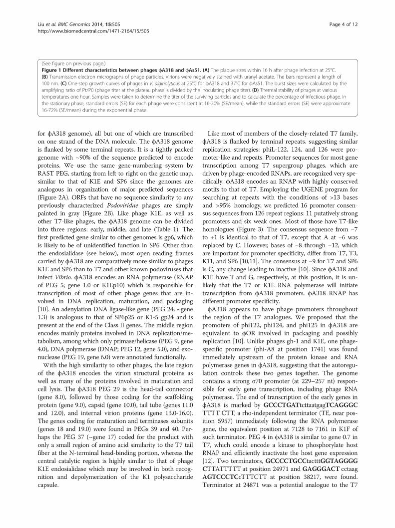
(See figure on previous page.)Figure 1 Different characteristics between phages ϕA318 and ϕAs51. (A) The plaque sizes within 16 h after phage infection at 25°C.(B) Transmission electron micrographs of phage particles. Virions were negatively stained with uranyl acetate. The bars represent a length of100 nm. (C) One-step growth curves of phages in V. alginolyticus at 25°C for ϕA318 and 37°C for ϕAs51. The burst sizes were calculated by theamplifying ratio of Pt/P0 (phage titer at the plateau phase is divided by the inoculating phage titer). (D) Thermal stability of phages at varioustemperatures one hour. Samples were taken to determine the titer of the surviving particles and to calculate the percentage of infectious phage. Inthe stationary phase, standard errors (SE) for each phage were consistent at 16-20% (SE/mean), while the standard errors (SE) were approximate16-72% (SE/mean) during the exponential phase.
Liu et al. BMC Genomics 2014, 15:505 Page 4 of 12http://www.biomedcentral.com/1471-2164/15/505
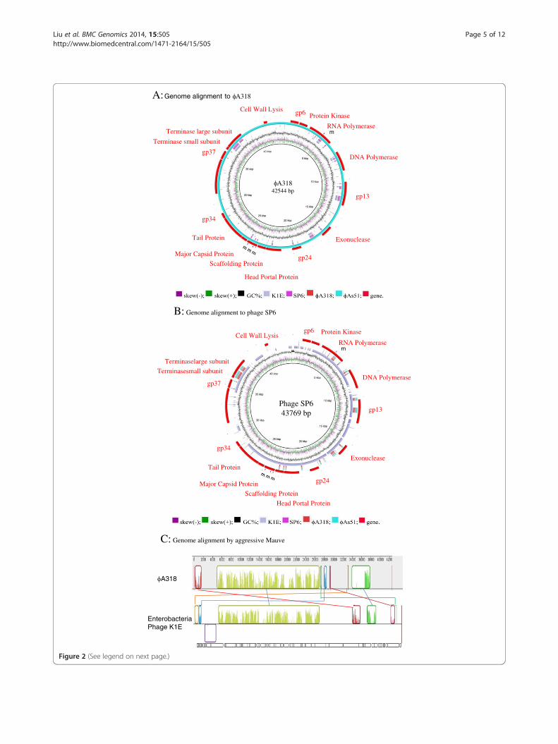
for ϕA318 genome), all but one of which are transcribedon one strand of the DNA molecule. The ϕA318 genomeis flanked by some terminal repeats. It is a tightly packedgenome with ~90% of the sequence predicted to encodeproteins. We use the same gene-numbering system byRAST PEG, starting from left to right on the genetic map,similar to that of K1E and SP6 since the genomes areanalogous in organization of major predicted sequences(Figure 2A). ORFs that have no sequence similarity to anypreviously characterized Podoviridae phages are simplypainted in gray (Figure 2B). Like phage K1E, as well asother T7-like phages, the ϕA318 genome can be dividedinto three regions: early, middle, and late (Table 1). Thefirst predicted gene similar to other genomes is gp6, whichis likely to be of unidentified function in SP6. Other thanthe endosialidase (see below), most open reading framescarried by ϕA318 are comparatively more similar to phagesK1E and SP6 than to T7 and other known podoviruses thatinfect Vibrio. ϕA318 encodes an RNA polymerase (RNAPof PEG 5; gene 1.0 or K1Ep10) which is responsible fortranscription of most of other phage genes that are in-volved in DNA replication, maturation, and packaging[10]. An adenylation DNA ligase-like gene (PEG 24, ~gene1.3) is analogous to that of SP6p25 or K1-5 gp24 and ispresent at the end of the Class II genes. The middle regionencodes mainly proteins involved in DNA replication/me-tabolism, among which only primase/helicase (PEG 9, gene4.0), DNA polymerase (DNAP; PEG 12, gene 5.0), and exo-nuclease (PEG 19, gene 6.0) were annotated functionally.With the high similarity to other phages, the late region
of the ϕA318 encodes the virion structural proteins aswell as many of the proteins involved in maturation andcell lysis. The ϕA318 PEG 29 is the head-tail connector(gene 8.0), followed by those coding for the scaffoldingprotein (gene 9.0), capsid (gene 10.0), tail tube (genes 11.0and 12.0), and internal virion proteins (gene 13.0-16.0).The genes coding for maturation and terminases subunits(genes 18 and 19.0) were found in PEGs 39 and 40. Per-haps the PEG 37 (~gene 17) coded for the product withonly a small region of amino acid similarity to the T7 tailfiber at the N-terminal head-binding portion, whereas thecentral catalytic region is highly similar to that of phageK1E endosialidase which may be involved in both recog-nition and depolymerization of the K1 polysaccharidecapsule.
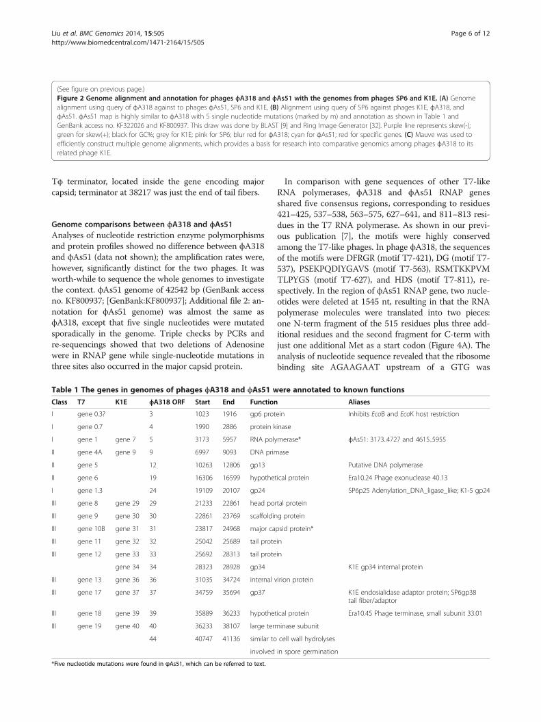
Like most of members of the closely-related T7 family,ϕA318 is flanked by terminal repeats, suggesting similarreplication strategies: phiL-122, 124, and 126 were pro-moter-like and repeats. Promoter sequences for most genetranscription among T7 supergroup phages, which aredriven by phage-encoded RNAPs, are recognized very spe-cifically. ϕA318 encodes an RNAP with highly conservedmotifs to that of T7. Employing the UGENE program forsearching at repeats with the conditions of >13 basesand >95% homology, we predicted 16 promoter consen-sus sequences from 126 repeat regions: 11 putatively strongpromoters and six weak ones. Most of those have T7-likehomologues (Figure 3). The consensus sequence from −7to +1 is identical to that of T7, except that A at −6 wasreplaced by C. However, bases of −8 through −12, whichare important for promoter specificity, differ from T7, T3,K11, and SP6 [10,11]. The consensus at −9 for T7 and SP6is C, any change leading to inactive [10]. Since ϕA318 andK1E have T and G, respectively, at this position, it is un-likely that the T7 or K1E RNA polymerase will initiatetranscription from ϕA318 promoters. ϕA318 RNAP hasdifferent promoter specificity.ϕA318 appears to have phage promoters throughout
the region of the T7 analogues. We proposed that thepromoters of phi122, phi124, and phi125 in ϕA318 areequivalent to φOR involved in packaging and possiblyreplication [10]. Unlike phages gh-1 and K1E, one phage-specific promoter (phi-A8 at position 1741) was foundimmediately upstream of the protein kinase and RNApolymerase genes in ϕA318, suggesting that the autoregu-lation controls these two genes together. The genomecontains a strong σ70 promoter (at 229–257 nt) respon-sible for early gene transcription, including phage RNApolymerase. The end of transcription of the early genes inϕA318 is marked by GCCCTGATtcttaatgagTCAGGGCTTTT CTT, a rho-independent terminator (TE, near pos-ition 5957) immediately following the RNA polymerasegene, the equivalent position at 7128 to 7161 in K1F ofsuch terminator. PEG 4 in ϕA318 is similar to gene 0.7 inT7, which could encode a kinase to phosphorylate hostRNAP and efficiently inactivate the host gene expression[12]. Two terminators, GCCCCTGCCtactttGGTAGGGGCTTATTTTT at position 24971 and GAGGGACT cctaagAGTCCCTCcTTTCTT at position 38217, were found.Terminator at 24871 was a potential analogue to the T7
Terminase large subunit
Cell Wall Lysis
RNA Polymerase
gp6
DNA Polymerase
gp13
gp37
Terminase small subunit
Exonuclease
Head Portal Protein
gp24Scaffolding Protein
Major Capsid Protein
Tail Protein
gp34
A: Genome alignment to A318
m
Protein Kinase
A31842544 bp
Terminaselarge subunit
Cell Wall LysisRNA Polymerase
gp6
DNA Polymerase
gp13
gp37
Terminasesmall subunit
Exonuclease
Head Portal Protein
gp24
Scaffolding Protein
Major Capsid Protein
Tail Protein
gp34
B: Genome alignment to phage SP6
m
Protein Kinase
Phage SP643769 bp
A318
EnterobacteriaPhage K1E
C: Genome alignment by aggressive Mauve
Figure 2 (See legend on next page.)
Liu et al. BMC Genomics 2014, 15:505 Page 5 of 12http://www.biomedcentral.com/1471-2164/15/505
(See figure on previous page.)Figure 2 Genome alignment and annotation for phages ϕA318 and ϕAs51 with the genomes from phages SP6 and K1E. (A) Genomealignment using query of ϕA318 against to phages ϕAs51, SP6 and K1E, (B) Alignment using query of SP6 against phages K1E, ϕA318, andϕAs51. ϕAs51 map is highly similar to ϕA318 with 5 single nucleotide mutations (marked by m) and annotation as shown in Table 1 andGenBank access no. KF322026 and KF800937. This draw was done by BLAST [9] and Ring Image Generator [32]. Purple line represents skew(-);green for skew(+); black for GC%; grey for K1E; pink for SP6; blur red for ϕA318; cyan for ϕAs51; red for specific genes. (C) Mauve was used toefficiently construct multiple genome alignments, which provides a basis for research into comparative genomics among phages ϕA318 to itsrelated phage K1E.
Liu et al. BMC Genomics 2014, 15:505 Page 6 of 12http://www.biomedcentral.com/1471-2164/15/505
Tφ terminator, located inside the gene encoding majorcapsid; terminator at 38217 was just the end of tail fibers.
Genome comparisons between ϕA318 and ϕAs51Analyses of nucleotide restriction enzyme polymorphismsand protein profiles showed no difference between ϕA318and ϕAs51 (data not shown); the amplification rates were,however, significantly distinct for the two phages. It wasworth-while to sequence the whole genomes to investigatethe context. ϕAs51 genome of 42542 bp (GenBank accessno. KF800937; [GenBank:KF800937]; Additional file 2: an-notation for ϕAs51 genome) was almost the same asϕA318, except that five single nucleotides were mutatedsporadically in the genome. Triple checks by PCRs andre-sequencings showed that two deletions of Adenosinewere in RNAP gene while single-nucleotide mutations inthree sites also occurred in the major capsid protein.
Table 1 The genes in genomes of phages ϕA318 and ϕAs51 w
Class T7 K1E ϕA318 ORF Start End Function
I gene 0.3? 3 1023 1916 gp6 prot
I gene 0.7 4 1990 2886 protein k
I gene 1 gene 7 5 3173 5957 RNA poly
II gene 4A gene 9 9 6997 9093 DNA prim
II gene 5 12 10263 12806 gp13
II gene 6 19 16306 16599 hypothet
I gene 1.3 24 19109 20107 gp24
III gene 8 gene 29 29 21233 22861 head por
III gene 9 gene 30 30 22861 23769 scaffoldin
III gene 10B gene 31 31 23817 24968 major ca
III gene 11 gene 32 32 25042 25689 tail prote
III gene 12 gene 33 33 25692 28313 tail prote
gene 34 34 28323 28928 gp34
III gene 13 gene 36 36 31035 34724 internal v
III gene 17 gene 37 37 34759 35694 gp37
III gene 18 gene 39 39 35889 36233 hypothet
III gene 19 gene 40 40 36233 38107 large term
44 40747 41136 similar to
involved
*Five nucleotide mutations were found in ϕAs51, which can be referred to text.
In comparison with gene sequences of other T7-likeRNA polymerases, ϕA318 and ϕAs51 RNAP genesshared five consensus regions, corresponding to residues421–425, 537–538, 563–575, 627–641, and 811–813 resi-dues in the T7 RNA polymerase. As shown in our previ-ous publication [7], the motifs were highly conservedamong the T7-like phages. In phage ϕA318, the sequencesof the motifs were DFRGR (motif T7-421), DG (motif T7-537), PSEKPQDIYGAVS (motif T7-563), RSMTKKPVMTLPYGS (motif T7-627), and HDS (motif T7-811), re-spectively. In the region of ϕAs51 RNAP gene, two nucle-otides were deleted at 1545 nt, resulting in that the RNApolymerase molecules were translated into two pieces:one N-term fragment of the 515 residues plus three add-itional residues and the second fragment for C-term withjust one additional Met as a start codon (Figure 4A). Theanalysis of nucleotide sequence revealed that the ribosomebinding site AGAAGAAT upstream of a GTG was
ere annotated to known functions
Aliases
ein Inhibits EcoB and EcoK host restriction
inase
merase* ϕAs51: 3173..4727 and 4615..5955
ase
Putative DNA polymerase
ical protein Era10.24 Phage exonuclease 40.13
SP6p25 Adenylation_DNA_ligase_like; K1-5 gp24
tal protein
g protein
psid protein*
in
in
K1E gp34 internal protein
irion protein
K1E endosialidase adaptor protein; SP6gp38tail fiber/adaptor
ical protein Era10.45 Phage terminase, small subunit 33.01
inase subunit
cell wall hydrolyses
in spore germination
Figure 3 The sixteen promoters in phage ϕA318 genome werepredicted and aligned with those of T7, SP6, K11, and T3phages. The position of each promoter in ϕA318 genome wasrecorded as the number following the name of phage. The namesof predicted promoters are given using their positions in ϕA318genome, while the positions are shifted by two nucleotides inϕAs51 genome.
Liu et al. BMC Genomics 2014, 15:505 Page 7 of 12http://www.biomedcentral.com/1471-2164/15/505
suitable for the second translation of ϕAs51 RNAP geneto a product of the exactly same sequence as the C-termof ϕA318 RNAP. With use of the threading method ofPHYRE2 to predict the 3-dimentional structures for thesetwo fragments of ϕAs51 RNAP, the result revealed thatthe truncation join was located in the junction of palmand fingers. As Figure 4B shows, the tail of ϕAs51 RNAPN-term fragment formed a clip (arrows) to hold theϕAs51 RNAP C-term fragment at the position on palm.Three mutations of single nucleotide were all in the
first codon of the residue, and therefore, totally alteredthe residues. The mutation at 23973th nucleotide resultedin mutating Histidine at 53th residue to be Asparagine(H53N); 24171th nucleotide mutation changed Isoleucineat 119th to Valine (I119V); and 245789th nucleotidemutation converted Glu-313 to Lys-313 (E313K). Thestructures of ϕA318 and ϕAs51 major capsid proteinswere predicted from models of HK97 and T7 capsidsby PHYRE2 with >93% confidence. In comparing withthe ϕA318 capsid, the mutation effects of Asn-53 onϕAs51 major capsid protein broke the presumed cationbridge between His-53 of two capsomere subunits inϕA318 capsid (Figure 5A and C). Glu-313 residue of βJ inP-domain formed two hydrogen bonds with the O =C of
the backbone of the Spine Helix to stabilize the link be-tween three beta-sheets and one helix (Figure 5B). How-ever, E313K mutation in ϕAs51 caused the βJ in P-domainto lose two hydrogen bonds so as to destabilize the link onthe Spine Helix (Figure 5D).
DiscussionThis is the first Podoviridae genomes of Vibrio alginoly-ticus phages that are documented in GenBank. 16 out of47 ORFs were annotated to known functions, whileother ORFs were similar to enterobacteria phages K1Eand SP6. 11 strong phage promoters and three termina-tors were predicted, which are located slightly differentlyfrom the closest partners, though genomic organizationswere highly homologous [10,12]. Most of promotershave T7-like homologues, except that A at −6 was re-placed by C and specificity region on −8 to −12 (Figure 3).In ϕA318 and ϕAs51 phage promoters, the consensussequence from −7 to +1 is CCCTATAG. However, basesof −8 through −12, which are important for promoter spe-cificity, differ from T7, T3, K11, and SP6. The consensusat −9 for T7 and SP6 is C; any change of this nucleotideinactivates the promoter function [10]. Since ϕA318 andK1E have T and G, respectively, at this position, it is un-likely that the T7 or K1E RNA polymerase will initiatetranscription from ϕA318 promoters. ϕA318 RNAP hasdifferent promoter specificity. This exclusivity of promoterspecificity seems to be the trend among the T7-likephages, and it seems that there must be some selectivepressure that resulted in this feature.Electron microscopy revealed that phage ϕA318 and
ϕAs51 particles were morphologically similar to type Cin Bradley’s classification of Podoviridae phage (Figure 1)[13]. The podovirus family is characterized by phageswith a nearly isometric icosahedron head and a shortnon-contractile tail [13,14]. Furthermore, the lengths ofthe icosahedral capsid and tail of phage ϕA318 weremorphologically indistinguishable from those typicallyobserved for ϕAs51.In phages ϕA318 and ϕAs51, the sequences of the mo-
tifs were DFRGR (motif T7-421), DG (motif T7-537),PSEKPQDIYGAVS (motif T7-563), RSMTKKPVMTLPYGS (motif T7-627), and HDS (motif T7-811), respectively.Strikingly, two nucleotides were deleted in the region ofϕAs51 RNAP gene, which resulted in the RNA polymer-ase molecules being translated into two pieces separatedat the 515th residues (1545 nt); the N-term fragment in-creased three additional residues in its C-terminus whilethe second fragment started from a suitable upstreamribosome binding site AGAAGAAT and added just onecodon GTG for Met to form exactly the same product asthe C-term of ϕA318 RNAP (Figure 4A). We suggestedthat the two fragments could form a 2-subunit-like
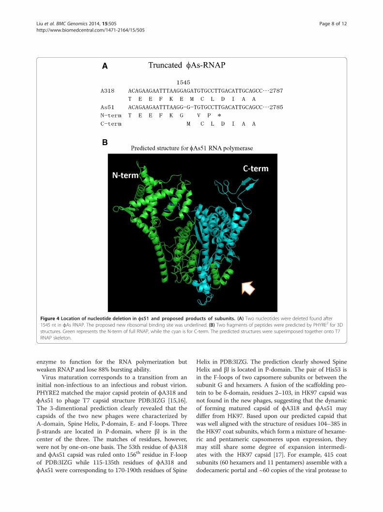
Figure 4 Location of nucleotide deletion in ϕs51 and proposed products of subunits. (A) Two nucleotides were deleted found after1545 nt in ϕAs RNAP. The proposed new ribosomal binding site was underlined. (B) Two fragments of peptides were predicted by PHYRE2 for 3Dstructures. Green represents the N-term of full RNAP, while the cyan is for C-term. The predicted structures were superimposed together onto T7RNAP skeleton.
Liu et al. BMC Genomics 2014, 15:505 Page 8 of 12http://www.biomedcentral.com/1471-2164/15/505
enzyme to function for the RNA polymerization butweaken RNAP and lose 88% bursting ability.Virus maturation corresponds to a transition from an
initial non-infectious to an infectious and robust virion.PHYRE2 matched the major capsid protein of ϕA318 andϕAs51 to phage T7 capsid structure PDB:3IZG [15,16].The 3-dimentional prediction clearly revealed that thecapsids of the two new phages were characterized byA-domain, Spine Helix, P-domain, E- and F-loops. Threeβ-strands are located in P-domain, where βJ is in thecenter of the three. The matches of residues, however,were not by one-on-one basis. The 53th residue of ϕA318and ϕAs51 capsid was ruled onto 156th residue in F-loopof PDB:3IZG while 115-135th residues of ϕA318 andϕAs51 were corresponding to 170-190th residues of Spine
Helix in PDB:3IZG. The prediction clearly showed SpineHelix and βJ is located in P-domain. The pair of His53 isin the F-loops of two capsomere subunits or between thesubunit G and hexamers. A fusion of the scaffolding pro-tein to be δ-domain, residues 2–103, in HK97 capsid wasnot found in the new phages, suggesting that the dynamicof forming matured capsid of ϕA318 and ϕAs51 maydiffer from HK97. Based upon our predicted capsid thatwas well aligned with the structure of residues 104–385 inthe HK97 coat subunits, which form a mixture of hexame-ric and pentameric capsomeres upon expression, theymay still share some degree of expansion intermedi-ates with the HK97 capsid [17]. For example, 415 coatsubunits (60 hexamers and 11 pentamers) assemble with adodecameric portal and ~60 copies of the viral protease to
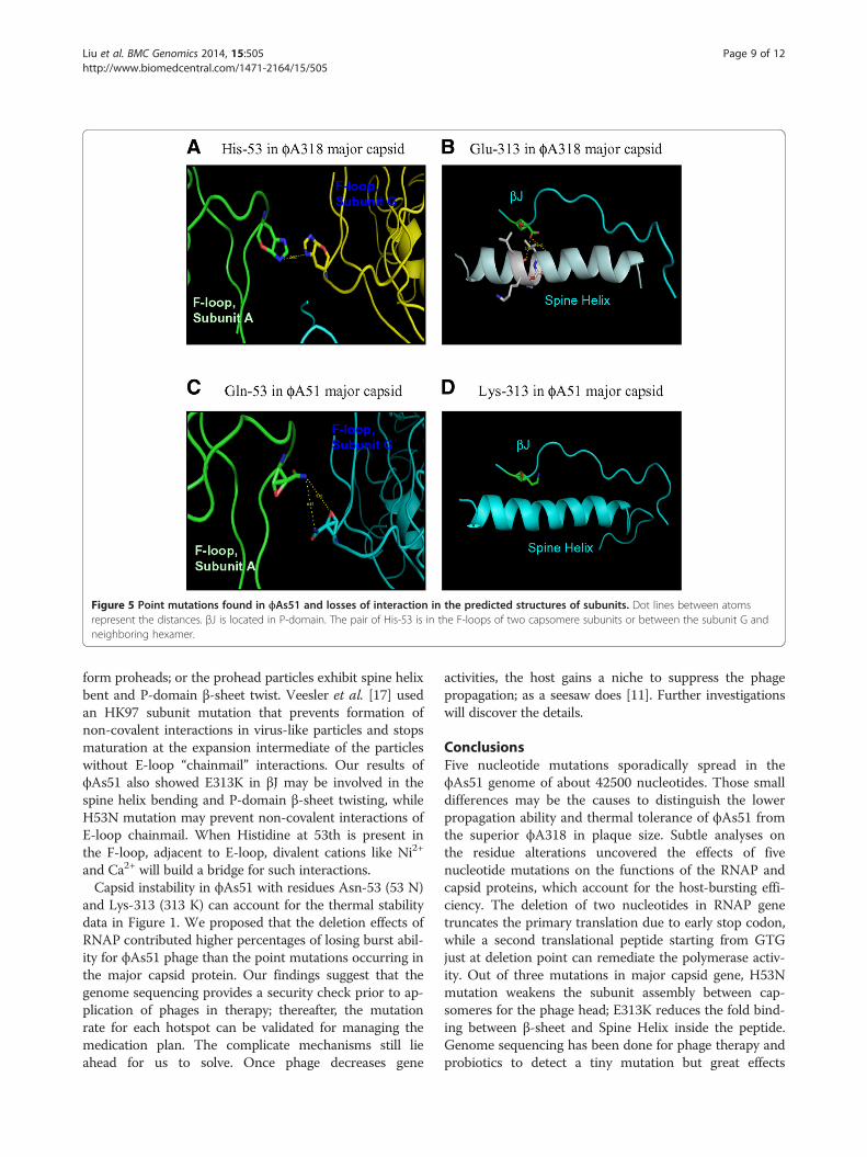
Figure 5 Point mutations found in ϕAs51 and losses of interaction in the predicted structures of subunits. Dot lines between atomsrepresent the distances. βJ is located in P-domain. The pair of His-53 is in the F-loops of two capsomere subunits or between the subunit G andneighboring hexamer.
Liu et al. BMC Genomics 2014, 15:505 Page 9 of 12http://www.biomedcentral.com/1471-2164/15/505
form proheads; or the prohead particles exhibit spine helixbent and P-domain β-sheet twist. Veesler et al. [17] usedan HK97 subunit mutation that prevents formation ofnon-covalent interactions in virus-like particles and stopsmaturation at the expansion intermediate of the particleswithout E-loop “chainmail” interactions. Our results ofϕAs51 also showed E313K in βJ may be involved in thespine helix bending and P-domain β-sheet twisting, whileH53N mutation may prevent non-covalent interactions ofE-loop chainmail. When Histidine at 53th is present inthe F-loop, adjacent to E-loop, divalent cations like Ni2+
and Ca2+ will build a bridge for such interactions.Capsid instability in ϕAs51 with residues Asn-53 (53 N)
and Lys-313 (313 K) can account for the thermal stabilitydata in Figure 1. We proposed that the deletion effects ofRNAP contributed higher percentages of losing burst abil-ity for ϕAs51 phage than the point mutations occurring inthe major capsid protein. Our findings suggest that thegenome sequencing provides a security check prior to ap-plication of phages in therapy; thereafter, the mutationrate for each hotspot can be validated for managing themedication plan. The complicate mechanisms still lieahead for us to solve. Once phage decreases gene
activities, the host gains a niche to suppress the phagepropagation; as a seesaw does [11]. Further investigationswill discover the details.
ConclusionsFive nucleotide mutations sporadically spread in theϕAs51 genome of about 42500 nucleotides. Those smalldifferences may be the causes to distinguish the lowerpropagation ability and thermal tolerance of ϕAs51 fromthe superior ϕA318 in plaque size. Subtle analyses onthe residue alterations uncovered the effects of fivenucleotide mutations on the functions of the RNAP andcapsid proteins, which account for the host-bursting effi-ciency. The deletion of two nucleotides in RNAP genetruncates the primary translation due to early stop codon,while a second translational peptide starting from GTGjust at deletion point can remediate the polymerase activ-ity. Out of three mutations in major capsid gene, H53Nmutation weakens the subunit assembly between cap-someres for the phage head; E313K reduces the fold bind-ing between β-sheet and Spine Helix inside the peptide.Genome sequencing has been done for phage therapy andprobiotics to detect a tiny mutation but great effects
Liu et al. BMC Genomics 2014, 15:505 Page 10 of 12http://www.biomedcentral.com/1471-2164/15/505
which can provide early prediction of treatment efficacyor potential side effects.
MethodsBacterial strains and growth conditionsVibrio strains were bought from the Bioresource Collectionand Research Center, Taiwan, including V. alginolyticusATCC 17749, V. carchariae ATCC 35084, V. damselaATCC 33536,V. harveyi ATCC 14126,V. parahaemolyticusATCC 17802, V. pelagius ATCC 25916, and V. vulnificusBCRC15431. The Vibrio strains were maintained in BactoBrain Heart Infusion medium (BHI; Becton-Dickinson Co,USA) supplemented with 3% NaCl (Panreac Quimica,France). For long-term preservation, bacteria were frozenat −80°C in BHI supplemented with 1% NaCl and 25% gly-cerol (Nihon Shiyaku, Japan). The strains were streakedonto modified seawater yeast extract (rich MSWYE) agarplates consisting of 23.4 g NaCl, 6.98 g MgSO4_7 H2O,and 0.75 g KCl in 1000 ml distilled water [18]. The pH wasadjusted to 7.6 with 1 N NaOH, followed by the additionof 5.0 g of proteose peptone (HIMEDIA Lab, India), 3.0 gof yeast extract (HIMEDIA Lab, India), and 20.0 g ofagar per liter.
Isolation and titer of bacteriophagesWater samples were collected from aquaculture water-ways in southern Taiwan between 2008 and 2010. Thewater was centrifuged at 10,000 × g for 30 minutes, andthe supernatants were filtered through a 0.45 μm micro-filter. After centrifugation and microfiltration, 20%MSWYE medium [7,8] and 1% overnight-grown Vibrioalginolyticus were added to the filtrate and incubated at25°C for 4–24 hours to enrich the phage growth. Thebacterial debris was removed by two centrifugations at10,000 × g for 30 minutes. After enriching the phagesin the field samples with the target bacterium Vibrioalginolyticus for 4–16 hours, phage-containing super-natants were incubated with the indicator strain for 5 mi-nutes, suspended in low-percentage agar (top agar), andplated onto solid agar plates, so the target strain wouldform a bacterial lawn with the formation of plaques. Theten largest plaques from each plate were picked and re-amplified three times in bacterium-containing broth usinga 5× volume ratio. The plaques were picked into 200 μlMSWYE broth for further titer counting or productionamplification. To prepare bacterial cells for determinationof phage concentrations, the host Vibrio alginolyticus wasfreshly inoculated as a 1% volume of seed from overnightculture into 10 ml of rich MSWYE broth, and it grew toan OD600 of 0.3-0.4 in about 2 hours. After a series ofdilutions, 10 μl of diluted phage were added to 200 μl ofbacterial cells, incubated for 5 minutes, mixed with 3 mlof top agar (rich MSWYE with 0.5% agar), and thenpoured onto the solid surface of a 2% agar plate. The
plaques were counted in 3–5 hours; the titer per ml wascalculated as 100 × (dilution factor) × (plaque count).
Electron microscopyPreparation of phage particles for electron microscopyhas been described elsewhere [19]. In brief, bacterio-phage particles were applied to parafilm to produce aspherical drop. Carbon-coated nitrocellulose films werefabricated on copper grids, which were placed face downon the sample drop for 1 min to absorb the particles.After being briefly washed twice in 10 μl of 10 mM Trisbuffer (pH 8) (Amresco, USA), the samples were thenrinsed twice with freshly prepared 2% uranyl acetate(UA; Sigma-Aldrich, USA) in Tris–HCl (pH 8.0) andstained for 60 seconds. Following each step of absorp-tion, washing, and UA staining, the grids were blottedwith filter paper until almost dry. The finished gridswere dried in vacuo overnight. Images of phage particleswere taken at a magnification of 40,000× and a defocusof 3 μm, using a 200-kV electron microscope (JEOLJEM-2010, equipped with a Gatan-832 CCD camera).
DNA Preparation for bacteriophageFor propagation of phage, 10 ml of phage stock wasadded to 100 ml of V. alginolyticus (3 × 10⁸ CFU ml−1)cultured in MSWYE and incubated in a shaker at 25°Cfor 3–5 hours until the lysate was clear. The remainingcells and debris were removed by two centrifugation cy-cles at 10,000 × g for 30 minutes. The supernatant, with atiter of 2 × 10¹⁰ PFU ml−1, was stored at 4°C as a phagestock. To concentrate phages using a standard PEG proto-col [7,20], solid NaCl and polyethylene glycol 8000 (Fluka,Germany) were added, and precipitation was performedovernight at 4°C. After centrifugation, the phage particleswere resuspended in 2 ml of SM buffer and treated withDNase I and RNase A (Sigma, USA) to remove contamin-ating nucleic acids from the host. The polyethylene glycolwas extracted by adding an equal volume of chloroform(TEDIA, USA) until the interface was clear. The aqueousphase containing the phage was treated with proteinase K(Invitrogen, USA) and sodium dodecyl sulfate (SDS; MDBio, Inc., USA) at 56°C for 1 h. Phenol extraction wascarried out three times at room temperature, and theaqueous phase was further extracted with a 1:1 mixture ofequilibrated phenol (Sigma, USA) and chloroform. DNAprecipitated by a 2x volume of cold ethanol was redis-solved in deionized water.
Adsorption and phage burst sizeAs described previously [7,8] for the adsorption of phageon bacteria, 1 ml of broth containing host cells andphages was taken at the 5th and 10th minutes and cen-trifuged to remove bacteria and bound phage, and freephage in the supernatants was then measured. Similarly,
Liu et al. BMC Genomics 2014, 15:505 Page 11 of 12http://www.biomedcentral.com/1471-2164/15/505
the burst size was measured as described previously [7].In brief, V. alginolyticus cells were grown in 20 ml ofmedium to mid-exponential phase (OD600 = 0.3-0.5) andmixed with 0.1 ml phage solution (MOI = 1). Samples of1 ml of the mixture were taken at intervals and immedi-ately subjected to centrifugation at 14,000 × g for 3 mi-nutes to remove bacteria. The phage titers in the solutionswere determined by the agar overlay technique. Using thegrowth curve for MOI = 1 according to the one-stepcriteria, the burst size (Bs) of phage was calculated asBs = Pt/P0, where Pt is the phage titer at that plateauphase and P0 is the initial infective titer.
Genome sequencing and annotationSimilar to the shotgun sequencing described elsewhere,approximately 5 μg of the bacteriophage genomic DNAwas randomly sheared by nebulization, and DNA sequen-cing was performed at Mission Biotech according to themanufacturer’s protocol for the Genome Sequencer GSJunior System (Roche Diagnostic). Low quality sequencesof the reads generated by the GS Junior sequencer weretrimmed off. De novo assembly of the shotgun reads wasperformed with the GS Assembler software. Sequenceassembly and analyses were performed essentially aspreviously described. Protein-encoding genes (PEG) werepredicted using The RAST Server (Rapid Annotationsusing Subsystems Technology; http://rast.nmpdr.org/) [21]and analyzed with the SEED-Viewer (http://www.theseed.org/wiki/Main_Page) [22]. Protein-coding genes were alsochecked using the ab initio gene-finding program Glimmerv3.02 [23]. rRNA and tRNA genes of the draftassembly were identified using RNAmmer [24] andtRNAscan-SE [25]. Automatic functional annotation re-sults obtained by the RAST were further compared withthe proteins in the GenBank database using PSI-BLAST(www.ncbi.nlm.nih.gov/blast/Blast.cgi). The repeat unitsin the phage genomes were found by UGENE tool [26]and further aligned to acquire the consensus regions. Thephage rho-independent terminators were predicted byARNold with the criteria of both Erpin and RNAmotifselections [27]. The genomes of ϕA318 and ϕAs51were submitted to GenBank with access no. KF322026and KF800937, respectively [GenBank:KF322026; GenBank:KF800937].
Multiple sequence alignmentTo determine the classification status of the newlyisolated ϕA318 and ϕAs51 phages, sequence data for thegenes encoding the DNA polymerase, DNA ligase, RNApolymerase, single-strand DNA binding protein, andcapsid proteins of enterobacteria phages (T7, SP6, K1E,and K1F) and Vibrio cholerae phages (N4, VP4, andICP3) were employed to find highly homologous regions.Complete genome sequences of four enterobacteria
phages and three Vibrio Podoviridae phages were acquiredfrom National Center for Biotechnology Information,NIH, USA: T7 (39,937 bp) [GenBank:NC_001604], SP6(43,769 bp) [GenBank:NC_004831], K1E (45,251 bp)[GenBank:NC_007637], K1F (39,704 bp) [GenBank:NC_007456], ICP3 (39,162 bp) [GenBank:NC_015159],N4 (38,497 bp) [GenBank:NC_013651], and VP4 (39,503 bp)[GenBank:NC_007149]. The T7 RNA polymerase se-quence is also the same as PDB:1QLN (RCSB-PDB ID),and SP6 RNA polymerase is from NC_004831 [GenBank:NC_004831]. The threading predictions by PHYRE ver2[15] were iterated to predict the tertiary structures ofϕA318 and ϕAs51 RNA polymerase.Sequences of individual genes which were retrieved
from the genome sets were then aligned using ClustalWwith default options [28]. The complete RNA polymer-ase sequences were analyzed by the neighbor-joiningmethod using the NEIGHBOR program in PhylogenyInference Package (PHYLIP) [29]. Distances were cal-culated using the DNADIST program of PHYLIP anddisplayed in TreeView. ClustalW, PHYLIP, and TreeViewwere bundled in the BioEdit program version 7.0.5 [30].The multiple sequence alignment for the phage genomeswas constructed in the Mauve using MUSCLE3.6algorithm [31].
Additional files
Additional file 1: phiA318.gbk. It is in GenBank format. Annotation forϕA318 genome.
Additional file 2: phiAs51.gbk. It is in GenBank format. Annotation forϕAs51 genome.
Competing interestsThe authors declare that they have no competing interests.
Authors’ contributionsCSL conducted the study and wrote the manuscript. CSL and YRL did theexperiments. CSL, WL, MWL, PJS and WHW analyzed the sequences andfunctions. All authors read and approved the final manuscript.
AcknowledgementsThis research fund is partially supported by grants from the NationalScience Council, Taiwan (NSC99-2313-B-110-002-MY3 and NSC102-2313-B-110-002-MY3), and the Ministry of Education, Taiwan (NSYSU95-99C031701;the second term of Top University Program: NSYSU00-02C030205 and NCHU100-S05-09) under the ATU plan. We thank Professors Hsueh-Wen Chang,MH Tai, Chih-Chuang Liaw, Chen-Tung Arthur Chen and Houng-Yung Chen,as the grant organizers of the ATU plan. Also we appreciate Kenneth B.Lin and Dr. Simon White for comments and editing.
Author details1Department of Marine Biotechnology and Resources, Asia-Pacific OceanResearch Center, National Sun Yat-sen University, Kaohsiung 80424, Taiwan.2Department of Biotechnology, Kaohsiung Medical University, Kaohsiung,Taiwan. 3Department of Aquaculture, National Taiwan Ocean University,Keelung, Taiwan. 4National Museum of Marine Biology and Aquarium,Pingtung 944, Taiwan.
Received: 18 November 2013 Accepted: 30 May 2014Published: 21 June 2014
Liu et al. BMC Genomics 2014, 15:505 Page 12 of 12http://www.biomedcentral.com/1471-2164/15/505
References1. Baffone W, Citterio B, Vittoria E, Casaroli A, Pianetti A, Campana A,
Bruscolini F: Determination of several potential virulence factors in Vibriospp. isolated from seawater. Food Microbiol 2001, 18:479–488.
2. Balebona MC, Andreu MJ, Bordas MA, Zorrilla I, Morinigo MA, Borrego JJ:Pathogenicity of Vibrio alginolyticus for cultured gilt-head sea bream(Sparus aurata L.). Appl Environ Microbiol 1998, 64(11):4269–427528.
3. Kahla-Nakbi AB, Chaieb K, Besbes A, Zmantar T, Bakhrouf A: Virulence andenterobacterial repetitive intergenic consensus PCR of Vibrio alginolyticusstrains isolated from Tunisian cultured gilthead sea bream and sea bassoutbreaks. Vet Microbiol 2006, 117(2–4):321–327.
4. Simidu U, Noguchi T, Hwang DF, Shida Y, Hashimoto K: Marinebacteria which produce tetrodotoxin. Appl Environ Microbol 1987,53(7):1714–1715.
5. Efrony R, Atad I, Rosenberg E: Phage therapy of coral white plaguedisease: properties of phage BA3. Curr Microbiol 2009, 58(2):139–145.
6. Seed KD, Bodi KL, Kropinski AM, Ackermann HW, Calderwood SB, Qadri F,Camilli A: Evidence of a dominant lineage of Vibrio cholerae-specific lyticbacteriophages shed by cholera patients over a 10-year period in Dhaka,Bangladesh. MBio 2011, 2(1):e00334-10.
7. Lin Y-R, Chiu C-W, Chang F-Y, Lin C-S: Characterization of a new phage,termed ϕA318, which is specific for Vibrio alginolyticus. Arch Virol 2012,157:917–926.
8. Lin Y-R, Lin C-S: Genome-wide characterization of Vibrio phage ϕpp2 withunique arrangements of the mob-like genes. BMC Genomics 2012,13:224–237.
9. Altschul SF, Gish W, Miller W, Myers EW, Lipman DJ: Basic local alignmentsearch tool. J Mol Biol 1990, 215:403–410.
10. Stummeyer K, Schwarzer D, Claus H, Vogel U, Gerardy-Schahn R,Mühlenhoff M: Evolution of bacteriophages infecting encapsulated bacteria:lessons from Escherichia coli K1-specific Phages. Mol Microbiol 2006,60(5):1123–1135.
11. Lind PA, Anderson DI: Whole-genome mutational biases in bacteria.Proc Natl Acad Sci U S A 2008, 105(46):7878–17883.
12. Cheetham GM, Steitz TA: Structure of a transcribing T7 RNA polymeraseinitiation complex. Science 1999, 286(5448):2305–2309.
13. Bradley DE: Ultrastructure of bacteriophages and bacteriocins. BacteriolRev 1967, 31(4):230–314.
14. Mitra S, Basu S: Some biophysical properties of a vibriophage and itsDNA. Biochim Biophys Acta 1968, 155(1):143–149.
15. Kelley LA, Sternberg MJE: Protein structure prediction on the web: a casestudy using the Phyre server. Nat Protoc 2009, 4:363–371.
16. Ionel A, Velázquez-Muriel JA, Luque D, Cuervo A, Castón JR, Valpuesta JM,Martín-Benito J, Carrascosa JL: Molecular rearrangements involved in thecapsid shell maturation of bacteriophage T7. J Biol Chem 2011,286:234–242.
17. Veesler D, Quispe J, Grigorieff N, Potter CS, Carragher B, Johnson JE:Maturation in action: cryoEM study of a viral capsid caught duringexpansion. Structure 2012, 20(8):1384–1390.
18. Schwarz JR, Colwell RR: Effect of hydrostatic pressure on growth andviability of Vibrio parahaemolyticus. Appl Microbiol 1974, 28:977–981.
19. Lu M-W, Liu W, Lin C-S: Infection competition against grouper nervousnecrosis virus by virus-like particles produced in Escherichia coli.J Gen Virol 2003, 84:1577–1582.
20. Sambrook J, Russell DW: Molecular cloning: a laboratory manual. 3rd edition.Cold Spring Harbor: Cold Spring Harbor Laboratory Press; 2001.
21. Aziz RK, Bartels D, Best AA, DeJongh M, Disz T, Edwards RA, Formsma K,Gerdes S, Glass EM, Kubal M, Meyer F, Olsen GJ, Olson R, Osterman AL,Overbeek RA, McNeil LK, Paarmann D, Paczian T, Parrello B, Pusch GD, ReichC, Stevens R, Vassieva O, Vonstein V, Wilke A, Zagnitko O: The RAST server:rapid annotations using subsystems technology. BMC Genomics 2008,9:75–89.
22. Overbeek R, Begley T, Butler RM, Choudhuri JV, Chuang HY, Cohoon M, deCrécy-Lagard V, Diaz N, Disz T, Edwards R, Fonstein M, Frank ED, Gerdes S,Glass EM, Goesmann A, Hanson A, Iwata-Reuyl D, Jensen R, Jamshidi N,Krause L, Kubal M, Larsen N, Linke B, McHardy AC, Meyer F, Neuweger H,Olsen G, Olson R, Osterman A, Portnoy V, et al: The subsystems approachto genome annotation and its use in the project to annotate 1000genomes. Nucleic Acids Res 2005, 33(17):5691–5702.
23. Delcher AL, Bratke KA, Powers EC, Salzberg SL: Identifying bacterialgenes and endosymbiont DNA with glimmer. Bioinformatics 2007,23(6):673–679.
24. Lagesen K, Hallin PF, Roland EA, Stafeldt HH, Rognes T, Ussery DW:RNammer: consistent annotation of rRNA genes in genomic sequences.Nucleic Acids Res 2007, 35(9):3100–3108.
25. Lowe TM, Eddy SR: tRNAscan-SE: a program for improved detection oftransfer RNA genes in genomic sequence. Nucleic Acids Res 1997,25:955–964.
26. Okonechnikov K, Golosova O, Fursov M: Unipro UGENE: a unifiedbioinformatics toolkit. Bioinformatics 2012, 28:1166–1167.
27. Macke T, Ecker D, Gutell R, Gautheret D, Case DA, Sampath R: RNAMotif – Anew RNA secondary structure definition and discovery algorithm.Nucleic Acids Res 2001, 29:4724–4735.
28. Thompson JD, Higgins DG, Gibson TJ: CLUSTAL W: improving thesensitivity of progressive multiple sequence alignment throughsequence weighting, position-specific gap penalties and weight matrixchoice. Nucleic Acids Res 1994, 22:4673–4680.
29. Felsenstein J: PHYLIP—Phylogeny Inference Package (Version 3.2).Cladistics 1989, 5:164–166.
30. Hall TA: BioEdit: a user-friendly biological sequence alignment editor andanalysis program for Windows 95/98/NT. Nucleic Acids Symp Ser 1999,41:95–98.
31. Darling AE, Mau B, Perna NT: progressiveMauve: Multiple GenomeAlignment with Gene Gain, Loss, and Rearrangement. PLoS One 2010,5(6):e11147.
32. Alikhan NF, Petty NK, Ben Zakour NL, Beatson SA: BLAST Ring ImageGenerator (BRIG): simple prokaryote genome comparisons.BMC Genomics 2011, 12:402.
doi:10.1186/1471-2164-15-505Cite this article as: Liu et al.: Genome sequences characterizing fivemutations in RNA polymerase and major capsid of phages ϕA318 andϕAs51 of Vibrio alginolyticus with different burst efficiencies. BMCGenomics 2014 15:505.
Submit your next manuscript to BioMed Centraland take full advantage of:
• Convenient online submission
• Thorough peer review
• No space constraints or color figure charges
• Immediate publication on acceptance
• Inclusion in PubMed, CAS, Scopus and Google Scholar
• Research which is freely available for redistribution
Submit your manuscript at www.biomedcentral.com/submit
![arXiv:1710.01950v1 [math.CA] 5 Oct 2017 · characterizing properties of the solutions to such extremal problems; see Theorem 9.2 below and [34, Theorem 7.3], respectively. The results](https://static.fdocument.org/doc/165x107/5e67a7f2c22ddb3d8115c6fb/arxiv171001950v1-mathca-5-oct-characterizing-properties-of-the-solutions-to.jpg)


![PFA(S)[S Spaces arXiv:1104.3471v1 [math.GN] 18 Apr 2011[45], [47], and [46] dealing with characterizing paracompactness and killing Dowker spaces in locally compact normal spaces,](https://static.fdocument.org/doc/165x107/60a0563f2ce08335df0bff54/pfass-spaces-arxiv11043471v1-mathgn-18-apr-2011-45-47-and-46-dealing.jpg)