Genetics: Published Articles Ahead of Print, published on ... · he n t i ons egi r eted el d he of...
36
γ Genetics: Published Articles Ahead of Print, published on September 15, 2004 as 10.1534/genetics.104.033654
Transcript of Genetics: Published Articles Ahead of Print, published on ... · he n t i ons egi r eted el d he of...
1
Transmissible and Nontransmissible Mutations Induced by Irradiating Arabidopsis
thaliana Pollen with γ-Rays and Carbon Ions
.
Ken Naito*,†, Makoto Kusaba*,1, Naoya Shikazono‡ Toshiya Takano*, Atsushi Tanaka‡,
Takatoshi Tanisaka† and Minoru Nishimura*
* Institute of Radiation Breeding, National Institute of Agrobiological Sciences,
Ohmiya-machi, Naka-gun, Ibaraki, 319-2293, Japan
†Division of Agronomy and Horticulture Science, Graduate School of Agriculture,
Kyoto University, Sakyouku, Kyoto, 606-8502, Japan
‡ Department of Ion Beam Applied Biology, Japan Atomic Energy Research Institute,
Watanuki-machi 1233, Takasaki, Gunma 370-1292, Japan
1 Corresponding author.
Genetics: Published Articles Ahead of Print, published on September 15, 2004 as 10.1534/genetics.104.033654
2
Short running title: Mutations from Pollen Irradiation
Keywords: deletions, transmissibility, LET, gametophytic lethality[MAT3], fertility
Corresponding author: Makoto Kusaba, Institute of Radiation Breeding, National
Institute of Agrobiological Sciences, P. O. Box 3, Ohmiya-machi, Naka-gun, Ibaraki,
319-2293, Japan.
Fax[MAT4]: +81-295-53-1075
E-mail: [email protected]
3
ABSTRACT
An early genetic study showed that most radiation-induced mutations are not
transmitted to progeny. In recent molecular studies in plants, M2 plants or their
progeny, which contain only transmissible mutations, have mainly been analyzed,
but the early results imply that these studies are insufficient as comprehensive
descriptions of radiation-induced mutations. To study radiation-induced mutations
caused by low-LET γ-rays and high-LET carbon ions at the molecular level, we used
the pollen irradiation method and the plant Arabidopsis thaliana to study various
mutations, including nontransmissible mutations. This analysis revealed that most
mutants induced with irradiation with γ-rays (150–600 Gy) or carbon ions (40–150
Gy) carried extremely large deletions of up to more than 6 Mbp, the majority of which
were not transmitted to progeny. Mutations containing 1- or 4-bp deletions, which
were transmitted normally, were also found. Comparison of the deleted regions in the
mutants showing various manners of transmission suggests that the
nontransmissibility of the large deletions may be due to the deletion of a particular
region that contains a gene or genes required for gamete development or viability.
4
INTRODUCTION
Since the discovery by Muller (1927) in Drosophila and by Stadler (1928) in barley
that X-rays can induce mutations, radiation-induced mutants have been extensively
studied and utilized in various ways, such as in the analysis of gene function and for
mutation breeding. Later, it was found that ionizing radiation causes DNA damage,
including base damage and single- and double-strand breaks (SSBs and DSBs,
respectively), and that mis-repair of DSBs is a major contributing factor to mutations
induced by ionizing radiation (BRITT 1999, GORBUNOVA and LEVY 1999, SACHS et al.
2000, LIEBER et al. 2003).
Ionizing radiation is typically classified into low linear energy transfer (LET)
radiation, such as γ-rays and X-rays, and high-LET radiation, such as α particles and
heavy ion particles. Low-LET radiation gives a relatively low energy per unit length
of the particle’s path and transfers energy evenly to the irradiated area. High-LET
radiation gives a relatively high energy per unit length of the particle’s path and
transfers energy to very limited regions. This difference is thought to cause different
types of damage to chromosomal DNA. For example, fluorescence in situ
hybridization (FISH) has shown that high-LET radiation generates complicated
rearrangements of chromosomal structures, whereas low-LET radiation generates
complicated structural alterations only at high doses (SACHS et al. 2000). However,
the difference in the effects of low-LET and high-LET radiation has not been
characterized well at the DNA sequence level.
In plants, sequence analyses of radiation-induced mutations have been widely

5
carried out. These mutations include deletions, translocations, and sometimes
insertions (SHIRLEY et al. 1992, BRUGGENMANN et al. 1996, BUSCHGES et al. 1997,
SHIKAZONO et al. 2000, YANO et al. 2000, NAKAZAKI et al. 2003, SHIKAZONO et al.
2003). In these studies, however, M2 plants or their progeny were mainly analyzed,
which could lead to biased results, because mutations with defective transmissibility
to progeny are excluded from this type of analysis.
In the infancy of genetic analysis using induced mutation, Stadler and Roman
(1948) studied X-ray-induced mutations by an interesting method. They irradiated
maize pollen with X-rays and used the pollen to fertilize a recessive mutant line
carrying the a mutation, which has a defect in anthocyanin synthesis, then screened
the M2
plants for the a phenotype (here we refer to the irradiated pollen as the M1
generation). This method enables the detection of even nontransmissible mutations,
because a complete set of normal genes from the female plant is retained in the M2
plants, and mutagenesis is performed after gamete formation is complete.
Large-scale screening revealed that only a few out of several hundred mutations
were transmitted to the progeny, implying that the molecular analyses of
radiation-induced mutations mentioned above are insufficient as descriptions of
mutations induced by ionizing radiation.
In this study, we employed Stadler and Roman’s sophisticated method to study
mutations caused by low-LET γ-rays and high-LET carbon ions. We call this ‘the
pollen irradiation method’. We chose Arabidopsis thaliana as the plant material,
because the availability of a complete genome sequence and a large collection of
6
mutants[MAT6] for this species make it feasible to analyze mutations at the DNA level.
In addition, the applicability of the pollen-irradiation method to Arabidopsis has
been demonstrated (CLARKE et al. 1993, VIZIR et al. 1994). Our results showed that
most mutants, whether induced by γ-ray (150–600 Gy) or carbon-ion (40–150 Gy)
irradiation, carried extremely large deletions, that is, up to more than 6 Mbp.
Furthermore, we found that most of these deletions were not transmitted to progeny
and that the nontransmissibility can be attributed to the deletion of a particular
region. These analyses provide a molecular basis for Stadler and Roman’s
observations.
MATERIALS AND METHODS
Plant materials and growth conditions: A gl1-1 ms1-1 double mutant line of
Arabidopsis thaliana was generated by crossing ms1-1 (Landsberg erecta (Ler)
background) with gl1-1 (Ler background). Both lines and the Columbia (Col-0) line
were obtained from the Arabidopsis Information Management System. The ttg1-1
ms1-1 double mutant line (Ler background) was provided by Prof. N. Goto (Miyagi
Education College).
Seeds suspended in 0.1% agar were sown one by one on moistened Golden Peat
Plates (Sakatanotane, Yokohama, Japan). After cold treatment at 4° for 4 days,
plants were grown at 20° with a 16 hr light/8 hr dark photoperiod.
Mutagenesis by γ-rays and carbon ions: Just before flowering, buds were harvested
and placed in a plastic bag to avoid desiccation during irradiation. Right after
7
harvesting, the buds in the plastic bag were placed under an acrylic pane (4 mm
thick) and were irradiated at room temperature with various doses of γ-rays from a
60Co source.
Carbon ions (220 MeV) penetrate only 1.1 mm in water (Tanaka et al. 1997,
Morishita et al. 2003). To ensure that the carbon ions would pass through pollen,
buds were taken just before flowering; their petals, sepals, and pistils were removed
with tweezers; and they were sandwiched between Kapton films (7.5 µm thickness)
and then irradiated. Other procedures for irradiation were as described by Tanaka et
al. (1997). The irradiated pollen was transferred to open flowers of gl1-1 ms1-1 or
ttg1-1 ms1-1 mutants within a day after irradiation.
Molecular markers and estimation of deletion size: All cleaved amplified
polymorphism (CAP) and derived cleaved amplified polymorphism (dCAP) markers
around GL1 and TTG1 were newly constructed. Polymorphisms between Col and Ler
were identified by comparing sequences of PCR products from Col and Ler or by
using polymorphism information from the Arabidopsis thaliana database
(http://mips.gsf.de/proj/thal/db/) at the Munich Information Center for Protein
Sequences and the Arabidopsis Information Resource (http://www.arabidopsis.org/).
The primers and the restriction enzymes for each marker are listed in Tables 1 and 2.
SSLP marker NGA139[MAT12], which is 50 kbp downstream of the TTG1 locus, was
designed by Bell and Ecker (1994). Because there is no DNA polymorphism in the
TTG1 gene between Col and Ler, the marker TTG1 was designed to distinguish
mutations in ttg1-1 from the wild type TTG1 (WALKER et al. 1999, Table 2). Deletion

8
sizes were estimated by the distance of the farthest Col markers that were lost in a
mutant.
Polymerase chain reaction and DNA sequence determination: The conditions for
PCR for the CAPS/dCAPS analyses were 94° for 3 min, 30 cycles of 95° for 30 s, 52°
for 30 s, and 72° for 1 min, followed by 72° for 5 min. The primers used for PCR
amplification and sequence determination of GL1 were GL1F1
[MAT13] (5′-TAGAATGTAAGTGTATACAGTGC-3′), GL1F2
(5′-CACTGGCCAATGGAACCGCATCG-3′), GL1R1
(5′-TTTATGATTCTTTTTATTATTTCG-3′), F3 (5′-TACTGGAACACTCATCTCAGC-3′),
and R2 (5′-CAGTATCCGCGGTAACTAAC-3′). Inverse PCR (OCHMAN et al. 1988) was
performed to identify the break points of the translocation in a gl1 mutant (G300f2).
Genomic DNA was triple-digested with Nhe I, Spe I, and Xba I and was subjected to
self-ligation. The resultant DNA was used for PCR amplification using the primer set
GL1F2 and GL1IR1 (5′-ACTATTTAACTAAATAATTGGAGG-3′). The conditions for
PCR were 95° for 1 min, 30 cycles of 98° for 10 s, 55° for 30 s, and 72° for 3 min,
followed by 72° for 5 min. To confirm the break/rejoining points in G300f2, genomic
PCR was performed with the primers GL1F2 (GL1) and mwf20F1 (BAC clone
MWF20), and GL1R3 (GL1) and mwf20R1 (BAC clone MWF20). The sequences of the
PCR primers were mwf20F1 (5′-ATGGATTAGAGACGAAACCAAACG-3′), mwf20R1
(5′-GTACCGACTAATTTGGTAAAGC-3′), and GL1R3
(5′-AGATTTGTTTTTGGAGGAAACACC-3′).
PCR products were purified with a QIAquick PCR purification kit (Qiagen, Tokyo,
9
Japan) and then directly sequenced with a Prism 377 genetic analyzer (Applied
Biosystems, Tokyo, Japan).
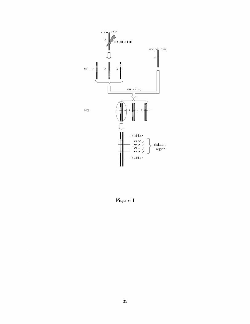
Principle of the experiment: To analyze various mutations induced by ionizing
radiation, including nontransmissible mutations, we employed the pollen-irradiation
method (STADLER and ROMAN, 1948). The experimental procedure was as follows
(Figure 1): (1[MAT14]) The pollen of wild type plants was irradiated. (2) The irradiated
pollen (M1 generation) was used to pollinate plants homozygous for a recessive
marker mutation. (3) M2 plants were screened for the recessive marker phenotype.
Because the whole body of an M2 plant is derived from a single fertilized egg, this
method can avoid chimeras, which make the molecular analysis of mutations difficult
(Figure 1).
In this experiment, Arabidopsis thaliana ecotype Columbia (Col) was used as the
pollen donor for mutagenesis, and ecotype Landsberg erecta (Ler) as the pollen
recipient. This approach enables the identification of radiation-induced mutations in
pollen by using molecular markers that distinguish the Col and Ler genomes. As
recessive visible marker mutations, the trichomeless mutations gl1-1 (chromosome 3)
and ttg1-1 (chromosome 5) on the Ler background were used. gl1-1 carries a ~10-kbp
deletion spanning the whole coding region (OPPENHEIMAER et al. 1991). ttg1-1
harbors a single nucleotide replacement that generates a premature stop codon
(WALKER et al. 1999). The male-sterile mutation ms1-1 on the Ler background
(WILSON et al. 2001) was also introduced into the pollen recipient lines to eliminate
the need for emasculation. SSLP markers F21B7-1334 on chromosome 1 and
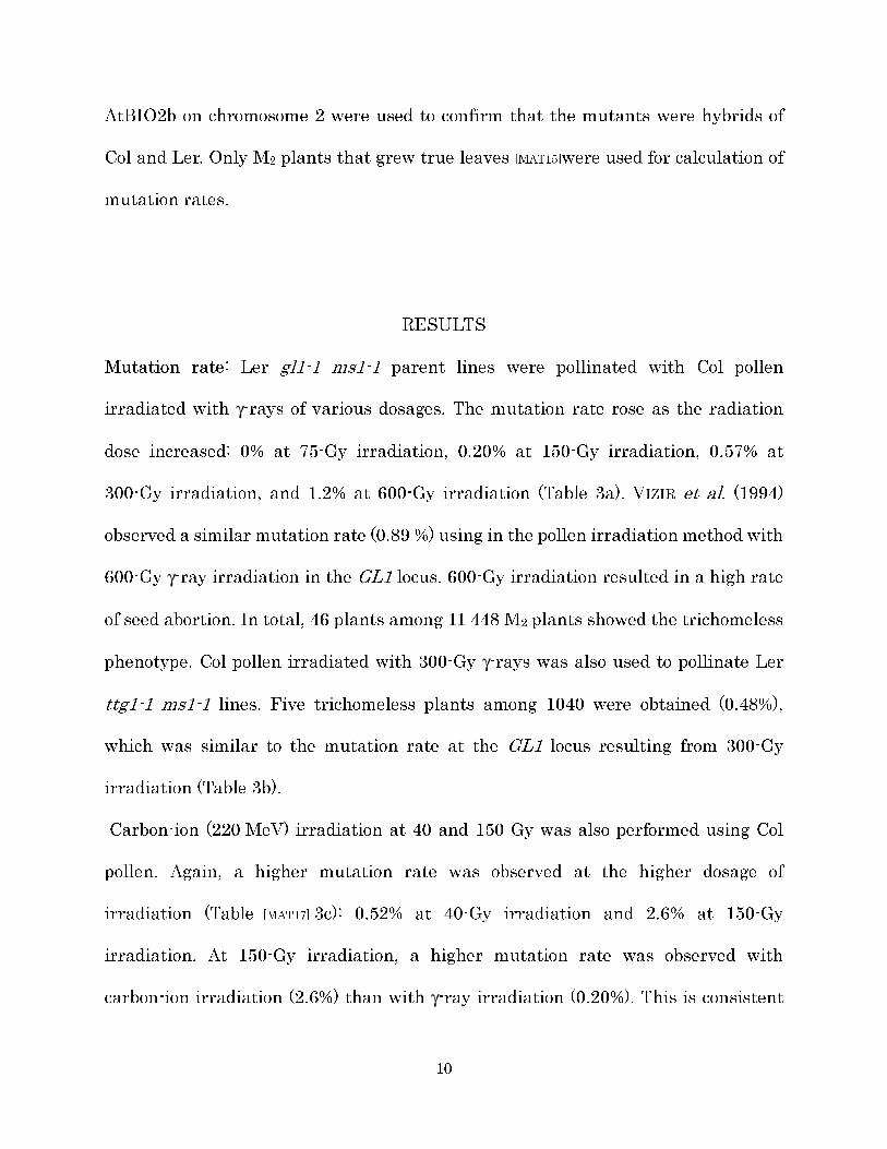
10
AtBIO2b on chromosome 2 were used to confirm that the mutants were hybrids of
Col and Ler. Only M2 plants that grew true leaves [MAT15]were used for calculation of
mutation rates.
RESULTS
Mutation rate: Ler gl1-1 ms1-1 parent lines were pollinated with Col pollen
irradiated with γ-rays of various dosages. The mutation rate rose as the radiation
dose increased: 0% at 75-Gy irradiation, 0.20% at 150-Gy irradiation, 0.57% at
300-Gy irradiation, and 1.2% at 600-Gy irradiation (Table 3a). VIZIR et al. (1994)
observed a similar mutation rate (0.89 %) using in the pollen irradiation method with
600-Gy γ-ray irradiation in the GL1 locus. 600-Gy irradiation resulted in a high rate
of seed abortion. In total, 46 plants among 11 448 M2 plants showed the trichomeless
phenotype. Col pollen irradiated with 300-Gy γ-rays was also used to pollinate Ler
ttg1-1 ms1-1 lines. Five trichomeless plants among 1040 were obtained (0.48%),
which was similar to the mutation rate at the GL1 locus resulting from 300-Gy
irradiation (Table 3b).
Carbon-ion (220 MeV) irradiation at 40 and 150 Gy was also performed using Col
pollen. Again, a higher mutation rate was observed at the higher dosage of
irradiation (Table [MAT17] 3c): 0.52% at 40-Gy irradiation and 2.6% at 150-Gy
irradiation. At 150-Gy irradiation, a higher mutation rate was observed with
carbon-ion irradiation (2.6%) than with γ-ray irradiation (0.20%). This is consistent
11
with the high relative biological effectiveness and mutagenic effect of carbon ions
(SHIKAZONO et al. 2003).
Shikazono et al. (2003) estimated that the mutation rate caused by irradiation of
Arabidopsis dry seeds with 150-Gy carbon ions was 0.029% in a large-scale screening
of an M2 population, but a mutation rate of 2.6% (9 out of 349 M2 plants) was
observed under the same irradiation conditions in this study. The different results
could be due to various factors, such as differing sensitivity to irradiation of different
tissues (dry seeds and pollen) (SHIKAZONO et al. 2002). However, the main reason for
the increased mutation rate in our study may be that we captured mutations that are
undetectable in ordinary M2 screening and which constitute the majority (more than
90%) of mutations induced by ionizing radiation (see below).
Mutations induced by γ-rays: In the gl1 mutants induced with γ-rays, only 3 of 46
mutants retained the GL1 sequence from the pollen donor (Col). CAPs/dCAPs
analysis revealed that all the remaining mutants carried large deletions, ranging
from 80 kbp (G600b1) to more than 6 Mbp (G300d1 and G300e3); the latter
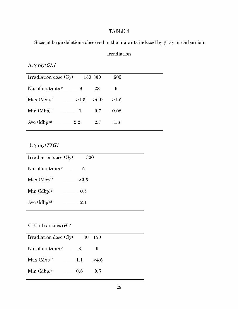
corresponds to more than 5% of the whole genome. The average sizes of the deletions
induced with 150-, 300-, and 600-Gy irradiation were 2.2 Mbp, 2.7 Mbp, and 1.8 Mbp,
respectively (Table 4a). In this paper, we define ‘large’ deletions as any deletions with
a size on the order of Mbp or kbp rather than bp.
In the ttg1 mutants induced with 300-Gy γ-ray irradiation, none of the mutants
retained the TTG1 sequence from Col. The size of the deletions ranged from 550 kbp
12
to more than 3.5 Mbp, and the average was about 2.1 Mbp (Table 4b).
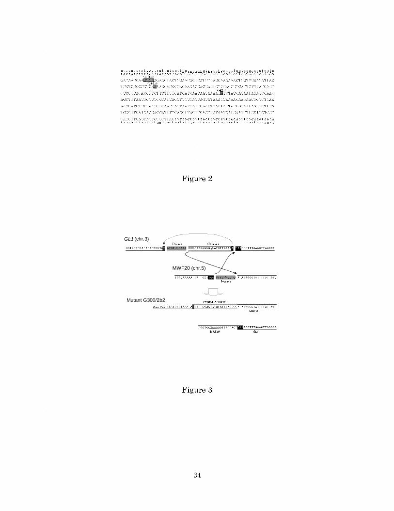
Three mutants (G150c3, G300e2, and G300f2) induced by γ-ray irradiation retained
all Col markers around the GL1 locus, including GL1 itself. To obtain more detailed
information about these mutations, the mutated GL1 gene in each mutant plant was
amplified by PCR, and its DNA sequence was determined. G150c3 and G300e2 had 4-
and 1-bp deletions, respectively, in the third exon which caused frame shifts and
premature stop codons (Figure 2).
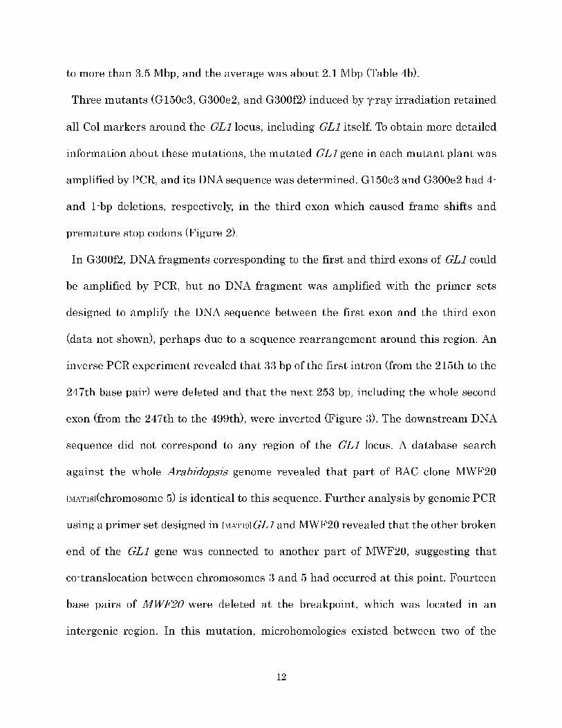
In G300f2, DNA fragments corresponding to the first and third exons of GL1 could
be amplified by PCR, but no DNA fragment was amplified with the primer sets
designed to amplify the DNA sequence between the first exon and the third exon
(data not shown), perhaps due to a sequence rearrangement around this region. An
inverse PCR experiment revealed that 33 bp of the first intron (from the 215th to the
247th base pair) were deleted and that the next 253 bp, including the whole second
exon (from the 247th to the 499th), were inverted (Figure 3). The downstream DNA
sequence did not correspond to any region of the GL1 locus. A database search
against the whole Arabidopsis genome revealed that part of BAC clone MWF20
[MAT18](chromosome 5) is identical to this sequence. Further analysis by genomic PCR
using a primer set designed in [MAT19]GL1 and MWF20 revealed that the other broken
end of the GL1 gene was connected to another part of MWF20, suggesting that
co-translocation between chromosomes 3 and 5 had occurred at this point. Fourteen
base pairs of MWF20 were deleted at the breakpoint, which was located in an
intergenic region. In this mutation, microhomologies existed between two of the
13
three rejoining sites (Figure 3). Similarly, in G150c3 and G300e2, a 1-bp homology
might be used for DSB repair (Figure 2).
Mutations induced by carbon-ion irradiation: Large deletions were frequently found
in carbon-ion-induced mutants: only 1 of 13 retained the GL1 sequence. This mutant
(C40a2) retained all Col markers around the GL1 locus. Sequence analysis of its GL1
locus revealed a 1-bp deletion in the third exon, which caused a frame-shift and a
premature stop codon (Figure 2). In this mutant, no microhomology was found at the
rejoining site. The average sizes of the remaining large deletions were 650 kbp at
40-Gy irradiation and 2.5 Mbp at 150-Gy irradiation. The minimum size of the
deletion was 495 kbp, and the maximum was more than 4.7 Mbp (Table 4c).
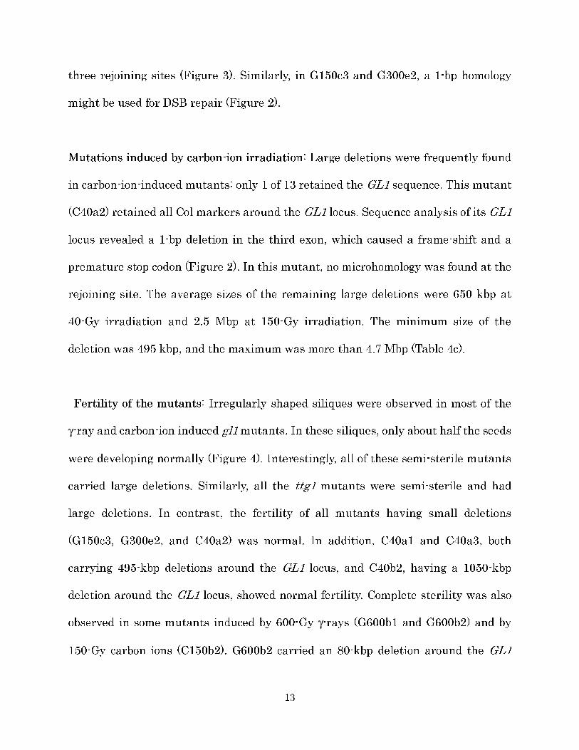
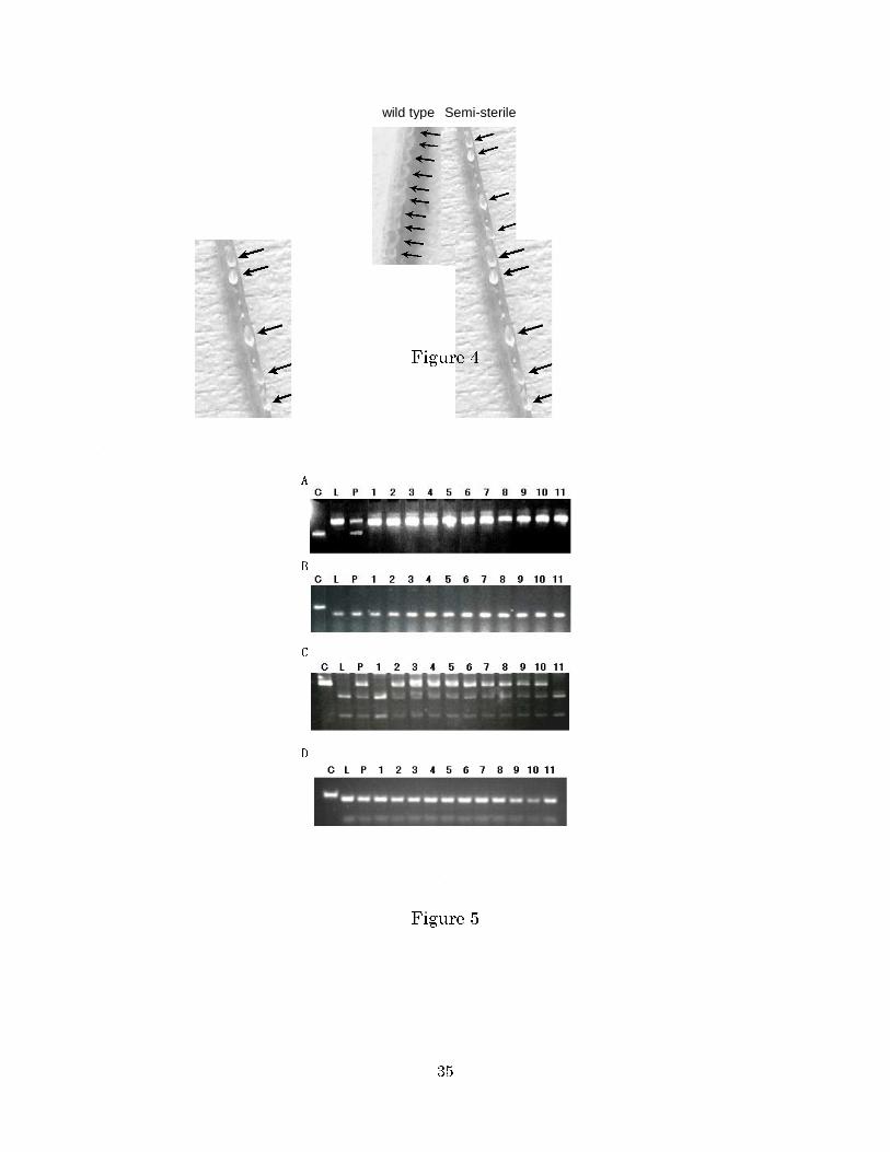
Fertility of the mutants: Irregularly shaped siliques were observed in most of the
γ-ray and carbon-ion induced gl1 mutants. In these siliques, only about half the seeds
were developing normally (Figure 4). Interestingly, all of these semi-sterile mutants
carried large deletions. Similarly, all the ttg1 mutants were semi-sterile and had
large deletions. In contrast, the fertility of all mutants having small deletions
(G150c3, G300e2, and C40a2) was normal. In addition, C40a1 and C40a3, both
carrying 495-kbp deletions around the GL1 locus, and C40b2, having a 1050-kbp
deletion around the GL1 locus, showed normal fertility. Complete sterility was also
observed in some mutants induced by 600-Gy γ-rays (G600b1 and G600b2) and by
150-Gy carbon ions (C150b2). G600b2 carried an 80-kbp deletion around the GL1
14
locus, the smallest among the large deletions. A high extent of chromosomal
aberration caused by high-dose irradiation might have prevented normal meiosis in
these mutants, thereby preventing normal gamete development. Unexpectedly, the
reciprocal translocation mutant (G300f2) was fully fertile, even though reciprocal
translocation generally causes reduced fertility in heterozygotes (data not shown,
VAN HARTEN 1998). It is possible that another reciprocal translocation between the
two chromosomes occurred close to the translocation point in this mutant and
compensated for its effects[MAT21] on semi-sterility.
Transmissibility of the radiation-induced mutations: Because all the selfed M3
progeny from a mutant M2 plant showed the trichomeless phenotype, the
transmissibility of the mutation was examined by genotyping the M3 plants with the
molecular markers[MAT22]. In the gl1 alleles having 1- and 4-bp deletions (G150c3,
G300e2, and C40a2), no distortion [MAT23]in transmissibility was observed among 40
M3 progeny of each line: about three-fourths of the M3 progeny had the Col GL1
sequence (data not shown). The reciprocal translocation mutation (G300f2) was also
transmitted normally among 40 M3 progeny (data not shown). However, when selfed
M3 progeny of the semi-sterile mutants were analyzed, no Col genotypes for markers
closely linked to the GL1 locus were detected. For example, in 40 M3 progeny of the
semi-sterile mutant G300a2, only Ler marker bands were detected in CAPS/dCAPS
analysis using t5m71-l, mdj14-2, and k1g2-1 (Figure 5a and data not shown),
suggesting that no or very few gl1 alleles associated with semi-sterility were
15
transmitted to the M3 plants. In addition, all the 40 M3 progeny examined carried the
Ler genotype for k24a2-4, whose Col allele was deleted in G300a2 (Figure 5b). In the
fully-fertile mutants having large deletions (C40a1, C40a3, and C40b2), no
Col-genotype homozygotes [MAT24]for mmg15-2, which is closely linked to the deletions,
were found in 40 M3 progeny of each mutant, although heterozygotes were found
(Figure 5c and data not shown). These observations suggest that these mutations
could be transmitted to progeny only heterozygously, [MAT25]in spite of their full
fertility. Among 40 M3 progeny of each mutant, all progeny had the Ler genotype for
k24a2-4, whose Col allele was deleted in these mutants, supporting this hypothesis
(Figure 5d and data not shown). One explanation for these observations is that these
deletions affect the viability of only male gametes. However, in germination tests,
only 73 out of 100 M3 seeds of C40a1 germinated, although 98 out of 100 F2 seeds of a
hybrid between Col and Ler germinated. Although we were not able to determine the
genotype of seeds that did not germinate, it appears that homozygotes for these
deletions cannot germinate. We should note that these transmissible large deletions
cannot be detected in ordinary M2 screening.
The semi-sterile ttg1 mutants also did not transmit the Col ttg1 mutation to the M3
progeny. For example, among 40 M3 progeny of tG300a1, which carried a 550-kbp
deletion from t32g24-1 through k18p6-3, no Col genotype was found for the marker
t11h3-3, which is closely linked to the deletion (Table 2 and data not shown).
DISCUSSION[MAT26]
16
Mutations induced by ionizing radiation: Although DSBs are not the major DNA
damage induced by irradiation, they have greater importance in mutations than do
other types of DNA damage (BRITT 1999, GORBUNOVA and LEVY 1999, TUTEJA et al.
2001). DSBs are thought to be repaired in three ways. The first is homologous
recombination (HR), which copies the homologous sequence from the undamaged
sister chromatid. The second is single-strand annealing (SSA), which is mainly
involved in the repair of repeated sequences. The third is nonhomologous end joining
(NHEJ), which rejoins one DNA broken end with another without specificity
(CRITCHLOW and JACKSON 1998, BRITT 1999, GORBUNOVA and LEVY 1999, LIEBER et
al. 2003). NHEJ can be precise but is often accompanied by these small deletions.
The deletion of one or a few base pairs observed in G150b3, G300d2, and C40a2 was
probably due to DSB repair by NHEJ of two broken ends that were originally linked.
Alternatively, replication slippage at damaged bases can be involved in the
generation of small deletions (KUNKEL 1990). [MAT27]
Most of the mutations identified in this study were very large deletions (larger than
80 kbp) in both the GL1 and TTG1 loci, at various irradiation doses (40–600 Gy), and
with both high- and low-LET irradiation (carbon ions and γ-rays, respectively). The
largest deletion was >6 Mbp around the GL1 locus, which should involve the deletion
of more than 1300 genes, based on the average gene density in Arabidopsis
chromosome 3 (a gene every 4.5 kbp) (THE ARABIDOPSIS GENOME INITIATIVE 2000).
VIZIR et al. (1994) also characterized the size of deletions in A. thaliana induced by
γ-rays using the pollen-irradiation method. In this classic genetic study, they
17
estimated that the average size of deletions was 160 kbp, which are large deletions,
but the average size of deletions in our study was much larger (more than 2 Mbp,
even if point mutations are included).
Very large deletions are thought to be generated by NHEJ (SACHS et al. 2000). When
both terminal DNA fragments of the three DNA fragments generated by two DSBs in
one chromosome are joined together, the consequent omission of the middle fragment
constitutes a deletion. When the omitted fragment is large, a large deletion occurs. In
this context, the observation that most of the mutations found in our study were
large deletions suggests that two DSBs were not frequently generated in nearby
positions, even with high-LET carbon-ion irradiation, despite its high density of
excited/ionized molecules. This could be a consequence of the higher-order structure
of chromatin.[MAT29] Importantly, the present study shows that both low-LET γ-rays
and high-LET carbon ions generate large deletions, but it does not necessarily
suggest that there is no difference between them regarding the mechanism
responsible for the generation of mutations. Furthermore, we should note that the
generation of large deletions vs. point-like mutations by the DSB repair process may
be less frequent than expected from the frequency of large deletions found in our
mutants, because large deletions have a higher mutagenic [MAT31]effect and are thus
detected more often; that is, it is more likely for a gene to be affected by one large
deletion than by one point mutation.
Transmissibility of mutations: All the semi-sterile gl1 mutants carried large
18
deletions around the GL1 locus, suggesting a correlation between large deletions and
semi-sterility. These large deletions did not appear to be transmitted even
heterozygously. The most likely explanation for these observations is that the large
deletions contained a gene or genes required for the formation or viability of pollen
and egg cells: only egg cells and pollen that do not carry the large deletions and are
able to survive can undergo successful fertilization, resulting in semi-sterility and
nontransmissibility of the deletions. Consistent with this, no seed development was
observed visually in the semi-sterile mutants in about half the placenta, implying
either the absence of fertilization or very early abortion of seeds (Figure 4). It is
unlikely that a dominant lethal mutation affecting embryogenesis is involved in the
semi-sterility, because this would result in no M2 seed formation[MAT32]. It is also
possible that DNA rearrangement in another region contributed to the semi-sterility,
but the distortion in transmissibility of the gl1 alleles strongly suggests that the
semi-sterility can be attributed to the large deletion around the GL1 locus. Similar
observations were made for the TTG1 locus, suggesting that genes involved in
gamete formation or viability are not rare and are dispersed throughout the genome.
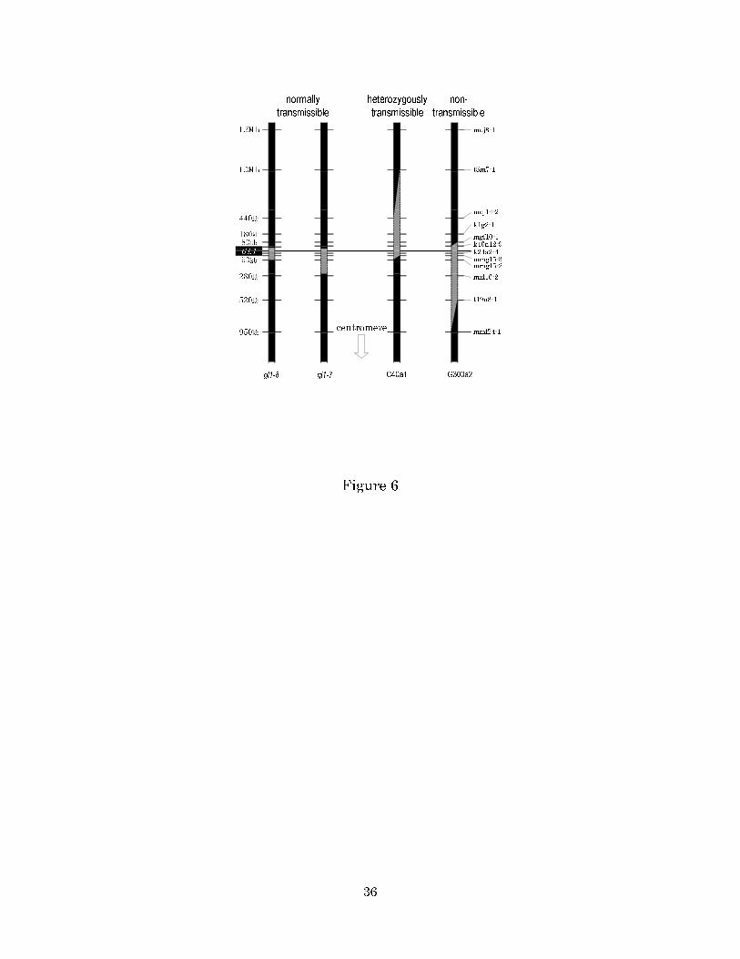
Interestingly, three mutants showed full fertility even though they carried large
deletions around the GL1 locus (Figure 6). These deletions were only transmitted
[MAT33] heterozygously. Taking into consideration the observation that one-fourth of
the M3 seeds of C40a1 did not germinate, we hypothesize that fertilized eggs
homozygous for the deletions can develop into seeds but lack the ability to germinate.
SHIKAZONO et al. (2003) isolated gl1 alleles carrying a large deletion, which were
19
transmitted normally (Figure 6 and N. SHIKAZONO, C. SUZUKI, S. KITAMURA, H.
WATANABE, S. TANO, and A. TANAKA, unpublished results). Comparison of the deleted
region in C40a1 with those in mutants gl1-7 and gl1-8 suggests that the gene is
located between 37 kbp upstream of GL1 and t5m7-1 (1 Mb upstream of GL1). On the
other hand, comparison of the deleted regions in the semi-sterile and
nontransmissible mutant, G300a2, with those in C40a1 and gl1-7 suggests that the
region between 194 kb downstream of GL1 and mmf24-1 (950 kbp downstream of
GL1) contains at least one gene essential for gamete development or viability.
By the pollen-irradiation method using X-rays and applied to maize, Stadler and
Roman (1948) obtained a very small number of transmissible mutations (2 out of 415),
and both mutations showed low transmissibility. Nuffer (1957) and Mottinger (1970)
made similar observations for another locus using the same method and concluded
that intragenic mutation, which usually shows complete transmissibility, cannot be
induced by X-rays. Our results are similar to those observations: most mutations
induced by ionizing radiation had defective transmissibility. However, some apparent
‘intragenic’ mutations were isolated (3 out of 64) in our study. The difference may be
attributed to the use of different species, because an intragenic mutation induced by
X-rays using the pollen-irradiation method has been reported in Arabidopsis
(CLARKE et al. 1993, 1997). Consistent with this, the DNA repair system may differ
between plant species having different genome sizes, e.g., very small genome size
such as Arabidopsis and large genome size such as tobacco (KIRIK et al. 2000)[MAT37].
Simple repair of a single DSB by NHEJ, which often results in deletion of a small
20
number of nucleotides, might be rare in maize in comparison with Arabidopsis.
In this study, we showed that most radiation-induced mutants carry extremely large
deletions, regardless of the irradiation dose, radiation type, or locus. Based on our
results, we hypothesize that most such deletions are not transmitted to progeny
because the deletions remove a gene or genes involved in gamete development or
viability. This means that most mutations induced by ionizing radiation have not
been used in the mutation breeding of seed-propagated crops. However,
'nontransmissible' mutations can survive over generations in vegetatively
propagated crops and have probably contributed to the genetic improvement of such
crops.
ACKNOWLEDGMENTS
This paper is dedicated to Toshiya Takano, who passed away in December 2003. We
thank to Prof. N. Goto for providing the ttg1-1 ms1-1 mutant line. This work was
supported by a grant for [MAT38]Nuclear Research of the Ministry of Education,
Culture, Sports, Science, and Technology of Japan.
LITERATURE CITED
BELL, C. J., and J. R. ECKER, 1994 Assignment of 30 microsatellite loci to the linkage
map of Arabidopsis. Genomics 19: 137–144.
BRITT, A. B., 1999 Molecular genetics of DNA repair in higher plants. Trends Plant
Sci. 4: 20–25.

21
BRUGGENMANN, E., K. HANDWERGER, C. ESSEX and G. STORZ, 1996 Analysis of fast
neutron-generated mutants at the Arabidopsis thaliana HY4 locus. Plant J. 10:
755–760.
BUSCHGES, R., K. HOLLRICHER, R. PANSTRUGA, G. SIMONS, M. WOLTER, et al., 1997
The barley Mlo gene: a novel control element of plant pathogen resistance. Cell 88:
695–705.
CLARK, S. E., M. P. RUNNING and E. M. MEYEROWITZ, 1993 CLAVATA1, a regulator of
meristem and flower development in Arabidopsis. Development 119: 397–418.
CLARK, S. E., R. W. WILLIAMS and E. M. MEYEROWITZ, 1997 The CLAVATA1 gene
encodes a putative receptor kinase that controls shoot and floral meristem size in
Arabidopsis. Cell 89: 575–585.
GORBNOVA[MAT39], V., and LEVY, A. A., 1999 How plants make ends meet: DNA double-
strand break repair. Trends Plant Sci. 4: 263–269.
KIRIK, A., S. SALOMON and H. PUCHTA, 2000 Species-specific double-strand break
repair and genome evolution in plants. EMBO J. 19: 5562–5566.
KUNKEL, T. A., 1990 Misalignment-mediated DNA synthesis errors. Biochemistry 29:
8003–8011.
LIEBER, M. R., Y. MA, U. PANNICKE and K. SCHWARZ, 2003 Mechanism and regulation
of human non-homologous DNA end-joining. Nat. Rev. Mol. Cell Biol. 4: 712–720.
22
MORISHITA, T., H. YAMAGUICHI, K. DEGI, N. SHIKAZONO, Y. HASE, A. TANAKA and T.
ABE, 2003 Dose response and mutation induction by ion beam irradiation in
buckwheat. Nucl. Inst. Meth. 206: 506–569.
MOTTINGER, J. P., 1970 The effects of X rays on the bronze and shrunken loci in maize.
Genetics 64: 259–271.
MULLER, H. J., 1927 Artificial transmutation of the gene. Science 66: 84–87.
NAKAZAKI, T., Y. OKUMOTO, A. HORIBATA, S. YAMAHIRA, M. TERAISHI et al., 2003
Mobilization of a transposon in the rice genome. Nature 421: 170–172.
NUFFER, M. G., 1957 Additional evidence on the effect [MAT41]of X-ray and ultraviolet
radiation on mutation in maize. Genetics 42: 273–282.
OCHMAN, H., A. S. GERBER and D. L. HARTL, 1988 Genetic applications of an inverse
polymerase chain reaction. Genetics 120: 621–623.
OPPENHEIMER, D. G., P. L. HERMAN, S. SIVAKUMARAN, J. ESCH and M. D. MARKS, 1991
A myb gene required for leaf trichome differentiation in Arabidopsis is expressed in
stipules. Cell 67: 483–493.
SACHS, R. K., L. R. HLATKY and B. J. TRASK, 2000 Radiation-produced chromosome
aberrations. Trends Genet. 16: 143–146.
SHIKAZONO, N., A. TANAKA, H. WATANABE and S. TANO, 2000 Rearrangements of the
DNA in carbon ion-induced mutants of Arabidopsis thaliana. Genetics 157: 379–387.

23
SHIKAZONO N., A. TANAKA, S. KITAYAMA, H. WATANABE and S. TANO, 2002 LET
dependence of lethality in Arabidopsis thaliana irradiated by heavy ions. Radiat.
Environ. Biophys. 41: 159–162.
SHIKAZONO, N., Y. YOKOTA, S. SATOSHI, G. SUZUKI, H. WATANABE et al., 2003
Mutation rate and novel tt mutants of Arabidopsis thaliana induced by carbon ions.
Genetics 163: 1449–1455.
SHIRLEY, B. W., S. HANLEY and H. M. GOODMAN, 1992 Effects of ionizing radiation on
a plant genome: analysis of two Arabidopsis transparent testa mutations. Plant Cell
4: 333–347.
STADLER, L. J., 1928 Mutation in barley induced by X-rays and radium. Science 67:
186–187.
STADLER, S. J., and H. ROMAN, 1948 The effect of X-rays upon mutation of the gene A
in maize. Genetics 33: 273–303.
TANAKA, A., N. SHIKAZONO, Y. YOKOTA, H. WATANABE and S. TANO, 1997 Effects of
heavy ions on the germination and survival of Arabidopsis thaliana. Int. J. Radiat.
Biol. 72: 121–127.
THE ARABIDOPSIS GENOME INITIATIVE, 2000 Analysis of the genome sequence of the
flowering plant Arabidopsis thaliana. Nature 408: 796–815.
VAN HARTEN, A. M., 1998 Mutation Breeding. Cambridge University Press,
Cambridge.
24
TUTEJA, N., M. B. SINGH, M. K. MISRA, P. L. BHALLA, R. TUTEJA, 2001 Molecular
mechanisms of DNA damage and repair: progress in plants. Crit Rev Biochem Mol
Biol. 36: 337-397.
VIZIR, I. Y., M. L. ANDERSON, Z. A. WILSON and B. J. MULLIGAN, 1994 Isolation of
deficiencies in the Arabidopsis genome by γ-irradiation of pollen. Genetics 137:
1111–1119.
WALKER, A. R., P. A. DAVISON, A. C. BOLOGNESI-WINFIELD, C. M. JAMES, N.
SRINIVASAN, et al., 1999 The TRANSPARENT TESTA GLABRA1 locus, which
regulates trichome differentiation and anthocyanin biosynthesis in Arabidopsis,
encodes a WD40 repeat protein. Plant Cell 11: 1337–1349.
WILSON, Z. A., S. M. MORROLL, J, DAWSON, R, SWARUP and P. TIGHE, 2001 The
Arabidopsis MALE STERILITY1 (MS1) gene is a transcriptional regulator of male
gametogenesis, with homology to the PHD-finger family of transcription factors.
Plant J. 28: 27–39.
YANO, M., Y. KATAYOSE, M, ASHIKARI, U, YAMANOUCHI, L. MONNA et al., 2000 Hd1, a
major photoperiod sensitivity quantitative trait locus in rice, is closely related to the
Arabidopsis flowering time gene CONSTANS. Plant Cell 12: 2473–2483.

25
26
TABLE 1
CAPs/dCAPs markers around the GL1 locus
Marker 5'-Forward Primer-3' 5'-Reverse Primer-3' Enzyme Distancea
moe17 agaatcataccccctacttagtgcaga tagagtttccatggtgaaatgtgg Mbo I 2.9 Mbp
k14b15 cgatacgtgtgaacgcgttggtc attacaaatttgcaactactgtttgagc Alu I 1.9 Mbp
muj8-1 tctagtggcagtaaaactacagagg acaacatctactgacacctgcacaaagt Afa I 1.6 Mbp
t5m7-1 cttctgaggctaactctgaaagcg attcttcgtgtctatatagtcgacc Hsp92 III 1.0 Mbp
mdj14-2 taaagtgtcttttaaccggaatcc cacgagaacaagcagagattacc Hsp92 III 440 kbp
k1g2-1 tgttggaaacattaggatcaatgaaactag atgcaaggaagcacaacatgatctg Spe I 180 kbp
mgf10-1 ggagtctgcgatccgaacc gaatcagtacagagcttgcc Afa I 80 kbp
k16n12-9 tgcctatttctcctccatgc gctctatatacatataaaacaaattaa PshB I 54 kbp
k16n12-8 ttttaacaacgtattatcctctcta aaatagcacaaattcctttggc Xsp I 47 kbp
k16n12-5 gaaagacgatgcaacggcg aacaagatcttgcttgtacgg Mbo I 23 kbp
k16n12-2 catgaagaagaccctatttgg atctctccttgttgtcctcg Afa I 5 kbp
GL1 tactggaacactcatctcagc cagtatccgcggtaactaac -[MAT43] 0 kbp
k16n12-3 gtctttgccattggcaatcc acgttttgttgactgaagtcc Afa I 5 kbp
k24a2-1 ggagtagggtcacaaagcc cttgatacattggtctcttcc Taq I 7 kbp
k24a2-2 aatgaatgcaaacccatgcg ctaggaatcttcaaccacac Alu I 13 kbp
k24a2-3 ggagaatcggatgcacgag ttgtcccaacaacatactcc Alu I 18 kbp
k24a2-4 cctatatgaatcgaacgcaaggatat ttggagaccgtgatgacgtg EcoR V 27 kbp
mmg15-3 agttttgttggatggggatgacctagttaa ccgccaacctatattacacc Hinc II 55 kbp
mmg15-2 agaatccttttaaatccatcgc atccttgtagatgtccttagg Alu I 90 kbp
mzf16-2 ggatctatgggagattatacttgc ttaaacactcctcaccgtgctagg Xsp I 280 kbp
t19n8-1 cggagctggagattctactgccag gtagcgcctccagctgtggcacc Msp I 520 kbp
mmf24-1 tttagccgttttctcgtggttacg cggatgtgttctgcacgagtgttattgtac Bst1407 I 950 kbp
mil15-1 tacatcacctagggaagtcaatcg tggaactcttgtgaggtgaactcc Hinc II 1.5 Mbp
f21a17-1 tgaagtccgtagaaatgaattcc gagcttacactcttacatagatgtcagc Hsp92 III 2.0 Mbp
f1d9-1 atctctccttctaccagaacgtgc gaagaatataccttcaacacaagc Acc II 3.1 Mbp
a Distance from the GL1 locus.
27
TABLE 2
CAPs/dCAPs and SSLP markers around the TTG1 locus
Marker 5'-Forward Primer-3' 5'-Reverse Primer-3' Enzyme Distancea
f5e19-2 atacaccatatatatacgtcatac gttctctatcaatattctaaactctggccc Hinf I 2.9 Mbp
t1a4-1 taaacatgttgggccgattatcct tgagtataatgttatttcttattttggtac Kpn I 2.0 Mbp
f5o24-1 tttcgggttatgaccgcattcgtt caaagtgttaaatgtggatatggatgaaat Ssp I 1.5 Mbp
mye28-1 attacatttccatcagaatgatta tatacaagggaggagatggc Sty I 1.1 Mbp
t32g24-1 ggcaggtcatggatgcattag cactcccaacgtgagggaggagtac Dra I 500 kbp
mzf18-1 ggccactctctgccacgtgg aagcgcaccaataacctacctgtgggctct Xho I 190 kbp
NGA139 accgaaccgattgatttcagaagg ggcggtacgacctcctatgg -[MAT44] 80 kbp
TTG1 tactggaacactcatctcagc cagtatccgcggtaactaac Mbo I 0 kbp
k18p6-3 ggtttcgtttcactatccagg agagctaccagatccgatgg - 50 kbp
t11h3-3 actagtaagacaacaatgtttacc tattctattcaaaatgttacgagg - 220 kbp
che24 1 tcaaaccggtttgaaggatgtatacgg ctcaagattctccatctgacccaacga Eco52 I 560 kbp
wue99-1 atgttattgagaagcacatctgct ttaggatgatgcggtaatagaagt Ssp I 1.0 Mbp
tig16-3 gaatttaagaggaaacttgatagg cacactgagtttgtctctaaaaac Hha I 1.5 Mbp
t8m17-1 tttattttaatgttttaaaacttatggatc attacttttagattgcaagtataaaa BamH I 2.1 Mbp
t3p01-1 atccaaaggaaccttatctccccgcttgcg tgaagcagagtgcagtagtaggtc Hha I 3.0 Mbp
a Distance from the TTG1 locus.
28
TABLE 3
Mutation rates in plants grown from pollen irradiated with γ-rays or carbon ions
A. γ-ray/GL1[MAT45]
Irradiation dose (Gy) 75 150 300 600
No. of mutants 0 10 30 6
No. of M2 plants 2542 4953 5272 485
Mutation rate (%) 0 0.20 0.57 1.24
B. γ-ray/TTG1
Irradiation dose (Gy) 300
No. of mutants 5
No. of M2 plants 1040
Mutation rate (%) 0.48
C. Carbon ions/GL1
Irradiation dose (Gy) 40 150
No. of mutants 4 9
No. of M2 plants 770 349
Mutation rate (%) 0.52 2.58
29
TABLE 4
Sizes of large deletions observed in the mutants induced by γ-ray or carbon-ion
irradiation
A. γ-ray/GL1
Irradiation dose (Gy) 150 300 600
No. of mutants a 9 28 6
Max (Mbp)b >4.5 >6.0 >4.5
Min (Mbp)c 1 0.7 0.08
Ave (Mbp)d 2.2 2.7 1.8
B. γ-ray/TTG1
Irradiation dose (Gy) 300
No. of mutants a 5
Max (Mbp)b >3.5
Min (Mbp)c 0.5
Ave (Mbp)d 2.1
C. Carbon ions/GL1
Irradiation dose (Gy) 40 150
No. of mutants a 3 9
Max (Mbp)b 1.1 >4.5
Min (Mbp)c 0.5 0.5

30
Ave (Mbp)d 0.7 2.5
a Number of mutants with a large deletion.
b Size of the largest deletion.
c Size of the smallest deletion among the large deletions.
d Average size of the large deletions.

31
FIGURE LEGENDS
FIGURE 1.—Principle of the experiment. Pollen from Arabidopsis thaliana Col plants
(M1) was irradiated and used to fertilize Ler plants carrying a recessive marker
mutant gene (a). The resultant M2 plants were screened for the marker phenotype.
These mutants were analyzed by using molecular markers around the marker locus
that distinguish the Col and Ler genomes. This enables the deleted regions of the Col
genome caused by pollen irradiation to be determined in the M2 plants. Col
chromosomes are shown in black, Ler chromosomes in gray, and deleted regions in
white.
FIGURE 2.—Positions of mutations in the gl1 alleles having small deletions. Small
letters indicate the second intron and the 3′ untranslated region of GL1. Capital
letters indicate the third exon. Nucleotides deleted in the mutants are shaded.
FIGURE 3.—Structural changes in the DNA of mutant G300b2. Deleted nucleotides
are shaded. Broken ends within chromosomes are shown as gaps, and the rejoinings
of broken ends are shown by arrows. White characters on black backgrounds indicate
microhomologies at the rejoining sites.
FIGURE 4.—Developing seeds in semi-sterile mutants produced by the
pollen-irradiation method. Opened siliques from a wild-type plant and from a typical
32
semi-sterile mutant are shown. Arrowheads indicate normally developing seeds.
FIGURE 5.—Transmissibility of the large deletions. (A, B) dCAPs analysis of the M3
plants of the semi-sterile mutant G300a2 (see Figure 6) using t5m7-1 (A) and
k24a2-4 (B). (C, D) dCAPs and CAPs analysis of the M3 progeny of the fully fertile
mutant C40a3 (see Figure 6) using k24a2-4 (C), and mmg15-2 (D). Lanes C, L, and P
indicate Col, Ler, and the mutant M2 plants, respectively. Lanes indicated by
numbers are the M3 progeny.
FIGURE 6.—Deleted regions in the gl1 mutants showing various manners of
transmission. Deleted regions are shaded[MAT46]. Positions of molecular markers are
shown on the left and the marker names are on the right.
33
Col/Ler
A
Ler onlyLer only
Col/Ler
Ler only
egg cell (Ler)
A A
Ler only
a
M1
M2
pollen (Col)
crossing
Ai r r adi at i on
A A Aa a a
deleted
region
Figure 1
34
a t a a a c a t a t g g t a t g t t a t a a t t t a t g t t a t a t g g t a t g a a t t t a a t a a a a a t g t c c t at a a t a t t t t t t c c c c a a a t t c a gATGGTCTTTGATAGCTAAAAGAGTACCGGGAAGAACA
GATAACCAAGTCAAGAACTACTGGAACACTCATCTCAGCAAAAAACTCGTCGGAGATTACG300e2
TCCTCCGCCGTCAAAACCACCGGAGAAGACGACGACTCTCCACCGTCATTGTTCATCACTC40a2
GCCGCCACACCTTCTTCTTGTCATCATCAACAAGAAAATATCTACGAGAATATAGCCAAG
AGCTTTAACGGCGTCGTATCAGCTTCGTACGAGGATAAACCAAAACAAGAACTGGCTCAA
AAAGATGTCCTAATGGCAACTACTAATGATCCAAGTCACTATTATGGCAATAACGCTTTA
TGGGTTCATGACGACGATTTTGAGCTTAGTTCACTCGTAATGATGAATTTTGCTTCTGGT
GATGTTGAGTACTGCCTTTAGt t g a t g t t t t g a a t t t g t c t t t g a a t t t t t g a ga t a a a at a a c a a t t a a t t a t g ga t a c t a a t a t c g a t a t c a a t a c t a c c a c t a t t a c a a t c t a g g a g
G150c3
Figure 2
GCTAGT TCA TA TA TAAA TA T A TCT GATA GGGCTAAAGAG ATCT TAAAAA T AA TCCT T TGAACT TAAAA T
T AAGAAAAA T T A T T ACT TAA CTCG CCTG ATA TAAAA T A T T T T A T T A TG
AGCTAGT TCA TA TA T AAA TA T T T T T AAGA T CTCT T T AGCCCATA TAAAA T A T T T T A T T A TG
MWF20
T AA TAAGAAAAA T T A T T ACT TAA TCCT T TGAACT TAAAA T
MWF20 GL1
253bases
MWF20 (chr.5)
-//-
-//-
inverted 253 bases
GL1 (chr.3)
Mutant G300/2b2
-//--//-
33bases
14bases
Figure 3
35
wild type Semi-sterile
Figure 4
A
B
C
D
Figure 5
36
1.6M b
1.0M b
440kb
950kb
280kb
180kb
80kb
520kb
G L1
90kb
gl1-8 C40a1 G300a2
muj8-1
t5m7-1
mdj14-2
k1g2-1
mgf10-1k16n12-9
mmg15-3mmg15-2
mzf16-2
t19n8-1
mmf24-1
k24a2-4
centromere
gl1-7
heterozygously
transmissible
normally
transmissible
non-
transmissible
Figure 6