Genetic determinants of cellular addiction to DNA ...
13
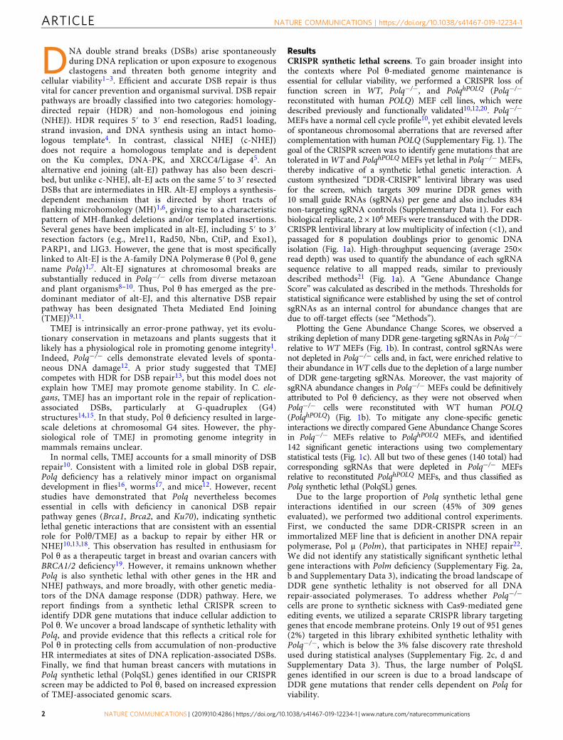
ARTICLE Genetic determinants of cellular addiction to DNA polymerase theta Wanjuan Feng 1 , Dennis A. Simpson 1 , Juan Carvajal-Garcia 1 , Brandon A. Price 1 , Rashmi J. Kumar 1 , Lisle E. Mose 1 , Richard D. Wood 2 , Naim Rashid 1,3 , Jeremy E. Purvis 4 , Joel S. Parker 1,4 , Dale A. Ramsden 1,5 & Gaorav P. Gupta 1,5,6 Polymerase theta (Pol θ, gene name Polq) is a widely conserved DNA polymerase that mediates a microhomology-mediated, error-prone, double strand break (DSB) repair path- way, referred to as Theta Mediated End Joining (TMEJ). Cells with homologous recombi- nation deficiency are reliant on TMEJ for DSB repair. It is unknown whether deficiencies in other components of the DNA damage response (DDR) also result in Pol θ addiction. Here we use a CRISPR genetic screen to uncover 140 Polq synthetic lethal (PolqSL) genes, the majority of which were previously unknown. Functional analyses indicate that Pol θ/TMEJ addiction is associated with increased levels of replication-associated DSBs, regardless of the initial source of damage. We further demonstrate that approximately 30% of TCGA breast cancers have genetic alterations in PolqSL genes and exhibit genomic scars of Pol θ/TMEJ hyperactivity, thereby substantially expanding the subset of human cancers for which Pol θ inhibition represents a promising therapeutic strategy. https://doi.org/10.1038/s41467-019-12234-1 OPEN 1 Lineberger Comprehensive Cancer Center, University of North Carolina at Chapel Hill, Chapel Hill, NC 27599, USA. 2 Department of Epigenetics and Molecular Carcinogenesis, University of Texas MD Anderson Cancer Center, Smithville, TX 78957, USA. 3 Department of Biostatistics, University of North Carolina at Chapel Hill, Chapel Hill, NC 27599, USA. 4 Department of Genetics, University of North Carolina at Chapel Hill, Chapel Hill, NC 27599, USA. 5 Department of Biochemistry and Biophysics, University of North Carolina at Chapel Hill, Chapel Hill, NC 27599, USA. 6 Department of Radiation Oncology, University of North Carolina at Chapel Hill, Chapel Hill, NC 27599, USA. Correspondence and requests for materials should be addressed to G.P.G. (email: [email protected]) NATURE COMMUNICATIONS | (2019)10:4286 | https://doi.org/10.1038/s41467-019-12234-1 | www.nature.com/naturecommunications 1 1234567890():,;
Transcript of Genetic determinants of cellular addiction to DNA ...
ARTICLE
Genetic determinants of cellular addiction to DNApolymerase thetaWanjuan Feng1, Dennis A. Simpson1, Juan Carvajal-Garcia 1, Brandon A. Price1, Rashmi J. Kumar1,
Lisle E. Mose1, Richard D. Wood 2, Naim Rashid1,3, Jeremy E. Purvis4, Joel S. Parker 1,4, Dale A. Ramsden1,5 &
Gaorav P. Gupta 1,5,6
Polymerase theta (Pol θ, gene name Polq) is a widely conserved DNA polymerase that
mediates a microhomology-mediated, error-prone, double strand break (DSB) repair path-
way, referred to as Theta Mediated End Joining (TMEJ). Cells with homologous recombi-
nation deficiency are reliant on TMEJ for DSB repair. It is unknown whether deficiencies in
other components of the DNA damage response (DDR) also result in Pol θ addiction. Here
we use a CRISPR genetic screen to uncover 140 Polq synthetic lethal (PolqSL) genes, the
majority of which were previously unknown. Functional analyses indicate that Pol θ/TMEJ
addiction is associated with increased levels of replication-associated DSBs, regardless of the
initial source of damage. We further demonstrate that approximately 30% of TCGA breast
cancers have genetic alterations in PolqSL genes and exhibit genomic scars of Pol θ/TMEJ
hyperactivity, thereby substantially expanding the subset of human cancers for which Pol θinhibition represents a promising therapeutic strategy.
https://doi.org/10.1038/s41467-019-12234-1 OPEN
1 Lineberger Comprehensive Cancer Center, University of North Carolina at Chapel Hill, Chapel Hill, NC 27599, USA. 2Department of Epigenetics and MolecularCarcinogenesis, University of Texas MD Anderson Cancer Center, Smithville, TX 78957, USA. 3Department of Biostatistics, University of North Carolina atChapel Hill, Chapel Hill, NC 27599, USA. 4Department of Genetics, University of North Carolina at Chapel Hill, Chapel Hill, NC 27599, USA. 5Department ofBiochemistry and Biophysics, University of North Carolina at Chapel Hill, Chapel Hill, NC 27599, USA. 6Department of Radiation Oncology, University of NorthCarolina at Chapel Hill, Chapel Hill, NC 27599, USA. Correspondence and requests for materials should be addressed to G.P.G. (email: [email protected])
NATURE COMMUNICATIONS | (2019) 10:4286 | https://doi.org/10.1038/s41467-019-12234-1 | www.nature.com/naturecommunications 1
1234
5678
90():,;
DNA double strand breaks (DSBs) arise spontaneouslyduring DNA replication or upon exposure to exogenousclastogens and threaten both genome integrity and
cellular viability1–3. Efficient and accurate DSB repair is thusvital for cancer prevention and organismal survival. DSB repairpathways are broadly classified into two categories: homology-directed repair (HDR) and non-homologous end joining(NHEJ). HDR requires 5′ to 3′ end resection, Rad51 loading,strand invasion, and DNA synthesis using an intact homo-logous template4. In contrast, classical NHEJ (c-NHEJ)does not require a homologous template and is dependenton the Ku complex, DNA-PK, and XRCC4/Ligase 45. Analternative end joining (alt-EJ) pathway has also been descri-bed, but unlike c-NHEJ, alt-EJ acts on the same 5′ to 3′ resectedDSBs that are intermediates in HR. Alt-EJ employs a synthesis-dependent mechanism that is directed by short tracts offlanking microhomology (MH)1,6, giving rise to a characteristicpattern of MH-flanked deletions and/or templated insertions.Several genes have been implicated in alt-EJ, including 5′ to 3′resection factors (e.g., Mre11, Rad50, Nbn, CtiP, and Exo1),PARP1, and LIG3. However, the gene that is most specificallylinked to Alt-EJ is the A-family DNA Polymerase θ (Pol θ, genename Polq)1,7. Alt-EJ signatures at chromosomal breaks aresubstantially reduced in Polq−/− cells from diverse metazoanand plant organisms8–10. Thus, Pol θ has emerged as the pre-dominant mediator of alt-EJ, and this alternative DSB repairpathway has been designated Theta Mediated End Joining(TMEJ)9,11.
TMEJ is intrinsically an error-prone pathway, yet its evolu-tionary conservation in metazoans and plants suggests that itlikely has a physiological role in promoting genome integrity1.Indeed, Polq−/− cells demonstrate elevated levels of sponta-neous DNA damage12. A prior study suggested that TMEJcompetes with HDR for DSB repair13, but this model does notexplain how TMEJ may promote genome stability. In C. ele-gans, TMEJ has an important role in the repair of replication-associated DSBs, particularly at G-quadruplex (G4)structures14,15. In that study, Pol θ deficiency resulted in large-scale deletions at chromosomal G4 sites. However, the phy-siological role of TMEJ in promoting genome integrity inmammals remains unclear.
In normal cells, TMEJ accounts for a small minority of DSBrepair10. Consistent with a limited role in global DSB repair,Polq deficiency has a relatively minor impact on organismaldevelopment in flies16, worms17, and mice12. However, recentstudies have demonstrated that Polq nevertheless becomesessential in cells with deficiency in canonical DSB repairpathway genes (Brca1, Brca2, and Ku70), indicating syntheticlethal genetic interactions that are consistent with an essentialrole for Polθ/TMEJ as a backup to repair by either HR orNHEJ10,13,18. This observation has resulted in enthusiasm forPol θ as a therapeutic target in breast and ovarian cancers withBRCA1/2 deficiency19. However, it remains unknown whetherPolq is also synthetic lethal with other genes in the HR andNHEJ pathways, and more broadly, with other genetic media-tors of the DNA damage response (DDR) pathway. Here, wereport findings from a synthetic lethal CRISPR screen toidentify DDR gene mutations that induce cellular addiction toPol θ. We uncover a broad landscape of synthetic lethality withPolq, and provide evidence that this reflects a critical role forPol θ in protecting cells from accumulation of non-productiveHR intermediates at sites of DNA replication-associated DSBs.Finally, we find that human breast cancers with mutations inPolq synthetic lethal (PolqSL) genes identified in our CRISPRscreen may be addicted to Pol θ, based on increased expressionof TMEJ-associated genomic scars.
ResultsCRISPR synthetic lethal screens. To gain broader insight intothe contexts where Pol θ-mediated genome maintenance isessential for cellular viability, we performed a CRISPR loss offunction screen in WT, Polq−/−, and PolqhPOLQ (Polq−/−
reconstituted with human POLQ) MEF cell lines, which weredescribed previously and functionally validated10,12,20. Polq−/−
MEFs have a normal cell cycle profile10, yet exhibit elevated levelsof spontaneous chromosomal aberrations that are reversed aftercomplementation with human POLQ (Supplementary Fig. 1). Thegoal of the CRISPR screen was to identify gene mutations that aretolerated inWT and PolqhPOLQ MEFs yet lethal in Polq−/− MEFs,thereby indicative of a synthetic lethal genetic interaction. Acustom synthesized “DDR-CRISPR” lentiviral library was usedfor the screen, which targets 309 murine DDR genes with10 small guide RNAs (sgRNAs) per gene and also includes 834non-targeting sgRNA controls (Supplementary Data 1). For eachbiological replicate, 2 × 10⁶ MEFs were transduced with the DDR-CRISPR lentiviral library at low multiplicity of infection (<1), andpassaged for 8 population doublings prior to genomic DNAisolation (Fig. 1a). High-throughput sequencing (average 250×read depth) was used to quantify the abundance of each sgRNAsequence relative to all mapped reads, similar to previouslydescribed methods21 (Fig. 1a). A “Gene Abundance ChangeScore” was calculated as described in the methods. Thresholds forstatistical significance were established by using the set of controlsgRNAs as an internal control for abundance changes that aredue to off-target effects (see “Methods”).
Plotting the Gene Abundance Change Scores, we observed astriking depletion of many DDR gene-targeting sgRNAs in Polq−/−
relative to WT MEFs (Fig. 1b). In contrast, control sgRNAs werenot depleted in Polq−/− cells and, in fact, were enriched relative totheir abundance inWT cells due to the depletion of a large numberof DDR gene-targeting sgRNAs. Moreover, the vast majority ofsgRNA abundance changes in Polq−/− MEFs could be definitivelyattributed to Pol θ deficiency, as they were not observed whenPolq−/− cells were reconstituted with WT human POLQ(PolqhPOLQ) (Fig. 1b). To mitigate any clone-specific geneticinteractions we directly compared Gene Abundance Change Scoresin Polq−/− MEFs relative to PolqhPOLQ MEFs, and identified142 significant genetic interactions using two complementarystatistical tests (Fig. 1c). All but two of these genes (140 total) hadcorresponding sgRNAs that were depleted in Polq−/− MEFsrelative to reconstituted PolqhPOLQ MEFs, and thus classified asPolq synthetic lethal (PolqSL) genes.
Due to the large proportion of Polq synthetic lethal geneinteractions identified in our screen (45% of 309 genesevaluated), we performed two additional control experiments.First, we conducted the same DDR-CRISPR screen in animmortalized MEF line that is deficient in another DNA repairpolymerase, Pol µ (Polm), that participates in NHEJ repair22.We did not identify any statistically significant synthetic lethalgene interactions with Polm deficiency (Supplementary Fig. 2a,b and Supplementary Data 3), indicating the broad landscape ofDDR gene synthetic lethality is not observed for all DNArepair-associated polymerases. To address whether Polq−/−
cells are prone to synthetic sickness with Cas9-mediated geneediting events, we utilized a separate CRISPR library targetinggenes that encode membrane proteins. Only 19 out of 951 genes(2%) targeted in this library exhibited synthetic lethality withPolq−/−, which is below the 3% false discovery rate thresholdused during statistical analyses (Supplementary Fig. 2c, d andSupplementary Data 3). Thus, the large number of PolqSLgenes identified in our screen is due to a broad landscape ofDDR gene mutations that render cells dependent on Polq forviability.
ARTICLE NATURE COMMUNICATIONS | https://doi.org/10.1038/s41467-019-12234-1
2 NATURE COMMUNICATIONS | (2019) 10:4286 | https://doi.org/10.1038/s41467-019-12234-1 | www.nature.com/naturecommunications
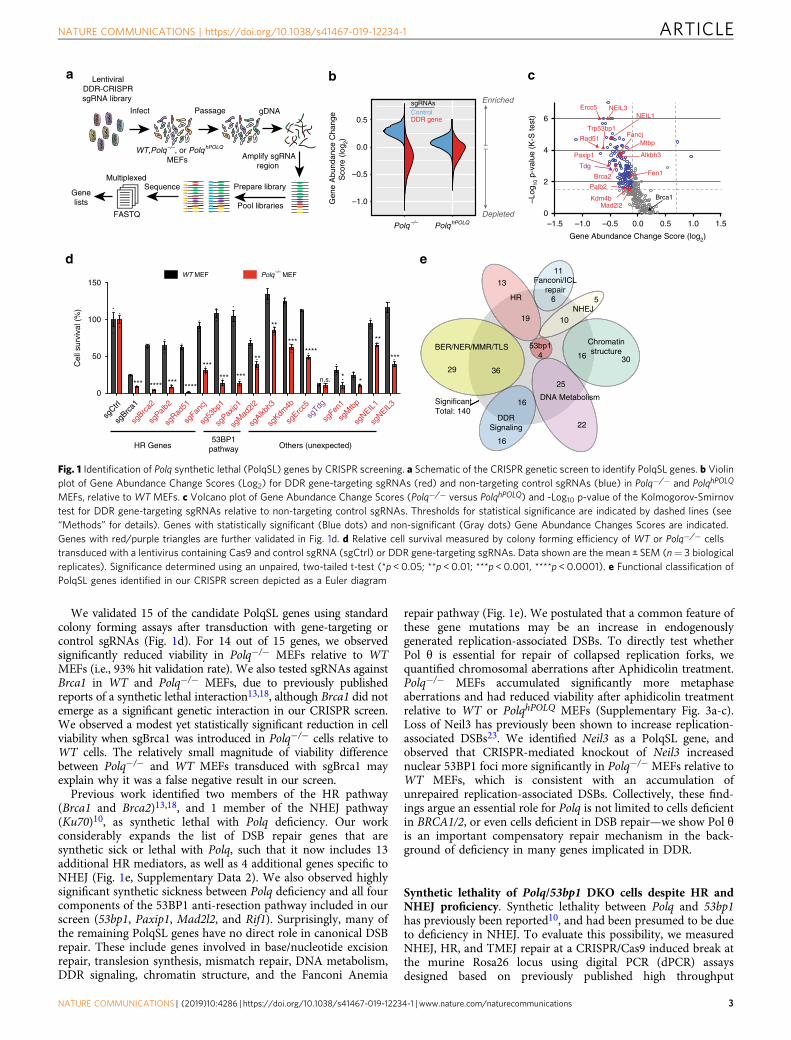
We validated 15 of the candidate PolqSL genes using standardcolony forming assays after transduction with gene-targeting orcontrol sgRNAs (Fig. 1d). For 14 out of 15 genes, we observedsignificantly reduced viability in Polq−/− MEFs relative to WTMEFs (i.e., 93% hit validation rate). We also tested sgRNAs againstBrca1 in WT and Polq−/− MEFs, due to previously publishedreports of a synthetic lethal interaction13,18, although Brca1 did notemerge as a significant genetic interaction in our CRISPR screen.We observed a modest yet statistically significant reduction in cellviability when sgBrca1 was introduced in Polq−/− cells relative toWT cells. The relatively small magnitude of viability differencebetween Polq−/− and WT MEFs transduced with sgBrca1 mayexplain why it was a false negative result in our screen.
Previous work identified two members of the HR pathway(Brca1 and Brca2)13,18, and 1 member of the NHEJ pathway(Ku70)10, as synthetic lethal with Polq deficiency. Our workconsiderably expands the list of DSB repair genes that aresynthetic sick or lethal with Polq, such that it now includes 13additional HR mediators, as well as 4 additional genes specific toNHEJ (Fig. 1e, Supplementary Data 2). We also observed highlysignificant synthetic sickness between Polq deficiency and all fourcomponents of the 53BP1 anti-resection pathway included in ourscreen (53bp1, Paxip1, Mad2l2, and Rif1). Surprisingly, many ofthe remaining PolqSL genes have no direct role in canonical DSBrepair. These include genes involved in base/nucleotide excisionrepair, translesion synthesis, mismatch repair, DNA metabolism,DDR signaling, chromatin structure, and the Fanconi Anemia
repair pathway (Fig. 1e). We postulated that a common feature ofthese gene mutations may be an increase in endogenouslygenerated replication-associated DSBs. To directly test whetherPol θ is essential for repair of collapsed replication forks, wequantified chromosomal aberrations after Aphidicolin treatment.Polq−/− MEFs accumulated significantly more metaphaseaberrations and had reduced viability after aphidicolin treatmentrelative to WT or PolqhPOLQ MEFs (Supplementary Fig. 3a-c).Loss of Neil3 has previously been shown to increase replication-associated DSBs23. We identified Neil3 as a PolqSL gene, andobserved that CRISPR-mediated knockout of Neil3 increasednuclear 53BP1 foci more significantly in Polq−/− MEFs relative toWT MEFs, which is consistent with an accumulation ofunrepaired replication-associated DSBs. Collectively, these find-ings argue an essential role for Polq is not limited to cells deficientin BRCA1/2, or even cells deficient in DSB repair—we show Pol θis an important compensatory repair mechanism in the back-ground of deficiency in many genes implicated in DDR.
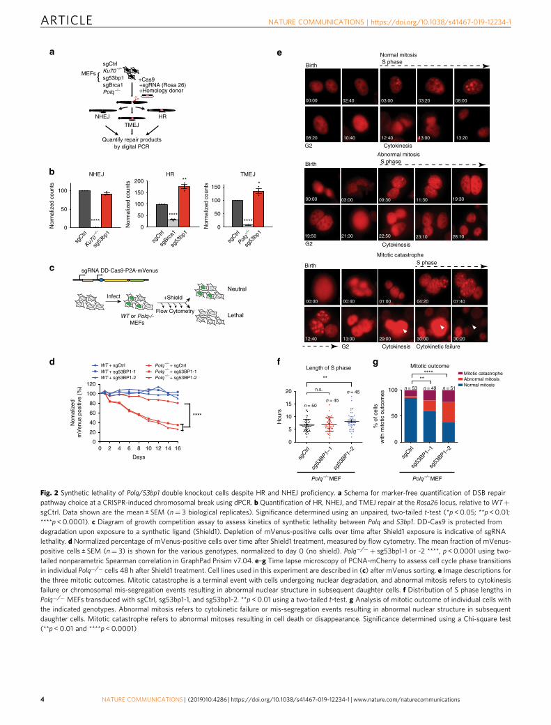
Synthetic lethality of Polq/53bp1 DKO cells despite HR andNHEJ proficiency. Synthetic lethality between Polq and 53bp1has previously been reported10, and had been presumed to be dueto deficiency in NHEJ. To evaluate this possibility, we measuredNHEJ, HR, and TMEJ repair at a CRISPR/Cas9 induced break atthe murine Rosa26 locus using digital PCR (dPCR) assaysdesigned based on previously published high throughput
LentiviralDDR-CRISPRsgRNA library
Infect Passage gDNA
Amplify sgRNAregion
Prepare library
Pool libraries
SequenceMultiplexed
FASTQ
Genelists
a b c
d
–1.5 –1.0 –0.5 0.0 0.5 1.0 1.50
2
4
6
Gene Abundance Change Score (log2)
–Log
10 p
-val
ue (
K-S
test
)
Ercc5
Rad51
Trp53bp1
Paxip1
Brca2
Mtbp
Alkbh3
Fancj
Fen1
Palb2
Kdm4bMad2l2
Brca1
HR
Fanconi/ICLrepair
NHEJ
Chromatinstructure
DNA Metabolism
DDRSignaling
BER/NER/MMR/TLS
SignificantTotal: 140
53bp14
3629
19
13
11
6 5
10
3016
22
25
16
16
0.0
0.5
–0.5
–1.0Gen
e A
bund
ance
Cha
nge
Sco
re (
log 2)
Polq–/– PolqhPOLQ
Enriched
Depleted
DDR geneControlsgRNAs
WT,Polq–/–, or PolqhPOLQ
MEFs
WT MEF Polq–/–
MEF
Tdg
e
NEIL3NEIL1
HR Genes53BP1
pathway Others (unexpected)
sgCtrl
sgBrc
a1
sgBrc
a2
sgPalb
2
sgRad
51
sgFan
cj
sg53
bp1
sgPax
ip1
sgM
ad2l2
sgAlkb
h3
sgKdm
4b
sgErc
c5
sgTdg
sgFen
1
sgM
tbp
sgNEIL
1
sgNEIL
3
Cel
l sur
viva
l (%
)
0
50
100
150
n.s.
Fig. 1 Identification of Polq synthetic lethal (PolqSL) genes by CRISPR screening. a Schematic of the CRISPR genetic screen to identify PolqSL genes. b Violinplot of Gene Abundance Change Scores (Log2) for DDR gene-targeting sgRNAs (red) and non-targeting control sgRNAs (blue) in Polq−/− and PolqhPOLQ
MEFs, relative to WT MEFs. c Volcano plot of Gene Abundance Change Scores (Polq−/− versus PolqhPOLQ) and -Log10 p-value of the Kolmogorov-Smirnovtest for DDR gene-targeting sgRNAs relative to non-targeting control sgRNAs. Thresholds for statistical significance are indicated by dashed lines (see“Methods” for details). Genes with statistically significant (Blue dots) and non-significant (Gray dots) Gene Abundance Changes Scores are indicated.Genes with red/purple triangles are further validated in Fig. 1d. d Relative cell survival measured by colony forming efficiency of WT or Polq−/− cellstransduced with a lentivirus containing Cas9 and control sgRNA (sgCtrl) or DDR gene-targeting sgRNAs. Data shown are the mean ± SEM (n= 3 biologicalreplicates). Significance determined using an unpaired, two-tailed t-test (*p < 0.05; **p < 0.01; ***p < 0.001, ****p < 0.0001). e Functional classification ofPolqSL genes identified in our CRISPR screen depicted as a Euler diagram
NATURE COMMUNICATIONS | https://doi.org/10.1038/s41467-019-12234-1 ARTICLE
NATURE COMMUNICATIONS | (2019) 10:4286 | https://doi.org/10.1038/s41467-019-12234-1 | www.nature.com/naturecommunications 3
c
a
b
d f
0 2 4 6 8 10 12 14 160
20
40
60
80
100
120
Days
Nom
aliz
ed
mV
enus
pos
itive
(%
)
WT + sgCtrlWT + sg53BP1-1WT + sg53BP1-2
Polq–/–
+ sgCtrlPolq
–/– + sg53BP1-1
Polq–/–
+ sg53BP1-2
e
sgCtrl
Ku70–/
–
sg53
bp1
****
sgCtrl
Polq–/
–
sg53
bp1
****
*
sgCtrl
sgBrc
a1
sg53
bp1
****
**
MEFs
sgCtrlKu70 –/–
sg53bp1sgBrca1Polq –/–
+Cas9+sgRNA (Rosa 26)+Homology donor
Quantify repair productsby digital PCR
NHEJ
TMEJ
HR
0
5
10
15
20
sg53
BP1–1
sg53
BP1–2
sgCtrl
Length of S phase
n.s.
Hou
rs
n = 50n = 45
n = 45
0
50
100
Mitotic outcome
Normal mitosisAbnormal mitosisMitotic catastrophe
% o
f cel
lsw
ith m
itotic
out
com
es n = 53 n = 49 n = 51
sg53
BP1–1
sg53
BP1–2
sgCtrl
Polq –/– MEFPolq –/– MEF
sgRNA DD-Cas9-P2A-mVenus
Infect +Shield
Flow Cytometry
Neutral
LethalWT or Polq-/-MEFs
Normal mitosis
Birth S phase
G2 Cytokinesis
00:00 03:00 03:20 08:00
08:20 10:40 12:40 13:00 13:20
02:40
g
00:00 03:00 09:30 11:30 19:30
19:50 21:30 22:50 23:10 28:10
00:00 00:40 01:00 04:20 07:40
12:40 13:00 29:00 30:00 30:20
Birth S phase
G2 Cytokinesis
Abnormal mitosis
Birth S phase
G2 Cytokinetic failure Cytokinesis
Mitotic catastrophe
0
50
100
150
Nor
mal
ized
cou
nts
TMEJ
0
50
100
Nor
mal
ized
cou
nts
NHEJ
0
50
100
150
200
Nor
mal
ized
cou
nts
HR
Fig. 2 Synthetic lethality of Polq/53bp1 double knockout cells despite HR and NHEJ proficiency. a Schema for marker-free quantification of DSB repairpathway choice at a CRISPR-induced chromosomal break using dPCR. b Quantification of HR, NHEJ, and TMEJ repair at the Rosa26 locus, relative toWT+sgCtrl. Data shown are the mean ± SEM (n= 3 biological replicates). Significance determined using an unpaired, two-tailed t-test (*p < 0.05; **p < 0.01;****p < 0.0001). c Diagram of growth competition assay to assess kinetics of synthetic lethality between Polq and 53bp1. DD-Cas9 is protected fromdegradation upon exposure to a synthetic ligand (Shield1). Depletion of mVenus-positive cells over time after Shield1 exposure is indicative of sgRNAlethality. d Normalized percentage of mVenus-positive cells over time after Shield1 treatment, measured by flow cytometry. The mean fraction of mVenus-positive cells ± SEM (n= 3) is shown for the various genotypes, normalized to day 0 (no shield). Polq−/−+ sg53bp1-1 or -2 ****, p < 0.0001 using two-tailed nonparametric Spearman correlation in GraphPad Prisim v7.04. e–g Time lapse microscopy of PCNA-mCherry to assess cell cycle phase transitionsin individual Polq−/− cells 48 h after Shield1 treatment. Cell lines used in this experiment are described in (c) after mVenus sorting. e Image descriptions forthe three mitotic outcomes. Mitotic catastrophe is a terminal event with cells undergoing nuclear degradation, and abnormal mitosis refers to cytokinesisfailure or chromosomal mis-segregation events resulting in abnormal nuclear structure in subsequent daughter cells. f Distribution of S phase lengths inPolq−/− MEFs transduced with sgCtrl, sg53bp1-1, and sg53bp1-2. **p < 0.01 using a two-tailed t-test. g Analysis of mitotic outcome of individual cells withthe indicated genotypes. Abnormal mitosis refers to cytokinetic failure or mis-segregation events resulting in abnormal nuclear structure in subsequentdaughter cells. Mitotic catastrophe refers to abnormal mitoses resulting in cell death or disappearance. Significance determined using a Chi-square test(**p < 0.01 and ****p < 0.0001)
ARTICLE NATURE COMMUNICATIONS | https://doi.org/10.1038/s41467-019-12234-1
4 NATURE COMMUNICATIONS | (2019) 10:4286 | https://doi.org/10.1038/s41467-019-12234-1 | www.nature.com/naturecommunications
sequencing analyses at this locus10 (Fig. 2a). 53bp1 deficiencyresulted in a nonsignificant reduction in NHEJ and increasedfrequencies of both HR and TMEJ repair (Fig. 2b), consistentwith previously described roles for 53bp1 in DSB repair pathwaychoice24. Based on these observations, NHEJ deficiency cannotexplain synthetic lethality between 53bp1 and Polq. However, itwas also surprising that HR could not compensate for TMEJdeficiency in 53bp1/Polq DKO cells, given that they both act onresected DSBs. We therefore investigated the synthetic lethalphenotype in greater detail.
To assess kinetics of synthetic lethality between 53bp1 and Polq,we utilized an inducible Cas9 expression system (DD-Cas925), andmonitored the relative growth rate of transduced cells by flowcytometry over time (Fig. 2c). Interestingly, although 53BP1expression was already diminished by 48 h after Shield1 treatment(Supplementary Fig. 4), the growth disadvantage of Polq−/−+sg53bp1 (i.e., Polq/53bp1 double knockout, DKO) cells persistedover at least 14 days (Fig. 2d). Time lapse microscopy usingPCNA-mCherry as a fluorescent cell cycle reporter26 revealed astatistically significant prolongation of S phase duration with oneout of two 53bp1-targeting sgRNAs in Polq−/− cells (Fig. 2f),although G1 and G2/M duration did not differ significantly for anyof the genotypes (Supplementary Fig. 5). More strikingly, there wasa significantly higher rate of aberrant mitoses (improperchromosomal segregation or abnormal cytokinesis) and mitoticcatastrophe when either of the 53bp1 targeting sgRNAs wasexpressed in Polq−/− cells (Fig. 2e, g).
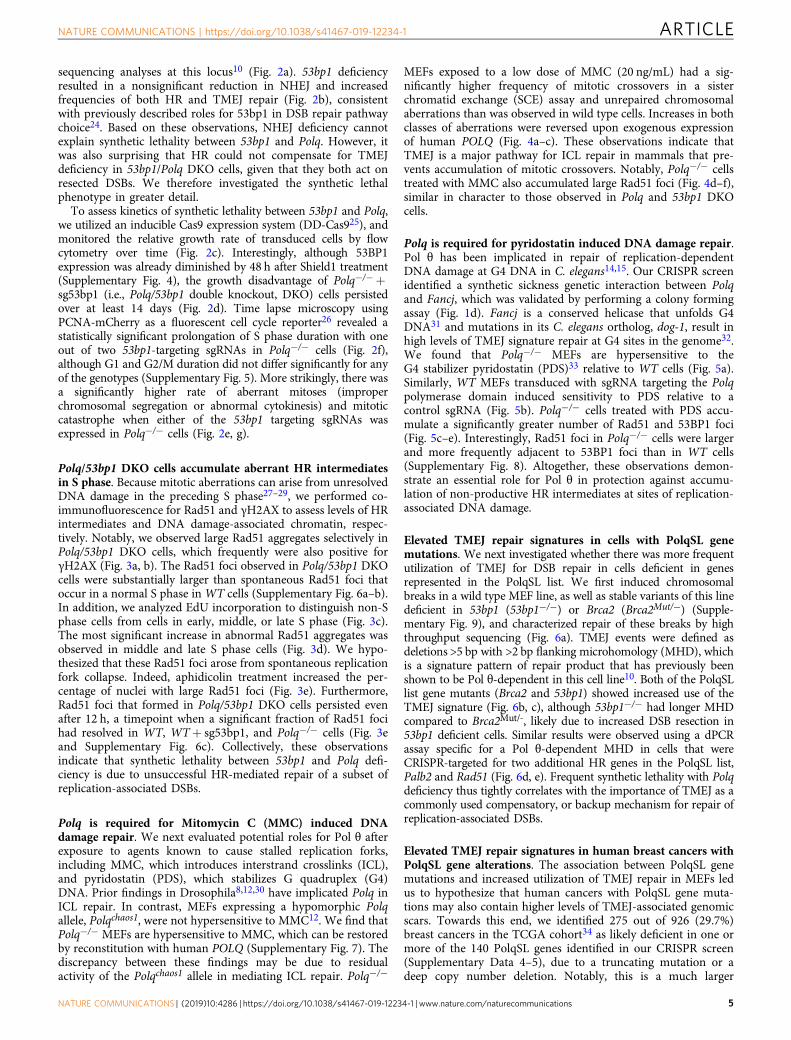
Polq/53bp1 DKO cells accumulate aberrant HR intermediatesin S phase. Because mitotic aberrations can arise from unresolvedDNA damage in the preceding S phase27–29, we performed co-immunofluorescence for Rad51 and γH2AX to assess levels of HRintermediates and DNA damage-associated chromatin, respec-tively. Notably, we observed large Rad51 aggregates selectively inPolq/53bp1 DKO cells, which frequently were also positive forγH2AX (Fig. 3a, b). The Rad51 foci observed in Polq/53bp1 DKOcells were substantially larger than spontaneous Rad51 foci thatoccur in a normal S phase inWT cells (Supplementary Fig. 6a–b).In addition, we analyzed EdU incorporation to distinguish non-Sphase cells from cells in early, middle, or late S phase (Fig. 3c).The most significant increase in abnormal Rad51 aggregates wasobserved in middle and late S phase cells (Fig. 3d). We hypo-thesized that these Rad51 foci arose from spontaneous replicationfork collapse. Indeed, aphidicolin treatment increased the per-centage of nuclei with large Rad51 foci (Fig. 3e). Furthermore,Rad51 foci that formed in Polq/53bp1 DKO cells persisted evenafter 12 h, a timepoint when a significant fraction of Rad51 focihad resolved in WT, WT+ sg53bp1, and Polq−/− cells (Fig. 3eand Supplementary Fig. 6c). Collectively, these observationsindicate that synthetic lethality between 53bp1 and Polq defi-ciency is due to unsuccessful HR-mediated repair of a subset ofreplication-associated DSBs.
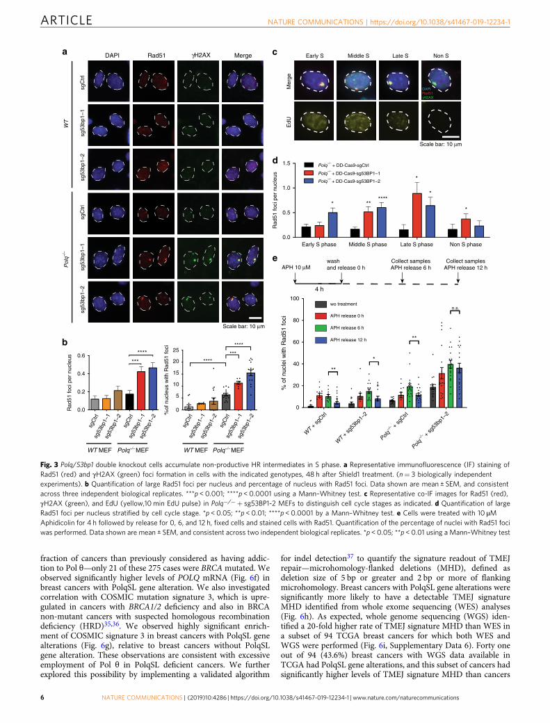
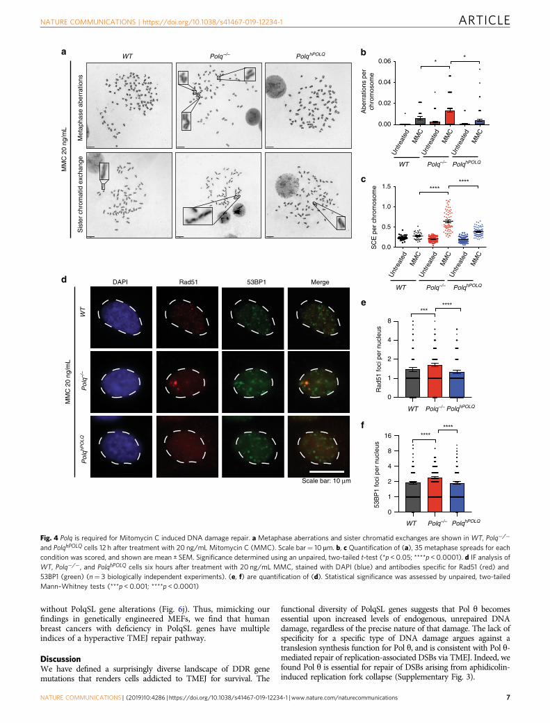
Polq is required for Mitomycin C (MMC) induced DNAdamage repair. We next evaluated potential roles for Pol θ afterexposure to agents known to cause stalled replication forks,including MMC, which introduces interstrand crosslinks (ICL),and pyridostatin (PDS), which stabilizes G quadruplex (G4)DNA. Prior findings in Drosophila8,12,30 have implicated Polq inICL repair. In contrast, MEFs expressing a hypomorphic Polqallele, Polqchaos1, were not hypersensitive to MMC12. We find thatPolq−/− MEFs are hypersensitive to MMC, which can be restoredby reconstitution with human POLQ (Supplementary Fig. 7). Thediscrepancy between these findings may be due to residualactivity of the Polqchaos1 allele in mediating ICL repair. Polq−/−
MEFs exposed to a low dose of MMC (20 ng/mL) had a sig-nificantly higher frequency of mitotic crossovers in a sisterchromatid exchange (SCE) assay and unrepaired chromosomalaberrations than was observed in wild type cells. Increases in bothclasses of aberrations were reversed upon exogenous expressionof human POLQ (Fig. 4a–c). These observations indicate thatTMEJ is a major pathway for ICL repair in mammals that pre-vents accumulation of mitotic crossovers. Notably, Polq−/− cellstreated with MMC also accumulated large Rad51 foci (Fig. 4d–f),similar in character to those observed in Polq and 53bp1 DKOcells.
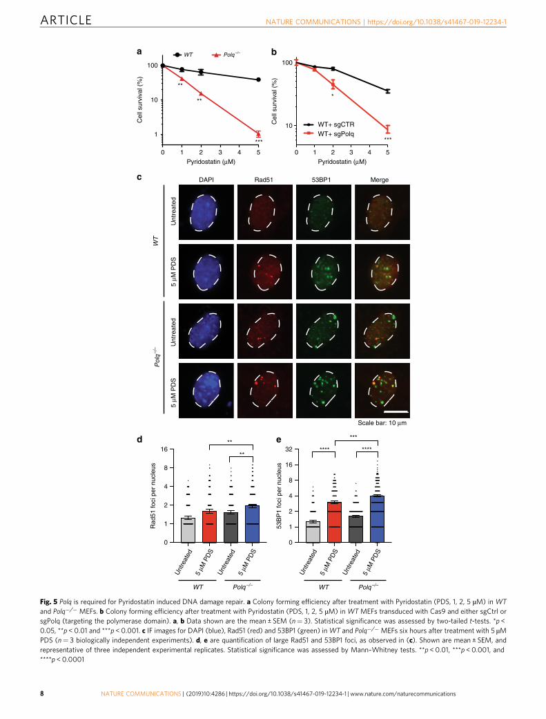
Polq is required for pyridostatin induced DNA damage repair.Pol θ has been implicated in repair of replication-dependentDNA damage at G4 DNA in C. elegans14,15. Our CRISPR screenidentified a synthetic sickness genetic interaction between Polqand Fancj, which was validated by performing a colony formingassay (Fig. 1d). Fancj is a conserved helicase that unfolds G4DNA31 and mutations in its C. elegans ortholog, dog-1, result inhigh levels of TMEJ signature repair at G4 sites in the genome32.We found that Polq−/− MEFs are hypersensitive to theG4 stabilizer pyridostatin (PDS)33 relative to WT cells (Fig. 5a).Similarly, WT MEFs transduced with sgRNA targeting the Polqpolymerase domain induced sensitivity to PDS relative to acontrol sgRNA (Fig. 5b). Polq−/− cells treated with PDS accu-mulate a significantly greater number of Rad51 and 53BP1 foci(Fig. 5c–e). Interestingly, Rad51 foci in Polq−/− cells were largerand more frequently adjacent to 53BP1 foci than in WT cells(Supplementary Fig. 8). Altogether, these observations demon-strate an essential role for Pol θ in protection against accumu-lation of non-productive HR intermediates at sites of replication-associated DNA damage.
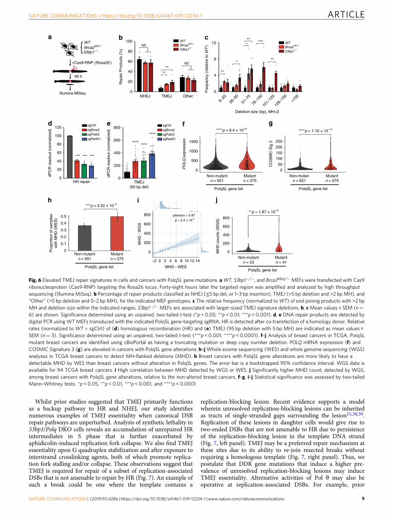
Elevated TMEJ repair signatures in cells with PolqSL genemutations. We next investigated whether there was more frequentutilization of TMEJ for DSB repair in cells deficient in genesrepresented in the PolqSL list. We first induced chromosomalbreaks in a wild type MEF line, as well as stable variants of this linedeficient in 53bp1 (53bp1−/−) or Brca2 (Brca2Mut/−) (Supple-mentary Fig. 9), and characterized repair of these breaks by highthroughput sequencing (Fig. 6a). TMEJ events were defined asdeletions >5 bp with >2 bp flanking microhomology (MHD), whichis a signature pattern of repair product that has previously beenshown to be Pol θ-dependent in this cell line10. Both of the PolqSLlist gene mutants (Brca2 and 53bp1) showed increased use of theTMEJ signature (Fig. 6b, c), although 53bp1−/− had longer MHDcompared to Brca2Mut/-, likely due to increased DSB resection in53bp1 deficient cells. Similar results were observed using a dPCRassay specific for a Pol θ-dependent MHD in cells that wereCRISPR-targeted for two additional HR genes in the PolqSL list,Palb2 and Rad51 (Fig. 6d, e). Frequent synthetic lethality with Polqdeficiency thus tightly correlates with the importance of TMEJ as acommonly used compensatory, or backup mechanism for repair ofreplication-associated DSBs.
Elevated TMEJ repair signatures in human breast cancers withPolqSL gene alterations. The association between PolqSL genemutations and increased utilization of TMEJ repair in MEFs ledus to hypothesize that human cancers with PolqSL gene muta-tions may also contain higher levels of TMEJ-associated genomicscars. Towards this end, we identified 275 out of 926 (29.7%)breast cancers in the TCGA cohort34 as likely deficient in one ormore of the 140 PolqSL genes identified in our CRISPR screen(Supplementary Data 4–5), due to a truncating mutation or adeep copy number deletion. Notably, this is a much larger
NATURE COMMUNICATIONS | https://doi.org/10.1038/s41467-019-12234-1 ARTICLE
NATURE COMMUNICATIONS | (2019) 10:4286 | https://doi.org/10.1038/s41467-019-12234-1 | www.nature.com/naturecommunications 5
fraction of cancers than previously considered as having addic-tion to Pol θ—only 21 of these 275 cases were BRCAmutated. Weobserved significantly higher levels of POLQ mRNA (Fig. 6f) inbreast cancers with PolqSL gene alteration. We also investigatedcorrelation with COSMIC mutation signature 3, which is upre-gulated in cancers with BRCA1/2 deficiency and also in BRCAnon-mutant cancers with suspected homologous recombinationdeficiency (HRD)35,36. We observed highly significant enrich-ment of COSMIC signature 3 in breast cancers with PolqSL genealterations (Fig. 6g), relative to breast cancers without PolqSLgene alteration. These observations are consistent with excessiveemployment of Pol θ in PolqSL deficient cancers. We furtherexplored this possibility by implementing a validated algorithm
for indel detection37 to quantify the signature readout of TMEJrepair—microhomology-flanked deletions (MHD), defined asdeletion size of 5 bp or greater and 2 bp or more of flankingmicrohomology. Breast cancers with PolqSL gene alterations weresignificantly more likely to have a detectable TMEJ signatureMHD identified from whole exome sequencing (WES) analyses(Fig. 6h). As expected, whole genome sequencing (WGS) iden-tified a 20-fold higher rate of TMEJ signature MHD than WES ina subset of 94 TCGA breast cancers for which both WES andWGS were performed (Fig. 6i, Supplementary Data 6). Forty oneout of 94 (43.6%) breast cancers with WGS data available inTCGA had PolqSL gene alterations, and this subset of cancers hadsignificantly higher levels of TMEJ signature MHD than cancers
APH release 12 h
APH release 0 h
wo treatment
APH release 6 h
Pol
q–/–
WT
sgC
trl
sg53
bp1–
1sg
53bp
1–2
sgC
trl
sg53
bp1–
1sg
53bp
1–2
DAPI Rad51 γH2AX Mergea
b
Early S Middle S Late S Non Sc
Mer
geE
dU
DAPIRad51γH2AX
d
Scale bar: 10 μm
Scale bar: 10 μm
APH 10 μM
4 h
washand release 0 h
Collect samplesAPH release 6 h
Collect samplesAPH release 12 h
e
0
20
40
60
80
100
n.s.
% o
f nuc
lei w
ith R
ad51
foci
WT +
sgCtrl
WT +
sg53
bp1–
2
Polq–/
– + sg
Ctrl
Polq
–/– +
sg53
bp1–
2
sgC
trlsg
53bp
1–1
sg53
bp1–
2sg
Ctrl
sg53
bp1–
1sg
53bp
1–2
0
5
10
15
20
25
%of
nuc
leus
with
Rad
51 fo
ci
Early S phase Middle S phase Late S phase Non S phase0.0
0.5
1.0
1.5
Rad
51 fo
ci p
er n
ucle
us
Polq–/–
+ DD-Cas9-sgCtrl
Polq–/–
+ DD-Cas9-sg53BP1–1
Polq–/–
+ DD-Cas9-sg53BP1–2
Rad
51 fo
ci p
er n
ucle
us
WT MEF Polq–/– MEFPolq–/–
MEF WT MEF
sgC
trlsg
53bp
1–1
sg53
bp1–
2sg
Ctrl
sg53
bp1–
1sg
53bp
1–2
0.0
0.2
0.4
0.6
Fig. 3 Polq/53bp1 double knockout cells accumulate non-productive HR intermediates in S phase. a Representative immunofluorescence (IF) staining ofRad51 (red) and γH2AX (green) foci formation in cells with the indicated genotypes, 48 h after Shield1 treatment. (n= 3 biologically independentexperiments). b Quantification of large Rad51 foci per nucleus and percentage of nucleus with Rad51 foci. Data shown are mean ± SEM, and consistentacross three independent biological replicates. ***p < 0.001; ****p < 0.0001 using a Mann–Whitney test. c Representative co-IF images for Rad51 (red),γH2AX (green), and EdU (yellow,10min EdU pulse) in Polq−/−+ sg53BP1-2 MEFs to distinguish cell cycle stages as indicated. d Quantification of largeRad51 foci per nucleus stratified by cell cycle stage. *p < 0.05; **p < 0.01; ****p < 0.0001 by a Mann–Whitney test. e Cells were treated with 10 µMAphidicolin for 4 h followed by release for 0, 6, and 12 h, fixed cells and stained cells with Rad51. Quantification of the percentage of nuclei with Rad51 fociwas performed. Data shown are mean ± SEM, and consistent across two independent biological replicates. *p < 0.05; **p < 0.01 using a Mann–Whitney test
ARTICLE NATURE COMMUNICATIONS | https://doi.org/10.1038/s41467-019-12234-1
6 NATURE COMMUNICATIONS | (2019) 10:4286 | https://doi.org/10.1038/s41467-019-12234-1 | www.nature.com/naturecommunications
without PolqSL gene alterations (Fig. 6j). Thus, mimicking ourfindings in genetically engineered MEFs, we find that humanbreast cancers with deficiency in PolqSL genes have multipleindices of a hyperactive TMEJ repair pathway.
DiscussionWe have defined a surprisingly diverse landscape of DDR genemutations that renders cells addicted to TMEJ for survival. The
functional diversity of PolqSL genes suggests that Pol θ becomesessential upon increased levels of endogenous, unrepaired DNAdamage, regardless of the precise nature of that damage. The lack ofspecificity for a specific type of DNA damage argues against atranslesion synthesis function for Pol θ, and is consistent with Pol θ-mediated repair of replication-associated DSBs via TMEJ. Indeed, wefound Pol θ is essential for repair of DSBs arising from aphidicolin-induced replication fork collapse (Supplementary Fig. 3).
WTa b
c
Met
apha
se a
berr
atio
nsS
iste
r ch
rom
atid
exc
hang
e
MM
C 2
0 ng
/mL
0.0
0.5
1.0
1.5
SC
E p
er c
hrom
osom
e
Abe
rrat
ions
per
chro
mos
ome
MM
C
Unt
reat
ed
MM
C
Unt
reat
ed
MM
C
Unt
reat
ed
WT PolqhPOLQ
PolqhPOLQ
PolqhPOLQ
PolqhPOLQ
53B
P1
foci
per
nuc
leus
d DAPI Rad51 53BP1 Merge
WT
MM
C 2
0 ng
/mL
e
f
Scale bar: 10 μm
Rad
51 fo
ci p
er n
ucle
us
0.00
0.02
0.04
0.06
MM
C
Unt
reat
ed
MM
C
Unt
reat
ed
MM
C
Unt
reat
ed
WT
0
1
2
4
8
0
1
2
4
8
16
WT
WT Polq–/–
Polq–/–
Polq–/–
Pol
qhPO
LQP
olq–/
–
Polq–/–
PolqhPOLQPolq–/–
Fig. 4 Polq is required for Mitomycin C induced DNA damage repair. a Metaphase aberrations and sister chromatid exchanges are shown in WT, Polq−/−
and PolqhPOLQ cells 12 h after treatment with 20 ng/mL Mitomycin C (MMC). Scale bar= 10 µm. b, c Quantification of (a), 35 metaphase spreads for eachcondition was scored, and shown are mean ± SEM. Significance determined using an unpaired, two-tailed t-test (*p < 0.05; ****p < 0.0001). d IF analysis ofWT, Polq−/−, and PolqhPOLQ cells six hours after treatment with 20 ng/mL MMC, stained with DAPI (blue) and antibodies specific for Rad51 (red) and53BP1 (green) (n= 3 biologically independent experiments). (e, f) are quantification of (d). Statistical significance was assessed by unpaired, two-tailedMann–Whitney tests (***p < 0.001; ****p < 0.0001)
NATURE COMMUNICATIONS | https://doi.org/10.1038/s41467-019-12234-1 ARTICLE
NATURE COMMUNICATIONS | (2019) 10:4286 | https://doi.org/10.1038/s41467-019-12234-1 | www.nature.com/naturecommunications 7
c
d
Rad
51 fo
ci p
er n
ucle
us
DAPI Rad51 53BP1 MergeU
ntre
ated
5 μM
PD
SU
ntre
ated
5 μM
PD
S
1
10
100
a WT Polq–/–
Cel
l sur
viva
l (%
)
Pyridostatin (μM)
0 1 2 3 4 5
10
100
Cel
l sur
viva
l (%
)
WT+ sgCTRWT+ sgPolq
Pyridostatin (μM)
b
0 1 2 3 4 5
Scale bar: 10 μm
e
WT Polq–/–
WT
Pol
q–/
–
WT Polq–/–
5 μM
PD
S
Unt
reat
ed5
μM P
DS
Unt
reat
ed
5 μM
PD
S
Unt
reat
ed5
μM P
DS
Unt
reat
ed
53B
P1
foci
per
nuc
leus
0
1
2
4
8
16
0
1
2
4
8
16
32
Fig. 5 Polq is required for Pyridostatin induced DNA damage repair. a Colony forming efficiency after treatment with Pyridostatin (PDS, 1, 2, 5 µM) in WTand Polq−/− MEFs. b Colony forming efficiency after treatment with Pyridostatin (PDS, 1, 2, 5 µM) in WT MEFs transduced with Cas9 and either sgCtrl orsgPolq (targeting the polymerase domain). a, b Data shown are the mean ± SEM (n= 3). Statistical significance was assessed by two-tailed t-tests. *p <0.05, **p < 0.01 and ***p < 0.001. c IF images for DAPI (blue), Rad51 (red) and 53BP1 (green) inWT and Polq−/− MEFs six hours after treatment with 5 µMPDS (n= 3 biologically independent experiments). d, e are quantification of large Rad51 and 53BP1 foci, as observed in (c). Shown are mean ± SEM, andrepresentative of three independent experimental replicates. Statistical significance was assessed by Mann–Whitney tests. **p < 0.01, ***p < 0.001, and****p < 0.0001
ARTICLE NATURE COMMUNICATIONS | https://doi.org/10.1038/s41467-019-12234-1
8 NATURE COMMUNICATIONS | (2019) 10:4286 | https://doi.org/10.1038/s41467-019-12234-1 | www.nature.com/naturecommunications
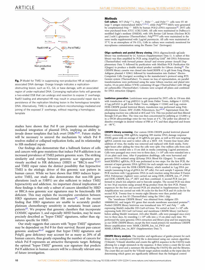
Whilst prior studies suggested that TMEJ primarily functionsas a backup pathway to HR and NHEJ, our study identifiesnumerous examples of TMEJ essentiality when canonical DSBrepair pathways are unperturbed. Analysis of synthetic lethality in53bp1/Polq DKO cells reveals an accumulation of unrepaired HRintermediates in S phase that is further exacerbated byaphidicolin-induced replication fork collapse. We also find TMEJessentiality upon G quadruplex stabilization and after exposure tointerstrand crosslinking agents, both of which promote replica-tion fork stalling and/or collapse. These observations suggest thatTMEJ is required for repair of a subset of replication-associatedDSBs that is not amenable to repair by HR (Fig. 7). An example ofsuch a break could be one where the template contains a
replication-blocking lesion. Recent evidence supports a modelwherein unresolved replication-blocking lesions can be inheritedas tracts of single-stranded gaps surrounding the lesion15,38,39.Replication of these lesions in daughter cells would give rise totwo-ended DSBs that are not amenable to HR due to persistenceof the replication-blocking lesion in the template DNA strand(Fig. 7, left panel). TMEJ may be a preferred repair mechanism atthese sites due to its ability to re-join resected breaks withoutrequiring a homologous template (Fig. 7, right panel). Thus, wepostulate that DDR gene mutations that induce a higher pre-valence of unresolved replication-blocking lesions may induceTMEJ essentiality. Alternative activities of Pol θ may also beoperative at replication-associated DSBs. For example, prior
NS
NS
NHEJ TMEJ Other
Rep
air
Pro
duct
s (%
)
a b c
d f g
h i j
e
Deletion size (bp), MH>2
Fre
quen
cy (
rela
tive
to W
T)
WTBrca2
Mut/–
53bp1–/–
WTBrca2
Mut/–
53bp1–/–
Brca2Mut/–
53bp1–/–
6–25
26–5
0
51–7
5
76–1
00
101–
125
126–
150
>150
PolqSL gene list
MH
D c
ount
s (W
GS
)**p = 1.87 × 10⁻³
400
200
800
600
0
MH
D -
WG
S
MHD - WES
–2
0
200
400
600
800
0 2 4 6 8 10 12 14
pearson = 0.47
p = 2.3 × 10⁻⁶
+Cas9-RNP (Rosa26 )
48 h
Illumina MiSeq
WT
dPC
R r
eado
ut (
norm
aliz
ed)
HR repair
0
20
40
60
80
100
0
4
8
12
0
20
40
60
80
100
120
0
200
400
600
800
dPC
R r
eado
ut (
norm
aliz
ed)
sgRad51
sgCtrlsgBrca2sgPalb2
sgRad51
sgCtrlsgBrca2sgPalb2
TMEJ(95 bp del)
PO
LQ e
xpre
ssio
n
****p = 9.4 × 10⁻⁸
PolqSL gene list
500
1000
1500
0
Non-mutann = 651
Mutantn = 275
Non-mutantn = 53
Mutantn = 41
Non-mutantn = 651
Mutantn = 275
PolqSL gene list
CO
SM
IC S
ig 3
****p = 1.10 × 10⁻¹¹
200
250
150
100
50
0
***p = 4.22 × 10⁻⁴
Non-mutantn = 651
Mutantn = 275
PolqSL gene list
Pro
port
ion
of s
ampl
esw
ith M
HD
(W
ES
)
0.2
0.1
0.3
0.4
0.5
0
Fig. 6 Elevated TMEJ repair signatures in cells and cancers with PolqSL gene mutations. aWT, 53bp1−/−, and Brca2Mut/− MEFs were transfected with Cas9ribonucleoprotein (Cas9-RNP) targeting the Rosa26 locus. Forty-eight hours later the targeted region was amplified and analyzed by high throughputsequencing (Illumina MiSeq). b Percentage of repair products classified as NHEJ (≤5 bp del, or 1–3 bp insertion), TMEJ (>5 bp deletion and >2 bp MH), and“Other” (>5 bp deletion and 0–2 bp MH), for the indicated MEF genotypes. c The relative frequency (normalized toWT) of end joining products with >2 bpMH and deletion size within the indicated ranges. 53bp1−/− MEFs are associated with larger-sized TMEJ signature deletions. b, c Mean values ± SEM (n=6) are shown. Significance determined using an unpaired, two-tailed t-test (*p < 0.05; **p < 0.01; ***p < 0.001). d, e DNA repair products are detected bydigital PCR using WT MEFs transduced with the indicated PolqSL gene-targeting sgRNA. HR is detected after co-transfection of a homology donor. Relativerates (normalized to WT+ sgCtrl) of (d) homologous recombination (HR) and (e) TMEJ (95 bp deletion with 5 bp MH) are indicated as mean values ±SEM (n= 3). Significance determined using an unpaired, two-tailed t-test (***p < 0.001; ****p < 0.0001). f–j Analysis of breast cancers in TCGA. PolqSLmutant breast cancers are identified using cBioPortal as having a truncating mutation or deep copy number deletion. POLQ mRNA expression (f) andCOSMIC Signature 3 (g) are elevated in cancers with PolqSL gene alterations. h–j Whole exome sequencing (WES) and whole genome sequencing (WGS)analyses in TCGA breast cancers to detect MH-flanked deletions (MHD). h Breast cancers with PolqSL gene alterations are more likely to have adetectable MHD by WES than breast cancers without alteration in PolqSL genes. The error bar is a bootstrapped 95% confidence interval. WGS data isavailable for 94 TCGA breast cancers. i High correlation between MHD detected by WGS or WES. j Significantly higher MHD count, detected by WGS,among breast cancers with PolqSL gene alterations, relative to the non-altered breast cancers. f–g, i–j Statistical significance was assessed by two-tailedMann–Whitney tests. *p < 0.05, **p < 0.01, ***p < 0.001, and ****p < 0.0001
NATURE COMMUNICATIONS | https://doi.org/10.1038/s41467-019-12234-1 ARTICLE
NATURE COMMUNICATIONS | (2019) 10:4286 | https://doi.org/10.1038/s41467-019-12234-1 | www.nature.com/naturecommunications 9
studies have shown that Pol θ can promote microhomology-mediated integration of plasmid DNA, implying an ability toinvade donor templates that lack overt DSBs40,41. Future studieswill be necessary to unravel the mechanism by which Pol θresolves stalled or collapsed replication forks, and its relationshipto HR-mediated repair.
Our findings also demonstrate that a hallmark feature of cellsand cancers with gene mutations that induce Pol θ addiction is anincreased prevalence of TMEJ pattern genomic scars. The strikingsimilarity and overlap between genomic scar signatures pre-viously ascribed to HR deficiency (HRD) or “BRCA-ness”42,43
and TMEJ repair raises the distinct possibility that hyperactiveTMEJ is the etiologic driver of this genomic scar pattern inhuman cancer. While we have shown that HRD induces hyper-active TMEJ, our study also demonstrates that non-HR genealterations (such as 53BP1) are also sufficient to induce TMEJhyperactivity and addiction. An important clinical implication ofthese findings is that only a subset of cancers identified by HRDor BRCA-ness genomic scar signatures may be functionally HRdeficient. This may explain the incomplete correlation betweenHRD signatures and functional HR assays44, and the recentfinding that HRD signatures are unable to accurately predictplatinum chemotherapy sensitivity in metastatic breast cancerpatients45. We propose that genomic classifiers that incorporateCOSMIC signature 3, and especially MHD burden, may be moreprecisely described as “hyper-TMEJ” signatures, rather than sig-natures specific for HRD.
Our study suggests that cancers with “hyper-TMEJ” signaturesmay be dependent on Pol θ for their survival. Recent pan-cancergenomic analyses46,47 suggest that hyper-TMEJ signatures andPolqSL gene deficiency may account for as many as 20% of allhuman cancers, thus greatly expanding the number of cancers forwhich Pol θ represents an attractive therapeutic target. Refiningthe optimal “hyper-TMEJ” genomic scar signature that predictsPol θ addiction in human cancers will be a clinically relevant areaof future investigation.
MethodsCell culture. WT (Polq+/+), Polq−/−, Polm−/−, and Polm+/+ cells were SV-40large T antigen immortalized MEFs12,20,22, while PolqhPOLQ MEFs were generatedby complemented Polq−/− MEFs by human POLQ cDNA expression20. 293T cellswere purchased from ATCC (CRL11268). All cells were maintained in Dulbecco’smodified Eagle’s medium (DMEM), with 10% Bovine Calf Serum (Hyclone BCS)and 2 mM L-glutamine (ThermoFisher). PolqhPOLQ cells were maintained in thesame media supplemented with 2 μg/ml puromycin. All cells were maintained at37 °C in an atmosphere of 5% CO2. Cells in culture were routinely monitored formycoplasma contamination using the Plasmo Test™ (Invivogen).
Oligo synthesis and pooled library cloning. DNA oligonucleotide sgGuidelibrary was synthesized by LC Sciences (Supplementary Data 1). A subset of thislibrary was then amplified by PCR using AmpliTaq Gold® 360 DNA Polymerase(ThermoFisher) with forward primer ArrayF and reverse primer ArrayR (Sup-plementary Data 7) followed by purification with MinElute PCR Purification Kit(Qiagen) to produce a double strand product suitable for Gibson cloning48. TheCRISPR library cassette was cloned into lentiCRISPR v2 (a gift from Feng Zhang,Addgene plasmid # 52961) followed by transformation into Endura™ Electro-Competent Cells (Lucigen) according to the manufacturer’s protocol using BTXGemini system (ThermoFisher). To ensure no loss of representation, six paralleltransformations were performed using the same Gibson reaction and plated intotwelve, 10 cm petri dishes (VWR) containing LB agar (ThermoFisher) with 100 µg/ml carbenicillin (ThermoFisher). Colonies were scraped off plates and combinedfor DNA extraction (Qiagen).
Lentivirus generation. Lentiviruses were generated by 293T cells in 150 mm dishwith transfection of 3 µg pMD2.G (a gift from Didier Trono, Addgene # 12259),4.5 µg psPAX2 (a gift from Didier Trono, Addgene # 12260) and 6 µg customDDR-CRISPR pooled lentiviral library, transfection was performed using Poly-ethylenimine (Linear, MW 25,000, Polysciences, Inc)49,50. Supernatant from thepackaging reaction was collected at 48 h and 72 h. This was pooled and then filteredthrough 0.45 μm filter. The virus was then concentrated by pelleting at 113,000 × gin a SW28 ultracentrifuge rotor for two hours at 4 °C. The pellet was allowed todissolve overnight in desired volume of PBS at 4 °C and then aliquoted and frozenat −80 °C.
CRISPR library screening. Our custom DDR-CRISPR pooled lentiviral plasmidlibrary containing 3908 sgRNAs targeting 309 murine DNA damage response(DDR) genes with an average of 10 sgRNAs per gene, as well as 834 non-targetingsgRNA controls was used to infect cells at a MOI ~0.8. Twenty-four hours afteraddition of virus, the media was removed and replaced with fresh media. Forty-eight hours after adding the virus the cells were split. One million cells from eachinfection was seeded into a 15 cm dish in media containing 2 μg/ml puromycin.Cells were passaged once every two to three days, 1 × 10⁶ cells were reseeded into a15 cm plate each time. After 8 population doublings, cells were harvested andgenomic DNA isolated using QIAamp DNA Blood Kit (Qiagen). To amplifylentiCRISPRv2 sgRNAs, PCR was performed in two steps: For the first PCR, theamount of input genomic DNA (gDNA) for each sample was calculated to achieve120 X coverage over the DDR-CRISPR library, which resulted in 5 μg DNA persample (assuming 6.6 μg of gDNA for 106 cells). For each sample 5 separate 100 μlPCR reactions with 1 μg genomic DNA in each reaction using Herculase II FusionDNA Polymerase (Agilent) were carried out using DDR_CRISPR_Ion_1st_FWDand DDR_CRISPR_Ion_1st_REV and then combined. A second PCR was per-formed to attach Ion adaptors and to barcode samples. The second PCR was donein two 50 μl reactions using around 80 ng product from the first PCR. Primersequences for the first and second PCR are attached in Supplementary Data 7.Amplification was carried out with 30 cycles for the first PCR and 10 cycles for thesecond PCR. Twenty-four to twenty-eight libraries were then pooled andsequenced on an Ion S5 (ThermoFisher) using the 530v1 chip.
The “membrane CRISPR library” was obtained from Addgene (ID:1000000124), and targets 951 genes that encode membrane associated proteins51.Pooled CRISPR library lentivirus was transduced into WT and Polq−/− cellsexpressing Shield1-inducible DD-Cas9-mVenus. We sorted at least 2 × 10⁶ cellsexpressing both DD-Cas9 (mVenus) and membrane CRISPR library (mCherry)before adding Shield1 treatment. 24 h after Shield1, cells were passaged once everytwo to three days, by reseeding 1 × 10⁶ cells into a 15 cm plate each time. Weperformed the same genomic DNA extraction and library prep and Ion sequencingas our DDR CRISPR library, except the first round primers for membrane CRISPRlibrary amplification were: MMB_CRISPR_Ion_1st_FWD andMMB_CRISPR_Ion_1st_REV (Supplementary Data 7).
CRISPR library analysis. The number and significance of guides present for eachlibrary in the multiplexed FASTQ file is determined using our custom algorithm(Völundr). Völundr identifies and counts the sgRNA sequence in the FASTQ readsallowing for a single mismatch in the sequence. It then writes a count file for eachlibrary in the pool and a summary file describing the FASTQ file and the libraries.The count files are used by the Völundr target analysis module as the input data fordetermining which genes are significantly different than the biological control
TMEJNon-productiveHR
Rad51/HR Pol θ/TMEJ
Fig. 7 Model for TMEJ in suppressing non-productive HR at replication-associated DNA damage. Orange triangle indicates a replication-obstructing lesion, such as ICL, G4, or base damage, with an associatedregion of under-replicated DNA. Converging replication forks will generatea two-ended DSB that can undergo end resection to expose 3′ overhangs.Rad51 loading and attempted HR may result in unsuccessful repair due topersistence of the replication blocking lesion in the homologous templateDNA. Alternatively, TMEJ is able to perform microhomology-mediated endjoining of the exposed 3′ overhangs, without requiring a homologoustemplate
ARTICLE NATURE COMMUNICATIONS | https://doi.org/10.1038/s41467-019-12234-1
10 NATURE COMMUNICATIONS | (2019) 10:4286 | https://doi.org/10.1038/s41467-019-12234-1 | www.nature.com/naturecommunications
sample. To accomplish this the counts in each file are first normalized to the totalcounts for its library (Supplementary Note 1 see Eq. 1). The normalized data fromthe replicate samples are then combined on a per guide basis by determining thegeometric mean for each guide across replicates (Supplementary Note 1 see Eq. 2).At this step the sgRNA TDNorm value and sgRNA Abundance Change Scores aredetermined as shown in Supplementary Note 1 see Eq. 3. Any guides with nocounts in the Plasmid sample are masked from all analysis steps at this point. The“Gene Abundance Change Scores” are determined as in Supplementary Note 1 seeEq. 4. For each sgRNA targeting a gene of interest, “ABC”, the experimental samplesgGuide TDNorm value is first subtracted from the corresponding biological samplecontrol sgGuide TDNorm value. The log2 transformed, geometric mean of this set isthe “Gene Abundance Change Scores”. These scores are also computed in the nextsection on the control guides to empirically estimate the distribution of the GeneAbundance Change Scores in the absence of real biological change using aresampling-based scheme.
The Völundr pipeline takes two different approaches to determine if a targetedgene is significantly different than the biological control. The first is to estimate anempirical null distribution for the Gene Abundance Change Scores by randomlysampling ten non-targeting guides (sgControl) sgRNA TDNorm values, from the834 sgControl guides a total of 100,000 times (Python NumPy, random choice).For each random set of control guides sampled, we repeat the procedure of theprior paragraph to calculate an empirical Gene Abundance Change Score. The99.99 percentile and 0.01 percentile values are used as the boundaries for the nullset. The second method uses the Kolmogorov-Smirnov test to determine if thesgRNA Abundance Change Scores for the genes were drawn from the samepopulation sgRNA Abundance Change Scores for the sgControls. The p-values ofthe Kolmogorov-Smirnov test (Python Scipy Stats, ks_2samp) are corrected formultiple tests using a false discovery of 3% with the two-stage linear step-upprocedure of Benjamini, Krieger and Yekutieli found in GraphPad Prisim v7.04.The first test evaluates the observed score distribution across the 10 guides for agene relative to the empirical null distribution across randomly sampled sets of 10control guides. The second score evaluates the overall gene sgRNA AbundanceChange Score relative to the overall sgControl sgRNA Abundance Change Score.For stringency, we require genes to pass both tests to be reported as significant.
Establishment of mammalian expression constructs and stable cell lines. DNAcorresponding to sgRNAs was cloned into LentiCRISPRv2 (a gift from Feng Zhang,Addgene # 52961), or DD-Cas9 (a gift from Raffaella Sordella, Addgene plasmid #90085), or pGL3-U6-sgRNA-PGK-puromycin (A gift of Xingxu Huang, Addgene #51133), using the same protocol described above. Cells were incubated with freshlentivirus for 24 h and then were recovered for another 24 h. Infected cells areselected by 2 μg/μl puromycin or mVenus by flow cytometry.
For 53BP1 and Brca2 mutant cell lines, we used the Alt-R CRISPR-Cas9 system(IDT). We performed transfection using the Neon transfection kit (Invitrogen)according to manufacturer’s protocol. Alt-R HiFi Cas9 nuclease, crRNA andtracrRNA were purchased from IDT and were used at the manufacturer’srecommended concentration; crRNA is designed using MIT CRISPR (http://crispr.mit.edu). Forty-eight hours after transfection, cells were seeded for single cloneoutgrowth, PCR screening and Sanger sequencing to confirm gene targeting, andsubsequent functional tests.
Synthetic lethal and colony formation assay. Two individual sgRNAs werechosen to target each gene. Forty-eight hours after infection, infected cells wereselected by puromycin and counted for colony formation efficiency. Cells wereincubated for 7–10 days at 37 °C to allow colony formation. Colonies were stainedby Coomassie blue. sgRNA sequences are attached in Supplementary Data 7.
Competitive growth assay. DD-Cas9-sgRNA was transduced into the indicatedcell lines by lentivirus infection. Forty-eight hours later, mVenus positive cells werequantified by flow cytometry. Normally, more than 50% cells are mVenus positivecells. Cells were treated with 200 nM Shield1 (Takara) and were collected for flowcytometry at the indicated time points.
Time-Lapse microscopy. Cells stably expressing Proliferating Cell Nuclear Anti-gen (PCNA)-mCherry were transduced with DD-Cas9-sgRNA. PCNA-mCherryfusion reporter is a gift from Dr. Jeremy Purvis and Hui Chao Xiao. Cells wereplated on Cell-Tak (Corning) coated glass-bottom 12-well plates (Cellvis) withPhenol-free DMEM (Invitrogen) supplemented with 10% FBS, and L-glutaminewith or without Shield1. Forty-eight hours post plating, cells were image capturedevery 20 min for 72 h in the mCherry and mVenus fluorescence channels. Fluor-escence images were obtained using a Nikon Ti Eclipse inverted microscope with a40x objective and Nikon Perfect Focus (PFS) system to maintain focus duringacquisition period. Cells were maintained at constant temperature (37 °C) andatmosphere (5% CO2). Image analysis was performed on ImageJ – Fiji.
DNA repair assay. Cell lines used in the assay are indicated in the figure. 2 × 10⁶
cells were transfected with 5 μg pGL3-U6-sgRNA-PGK-puromycin (A gift ofXingxu Huang, Addgene # 51133), 5 μg Flag-Cas9 (A gift of Xingxu Huang,Addgene # 44758), with or without 10 μg HR long donor10 and 1 μg pEGFP-N2
(Takara) by Neon transfection kit (Invitrogen) using a 1350 V, 30 ms pulse in a100 μL chamber. Forty-eight hours post transfection, a portion of the cells wereanalyzed by flow cytometry to quantify the transfection efficiency, and theremaining cells were harvested for genomic DNA extraction (Qiagen). Digital PCR(QX-200, Bio-Rad) was performed to quantify the frequency of gene conversionevents using the primers and Taqman probes listed in Supplementary Data 7. Therepair signal was normalized to 5000 copies of genomic DNA, measured using aChromosome 6 control dPCR assay, using primers/probes sequences listed inSupplementary Data 7. Analysis of dPCR data was performed using QuantaSoft(Bio-Rad).
Immunofluorescence. Cells were permeabilized by CSK buffer (10 mM Hepes,300 mM Sucrose, 100 mM NaCl, 3 mM MgCl2, and 0.5% Triton X-100, pH= 7.4)for 2 min followed by fixation for 15 min in 3% paraformaldehyde. Cells weresubsequently processed for immunostaining experiments using the indicatedantibodies. Nuclei were visualized by staining with DAPI. The primary antibodiesused were: Rad51 (1:500, Novus Biologicals, NB100-148), γH2AX (1:500, Trevigen,4418-APC-100), and 53BP1 (1:500 for immunofluorescence, 1:5000 for westernblot, Bethyl, A300-272A). The secondary antibodies were: Rhodamine Goat Anti-Mouse IgG (H+ L) (1:500, Jackson ImmunoResearch, 115-025-146) and FITCGoat Anti Rabbit IgG (H+ L) (1:500, Jackson ImmunoResearch, 111-095-144). ForS phase stain, we incubated cells with 10 μM EdU for 10 min, EdU was detectedaccording to the EdU-Click 647 kit protocol (baseclick). Images were acquiredusing an Olympus BX61 fluorescence microscope or Zeiss 880 with Airyscanprocessing for the super-resolution images.
Metaphase and sister chromatin exchange assay. Metaphases were prepared bya previously published method52 with the noted changes. Cells were treated with100 ng/ml of Colcemid (KaryoMAX® 15210-040 from Gibco) for 1 h prior toharvest and swelling in 75 mM potassium chloride for 20 min at 37 °C. Once themetaphases were dropped onto slides, the slides were stored at room temperaturefor at least two days prior to staining with Giemsa (KaryoMAX® 10092-013 fromGibco) for 2–3 min. After staining, the slides are rinsed with distilled water andallowed to air dry completely before mounting the coverslips with DPX Mountant(Millipore Sigma). Spreads were imaged under a 100× objective using an OlympusBX61 Light Microscope with QImaging RETIGA 4000R camera.
The SCE assay was performed as previously described53. Briefly, 24 h after cellswere plated, 10 μM bromodeoxyuridine (BrdU) (Millipore-Sigma) was added to theplates for 24 h. MMC (20 ng/ml, Millipore-Sigma) was added for the final 12 h inBrdU. For the final hour 100 ng/ml of Colcemid (KaryoMAX® 15210-040 fromGibco) was added to the media. The cells were harvested by trypsinization andprocessed for metaphase spreads as described above. After 2–3 days the metaphaseswere stained for 30 min by placing the slides in a Coplin jar containing 10 µg/mlHoechst 33342 (Thermo Fisher) in PBS. The slides were then removed from theHoechst solution and placed in a tray of 2 × SSC (20 × SSC Stock: 3 M sodiumchloride with 300 mM sodium citrate) on a 45 °C heat block. While in the warmSSC the metaphases where exposed to UVB radiation from a Danmar UVBcompact fluorescent bulb (peak emission 365 nM) at a distance of 5 cm for 20 min.After exposure to UVB place the slides in a Coplin jar of 2 × SSC for ≥10 min to letthe Hoechst and degraded DNA wash away. The slides were then stained withGiemsa (KaryoMAX® 10092-013 from Gibco) for 5 min. The slides were coverslipped and imaged as described above for metaphases.
High throughput sequencing. Two hundred nanograms of genomic DNA wereamplified using a two-step PCR that added unique library bar-codes, heterogeneityspacers and Illumina MiSeq adapters (as in54). Two-step PCR primers are attachedin Supplementary Data 7. Samples were sequenced using a 2 × 300 MiSeq kit55.Quantification and classification of the sequences was done in R and excel.
Analysis of MHD in human breast cancers. TCGAWES reads were aligned usingbwa-mem (Li, 2013, arXiv:1303.3997 [q-bio.GN]) and realigned using ABRA2(https://github.com/mozack/abra2)37. Read duplicates were marked by bio-bambam2 (https://github.com/gt1/biobambam256,). Variants were called withStrelka57, UNCeqR58 and Cadabra37 (https://cadabra.science). Variant calls wereannotated with the Variant Effect Predictor (VEP)59. For TCGA cases where WGSdata were available, reads were trimmed using SeqPurge60, aligned using bwa-memand realigned using ABRA2. Read duplicates were marked by biobambam2 andindels were called using Cadabra. Variant calls were annotated with VEP. TCGAmRNA reads were aligned to human reference genome hg38 using STAR61 andquantified with Salmon62 which was run against STAR’s transcriptome alignments.Quantification values were upper quantile normalized.
MHD were defined as deletions ≥ 5 bp in length with ≥ 2 bp of flankingmicrohomology. To determine flanking microhomology, the 3′ end of the deletionwas matched to the sequence directly upstream of the deletion junction. Deletionslocated in regions enriched in short repeats were ignored. MHD in TCGA breastcancer samples were summed per sample with a sample being classified as havingMHD if at least one occurrence of MHD was observed.
PolqSL mutant TCGA samples were identified as having at least one deep copynumber deletion or truncating mutation in a PolqSL gene using cBioPortal. POLQ
NATURE COMMUNICATIONS | https://doi.org/10.1038/s41467-019-12234-1 ARTICLE
NATURE COMMUNICATIONS | (2019) 10:4286 | https://doi.org/10.1038/s41467-019-12234-1 | www.nature.com/naturecommunications 11
mRNA expression, COSMIC mutation signature 3 scores63, and proportion ofsamples with MHD were compared between PolqSL mutant and non-mutantgroups using a two-tailed Mann–Whitney test.
Reporting summary. Further information on research design is available inthe Nature Research Reporting Summary linked to this article.
Data availabilitySequencing data is available at [https://www.ncbi.nlm.nih.gov/sra/PRJNA556352].Unanalyzed raw data is available at [https://figshare.com/projects/Genetic_Determinants_of_Cellular_Addiction_to_DNA_Polymerase_Theta/67331]. All data is available from thecorresponding author upon request. The source data underlying Figs. 1d, 2b, d, f, g, 3b, d, e,4b, c, e, f, 5a, b, d, e, 6b–e and Supplementary Figs. 1, 3, 5, 6, 7a and 8 are provided as asource data file.
Code availabilityCode for Völundr has been made publicly available at [https://github.com/pkMyt1/Volundr]. Other software for statistical analysis is publicly available and referencedas noted.
Received: 20 December 2018 Accepted: 22 August 2019
References1. Wood, R. D. & Doublié, S. DNA polymerase theta (POLQ), double-strand
break repair, and cancer. DNA Repair 44, 22–32 (2016).2. Chapman, J. R., Taylor, M. R. & Boulton, S. J. Playing the end game: DNA
double-strand break repair pathway choice. Mol. Cell 47, 497–510 (2012).3. Kass, E. M., Moynahan, M. E. & Jasin, M. When genome maintenance goes
badly awry. Mol. Cell 62, 777–787 (2016).4. Mehta, A. & Haber, J. E. Sources of DNA double-strand breaks and models of
recombinational DNA repair. Cold Spring Harb. Perspect. Biol. 6, a016428(2014).
5. Chang, H. H. Y., Pannunzio, N. R., Adachi, N. & Lieber, M. R. Non-homologous DNA end joining and alternative pathways to double-strandbreak repair. Nat. Rev. Mol. Cell Biol. 18, 495–506 (2017).
6. Sallmyr, A. & Tomkinson, A. E. Repair of DNA double-strand breaks bymammalian alternative end-joining pathways. J. Biol. Chem. 293,10536–10546 (2018).
7. Sfeir, A. & Symington, L. S. Microhomology-mediated end joining: a back-upsurvival mechanism or dedicated pathway? Trends Biochem Sci. 40, 701–714(2015).
8. Chan, S. H., Yu, A. M. & McVey, M. Dual roles for DNA polymerase theta inalternative end-joining repair of double-strand breaks in Drosophila. PLoSGenet 6, e1001005 (2010).
9. van Schendel, R., Roerink, S. F., Portegijs, V., van den Heuvel, S. & Tijsterman,M. Polymerase theta is a key driver of genome evolution and of CRISPR/Cas9-mediated mutagenesis. Nat. Commun. 6, 7394 (2015).
10. Wyatt, D. W. et al. Essential roles for polymerase theta-mediated end joiningin the repair of chromosome breaks. Mol. Cell 63, 662–673 (2016).
11. Roerink, S. F., van Schendel, R. & Tijsterman, M. Polymerase theta-mediatedend joining of replication-associated DNA breaks in C. elegans. Genome Res.24, 954–962 (2014).
12. Shima, N., Munroe, R. J. & Schimenti, J. C. The mouse genomic instabilitymutation chaos1 is an allele of Polq that exhibits genetic interaction with Atm.Mol. Cell Biol. 24, 10381–10389 (2004).
13. Mateos-Gomez, P. A. et al. Mammalian polymerase theta promotes alternativeNHEJ and suppresses recombination. Nature 518, 254–257 (2015).
14. Koole, W. et al. A polymerase theta-dependent repair pathway suppressesextensive genomic instability at endogenous G4 DNA sites. Nat. Commun. 5,3216 (2014).
15. Lemmens, B., van Schendel, R. & Tijsterman, M. Mutagenic consequences of asingle G-quadruplex demonstrate mitotic inheritance of DNA replication forkbarriers. Nat. Commun. 6, 8909 (2015).
16. Harris, P. V. et al. Molecular cloning of Drosophila mus308, a gene involved inDNA cross-link repair with homology to prokaryotic DNA polymerase Igenes. Mol. Cell Biol. 16, 5764–5771 (1996).
17. Muzzini, D. M., Plevani, P., Boulton, S. J., Cassata, G. & Marini, F.Caenorhabditis elegans POLQ-1 and HEL-308 function in two distinct DNAinterstrand cross-link repair pathways. DNA Repair 7, 941–950 (2008).
18. Ceccaldi, R. et al. Homologous-recombination-deficient tumours aredependent on Poltheta-mediated repair. Nature 518, 258–262 (2015).
19. Higgins, G. S. & Boulton, S. J. Beyond PARP-POLtheta as an anticancer target.Science 359, 1217–1218 (2018).
20. Yousefzadeh, M. J. et al. Mechanism of suppression of chromosomalinstability by DNA polymerase POLQ. PLoS Genet. 10, e1004654 (2014).
21. Sanjana, N. E., Shalem, O. & Zhang, F. Improved vectors and genome-widelibraries for CRISPR screening. Nat. Methods 11, 783–784 (2014).
22. Pryor, J. M. et al. Essential role for polymerase specialization in cellularnonhomologous end joining. Proc. Natl Acad. Sci. USA 112, E4537–E4545(2015).
23. Klattenhoff, A. W. et al. Loss of NEIL3 DNA glycosylase markedly increasesreplication associated double strand breaks and enhances sensitivity to ATRinhibitor in glioblastoma cells. Oncotarget 8, 112942–112958 (2017).
24. Bunting, S. F. et al. 53BP1 inhibits homologous recombination in Brca1-deficient cells by blocking resection of DNA breaks. Cell 141, 243–254 (2010).
25. Senturk, S. et al. Rapid and tunable method to temporally control gene editingbased on conditional Cas9 stabilization. Nat. Commun. 8, 14370 (2017).
26. Chao, H. X. et al. Orchestration of DNA damage checkpoint dynamics acrossthe human cell cycle. Cell Syst. 5, 445–459, e445 (2017).
27. Feng, W. & Jasin, M. BRCA2 suppresses replication stress-induced mitoticand G1 abnormalities through homologous recombination. Nat. Commun. 8,525 (2017).
28. Lukas, C. et al. 53BP1 nuclear bodies form around DNA lesions generated bymitotic transmission of chromosomes under replication stress. Nat. Cell Biol.13, 243–253 (2011).
29. Mankouri, H. W., Huttner, D. & Hickson, I. D. How unfinished business fromS-phase affects mitosis and beyond. EMBO J. 32, 2661–2671 (2013).
30. Beagan, K. et al. Drosophila DNA polymerase theta utilizes both helicase-likeand polymerase domains during microhomology-mediated end joining andinterstrand crosslink repair. PLoS Genet. 13, e1006813 (2017).
31. Wu, C. G. & Spies, M. G-quadruplex recognition and remodeling by theFANCJ helicase. Nucleic Acids Res. 44, 8742–8753 (2016).
32. Castillo Bosch, P. et al. FANCJ promotes DNA synthesis through G-quadruplex structures. EMBO J. 33, 2521–2533 (2014).
33. Rodriguez, R. et al. Small-molecule-induced DNA damage identifiesalternative DNA structures in human genes. Nat. Chem. Biol. 8, 301–310(2012).
34. Cancer Genome Atlas, N. Comprehensive molecular portraits of humanbreast tumours. Nature 490, 61–70 (2012).
35. Alexandrov, L. B. et al. Signatures of mutational processes in human cancer.Nature 500, 415–421 (2013).
36. Nik-Zainal, S. et al. Mutational processes molding the genomes of 21 breastcancers. Cell 149, 979–993 (2012).
37. Mose, L. E., Wilkerson, M. D., Hayes, D. N., Perou, C. M. & Parker, J. S.ABRA: improved coding indel detection via assembly-based realignment.Bioinformatics 30, 2813–2815 (2014).
38. Moreno, A. et al. Unreplicated DNA remaining from unperturbed S phasespasses through mitosis for resolution in daughter cells. Proc Natl. Acad. Sci.USA 113, E5757–E5764 (2016).
39. Spies, J. et al. 53BP1 nuclear bodies enforce replication timing at under-replicated DNA to limit heritable DNA damage. Nat. Cell Biol. 21, 487–497(2019).
40. Saito, S., Maeda, R. & Adachi, N. Dual loss of human POLQ and LIG4abolishes random integration. Nat. Commun. 8, 16112 (2017).
41. Zelensky, A. N., Schimmel, J., Kool, H., Kanaar, R. & Tijsterman, M.Inactivation of Pol theta and C-NHEJ eliminates off-target integration ofexogenous DNA. Nat. Commun. 8, 66 (2017).
42. Davies, H. et al. HRDetect is a predictor of BRCA1 and BRCA2 deficiencybased on mutational signatures. Nat. Med 23, 517–525 (2017).
43. Lord, C. J. & Ashworth, A. BRCAness revisited. Nat. Rev. Cancer 16, 110–120(2016).
44. Tumiati, M. et al. A functional homologous recombination assay predictsprimary chemotherapy response and long-term survival in ovarian cancerpatients. Clin. Cancer Res. 24, 4482–4493 (2018).
45. Tutt, A. et al. Carboplatin in BRCA1/2-mutated and triple-negative breastcancer BRCAness subgroups: the TNT Trial. Nat. Med. 24, 628–637 (2018).
46. Knijnenburg, T. A. et al. Genomic and molecular landscape of DNA damagerepair deficiency across The Cancer Genome Atlas. Cell Rep. 23, 239–254,e236 (2018).
47. Riaz, N. et al. Pan-cancer analysis of bi-allelic alterations in homologousrecombination DNA repair genes. Nat. Commun. 8, 857 (2017).
48. Gibson, D. G. et al. Enzymatic assembly of DNA molecules up to severalhundred kilobases. Nat. Methods 6, 343–345 (2009).
49. Gupta, G. P. et al. The Mre11 complex suppresses oncogene-driven breasttumorigenesis and metastasis. Mol. Cell 52, 353–365 (2013).
50. Zufferey, R. et al. Self-inactivating lentivirus vector for safe and efficientin vivo gene delivery. J. Virol. 72, 9873–9880 (1998).
51. Morgens, D. W. et al. Genome-scale measurement of off-target activity usingCas9 toxicity in high-throughput screens. Nat. Commun. 8, 15178 (2017).
ARTICLE NATURE COMMUNICATIONS | https://doi.org/10.1038/s41467-019-12234-1
12 NATURE COMMUNICATIONS | (2019) 10:4286 | https://doi.org/10.1038/s41467-019-12234-1 | www.nature.com/naturecommunications
52. Filatov, L. et al. Chromosomal instability is correlated with telomere erosion andinactivation of G2 checkpoint function in human fibroblasts expressing humanpapillomavirus type 16 E6 oncoprotein. Oncogene 16, 1825–1838 (1998).
53. Wolff, S. & Perry, P. Differential Giemsa staining of sister chromatids and thestudy of chromatid exchanges without autoradiography. Chromosoma 48,341–353 (1974).
54. de Muinck, E. J., Trosvik, P., Gilfillan, G. D., Hov, J. R. & Sundaram, A. Y. M. Anovel ultra high-throughput 16S rRNA gene amplicon sequencing librarypreparation method for the Illumina HiSeq platform. Microbiome 5, 68 (2017).
55. Waters, C. A. et al. The fidelity of the ligation step determines how ends areresolved during nonhomologous end joining. Nat. Commun. 5, 4286 (2014).
56. Tischler, G. & Leonard, S. biobambam: tools for read pair collation basedalgorithms on BAM files. Source Code Biol. Med. 9, 13 (2014).
57. Saunders, C. T. et al. Strelka: accurate somatic small-variant calling fromsequenced tumor-normal sample pairs. Bioinformatics 28, 1811–1817 (2012).
58. Wilkerson, M. D. et al. Integrated RNA and DNA sequencing improvesmutation detection in low purity tumors. Nucleic Acids Res. 42, e107 (2014).
59. McLaren, W. et al. The Ensembl Variant Effect Predictor. Genome Biol. 17,122 (2016).
60. Sturm, M., Schroeder, C. & Bauer, P. SeqPurge: highly-sensitive adaptertrimming for paired-end NGS data. BMC Bioinforma. 17, 208 (2016).
61. Dobin, A. et al. STAR: ultrafast universal RNA-seq aligner. Bioinformatics 29,15–21 (2013).
62. Patro, R., Duggal, G., Love, M. I., Irizarry, R. A. & Kingsford, C. Salmonprovides fast and bias-aware quantification of transcript expression. Nat.Methods 14, 417–419 (2017).
63. Polak, P. et al. A mutational signature reveals alterations underlying deficienthomologous recombination repair in breast cancer. Nat. Genet 49, 1476–1486(2017).
AcknowledgementsThis work is dedicated to the memory of Alexander Kenan, who was instrumental in thedesign of the DDR-CRISPR library. We thank S. Kumar, H.X. Chao, M. Kapustina, V.Roberts, and S. Connor for technical assistance and/or data analysis support. We are gratefulto Gupta and Ramsden Lab members for helpful discussions. UNC Core labs (MicroscopyServices Laboratory, Hooker Imaging Core, Flow Cytometry Core Facility, and HighThroughput Sequencing Facility) used in this study are supported in part by P30 CA016086Cancer Center Core Support Grant to the UNC Lineberger Comprehensive Cancer Center.G.P.G. holds a Career Award for Medical Scientists from the Burroughs Wellcome Fund.Additional funding support was provided by the University Cancer Research Fund (G.P.G.,D.A.R.), NCI/NIH (CA222092, D.A.R. and G.P.G., CA193124, R.D.W.), Dept of Defense(W81XWH-18-1-0047, G.P.G. and D.A.R.), the Grady F. Saunders, PhD DistinguishedResearch Professorship (R.D.W.) and NCI P30 CA016086 (J.S.P.).
Author contributionsG.P.G. conceived and supervised the study. G.P.G., D.A.R. and W.F. designed theexperiments. R.D.W. provided critical reagents and project guidance. W.F. performed theCRISPR screens, D.A.S. developed and implemented the bioinformatics analysis pipeline,and N.R. provided statistical input and review. Unless otherwise stated, W.F. performedall additional experiments and data analyses, with statistical review by N.R. R.J.K. per-formed live cell imaging experiments with guidance from J.E.P. D.A.S. performed mitoticaberration and SCE experiments. J.C.G. performed Rosa26 high throughput sequencingand data analysis with supervision from D.A.R. B.A.P. and L.E.M. performed analyses ofhuman breast cancer genomic datasets with supervision from J.S.P. G.P.G., D.A.S., W.F.and D.A.R. wrote the manuscript, with contributions from all authors. All authors readand accepted the manuscript.
Additional informationSupplementary Information accompanies this paper at https://doi.org/10.1038/s41467-019-12234-1.
Competing interests: The authors declare no competing interests.
Reprints and permission information is available online at http://npg.nature.com/reprintsandpermissions/
Peer review information Nature Communications thanks the anonymous reviewer(s) fortheir contribution to the peer review of this work. Peer reviewer reports are available.
Publisher’s note Springer Nature remains neutral with regard to jurisdictional claims inpublished maps and institutional affiliations.
Open Access This article is licensed under a Creative CommonsAttribution 4.0 International License, which permits use, sharing,
adaptation, distribution and reproduction in any medium or format, as long as you giveappropriate credit to the original author(s) and the source, provide a link to the CreativeCommons license, and indicate if changes were made. The images or other third partymaterial in this article are included in the article’s Creative Commons license, unlessindicated otherwise in a credit line to the material. If material is not included in thearticle’s Creative Commons license and your intended use is not permitted by statutoryregulation or exceeds the permitted use, you will need to obtain permission directly fromthe copyright holder. To view a copy of this license, visit http://creativecommons.org/licenses/by/4.0/.
© The Author(s) 2019
NATURE COMMUNICATIONS | https://doi.org/10.1038/s41467-019-12234-1 ARTICLE
NATURE COMMUNICATIONS | (2019) 10:4286 | https://doi.org/10.1038/s41467-019-12234-1 | www.nature.com/naturecommunications 13