Generation of potent cytotoxic T lymphocytes against in ... · fully induced by αDC1s loaded with...
13
ORIGINAL ARTICLE 615 Generation of potent cytotoxic T lymphocytes against in male patients with non-muscle invasive bladder cancer by dendritic cells loaded with dying T24 bladder cancer cells _______________________________________________ Eu Chang Hwang 1,2 , Seung Il Jung 2 , Hyun-Ju Lee 1 , Je-Jung Lee 1,3,4,5 , Dong Deuk Kwon 2 1 Research Center for Cancer Immunotherapy, Chonnam National University Hwasun Hospital, Hwasun, Jeollanamdo, Republic of Korea; 2 Department of Urology and 3 Department of Hematology-Oncology, Chonnam National University Hwasun Hospital, Hwasun, Jeollanamdo, Republic of Korea; 4 Vaxcell- Bio Therapeutics, Hwasun, Jeollanamdo, Republic of Korea; 5 The Brain Korea 21 Project, Center for Biomedical Human Resources at Chonnam National University, Gwangju, Republic of Korea ABSTRACT ARTICLE INFO ______________________________________________________________ ______________________ Background: In order to induce a potent cytotoxic T lymphocyte (CTL) response in dendritic cell (DC)-based immunotherapy for bladder cancer, various tumor antigens can be loaded onto DCs. Objective: The aim of this study was to establish a method of immunotherapy for male patients with non-muscle invasive bladder cancer (NMIBC), using bladder cancer-spe- cific CTLs generated in vitro by DCs. Materials and Methods: Monocyte-derived DCs from bladder cancer patients were in- duced to mature in a standard cytokine cocktail (IL-1β, TNF-α, IL-6, and PGE 2 : standard DCs, sDCs) or anα-type 1-polarized DC (αDC1) cocktail (IL-1β, TNF-α, IFN-α, IFN-γ, and polyinosinic:polycytidylic acid) and loaded with the UVB-irradiated bladder cancer cell line, T24. Antigen-loaded αDC1s were evaluated by morphological and functional assays, and the bladder cancer-specific CTL response was analyzed by cytotoxic assay. Results: The αDC1s significantly increased the expression of several molecules pertain- ing to DC maturation, regardless of whether or not the αDC1s were loaded with tumor antigens, relative to sDCs. The αDC1s demonstrated increased production of interleu- kin-12 both during maturation and after subsequent stimulation with CD40L that was not significantly affected by loading with tumor antigens as compared to that of sDCs. Bladder cancer-specific CTLs targeting autologous bladder cancer cells were success- fully induced by αDC1s loaded with dying T24 cells. Conclusion: Autologous αDC1s loaded with an allogeneic bladder cancer cell line re- sulted in increased bladder cancer-specific CTL responses as compared to that with sDCs, and therefore, may provide a novel source of DC-based vaccines that canbe used in immunotherapy for male patients with NMIBC. Keywords: Dendritic Cells; Urinary Bladder Neoplasms; T-Lymphocytes, Cytotoxic Int Braz J Urol. 2017; 43: 615-27 _____________________ Submitted for publication: May 15, 2016 _____________________ Accepted after revision: October 01, 2016 _____________________ Published as Ahead of Print: February 13, 2017 INTRODUCTION Urothelial carcinoma (UC) can be defined as neoplasms that arise from the epithelial lining of the urinary tract, from the minor calyces to the urinary bladder and even to the prostatic urethra. Based on histological evidence, majority of UC is bladder cancer (1). UC accounts for 90% of bladder Vol. 43 (4): 615-627, July - August, 2017 doi: 10.1590/S1677-5538.IBJU.2016.0274
Transcript of Generation of potent cytotoxic T lymphocytes against in ... · fully induced by αDC1s loaded with...
ORIGINAL ARTICLE
615
Generation of potent cytotoxic T lymphocytes against in male patients with non-muscle invasive bladder cancer by dendritic cells loaded with dying T24 bladder cancer cells_______________________________________________Eu Chang Hwang 1,2, Seung Il Jung 2, Hyun-Ju Lee 1, Je-Jung Lee 1,3,4,5, Dong Deuk Kwon 2
1 Research Center for Cancer Immunotherapy, Chonnam National University Hwasun Hospital, Hwasun, Jeollanamdo, Republic of Korea; 2 Department of Urology and 3 Department of Hematology-Oncology, Chonnam National University Hwasun Hospital, Hwasun, Jeollanamdo, Republic of Korea; 4 Vaxcell-Bio Therapeutics, Hwasun, Jeollanamdo, Republic of Korea; 5 The Brain Korea 21 Project, Center for Biomedical Human Resources at Chonnam National University, Gwangju, Republic of Korea
ABsTRAcT ARTIcLE InfO______________________________________________________________ ______________________
Background: In order to induce a potent cytotoxic T lymphocyte (CTL) response in dendritic cell (DC)-based immunotherapy for bladder cancer, various tumor antigens can be loaded onto DCs.Objective: The aim of this study was to establish a method of immunotherapy for male patients with non-muscle invasive bladder cancer (NMIBC), using bladder cancer-spe-cific CTLs generated in vitro by DCs.Materials and Methods: Monocyte-derived DCs from bladder cancer patients were in-duced to mature in a standard cytokine cocktail (IL-1β, TNF-α, IL-6, and PGE2: standard DCs, sDCs) or anα-type 1-polarized DC (αDC1) cocktail (IL-1β, TNF-α, IFN-α, IFN-γ, and polyinosinic:polycytidylic acid) and loaded with the UVB-irradiated bladder cancer cell line, T24. Antigen-loaded αDC1s were evaluated by morphological and functional assays, and the bladder cancer-specific CTL response was analyzed by cytotoxic assay.Results: The αDC1s significantly increased the expression of several molecules pertain-ing to DC maturation, regardless of whether or not the αDC1s were loaded with tumor antigens, relative to sDCs. The αDC1s demonstrated increased production of interleu-kin-12 both during maturation and after subsequent stimulation with CD40L that was not significantly affected by loading with tumor antigens as compared to that of sDCs. Bladder cancer-specific CTLs targeting autologous bladder cancer cells were success-fully induced by αDC1s loaded with dying T24 cells.Conclusion: Autologous αDC1s loaded with an allogeneic bladder cancer cell line re-sulted in increased bladder cancer-specific CTL responses as compared to that with sDCs, and therefore, may provide a novel source of DC-based vaccines that canbe used in immunotherapy for male patients with NMIBC.
Keywords:Dendritic Cells; Urinary Bladder Neoplasms; T-Lymphocytes, Cytotoxic
Int Braz J urol. 2017; 43: 615-27
_____________________Submitted for publication:May 15, 2016_____________________Accepted after revision:October 01, 2016_____________________Published as Ahead of Print:February 13, 2017
InTRODucTIOn
Urothelial carcinoma (UC) can be defined as neoplasms that arise from the epithelial lining
of the urinary tract, from the minor calyces to the urinary bladder and even to the prostatic urethra. Based on histological evidence, majority of UC is bladder cancer (1). UC accounts for 90% of bladder
Vol. 43 (4): 615-627, July - August, 2017
doi: 10.1590/S1677-5538.IBJU.2016.0274
ibju | Immunotherapy and bladder cancer
616
tumors, of which approximately 70% are confined to layers above the muscularis propria; these compri-se the so-called non-muscle invasive bladder cancer (NMIBC). These tumors (previously termed “superfi-cial bladder tumors”) include stages Ta, T1, and Tis, which occur in 70, 20, and 10% of NMIBC cases, res-pectively (2). Standard primary treatment for NMIBC is transurethral resection; however, a problem in the management of NMIBC is its high intravesical re-currence rate, which ranges from 30 to nearly 80%, depending on the risk profile. Several mechanisms for intravesical recurrence have been proposed, in-cluding microscopic persistence of tumor, cancer cell implantation, and new tumor formation (1). More importantly, NMIBC may progress to muscle-inva-sive cancer during repeated episodes of intravesical recurrence. High rates of recurrence and progression of NMIBC have prompted investigation into a myriad of treatments attempting to decrease the burden of this tumor. Typically, treatment for high-risk NMIBC involves transurethral resection of the bladder tumor, and subsequent adjuvant therapy with Bacillus Cal-mette-Guerin (BCG) as one of feasible choices. The precise immunological mechanism of BCG has not been determined, but it is assumed that BCG is dependent on T cells. The role of Th1-mediated immunity, including CD4+ T cells and CD8+ cytotoxic T lymphocytes (CTLs), is well kno-wn. Ratliff et al.(3) showed that athymic nude mice did not undergo BGC-mediated antitumor activity. BCG treatment can reduce the risk of recurrence and progression of NMIBC, and is regarded as the most successful immunotherapy to date (4). However, 30-45% of patients are BCG failures, and its use is limited by its adver-se effect profile and an intolerance that occurs in 20% of patients (5). Thus, new effective bla-dder-sparing treatments are needed in patients with NMIBC following BCG failure.
Dendritic cells (DCs) have the unique ca-pacity to establish a primary immune response against tumor-associated antigens (TAA). This essential role of DCs in cellular immunity has led to the development of feasible and effective DC-based vaccines against tumor antigens to eliminate tumor cells (6). As a result, clinical trials using DC-based immunotherapeutic tar-geting of tumors are now underway (7).
Previously, Jonuleit et al.(8) introduced sDCs induced by a cytokine cocktail containing tumor necrosis factor (TNF), α/interleukin (IL)-1β/IL-6, and prostaglandin E2 (PGE2),which have been used in several clinical studies (9). However, the main disadvantage of sDCs is the absence of IL-12p70 secretion(10).This is important for the induction of effective Th1 and cytotoxic T lym-phocyte (CTL) responses, which are assumed to be essential to cancer vaccination therapy. In an attempt to increase the potency of DCs, αDC1s were developed through the use of cytokine combinations. The αDC1s are induced to mature by the addition of an αDC1-polarizing cytokine cocktail containing IL-1β, TNF-α, IFN-α, IFN-γ, and polyinosinic:polycytidylic acid [poly(I:C)]. Compared to sDCs, αDC1s generate stronger and more functional CTLs for several diseases (11).
Effective tumor antigens are another important consideration of DC-based immuno-therapy. Several studies have demonstrated that DCs pulsed with peptide and genescan genera-te potent bladder cancer-specific CTLs (12,13). The use of whole tumor cells rather than single antigens has the advantage of targeting multi-ple tumor antigens at once, which should avoid antigen escape mechanisms and allow for effec-tive targeting of the majority of tumor cells wi-thin a growing tumor. However, this approach is technically limited, because harvesting tumor cells and generating a vaccine line that expres-ses a standardized amount of cytokine is not always feasible. It can be expensive, time-con-suming, and unsuitable for those with a lower tumor burden status (e.g., carcinoma in situ).
To overcome this limitation, allogeneic tumor cells or established cancer cell lines from various tumors have been used as an alternati-ve source of tumor-relevant antigens (14). Vac-cines made from allogeneic cells circumvent the issue of individualizing each patient’s the-rapy, and by using several cell lines derived from different tumors in the vaccine, there is an increased likelihood that the patient’s tumor will share antigens expressed by the vaccine cells, including important tumor antigens that are frequently overexpressed or mutated in that particular cancer (15).
ibju | Immunotherapy and bladder cancer
617
In the present study, we investigated the feasibility of DC-based immunotherapy for male patients with NMIBC. To achieve this purpose, αDC1s were loaded with UVB-irradiated alloge-neic bladder cancer cells as tumor antigens. Their ability to elicit specific immune responses media-ted by CTLs in vitro relative to sDCs loaded with tumor antigens was then assessed by IFN-γ enzy-me-linked immunospot (ELISPOT) assay.
MATERIALs AnD METhODs
Patient characteristics A total of five patients with initial NMIBC
treated by transurethral resection were enrolled for blood and tumor cell sampling. Patient demo-graphics are shown in Table-1.
The study protocol was approved by the institutional review board at the Chonnam Natio-nal University Hwasun Hospital (IRB registration number-2010-88; Hwasun, Korea). Informed con-sent was obtained from each patient.
Generation of αDC1s from patients with bladder cancer
Peripheral blood mononuclear cells were collected from bladder cancer patients. CD14+monocytes were isolated by positive selec-tion using the magnetic-activated cell sorting sys-tem (MACS; Miltenyi Biotec, Auburn, CA, USA). The purity of the CD14+ cells was >90%. To gene-rate immature DCs (iDCs), CD14+monocytes were cultured in IMDM (Gibco-BRL, Seoul, Korea) with 10% heat-inactivated FBS (Hyclone) and 1% peni-cillin/streptomycin (Gibco-BRL) for 6 d in 24-well
plates with 5×10⁵ cells per well in the presence of 50ng/mL granulocyte-macrophage colony-sti-mulating factor (GM-CSF) (PEPROTECH, Rocky Hill, NJ) and 20ng/mL IL-4 (PEPROTECH). On day 6, the iDCs were matured with either conventio-nal cytokine cocktail composed of IL-1ß (25ng/mL, PEPROTECH), TNF-α (50ng/mL, PEPRO-TECH), IL-6 (1.000units/mL, PEPROTECH), and PGE2(106M/L, Sigma-Aldrich, St Louis, MO, USA) to produce sDCs(8), or αDC1-polarizing cytoki-ne cocktail composed of IL-1ß (25ng/mL), TNF-α (50ng/mL), IFN-α (3.000IU/mL, Intron-A-IFN-α-2b, Schering-Plough International, Kenilworth, NJ, USA), IFN-γ (1.000units/mL, Strathmann Bio-tech GmbH, Hannover, Germany) and poly(I:C) (20μg/mL, Sigma-Aldrich) to produce αDC1s (11). The DCs were then loaded with the UVB-irradiated T24bladder cancer cell line at a ratio of 2:1 at 2h after the addition of the maturation cytokinesas described previously (16). The matured DCs loaded with dying T24 tumor cells were harvested on day 8, washed, and analyzed by functional assay.
Preparation of the UVB-irradiated tumor cells as a source of tumor antigen
To load the tumor cells ontoDCs, T24 cells were irradiated with UVB (30mJ/cm2) (International Light, Newburyport, MA, USA), cultured overnight in RPMI-1640 (Gibco-BRL) supplemented with FBS to induce apoptosis, and then thoroughly washed. The irradiated tumor cells were loaded onto DCs 2h after the addition of maturation cytokines, ac-cording to the previous reports (16). The irradia-ted dying cells were immediately confirmed using annexin-V and propidium iodide (PI).
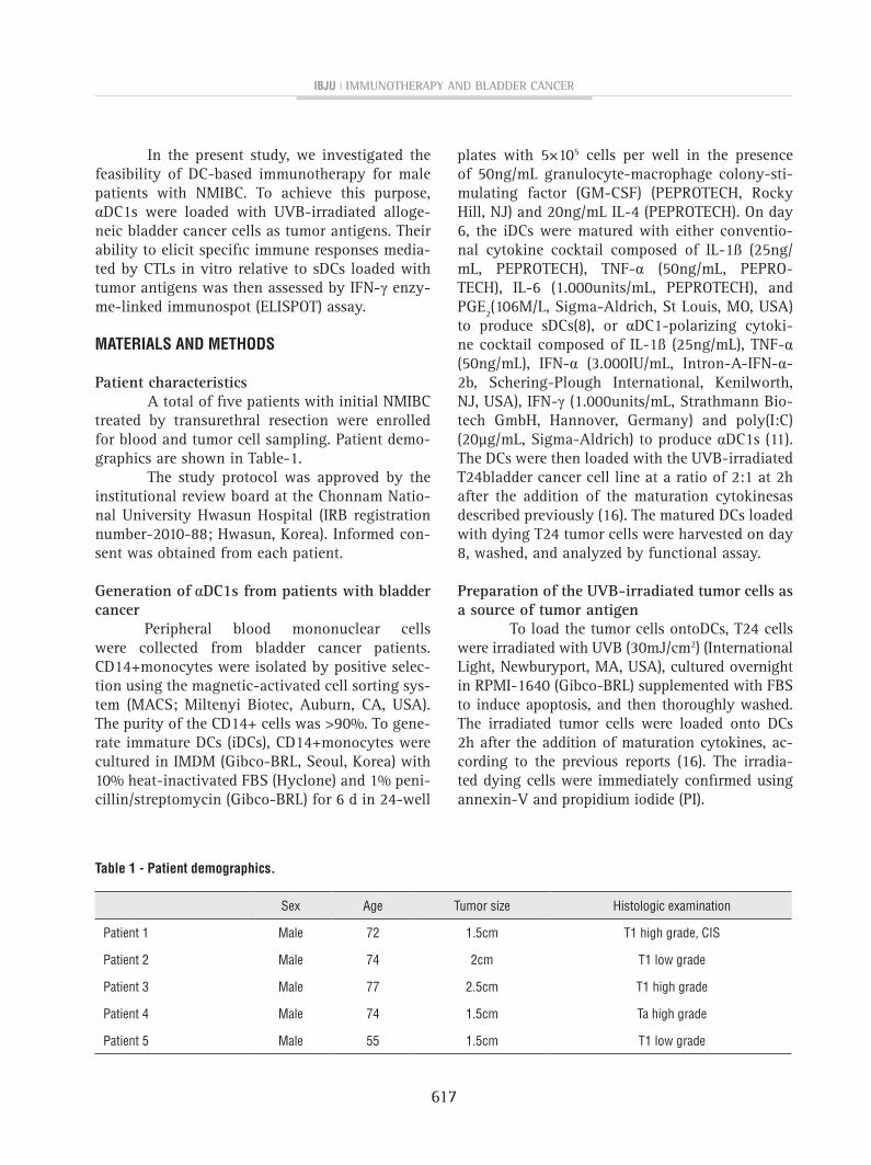
Table 1 - patient demographics.
Sex Age Tumor size Histologic examination
Patient 1 Male 72 1.5cm T1 high grade, CIS
Patient 2 Male 74 2cm T1 low grade
Patient 3 Male 77 2.5cm T1 high grade
Patient 4 Male 74 1.5cm Ta high grade
Patient 5 Male 55 1.5cm T1 low grade
ibju | Immunotherapy and bladder cancer
618
Preparation of bladder cancer cells from patients Tumor tissues obtained from bladder can-
cer patients by transurethral resection were min-ced and lysed for 2-4h at 37ºC in AIM-V medium containing 0.4% collagenase type III. The mononu-clear cells were separated by density gradient cen-trifugation with Ficoll-Hypaque (Lymphoprep) and cryopreserved until their use as target cells in the cytotoxic assay.
Tumor antigen uptake by DCs To measure tumor antigen uptake by the
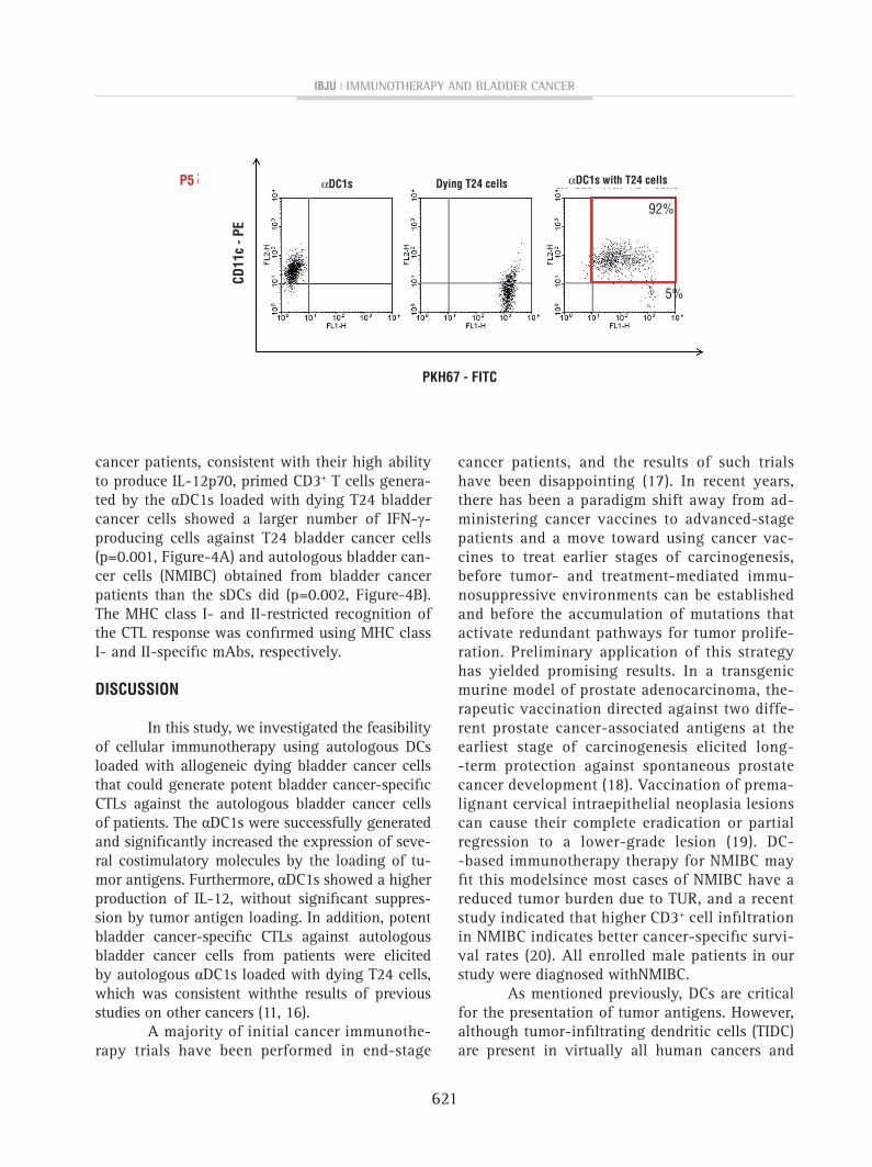
DCs, T24 cells were labeled with PKH67-GL- flu-orescein isothiocyanate (FITC) (Sigma-Aldrich) before UVB irradiation. After the loading of tu-mor antigen onto DCs at a ratio of 1:2 on day 6, the αDC1s loaded with dying T24 tumor cells were stained with CD11c-phycoerythrin (PE) and analyzed by flow cytometry for tumor antigen up-take (CD11c+/PKH67+).
Immunophenotyping of DCs To characterize the cell surface pheno-
types on DCs, flow cytometry was performed using a FACSAria cell sorter (Becton Dickinson, San Jose, CA, USA) after labeling of the cells with CD86-PE, CD83-FITC, CCR7-FITC (PharMingen, San Diego, CA, USA), and the relevant isotype controls (mouse IgG1 and IgG2a, PharMingen). Cell debris was eliminated from the analysis by forward and side-scatter gating, and the data were analyzed with WinMDI Version 2.9 software (Biology Software Net).
Cytokine analyses by enzyme-linked immuno-sorbent assay (ELISA)
The levels of IL-12p70 and IL-10 in the primary culture supernatants of the DCs were me-asured using Quantikine Immunoassay Kits (R&D Systems, Minneapolis, MN, USA). Additionally, DCs harvested on day 8 were plated in 96-well plates at 2×10⁴ cells/well and stimulated to secrete IL-12 with CD40 ligand (CD40L)-transfected J558 cells (as an analogue of CD40L-expressing Th cells; a gift from Dr. P. Lane, University of Birmingham, UK) at a density of 5×10⁴cells/well. After 24h, the supernatant was harvested and the production of IL-12p70 determined by ELISA (R&D Systems).
Induction of bladder cancer-specific CTLs Autologous CD3+ (purity >90%) cells were
positively isolated from the lymphocyte fraction af-ter Percoll isolation using MACS (Miltenyi Biotec). T cells (1×10⁶cells) were sensitized by autologous αDC1s (1×10⁵ cells) loaded with dying T24 tumor cells. On day 3, rhuIL-2 (5ng/mL, R&D Systems) and IL-7 (10ng/mL, R&D Systems) were added. The CTLs were re-stimulated with the same DCs on day 10. On day 20, the number of antigen-specific T cells was analyzed by IFN-γ enzyme-linked immu-ne spot (ELISPOT) assay. T24 and autologous blad-der cancer cells from bladder cancer patients were used as target cells. MHC class I- and II-restricted recognition of the prostate cancer-specific CTLs was analyzed using MHC class I- and II-specific mAbs (clone W6/32 and clone CR3/43, respectively). The ELISPOT data were expressed as the mean number of spots (±SD) per 0.5-2×10⁵ T cells. CTL alone was used as the control.
statistical analysis
Data presented are mean plus or minus SD. The statistical significance of differences was assessed using the unpaired t-test. P values <0.05 were considered significant. All statistical analy-ses were performed with SPSS 17.0 for Windows software (SPSS Inc., Chicago, IL, USA).
REsuLTs
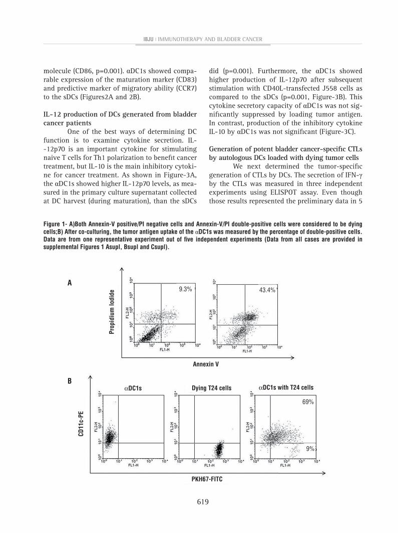
Preparation of dying T24 cells as tumor antigens and antigen uptake by αDC1s. About 43.4% of T24 cells were shown as dying cells after UVB irradiation (Figure-1A). As shown in Figure--1B, αDC1s efficiently incorporated the dying T24 tumor antigen (65.8±14.9%; n=5) as measured by flow cytometry (CD11c+/PKH-67). The efficacy of antigen uptake ofthe αDC1s and sDCs was similar (data not shown).
Characteristics of DCs generated from bladder cancer patients
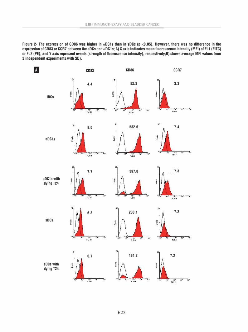
The αDC1s showed typical morphology, with large and branching structures aggregated among the cells (not shown). Phenotypically, αDC1s exhibited higher expression of the co-stimulatory
ibju | Immunotherapy and bladder cancer
619
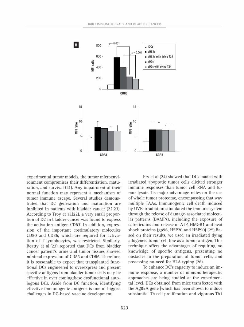
molecule (CD86, p=0.001). αDC1s showed compa-rable expression of the maturation marker (CD83) and predictive marker of migratory ability (CCR7) to the sDCs (Figures2A and 2B).
IL-12 production of DCs generated from bladder cancer patients
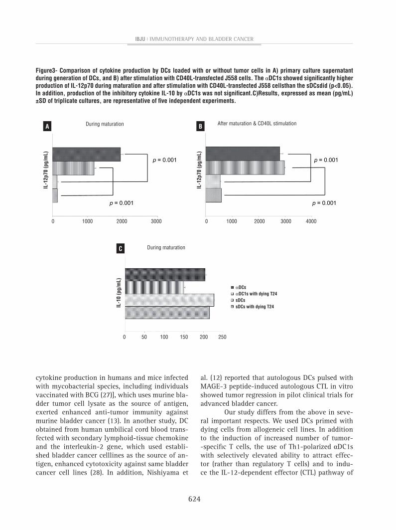
One of the best ways of determining DC function is to examine cytokine secretion. IL--12p70 is an important cytokine for stimulating naive T cells for Th1 polarization to benefit cancer treatment, but IL-10 is the main inhibitory cytoki-ne for cancer treatment. As shown in Figure-3A, the αDC1s showed higher IL-12p70 levels, as mea-sured in the primary culture supernatant collected at DC harvest (during maturation), than the sDCs
did (p=0.001). Furthermore, the αDC1s showed higher production of IL-12p70 after subsequent stimulation with CD40L-transfected J558 cells as compared to the sDCs (p=0.001, Figure-3B). This cytokine secretory capacity of αDC1s was not sig-nificantly suppressed by loading tumor antigen. In contrast, production of the inhibitory cytokine IL-10 by αDC1s was not significant (Figure-3C).
Generation of potent bladder cancer-specific CTLs by autologous DCs loaded with dying tumor cells
We next determined the tumor-specific generation of CTLs by DCs. The secretion of IFN-γ by the CTLs was measured in three independent experiments using ELISPOT assay. Even though those results represented the preliminary data in 5
figure 1- A)Both Annexin-v positive/pI negative cells and Annexin-v/pI double-positive cells were considered to be dying cells;B) After co-culturing, the tumor antigen uptake of the αDc1s was measured by the percentage of double-positive cells. Data are from one representative experiment out of five independent experiments (Data from all cases are provided in supplemental figures 1 Asupl, Bsupl and csupl).
A
B
cD11
c-pE
prop
idiu
m lo
dide
pKh67-fITc
Annexin v
Dying T24 cells αDc1s with T24 cells
69%
43.4%9.3%
9%
αDc1s
ibju | Immunotherapy and bladder cancer
620
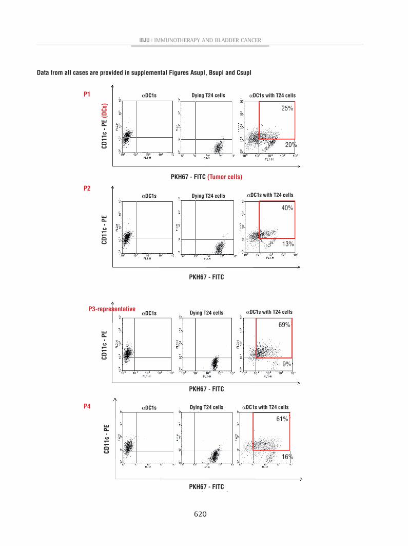
Data from all cases are provided in supplemental figures Asupl, Bsupl and csupl
cD11
c - p
E (D
cs)
cD11
c - p
EcD
11c
- pE
cD11
c - p
E
p1
p2
p4
p3-representative
Dying T24 cells
Dying T24 cells
Dying T24 cells
Dying T24 cells
αDc1s with T24 cells
αDc1s with T24 cells
αDc1s with T24 cells
αDc1s with T24 cells
αDc1s
αDc1s
αDc1s
αDc1s
pKh67 - fITc (Tumor cells)
pKh67 - fITc
pKh67 - fITc
pKh67 - fITc
25%
61%
40%
69%
20%
16%
13%
9%
ibju | Immunotherapy and bladder cancer
621
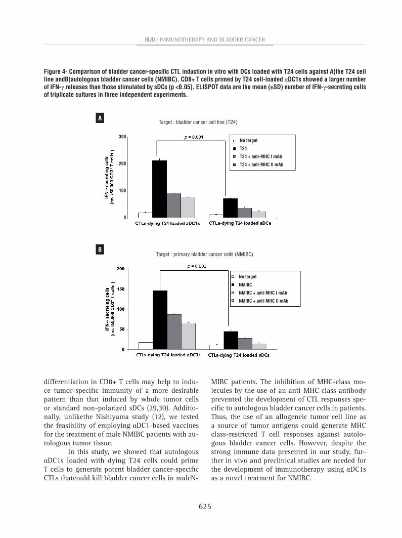
cancer patients, consistent with their high ability to produce IL-12p70, primed CD3+ T cells genera-ted by the αDC1s loaded with dying T24 bladder cancer cells showed a larger number of IFN-γ-producing cells against T24 bladder cancer cells (p=0.001, Figure-4A) and autologous bladder can-cer cells (NMIBC) obtained from bladder cancer patients than the sDCs did (p=0.002, Figure-4B). The MHC class I- and II-restricted recognition of the CTL response was confirmed using MHC class I- and II-specific mAbs, respectively.
DIscussIOn
In this study, we investigated the feasibility of cellular immunotherapy using autologous DCs loaded with allogeneic dying bladder cancer cells that could generate potent bladder cancer-specific CTLs against the autologous bladder cancer cells of patients. The αDC1s were successfully generated and significantly increased the expression of seve-ral costimulatory molecules by the loading of tu-mor antigens. Furthermore, αDC1s showed a higher production of IL-12, without significant suppres-sion by tumor antigen loading. In addition, potent bladder cancer-specific CTLs against autologous bladder cancer cells from patients were elicited by autologous αDC1s loaded with dying T24 cells, which was consistent withthe results of previous studies on other cancers (11, 16).
A majority of initial cancer immunothe-rapy trials have been performed in end-stage
cancer patients, and the results of such trials have been disappointing (17). In recent years, there has been a paradigm shift away from ad-ministering cancer vaccines to advanced-stage patients and a move toward using cancer vac-cines to treat earlier stages of carcinogenesis, before tumor- and treatment-mediated immu-nosuppressive environments can be established and before the accumulation of mutations that activate redundant pathways for tumor prolife-ration. Preliminary application of this strategy has yielded promising results. In a transgenic murine model of prostate adenocarcinoma, the-rapeutic vaccination directed against two diffe-rent prostate cancer-associated antigens at the earliest stage of carcinogenesis elicited long--term protection against spontaneous prostate cancer development (18). Vaccination of prema-lignant cervical intraepithelial neoplasia lesions can cause their complete eradication or partial regression to a lower-grade lesion (19). DC--based immunotherapy therapy for NMIBC may fit this modelsince most cases of NMIBC have a reduced tumor burden due to TUR, and a recent study indicated that higher CD3+ cell infiltration in NMIBC indicates better cancer-specific survi-val rates (20). All enrolled male patients in our study were diagnosed withNMIBC.
As mentioned previously, DCs are critical for the presentation of tumor antigens. However, although tumor-infiltrating dendritic cells (TIDC) are present in virtually all human cancers and
cD11
c - p
E
p5 Dying T24 cells αDc1s with T24 cellsαDc1s
pKh67 - fITc
92%
5%
ibju | Immunotherapy and bladder cancer
622
figure 2- The expression of cD86 was higher in αDc1s than in sDcs (p <0.05). however, there was no difference in the expression of cD83 or ccR7 between the sDcs and αDc1s; A) X axis indicates mean fluorescence intensity (MfI) of fL1 (fITc) or fL2 (pE), and Y axis represent events (strength of fluorescence intensity), respectively;B) shows average MfI values from 3 independent experiments with sD).
A cD83
8.0
4.4
7.7
6.8
6.7
cD86
582.6
82.3
397.0
ccR7
7.4
3.3
7.3
7.2
7.2
230.1
184.2
iDcs
aDc1s
aDc1s withdying T24
sDcs withdying T24
sDcs
ibju | Immunotherapy and bladder cancer
623
experimental tumor models, the tumor microenvi-ronment compromises their differentiation, matu-ration, and survival (21). Any impairment of their normal function may represent a mechanism of tumor immune escape. Several studies demons-trated that DC generation and maturation are inhibited in patients with bladder cancer (22,23). According to Troy et al.(22), a very small propor-tion of DC in bladder cancer was found to express the activation antigen CD83. In addition, expres-sion of the important costimulatory molecules CD80 and CD86, which are required for activa-tion of T lymphocytes, was restricted. Similarly, Beatty et al.(23) reported that DCs from bladder cancer patient’s urine and tumor tissues showed minimal expression of CD83 and CD86. Therefore, it is reasonable to expect that transplanted func-tional DCs engineered to overexpress and present specific antigens from bladder tumor cells may be effective in over comingthese dysfunctional auto-logous DCs. Aside from DC function, identifying effective immunogenic antigens is one of biggest challenges in DC-based vaccine development.
Fry et al.(24) showed that DCs loaded with irradiated apoptotic tumor cells elicited stronger immune responses than tumor cell RNA and tu-mor lysate. Its major advantage relies on the use of whole tumor proteome, encompassing that way multiple TAAs. Immunogenic cell death induced by UVB-irradiation stimulated the immune system through the release of damage-associated molecu-lar patterns (DAMPs), including the exposure of calreticulins and release of ATP, HMGB1 and heat shock proteins (gp96, HSP70 and HSP90) (25).Ba-sed on their results, we used an irradiated dying allogeneic tumor cell line as a tumor antigen. This technique offers the advantages of requiring no knowledge of specific antigens, presenting no obstacles to the preparation of tumor cells, and possessing no need for HLA typing (26).
To enhance DC’s capacity to induce an im-mune response, a number of immunotherapeutic approaches are being studied at the experimen-tal level. DCs obtained from mice transfected with the Ag85A gene [which has been shown to induce substantial Th cell proliferation and vigorous Th1
B iDcs
aDc1s
aDc1s with dying T24
sDcs
sDcs with dying T24
p = 0.001
cD86
cD83 ccR7
MfI
ratio
800
600
400
200
0
15
10
5
0
15
10
5
0
p = 0.001
MfI
ratio
MfI
ratio
ibju | Immunotherapy and bladder cancer
624
cytokine production in humans and mice infected with mycobacterial species, including individuals vaccinated with BCG (27)], which uses murine bla-dder tumor cell lysate as the source of antigen, exerted enhanced anti-tumor immunity against murine bladder cancer (13). In another study, DC obtained from human umbilical cord blood trans-fected with secondary lymphoid-tissue chemokine and the interleukin-2 gene, which used establi-shed bladder cancer celllines as the source of an-tigen, enhanced cytotoxicity against same bladder cancer cell lines (28). In addition, Nishiyama et
al. (12) reported that autologous DCs pulsed with MAGE-3 peptide-induced autologous CTL in vitro showed tumor regression in pilot clinical trials for advanced bladder cancer.
Our study differs from the above in seve-ral important respects. We used DCs primed with dying cells from allogeneic cell lines. In addition to the induction of increased number of tumor--specific T cells, the use of Th1-polarized αDC1s with selectively elevated ability to attract effec-tor (rather than regulatory T cells) and to indu-ce the IL-12-dependent effector (CTL) pathway of
figure3- comparison of cytokine production by Dcs loaded with or without tumor cells in A) primary culture supernatant during generation of Dcs, and B) after stimulation with cD40L-transfected J558 cells. The αDc1s showed significantly higher production of IL-12p70 during maturation and after stimulation with cD40L-transfected J558 cellsthan the sDcsdid (p<0.05). In addition, production of the inhibitory cytokine IL-10 by αDc1s was not significant.c)Results, expressed as mean (pg/mL) ±sD of triplicate cultures, are representative of five independent experiments.
A B
c
lL-1
2p70
(pg/
mL)
lL-1
2p70
(pg/
mL)
During maturation
During maturation
After maturation & CD40L stimulation
0 1000 2000 3000 0 1000 2000 3000 4000
0 50 100 150 200 250
IL-1
0 (p
g/m
L)
αDcsαDc1s with dying T24sDcssDcs with dying T24
ibju | Immunotherapy and bladder cancer
625
figure 4- comparison of bladder cancer-specific cTL induction in vitro with Dcs loaded with T24 cells against A)the T24 cell line andB)autologous bladder cancer cells (nMIBc). cD8+ T cells primed by T24 cell-loaded αDc1s showed a larger number of Ifn-γ releases than those stimulated by sDcs (p <0.05). ELIspOT data are the mean (±sD) number of Ifn-γ-secreting cells of triplicate cultures in three independent experiments.
A
B
differentiation in CD8+ T cells may help to indu-ce tumor-specific immunity of a more desirable pattern than that induced by whole tumor cells or standard non-polarized sDCs (29,30). Additio-nally, unlikethe Nishiyama study (12), we tested the feasibility of employing αDC1-based vaccines for the treatment of male NMIBC patients with au-tologous tumor tissue.
In this study, we showed that autologous αDC1s loaded with dying T24 cells could prime T cells to generate potent bladder cancer-specific CTLs thatcould kill bladder cancer cells in maleN-
MIBC patients. The inhibition of MHC-class mo-lecules by the use of an anti-MHC class antibody prevented the development of CTL responses spe-cific to autologous bladder cancer cells in patients. Thus, the use of an allogeneic tumor cell line as a source of tumor antigens could generate MHC class-restricted T cell responses against autolo-gous bladder cancer cells. However, despite the strong immune data presented in our study, fur-ther in vivo and preclinical studies are needed for the development of immunotherapy using αDC1s as a novel treatment for NMIBC.
Target : bladder cancer cell line (T24)
Target : primary bladder cancer cells (NMIBC)
no target
T24
T24 + anti-Mhc I mAb
T24 + anti-Mhc II mAb
no target
nMIBc
nMIBc + anti-Mhc I mAb
nMIBc + anti-Mhc II mAb
300
200
100
0
ibju | Immunotherapy and bladder cancer
626
cOncLusIOns
The present study showed that autologous αDC1s loaded with allogeneic dying bladder cancer cells could generate strong bladder cancer-specific CTLs against autologous bladder cancer cells. This technique may offer a highly feasible and effecti-ve method for DC-based cancer immunotherapy in male NMIBC patients. Further studies are necessary to enhance the in vivo anti-tumor effect of bladder cancer-specific CTLs generated by autologous dying bladder cancer cells against bladder cancer before cli-nical application.
cOnfLIcT Of InTEREsT
None declared.
REfEREncEs
1. Wood DP Jr. Urothelial tumors of the Bladder. In: Wein AJ, Kavoussi LR, Novick AC, et al., editors. Campbell-Walsh Urology, 10th ed., Vol. 3. Philadelphia: Saunders, Elsevier, 2011; pp. 2309–34.
2. Ro JY, Staerkel GA, Ayala AG. Cytologic and histologic features of superficial bladder cancer. Urol Clin North Am. 1992;19:435-53.
3. Ratliff TL, Gillen D, Catalona WJ. Requirement of a thymus dependent imune response for BCG-mediated antitumor activity. J Urol. 1987;137:155-8.
4. Redelman-Sidi G, Glickman MS, Bochner BH. The mechanism of action of BCG therapy for bladder cancer--a current perspective. Nat Rev Urol. 2014;11:153-62.
5. Sylvester RJ, van der MEIJDEN AP, Lamm DL. Intravesical bacillus Calmette-Guerin reduces the risk of progression in patients with superficial bladder cancer: a meta-analysis of the published results of randomized clinical trials. J Urol. 2002;168:1964-70.
6. Gilboa E. DC-based cancer vaccines. J Clin Invest. 2007;117:1195-203.
7. Steinman RM, Pope M. Exploiting dendritic cells to improve vaccine efficacy. J Clin Invest. 2002;109:1519-26.
8. Jonuleit H, Kühn U, Müller G, Steinbrink K, Paragnik L, Schmitt E, et al. Pro-inflammatory cytokines and prostaglandins induce maturation of potente immunostimulatory dendritic cells under fetal calf serum-free conditions. Eur J Immunol. 1997;27:3135-42.
9. Ridolfi L, Petrini M, Fiammenghi L, Granato AM, Ancarani V, Pancisi E, et al. Dendritic cell-based vaccine in advanced melanoma: update of clinical outcome. Melanoma Res. 2011;21:524-9.
10. Kyte JA, Mu L, Aamdal S, Kvalheim G, Dueland S, Hauser M, et al. Phase I/II trial of melanoma therapy with dendritic cells transfected with autologous tumor-mRNA. Cancer Gene Ther. 2006;13:905-18.
11. Mailliard RB, Wankowicz-Kalinska A, Cai Q, Wesa A, Hilkens CM, Kapsenberg ML, et al. alpha-type-1 polarized dendritic cells: a novel immunization tool with optimized CTL-inducing activity. Cancer Res. 2004;64:5934-7.
12. Nishiyama T, Tachibana M, Horiguchi Y, Nakamura K, Ikeda Y, Takesako K, et al. Immunotherapy of bladder cancer using autologous dendritic cells pulsed with human lymphocyte antigen-A24-specific MAGE-3 peptide. Clin Cancer Res. 2001;7:23-31.
13. Zhang P, Wang J, Wang D, Wang H, Shan F, Chen L, et al. Dendritic cell vaccine modified by Ag85A gene enhances anti-tumor immunity against bladder cancer. Int Immunopharmacol. 2012;14:252-60.
14. Palucka AK, Ueno H, Connolly J, Kerneis-Norvell F, Blanck JP, Johnston DA, et al. Dendritic cells loaded with killed allogeneic melanoma cells can induce objective clinical responses and MART-1 specific CD8+ T-cell immunity. J Immunother. 2006;29:545-57.
15. Keenan BP, Jaffee EM. Whole cell vaccines--past progress and future strategies. Semin Oncol. 2012;39:276-86.
16. Hwang EC, Lim MS, Im CM, Kwon DD, Lee HJ, Nguyen-Pham TN, et al. Generation of potent cytotoxic T lymphocytes against castration-resistant prostate cancer cells by dendritic cells loaded with dying allogeneic prostate cancer cells. Scand J Immunol. 2013;77:117-24.
17. Gray A, Raff AB, Chiriva-Internati M, Chen SY, Kast WM. A paradigm shift in therapeutic vaccination of cancer patients: the need to apply therapeutic vaccination strategies in the preventive setting. Immunol Rev. 2008;222:316-27.
18. Garcia-Hernandez Mde L, Gray A, Hubby B, Klinger OJ, Kast WM. Prostate stem cell antigen vaccination induces a long-term protective immune response against prostate cancer in the absence of autoimmunity. Cancer Res. 2008;68:861-9.
19. Einstein MH, Kadish AS, Burk RD, Kim MY, Wadler S, Streicher H, et al. Heat shock fusion protein-based immunotherapy for treatment of cervical intraepithelial neoplasia III. Gynecol Oncol. 2007;106:453-60.
20. Otto W, Denzinger S, Wieland WF, Hartmann A. First analysis of immune cell infiltration in stage pT1 urothelial bladder carcinoma: CD3 positivity as a prognostic marker for cancer-specific survival. World J Urol. 2012;30:875-7.
21. Bennaceur K, Chapman JA, Touraine JL, Portoukalian J. Immunosuppressive networks in the tumour environment and their effect in dendritic cells. Biochim Biophys Acta. 2009;1795:16-24. Retraction in: Biochim Biophys Acta. 2013;1835:259.
22. Troy AJ, Davidson PJ, Atkinson CH, Hart DN. CD1a dendritic cells predominate in transitional cell carcinoma of bladder and kidney but are minimally activated. J Urol. 1999;161:1962-7.
23. Beatty JD, Islam S, North ME, Knight SC, Ogden CW. Urine dendritic cells: a noninvasive probe for immune activity in bladder cancer? BJU Int. 2004;94:1377-83.
ibju | Immunotherapy and bladder cancer
627
24. Fry TJ, Shand JL, Milliron M, Tasian SK, Mackall CL. Antigen loading of DCs with irradiated apoptotic tumor cells induces improved anti-tumor immunity compared to other approaches. Cancer Immunol Immunother. 2009;58:1257-64.
25. Garg AD, Agostinis P. Editorial: Immunogenic Cell Death in Cancer: From Benchside Research to Bedside Reality. Front Immunol. 2016;7:110.
26. Salcedo M, Bercovici N, Taylor R, Vereecken P, Massicard S, Duriau D, et al. Vaccination of melanoma patients using dendritic cells loaded with an allogeneic tumor cell lysate. Cancer Immunol Immunother. 2006;55:819-29.
27. Huygen K, Content J, Denis O, Montgomery DL, Yawman AM, Deck RR, et al. Immunogenicity and protective efficacy of a tuberculosis DNA vaccine. Nat Med. 1996;2:893-8.
28. Li YG, Wang ZP, Tian JQ, Tian BQ, Rodrigues R, Shang PF, et al. Dendritic cell transfected with secondary lymphoid-tissue chemokine and/or interleukin-2 gene-enhanced cytotoxicity of T-lymphocyte in human bladder tumor cell S in vitro. Cancer Invest. 2009;27:909-17.
29. Muthuswamy R, Urban J, Lee JJ, Reinhart TA, Bartlett D, Kalinski P. Ability of mature dendritic cells to interact with regulatory T cells is imprinted during maturation. Cancer Res. 2008;68:5972-8.
30. Watchmaker PB, Berk E, Muthuswamy R, Mailliard RB, Urban JA, Kirkwood JM, et al. Independent regulation of chemokine responsiveness and cytolytic function versus CD8+ T cell expansion by dendritic cells. J Immunol. 2010;184:591-7.
_______________________Correspondence address:
Dong Deuk Kwon, MD, PhDDepartment of Urology,
Chonnam National University Medical School42, Jebong-ro, Donggu,
Gwangju, 501-757, Republic of Korea Fax: +82 62 227-1643
E-mail: [email protected]