genePix 4000B microarray scanner - moleculardevices.com · minutes for a full scan at 10 μm...
2
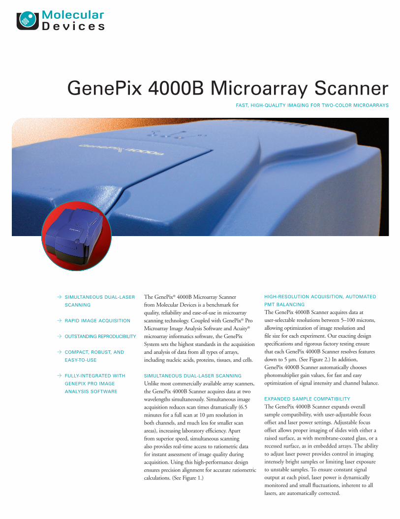
HIGH-RESOLUTION ACQUISITION, AUTOMATED PMT BALANCING The GenePix 4000B Scanner acquires data at user-selectable resolutions between 5–100 microns, allowing optimization of image resolution and file size for each experiment. Our exacting design specifications and rigorous factory testing ensure that each GenePix 4000B Scanner resolves features down to 5 μm. (See Figure 2.) In addition, GenePix 4000B Scanner automatically chooses photomultiplier gain values, for fast and easy optimization of signal intensity and channel balance. EXPANDED SAMPLE COMPATIBILITY The GenePix 4000B Scanner expands overall sample compatibility, with user-adjustable focus offset and laser power settings. Adjustable focus offset allows proper imaging of slides with either a raised surface, as with membrane-coated glass, or a recessed surface, as in embedded arrays. The ability to adjust laser power provides control in imaging intensely bright samples or limiting laser exposure to unstable samples. To ensure constant signal output at each pixel, laser power is dynamically monitored and small fluctuations, inherent to all lasers, are automatically corrected. FAST, HIGH-QUALITY IMAGING FOR TWO-COLOR MICROARRAYS GenePix 4000B Microarray Scanner The GenePix ® 4000B Microarray Scanner from Molecular Devices is a benchmark for quality, reliability and ease-of-use in microarray scanning technology. Coupled with GenePix ® Pro Microarray Image Analysis Software and Acuity ® microarray informatics software, the GenePix System sets the highest standards in the acquisition and analysis of data from all types of arrays, including nucleic acids, proteins, tissues, and cells. SIMULTANEOUS DUAL-LASER SCANNING Unlike most commercially available array scanners, the GenePix 4000B Scanner acquires data at two wavelengths simultaneously. Simultaneous image acquisition reduces scan times dramatically (6.5 minutes for a full scan at 10 μm resolution in both channels, and much less for smaller scan areas), increasing laboratory efficiency. Apart from superior speed, simultaneous scanning also provides real-time access to ratiometric data for instant assessment of image quality during acquisition. Using this high-performance design ensures precision alignment for accurate ratiometric calculations. (See Figure 1.) > SIMULTANEOUS DUAL-LASER SCANNING > RAPID IMAGE ACQUISITION > OUTSTANDING REPRODUCIBILITY > COMPACT, ROBUST, AND EASY-TO-USE > FULLY-INTEGRATED WITH GENEPIX PRO IMAGE ANALYSIS SOFTWARE
Transcript of genePix 4000B microarray scanner - moleculardevices.com · minutes for a full scan at 10 μm...
HigH-Resolution Acquisition, AutomAted
Pmt BAlAncing
The GenePix 4000B Scanner acquires data at user-selectable resolutions between 5–100 microns, allowing optimization of image resolution and file size for each experiment. Our exacting design specifications and rigorous factory testing ensure that each GenePix 4000B Scanner resolves features down to 5 μm. (See Figure 2.) In addition, GenePix 4000B Scanner automatically chooses photomultiplier gain values, for fast and easy optimization of signal intensity and channel balance.
exPAnded sAmPle comPAtiBility
The GenePix 4000B Scanner expands overall sample compatibility, with user-adjustable focus offset and laser power settings. Adjustable focus offset allows proper imaging of slides with either a raised surface, as with membrane-coated glass, or a recessed surface, as in embedded arrays. The ability to adjust laser power provides control in imaging intensely bright samples or limiting laser exposure to unstable samples. To ensure constant signal output at each pixel, laser power is dynamically monitored and small fluctuations, inherent to all lasers, are automatically corrected.
fAst, HigH-quAlity imAging foR two-coloR micRoARRAys
genePix 4000B microarray scanner
The GenePix® 4000B Microarray Scanner from Molecular Devices is a benchmark for quality, reliability and ease-of-use in microarray scanning technology. Coupled with GenePix® Pro Microarray Image Analysis Software and Acuity® microarray informatics software, the GenePix System sets the highest standards in the acquisition and analysis of data from all types of arrays, including nucleic acids, proteins, tissues, and cells.
simultAneous duAl-lAseR scAnning
Unlike most commercially available array scanners, the GenePix 4000B Scanner acquires data at two wavelengths simultaneously. Simultaneous image acquisition reduces scan times dramatically (6.5 minutes for a full scan at 10 μm resolution in both channels, and much less for smaller scan areas), increasing laboratory efficiency. Apart from superior speed, simultaneous scanning also provides real-time access to ratiometric data for instant assessment of image quality during acquisition. Using this high-performance design ensures precision alignment for accurate ratiometric calculations. (See Figure 1.)
> simultAneous duAl-lAseR
scAnning
> RAPid imAge Acquisition
> outstAnding RePRoduciBility
> comPAct, RoBust, And
eAsy-to-use
> fully-integRAted witH
genePix PRo imAge
AnAlysis softwARe
genePix 4000B micRoARRAy scAnneR
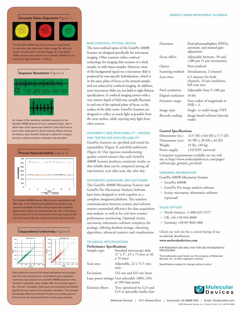
field uniformity ensures that signal calculations are accurate
over the entire arrayed area. A hybridized gene expression
microarray was scanned on a genePix 4000B scanner in the
“forward” orientation, then rotated 180° and scanned again in
the “reverse” orientation. each spot was quantified and plotted
against the same spot in the opposite orientation. the average
percentage difference between forward and reverse scans is
6.4% for the red channel and 4.5% for the green channel.
An image of the resolution standard scanned on the
genePix 4000B scanner at 5 μm resolution (top), with a
detail view of the same scan (bottom). each short bar is
5 μm wide, separated by 20 μm spacing. Before leaving
the factory, each genePix scanner is tested for imaging
accuracy using a precision resolution test standard.
Detection: Dual photomultipliers (PMTs), automatic and manual gain adjustment
Focus offset: Adjustable between -50 and +200 μm (5 μm increments)
Optics: Non-confocal
Scanning method: Simultaneous, 2-channel
Scan time: 6.5 minutes for both channels, 10 μm resolution, full scan area
Pixel resolution: Adjustable from 5–100 μm
Digital resolution: 16-bit
Dynamic range: Four orders of magnitude at SNR > 3
Image type: Single- or multi-image TIFF
Barcode reading: Image-based software barcode reader
General SpecificationsDimensions (in.): 13.5 (W) x 8.0 (H) x 17.5 (D) Dimensions (cm): 34 (W) x 20 (H) x 44 (D)Weight: 45 lbs. (20 kg)Power supply: 110/220V universal
Computer requirements available on our web site at http://www.moleculardevices.com/pages/software/gn_genepix_pro.html oRdeRing infoRmAtion
GenePix 4000B Microarray Scanner> GenePix 4000B> GenePix Pro image analysis software> Acuity microarray informatics software
(optional)
sAles offices
> North America +1-800-635-5577> UK +44-118-944-8000> Germany +49-89-9605-880
Check our web site for a current listing of our worldwide distributors. www.moleculardevices.com
foR ReseARcH use only. not foR use in diAgnostic PRoceduRes.
the trademarks used herein are the property of molecular devices, inc. or their respective owners.
specifications subject to change without notice.
non-confocAl oPticAl design
The non-confocal optics of the GenePix 4000B Scanner are designed specifically for microarray imaging. Other scanners utilize confocal technology for imaging thin sections of a thick sample, as with tissue samples. However, most of the background signal on a microarray slide is produced by non-specific hybridization, which is in the same plane of focus as the arrayed sample, and not reduced by confocal imaging. In addition, most microarray slides are not held to tight flatness specifications. A confocal imaging system with a very narrow depth of field may actually fluctuate in and out of the optimal plane of focus, as the surface of the slide varies. GenePix Scanners are designed to collect as much light as possible from the array surface, while rejecting stray light from other sources.
unifoRmity And RePeAtABility—PRoven
And tested foR dAtA ReliABility
GenePix Scanners are specified and tested for repeatability (Figure 3) and field uniformity (Figure 4). Our rigorous manufacturing quality control ensures that each GenePix 4000B Scanner produces consistent results, so that reliable data can be compared among all experiments, scan after scan, day after day.
integRAted HARdwARe And softwARe
The GenePix 4000B Microarray Scanner and GenePix Pro Microarray Analysis Software have been designed to work together as a complete integrated platform. The seamless communication between scanner and software ensures unmatched efficiency for data acquisition and analysis, as well as for real-time scanner performance monitoring. Optional Acuity microarray informatics software completes the package, offering database storage, clustering algorithms, advanced statistics and visualizations.
tecHnicAl sPecificAtions
Performance SpecificationsSample type: Standard microscope slide
(1" x 3", 25 x 75 mm or 26 x 76 mm)
Scan area: Adjustable, 22 x 71.5 mm max.
Excitation: 532 nm and 635 nm lasers
Laser power settings: User-selectable 100%, 33% or 10% laser power
Emission filters: Two, optimized for Cy3 and Cy5 or spectrally similar dyes
the genePix 4000B scanner is precision engineered
for accurate ratio alignment. Ratio image (far left) and
single red and green channel images of a hybridized
cdnA arrayed spot scanned on a genePix 4000B at 5 μm
resolution. spot diameter = 150 μm.
Accurate Ratio Alignment (Figure 1)
Superior Resolution (Figure 2)
Unparalleled Uniformity (Figure 4)
Proven Reproducibility (Figure 3)
the genePix 4000B scanner offers proven repeatability, scan
after scan. A non-bleaching fluorescent test standard was
scanned repeatedly and the average signal was quantified.
signal value variance among all scans was 1.6% in the green
channel and 2.1% in the red channel. error bars cover two sd
from the mean of the four measurements at each time point.
molecular devices | 1311 orleans drive | sunnyvale, cA 94089 usA | email: [email protected]©2010 molecular devices, inc. Printed in u.s.A. 6/10 #0120-1392B