gamma-Secretase mediated proteolytic processing of the ...hss.ulb.uni-bonn.de/2011/2539/2539.pdf ·...
121
γ-Secretase mediated proteolytic processing of the triggering receptor expressed on myeloid cells-2 – functional implications for intracellular signaling DISSERTATION zur Erlangung des Doktorgrades (Dr. rer. nat.) der Mathematisch-Naturwissenschaftlichen-Fakultät der Rheinischen Friedrich-Wilhelms-Universität Bonn vorgelegt von Dipl. Biochem. Patrick Wunderlich aus Brühl Bonn, 21.12.2010
Transcript of gamma-Secretase mediated proteolytic processing of the ...hss.ulb.uni-bonn.de/2011/2539/2539.pdf ·...
γ-Secretase mediated proteolytic processing of the
triggering receptor expressed on myeloid cells-2 –
functional implications for intracellular signaling
DISSERTATION
zur
Erlangung des Doktorgrades (Dr. rer. nat.)
der
Mathematisch-Naturwissenschaftlichen-Fakultät
der
Rheinischen Friedrich-Wilhelms-Universität Bonn
vorgelegt von
Dipl. Biochem. Patrick Wunderlich
aus
Brühl
Bonn, 21.12.2010

Angefertigt mit Genehmigung der Mathematisch-Naturwissenschaftlichen Fakultät
der Rheinischen Friedrich-Wilhelms-Universität Bonn
1. Gutachter: Prof. Dr. rer. nat. Jochen Walter
2. Gutachter: Prof. Dr. rer. nat. Sven Burgdorf
Tag der Abgabe: 21.12.2010
Tag der Promotion: 06.04.2011
Erscheinungsjahr: 2011

An Eides statt versichere ich, dass ich die Dissertation “γ-Secretase mediated proteolytic processing of the
triggering receptor expressed on myeloid cells-2 – functional implications for intracellular signaling“ selbst
und ohne jede unerlaubte Hilfe angefertigt habe und dass diese oder eine ähnliche Arbeit noch an keiner
anderen Stelle als Dissertation eingereicht worden ist.
Promotionsordnung vom 7. Januar 2004 _________________________
Patrick Wunderlich
INDEX IV
CONTENTS
ABBREVIATIONS...................................................................................................................IX
1 INTRODUCTION.................................................................................................................14
1.1 Alzheimer's disease (AD) ............................................................................................................14
1.1.1 Neuropathological hallmarks of AD.................................................................................15
1.1.1.1 Neurofibrillary tangles.................................................................................................15
1.1.1.2 β-amyloid plaques.........................................................................................................17
1.1.2 Genetic factors involved in AD .........................................................................................18
1.1.2.1 Late onset or sporadic AD..........................................................................................18
1.1.2.2 Early onset or familiar AD..........................................................................................19
1.1.3 Characteristics of the β-amyloid precursor protein (APP).............................................20
1.1.4 APP processing.....................................................................................................................21
1.1.5 Structural and functional insights in the APP processing enzymes...............................22
1.1.5.1 The α-secretase: ADAM-10, ADAM-17 and ADAM-9..........................................22
1.1.5.2 The β-secretase: BACE-1............................................................................................23
1.1.5.3 The γ-secretase..............................................................................................................25
1.2 Aβ clearance in the brain..............................................................................................................28
1.3 Microglia.........................................................................................................................................30
1.4 The role of microglia & inflammation in AD...........................................................................31
1.4.1 Beneficial roles of microglia in Aβ clearance...................................................................31
1.4.2 Detrimental roles of microglia by chronic enhancement of inflammatory
processes................................................................................................................................33
1.5 Triggering receptors expressed on myeloid cells (TREMs).....................................................34
1.5.1 TREM1...................................................................................................................................35
1.5.2 TREM2...................................................................................................................................35
1.6 DAP12 – structure and signaling................................................................................................37
1.7 Rationale.........................................................................................................................................39
2 MATERIAL & METHODS...................................................................................................40
2.1 Cell biological techniques.............................................................................................................41
2.1.1 Cell culture ............................................................................................................................41
2.1.2 Cell culture of ES cell derived microglia...........................................................................42
2.1.3 Transient transfection...........................................................................................................42
INDEX V
2.1.4 Immunocytochemistry (ICC)..............................................................................................43
2.2 Molecularbiological techniques...................................................................................................44
2.2.1 Polymerase chain reaction (PCR)........................................................................................44
2.2.2 Separation and visualization of DNA fragments.............................................................47
2.2.3 Isolation of DNA fragments from agarose gels..............................................................47
2.2.4 Restriction digestion.............................................................................................................47
2.2.5 Dephosphorylation of linearized DNA............................................................................47
2.2.6 Ligation...................................................................................................................................48
2.2.7 Generation of chemo competent E. coli Top10..............................................................48
2.2.8 Transformation of E. coli Top10.......................................................................................48
2.2.9 Cryo conservation of transformed E. coli........................................................................49
2.2.10 Extraction of plasmid DNA from E. coli.......................................................................49
2.2.11 Precipitation of DNA by isopropanol.............................................................................50
2.2.12 Precipitation of DNA by ethanol.....................................................................................50
2.2.13 DNA sequencing................................................................................................................50
2.2.14 Photometric determination of DNA concentration.....................................................50
2.3 Proteinbiochemical techniques....................................................................................................51
2.3.1 Extraction of membrane proteins out of eukaryotic cells.............................................51
2.3.2 Protein extraction out of eukaryotic cells.........................................................................51
2.3.3 Protein estimation.................................................................................................................52
2.3.4 Immunoprecipitation (IP)....................................................................................................52
2.3.5 Co-immunoprecipitation of DAP12 and TREM2...........................................................53
2.3.6 Sodium dodecyl sulfate polyacrylamide gel electrophoresis (SDS-PAGE)...................53
2.3.7 Western immunoblotting (WB)...........................................................................................54
2.3.8 Coomassie staining of proteins in polyacrylamide gels...................................................56
2.3.9 Precipitation of soluble proteins from cell culture supernatants by trichloroacetic
acid (TCA)..............................................................................................................................56
2.3.10 Expression and isolation of glutathione S-transferase (GST) fusion proteins..........57
2.3.11 Biotinylation of cell surface proteins...............................................................................58
2.3.12 Radio-labeling with 32P-orthophosphate........................................................................58
2.3.13 TREM2 shedding assay......................................................................................................59
2.3.14 TREM2 activation assay.....................................................................................................59
2.3.15 Aβ-Phagocytosis assay.......................................................................................................60
2.4 Densitometric quantification of signals and statistical analysis..............................................60
INDEX VI
3 RESULTS............................................................................................................................61
3.1 Proteolytic processing of TREM2 by γ-secretase....................................................................61
3.1.1 Expression of PS1 in microglia..........................................................................................61
3.1.2 γ-secretase dependent processing of TREM2..................................................................62
3.2 Characterization of γ-secretase dependent processing and localization of TREM2.........65
3.2.1 Cleavage of TREM2 occurs at the cell surface................................................................65
3.2.2 γ-secretase mediated processing in response to the activation of TREM2..................69
3.2.3 TREM2 ICD is not translocated to the nucleus...............................................................70
3.2.4 TREM2 ectodomain shedding by an protease of the ADAM or MMP family...........71
3.3 Role of TREM2 processing in the interaction with DAP12...................................................78
3.3.1 Inhibition of γ-secretase facilitates co-localization of TREM2 and DAP12 at
the cell surface.......................................................................................................................78
3.3.2 Processing of TREM2 alters the interaction with DAP12.............................................79
3.3.3 Inhibition of γ-secretase suppresses phosphorylation of DAP12 upon TREM2
activation................................................................................................................................80
3.4 Inhibition of γ-secretase inhibition in microglia.......................................................................82
3.4.1 Inhibition of γ-secretase in PS1 FAD mutants decrease phagocytosis of Aβ.............82
4 DISCUSSION.......................................................................................................................84
4.1 Proteolytic processing of TREM2..............................................................................................85
4.1.1 Shedding of the TREM2 ectodomain...............................................................................85
4.1.2 Cleavage of TREM2 CTF by γ-secretase..........................................................................88
4.2 γ-secretase dependent interaction of TREM2 and DAP12....................................................91
4.2.1 Inhibition of γ-secretase cleavage alters interaction of TREM2 with DAP12............91
4.2.2 Inhibition of γ-secretase impairs DAP12 phosphorylation............................................93
4.3 Potential effects of impaired TREM2/DAP12 signaling on phagocytosis and
degradation of Aβ.........................................................................................................................97
5 OUTLOOK........................................................................................................................100
6 ABSTRACT........................................................................................................................102
7 REFERENCES...................................................................................................................104
8 ACKNOWLEDGMENT........................................................................................................120
9 CURRICULUM VITAE........................................................................................................121
INDEX VII
LIST OF FIGURES
Figure 1: Neuropathological hallmarks of AD...................................................................................16
Figure 2: Scheme of the APP structure..............................................................................................21
Figure 3: The APP processing..............................................................................................................22
Figure 4: Scheme of some components of the inflammatory cascade..........................................34
Figure 5: General understanding of the TREM2/DAP12 signaling pathway...............................37
Figure 6: Schematic drawings of the different constructs used in this study.................................45
Figure 7: Detection of functional γ-secretase in microglia...............................................................62
Figure 8: Pharmacological inhibition of γ-secretase leads to the accumulation of a
TREM2 C-terminal fragment...............................................................................................63
Figure 9: TREM2 is proteolytically processed by γ-secretase..........................................................64
Figure 10: FAD mutations of PS1 lead to accumulation of TREM2 CTFs....................................65
Figure 11: Inhibition of γ-secretase changes the localization of TREM2.......................................66
Figure 12: Inhibition of γ-secretase alters the distribution of TREM2 at the cell surface............67
Figure 13: TREM2 CTFs accumulate at the cell surface after γ-secretase inhibition.....................68
Figure 14: Interaction with DAP12 is not prerequisite for γ-secretase mediated
TREM2 cleavage....................................................................................................................69
Figure 15: γ-secretase cleavage in response to activation of TREM2...............................................70
Figure 16: TREM2 ICD is not translocated to the nucleus................................................................71
Figure 17: TREM2 ECD can be detected in supernatant of TREM2 overexpressing cells..........72
Figure 18: BACE-1 overexpression increases the ratio of TREM2 CTF/TREM2 FL..................73
Figure 19: ADAM-10 overexpression increases ratio of TREM2 CTF/TREM2 FL....................74
Figure 20: Pharmacological modulation of TREM2 shedding..........................................................75
Figure 21: Treatment with Batimastat increases the TREM2 surface expression...........................77
Figure 22: Co-localization of TREM2∆ECD and DAP12 upon cell treatment with DAPT........79
Figure 23: Inhibition of γ-secretase alters the association of TREM2 and DAP12.......................81
Figure 24: Inhibition of γ-secretase impairs the phosphorylation of DAP12.................................82
Figure 25: γ-secretase is involved in the phagocytosis of fibrillar Aβ...............................................83
Figure 26: Hypothetical scheme for the sequential processing of TREM2.....................................92
Figure 27: Hypothetical scheme of impaired TREM2 processing upon γ-secretase inhibition....93
Figure 28: Impaired DAP12 signaling stabilizes TLR response via PLCγ.......................................95
Figure 29: Impaired DAP12 signaling stabilizes TLR response via PI3K........................................96
INDEX VIII
Figure 30: Hypothetical link between impaired TREM2 cleavage and attenuated Aβ
phagocytosis and degeneration............................................................................................99
LIST OF TABLES
Table 1: Equipment....................................................................................................................................40
Table 2: Cell lines.......................................................................................................................................42
Table 3: List of the DNA constructs used for the project..................................................................45
Table 4: Overview of cloning primers....................................................................................................46
Table 5: Detailed PCR programs.............................................................................................................46
Table 6: Composition of the SDS-PAGE gels.......................................................................................54
Table 7: Primary antibodies that were used for western immunoblotting (WB),
immunocytochemistry (ICC), surface stainings (SurfS), immunoprecipitation (IP) and
co-immunoprecipitation (CoIP)...............................................................................................55
Table 8: Secondary antibodies that were used for western immunoblotting (WB), immunocyto-
chemistry (ICC) and surface stainings (SurfS)........................................................................56
INDEX IX
ABBREVIATIONS
aa Amino acid
Ab Antibody
ACE Angiotensin converting enzyme
AD Alzheimer´s disease
ADAM A disintegrin and metalloprotease
AICD APP intracellular domain
APH-1 Anterior pharynx defective-1
APLP Amyloid precursor like protein
APP Amyloid precursor protein
APS Ammonium persulfate
Aβ β-Amyloid
BACE β-Site APP cleaving enzyme
BBB Blood brain barrier
BSA Bovine serum albumin
Cdk5 Cyclin dependent kinase 5
CK-1 Casein kinase-1
CNS Central nervous system
co-IP Co-immunoprecipitation
CR3 Complement receptor-3
CS Cover slip
CT C-terminus
CTF C-terminal fragment
DAMPs Damage associated molecular pattern molecules
DAP12 DNAX activating protein of 12 kDa
DAPT N-[(3,5-Difluorophenyl)acetyl]-L-alanyl-2-phenyl]gly-cine-1,1-dimethylethyl ester
DC Dendritic cell
dko Double knock-out
DMEM Dulbecco´s Modified Eagle´s Medium
DMSO Dimethyl sulfoxide
DN Dominant negative
DOC Deoxycholic acid
DR-6 Death receptor-6
DTT Dithiothreitol
ECD Extra cellular domain
ECE Endothelin converting enzyme
ECL Enhanced chemiluminescence
INDEX X
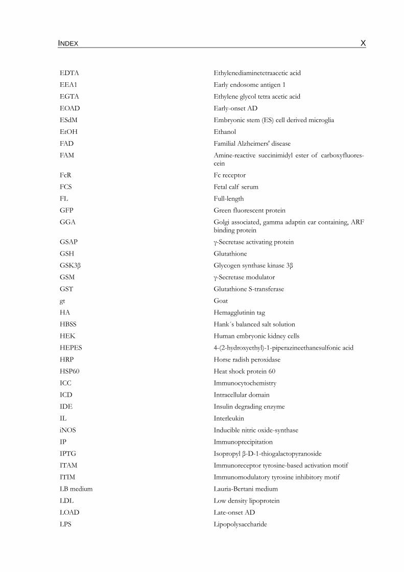
EDTA Ethylenediaminetetraacetic acid
EEA1 Early endosome antigen 1
EGTA Ethylene glycol tetra acetic acid
EOAD Early-onset AD
ESdM Embryonic stem (ES) cell derived microglia
EtOH Ethanol
FAD Familial Alzheimers' disease
FAM Amine-reactive succinimidyl ester of carboxyfluores-cein
FcR Fc receptor
FCS Fetal calf serum
FL Full-length
GFP Green fluorescent protein
GGA Golgi associated, gamma adaptin ear containing, ARF binding protein
GSAP γ-Secretase activating protein
GSH Glutathione
GSK3β Glycogen synthase kinase 3β
GSM γ-Secretase modulator
GST Glutathione S-transferase
gt Goat
HA Hemagglutinin tag
HBSS Hank´s balanced salt solution
HEK Human embryonic kidney cells
HEPES 4-(2-hydroxyethyl)-1-piperazineethanesulfonic acid
HRP Horse radish peroxidase
HSP60 Heat shock protein 60
ICC Immunocytochemistry
ICD Intracellular domain
IDE Insulin degrading enzyme
IL Interleukin
iNOS Inducible nitric oxide-synthase
IP Immunoprecipitation
IPTG Isopropyl β-D-1-thiogalactopyranoside
ITAM Immunoreceptor tyrosine-based activation motif
ITIM Immunomodulatory tyrosine inhibitory motif
LB medium Lauria-Bertani medium
LDL Low density lipoprotein
LOAD Late-onset AD
LPS Lipopolysaccharide
INDEX XI
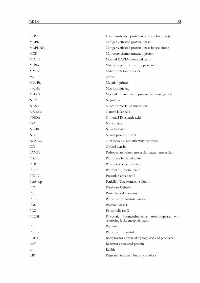
LRP Low density lipid protein receptor related protein
MAPK Mitogen activated protein kinase
MAPKKK Mitogen activated protein kinase kinase kinase
MCP Monocyte chemo attractant protein
MDL-1 Myeloid DAP12-associated lectin
MIP1α Macrophage inflammatory protein-1α
MMP9 Matrix metalloprotease-9
ms Mouse
Mut. Pr. Mutation primer
mycHis Myc-histidine tag
MyD88 Myeloid differentiation primary response gene 88
NEP Neprilysin
NEXT Notch extracellular truncation
NK cells Natural killer cells
NMDA N-methyl-D-aspartic acid
NO Nitric oxide
NP-40 Nonidet P-40
NPC Neural progenitor cell
NSAIDs Non steroidal anti inflammatory drugs
OD Optical density
PAMPs Pathogen associated molecular pattern molecules
PBS Phosphate buffered saline
PCR Polymerase chain reaction
PDBu Phorbol 12,13-dibutyrate
PEN-2 Presenilin enhancer-2
PenStrep Penicillin/Streptomycin solution
PFA Paraformaldehyde
PHF Paired helical filaments
PI3K Phosphatidylinositol-3 kinase
PKC Protein kinase C
PLC Phospholipase C
PLOSL Polycystic lipomembraneous osteodysplasia with sclerosing leukoencephalopathy
PS Presenilin
PtdIns Phosphatidylinositol
RAGE Receptor for advanced glycosylated end products
RAP Receptor-associated protein
rb Rabbit
RIP Regulated intramembrane proteolysis
INDEX XII
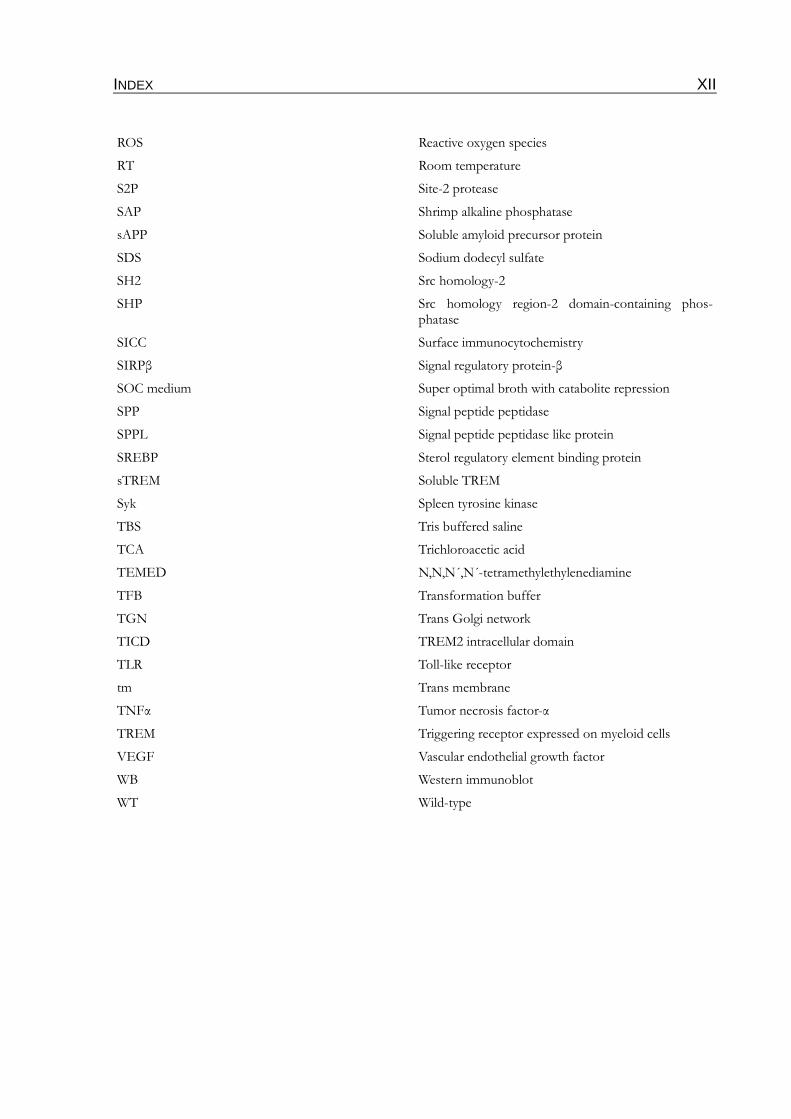
ROS Reactive oxygen species
RT Room temperature
S2P Site-2 protease
SAP Shrimp alkaline phosphatase
sAPP Soluble amyloid precursor protein
SDS Sodium dodecyl sulfate
SH2 Src homology-2
SHP Src homology region-2 domain-containing phos-phatase
SICC Surface immunocytochemistry
SIRPβ Signal regulatory protein-β
SOC medium Super optimal broth with catabolite repression
SPP Signal peptide peptidase
SPPL Signal peptide peptidase like protein
SREBP Sterol regulatory element binding protein
sTREM Soluble TREM
Syk Spleen tyrosine kinase
TBS Tris buffered saline
TCA Trichloroacetic acid
TEMED N,N,N´,N´-tetramethylethylenediamine
TFB Transformation buffer
TGN Trans Golgi network
TICD TREM2 intracellular domain
TLR Toll-like receptor
tm Trans membrane
TNFα Tumor necrosis factor-α
TREM Triggering receptor expressed on myeloid cells
VEGF Vascular endothelial growth factor
WB Western immunoblot
WT Wild-type
INDEX XIII
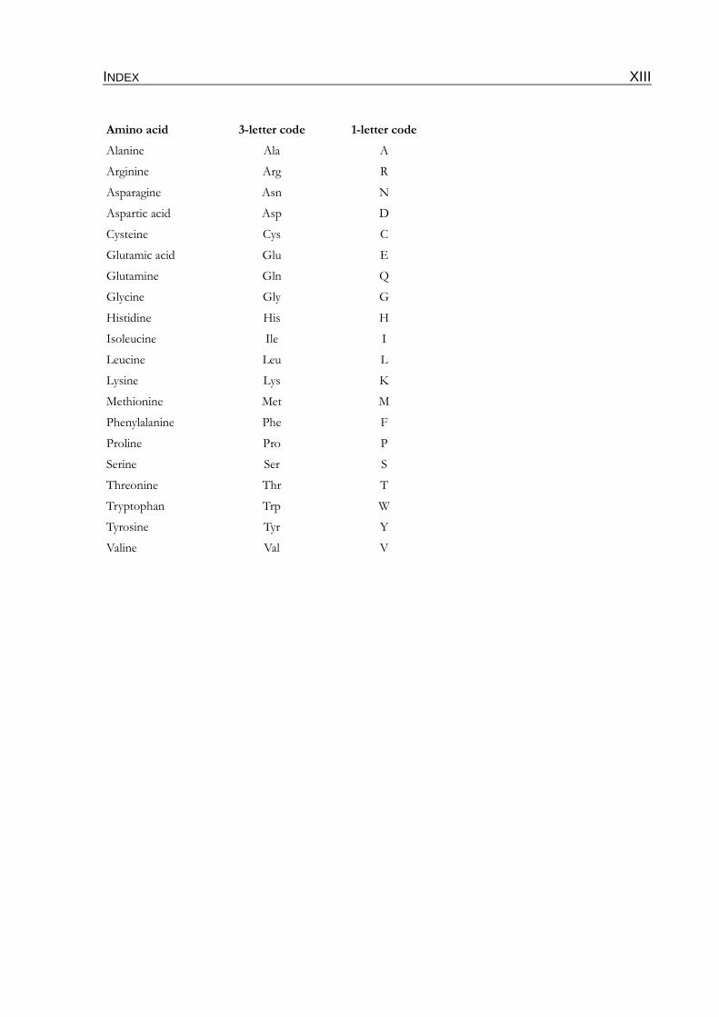
Amino acid 3-letter code 1-letter code
Alanine Ala A
Arginine Arg R
Asparagine Asn N
Aspartic acid Asp D
Cysteine Cys C
Glutamic acid Glu E
Glutamine Gln Q
Glycine Gly G
Histidine His H
Isoleucine Ile I
Leucine Leu L
Lysine Lys K
Methionine Met M
Phenylalanine Phe F
Proline Pro P
Serine Ser S
Threonine Thr T
Tryptophan Trp W
Tyrosine Tyr Y
Valine Val V
INTRODUCTION 14
1 INTRODUCTION
1.1 Alzheimer's disease (AD)
Alzheimer's disease (AD) is a neurodegenerative disorder, characterized by a progressive cognit-
ive decline. According to recent studies, more than 35 million people worldwide (Wimo & Prince
2010) or rather more than 800.000 people in Germany suffer from AD (Bickel 2010). Because of
an increased life expectancy in western industrial countries the number of dementia cases is
estimated to double every 20 years (Wimo & Prince 2010). AD is the most common form of
dementia diagnosed in mid-to-late life, affecting 7–10 % of all individuals over 65 years of age
and approximately 40 % of all persons over 80 years of age (Sisodia 1999; Prince & Jackson
2009). In the beginning of the disease, affected people show an impairment of short term
memory, language and cognitive functions. Further they have paranoia, delusions and show a loss
of social appropriateness. Later all these hallmarks become intensified and additional problems
with motor and sensory functions become manifested. AD is a terminal disease, but the most fre-
quent cause of death are pneumonia and myocardial infarction and not the disease itself (Forstl
& Kurz 1999). The symptoms are attributed to alterations or loss of neurons in several brain
areas or neural systems including the cortex, hippocampus, amygdala, anterior thalamus, basal
forebrain and several brainstem regions (Sisodia 1999; Wenk 2003; Jalbert et al., 2008).
More than 100 years after the first description of AD by the german psychiatrist Alois
Alzheimer, this severe form of dementia is still not fully understood. However, there is evidence
that besides age also high cholesterol levels and diabetes are risk factors for AD (Tan et al., 2003;
Akomolafe et al., 2006). AD cases can be classified into the so-called early onset forms and the
late onset forms. Only about 5 % of all AD cases account for early-onset AD (EOAD) in which
genetic mutations either in the amyloid precursor protein (APP) or in presenilins 1 and 2 (PS1,
PS2) are associated with an age-of-onset below 65 years, with some really severe mutations
already between 20-40 (Selkoe 2001).
In contrast, cases of late-onset AD (LOAD) which are also called sporadic AD are not linked to
mutations in APP or PS, but genetic determinations like ApoE4 are known to increase the risk
for developing AD.
INTRODUCTION 15
1.1.1 Neuropathological hallmarks of AD
Both the sporadic and the familiar forms exhibit the same histopathological characteristics: neur-
ofibrillary tangles and β-amyloid plaques, appearing mostly in the neocortex, hippocampus and in
the limbic system (Braak & Braak 1996). There are several hypotheses how these hallmarks can
account for the very complex pathology of AD and how they could be linked.
1.1.1.1 Neurofibrillary tangles
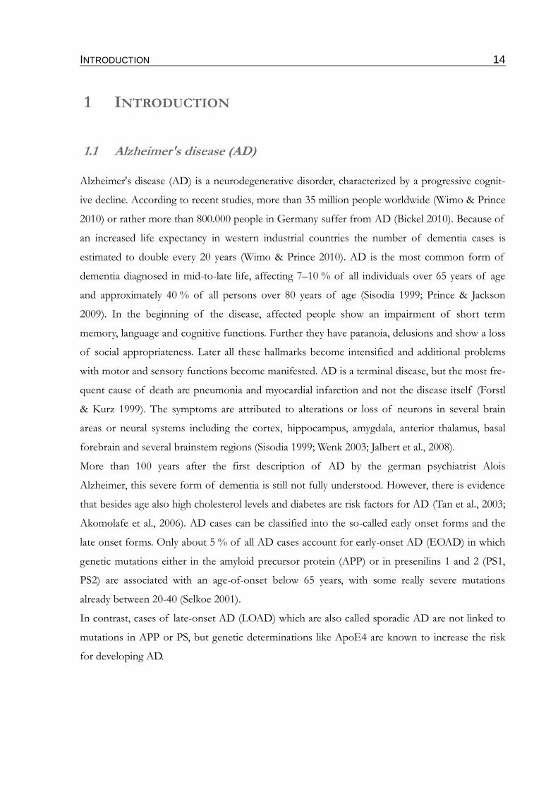
Neurofibrillary tangles (see fig. 1) are intraneuronal accumulations of hyperphosphorylated forms
of the tau protein. Under physiological conditions tau is associated with microtubules in axonal
compartments, forming and stabilizing their structure (Grundke-Iqbal et al., 1986a; Goedert et
al., 1988; Friedhoff et al., 2000). Aside this, tau might have additional functions due to its interac-
tion with the plasma membrane (Brandt et al., 1995; Maas et al., 2000) and the actin cytoskeleton
(Fulga et al., 2007). Furthermore, a recent paper also highlights a localization and function of tau
in dendritic compartments without association to microtubules (Ittner et al., 2010). The associ-
ation of tau with microtubules is regulated by phosphorylation (Drewes et al., 1995). Several
kinases including the glycogen synthase kinase 3β (GSK3β) also known as tau kinase 1, the cyclin
dependent kinase 5 (Cdk5) and the mitogen activated proteinkinase (MAPK) are known to phos-
phorylate tau (Ishiguro et al., 1993; Chung 2009). Under conditions present in AD, tau becomes
hyperphosphorylated. The hyperphosphorylated tau loses its binding capacity to the microtu-
bules, thereby destabilizing their structure. Additionally, the hyperphosphorylation enables tau to
aggregate into so-called paired helical filaments (PHFs) (Alonso et al., 2001). Continuous aggreg-
ation of PHFs finally forms the neurofibrillary tangles in the soma of neurons. The combination
of the loss of function, compromising axonal transport through changes of microtubules
dynamics and the toxic gain of function forming intracellular protein aggregates results finally in
cell death of the affected neurons (Grundke-Iqbal et al., 1986b; Ishiguro et al., 1993; Li et al.,
2006; Li & Paudel 2006; Ballatore et al., 2007). Thus, the intracellular neurofibrillary tangles
become detectable in the extracellular environment (Bondareff et al., 1994).
There is strong evidence that the activity of protein phosphatase 2A, responsible for the dephos-
phorylation of tau, is reduced in the brain of AD patients (Gong et al., 1994; Tian & Wang 2002;
Chung 2009). However, to date it is not clear whether the hyperphosphorylation of tau is con-
nected with decreased dephosphorylation, hyperactivity of the tau related kinases or both (Man-
delkow & Mandelkow 1998).
INTRODUCTION 16
In the tau hypothesis neuronal loss as a consequence of the accumulation of neurofibrillary
tangles was described as the initial events in the development of AD. However, there are some
findings that challenge this. The presence of hyperphosphorylated tau and tangles is not limited
to AD (Joachim & Selkoe 1992; Morris et al., 2001). Other neurodegenerative diseases character-
ized by the aggregation of tau are classified as tauopathies. Important members of this group are
the frontotemporal dementia (Dickson 2009). Moreover, there is no way known in which tau
could cause amyloid plaques. However, it was shown that tau is mediating Aβ induced cognitive
impairments and excitotoxicity. The reduction of endogenous tau in APP transgenic mice pre-
vents behavioral deficits without affecting Aβ levels by blocking Aβ- and excitotoxin-induced
neuronal dysfunction (Roberson et al., 2007). Although the exact mechanism for the rescue is
unclear, it might be linked to the prevention of Aβ-induced axonal transport defects (Vossel et
al., 2010). While incubation of primary hippocampal neurons with oligomeric Aβ rapidly inhib-
ited axonal transport of mitochondria and the neutrotrophin receptor TrkA, these transport
defects were rescued in primary neurons of tau -/- and tau +/- mice (Vossel et al., 2010). Another
recent study suggests a link by which tau could facilitate Aβ toxicity to synapses. Dendritic local-
ized tau supports the recruitment of the Src-family kinase Fyn to NMDA (N-methyl-D-aspartic
acid) receptors on postsynaptic membranes. The resulting phosphorylation of NMDA receptors
by Fyn renders them more susceptible to Aβ (Ittner et al., 2010).
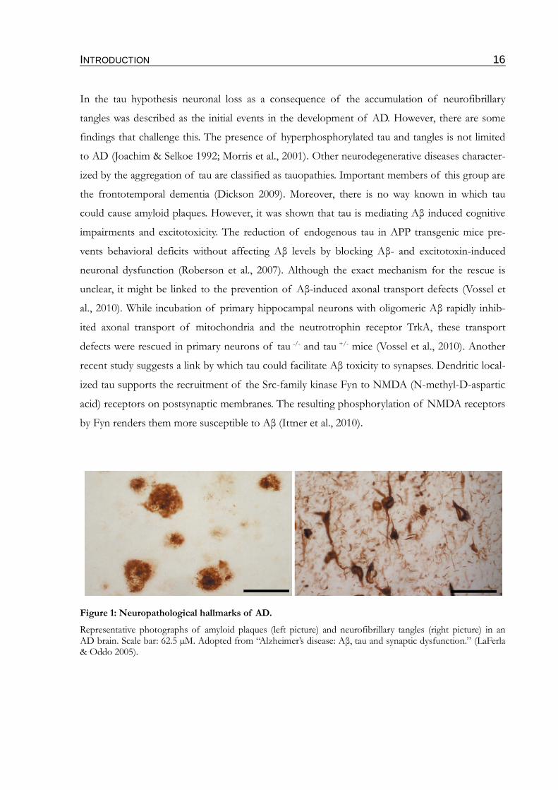
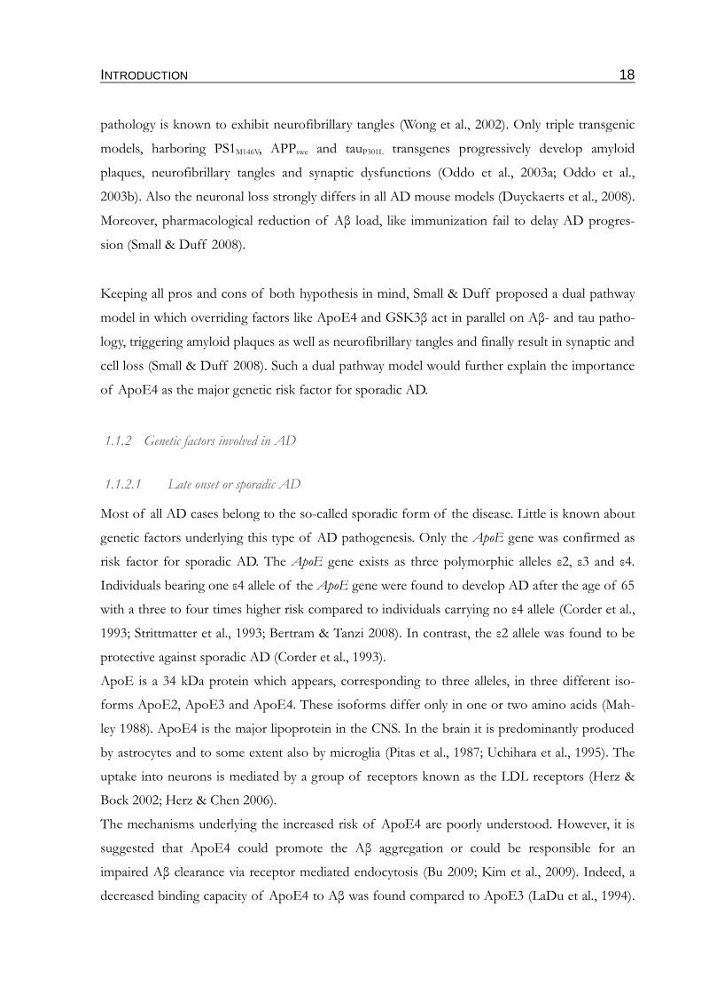
Figure 1: Neuropathological hallmarks of AD.
Representative photographs of amyloid plaques (left picture) and neurofibrillary tangles (right picture) in an AD brain. Scale bar: 62.5 µM. Adopted from “Alzheimer’s disease: Aβ, tau and synaptic dysfunction.” (LaFerla & Oddo 2005).
INTRODUCTION 17
1.1.1.2 β-amyloid plaques
In contrast to neurofibrillary tangles, amyloid plaques (fig. 1) are extracellular deposits that are
also detectable in the basement membranes of cerebral vessels. One main component of β-
amyloid plaques is the small hydrophobic β-amyloid peptide (Aβ) which is derived from the
amyloid precursor protein (APP), as explained in 1.1.3. Plaques can be microscopically subclassi-
fied into neuritic, also called senile plaques, and diffuse plaques. Neuritic plaques are spherical
multicellular lesions, consisting of Aβ fibrils intermixed with non fibrillar variants of the peptide
as well as degenerated axons and neurites (Braak & Braak 1996). These compact structures also
contain a variable number of activated microglia within and near the fibrillar core. Furthermore,
the plaques are often surrounded by reactive astrocytes (Mattiace et al., 1990; Pike et al., 1994;
Jefferies et al., 1996; Stalder et al., 1999).
Diffuse plaques appear mainly in the cerebellum (Yamaguchi et al., 1989). They are mainly com-
posed of non-fibrillar Aβ (Tagliavini et al., 1988; Yamaguchi et al., 1988), thereby representing
most likely initial stages of the plaque pathology in AD (Tagliavini et al., 1988; Giaccone et al.,
1989). In contrast to neuritic plaques which are only found in brains of AD patients, diffuse
plaques are also present in brains of healthy older people (Hardy & Selkoe 2002). Although
plaques are strong characteristics of AD their density and distribution pattern turned out to be
of limited significance for differentiation of neuropathological stages (Braak & Braak 1991).
According to the amyloid-cascade hypothesis, Aβ is the triggering factor of a severe pathological
cascade. The accumulation of Aβ leads to neuronal dysfunction, followed by inflammatory pro-
cesses. The resulting progressive synaptic and neuritic injury entails impaired neuronal homeo-
stasis, thereby altering the activity of several kinases and phosphatases leading to hyperphos-
phorylation of tau and favoring the generation of neurofibrillary tangles. Aβ plaques and neur-
ofibrillary tangles together cause widespread neuronal deficits which end up in dementia (Hardy
& Selkoe 2002). There are a lot of in vitro findings supporting this Aβ cascade hypothesis: the
impairment of synaptic activity in hippocampal slice cultures through treatment with naturally
secreted Aβ oligomers (Lambert et al., 1998; Walsh et al., 2002) and Aβ oligomeric species
extracted from human brains (Shankar et al., 2008) as well as the loss of memory and cognitive
functions of mice and rats injected with natural Aβ oligomers (Cleary et al., 2005; Lesne et al.,
2006; Shankar et al., 2008). There are also some in vitro evidences that Aβ can modulate the
activity of tau phosphorylating kinases (Hooper et al., 2008; Magdesian et al., 2008; De Felice et
al., 2009; Lee et al., 2009). However, these findings could not be confirmed in animal models.
No APP single transgenic or APP/PS1 double transgenic animal model which develop plaque
INTRODUCTION 18
pathology is known to exhibit neurofibrillary tangles (Wong et al., 2002). Only triple transgenic
models, harboring PS1M146V, APPswe and tauP301L transgenes progressively develop amyloid
plaques, neurofibrillary tangles and synaptic dysfunctions (Oddo et al., 2003a; Oddo et al.,
2003b). Also the neuronal loss strongly differs in all AD mouse models (Duyckaerts et al., 2008).
Moreover, pharmacological reduction of Aβ load, like immunization fail to delay AD progres-
sion (Small & Duff 2008).
Keeping all pros and cons of both hypothesis in mind, Small & Duff proposed a dual pathway
model in which overriding factors like ApoE4 and GSK3β act in parallel on Aβ- and tau patho-
logy, triggering amyloid plaques as well as neurofibrillary tangles and finally result in synaptic and
cell loss (Small & Duff 2008). Such a dual pathway model would further explain the importance
of ApoE4 as the major genetic risk factor for sporadic AD.
1.1.2 Genetic factors involved in AD
1.1.2.1 Late onset or sporadic AD
Most of all AD cases belong to the so-called sporadic form of the disease. Little is known about
genetic factors underlying this type of AD pathogenesis. Only the ApoE gene was confirmed as
risk factor for sporadic AD. The ApoE gene exists as three polymorphic alleles ε2, ε3 and ε4.
Individuals bearing one ε4 allele of the ApoE gene were found to develop AD after the age of 65
with a three to four times higher risk compared to individuals carrying no ε4 allele (Corder et al.,
1993; Strittmatter et al., 1993; Bertram & Tanzi 2008). In contrast, the ε2 allele was found to be
protective against sporadic AD (Corder et al., 1993).
ApoE is a 34 kDa protein which appears, corresponding to three alleles, in three different iso-
forms ApoE2, ApoE3 and ApoE4. These isoforms differ only in one or two amino acids (Mah-
ley 1988). ApoE4 is the major lipoprotein in the CNS. In the brain it is predominantly produced
by astrocytes and to some extent also by microglia (Pitas et al., 1987; Uchihara et al., 1995). The
uptake into neurons is mediated by a group of receptors known as the LDL receptors (Herz &
Bock 2002; Herz & Chen 2006).
The mechanisms underlying the increased risk of ApoE4 are poorly understood. However, it is
suggested that ApoE4 could promote the Aβ aggregation or could be responsible for an
impaired Aβ clearance via receptor mediated endocytosis (Bu 2009; Kim et al., 2009). Indeed, a
decreased binding capacity of ApoE4 to Aβ was found compared to ApoE3 (LaDu et al., 1994).
INTRODUCTION 19
Besides ApoE, recent genome wide association studies identified variants of CLU and PICALM
to be associated with AD risk (Harold et al., 2009; Corneveaux et al., 2010; Kamboh et al., 2010).
Nevertheless, all of them have much lower significance compared to ApoE.
1.1.2.2 Early onset or familiar AD
About 5 % of all AD cases are linked to mutations in AD relevant genes. Since these forms
appear before the age of 65, they are often referred as EOAD (Selkoe 2001). All known muta-
tions linked with EOAD are located in the genes coding for APP or presenilin 1 and 2 and are
mainly associated with increased Aβ42/Aβ40 ratio and/or alteration of the Aβ aggregation (Goate
et al., 1991; Citron et al., 1992; Cai et al., 1993; Borchelt et al., 1996). All mutations in APP are
located within or nearby the Aβ domain (Wolfe 2007). A well analyzed mutation, which belongs
to this group is the swedish double mutation. This mutation was initially found in a swedish fam-
ily with EOAD (Mullan et al., 1992). The mutations K595N/M596L in APP increases the affinity
of BACE-1 to its substrate APP (Citron et al., 1992; Cai et al., 1993; Vassar et al., 1999). Thus,
APP bearing this mutation is cleaved earlier and more efficiently in the secretory pathway by
BACE-1 than APP WT (Haass et al., 1995; Thinakaran et al., 1996). Other mutations in APP near
the γ-secretase cleavage site lead to increased production of the more aggregation-prone Aβ42 rel-
ative to Aβ40. Mutations in the Aβ region itself alter the biophysical properties of the peptide,
thereby changing its aggregation and degradation behavior (Wolfe 2007).
So far, more than 160 mutations were identified in the genes encoding PS1 and 2. Most of them
are simple missense mutations resulting in the exchange of one amino acid in PS1, but there are
also more complex ones, for example small deletions or insertions. The most severe mutation is a
splice mutation leading to the deletion of exon 9 (∆exon9) (De Strooper 2007). Such AD causing
mutations in PS1 lead both to increased Aβ42/Aβ40 ratios (Goate et al., 1991; Citron et al., 1992;
Cai et al., 1993; Borchelt et al., 1996) and partial loss of γ-secretase function (Song et al., 1999;
Bentahir et al., 2006; Tamboli et al., 2008). To explain this apparent paradox at least two different
cleavage sites of γ-secretase in APP were assumed. Cleavage at the so-called ε-site produces the
C-terminus of Aβ and releases the AICD. Afterwards the cleavage at the γ-site releases Aβ. This
cleavage site can vary in its position, leading to the various Aβ-species (Aβ40 and Aβ42). Longer
forms of Aβ (e.g. Aβ42) are thought to be retained in the active site of the γ-secretase because of
their larger hydrophobic tm-domain. Shorter variants (e.g. Aβ40) in contrast are more likely to be
released. A less active γ-secretase would thereby allow more time for the release of the longer,
more hydrophobic Aβ42 (Qi-Takahara et al., 2005; Wolfe 2007).
INTRODUCTION 20
1.1.3 Characteristics of the β-amyloid precursor protein (APP)
As mentioned above, Aβ originates from a sequential processing of APP, a large type I trans-
membrane protein, expressed in several human tissues. APP and its homologues APLP-1 and
APLP-2 (amyloid precursor like protein-1 and -2) form a small family of proteins of 100-
140 kDa in size, which are ubiquitously expressed throughout mammalians as well as Caenorhab-
ditis elegans and Drosophila melanogaster (Goldgaber et al., 1987; Rosen et al., 1989; Luo et al., 1990;
Daigle & Li 1993; Zheng & Koo 2006). Additional heterogeneity is caused on the one hand by
alternative splicing (Weidemann et al., 1989; Oltersdorf et al., 1990; Hung & Selkoe 1994), produ-
cing several isoforms of which APP 695, 751 and 770 are the most abundant in the brain (Wer-
tkin et al., 1993). On the other hand a couple of posttranslational modifications as O- and N-
glycosylation (Tomita et al., 1998), sulfation as well as phosphorylation (Hung & Selkoe 1994;
Walter et al., 1997) raises the heterogeneity of this group of proteins. APP can be divided in
three main parts (see fig. 2), a 47 aa long C-terminal cytoplasmic domain, a 24 aa long transmem-
brane domain (tm-domain) and a N-terminal extracellular domain, varying in size between the
different isoforms.
There are several physiological functions discussed for APP, including modulation of cell-cell
interaction, cell adhesion, signal transduction and neurite outgrowth (Milward et al., 1992;
Nishimoto et al., 1993; Koo 2002). Cell-cell contacts and cell adhesion could be mediated by the
E1 domain of the extracellular part by which APP is able to form homodimers or heterodimers
with its homologues APLP-1 and -2 (Soba et al., 2005).
While single gene knock out mice for APP, APLP-1 or APLP-2 show only mild phenotypes,
double knockout models (APP/APLP-2; APLP-1/APLP-2) are lethal, showing that the APP pro-
tein family plays important physiological roles, that are partly redundant (Zheng et al., 1995; von
Koch et al., 1997; Heber et al., 2000). Particularly the Aβ domain, which consists of the last 28 aa
of the APP ectodomain and the first 12-14 aa of the tm-domain and is not present in APLPs,
cannot play a pivotal physiological role. However, a recent study demonstrates a ferroxidase activ-
ity of APP and a role in neuronal iron-export. Intracellular iron accumulates in primary neurons
of APP knock out mice. Moreover, APP knock out mice fed with an iron dietary exhibit
increased Fe2+ levels, resulting in oxidative stress in cortical neurons (Duce et al., 2010).
INTRODUCTION 21
1.1.4 APP processing
The release of Aβ from APP is mediated by two different proteases. These so-called secretases
cleave APP sequentially in the lumen of cellular organelles e.g. Golgi, endosomes and lysosomes
or at the cell surface. The first cleavage is catalyzed by β-secretase at position 1 of the Aβ domain
(see fig. 2 and 3), thereby releasing the soluble APP fragment (sAPPβ) and a membrane bound C-
terminal fragment (CTF) with a size of 99 aa (CTFβ or C99). Further processing of CTFβ by a
multienzyme complex called γ-secretase leads to the production of Aβ and the release of the
APP intracellular domain (AICD) into the cytosol (Haass et al., 1992b; Sastre et al., 2001; Haass
& Steiner 2002). Since γ-secretase can cleave C99 at different sites, several Aβ variants with sizes
of 37 aa–42 aa arise. However, Aβ40 (~90 %) and Aβ42 (~10 %) are the variants most frequently
released from the membrane (Citron et al., 1996; Weggen et al., 2001; Wiltfang et al., 2002).
Importantly, Aβ42 is, based on its hydrophobicity, most prone to aggregate and form plaques
(Barrow & Zagorski 1991; Jarrett et al., 1993). Alternatively, APP can be processed in the non
amyloidogenic pathway which results in the generation of a small, non amyloidogenic peptide,
named p3. In this pathway the first cleavage of APP within the Aβ domain at aa 16 (see fig. 2 and
11) is catalyzed by the α-secretase (Esch et al., 1990; Sisodia et al., 1990) releasing sAPPα into the
extracellular space and a CTF which is 83 aa in size (CTFα or C83).
The subsequent γ-secretase cleavage of CTFα results in the release of the non amyloidogenic p3
peptide (Haass et al., 1993; Sastre et al., 2001) and AICD, like in the amyloidogenic pathway. The
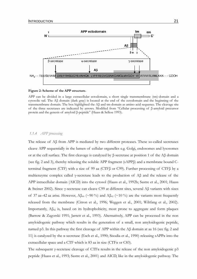
Figure 2: Scheme of the APP structure.
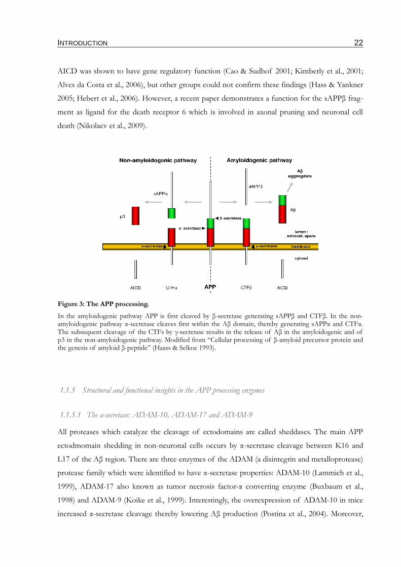
APP can be divided in a large extracellular ectodomain, a short single transmembrane (tm)-domain and a cytosolic tail. The Aβ domain (dark gray) is located at the end of the ectodomain and the beginning of the transmembrane domain. The box highlighted the Aβ and tm-domain as amino acid sequence. The cleavage site of the three secretases are indicated by arrows. Modified from “Cellular processing of β-amyloid precursor protein and the genesis of amyloid β-peptide” (Haass & Selkoe 1993).
INTRODUCTION 22
AICD was shown to have gene regulatory function (Cao & Sudhof 2001; Kimberly et al., 2001;
Alves da Costa et al., 2006), but other groups could not confirm these findings (Hass & Yankner
2005; Hebert et al., 2006). However, a recent paper demonstrates a function for the sAPPβ frag-
ment as ligand for the death receptor 6 which is involved in axonal pruning and neuronal cell
death (Nikolaev et al., 2009).
1.1.5 Structural and functional insights in the APP processing enzymes
1.1.5.1 The α-secretase: ADAM-10, ADAM-17 and ADAM-9
All proteases which catalyze the cleavage of ectodomains are called sheddases. The main APP
ectodmomain shedding in non-neuronal cells occurs by α-secretase cleavage between K16 and
L17 of the Aβ region. There are three enzymes of the ADAM (a disintegrin and metalloprotease)
protease family which were identified to have α-secretase properties: ADAM-10 (Lammich et al.,
1999), ADAM-17 also known as tumor necrosis factor-α converting enzyme (Buxbaum et al.,
1998) and ADAM-9 (Koike et al., 1999). Interestingly, the overexpression of ADAM-10 in mice
increased α-secretase cleavage thereby lowering Aβ production (Postina et al., 2004). Moreover,
Figure 3: The APP processing.
In the amyloidogenic pathway APP is first cleaved by β-secretase generating sAPPβ and CTFβ. In the non-amyloidogenic pathway α-secretase cleaves first within the Aβ domain, thereby generating sAPPα and CTFα. The subsequent cleavage of the CTFs by γ-secretase results in the release of Aβ in the amyloidogenic and of p3 in the non-amyloidogenic pathway. Modified from “Cellular processing of β-amyloid precursor protein and the genesis of amyloid β-peptide” (Haass & Selkoe 1993).
INTRODUCTION 23
the ectodomain shedding of APP can be increased by activation of protein kinase C (Etcheber-
rigaray et al., 2004).
All ADAMs are type I transmembrane zinc metalloproteases and cleave APP on the secretory
pathway from the trans Golgi network (TGN) to the plasma membrane or directly at the plasma
membrane (Haass et al., 1992a; Sisodia 1992; Chyung & Selkoe 2003). During the past years the
ADAM family appeared to be one of the most important groups of sheddases, with several sub-
strates involved in various signaling pathways. In addition to APP, the type I transmembrane pro-
teins TNFα, the Notch receptors, p75, TNFα-receptor and L-selectin are substrates of the
ADAM family (Seals & Courtneidge 2003). Mice deficient for either ADAM-10 or ADAM-17 are
lethal already in embryonic state, confirming the important physiological role of these sheddases
(Peschon et al., 1998; Hartmann et al., 2002).
1.1.5.2 The β-secretase: BACE-1
The shedding of the APP ectodomain is essential for the release of Aβ, but for a long time the
exact protein acting as β-secretase was unknown. In the late '90s a type I transmembrane aspartyl
protease, the β-site APP cleaving enzyme-1 (BACE-1), also known as aspartic protease-2 or
memapsin-2 (membrane-associated aspartic protease-2) could be identified as β-secretase (Hus-
sain et al., 1999; Sinha et al., 1999; Vassar et al., 1999; Yan et al., 1999; Lin et al., 2000). Like all
type I transmembrane proteins BACE-1 consists of an ectodomain, a transmembrane domain
and a C-terminal cytosolic tail (Hussain et al., 1999; Sinha et al., 1999; Vassar et al., 1999; Yan et
al., 1999; Lin et al., 2000). The ectodomain bears the typical DTGS and DSGT amino acid
motifs, forming the catalytic center of aspartyl proteases (Hussain et al., 1999; Vassar et al., 1999;
Yan et al., 1999). Mutation of one of these motifs abolishes the protease activity completely
(Hussain et al., 1999).
As other aspartic proteases, BACE-1 contains a propeptide, located at the N-terminus of the
enzyme, which is cleaved during maturation by furin or a furin-like protease (Bennett et al.,
2000b; Capell et al., 2000; Creemers et al., 2001). Furthermore, the ectodomain contains four
asparagine residues which undergo complex N-glycosylation during the transport from the ER to
the Golgi (Capell et al., 2000; Huse et al., 2000). Fully matured BACE-1 is transported from the
Golgi to the cell surface. The transport of BACE-1 is mediated by motifs located in the cytoplas-
mic domain, since the deletion of this part results in an accumulation of BACE-1 in the ER
(Capell et al., 2000). From the cell surface, BACE-1 can be re-internalized into endosomal com-
partments from which it is either recycled back to the plasma membrane or transported to the
INTRODUCTION 24
TGN (Walter et al., 2001a) or lysosomes (Koh et al., 2005). While the re-internalization is medi-
ated by a di-leucine motif in the C-terminus (Huse et al., 2000; Pastorino et al., 2002), the trans-
port to the TGN is controlled by the phosphorylation state of the adjacent serine residue S498
(Walter et al., 2001a). Both the di-leucine motif and serine 498 are part of a DISLL motif which
follows the structure DxxLL, known to serve as the binding site for a particular group of trans-
port proteins, the GGAs (Golgi associated, γ-adaptin ear containing, ARF binding proteins).
These are monomeric adapter proteins which sort specific cargo-proteins like the mannose-6-
phosphate receptor from the TGN to endosomal/lysosomal compartments (Bonifacino 2004;
Robinson 2004). BACE-1 was shown to bind GGAs (He et al., 2002; Shiba et al., 2004).
Moreover, it was shown that the DxxLL motif regulates the retrograde transport of internalized
BACE-1 from endosomal compartments to the TGN in a phosphorylation dependent manner
(Wahle et al., 2005). In addition to the critical DxxLL motif, the C-terminus of BACE-1 contains
also three cysteine residues which can be palmitoylated (Benjannet et al., 2001). The palmitoyla-
tion leads to the insertion of a second membrane anchor which might take part in regulating the
subcellular localization (Schweizer et al., 1996; Vetrivel et al., 2009). Although β-secretase activity
was detected in various cell types and tissues, the highest BACE-1 activity was measured in neur-
onal tissue, particularly in neurons (Haass et al., 1992b; Shoji et al., 1992; Seubert et al., 1993).
Biochemical studies revealed that BACE-1 shows highest activity at pH 5 (Vassar & Citron 2000;
Walter et al., 2001b). Accordingly, BACE-1 was mainly found in endosomal and lysosomal com-
partments.
Less is known about the physiological role of BACE-1, since BACE-1 knock out mice show no
overt phenotype. However, more careful analysis of BACE-1 deficient mice revealed a decreased
myelination, suggesting a role of BACE-1 in the formation of myelin sheets. Responsible for this
defect might be the impaired cleavage of the transmembrane protein neuregulin-1 by BACE-1
(Aoki et al., 2004; Hu et al., 2006; Willem et al., 2006; Hu et al., 2008). Physiologically neuregulin-
1 plays a role in the maintenance of heart function and in myelination of axons. Other known
BACE-1 substrates are beside APP also the APLPs (Li & Sudhof 2004), the sialyltransferase
ST6Gal-I (Kitazume et al., 2003), the P-selectine glycoprotein ligand-1 (PSGL-1) (Lichtenthaler
et al., 2003) and LRP (low density lipoprotein receptor related protein) (von Arnim et al., 2005).
In parallel to BACE-1 a homologue protein was identified as β-site APP cleaving enzyme-2. Both
proteins have similar structural organization and share more than 50 % sequence identity on the
amino acid level (Vassar et al., 1999; Yan et al., 1999; Acquati et al., 2000; Hussain et al., 2000; Lin
et al., 2000). Consistent with the high molecular structure identity to BACE-1, BACE-2 under-
goes comparable posttranslational modifications like complex N-glycosylation at asparagine
INTRODUCTION 25
residues located in the ectodomain (Vassar & Citron 2000; Walter et al., 2001b). Immature
BACE-2 also contains a propeptide which is cleaved during the maturation. However, while this
maturation step is catalyzed by furin proteases in BACE-1, the propeptide of BACE-2 appears to
be removed autocatalytically (Hussain et al., 2001; Yan et al., 2001). Although both enzymes are
very similar, BACE-2 shows striking differences regarding stability and intracellular transport
(Fluhrer et al., 2002). Compared to BACE-1 that is highly expressed in neurons, there is relative
low expression of BACE-2 in the nervous system, but higher in peripheral tissues (Vassar et al.,
1999; Bennett et al., 2000a). The highest expression in the nervous system is observed in glia cells
(Dominguez et al., 2005). Since the bace2 gene is localized on chromosome 21, a contribution of
BACE-2 to the AD-like pathology in Down's syndrome and in AD was suggested (Yan et al.,
1999; Solans et al., 2000; Vassar & Citron 2000; Walter et al., 2001b). Interestingly, BACE-2
mRNA and protein levels were increased in individuals with Down's syndrome (Motonaga et al.,
2002; Barbiero et al., 2003).
As shown by cell biological studies, BACE-2 is able to cleave APP, but the cleavage efficiency at
the β-secretase site is very low (Bayer et al., 1999; Farzan et al., 2000; Hussain et al., 2000; Lin et
al., 2000; Yan et al., 2001). Rather, BACE-2 cleaves APP in an α-secretase-like manner within the
Aβ domain between phe 19 and phe 20 as well as between phe 20 and ala 21 (Farzan et al., 2000;
Yan et al., 2001; Fluhrer et al., 2002). Accordingly, protein expression studies in human brain of
AD patients and aged-matched controls, can detect BACE-2, but the expression and activity is
not significantly altered in AD brains (Stockley et al., 2006; Ahmed et al., 2010).
1.1.5.3 The γ-secretase
The γ-secretase is a multiprotein complex consisting of four essential components. The catalytic
center of the complex is formed by PS1 or its homologue PS2 (Levy-Lahad et al., 1995; Sherring-
ton et al., 1995), supported by three additional co-factors: nicastrin (Yu et al., 2000; Edbauer et
al., 2002b; Kopan & Goate 2002; Lai 2002), APH-1 (anterior pharynx-defective-1) (Francis et al.,
2002; Goutte et al., 2002; Lee et al., 2002; Luo et al., 2003) and PEN-2 (presenilin enhancer-2)
(Francis et al., 2002; Steiner et al., 2002; Luo et al., 2003). These components appear in a stoi-
chiometric ratio of 1:1:1:1 in the mature γ-secretase complex (Sato et al., 2007). Since the total
molecular mass of the γ-secretase complex is higher than the summarized molecular masses of
the single components, it was early considered that other unidentified proteins take part in the
complex. In fact, it was shown that γ-secretase modulators (GSMs) like TMP21 (Chen et al.,
2006; Pardossi-Piquard et al., 2009), CD147 (Zhou et al., 2005) and the recently found GSAP (γ-
INTRODUCTION 26
secretase activating protein) (He et al., 2010) are associated with the γ-secretase complex and can
modulate γ-secretase activity. However, purification of γ-secretase during which the GSMs are
separated early from the rest of the γ-secretase complex, and subsequent activity assays with the
purified γ-secretase, revealed that GSMs are not relevant for the basal activity of the γ-secretase
complex. Thus, the four main components PS, nicastrin, APH-1 and PEN-2 are sufficient for an
active γ-secretase complex (Winkler et al., 2009).
PS1 and PS2 are membrane proteins with nine transmembrane domains, bearing the two catalytic
aspartate residues (Spasic et al., 2006). In the γ-secretase complex, presenilins are present as a het-
erodimer formed by a C-terminal and an N-terminal fragment, each of them containing one cata-
lytic aspartate residue. The two fragments are most likely generated by autocatalytic endoproteo-
lysis between tm-domain 6 and tm-domain 7 (Ratovitski et al., 1997; Capell et al., 1998; Wolfe et
al., 1999a; Wolfe et al., 1999b; Kimberly et al., 2000).
Nicastrin, a glycosylated type I transmembrane protein was shown to be involved in the substrate
recognition. Nicastrin binds the extracellular/lumenal N-terminus, generated upon ectodomain
shedding of type I transmembrane proteins, thereby recruiting the substrate into the catalytic
center. It was found that the recognition is not mediated by the amino acid sequence of the sub-
strates. Only a short free N-terminal part above the cell membrane seems to be necessary (Yu et
al., 2000; Shah et al., 2005).
PEN-2 and APH-1 are both polytopic transmembrane proteins. Only little is known about the
function of these proteins. However, PEN-2 is supposed to support the endoproteolytic cleavage
of presenilin, because PEN-2 deficiency stabilized the presenilin holoprotein (Hu & Fortini 2003;
Luo et al., 2003; Takasugi et al., 2003; Prokop et al., 2004). Although the assembly of the whole
γ-secretase complex is not yet fully understood, there is evidence that APH-1 supports this pro-
cess and is involved in maturation and transport (Luo et al., 2003; Takasugi et al., 2003; Kaether
et al., 2006a). There are studies showing that only fully assembled complexes can be transported
via the secretory pathway to the plasma membrane and early endosomes where most of the γ-
secretase is located (Walter et al., 1998; Annaert et al., 1999; Kaether et al., 2006a), while unas-
sembled, single components were retained in the ER (Kaether et al., 2006a; Dries & Yu 2008).
To date a couple of different γ-secretase substrates are identified. Among these are APP, Notch
(De Strooper et al., 1999; Saxena et al., 2001) and other proteins which have important physiolo-
gical functions e.g. APLP-1 and -2 (Walsh et al., 2003), E- and N-cadherin (Marambaud et al.,
2002), EphB (Georgakopoulos et al., 2006) and LRP (May et al., 2002). Another important pro-
tein which is processed by γ-secretase is the widely expressed cell-adhesion protein CD44
(Okamoto et al., 2001; Lammich et al., 2002; Murakami et al., 2003), implicated amongst others in
INTRODUCTION 27
leukocyte homing and activation as well as cell migration.
Since the cleavage reaction occurs in all cases inside the plasma membrane and not in a water
containing environment, the process is called regulated intramembrane proteolysis (RIP). To
afford proteolysis within the membrane, it was suggested that the γ-secretase complex forms a
water containing pore in which the cleavage reaction is catalyzed. Because of the high number of
γ-secretase substrates and its less cleavage specificity, the γ-secretase was long time assumed to be
a “proteasome of the membrane” (Kopan & Ilagan 2004). However, an unbiased proteomic
screen revealed exact substrate characteristics, as the short ectodomain and permissive transmem-
brane and cytoplasmic domain (Hemming et al., 2008). Besides the release of gene regulatory
intracellular domains, like the Notch ICD (De Strooper et al., 1999; Saxena et al., 2001), γ-
secretase can directly affect the Wnt signaling pathway by direct interaction with GSK3β and β-
catenin (Prager et al., 2007). PS1 mediated processing of Notch, which is involved in transcrip-
tional regulation of developmental genes is a crucial function of γ-secretase. Because of this
PS -/- mice and PS1/PS2 knockout mice resemble a Notch knock out phenotype to some extent,
and are lethal directly after birth (Shen et al., 1997). However, PS2 -/- mice are viable, indicating
that PS2 could not compensate PS1 deficiency.
There is evidence that γ-secretase function is also important for endocytosis. In presenilin defi-
cient cells the endocytosis of the LDL-receptor is impaired, leading to upregulation of choles-
terol biosynthesis (Tamboli et al., 2008).
Recently, a very interesting function of γ-secretase in neurogenesis was published. Neural pro-
genitor cells (NPCs) from mice expressing a variant of presenilin linked to familiar AD (PS FAD)
show a defect in proliferation (Choi et al., 2008). Moreover, NPCs cultured in medium of
microglia from PS FAD mice exhibit a significant less proliferation rate than the NPCs in
medium of control microglia. Based on these results a non-cell-autonomous mechanism was sup-
posed by which γ-secretase modulates neurogenesis (Choi et al., 2008). In a subsequent study the
authors could show that microglia from PS1 FAD mice produce altered chemokine levels and
that these chemokines are closely linked to neurogenesis (Veeraraghavalu et al., 2010). Although
γ-secretase expression was mainly shown in neurons. These results give the first hints for γ-
secretase function in microglia. According to this, the presence of presenilin and nicastrin in
microglia and reactive astrocytes following traumatic brain injury (Nadler et al., 2008) might be
related to the modulation of neurogenesis.
INTRODUCTION 28
1.2 Aβ clearance in the brain
While EOAD is predominantly caused by increased Aβ42/Aβ40 ratio or altered aggregation beha-
vior of Aβ, the reasons for developing sporadic AD are largely unclear. Most likely, a misregula-
tion of either Aβ production, Aβ clearance or both could contribute to sporadic pathogenesis. In
fact, the activity of BACE-1 which catalyzes the rate limiting step in the generation of Aβ is
increasing with age in humans (Tyler et al., 2002; Kern et al., 2006; Stockley et al., 2006) as well as
in mice (Apelt et al., 2004). In contrast, the amount and activity of Aβ degrading enzymes is
reduced with age (Wang et al., 2006). Moreover, there are findings that the ability of microglia to
clear Aβ decreases also with age (Hickman et al., 2008).
There are different pathways by which Aβ can be cleared from the brain:
Efflux of soluble Aβ to the peripheral circulation through the blood brain barrier.
The blood brain barrier (BBB) is a highly specialized endothelial structure of the brain cells, sep-
arating together with astrocytes, pericytes and microglia components of the blood stream from
neurons (Zlokovic 2008). To clear sAβ from the brain, it can be transported across the BBB into
brain blood vessels by LRP1 mediated transcytosis (Shibata et al., 2000; Deane et al., 2008b;
Deane et al., 2009). Therefore sAβ either binds directly (Deane et al., 2004) or in complex with
ApoE (DeMattos et al., 2004; Bell et al., 2007; Deane et al., 2008a) or α2-macroglobulin (Narita et
al., 1997; Qiu et al., 1999) to LRP-1, located on brain endothelial cells. A relatively minor part
(10-15 % of total Aβ clearance) is directly transported by bulk flow of interstitial fluid into
cerebrospinal fluid, followed by drainage into the blood stream (Shibata et al., 2000; Deane et al.,
2009). Furthermore, there is an active transport of Aβ across the BBB, mediated by the P-gly-
coprotein efflux pump, highly expressed on the lumenal surface of brain capillary endothelial
cells (Lam et al., 2001; Cirrito et al., 2005; Kuhnke et al., 2007).
Proteolytic degradation of Aβ.
sAβ has been shown to be sensitive to proteolytic degradation mediated by proteases which
cleave Aβ additionally to their physiological substrates. The known proteases are neprilysin
(NEP), insulin degrading enzyme (IDE), endothelin converting enzyme-1 (ECE1), angiotensin
converting enzyme (ACE), plasmin and matrix metalloprotease-9 (MMP9) (Miners et al., 2008).
With respect to Aβ degradation, NEP and IDE are the best characterized intracellular and extra-
cellular enzymes in microglia and other cells (Iwata et al., 2000; Mukherjee & Hersh 2002; Jiang et
INTRODUCTION 29
al., 2008).
NEP is a type II transmembrane protein of the zinc metalloprotease family, so its C-terminal
domain is oriented to the extracellular space (Malito et al., 2008). NEP is able to degrade sAβ in
the extracellular space while Aβ40 can be degraded more efficiently than Aβ42. There are several
studies supporting the role of NEP as a rate-limiting Aβ degrading enzyme. NEP knockout in
mice results in decreased degradation of injected radiolabeled Aβ as well as suppression of endo-
genous Aβ levels (Iwata et al., 2001). Furthermore, overexpression of NEP (8-fold) in APP trans-
genic mice revealed a 50 % decrease of soluble and insoluble Aβ40 and Aβ42 (Leissring et al.,
2003).
IDE is also a zinc metalloprotease which is normally expressed in the cytosol of microglia, neur-
ons and astrocytes but can be also found membrane localized or as a secreted protein. Microglia
and astrocytes were found to secrete IDE (Malito et al., 2008) via an exosome mediated pathway,
since IDE lack any signal sequence (Bulloj et al., 2010; Tamboli et al., 2010). In addition to insulin
and numerous other substrates, sAβ has been reported to be a canonical substrate of IDE. The
strongest evidence that IDE can indeed degrade sAβ has come from studies with IDE -/- and
IDE +/- mice. While homozygous mice showed < 50 % increased Aβ levels compared to controls,
heterozygous mice exhibit Aβ levels that were intermediate between the IDE -/- and the control
mice (Farris et al., 2003). Further support came from the overexpression experiments of Leiss-
ring and colleagues as described already for NEP. In this study a 2-fold overexpression of IDE
resulted in a 50 % decrease of Aβ (Leissring et al., 2003), suggesting that IDE is the more effi-
cient Aβ degrading enzyme. Interestingly, the levels of IDE were found to be elevated in an AD
mouse model, overexpressing APPswe and PS1 ∆exon 9 (Lazarov et al., 2005). However, it was
also reported that levels of NEP, IDE and MMP9 were massively reduced in older mice concom-
itant with the upregulation of pro inflammatory cytokines (Hickman et al., 2008).
Uptake and degradation by microglia
Microglia were shown to internalize fibrillar Aβ (fAβ) and sAβ by phagocytosis and a process
called macropinocytosis, respectively. The exact mechanisms are discussed in 1.4.1. However, by
yet unknown mechanisms the phagocytosis efficiency of microglia is decreased in AD (discussed
below).
INTRODUCTION 30
1.3 Microglia
Microglia are resident macrophages of the central nervous system (CNS) and account for 5-10 %
of the total cell population in the brain. They are derived from myeloid precursors, which was
conclusively shown by absence of microglia in PU.1 null mice (McKercher et al., 1996), a tran-
scription factor controlling gene expression during myeloid development. These precursors enter
the CNS during development, proliferate and mature under the influence of the CNS microen-
vironment. In the adult brain microglia have a small cell soma, little perinuclear cytoplasm and a
number of fine branched protrusions (Ransohoff & Perry 2009). By in vivo two-photon micro-
scopy of mice expressing GFP in the gene coding for the fractalkine receptor it was shown that
microglia in the “ramified”, also known as “resting” state are very active. The processes and
arborization of such cells are highly motile. De novo synthesis of the protrusions and withdrawal
of the protrusions lead to permanent reorganization of the processes (Davalos et al., 2005; Nim-
merjahn et al., 2005). Such dynamic structures enable the microglia to monitor their local envir-
onment without disturbing other cells in the CNS in particular neurons (Hanisch & Kettenmann
2007). This distinct phenotype is maintained by intensive contact of microglia to the cells in their
immediate environment. A couple of receptor-ligand pairs, which are expressed on microglia and
their neighboring cells, play a crucial role in this process. CD200 expressed on neurons binds to
the CD200 receptor (CD200R) located on the microglia surface (Hoek et al., 2000; Wright et al.,
2000). CD200R contains an immunomodulatory tyrosine inhibitory motif (ITIM) that averts
activation of microglia via Src activation and recruitment of SHP-1 & 2 (Src homology region 2
domain-containing phosphatase). Other ligand–receptor pairs which can modulate the phenotype
of microglia are CX3CL1–CX3CR1 (Cardona et al., 2006) and SIRPα–CD47 (Vernon-Wilson et
al., 2000). Disruption of these cell–cell contacts through brain injury or disease results in a shift
of the microglia activation state towards a phenotype characterized by shortened and extensively
branched processes and hypertrophy of the cell body (Perry et al., 2010). The so-called “off-sig-
naling” is only one important signaling principle which organizes the microglia responsiveness.
The other principle, the “on-signaling” relies on receptors recognizing pathogen-associated
molecular pattern molecules (PAMPs) or damage-associated molecular pattern molecules
(DAMPs). PAMPs are proteins of microbiological origin, like lipopolysaccharide, peptidoglycan
and nucleic acid variants associated with viruses. These molecules are recognized by different toll-
like receptors (TLRs) or other pattern recognition receptors. DAMPs are mostly intracellular
molecules which are released due to cell damage or cell death. Examples are heat shock proteins,
HMGB1 (high-mobility group box 1) (Scaffidi et al., 2002), high concentrations of ATP (Davalos
INTRODUCTION 31
et al., 2005) or UDP as well as DNA. Some of these DAMPs are also recognized by TLRs (for
example DNA by TLR9) whereas the detection of ATP and UDP is accomplished by P2Y
metabotropic receptors (Haynes et al., 2006; Koizumi et al., 2007). Another important DAMP
receptor expressed on microglia is the receptor for advanced glycosylated end products (RAGE).
Detection of PAMPs and DAMPs results in phagocytosis. The uptake of bacterial molecules is
thereby accompanied by release of inflammatory-mediators (Hanisch et al., 2001; Hausler et al.,
2002) whereas microglia which engulf apoptotic cells or myelin debris release anti-inflammatory
factors (Magnus et al., 2001; Liu et al., 2006).
1.4 The role of microglia & inflammation in AD
Microglia play a multifaceted role in the pathogenesis of AD. To date, there is a controversial
debate going on whether they are beneficial or detrimental. Their beneficial contribution is
mostly linked with Aβ phagocytosis, as discussed in detail in 1.4.1 whereas the detrimental effects
are connected with amplification of as well as response to inflammatory processes.
In brains of AD patients and in a well known mouse disease model the Tg2576 mouse, amyloid
plaques are found associated with microglia, exhibiting an “activated” proinflammatory pheno-
type (Perlmutter et al., 1990; Frautschy et al., 1998). Moreover, in vivo two-photon microscopy
revealed that surveying microglia rapidly extend their processes and finally migrate towards a new
appeared plaque (Bolmont et al., 2008; Meyer-Luehmann et al., 2008). The number and size of
microglia increase thereby in proportion to the size of plaques, suggesting a role of microglia in
plaque maintenance (Bolmont et al., 2008; Meyer-Luehmann et al., 2008; Yan et al., 2009). In
contrast, it was shown that the ablation of endogenous microglia in an AD mouse model has no
effect on the plaque number and size over a period of 2-4 weeks. However, there was a 3-4 fold
increase in soluble Aβ40 and Aβ42 measurable, meaning that microglia contribute to the removal
of soluble Aβ species (Grathwohl et al., 2009).
1.4.1 Beneficial roles of microglia in Aβ clearance
Several cells and pathways were described to be necessary for the uptake of sAβ. The best
described one is the uptake of sAβ by brain capillary endothelial cells in a LRP1 mediated path-
way. Although, microglia expresses LRP (Marzolo et al., 2000) it is not the major pathway for the
uptake of sAβ since treatment of microglia with the LRP antagonist receptor-associated protein
(RAP) had no effect on the sAβ uptake (Mandrekar et al., 2009). However, it was also shown that
INTRODUCTION 32
intracellular degradation of sAβ by microglia was induced in presence of ApoE, a ligand for
LRP1 (Jiang et al., 2008), supporting the role of LRP1 in the microglia sAβ uptake. The receptor
complex including scavenger receptors, CD36 and CD47 which is believed to be responsible for
internalization of fAβ, plays no role in uptake of sAβ because the inhibition of individual
receptor components didn't significantly impair the incorporation of sAβ in microglia (Man-
drekar et al., 2009). In contrast, a macropinocytosis mediated uptake process was suggested,
where sAβ is taken up to pinocytotic vesicles which are transported rapidly to endolysosomal
compartments where the soluble peptides undergo degradation (Mandrekar et al., 2009).
Although some studies show no effect of microglia on plaque size, there are some findings indic-
ating that microglia can ingest fAβ via receptor-mediated phagocytosis (Paresce et al., 1996;
Koenigsknecht & Landreth 2004). However, it is not conclusively evidenced yet, whether
microglia can degrade fAβ intracellularly. The findings vary from release of fAβ after internaliza-
tion (Chung et al., 1999) to the retention of fAβ for some weeks before degradation (Paresce et
al., 1997). The degradation of Aβ might depend on the activation status of microglia, since
microglia activated with the macrophage colony-stimulating factor were able to degrade fAβ effi-
ciently (Majumdar et al., 2007). Moreover, microglia displaying an activation status in which they
produce proinflammatory cytokines, were unable to take up and degrade fAβ (Koenig-
sknecht-Talboo & Landreth 2005).
Several receptors have been shown to interact with fAβ. Contribution of Fc receptors (FcRs) was
suggested, because opsonization of Aβ with anti-Aβ antibodies in the brain of mice resulted in a
robust phagocytic response (Bard et al., 2000; Wilcock et al., 2003). Additionally, TLRs (Reed-
Geaghan et al., 2009; Stewart et al., 2010), scavenger receptors of class A and B as well as integ-
rins play an important role in the uptake of fAβ. All these receptors were thought to form a
receptor complex which leads to induction of phagocytosis subsequent to receptor activation
(Bamberger et al., 2003). Although a lot of studies indicate phagocytosis of Aβ in vitro, the
unsolved question remains why they fail to clear amyloid deposits in the brain.
Besides their function as phagocytes, microglia also express Aβ degrading enzymes as explained
in 1.2, which can degrade Aβ species directly in the extracellular environment.
Furthermore, microglia can contribute to neurogenesis when they are activated by T helper cell
cytokines like IL-4 and low concentrations of interferon-γ (Butovsky et al., 2006).
INTRODUCTION 33
1.4.2 Detrimental roles of microglia by chronic enhancement of inflammatory processes
Inflammation is an active defense mechanism against diverse insults, designed to remove the
injurious stimuli and to inhibit or reverse their detrimental effects. Evidences that inflammatory
processes play a role in AD came initially from epidemiological studies showing that people
treated with non steroidal anti inflammatory drugs (NSAIDs) over a long period of time, have a
reduced risk to develop AD (Andersen et al., 1995; Breitner et al., 1995). The induction of the
inflammatory processes is ascribed to the accumulation of Aβ aggregates. These aggregates trig-
ger cellular stress finally resulting in cell death. The binding of Aβ to RAGE on neurons results
in oxidative stress and cytotoxicity (Yan et al., 1996). Moreover, the binding of Aβ to various
microglial surface receptors, like RAGE, scavenger receptors as well as TLRs and their co-re-
ceptor CD14 can activate microglia (Liu et al., 2005). Such activated microglia secrete proinflam-
matory cytokines and produce reactive oxygen species (ROS) and nitric oxide (NO) (Mantovani
et al., 2004; Cameron & Landreth 2010). These toxic substances can augment the oxidative stress
and neuronal loss observed in AD brains (fig. 4).
The activation of the complement system by Aβ could further increase the inflammatory
response of microglia and could directly attack neurons by forming membrane attack complexes
(Rogers et al., 1992; Webster et al., 1997a; Webster et al., 1997b).
Massive cell death could then facilitate the inflammatory response of microglia through the reac-
tion to DAMPs as explained in chapter 1.3 or by loss of microglia-neuron contacts needed for
microglia suppression. Besides that it could be shown, that the release of proinflammatory
cytokines can inhibit neurogenesis (Ekdahl et al., 2003; Monje et al., 2003).
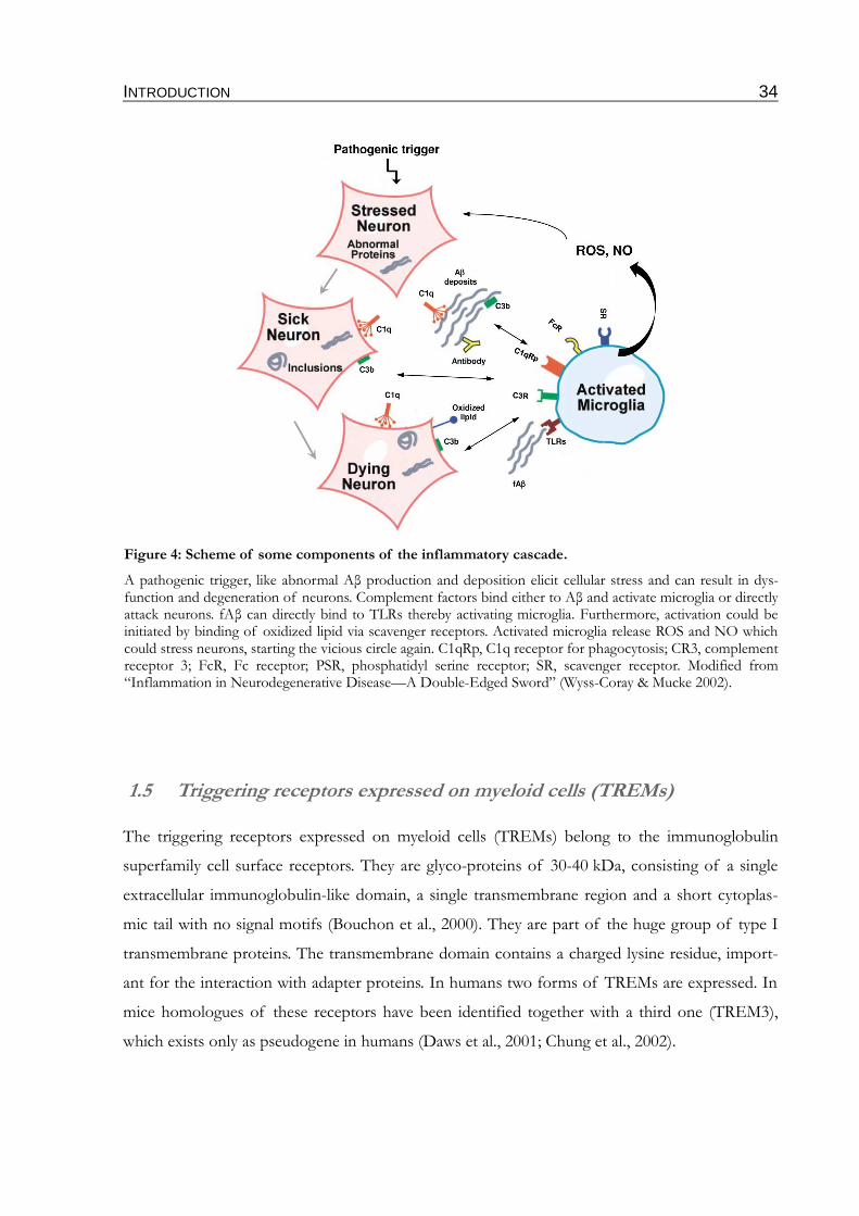
INTRODUCTION 34
1.5 Triggering receptors expressed on myeloid cells (TREMs)
The triggering receptors expressed on myeloid cells (TREMs) belong to the immunoglobulin
superfamily cell surface receptors. They are glyco-proteins of 30-40 kDa, consisting of a single
extracellular immunoglobulin-like domain, a single transmembrane region and a short cytoplas-
mic tail with no signal motifs (Bouchon et al., 2000). They are part of the huge group of type I
transmembrane proteins. The transmembrane domain contains a charged lysine residue, import-
ant for the interaction with adapter proteins. In humans two forms of TREMs are expressed. In
mice homologues of these receptors have been identified together with a third one (TREM3),
which exists only as pseudogene in humans (Daws et al., 2001; Chung et al., 2002).
Figure 4: Scheme of some components of the inflammatory cascade.
A pathogenic trigger, like abnormal Aβ production and deposition elicit cellular stress and can result in dys-function and degeneration of neurons. Complement factors bind either to Aβ and activate microglia or directly attack neurons. fAβ can directly bind to TLRs thereby activating microglia. Furthermore, activation could be initiated by binding of oxidized lipid via scavenger receptors. Activated microglia release ROS and NO which could stress neurons, starting the vicious circle again. C1qRp, C1q receptor for phagocytosis; CR3, complement receptor 3; FcR, Fc receptor; PSR, phosphatidyl serine receptor; SR, scavenger receptor. Modified from “Inflammation in Neurodegenerative Disease—A Double-Edged Sword” (Wyss-Coray & Mucke 2002).
INTRODUCTION 35
1.5.1 TREM1
TREM1 was the first receptor found of this family and was initially cloned by a cDNA screening
from human natural killer cells (Bouchon et al., 2000). TREM1 is mainly expressed on blood
neutrophils and monocytes/macrophages. Activation of the receptor by a yet unknown ligand
and engagement with its adapter protein DNAX activating protein of 12 kDa (DAP12) triggers
the generation of proinflammatory cytokines like interleukin-8 (IL-8), monocyte chemo attract-
ant protein 1 (MCP1), MCP3 and macrophage inflammatory protein-1α (MIP1α) (Bouchon et al.,
2000; Bleharski et al., 2003; Colonna & Facchetti 2003), but doesn't induce phagocytosis (Ble-
harski et al., 2003). Furthermore, TREM1 is highly expressed by neutrophils infiltrating tissues
that are infected with bacteria or fungi as well as epithelial cells in these tissues (Bouchon et al.,
2001a). According to this, the DAP12/TREM1 receptor system plays a crucial role in the ampli-
fication of inflammatory processes caused by infection.
1.5.2 TREM2
TREM2 was originally identified on monocyte-derived dendritic cells (DCs), where it promotes
migration (Bouchon et al., 2001b). However, as TREM2 has not yet been detected on primary
DCs, the involvement in DC biology remains unclear (Klesney-Tait et al., 2006). In contrast, it
has clearly been showed that TREM2 is negatively regulating the response of macrophages to
TLR activation. The concentrations of inflammatory cytokines like tumor necrosis factor (TNF)
and IL-6 are exaggerated in macrophages of TREM2 deficient mice (Turnbull et al., 2006) as well
as after knockdown of TREM2 by small interfering RNAs (siRNA) (Hamerman et al., 2006).
Insight into two more functions of TREM2 was gained by studies of the rare genetic disorder,
polycystic lipomembraneous osteodysplasia with sclerosing leukoencephalopathy (PLOSL or
Nasu-Hakola disease). Patients exhibit multiple bone cysts and develop a progressive presenile
dementia in the fourth decade of life. In the white matter of brains of autopsied patients scler-
otic lesions including activated microglia were identified (Kitajima et al., 1989; Verloes et al., 1997;
Tanaka 2000). Genetic analysis revealed that loss of function mutations in the TREM2 gene and
in the gene for the adapter protein DAP12 are causative for this disease (Paloneva et al., 2000;
Paloneva et al., 2002). It was shown that TREM2 and DAP12 are involved in the genesis of
osteoclasts which are multinucleated cells of myeloid origin involved in bone resorption and
homeostasis. Peripheral mononuclear blood cells from TREM2 or DAP12 deficient PLOSL
patients failed to differentiate into osteoclasts (Paloneva et al., 2003). However, these findings
from humans could not be reproduced in TREM2 deficient mice, so the function of TREM2 in
INTRODUCTION 36
osteoclastogenesis remains unclear. The finding that TREM2 mutations can also cause early-on-
set dementia without bone cysts, suggests that TREM2 is active in the CNS (Chouery et al.,
2008). Indeed TREM2 was found by in situ hybridization to be expressed in microglia, but the
levels of expression and the percentages of expressing cells vary in different brain regions. The
highest percentages were found in cortical regions (Schmid et al., 2002). The receptor is
expressed at the cell surface but predominantly distributed into two pools: one in the Golgi com-
plex and one population in exocytic vesicles, which are continuously shuttling to and from the
cell surface (Sessa et al., 2004). Activation of TREM2 in microglia induces the expression of
chemokine receptors and cell migration. Additionally, a decrease in phagocytosis of apoptotic
neurons was observed after knockdown with siRNA (Takahashi et al., 2005; Takahashi et al.,
2007). Interestingly, this knockdown resulted in an upregulation of inflammatory cytokines, sug-
gesting an anti-inflammatory function (Takahashi et al., 2007). Since the ligand(s) for TREM2 are
still unknown it was unclear how TREM2 recognizes apoptotic cells, till Hsieh et al. described a
ligand expressed on apoptotic neurons (Hsieh et al., 2009). Another endogenous ligand was
described as heat shock protein 60 (Hsp60) a mitochondrial chaperon which can be released from
both healthy and dying cells (Stefano et al., 2009). Moreover, other potential binding partners
have been reported, like polyanionic microbial products such as LPS or peptidoglycan (Daws et
al., 2003). This was supported by the finding that TREM2 can bind several species of bacteria
and fungi (Daws et al., 2003) and that TREM2 can serve as an phagocytic receptor for these
microbes (N'Diaye et al., 2009). Nevertheless, specific molecules mediating the activation have
not been identified. One possible explanation could be that TREM2 participates in a multimeric
receptor complex, containing TREM2, a ligand, DAP12, plexin-A1 – a receptor known to con-
trol axon guidance – and the plexin-A1 ligand semaphorin 6D (Takegahara et al., 2006; Ford &
McVicar 2009). However, in all scenarios DAP12 is indispensable for the function of TREM2.
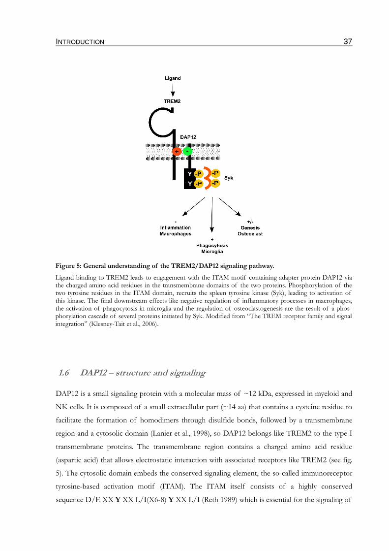
INTRODUCTION 37
1.6 DAP12 – structure and signaling
DAP12 is a small signaling protein with a molecular mass of ~12 kDa, expressed in myeloid and
NK cells. It is composed of a small extracellular part (~14 aa) that contains a cysteine residue to
facilitate the formation of homodimers through disulfide bonds, followed by a transmembrane
region and a cytosolic domain (Lanier et al., 1998), so DAP12 belongs like TREM2 to the type I
transmembrane proteins. The transmembrane region contains a charged amino acid residue
(aspartic acid) that allows electrostatic interaction with associated receptors like TREM2 (see fig.
5). The cytosolic domain embeds the conserved signaling element, the so-called immunoreceptor
tyrosine-based activation motif (ITAM). The ITAM itself consists of a highly conserved
sequence D/E XX Y XX L/I(X6-8) Y XX L/I (Reth 1989) which is essential for the signaling of
Figure 5: General understanding of the TREM2/DAP12 signaling pathway.
Ligand binding to TREM2 leads to engagement with the ITAM motif containing adapter protein DAP12 via the charged amino acid residues in the transmembrane domains of the two proteins. Phosphorylation of the two tyrosine residues in the ITAM domain, recruits the spleen tyrosine kinase (Syk), leading to activation of this kinase. The final downstream effects like negative regulation of inflammatory processes in macrophages, the activation of phagocytosis in microglia and the regulation of osteoclastogenesis are the result of a phos-phorylation cascade of several proteins initiated by Syk. Modified from “The TREM receptor family and signal integration” (Klesney-Tait et al., 2006).
INTRODUCTION 38
DAP12 containing receptor complexes. Besides DAP12 there are more adapter proteins known
which signal via an ITAM domain. However, the general signaling pathway is similar for all of
these proteins. The binding of a ligand leads to the clustering of the receptor e.g. TREM2 with
DAP12. As a result, Src-family kinases become activated by dephosphorylation of their regulat-
ory C-terminal tyrosine and subsequently phosphorylate DAP12 at the two tyrosines in the
ITAM domain thereby recruiting the spleen tyrosine kinase (Syk) in myeloid cells or the Zeta-
chain associated kinase of 70 kDa (Zap-70) in NK cells. Interaction of the kinase Src homology
2 (SH2) domains with the phosphorylated tyrosine residues induces the activation of the kinases.
They in turn phosphorylate several downstream substrates forming signaling complexes that
communicate to a number of pathways in the cell, like mitogen-activated protein kinase (MAPK)
or phospholipase Cγ (PLCγ) leading to different cellular responses (fig. 5), for review see
(Hamerman et al., 2009; Mocsai et al., 2010). The exact member of the Src-family kinases
involved in the DAP12/TREM2 signaling in microglia is unknown. Mutations of either ITAM or
the SH2 domains in the kinases completely block the signaling function, showing that both are
absolutely necessary for the function of the associated immunoreceptors (Hamerman et al.,
2009). It is known that DAP12 can mediate either activatory or inhibitory processes. The regula-
tion of multinucleated giant cell formation (Helming et al., 2008), osteoclast fusion and the initi-
ation of phagocytosis are three examples for an activatory signaling (see 1.5.2). The inhibitory
signaling is generally observed when cells are stimulated simultaneously by a DAP12-associated
receptor and a distinct receptor system, like TLRs. DAP12 deficient macrophages produce
increased levels of inflammatory cytokines after stimulation of TLRs (Hamerman et al., 2005).
As previously described for TREM2, DAP12 negatively regulates the transcription of genes cod-
ing for TNF, IL-1β and inducible nitric oxide-synthase (iNOS) (Takahashi et al., 2005). DAP12
signals not only in concert with immunoreceptors, also a signaling with integrins has been
described. The interaction of DAP12 with the microglial CD11b integrin for example regulates
neuronal apoptosis during development. Since the TREM2/DAP12 system regulates several
functions in microglia, it is interesting to study this regulation in context to AD.
INTRODUCTION 39
1.7 Rationale
Microglia have been shown to phagocytose Aβ in a FcR-mediated process upon passive immun-
ization with anti-Aβ antibodies (Bard et al., 2000; Bacskai et al., 2002; Wilcock et al., 2003) as well
as active immunization with Aβ42 (Schenk et al., 1999). However, phagocytosis of Aβ without
vaccination appears to be less efficient. The reason for this defect remains unclear. Since phago-
cytosis is a specific form of endocytosis and γ-secretase has been shown to regulate endocytosis
(Tamboli et al., 2008), a contribution of γ-secretase in phagocytotic processes might be possible.
There are some studies suggesting expression of γ-secretase components in microglia. However,
a function of γ-secretase in these cells is still unknown.
Thus, the main goal of this project was to investigate the expression and function of γ-secretase
in microglia. By pharmacological and genetic approaches it should also be tested whether γ-
secretase is functionally involved in phagocytosis.
One potential candidate might be the microglial receptor TREM2. As type I transmembrane pro-
tein TREM2 might represent a γ-secretase substrate. Since TREM2 has been shown to be
involved in phagocytotic processes of apoptotic neurons in the brain, processing of this receptor
might alter phagocytosis of Aβ by microglia.
MATERIAL & METHODS 40
2 MATERIAL & METHODS
Except as noted otherwise, all chemicals used for the described experiments were purchased with
a purity grade of “per analysi” from Sigma-Aldrich (Steinheim, Germany), Roche (Basel, Switzer-
land), Roth (Karlsruhe, Germany) or Applichem (Darmstadt, Germany). The specific cloning
primers were obtained from Sigma-Aldrich, and the restriction enzymes from Fermentas (St.
Leon-Rot, Germany). The radiochemicals were from Hartmann Analytic (Braunschweig, Ger-
many). Cell culture media and additives were purchased from Invitrogen, cell culture plastics
from Corning.
Table 1: Equipment
Cell culture equipment
-80 °C Freezer Thermo
Cell culture hood Thermo
37 °C CO2 incubator Binder
Water bath Medigen
Centrifuge (5804) Eppendorf
Blotting- and cloning equipment
Protein-electrophoresis chamber Höfer
Blotting chamber Höfer
Chemiluminescence Imager (ChemiDoc XRS)with software Quantity One
Bio-Rad
Cooling system (E100) Lauda
DNA-electrophoresis chamber Amersham
Trans-UV illuminator (GVM 20) Syngene
General laboratory devices
Microcentrifuge(5415D) Eppendorf
refrigerated Microcentrifuge (5415R) Eppendorf
refrigerated Centrifuge (5804R) Eppendorf
Cycler (Mastercycler Personal) Eppendorf
Gradientcycler (Mastercycler Gradient) Eppendorf
37 °C incubator Binder
Autoclave H+P
Heeting Block (SBH 130 D) Stuard Scientific
Magnetic stirrer Velp Scientifica
pH Meter (MP 225) Mettler Toledo
MATERIAL & METHODS 41
Photometer (Genesis) Thermo
Sonifier (Sonopuls, UW 2070) Bandelin
Thermomixer (Thermomixer compact) Eppendorf
Overhead rotor Scientific Industries
Vortex (MS 2 Minishaker) IKA
Analytical Balance (Labstyle 204) Mettler Toledo
Balance (PL 202-S) Mettler Toledo
Microtiterplate Reader (Multiskan RC) Thermo
Special devices
Fluorescence microscope (AxioVert 200)with software AxioVision
Zeiss
Phosphoimager Fuji
2.1 Cell biological techniques
2.1.1 Cell culture
Cell culture medium (DMEM +/+)Dulbecco´s Modified Eagle´s Medium (DMEM) GlutamaxTM containing 4.5 g/l D-glucose supplemented with 10 % heat inactivated fetal calf serum (FCS) and 1 % PenStrep solution (50 U/ml Penicillin, 50 µg/ml Streptomycin)Phosphate Buffered Saline (PBS)140 mM NaCl, 10 mM Na2HPO4, 1.75 mM KH2PO4, dH2O, pH 7.4Trypsin-EDTA Solution0.05 % (w/v) trypsin (Invitrogen), 0.53 mM EDTA, dH2OCryo medium90 % FCS supplemented with 10 % dimethylsufoxide (DMSO)
Frozen cells were rapidly thawed in a water bath. Cells were washed once with cell culture
medium (300 x g, 3 min) and plated on a new dish in fresh cell culture medium. Cells were main-
tained in a CO2 incubator at 37 °C, 95 % humidity and 5 % CO2. When cells reached confluence
of 70-90 %, they were split. Cells were therefore washed once with PBS before they were
detached from the surface by 1 ml of Trypsin-EDTA solution per 10 cm dish and plated onto a
new culture dish in a ratio of 1:20-1:2. If an exact cell number was needed, cells were counted in
a Neubauer counting-chamber. For cryo conservation cells of a 90 % confluent 6 cm dish were
detached as described above, pelleted (300 x g, 3 min), resuspended in 1-2 ml cryo medium and
finally stored in a -80 °C freezer or for long term storage in liquid nitrogen.
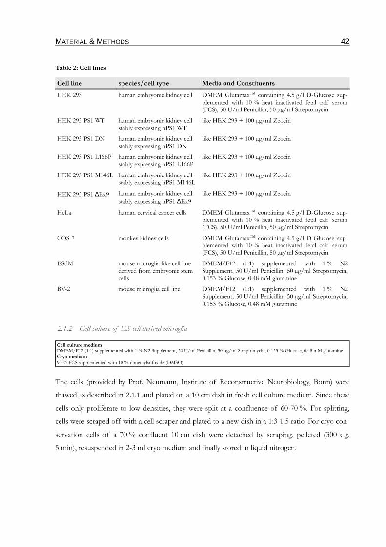
MATERIAL & METHODS 42
Table 2: Cell lines
Cell line species/cell type Media and Constituents
HEK 293 human embryonic kidney cell DMEM GlutamaxTM containing 4.5 g/l D-Glucose sup-plemented with 10 % heat inactivated fetal calf serum (FCS), 50 U/ml Penicillin, 50 µg/ml Streptomycin
HEK 293 PS1 WT human embryonic kidney cellstably expressing hPS1 WT
like HEK 293 + 100 µg/ml Zeocin
HEK 293 PS1 DN human embryonic kidney cellstably expressing hPS1 DN
like HEK 293 + 100 µg/ml Zeocin
HEK 293 PS1 L166P human embryonic kidney cellstably expressing hPS1 L166P
like HEK 293 + 100 µg/ml Zeocin
HEK 293 PS1 M146L human embryonic kidney cellstably expressing hPS1 M146L
like HEK 293 + 100 µg/ml Zeocin
HEK 293 PS1 ∆Ex9 human embryonic kidney cell
stably expressing hPS1 ∆Ex9
like HEK 293 + 100 µg/ml Zeocin
HeLa human cervical cancer cells DMEM GlutamaxTM containing 4.5 g/l D-Glucose sup-plemented with 10 % heat inactivated fetal calf serum (FCS), 50 U/ml Penicillin, 50 µg/ml Streptomycin
COS-7 monkey kidney cells DMEM GlutamaxTM containing 4.5 g/l D-Glucose sup-plemented with 10 % heat inactivated fetal calf serum (FCS), 50 U/ml Penicillin, 50 µg/ml Streptomycin
ESdM mouse microglia-like cell line derived from embryonic stem cells
DMEM/F12 (1:1) supplemented with 1 % N2 Supplement, 50 U/ml Penicillin, 50 µg/ml Streptomycin, 0.153 % Glucose, 0.48 mM glutamine
BV-2 mouse microglia cell line DMEM/F12 (1:1) supplemented with 1 % N2 Supplement, 50 U/ml Penicillin, 50 µg/ml Streptomycin, 0.153 % Glucose, 0.48 mM glutamine
2.1.2 Cell culture of ES cell derived microglia
Cell culture mediumDMEM/F12 (1:1) supplemented with 1 % N2 Supplement, 50 U/ml Penicillin, 50 µg/ml Streptomycin, 0.153 % Glucose, 0.48 mM glutamineCryo medium90 % FCS supplemented with 10 % dimethylsufoxide (DMSO)
The cells (provided by Prof. Neumann, Institute of Reconstructive Neurobiology, Bonn) were
thawed as described in 2.1.1 and plated on a 10 cm dish in fresh cell culture medium. Since these
cells only proliferate to low densities, they were split at a confluence of 60-70 %. For splitting,
cells were scraped off with a cell scraper and plated to a new dish in a 1:3-1:5 ratio. For cryo con-
servation cells of a 70 % confluent 10 cm dish were detached by scraping, pelleted (300 x g,
5 min), resuspended in 2-3 ml cryo medium and finally stored in liquid nitrogen.
MATERIAL & METHODS 43
2.1.3 Transient transfection
Opti-MEM ® I (Invitrogen)DMEM +/-Dulbecco´s Modified Eagle´s Medium (DMEM) GlutamaxTM containing 4.5 g/l D-glucose supplemented with 10 % FCS
Cells were grown to 80 % confluence and transfected with Lipofectamin 2000 (Invitrogen)
according to manufacturer´s instruction (for each 6 cm dish 5 µg DNA and 7.5 µl Lipofectamin
2000). During the transfection time of 5-6 h, cells were incubated in DMEM +/-. Medium was
changed and cells were incubated for another 24 h at 37 °C, 5 % CO2. To achieve good transfec-
tion results the concentration of the used DNA solution should be > 0.5 µg/ml.
2.1.4 Immunocytochemistry (ICC)
Phosphate Buffered Saline (PBS), sterile140 mM NaCl, 10 mM Na2HPO4, 1.75 mM KH2PO4, dH2O, pH 7.4DMEM -/-Dulbecco´s Modified Eagle´s Medium (DMEM) GlutamaxTM containing 4.5 g/l D-glucosePoly-L-lysine solution100 µg/ml sterile poly-L-lysine in PBS Distilled Water, sterile (dH2O) (Invitrogen)Paraformaldehyde solution (PFA; 4 %)4 % (w/v) Paraformaldehyde in PBS Triton X-100 solution (0.25 %)0.25 % (v/v) Triton X-100 (Sigma-Aldrich) in PBS Blocking solution10 % (w/v) BSA in PBS (for neurons: 10 % BSA in PBS + 0.25 % Triton X-100) or DMEMPrimary antibody solutionPrimary antibody in appropriate concentration (see Table 7) in PBS or DMEM supplemented with 5 % BSA (for neurons: additionally with 0.125 % Triton X-100)Secondary antibody solutionSecondary antibody in appropriate concentration (see Table 8) in PBS or DMEM with 5 % BSA (for neurons: additionally with 0.125 % Tri-ton X-100)
For immunocytochemistry cover slips (CS) were used which were coated with poly-L-lysine for
1 h at 37 °C, washed three times with sterile distilled water (dH2O) and once with PBS. 150000
cells were seeded in one 6 well (containing 3 CS). After 24 h cells were transfected as described in
2.1.3. Cells were then washed three times with PBS and fixed in 4 % Paraformaldehyde (Birn-
boim & Doly 1979) for 10 min. To remove residual PFA cells were washed three times with PBS.
Cells were then permeabilized by incubation in Triton X-100 for 10 min at room temperature
(RT) and incubated in incubated in blocking solution for 1 h at RT. The protein of interest was
then stained by incubation in 100 µl primary antibody solution for 1 h at RT, followed by intens-
ive washing 3 x with PBS. The washing was repeated 3 times with 5 min incubation in between.
After incubation with secondary antibody solution (100 µl) for 1 h at RT, the unbound secondary
antibody was removed by intensive washing as described above. Finally CS were washed once
with dH2O and embedded on a slide with ImmuMount (Thermo). The slides were kept dark at
4 °C. Slides were analyzed with a fluorescence microscope.
MATERIAL & METHODS 44
To selectively detect cell surface proteins, cells were stained prior to fixation. For this purpose,
cells were kept on ice for the whole staining procedure. As blocking solution 10 % BSA in
DMEM -/- was used. The primary and the secondary antibody was diluted in DMEM -/- with
5 % BSA. All washing steps were conducted in DMEM -/-. After the last washing, the cells were
fixed in 4 % PFA. For visualization of the total protein expression, standard immunostaining as
described in 2.1.4 was used.
2.2 Molecularbiological techniques
2.2.1 Polymerase chain reaction (PCR)
PCR reaction mix10X Pfu Buffer with MgSO4 5 µl dNTP Mix, 10 mM each 1 µl (0.2 mM of each) Forward primer 1.0 µM Reverse primer 1.0 µM Template DNA 10-100 ng Pfu DNA Polymerase 2.5 U (1 µl)add to 50 µl total volume with nuclease-free water
The DNA constructs used in this project (see Table 3) were generated by PCR using the primers
listed in Table 4. The PCR reaction mix was prepared on ice. For amplification the PCR program
was run for 30 cycles after one initial DNA denaturating step (2 min, 95 °C). Every program
included the following steps:
1. Denaturation: 95 °C, 0.5 min
2. Annealing: 50-80 °C (depends on primer), 0.5 min
3. Elongation: 72 °C, 0.5-2 min (depends on construct length)
The exact PCR programs are listed in Table 5.
The point mutations in DAP12 were generated by a three-step cloning strategy. At first two PCRs
were conducted using the cloning primers in combination with the opposite mutation primers
(step 1 & 2). The products of step 1 and 2 were then used as template which was amplified using
the cloning primers.
To continue the cloning the amplified PCR products were analyzed according to their size in a 1-
2 % agarose gel (see 2.2.2). The bands were cut out of the gel and the DNA was isolated as
described in 2.2.3.
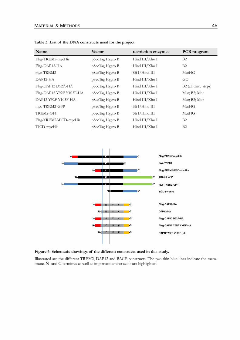
MATERIAL & METHODS 45
Table 3: List of the DNA constructs used for the project
Name Vector restriction enzymes PCR program
Flag-TREM2-mycHis pSecTag Hygro B Hind III/Xho I B2
Flag-DAP12-HA pSecTag Hygro B Hind III/Xho I B2
myc-TREM2 pSecTag Hygro B Sfi I/Hind III MutHG
DAP12-HA pSecTag Hygro B Hind III/Xho I GC
Flag-DAP12 D52A-HA pSecTag Hygro B Hind III/Xho I B2 (all three steps)
Flag-DAP12 Y92F Y103F-HA pSecTag Hygro B Hind III/Xho I Mut; B2; Mut
DAP12 Y92F Y103F-HA pSecTag Hygro B Hind III/Xho I Mut; B2; Mut
myc-TREM2-GFP pSecTag Hygro B Sfi I/Hind III MutHG
TREM2-GFP pSecTag Hygro B Sfi I/Hind III MutHG
Flag-TREM2∆ECD-mycHis pSecTag Hygro B Hind III/Xho I B2
TICD-mycHis pSecTag Hygro B Hind III/Xho I B2
Figure 6: Schematic drawings of the different constructs used in this study.
Illustrated are the different TREM2, DAP12 and BACE constructs. The two thin blue lines indicate the mem-brane. N- and C-terminus as well as important amino acids are highlighted.
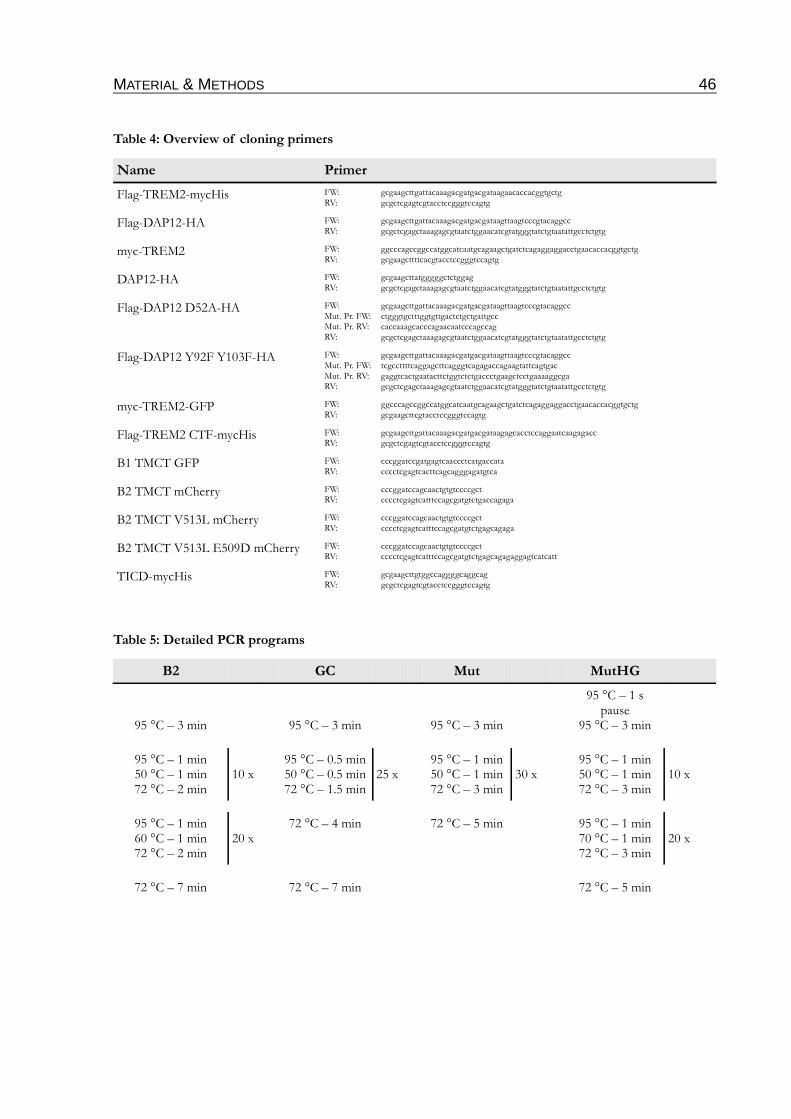
MATERIAL & METHODS 46
Table 4: Overview of cloning primers
Name Primer
Flag-TREM2-mycHis FW: gcgaagcttgattacaaagacgatgacgataagaacaccacggtgctgRV: gcgctcgagtcgtacctccgggtccagtg
Flag-DAP12-HA FW: gcgaagcttgattacaaagacgatgacgataagttaagtcccgtacaggccRV: gcgctcgagctaaagagcgtaatctggaacatcgtatgggtatctgtaatattgcctctgtg
myc-TREM2 FW: ggcccagccggccatggcatcaatgcagaagctgatctcagaggaggacctgaacaccacggtgctgRV: gcgaagcttttcacgtacctccgggtccagtg
DAP12-HA FW: gcgaagcttatgggggctctggagRV: gcgctcgagctaaagagcgtaatctggaacatcgtatgggtatctgtaatattgcctctgtg
Flag-DAP12 D52A-HA FW: gcgaagcttgattacaaagacgatgacgataagttaagtcccgtacaggccMut. Pr. FW: ctgggtgctttggtgttgactctgctgattgccMut. Pr. RV: caccaaagcacccagaacaatcccagccagRV: gcgctcgagctaaagagcgtaatctggaacatcgtatgggtatctgtaatattgcctctgtg
Flag-DAP12 Y92F Y103F-HA FW: gcgaagcttgattacaaagacgatgacgataagttaagtcccgtacaggccMut. Pr. FW: tcgccttttcaggagcttcagggtcagagaccagaagtattcagtgacMut. Pr. RV: gaggtcactgaatacttctggtctctgaccctgaagctcctgaaaaggcga RV: gcgctcgagctaaagagcgtaatctggaacatcgtatgggtatctgtaatattgcctctgtg
myc-TREM2-GFP FW: ggcccagccggccatggcatcaatgcagaagctgatctcagaggaggacctgaacaccacggtgctgRV: gcgaagcttcgtacctccgggtccagtg
Flag-TREM2 CTF-mycHis FW: gcgaagcttgattacaaagacgatgacgataagagcacctccaggaatcaagagaccRV: gcgctcgagtcgtacctccgggtccagtg
B1 TMCT GFP FW: cccggatccgatgagtcaaccctcatgaccataRV: cccctcgagtcacttcagcagggagatgtca
B2 TMCT mCherry FW: cccggatccagcaactgtgtccccgctRV: cccctcgagtcatttccagcgatgtctgaccagaga
B2 TMCT V513L mCherry FW: cccggatccagcaactgtgtccccgctRV: cccctcgagtcatttccagcgatgtctgagcagaga
B2 TMCT V513L E509D mCherry FW: cccggatccagcaactgtgtccccgctRV: cccctcgagtcatttccagcgatgtctgagcagagaggagtcatcatt
TICD-mycHis FW: gcgaagcttgtggccaggggcaggcagRV: gcgctcgagtcgtacctccgggtccagtg
Table 5: Detailed PCR programs
B2 GC Mut MutHG
95 °C – 3 min 95 °C – 3 min 95 °C – 3 min
95 °C – 1 spause
95 °C – 3 min
95 °C – 1 min50 °C – 1 min72 °C – 2 min
10 x95 °C – 0.5 min50 °C – 0.5 min72 °C – 1.5 min
25 x95 °C – 1 min50 °C – 1 min72 °C – 3 min
30 x95 °C – 1 min50 °C – 1 min72 °C – 3 min
10 x
95 °C – 1 min60 °C – 1 min72 °C – 2 min
20 x72 °C – 4 min 72 °C – 5 min 95 °C – 1 min
70 °C – 1 min72 °C – 3 min
20 x
72 °C – 7 min 72 °C – 7 min 72 °C – 5 min
MATERIAL & METHODS 47
2.2.2 Separation and visualization of DNA fragments
TBE Buffer9 mM Tris-Borat, 2 mM EDTA, dH2O, pH 8.0 6 x orange loading dye60 % Glycerin, 0.15 % Orange G, 60 mM EDTA, 10 mM Tris, dH2O, pH 7.6GelRed (Biotium Inc.)10000 x in water
Agarose gels allow the separation of DNA by their fragment length in an electric field. To separ-
ate 100-10000 bp gels of 1-2 % were used. For visualization of the DNA the agarose gels were
supplemented with 2 µl GelRed/100 ml gel. The DNA samples were mixed with 6 x orange load-
ing dye before they were loaded in the gel. The gel was then run in TBE buffer at 110 V.
2.2.3 Isolation of DNA fragments from agarose gels
Wizard® SV Gel and PCR Clean up system (Promega)dH2O
The desired bands were cut out of the gel on an UV illuminator. The DNA was isolated using
the Wizard® SV Gel and PCR Clean up system (Promega) according to the manufacturers' instruc-
tions. After elution in nuclease free dH2O, the DNA was used directly for further cloning steps.
2.2.4 Restriction digestion
Restriction enzymes (see Table 3, Fermentas)dH2O
Restriction digestion of DNA was conducted in the buffer recommended for each enzyme. For
double digestion the ideal buffer for both enzymes was chosen by the Double Digest tool available
on the Fermentas web site. For preparative purposes 2-5 µg DNA were digested with 5-10 U of
the appropriate enzyme in 20 µl at 37 °C for 4-16 h.
2.2.5 Dephosphorylation of linearized DNA
Shrimp alkaline phosphatase (SAP, 1 U/µl, Fermentas)dH2O
To prevent self re-ligation of the linearized plasmid DNA, the free 5'-phosphate group was
removed by adding 2 U shrimp alkaline phosphatase for the last hour directly to the restriction
digestion.
MATERIAL & METHODS 48
2.2.6 Ligation
T4 DNA-ligase (5 U/µl, Fermentas)dH2O
The insertion of passenger DNA into the dephosphorylated linearized vector via the DNA-ligase
of the bacteriophage T4 was carried out in a molar ratio of 1:3 (vector:insert). At least 100 ng
insert were incubated together with the reaction buffer and 5 U T4 ligase for 16 h at RT. 1/3 of
the ligation mixture was then used for the transformation in competent E. coli cells (see 2.2.8).
2.2.7 Generation of chemo competent E. coli Top10
TFB I100 mM RbCl, 50 mM MnCl2, 30 mM potassium acetate, 10 mM CaCl2, 15 % Glycerol, pH 5.8TFB II10 mM MOPS, 10 mM RbCl, 75 mM CaCl2, 15 % Glycerol, pH 6.8Low salt Lauria-Bertani (LB) medium1 % (w/v) tryptone, 0.5 % (w/v) yeast extract, 0.5 % (w/v) NaCl, dH2O, pH 7.0 (autoclaved)
At first, 200 ml LB medium in a 1 l flask were inoculated with 2 ml of an overnight culture. The
culture was incubated for 2 h under rotation (250 rpm) at 37 °C. Cells were pelleted by centrifu-
gation (1500 x g, 10 min, 4 °C). After resuspending the pellet in 30 ml ice cold TFB I by pipet-
ting, a centrifugation step followed to pellet the cells again. This pellet was then resuspended in
4 ml ice cold TFB II. Finally, the solution was divided into 50 µl aliquots and freezed at -80 °C
for long term storage.
2.2.8 Transformation of E. coli Top10
SOC medium0.5 % (w/v) yeast extract, 2 % (w/v) tryptone, 10 mM NaCl, 2.5 mM KCl, 20 mM MgSO4, 20 mM glucoseLB agar plates1 % (w/v) tryptone, 0.5% (w/v) yeast extract, 0.5% (w/v) NaCl, 15 g/l agar, dH2O, pH 7.0 (autoclaved); before pouring the plates the antibiotics were added to the warm (~ 40-50 °C) solution.Selection antibioticsKanamycin: 30 µg/ml (Stock: 30 mg/ml, dH2O); Ampicillin: 100 µg/ml (Stock: 100 mg/ml, dH2O)
For transformation of the chemo competent bacteria with plasmid DNA, the thawed cells were
mixed with 1/3 of the ligation mixture, or in case of re-transformation with 0.5 µg plasmid
DNA and incubated for 30 min on ice. After a heat shock of exact 30 s at 42 °C the cells were
placed back on ice. 250 µl of pre-warmed SOC medium were added to each vial and the cells
were incubated for 1 h at 37 °C at 500 rpm in a shaking incubator. To spread the transformed
cells onto a LB agar plate, the cells were pelleted at 300 x g for 3 min, the supernatant was
removed except 50 µl. The pellet was resuspended in these 50 µl and the solution was plated on
an agar plate supplemented with the appropriate antibiotic.
MATERIAL & METHODS 49
2.2.9 Cryo conservation of transformed E. coli
Conservation medium70 % (v/v) Glycerol, dH2O (autoclaved)Low salt Lauria-Bertani (LB) medium1 % (w/v) tryptone, 0.5 % (w/v) yeast extract, 0.5 % (w/v) NaCl, dH2O, pH 7.0 (autoclaved)
0.8 ml of a fresh bacteria culture was transferred into a screw cap tube, 0.5 ml conservation
medium was added, gently mixed and freezed at -80 °C. For re-utilization of the transformed
bacteria a bit of the frozen medium was scratched with a sterile pipette tip and transferred into a
test tube filled with 3 ml LB medium. For an overnight culture the test tube was then incubated
overnight (~16 h) in a shaking incubator at 37 °C and 250 rpm.
2.2.10 Extraction of plasmid DNA from E. coli
Resuspension buffer50 mM Tris, 10 mM EDTA, 100 µg/ml RNase A, pH 8.0Lysis buffer200 mM NaOH, 1 % (w/v) SDSNeutralization buffer3 M Potassium acetate, pH 5.5PureYield Plasmid Midiprep System (Promega)GeneJET™ Plasmid Miniprep Kit (Fermentas)
For the extraction of DNA out of E. coli cells an alkaline procedure was used as described by
Birnboim & Doly (Birnboim & Doly 1979). Briefly, 1 ml of the overnight culture was centrifuged
(1 min, 12000 x g) and the pellet resuspended in 100 µl resuspension buffer. 100 µl lysis buffer
were added. After mixing by inverting and an incubation of 3 min, 175 µl neutralization buffer
were added to precipitate proteins and associated genomic DNA. Solution was cleared by centri-
fugation (10 min, 12000 x g) and the DNA precipitated as described in 2.2.11. The obtained
DNA solution was used for restriction digestion (see 2.2.4).
For preparative extraction the PureYield Plasmid Midiprep System (Promega) or for small amounts
the GeneJET™ Plasmid Miniprep Kit (Fermentas) was used. Both kits were used as described in the
manufacturers' manual. The DNA was general eluted in nuclease-free dH2O. In case that the con-
centration of the gained DNA solution was too low for effective transfection (2.1.3), the DNA
was concentrated by ethanol precipitation (2.2.12).
MATERIAL & METHODS 50
2.2.11 Precipitation of DNA by isopropanol
Isopropanol70 % EthanolRNase-H2OSodium acetate 3 M Sodium acetate, dH2O, pH 5.2RNase waterRNase 10 µg/ml, dH2O
To isolate the pure DNA from the crude solution obtained from the alkaline extraction (2.2.10)
the cleared supernatant was supplemented with 420 µl isopropanol and 35 µl sodium acetate.
After a spin of 10 min at 12000 x g, the supernatant was discarded and the pellet washed with
600 µl 70 % ethanol (EtOH). After a further spin (10 min, 12000 x g ) the pellet was air dried.
Finally, the dried DNA pellet was resuspended in 50 µl RNase-H2O.
2.2.12 Precipitation of DNA by ethanol
absolute EthanolSodium acetate 3 M Sodium acetate, dH2O, pH 5.270 % EthanolNuclease-free dH2O
To concentrate diluted DNA solutions, the DNA solution was mixed by vortexing with 1/10
volume with sodium acetate and 3 volumes of absolute ethanol and aliquoted to 2 ml tubes. After
incubation for 15 min at -20 °C and a 30 min spin (12000 x g, 4 °C), the pellets were washed with
70 % EtOH and again centrifuged (5 min, 12000 x g, 4 °C). In the end the pellets were resuspen-
ded in the required amount of nuclease-free water to gain an applicable concentration for transi-
ent transfection (2.1.3).
2.2.13 DNA sequencing
BigDye Terminator v1.1 Cycle Sequencing Kit (Applied Biosystems)
The cDNA-constructs were sequenced using the BigDye Terminator v1.1 Cycle Sequencing Kit
(Applied Biosystems) as described in the manufacturers' guidelines.
2.2.14 Photometric determination of DNA concentration
The DNA was diluted 1:20 in dH2O and measured at 260 nm in a quartz cuvette (A260). The
blank value was determined using dH2O as well. The concentration was then calculated using the
following equation:
MATERIAL & METHODS 51
c[µg/ml]=A260⋅dilution factorDF ⋅multiplikation factorF
(F=50, V=20)
2.3 Proteinbiochemical techniques
2.3.1 Extraction of membrane proteins out of eukaryotic cells
Phosphate Buffered Saline (PBS)140 mM NaCl, 10 mM Na2HPO4, 1.75 mM KH2PO4, dH2O, pH 7.4Hypoton buffer:10 mM Tris, 1 mM EDTA, 1 mM EGTA, dH2O, pH 7.6 STEN-lysis buffer:50 mM Tris, 150 mM NaCl, 2 mM EDTA, 1 % (v/v) NP40 (Sigma-Aldrich), 1 % (v/v) Triton X-100 (Sigma-Aldrich), dH2O, pH 7.425 x Protease inhibitor (PI) cocktail (Complete, Roche), dH2O
Cells were washed once with PBS, scraped and pelleted in 1 ml PBS. The cells were incubated in
0.8 ml hypoton buffer for 15 min on ice and lysed by drawing 20 x through a 0.6 mm needle. To
clear the homogenate from nuclei and mitochondria it was spun for 10 min at 300 x g and 4 °C.
The separation of the cytosol fraction and the membrane vesicles was accomplished by a second
centrifugation step (60 min, 12000 x g, 4 °C). Before the protein concentration was measured by
the BCA assay, as described in 2.3.3, the membrane pellet was lysed in 50 µl STEN lysis buffer,
incubated for another 15 min on ice and sedimented by a 15 min spin at 12000 x g and 4 °C. Pro-
tein extracts were either stored at -20 °C or directly used for SDS electrophoresis (see 2.3.6).
2.3.2 Protein extraction out of eukaryotic cells
PBS140 mM NaCl, 10 mM Na2HPO4, 1.75 mM KH2PO4, dH2O, pH 7.4RIPA lysis buffer50 mM Tris, 150 mM NaCl, 0.5 % (w/v) Sodium deoxycholic acid, 0.1 % (w/v) SDS, 1 % (v/v) NP-40, pH 7.4 STEN lysis buffer50 mM Tris, 150 mM NaCl, 2 mM EDTA, 1 % (v/v) NP40 (Sigma-Aldrich), 1 % (v/v) Triton X-100 (Sigma-Aldrich), dH2O, pH 7.4
Cells were washed once with PBS and lysed in 0.8 ml RIPA or STEN lysis buffer for 15 min on
ice. The homogenates were cleared from the insoluble material by centrifugation for 15 min at
12000 x g and 4 °C. For further use protein extracts were stored at -20 °C or loaded on gels for
SDS electrophoresis (see 2.3.6).
MATERIAL & METHODS 52
2.3.3 Protein estimation
BCA protein assay kit (Thermo Scientific)Bradford reagent (Bio-Rad)
Two different methods for protein estimation were used depending on the detergents exerted for
protein extraction.
I) BCA method, first described by Smith and co-workers (Smith et al., 1985)
This method is divided into two reactions. In a first reaction, the so-called Biuret reac-
tion are Cu2+ ions reduced by negatively charged hydroxyl groups to Cu+. These copper
ions complex then two bicinchoninic acid molecules, resulting in a violet complex which
can be photometrically measured.
For the estimation the BCA protein assay kit (Thermo Scientific) was used. The protein
extracts were diluted 1:10-1:20 with water and mixed with the kit reagents according to
the manual. After incubation for 30 min the absorption at 562 nm was measured. The
protein concentration was finally calculated by means of a standard curve.
II) Bradford method, first described by (Bradford 1976)
In this method basic and aromatic protein side chains react with Coomassie G-250, which
is part of the Bradford reagent (Bio-Rad), shifting its absorption maximum to 595 nm.
For estimation protein extracts in different dilutions were mixed 1:5 with Bradford
reagent, incubated for 5 min at RT and subsequently the absorption measured at
595 nm. The protein concentration was finally calculated by means of a standard curve.
2.3.4 Immunoprecipitation (IP)
STEN buffer50 mM Tris, 150 mM NaCl, 2 mM EDTA, 0.2 % (v/v) NP40 (Sigma-Aldrich), pH 7.6 STEN-NaCl50 mM Tris, 500 mM NaCl, 2 mM EDTA, 0.2 % (v/v) NP40 (Sigma-Aldrich), dH2O, pH 7.6 PBS140 mM NaCl, 10 mM Na2HPO4, 1.75 mM KH2PO4, dH2O, pH 7.4Loading dye (5 x)50 % (v/v) Glycerin, 7.5 % (w/v) SDS, 0.1 M DTT, 0.025 mg/ml Bromphenol blue in stacking gel bufferProtein A sepharose (Zymed)Protein G sepharose (Invitrogen)
Specific proteins can be isolated by specific antibodies coupled to protein A (against rabbit IgG)
or protein G sepharose (against mouse IgG) from cell lysates (see 2.3.2). To prevent unspecific
binding of proteins the lysates were pre-cleared with 30 µl uncoupled protein A sepharose beads
for 1 h at 4 °C on a rotatory shaker. The sepharose was sedimented (2 min, 9300 x g, 4 °C) and
the supernatant incubated with fresh sepharose beads and generally 1-3 µg of the specific anti-
body for 2-16 h at 4 °C under constant rotation. Afterwards the beads were sedimented (2 min,
MATERIAL & METHODS 53
9300 x g, 4 °C) and the supernatant discarded. The beads were then washed once with STEN-
NaCl and three times with STEN buffer. Therefore the beads were incubated for 5 min on a
rotatory shaker at 4 °C before they were pelleted (2 min, 9300 x g, 4 °C). Finally the beads were
sucked dry by the use of a syringe and 0.4 mm needle, taken up in 20 µl of 2 x loading dye and
boiled 5 min at 95 °C. The samples were then used for SDS electrophoresis (see 2.3.6).
2.3.5 Co-immunoprecipitation of DAP12 and TREM2
Brij97 lysis buffer0.875 % Brij 97, 0.125 % Igepal, 10 mM Tris, 150 mM NaClProtein G sepharose (Invitrogen)
Co-immunoprecipitation (co-IP) is a technique to detect protein-protein interactions. For co-IP
of DAP12 and TREM2, the Brij97 lysis buffer described by Voehringer and colleagues
(Voehringer et al., 2004) was used. Cells were lysed, according to 2.3.2 in 800 µl Brij97 lysis buf-
fer. 20 µl of the cell lysate was taken and the rest pre-cleared as described in 2.3.4. During the
pre-clearing the protein concentration was estimated by BCA according to 2.3.3. For IP, which
was accomplished for 16 h, as described in 2.3.4, 1 mg protein per sample was used in equal
volumes. After the IP the procedure was continued as described in 2.3.4 with the exception that
the washing steps were performed with Brij97 lysis buffer.
2.3.6 Sodium dodecyl sulfate polyacrylamide gel electrophoresis (SDS-PAGE)
Stacking gel buffer (4 x) (Upper-Tris)500 mM Tris, 0.4 % (w/v) SDS, dH2O, pH 6.8Separation gel buffer (4 x)1.5 M Tris, 0.4 % (w/v) SDS, dH2O, pH 8.8SDS loading dye (5 x)50 % (v/v) Glycerin, 7.5 % (w/v) SDS, 0.1 M DTT, 0.025 mg/ml Bromphenol blue in stacking gel bufferAmmonium persulfate (APS, Sigma)10 % (w/v) Ammonium persulfate, dH2ON,N,N´,N´-Tetramethylethylenediamine (TEMED, Roth)Running buffer25 mM Tris, 200 mM Glycine, 0.1 % (w/v) SDS, dH2OAcrylamide/Bisacrylamide solution30 % (v/v) Acrylamide/Bisacrylamide in the ratio of 37.5:1PageRuler™ protein ladder (Fermentas)PageRuler™ prestained protein ladder (Fermentas)SeeBlue® prestained standard (Invitrogen)SeeBlue® Plus prestained standard (Invitrogen)
The separation of proteins on the basis of their molecular weight was carried out in 1.5 mm
thick gels which were casted under the conditions listed in Table 6.
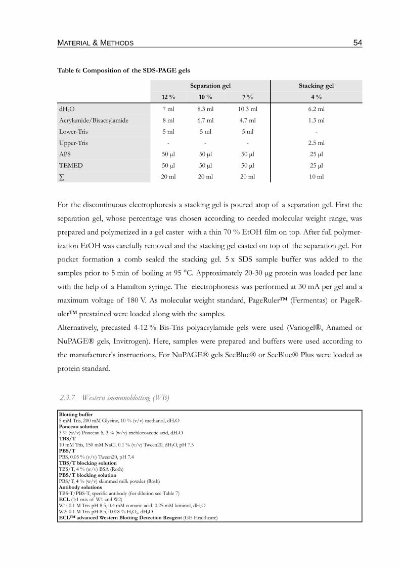
MATERIAL & METHODS 54
Table 6: Composition of the SDS-PAGE gels
Separation gel Stacking gel
12 % 10 % 7 % 4 %
dH2O 7 ml 8.3 ml 10.3 ml 6.2 ml
Acrylamide/Bisacrylamide 8 ml 6.7 ml 4.7 ml 1.3 ml
Lower-Tris 5 ml 5 ml 5 ml -
Upper-Tris - - - 2.5 ml
APS 50 µl 50 µl 50 µl 25 µl
TEMED 50 µl 50 µl 50 µl 25 µl
∑ 20 ml 20 ml 20 ml 10 ml
For the discontinuous electrophoresis a stacking gel is poured atop of a separation gel. First the
separation gel, whose percentage was chosen according to needed molecular weight range, was
prepared and polymerized in a gel caster with a thin 70 % EtOH film on top. After full polymer-
ization EtOH was carefully removed and the stacking gel casted on top of the separation gel. For
pocket formation a comb sealed the stacking gel. 5 x SDS sample buffer was added to the
samples prior to 5 min of boiling at 95 °C. Approximately 20-30 µg protein was loaded per lane
with the help of a Hamilton syringe. The electrophoresis was performed at 30 mA per gel and a
maximum voltage of 180 V. As molecular weight standard, PageRuler™ (Fermentas) or PageR-
uler™ prestained were loaded along with the samples.
Alternatively, precasted 4-12 % Bis-Tris polyacrylamide gels were used (Variogel®, Anamed or
NuPAGE® gels, Invitrogen). Here, samples were prepared and buffers were used according to
the manufacturer's instructions. For NuPAGE® gels SeeBlue® or SeeBlue® Plus were loaded as
protein standard.
2.3.7 Western immunoblotting (WB)
Blotting buffer5 mM Tris, 200 mM Glycine, 10 % (v/v) methanol, dH2OPonceau solution3 % (w/v) Ponceau S, 3 % (w/v) trichloroacetic acid, dH2O TBS/T10 mM Tris, 150 mM NaCl, 0.1 % (v/v) Tween20, dH2O, pH 7.5 PBS/TPBS, 0.05 % (v/v) Tween20, pH 7.4TBS/T blocking solutionTBS/T, 4 % (w/v) BSA (Roth) PBS/T blocking solutionPBS/T, 4 % (w/v) skimmed milk powder (Roth)Antibody solutionsTBS-T/PBS-T, specific antibody (for dilution see Table 7)ECL (1:1 mix of W1 and W2)W1: 0.1 M Tris pH 8.5, 0.4 mM cumaric acid, 0.25 mM luminol, dH2OW2: 0.1 M Tris pH 8.5, 0.018 % H2O2, dH2OECL™ advanced Western Blotting Detection Reagent (GE Healthcare)
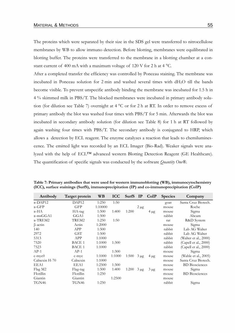
MATERIAL & METHODS 55
The proteins which were separated by their size in the SDS gel were transferred to nitrocellulose
membranes by WB to allow immuno detection. Before blotting, membranes were equilibrated in
blotting buffer. The proteins were transferred to the membrane in a blotting chamber at a con-
stant current of 400 mA with a maximum voltage of 120 V for 2 h at 4 °C.
After a completed transfer the efficiency was controlled by Ponceau staining. The membrane was
incubated in Ponceau solution for 2 min and washed several times with dH2O till the bands
become visible. To prevent unspecific antibody binding the membrane was incubated for 1.5 h in
4 % skimmed milk in PBS/T. The blocked membranes were incubated in primary antibody solu-
tion (for dilution see Table 7) overnight at 4 °C or for 2 h at RT. In order to remove excess of
primary antibody the blot was washed four times with PBS/T for 5 min. Afterwards the blot was
incubated in secondary antibody solution (for dilution see Table 8) for 1 h at RT followed by
again washing four times with PBS/T. The secondary antibody is conjugated to HRP, which
allows a detection by ECL reagent. The enzyme catalyzes a reaction that leads to chemilumines-
cence. The emitted light was recorded by an ECL Imager (Bio-Rad). Weaker signals were ana-
lyzed with the help of ECL™ advanced western Blotting Detection Reagent (GE Healthcare).
The quantification of specific signals was conducted by the software Quantity One®.
Table 7: Primary antibodies that were used for western immunoblotting (WB), immunocytochemistry (ICC), surface stainings (SurfS), immunoprecipitation (IP) and co-immunoprecipitation (CoIP)
Antibody Target protein WB ICC SurfS IP CoIP Species Company
α-DAP12 DAP12 1:250 1:50 goat Santa Cruz Biotech.α-GFP GFP 1:10000 2 µg mouse Rocheα-HA HA-tag 1:500 1:400 1:200 4 µg mouse Sigmaα-msGGA1 GGA1 1:500 rabbit Abcamα-TREM2 TREM2 1:250 1:50 rat R&D Systemβ-actin Actin 1:2000 mouse Sigma140 APP 1:500 rabbit Lab AG Walter2972 GST 1:500 rabbit Lab AG Walter5313 APP 1:1000 rabbit (Walter et al., 2000)7520 BACE 1 1:1000 1:300 rabbit (Capell et al., 2000)7523 BACE 1 1:1000 rabbit (Capell et al., 2000)AP-1 AP-1 1:300 mouse Sigmac-myc0 c-myc 1:1000 1:1000 1:500 3 µg 4 µg mouse (Wahle et al., 2005)Calnexin H-70 Calnexin 1:1000 mouse Santa Cruz Biotech.EEA1 EEA1 1:2500 1:300 mouse BD BiosciencesFlag M2 Flag-tag 1:500 1:400 1:200 3 µg 3 µg mouse SigmaFlotillin Flotillin 1:250 mouse BD BiosciencesGiantin Giantin 1:2500 mouseTGN46 TGN46 1:250 rabbit Sigma

MATERIAL & METHODS 56
Table 8: Secondary antibodies that were used for western immunoblotting (WB), immunocyto-chemistry (ICC) and surface stainings (SurfS)
Antibody Target protein WB ICC SurfS -conjugated Company
α-gt-HRP goat IgG 1:1000 HRP Sigma
α-ms-HRP mouse IgG 1:50000 HRP Sigma
α-rat-HRP rat IgG 1:50000 HRP Sigma
α-rb-HRP rabbit IgG 1:50000 HRP Sigma
ms-Alexa 350 mouse IgG 1:250 1:100 Alexa 350 Invitrogen
ms-Alexa 488 mouse IgG 1:750 1:400 Alexa 488 Invitrogen
ms-Alexa 594 mouse IgG 1:750 1:400 Alexa 594 Invitrogen
rb-α-ms mouse IgG 1:500 Sigma
rb-Alexa 350 rabbit IgG 1:250 1:100 Alexa 350 Invitrogen
rb-Alexa 488 rabbit IgG 1:750 1:400 Alexa 488 Invitrogen
rb-Alexa 594 rabbit IgG 1:750 1:400 Alexa 594 Invitrogen
2.3.8 Coomassie staining of proteins in polyacrylamide gels
Coomassie staining solution50 % (v/v) Isopropanol, 10 % (v/v) acetic acid, 0.5 % (w/v) Coomassie brilliant-blue R, dH2ODestaining solution20 % (v/v) Isopropanol, 10 % (v/v) acetic acid, dH2O
Alternatively to WB the polyacrylamide gels were directly stained with Coomassie. Therefore the
gels were incubated in Coomassie staining solution for 30 min at RT. To remove excess of bound
Coomassie the gels were incubated for another 2-4 h in destaining solution.
2.3.9 Precipitation of soluble proteins from cell culture supernatants by trichloroacetic acid (TCA)
DOC solution2 % (w/v) Sodium deoxycholic acidTrichloroacetic acid (TCA)100 % (v/v) TCA, dH2OAcetoneTris/SDS Buffer50 mM Tris, 1 % SDS, dH2OSDS loading dye (5 x)50 % (v/v) Glycerin, 7.5 % (w/v) SDS, 0.1 M DTT, 0.025 mg/ml bromphenol blue in stacking gel buffer
1.5 ml supernatant was collected from cultured cells, cleared from cell debris by centrifugation
(10 min, 300 x g) and 1.3 ml transferred into a new tube. DOC solution was added to a final con-
centration of 0.02 %, mixed vigorously and incubated for 15 min at RT. TCA was added to an
end concentration of 10 %. After an incubation of 1 h at RT a spin of 10 min at 12000 x g and
4 °C followed. The supernatant was discarded and the pellet washed twice with ice cold acetone.
MATERIAL & METHODS 57
Therefore, 200 µl of acetone was added to the pellet, incubated for 15 min on ice and spun
10 min at 12000 x g and 4 °C. The washed pellet was air-dried, resuspended in 35 µl Tris/SDS
buffer and incubated for 10 min at 50 °C. Finally, 10 µl 5 x loading dye were added.
2.3.10 Expression and isolation of glutathione S-transferase (GST) fusion proteins
IPTG solution (1000 x)100 mM IPTG, dH2OLysozyme solution (stock)100 mg/ml, dH2OElution buffer50 mM Tris, 10 mM glutathione (reduced), dH2O, pH 8.0 Equilibration buffer50 mM Tris, dH2O, pH 8.0 GSH-sepharose (Amersham)Triton solution25 % Triton X-100, dH2O
400 ml LB medium were inoculated with 10 ml of an overnight culture and incubated at 250 rpm
and 37 °C to an OD600 of 0.6-0.8. The expression of the fusion protein was induced by adding
IPTG to a final concentration of 0.1 mM. To reach a good amount of fusion protein the culture
was incubated for another 5 h under the conditions mentioned above. The cells were pelleted
(30 min, 3500 x g, 4 °C) and frozen at -20 °C. Later the pellet was resuspended in 20 ml PBS con-
taining 5 mg/ml lysozyme and incubated for 30 min on ice. The cell suspension was then 10
times sonicated for 30 s with an idle period of 20 s each plus one final sonification step for
1.5 min. 400 µl Triton solution was added and the mixture incubated 45 min under constant agit-
ation. The solution was then cleared from the insoluble fragments by centrifugation
(30 min, 3500 x g, 4 °C). In the next step the supernatant was diluted 1:5 with PBS, mixed with
2 ml GSH-sepharose and incubated at 4 °C on an overhead shaker for 2 h. The GSH-beads were
pelleted (5 min, 2000 x g, 4 °C) and washed in the same way five times with PBS. After the last
washing step, beads were taken up in 10 ml PBS and carefully packed to a poly-prep chromato-
graphy column (Amersham). In the column the beads were washed two times with equilibration
buffer. Finally the fusion proteins were eluted from the beads by adding 10 ml elution buffer,
thereby the eluate was collected in 0.5 ml fractions. Using the Bradford method (see 2.3.3) the
protein concentration of each fraction was measured and the purity controlled by Coomassie
staining (2.3.8) of an polyacrylamide gel.
MATERIAL & METHODS 58
2.3.11 Biotinylation of cell surface proteins
Poly-L-lysine solution100 µg/ml sterile poly-L-lysine in PBSPBS140 mM NaCl, 10 mM Na2HPO4, 1.75 mM KH2PO4, dH2O, pH 7.4Biotin solution0.5 mg/ml sulfo-NHS-biotin (Pierce) in PBS Glycine solution 20 mM glycine in PBS SDS loading dye (5 x)50 % (v/v) Glycerin, 7.5 % (w/v) SDS, 0.1 M DTT, 0.025 mg/ml bromphenol blue in stacking gel buffer STEN lysis buffer50 mM Tris, 150 mM NaCl, 2 mM EDTA, 1 % (v/v) NP40 (Sigma-Aldrich), 1 % (v/v) Triton X-100 (Sigma-Aldrich), dH2O, pH 7.4
Cells were cultured in poly-L-lysine coated 6 cm dishes to a confluence of 80-90 %. The cells
were washed twice with ice cold PBS and incubated in 2 ml biotin solution for 30 min on ice
under constant gentle shaking. The biotin solution was removed and the cells were washed three
times with glycine solution. The glycine solution of the last wash was left on cells for 15 min on
ice. The cells were then washed once with ice cold PBS prior to lysis by addition of 900 µl STEN
lysis buffer and incubation for 15 min on ice. After the removal of insoluble cell fractions by
centrifugation (10 min, 12000 x g, 4 °C), 50 µl of washed streptavidin-sepharose was added to
the lysates and the mixture was incubated overnight at 4 °C on a overhead shaker. Finally, the
streptavidin-sepharose was washed four times for 10 min each with STEN buffer, pelleted by
centrifugation (3 min, 600 x g, 4 °C) and boiled in 20 µl of 2 x SDS loading dye. All samples were
analyzed by SDS-PAGE and WB as described in sections 2.3.6 and 2.3.7.
2.3.12 Radio-labeling with 32P-orthophosphate
Poly-L-lysine solution100 µg/ml sterile poly-L-lysine in PBSPBS140 mM NaCl, 10 mM Na2HPO4, 1.75 mM KH2PO4, dH2O, pH 7.4Phosphate free DMEM (Invitrogen) Labeling mediumPhosphate free DMEM, 0.5 mCi/dish [32P]-ortho phosphateRIPA lysis buffer50 mM Tris, 150 mM NaCl, 0.5 % (w/v) Sodium deoxycholic acid, 0.1 % (w/v) SDS, 1 % (v/v) NP-40, pH 7.4
Cells were grown to a confluence of 80-90 % on poly-L-lysine coated 6 cm dishes and incubated
for 1 h in phosphate-free medium. After incubation for 1 h in labeling medium in a CO2 incub-
ator, cells were washed once with PBS and lysed in 1 ml RIPA lysis buffer. The protein of
interest was finally isolated by IP as described in 2.3.4. The [32P]-phosphate incorporation was
visualized by autoradiography.
MATERIAL & METHODS 59
2.3.13 TREM2 shedding assay
PBS140 mM NaCl, 10 mM Na2HPO4, 1.75 mM KH2PO4, dH2O, pH 7.4DMEM -/-PDBu (stock solution)10 mM PDBu in DMSOBatimastat (stock solution)10 mM Batimastat in DMSODMSOPoly-L-lysine solution100 µg/ml sterile poly-L-lysine in PBS
80-90 % confluent cells were washed once carefully with warm PBS and incubated then for 4 h in
2 ml DMEM -/- supplemented with 1 µM PDBu, 10 µM Batimastat or DMSO as control
respectively. After the incubation supernatants were collected, cleared and used for TCA precipit-
ation (see 2.3.9). Cells were scraped and used for membrane preparation as shown in 2.3.1.
Alternatively the assay was combined with cell surface protein staining as described in 2.1.4.
Therefore the cells were grown on poly-L-lysine coated CS to the required confluence. CS were
then placed on parafilm and the cells were incubated with 150 µl DMEM -/- with the chemicals
mentioned above for 4 h. Finally the parafilm with the CS was transferred on ice prior to follow-
ing the cell surface staining protocol 2.1.4.
2.3.14 TREM2 activation assay
Poly-L-lysine solution100 µg/ml sterile poly-L-lysine in PBSPBS140 mM NaCl, 10 mM Na2HPO4, 1.75 mM KH2PO4, dH2O, pH 7.4Phosphate free DMEM (Invitrogen) Labeling mediumPhosphate free DMEM, 0.5 mCi/dish [32P]-ortho phosphateRIPA lysis buffer50 mM Tris, 150 mM NaCl, 0.5 % (w/v) Sodium deoxycholic acid, 0.1 % (w/v) SDS, 1 % (v/v) NP-40, pH 7.4 Orthovanadate (stock solution)20 mM sodium orthovanadate, dH2ODMEM -/-
In principle the TREM2 activation assay follows the protocol for Radio-labeling with 32P-ortho-
phosphate (2.3.12). Since the natural ligand of TREM2 is still unknown, the activation of the
TREM2/DAP12 system was achieved by cross-linking of two TREM2 molecules via the N-ter-
minal located myc-tag by an anti-myc antibody. Thus, after 1 h incubation in labeling medium,
cells were treated with 10 µg/ml anti-myc antibody. To stabilize the phosphorylation of DAP12
some dishes (as indicated in fig. 7) were also treated with orthovanadate to a final concentration
of 200 µM. The cells were then incubated for another hour and it was continued as described in
2.3.12.
Alternatively the TREM2 activation assay was performed as a non radioactive variant. Therefore,
MATERIAL & METHODS 60
after reaching 80-90 % confluence, the cells were washed once carefully with PBS prior to incub-
ation for 1 h with 2 ml of DMEM -/- supplemented with 10 µg/ml anti-myc antibody. After-
wards, the supernatants were collected, cleared by centrifugation (300 x g; 10 min) and used for
TCA precipitation as described in 2.3.9. The cells were used for membrane preparation (2.3.1).
2.3.15 Aβ-Phagocytosis assay
FAM-Aβ42 15 µM FAM-Aβ42 (AnaSpec) in PBS, aged 3 days at 37 °C0.2 % Trypan Blue125 µg/ml Trypan Blue in PBS, pH 4.4H33342 solution50 µg/ml Hoechst 33342 in PBSDMEM +/+
10000 BV-2 cells/well for 4 h incubation or 5000 cells/well for 18 h incubation were plated into a
96-well plate. Cells were incubated for 1 h at 37 °C prior to adding the Aβ solution to a final con-
centration of 0.5 µM. After incubation for 4-18 h the medium was aspirated using a multichannel
pipette. 100 µl Trypan Blue solution was added to each well and the plate was incubated for
1 min at RT. The Trypan Blue solution was removed again and the emission at ~535 nm (excita-
tion ~485 nm) was measured in a plate reader. 100 µl H33342 solution was added to each well
and the plate incubated for 30 min. After complete removal of the H33342 solution the emission
at ~465 nm (excitation ~360 nm) was measured in a plate reader.
2.4 Densitometric quantification of signals and statistical analysis
Protein signals were quantified by densitometric analysis using Quantity One® software. Statist-
ical analysis of the experiments were carried out by a two-sided student’s t-test or to compare
more than two individual groups by an One-way Anova test. If not indicated differently in the
figure caption in the result part, the diagrams show the mean values and the corresponding stand-
ard error (SEM) of three independent experiments. To classify and indicate significant values the
following P-values were used: P < 5 %, *; P < 1 %, **; P < 0,1 %, ***.
RESULTS 61
3 RESULTS
3.1 Proteolytic processing of TREM2 by γ-secretase
3.1.1 Expression of PS1 in microglia
While PS1 expression in neurons (Naruse et al., 1998) and astrocytes (Weggen et al., 1998; Diehl-
mann et al., 1999) is well established, less is known about the role of PS1 in microglia. Only the
upregulation of PS1 and nicastrin in microglia after traumatic brain injury was shown, suggesting
the presence of a functional γ-secretase complex in these cells (Nadler et al., 2008). In the active
γ-secretase, PS is present as a heterodimer consisting of an C-terminal and a N-terminal part (see
1.1.5.3). To test the expression of PS1 and PS2 in microglia, the microglial cell line BV-2 as well
as primary murine microglia were tested. Western immunoblotting demonstrates the expression
of PS1 CTF in BV-2 cells, mouse embryonic fibroblasts as well as primary microglia (fig. 7 A).
The detection of a CTF and N-terminal fragment (NTF) of PS1 and PS2 indicates the presence
of a functional γ-secretase in these cells (fig. 7 B). Accordingly, the treatment of BV-2 cells with
the γ-secretase inhibitor DAPT led to accumulation of APP CTF (fig. 7 C). As indicated by
immunofluorescence microscopy, APP CTF accumulates at the cell surface upon treatment of
BV-2 cells with the γ-secretase inhibitor DAPT (fig. 7 D). All together these experiments indicate
presence of functional γ-secretase complex in microglia.
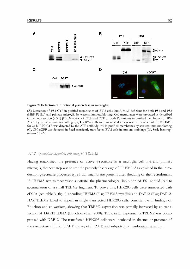
RESULTS 62
3.1.2 γ-secretase dependent processing of TREM2
Having established the presence of active γ-secretase in a microglia cell line and primary
microglia, the next step was to test the proteolytic cleavage of TREM2. As explained in the intro-
duction γ-secretase processes type I transmembrane proteins after shedding of their ectodomain.
If TREM2 acts as γ-secretase substrate, the pharmacological inhibition of PS1 should lead to
accumulation of a small TREM2 fragment. To prove this, HEK293 cells were transfected with
cDNA (see table 3, fig. 6) encoding TREM2 (Flag-TREM2-mycHis) and DAP12 (Flag-DAP12-
HA). TREM2 failed to appear in single transfected HEK293 cells, consistent with findings of
Bouchon and co-workers, showing that TREM2 expression was partially increased by co-trans-
fection of DAP12 cDNA (Bouchon et al., 2000). Thus, in all experiments TREM2 was co-ex-
pressed with DAP12. The transfected HEK293 cells were incubated in absence or presence of
the γ-secretase inhibitor DAPT (Dovey et al., 2001) and subjected to membrane preparation.
Figure 7: Detection of functional γ-secretase in microglia.
(A) Detection of PS1 CTF in purified membranes of BV-2 cells, MEF, MEF deficient for both PS1 and PS2 (MEF PSdko) and primary microglia by western immunoblotting. Cell membranes were prepared as described in methods section (2.3.1) (B) Detection of NTF and CTF of both PS variants in purified membranes of BV-2 cells by western immunoblotting. (C, D) BV-2 cells were incubated in absence or presence of 1 µM DAPT for 24 h. APP CTF was detected by the APP antibody 140 in purified membranes by western immunoblotting (C). C99-eGFP was detected in fixed transiently transfected BV-2 cells in immuno stainings (D). Scale bars rep-resents 10 µM
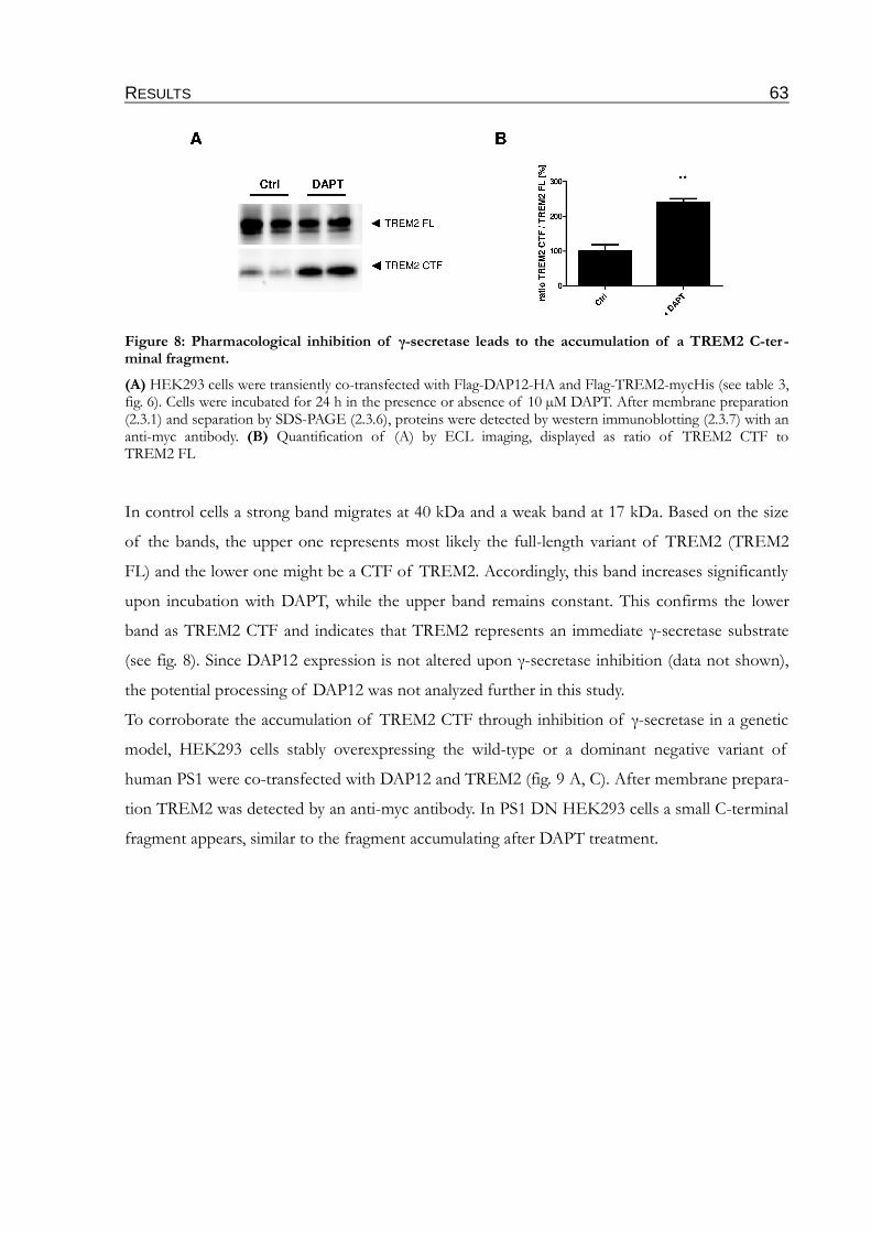
RESULTS 63
In control cells a strong band migrates at 40 kDa and a weak band at 17 kDa. Based on the size
of the bands, the upper one represents most likely the full-length variant of TREM2 (TREM2
FL) and the lower one might be a CTF of TREM2. Accordingly, this band increases significantly
upon incubation with DAPT, while the upper band remains constant. This confirms the lower
band as TREM2 CTF and indicates that TREM2 represents an immediate γ-secretase substrate
(see fig. 8). Since DAP12 expression is not altered upon γ-secretase inhibition (data not shown),
the potential processing of DAP12 was not analyzed further in this study.
To corroborate the accumulation of TREM2 CTF through inhibition of γ-secretase in a genetic
model, HEK293 cells stably overexpressing the wild-type or a dominant negative variant of
human PS1 were co-transfected with DAP12 and TREM2 (fig. 9 A, C). After membrane prepara-
tion TREM2 was detected by an anti-myc antibody. In PS1 DN HEK293 cells a small C-terminal
fragment appears, similar to the fragment accumulating after DAPT treatment.
Figure 8: Pharmacological inhibition of γ-secretase leads to the accumulation of a TREM2 C-ter-minal fragment.
(A) HEK293 cells were transiently co-transfected with Flag-DAP12-HA and Flag-TREM2-mycHis (see table 3, fig. 6). Cells were incubated for 24 h in the presence or absence of 10 µM DAPT. After membrane preparation (2.3.1) and separation by SDS-PAGE (2.3.6), proteins were detected by western immunoblotting (2.3.7) with an anti-myc antibody. (B) Quantification of (A) by ECL imaging, displayed as ratio of TREM2 CTF to TREM2 FL
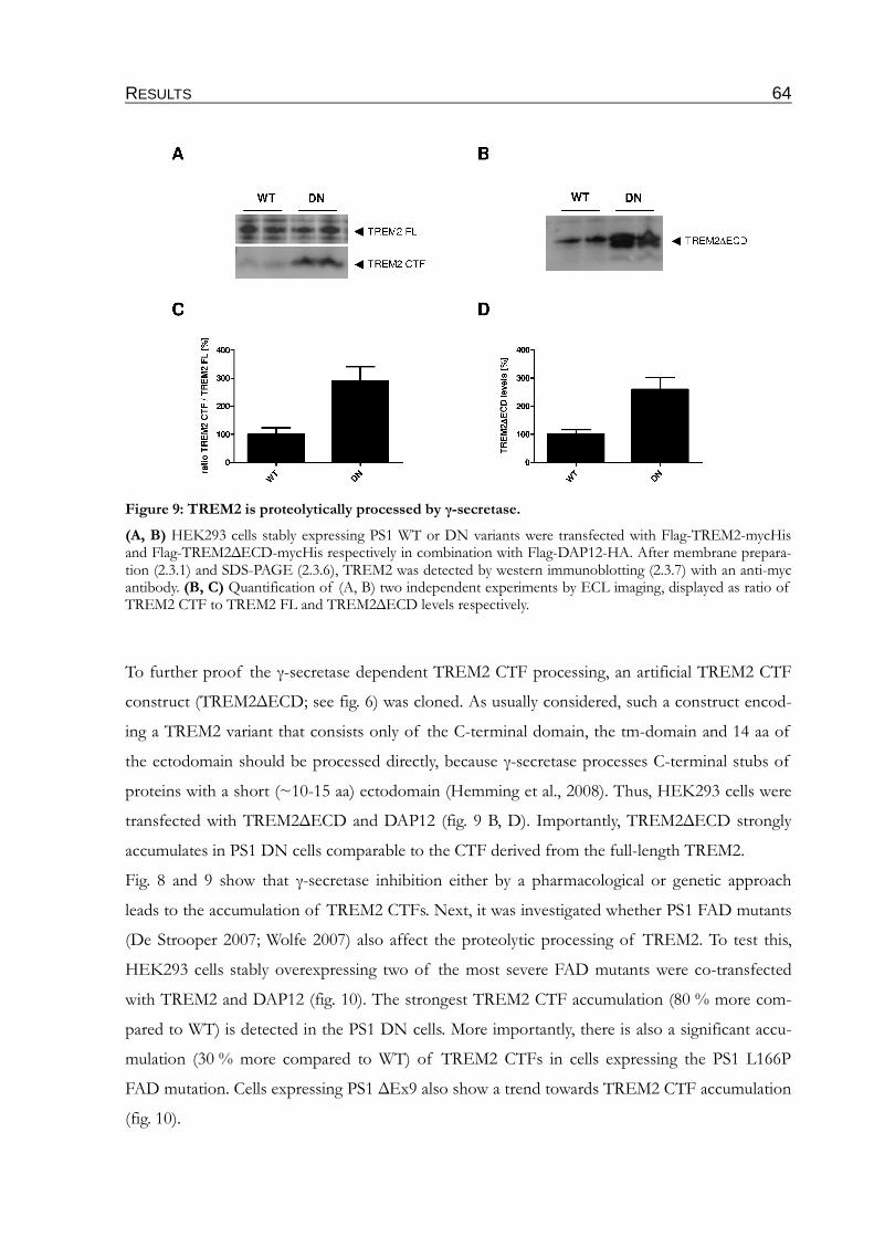
RESULTS 64
To further proof the γ-secretase dependent TREM2 CTF processing, an artificial TREM2 CTF
construct (TREM2∆ECD; see fig. 6) was cloned. As usually considered, such a construct encod-
ing a TREM2 variant that consists only of the C-terminal domain, the tm-domain and 14 aa of
the ectodomain should be processed directly, because γ-secretase processes C-terminal stubs of
proteins with a short (~10-15 aa) ectodomain (Hemming et al., 2008). Thus, HEK293 cells were
transfected with TREM2∆ECD and DAP12 (fig. 9 B, D). Importantly, TREM2∆ECD strongly
accumulates in PS1 DN cells comparable to the CTF derived from the full-length TREM2.
Fig. 8 and 9 show that γ-secretase inhibition either by a pharmacological or genetic approach
leads to the accumulation of TREM2 CTFs. Next, it was investigated whether PS1 FAD mutants
(De Strooper 2007; Wolfe 2007) also affect the proteolytic processing of TREM2. To test this,
HEK293 cells stably overexpressing two of the most severe FAD mutants were co-transfected
with TREM2 and DAP12 (fig. 10). The strongest TREM2 CTF accumulation (80 % more com-
pared to WT) is detected in the PS1 DN cells. More importantly, there is also a significant accu-
mulation (30 % more compared to WT) of TREM2 CTFs in cells expressing the PS1 L166P
FAD mutation. Cells expressing PS1 ∆Ex9 also show a trend towards TREM2 CTF accumulation
(fig. 10).
Figure 9: TREM2 is proteolytically processed by γ-secretase.
(A, B) HEK293 cells stably expressing PS1 WT or DN variants were transfected with Flag-TREM2-mycHis and Flag-TREM2∆ECD-mycHis respectively in combination with Flag-DAP12-HA. After membrane prepara-tion (2.3.1) and SDS-PAGE (2.3.6), TREM2 was detected by western immunoblotting (2.3.7) with an anti-myc antibody. (B, C) Quantification of (A, B) two independent experiments by ECL imaging, displayed as ratio of TREM2 CTF to TREM2 FL and TREM2∆ECD levels respectively.
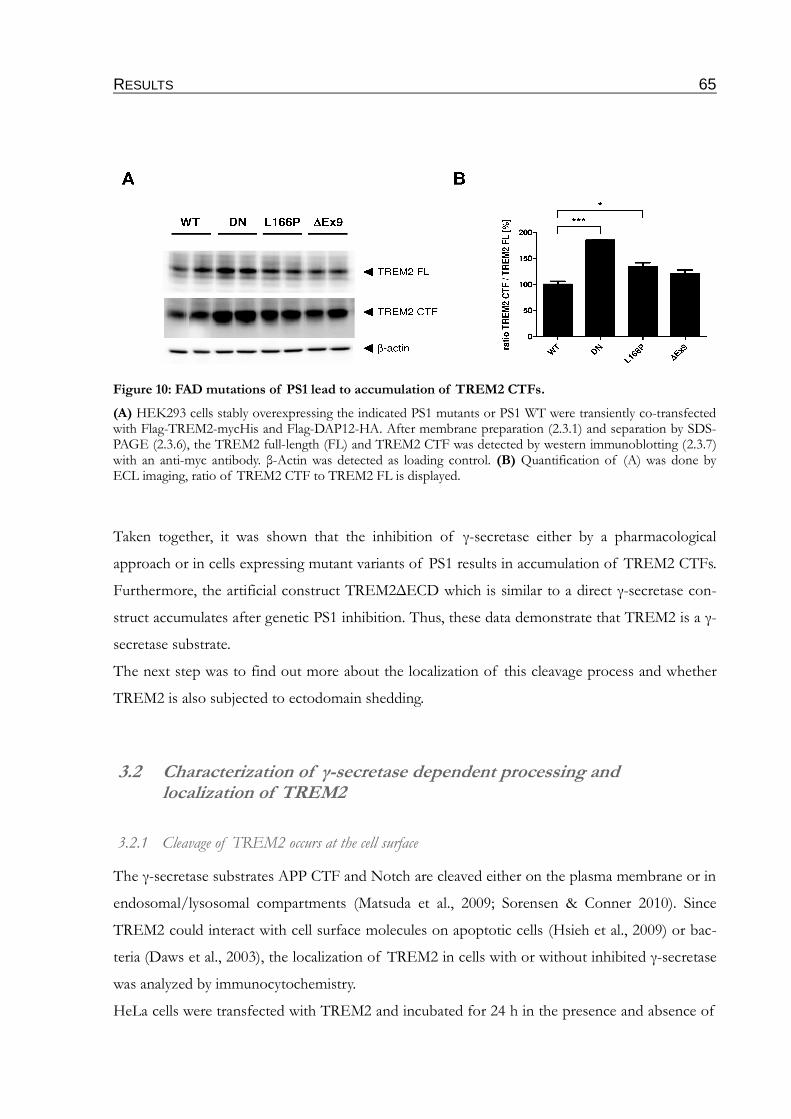
RESULTS 65
Taken together, it was shown that the inhibition of γ-secretase either by a pharmacological
approach or in cells expressing mutant variants of PS1 results in accumulation of TREM2 CTFs.
Furthermore, the artificial construct TREM2∆ECD which is similar to a direct γ-secretase con-
struct accumulates after genetic PS1 inhibition. Thus, these data demonstrate that TREM2 is a γ-
secretase substrate.
The next step was to find out more about the localization of this cleavage process and whether
TREM2 is also subjected to ectodomain shedding.
3.2 Characterization of γ-secretase dependent processing and localization of TREM2
3.2.1 Cleavage of TREM2 occurs at the cell surface
The γ-secretase substrates APP CTF and Notch are cleaved either on the plasma membrane or in
endosomal/lysosomal compartments (Matsuda et al., 2009; Sorensen & Conner 2010). Since
TREM2 could interact with cell surface molecules on apoptotic cells (Hsieh et al., 2009) or bac-
teria (Daws et al., 2003), the localization of TREM2 in cells with or without inhibited γ-secretase
was analyzed by immunocytochemistry.
HeLa cells were transfected with TREM2 and incubated for 24 h in the presence and absence of
Figure 10: FAD mutations of PS1 lead to accumulation of TREM2 CTFs.
(A) HEK293 cells stably overexpressing the indicated PS1 mutants or PS1 WT were transiently co-transfected with Flag-TREM2-mycHis and Flag-DAP12-HA. After membrane preparation (2.3.1) and separation by SDS-PAGE (2.3.6), the TREM2 full-length (FL) and TREM2 CTF was detected by western immunoblotting (2.3.7) with an anti-myc antibody. β-Actin was detected as loading control. (B) Quantification of (A) was done by ECL imaging, ratio of TREM2 CTF to TREM2 FL is displayed.
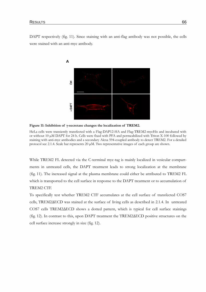
RESULTS 66
DAPT respectively (fig. 11). Since staining with an anti-flag antibody was not possible, the cells
were stained with an anti-myc antibody.
While TREM2 FL detected via the C-terminal myc-tag is mainly localized in vesicular compart-
ments in untreated cells, the DAPT treatment leads to strong localization at the membrane
(fig. 11). The increased signal at the plasma membrane could either be attributed to TREM2 FL
which is transported to the cell surface in response to the DAPT treatment or to accumulation of
TREM2 CTF.
To specifically test whether TREM2 CTF accumulates at the cell surface of transfected COS7
cells, TREM2∆ECD was stained at the surface of living cells as described in 2.1.4. In untreated
COS7 cells TREM2∆ECD shows a dotted pattern, which is typical for cell surface stainings
(fig. 12). In contrast to this, upon DAPT treatment the TREM2∆ECD positive structures on the
cell surface increase strongly in size (fig. 12).
Figure 11: Inhibition of γ-secretase changes the localization of TREM2.
HeLa cells were transiently transfected with a Flag-DAP12-HA and Flag-TREM2-mycHis and incubated with or without 10 µM DAPT for 24 h. Cells were fixed with PFA and permeabilized with Triton X-100 followed by staining with anti-myc antibodies and a secondary Alexa 594-coupled antibody to detect TREM2. For a detailed protocol see 2.1.4. Scale bar represents 20 µM. Two representative images of each group are shown.
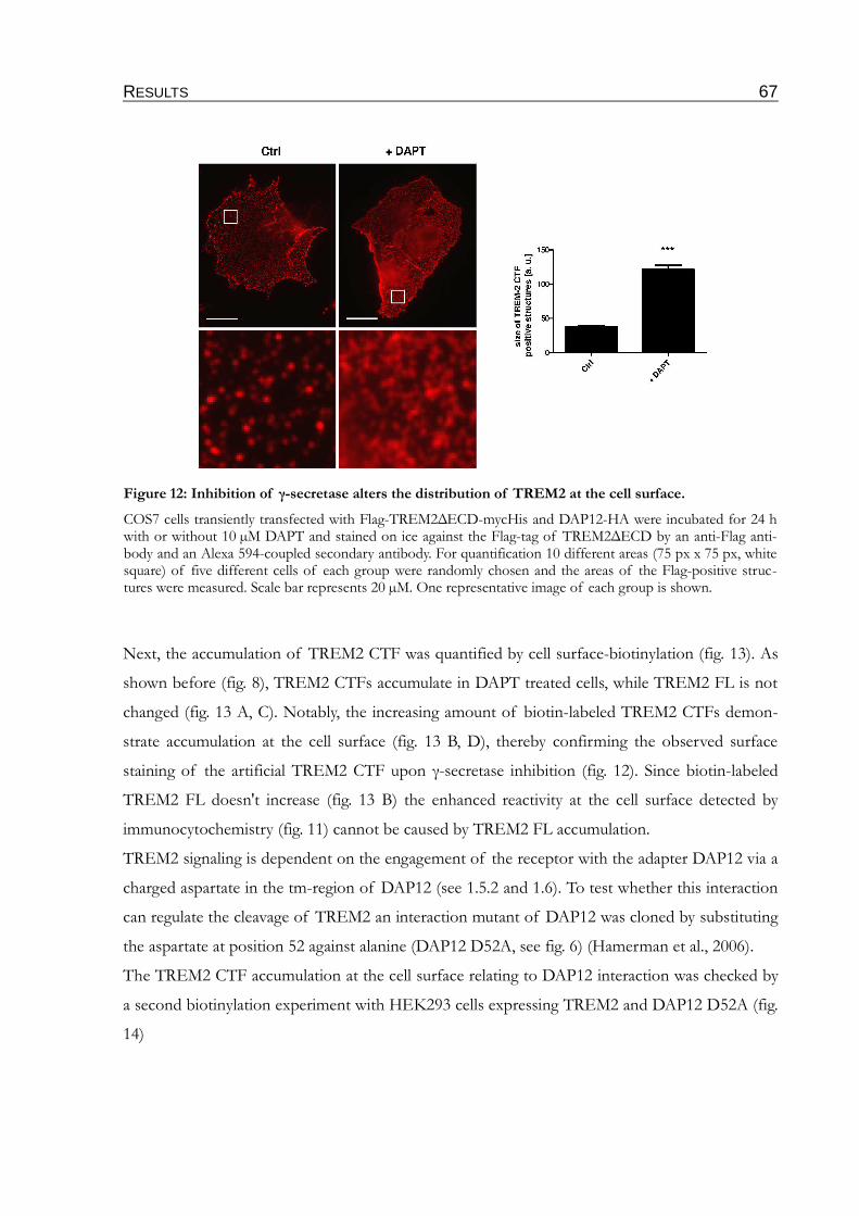
RESULTS 67
Next, the accumulation of TREM2 CTF was quantified by cell surface-biotinylation (fig. 13). As
shown before (fig. 8), TREM2 CTFs accumulate in DAPT treated cells, while TREM2 FL is not
changed (fig. 13 A, C). Notably, the increasing amount of biotin-labeled TREM2 CTFs demon-
strate accumulation at the cell surface (fig. 13 B, D), thereby confirming the observed surface
staining of the artificial TREM2 CTF upon γ-secretase inhibition (fig. 12). Since biotin-labeled
TREM2 FL doesn't increase (fig. 13 B) the enhanced reactivity at the cell surface detected by
immunocytochemistry (fig. 11) cannot be caused by TREM2 FL accumulation.
TREM2 signaling is dependent on the engagement of the receptor with the adapter DAP12 via a
charged aspartate in the tm-region of DAP12 (see 1.5.2 and 1.6). To test whether this interaction
can regulate the cleavage of TREM2 an interaction mutant of DAP12 was cloned by substituting
the aspartate at position 52 against alanine (DAP12 D52A, see fig. 6) (Hamerman et al., 2006).
The TREM2 CTF accumulation at the cell surface relating to DAP12 interaction was checked by
a second biotinylation experiment with HEK293 cells expressing TREM2 and DAP12 D52A (fig.
14)
Figure 12: Inhibition of γ-secretase alters the distribution of TREM2 at the cell surface.
COS7 cells transiently transfected with Flag-TREM2∆ECD-mycHis and DAP12-HA were incubated for 24 h with or without 10 µM DAPT and stained on ice against the Flag-tag of TREM2∆ECD by an anti-Flag anti-body and an Alexa 594-coupled secondary antibody. For quantification 10 different areas (75 px x 75 px, white square) of five different cells of each group were randomly chosen and the areas of the Flag-positive struc-tures were measured. Scale bar represents 20 µM. One representative image of each group is shown.
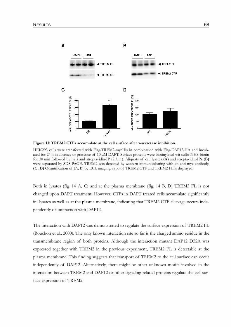
RESULTS 68
Both in lysates (fig. 14 A, C) and at the plasma membrane (fig. 14 B, D) TREM2 FL is not
changed upon DAPT treatment. However, CTFs in DAPT treated cells accumulate significantly
in lysates as well as at the plasma membrane, indicating that TREM2 CTF cleavage occurs inde-
pendently of interaction with DAP12.
The interaction with DAP12 was demonstrated to regulate the surface expression of TREM2 FL
(Bouchon et al., 2000). The only known interaction site so far is the charged amino residue in the
transmembrane region of both proteins. Although the interaction mutant DAP12 D52A was
expressed together with TREM2 in the previous experiment, TREM2 FL is detectable at the
plasma membrane. This finding suggests that transport of TREM2 to the cell surface can occur
independently of DAP12. Alternatively, there might be other unknown motifs involved in the
interaction between TREM2 and DAP12 or other signaling related proteins regulate the cell-sur-
face expression of TREM2.
Figure 13: TREM2 CTFs accumulate at the cell surface after γ-secretase inhibition.
HEK293 cells were transfected with Flag-TREM2-mycHis in combination with Flag-DAP12-HA and incub-ated for 24 h in absence or presence of 10 µM DAPT. Surface proteins were biotinylated wit sulfo-NHS-biotin for 30 min followed by lysis and streptavidin-IP (2.3.11). Aliquots of cell lysates (A) and streptavidin-IPs (B) were separated by SDS-PAGE. TREM2 was detected by western immunoblotting with an anti-myc antibody. (C, D) Quantification of (A, B) by ECL imaging, ratio of TREM2 CTF and TREM2 FL is displayed.

RESULTS 69
3.2.2 γ-secretase mediated processing in response to the activation of TREM2.
There are several ways to inactivate receptors upon ligand binding and activation. The ligand-re-
ceptor complexes could be taken up by endocytosis and be further degraded. Alternatively, the
complexes could be cleaved directly at the membrane. It is known that ligand binding could
induce the shedding of the ECD of Notch, allowing subsequent γ-secretase cleavage which leads
to an increase in Notch ICD, a small fragment transported to the nucleus where it has gene regu-
latory function (Schroeder et al., 1998; Mumm et al., 2000). The induced receptor cleavage is also
known for G-protein coupled receptors like the V2 vasopressin receptor (Kojro & Fahrenholz
1995), thereby downregulating the receptors.
However, nothing is known about the downregulation of the TREM2/DAP12 signaling. To test
whether TREM2 activation alters γ-secretase processing HEK293 PS1 WT and DN cells,
expressing myc-TREM2-GFP/DAP12-HA were subjected to a non-radioactive TREM2 activa-
tion assay as described in 2.3.14 (fig. 15). Since no ligand of TREM2 is known, myc-tagged
TREM2 was activated by ligation with an anti-myc antibody.
Figure 14: Interaction with DAP12 is not prerequisite for γ-secretase mediated TREM2 cleavage.
HEK293 expressing Flag-TREM2-mycHis in combination with Flag-DAP12 D52A-HA were incubated for 24 h in absence and presence of 10 µM DAPT. After biotinylation with sulfo-NHS-biotin, lysis and streptavid-in-IP (2.3.11) aliquots of cell lysates (A) and streptavidin-IPs (B) were separated by SDS-PAGE. TREM2 was detected by western immunoblotting with an anti-myc antibody. (C, D) Quantification of (A, B) was done by ECL imaging.

RESULTS 70
Upon activation, levels of TREM2 FL changes neither in PS1 WT nor in PS1 DN cells. How-
ever, as previously shown, TREM2 CTF accumulates in HEK293 PS1 DN cells (fig. 15). Notably,
after TREM2 activation by antibody cross-linking a slight increase in TREM2 CTFs is observed
in HEK293 PS1 DN as compared to PS1 WT cells, but this difference doesn't reach significance.
Taken together, all these data indicate that proteolytic processing of TREM2 CTFs occurs at the
plasma membrane dependent on the activation state of TREM2.
3.2.3 TREM2 ICD is not translocated to the nucleus
γ-secretase cleavage of substrate CTFs results in release of ICDs to the cytosol (see 1.1.4). The
ICD of Notch is known to translocate to the nucleus upon generation, regulating expression of
various target genes. The ICDs of several other γ-secretase substrates are very unstable due to
efficient proteolytic degradation. Since detection of TREM2 ICD (TICD) after overexpression
of TREM2 FL by western immunoblotting and immunostainings was not possible (not shown),
an artificial TICD construct was generated starting one amino acid behind the putative tm-do-
main (see table 3, fig. 6). To test the localization of this fragment, HeLa cells were transiently
transfected and subjected to immunofluorescence stainings (fig. 16). The transfected TICDs
reveal no nuclear staining, but a vesicular stain, distributed over the whole cell.
Figure 15: γ-secretase cleavage in response to activation of TREM2.
(A) HEK293 cells stably expressing PS1 WT and DN were transiently transfected with myc-TREM2-GFP. 24 h after transfection medium was changed against DMEM -/- or DMEM -/- supplemented with 10 µg/ml anti-myc antibody and cells were incubated for 1 h (for detailed protocol see 2.3.14). Membrane proteins were isolated as described in 2.3.1. After SDS-PAGE TREM2 FL & CTF were detected by western immunoblotting with an anti-GFP antibody. (B) Quantification of TREM2 FL and CTF by ECL imaging, normalized to β-actin which served as loading control. Ratio of TREM2 CTF/FL is shown.
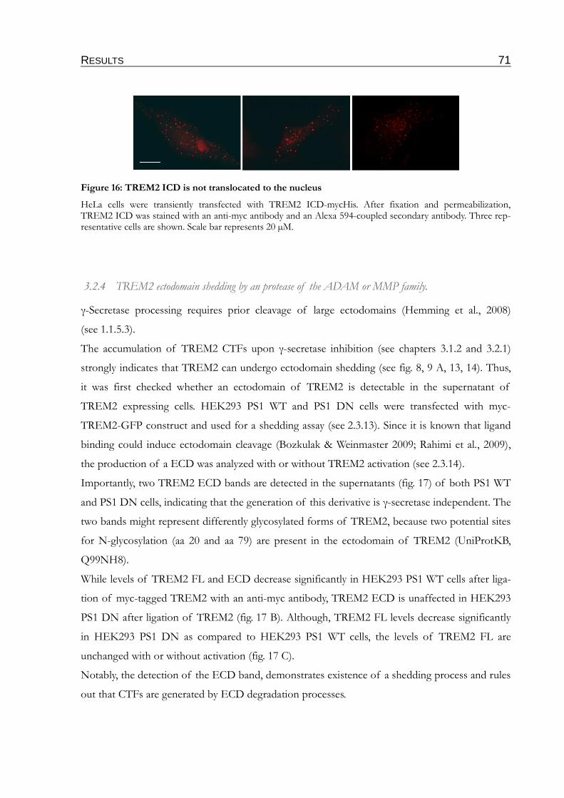
RESULTS 71
3.2.4 TREM2 ectodomain shedding by an protease of the ADAM or MMP family.
γ-Secretase processing requires prior cleavage of large ectodomains (Hemming et al., 2008)
(see 1.1.5.3).
The accumulation of TREM2 CTFs upon γ-secretase inhibition (see chapters 3.1.2 and 3.2.1)
strongly indicates that TREM2 can undergo ectodomain shedding (see fig. 8, 9 A, 13, 14). Thus,
it was first checked whether an ectodomain of TREM2 is detectable in the supernatant of
TREM2 expressing cells. HEK293 PS1 WT and PS1 DN cells were transfected with myc-
TREM2-GFP construct and used for a shedding assay (see 2.3.13). Since it is known that ligand
binding could induce ectodomain cleavage (Bozkulak & Weinmaster 2009; Rahimi et al., 2009),
the production of a ECD was analyzed with or without TREM2 activation (see 2.3.14).
Importantly, two TREM2 ECD bands are detected in the supernatants (fig. 17) of both PS1 WT
and PS1 DN cells, indicating that the generation of this derivative is γ-secretase independent. The
two bands might represent differently glycosylated forms of TREM2, because two potential sites
for N-glycosylation (aa 20 and aa 79) are present in the ectodomain of TREM2 (UniProtKB,
Q99NH8).
While levels of TREM2 FL and ECD decrease significantly in HEK293 PS1 WT cells after liga-
tion of myc-tagged TREM2 with an anti-myc antibody, TREM2 ECD is unaffected in HEK293
PS1 DN after ligation of TREM2 (fig. 17 B). Although, TREM2 FL levels decrease significantly
in HEK293 PS1 DN as compared to HEK293 PS1 WT cells, the levels of TREM2 FL are
unchanged with or without activation (fig. 17 C).
Notably, the detection of the ECD band, demonstrates existence of a shedding process and rules
out that CTFs are generated by ECD degradation processes.
Figure 16: TREM2 ICD is not translocated to the nucleus
HeLa cells were transiently transfected with TREM2 ICD-mycHis. After fixation and permeabilization, TREM2 ICD was stained with an anti-myc antibody and an Alexa 594-coupled secondary antibody. Three rep-resentative cells are shown. Scale bar represents 20 µM.
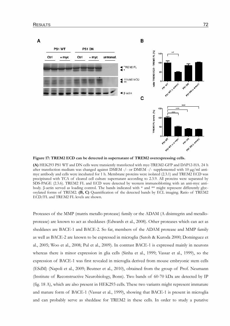
RESULTS 72
Proteases of the MMP (matrix metallo protease) family or the ADAM (A disintegrin and metallo-
protease) are known to act as sheddases (Edwards et al., 2008). Other proteases which can act as
sheddases are BACE-1 and BACE-2. So far, members of the ADAM protease and MMP family
as well as BACE-2 are known to be expressed in microglia (Satoh & Kuroda 2000; Dominguez et
al., 2005; Woo et al., 2008; Pul et al., 2009). In contrast BACE-1 is expressed mainly in neurons
whereas there is minor expression in glia cells (Sinha et al., 1999; Vassar et al., 1999), so the
expression of BACE-1 was first revealed in microglia derived from mouse embryonic stem cells
(ESdM) (Napoli et al., 2009; Beutner et al., 2010), obtained from the group of Prof. Neumann
(Institute of Reconstructive Neurobiology, Bonn). Two bands of 60-70 kDa are detected by IP
(fig. 18 A), which are also present in HEK293 cells. These two variants might represent immature
and mature form of BACE-1 (Vassar et al., 1999), showing that BACE-1 is present in microglia
and can probably serve as sheddase for TREM2 in these cells. In order to study a putative
Figure 17: TREM2 ECD can be detected in supernatant of TREM2 overexpressing cells.
(A) HEK293 PS1 WT and DN cells were transiently transfected with myc-TREM2-GFP and DAP12-HA. 24 h after transfection medium was changed against DMEM -/- or DMEM -/- supplemented with 10 µg/ml anti-myc antibody and cells were incubated for 1 h. Membrane proteins were isolated (2.3.1) and TREM2 ECD was precipitated with TCA of cleared cell culture supernatant according to 2.3.9. All proteins were separated by SDS-PAGE (2.3.6). TREM2 FL and ECD were detected by western immunoblotting with an anti-myc anti-body. β-actin served as loading control. The bands indicated with * and ** might represent differently glyc-osylated forms of TREM2. (B, C) Quantification of the detected bands by ECL imaging. Ratio of TREM2 ECD/FL and TREM2 FL levels are shown.
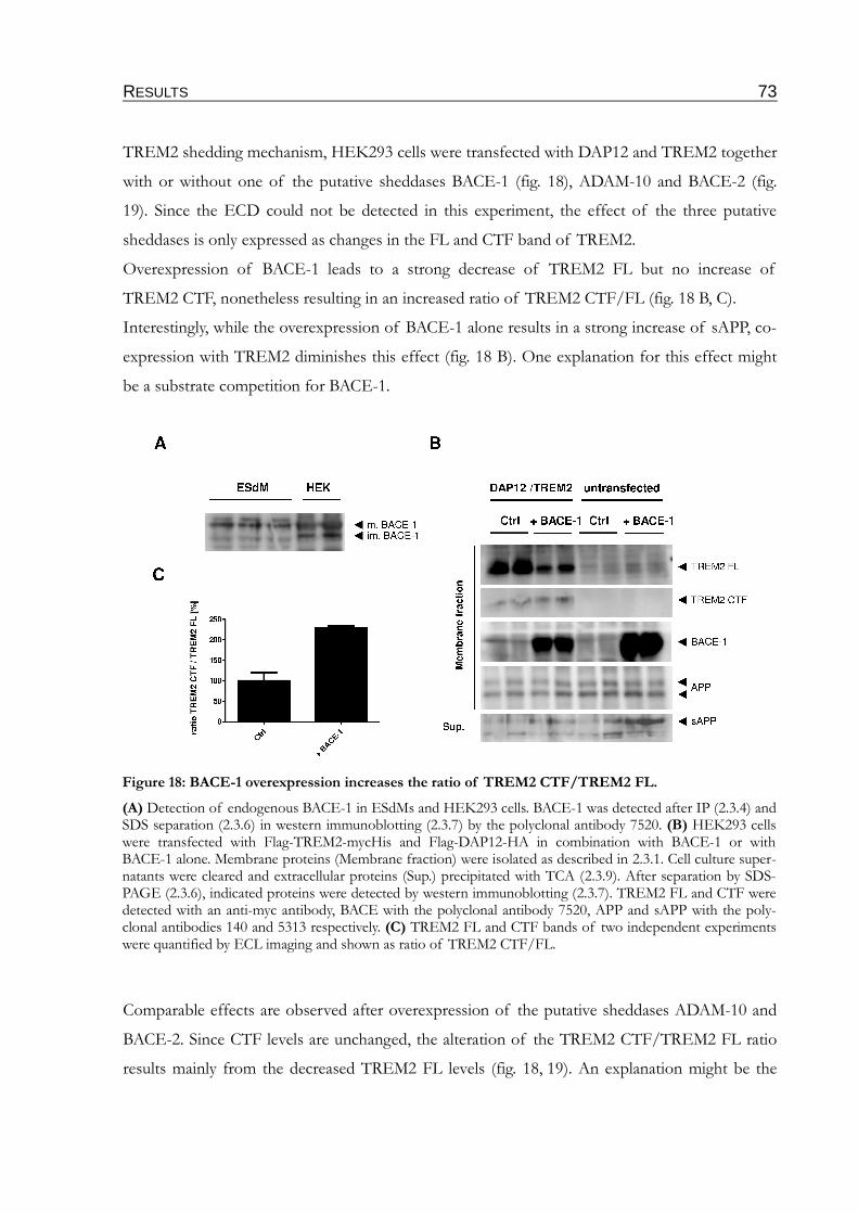
RESULTS 73
TREM2 shedding mechanism, HEK293 cells were transfected with DAP12 and TREM2 together
with or without one of the putative sheddases BACE-1 (fig. 18), ADAM-10 and BACE-2 (fig.
19). Since the ECD could not be detected in this experiment, the effect of the three putative
sheddases is only expressed as changes in the FL and CTF band of TREM2.
Overexpression of BACE-1 leads to a strong decrease of TREM2 FL but no increase of
TREM2 CTF, nonetheless resulting in an increased ratio of TREM2 CTF/FL (fig. 18 B, C).
Interestingly, while the overexpression of BACE-1 alone results in a strong increase of sAPP, co-
expression with TREM2 diminishes this effect (fig. 18 B). One explanation for this effect might
be a substrate competition for BACE-1.
Comparable effects are observed after overexpression of the putative sheddases ADAM-10 and
BACE-2. Since CTF levels are unchanged, the alteration of the TREM2 CTF/TREM2 FL ratio
results mainly from the decreased TREM2 FL levels (fig. 18, 19). An explanation might be the
Figure 18: BACE-1 overexpression increases the ratio of TREM2 CTF/TREM2 FL.
(A) Detection of endogenous BACE-1 in ESdMs and HEK293 cells. BACE-1 was detected after IP (2.3.4) and SDS separation (2.3.6) in western immunoblotting (2.3.7) by the polyclonal antibody 7520. (B) HEK293 cells were transfected with Flag-TREM2-mycHis and Flag-DAP12-HA in combination with BACE-1 or with BACE-1 alone. Membrane proteins (Membrane fraction) were isolated as described in 2.3.1. Cell culture super-natants were cleared and extracellular proteins (Sup.) precipitated with TCA (2.3.9). After separation by SDS-PAGE (2.3.6), indicated proteins were detected by western immunoblotting (2.3.7). TREM2 FL and CTF were detected with an anti-myc antibody, BACE with the polyclonal antibody 7520, APP and sAPP with the poly-clonal antibodies 140 and 5313 respectively. (C) TREM2 FL and CTF bands of two independent experiments were quantified by ECL imaging and shown as ratio of TREM2 CTF/FL.
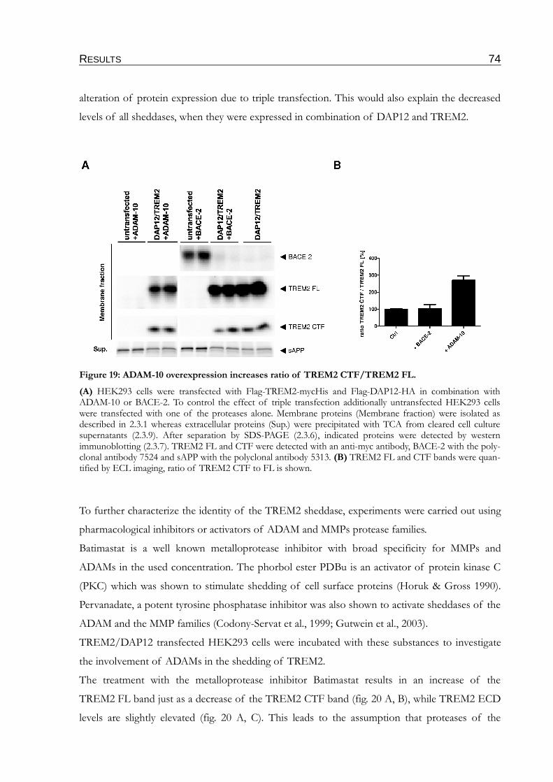
RESULTS 74
alteration of protein expression due to triple transfection. This would also explain the decreased
levels of all sheddases, when they were expressed in combination of DAP12 and TREM2.
To further characterize the identity of the TREM2 sheddase, experiments were carried out using
pharmacological inhibitors or activators of ADAM and MMPs protease families.
Batimastat is a well known metalloprotease inhibitor with broad specificity for MMPs and
ADAMs in the used concentration. The phorbol ester PDBu is an activator of protein kinase C
(PKC) which was shown to stimulate shedding of cell surface proteins (Horuk & Gross 1990).
Pervanadate, a potent tyrosine phosphatase inhibitor was also shown to activate sheddases of the
ADAM and the MMP families (Codony-Servat et al., 1999; Gutwein et al., 2003).
TREM2/DAP12 transfected HEK293 cells were incubated with these substances to investigate
the involvement of ADAMs in the shedding of TREM2.
The treatment with the metalloprotease inhibitor Batimastat results in an increase of the
TREM2 FL band just as a decrease of the TREM2 CTF band (fig. 20 A, B), while TREM2 ECD
levels are slightly elevated (fig. 20 A, C). This leads to the assumption that proteases of the
Figure 19: ADAM-10 overexpression increases ratio of TREM2 CTF/TREM2 FL.
(A) HEK293 cells were transfected with Flag-TREM2-mycHis and Flag-DAP12-HA in combination with ADAM-10 or BACE-2. To control the effect of triple transfection additionally untransfected HEK293 cells were transfected with one of the proteases alone. Membrane proteins (Membrane fraction) were isolated as described in 2.3.1 whereas extracellular proteins (Sup.) were precipitated with TCA from cleared cell culture supernatants (2.3.9). After separation by SDS-PAGE (2.3.6), indicated proteins were detected by western immunoblotting (2.3.7). TREM2 FL and CTF were detected with an anti-myc antibody, BACE-2 with the poly-clonal antibody 7524 and sAPP with the polyclonal antibody 5313. (B) TREM2 FL and CTF bands were quan-tified by ECL imaging, ratio of TREM2 CTF to FL is shown.
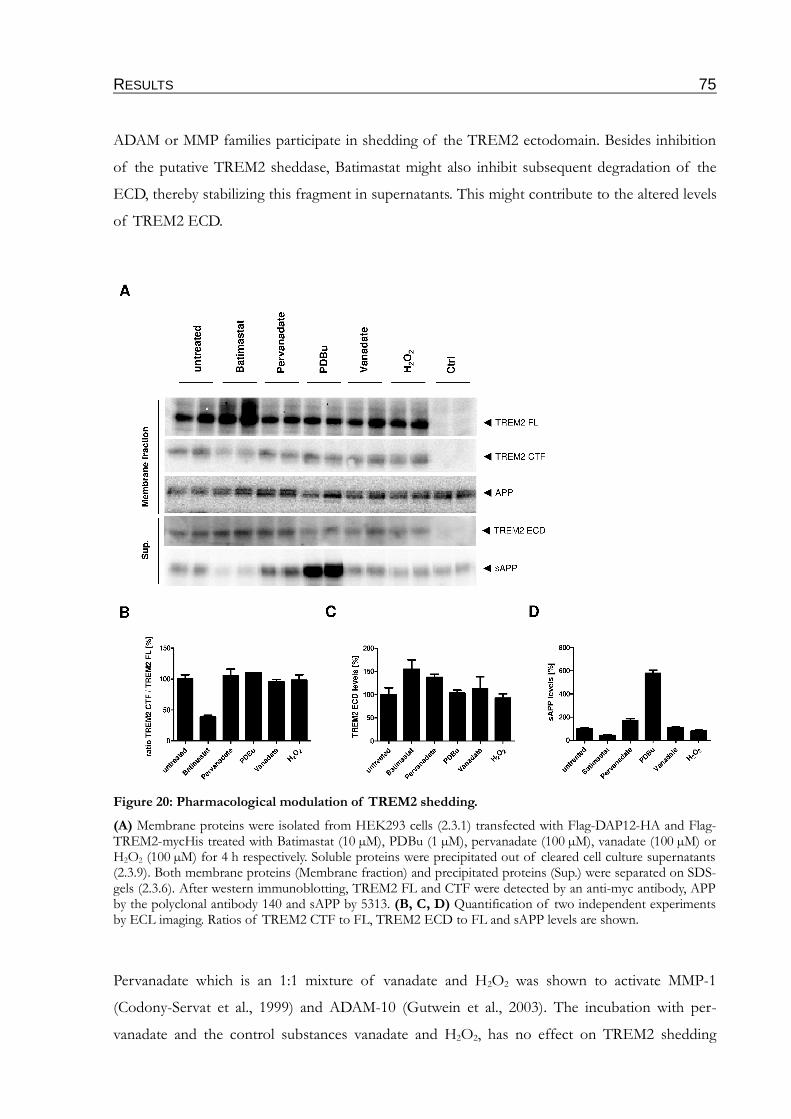
RESULTS 75
ADAM or MMP families participate in shedding of the TREM2 ectodomain. Besides inhibition
of the putative TREM2 sheddase, Batimastat might also inhibit subsequent degradation of the
ECD, thereby stabilizing this fragment in supernatants. This might contribute to the altered levels
of TREM2 ECD.
Pervanadate which is an 1:1 mixture of vanadate and H2O2 was shown to activate MMP-1
(Codony-Servat et al., 1999) and ADAM-10 (Gutwein et al., 2003). The incubation with per-
vanadate and the control substances vanadate and H2O2, has no effect on TREM2 shedding
Figure 20: Pharmacological modulation of TREM2 shedding.
(A) Membrane proteins were isolated from HEK293 cells (2.3.1) transfected with Flag-DAP12-HA and Flag-TREM2-mycHis treated with Batimastat (10 µM), PDBu (1 µM), pervanadate (100 µM), vanadate (100 µM) or H2O2 (100 µM) for 4 h respectively. Soluble proteins were precipitated out of cleared cell culture supernatants (2.3.9). Both membrane proteins (Membrane fraction) and precipitated proteins (Sup.) were separated on SDS-gels (2.3.6). After western immunoblotting, TREM2 FL and CTF were detected by an anti-myc antibody, APP by the polyclonal antibody 140 and sAPP by 5313. (B, C, D) Quantification of two independent experiments by ECL imaging. Ratios of TREM2 CTF to FL, TREM2 ECD to FL and sAPP levels are shown.
RESULTS 76
(fig. 20 A, B), suggesting that MMP-1 and ADAM-10 might not represent major TREM2 shed-
dases. However, TREM2 ECD levels are slightly increased with pervanadate (fig. 20 A, C). Para-
doxically, the well established shedding activators PDBu and pervanadate don't alter the shedding
of TREM2, demonstrated by the unaltered TREM2 CTF/FL ratio and ECD levels. However, as
expected sAPP levels increase upon treatment with PDBu and pervanadate, confirming the activ-
ity of both modulators.
In fig. 20 the effect of different shedding modulators is addressed, but the levels of TREM2
ECD don't match to the TREM2 CTF/FL ratio. To further establish an effect of these modulat-
ors on the TREM2 FL-expression at the cell surface, myc-TREM2-GFP was analyzed by a
microscopic approach. In the used construct, the myc-tag allows the specific detection of
TREM2 FL at the plasma membrane, whereas the GFP-tag allows estimation of the total expres-
sion.
COS7 cells transfected with DAP12-HA and myc-TREM2-GFP were incubated with the indic-
ated substances and myc-tagged extracellular part of TREM2 was stained at the surface of living
cells as described in 2.1.4.
In untreated and DMSO treated cells, only minor amounts of TREM2 are detected at the cell
surface, suggesting that TREM2 undergoes efficient constitutive shedding in COS7 cells (fig. 21
G, H, I). The amount of surface localized TREM2 FL is significantly decreased when cells were
incubated for one hour with PDBu (fig. 21 D, E, F). In contrast, the treatment with the broad
sheddase inhibitor Batimastat results in a markedly increase of myc-positive dots on the cell sur-
face (fig. 21 A, B, C). The quantification of 10 cells with five randomly chosen squares of
75 x 75 px each, yield an averaged number of 25 dots for Batimastat. Both untreated and DMSO
treated cells show less than 10 dots per square.
Interestingly, the GFP staining which represents the C-terminal part of TREM2 is similar under
all tested conditions. This suggests that large fractions of TREM2 are localized in intracellular
compartments which would be consistent to the localization pattern of TREM2 observed in
microglia (Sessa et al., 2004; Prada et al., 2006). However, it is also possible that TREM2 exists
predominantly in a variant lacking the lumenal domain on the cell surface (fig. 21 B, E, H, K).
Taken together, these experiments clearly show that TREM2 undergoes shedding. However, the
nature of the involved protease could not be clarified completely. The sensitivity to PDBu and
Batimastat substantiate the suspicion that ADAMs, most likely ADAM-17 are involved in the
shedding process, but involvement of other proteases like BACE-1 or MMPs could not be ruled
out.
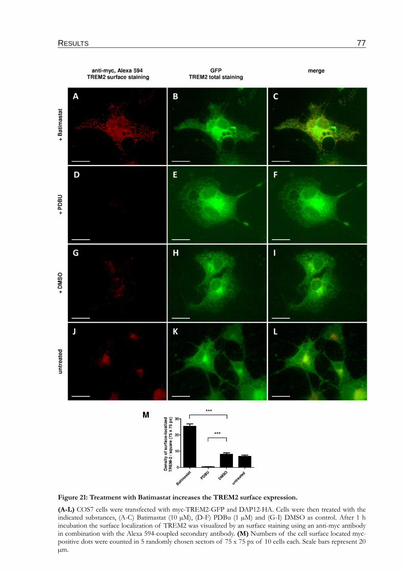
RESULTS 77
Figure 21: Treatment with Batimastat increases the TREM2 surface expression.
(A-L) COS7 cells were transfected with myc-TREM2-GFP and DAP12-HA. Cells were then treated with the indicated substances, (A-C) Batimastat (10 µM), (D-F) PDBu (1 µM) and (G-I) DMSO as control. After 1 h incubation the surface localization of TREM2 was visualized by an surface staining using an anti-myc antibody in combination with the Alexa 594-coupled secondary antibody. (M) Numbers of the cell surface located myc-positive dots were counted in 5 randomly chosen sectors of 75 x 75 px of 10 cells each. Scale bars represent 20 µm.
RESULTS 78
3.3 Role of TREM2 processing in the interaction with DAP12
3.3.1 Inhibition of γ-secretase facilitates co-localization of TREM2 and DAP12 at the cell surface.
After showing that TREM2 is cleaved by a sheddase generating a C-terminal fragment which is
subsequently cleaved by the γ-secretase (see chapters 3.1 and 3.2), it was next investigated
whether processing alters the interaction of TREM2 with its co-repressor DAP12.
To test this, COS7 cells transfected with Flag-TREM2∆ECD-mycHis and DAP12-HA, were
treated with or without DAPT and subjected to surface immunostaining (see 2.1.4). To visualize a
putative co-localization of DAP12 with the accumulated TREM2∆ECD, cells were fixed and per-
meabilized after surface staining and stained for total DAP12 expression via the C-terminal HA-
tag of the DAP12 construct.
The total DAP12 stain shows a diffuse but in some areas dotted pattern (fig. 22 B, E ). The co-
localization between TREM2∆ECD and DAP12 is very limited (fig. 22 C, F ). Upon γ-secretase
inhibition with DAPT, the surface staining of TREM2 increases and changes from a punctuated
to a more elongated, spiry shape, distributed over the whole cell. Interestingly, DAP12 shows
very similar changes in the localization (fig. 22 H, K ), reaching complete co-localization (fig. 22 I,
L ), indicating that TREM2 CTF interacts with DAP12 at the cell surface and affect its localiza-
tion upon γ-secretase inhibition.
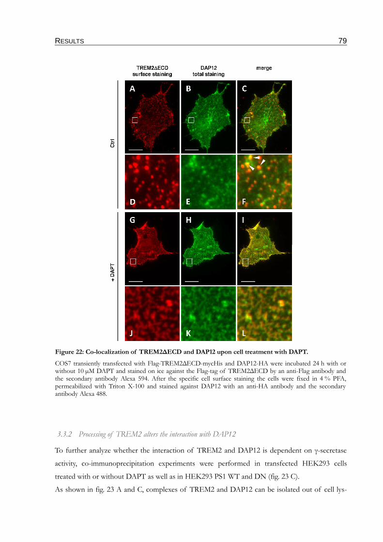
RESULTS 79
3.3.2 Processing of TREM2 alters the interaction with DAP12
To further analyze whether the interaction of TREM2 and DAP12 is dependent on γ-secretase
activity, co-immunoprecipitation experiments were performed in transfected HEK293 cells
treated with or without DAPT as well as in HEK293 PS1 WT and DN (fig. 23 C).
As shown in fig. 23 A and C, complexes of TREM2 and DAP12 can be isolated out of cell lys-
Figure 22: Co-localization of TREM2∆ECD and DAP12 upon cell treatment with DAPT.
COS7 transiently transfected with Flag-TREM2∆ECD-mycHis and DAP12-HA were incubated 24 h with or without 10 µM DAPT and stained on ice against the Flag-tag of TREM2∆ECD by an anti-Flag antibody and the secondary antibody Alexa 594. After the specific cell surface staining the cells were fixed in 4 % PFA, permeabilized with Triton X-100 and stained against DAP12 with an anti-HA antibody and the secondary antibody Alexa 488.
RESULTS 80
ates. Interestingly, the interaction of TREM2 FL with DAP12 markedly decreases in cells
expressing the dominant negative form of presenilin (fig. 23 A, B), indicating that loss of γ-
secretase function impairs interaction of TREM2 FL with DAP12. Decreased complex forma-
tion is also detected upon pharmacological γ-secretase inhibition with DAPT (fig. 23 C, D). How-
ever, these findings are completely contrary to trapping of DAP12 at the cell surface after DAPT
treatment as shown in fig. 22.
To control the identity of the precipitated proteins, in one lane of each blot the protein was pre-
cipitated with the antibody later used for detection (lane labeled with IP control). Precipitation
out of single transfected HEK293 cells results in a clean lane, evidencing the specificity of the
performed co-IPs.
One explanation for the contrary findings might base on the experimental conditions. The main
pool of TREM2 is located in the Golgi and in exocytic vesicles and only a minor part is located
at the membrane. While the surface staining experiment in fig. 22 visualizes TREM2/DAP12
interaction at the cell surface, the Co-IP strategy addresses TREM2/DAP12 interaction in the
whole cell. Thus, if binding of DAP12 by TREM2 CTF occurs only at the cell surface, precipita-
tion of the intracellular TREM2 would mask the cell surface effect. Moreover, the decrease of
TREM2/DAP12 interaction upon γ-secretase inhibition might be explained by massive transloca-
tion of DAP12 from intracellular pools to the cell membrane.
3.3.3 Inhibition of γ-secretase suppresses phosphorylation of DAP12 upon TREM2 activation
Ligand-dependent activation of TREM2 causes the interaction with the co-receptor DAP12
which subsequently induces phosphorylation of DAP12 (Takaki et al., 2006; Lanier 2009), trig-
gering activation of microglia or inducing phagocytosis (Neumann & Takahashi 2007; Takahashi
et al., 2007). Therefore, a potential involvement of γ-secretase in the regulation of phosphoryla-
tion of DAP12 was analyzed.
Cells co-expressing TREM2 and DAP12 were labeled with 32P-orthophosphate in absence or
presence of DAPT to inhibit γ-secretase. The two weak bands in the first lane of fig. 24. repres-
ent the phosphate incorporation of DAP12 one hour after activation of TREM2. As compared
to control cells, presence of orthovanadate strongly increases phosphorylation of DAP12, indic-
ating that DAP12 is efficiently dephosphorylated by phospho-tyrosine phosphatases. This in fact
also indicates that the phosphorylation of DAP12 takes place at tyrosine residues. Two tyrosine

RESULTS 81
residues in DAP12 are located within the ITAM domain, which have been shown to be important
for DAP12 signaling (Lanier et al., 1998; McVicar et al., 1998).
Notably, DAPT treatment selectively decreases the phosphorylation of the lower migrating vari-
ant of DAP12 (fig. 24). The nature of these two bands is unclear but they might represent differ-
entially phosphorylated forms of DAP12. Thus, inhibition of γ-secretase might selectively affect
the phosphorylation of a single Tyr residue of DAP12. As a control, a DAP12 mutant in which
both tyrosines (Y92, Y103) of the ITAM domain were mutated against phenylalanine (Hamer-
Figure 23: Inhibition of γ-secretase alters the association of TREM2 and DAP12.
(A) HEK293 PS1 WT and DN cells were transiently co-transfected with Flag-TREM2-mycHis and Flag-DAP12-HA. Cell lysates were precipitated with an anti-myc monoclonal antibody (2.3.5). Co-precipitated DAP12 was detected by western immunoblotting by an anti-HA antibody. Specificity of the co-IPs is indicated by the lack of signals when DAP12 alone was transfected (lane labeled with DAP12). Last lane (labeled with IP control) represents a control in which the protein was detected with the antibody used for precipitation. (B) HEK293 cells were co-transfected with TREM2-mycHis and DAP12-HA and incubated for 24 h with or without 10 µM DAPT. TREM2 was immunoprecipitated (2.3.5) with an anti-myc antibody and separated by SDS-PAGE (2.3.6). DAP12 was visualized with an anti-HA antibody. Lane 5 (labeled with IP Ctrl) represents a control in which DAP12 was immunoprecipitated and detected with an anti-HA antibody. To test the spe-cificity of the co-IP two lanes represent control cells in which only DAP12 was transfected (labeled with DAP12). The last lane (labeled with D52A/TREM2) represents a control were the interaction site mutant of DAP12, in which the charged aspartate is mutated against alanine was transfected. (C, D) The DAP12 bands of two independent experiments (A, B) were quantified by ECL imaging.
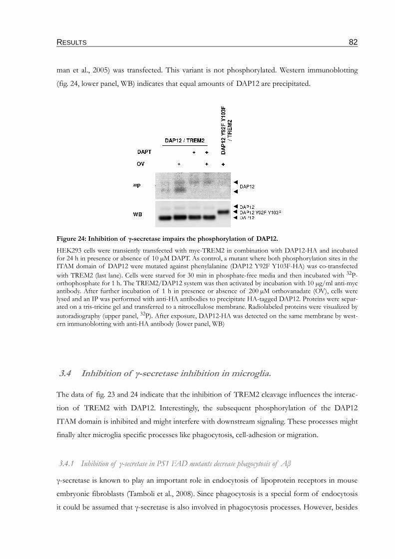
RESULTS 82
man et al., 2005) was transfected. This variant is not phosphorylated. Western immunoblotting
(fig. 24, lower panel, WB) indicates that equal amounts of DAP12 are precipitated.
3.4 Inhibition of γ-secretase inhibition in microglia.
The data of fig. 23 and 24 indicate that the inhibition of TREM2 cleavage influences the interac-
tion of TREM2 with DAP12. Interestingly, the subsequent phosphorylation of the DAP12
ITAM domain is inhibited and might interfere with downstream signaling. These processes might
finally alter microglia specific processes like phagocytosis, cell-adhesion or migration.
3.4.1 Inhibition of γ-secretase in PS1 FAD mutants decrease phagocytosis of Aβ
γ-secretase is known to play an important role in endocytosis of lipoprotein receptors in mouse
embryonic fibroblasts (Tamboli et al., 2008). Since phagocytosis is a special form of endocytosis
it could be assumed that γ-secretase is also involved in phagocytosis processes. However, besides
Figure 24: Inhibition of γ-secretase impairs the phosphorylation of DAP12.
HEK293 cells were transiently transfected with myc-TREM2 in combination with DAP12-HA and incubated for 24 h in presence or absence of 10 µM DAPT. As control, a mutant where both phosphorylation sites in the ITAM domain of DAP12 were mutated against phenylalanine (DAP12 Y92F Y103F-HA) was co-transfected
with TREM2 (last lane). Cells were starved for 30 min in phosphate-free media and then incubated with 32P-orthophosphate for 1 h. The TREM2/DAP12 system was then activated by incubation with 10 µg/ml anti-myc antibody. After further incubation of 1 h in presence or absence of 200 µM orthovanadate (OV), cells were lysed and an IP was performed with anti-HA antibodies to precipitate HA-tagged DAP12. Proteins were separ-ated on a tris-tricine gel and transferred to a nitrocellulose membrane. Radiolabeled proteins were visualized by
autoradiography (upper panel, 32P). After exposure, DAP12-HA was detected on the same membrane by west-ern immunoblotting with anti-HA antibody (lower panel, WB)
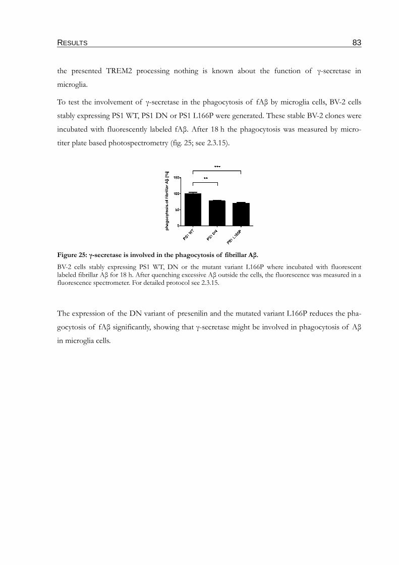
RESULTS 83
the presented TREM2 processing nothing is known about the function of γ-secretase in
microglia.
To test the involvement of γ-secretase in the phagocytosis of fAβ by microglia cells, BV-2 cells
stably expressing PS1 WT, PS1 DN or PS1 L166P were generated. These stable BV-2 clones were
incubated with fluorescently labeled fAβ. After 18 h the phagocytosis was measured by micro-
titer plate based photospectrometry (fig. 25; see 2.3.15).
The expression of the DN variant of presenilin and the mutated variant L166P reduces the pha-
gocytosis of fAβ significantly, showing that γ-secretase might be involved in phagocytosis of Aβ
in microglia cells.
Figure 25: γ-secretase is involved in the phagocytosis of fibrillar Aβ.
BV-2 cells stably expressing PS1 WT, DN or the mutant variant L166P where incubated with fluorescent labeled fibrillar Aβ for 18 h. After quenching excessive Aβ outside the cells, the fluorescence was measured in a fluorescence spectrometer. For detailed protocol see 2.3.15.
DISCUSSION 84
4 DISCUSSION
Phagocytosis of cell debris, apoptotic cells, lipids, aggregated proteins or other waste material is
one of the main function of microglia. Depending on the ingested material they exert either
pro- or an anti-inflammatory responses.
In the AD brain, microglia with a proinflammatory phenotype are found in proximity to Aβ
plaques, suggesting Aβ phagocytosis (Perlmutter et al., 1990; Frautschy et al., 1998; Bolmont et
al., 2008; Meyer-Luehmann et al., 2008). There are several in vitro studies showing the ability of
microglia to phagocytose Aβ (Koenigsknecht & Landreth 2004; Mandrekar et al., 2009; Hjorth et
al., 2010; Nagano et al., 2010; Smith et al., 2010). In vivo uptake of injected sAβ and fAβ by
microglia has also been demonstrated (Weldon et al., 1998; Takata et al., 2003). However, the
ablation of microglia don't result in significant decrease of plaque growth or plaque development
(Grathwohl et al., 2009), suggesting only a minor role of microglia in plaque formation, mainten-
ance and clearance in vivo. Nevertheless, activation of microglia and phagocytosis of Aβ play an
important role in AD as demonstrated by studies with APP transgenic mice lacking the
chemokine receptor CCR2. These mice show less microglia activation and Aβ phagocytosis, res-
ulting in increased AD progression and mortality (El Khoury et al., 2007).
Since Aβ accumulates in the brain of AD patients, there is obviously an insufficient clearance of
Aβ by microglia under physiological conditions, because one function of microglia is the removal
of waste material from the CNS (see 1.3). However, whether microglial phagocytosis in AD brain
is defective is unclear..
Since γ-secretase has been linked to endocytosis processes previously (Tamboli et al., 2008), it
might also be implicated in the regulation of phagocytosis in microglia.
Indeed, a very recent paper demonstrated a role of the γ-secretase component PS in the phago-
cytosis of Aβ. The treatment of the microglial cell line N9 with γ-secretase inhibitors decreased
the phagocytosis of fAβ42. The phagocytosis was further diminished after shRNA mediated
knock down of PS1 as well as in primary microglia of PS2 knock out mice (Farfara et al., 2010).
However, the molecular mechanisms underlying γ-secretase dependent regulation of phagocyt-
osis is unknown.
In this work it it shown that the triggering receptor TREM2 which is expressed on several myel-
oid cells including microglia is cleaved by γ-secretase. Prior to the γ-secretase cleavage the ectodo-
main of TREM2 is shed by a metalloprotease. The inhibition of TREM2 processing alters the
interaction with its co-receptor DAP12. Moreover, blocking of the γ-secretase cleavage impairs
DISCUSSION 85
phosphorylation of the ITAM motif of DAP12, which is necessary for downstream signaling of
TREM2 and DAP12.
Thus, cleavage of TREM2 and impaired DAP12 signaling could be a link between PS deficiency
and phagocytosis defects, as observed in PS deficient microglia cells (Farfara et al., 2010)(see also
chapter 3.4.1).
In accordance with this hypothesis TREM2/DAP12-dependent signaling was previously shown
to regulate levels of proinflammatory cytokines after activation of TLRs (Hamerman et al.,
2006). and linked to phagocytosis processes (Takahashi et al., 2005; Takahashi et al., 2007; Hsieh
et al., 2009; N'Diaye et al., 2009).
4.1 Proteolytic processing of TREM2
Regulated intramembrane proteolysis (RIP) is a sequential mechanism liberating proteins from
the lipid-bilayer. In the first step, cleavage by a sheddase, results in release of the ectodomain and
the generation of a membrane-bound fragment of the protein. This part which consists of a
small lumenal stub, a tm-domain and a cytoplasmic domain is then cleaved by a protease within
the tm-domain, releasing both the lumenal part and the C-terminal domain from the membrane.
4.1.1 Shedding of the TREM2 ectodomain
Detection of soluble TREM2 ectodomain in cell culture supernatants (see chapter 3.2.4) estab-
lished the shedding of TREM2. Well established sheddases are the members of the ADAM and
the MMP families. Additionally, aspartate proteases like BACE-1 and -2 are also known to act as
sheddases. While the members of the APP family (APP and APLPs) are the only known sub-
strate for BACE-2, BACE-1 has several substrates. One of the first identified substrates of
BACE-1 was the sialyltransferase ST6Gal I (Kitazume et al., 2003). However, the most studied
substrates are APP (Hussain et al., 1999; Sinha et al., 1999; Vassar et al., 1999; Yan et al., 1999;
Lin et al., 2000) and neuregulin which is involved in myelination processes (Hu et al., 2006;
Willem et al., 2006; Hu et al., 2008). By the use of different protease modulators this work indic-
ates that a protease of the ADAM or MMP family can shed TREM2 ectodomain (fig. 20, 21).
However, these data don't rule out that other proteases, which might not be affected by the used
modulators, contribute to TREM2 shedding. It might also be possible that TREM2 ectodomain
shedding occurs by different proteases dependent on the intracellular localization and transport
of both the protease and TREM2. This is known for APP, which ectodomain can either be shed
DISCUSSION 86
by ADAMs (Buxbaum et al., 1998; Koike et al., 1999; Lammich et al., 1999), BACE-1 or -2 (Hus-
sain et al., 1999; Sinha et al., 1999; Vassar et al., 1999; Yan et al., 1999; Lin et al., 2000) or MMPs
(Higashi & Miyazaki 2003; Ahmad et al., 2006; Yin et al., 2006; Talamagas et al., 2007). To further
identity the TREM2 sheddase, surface stainings as shown in fig. 21 after treatment with more
specific inhibitors are required. Alternatively, the generation of the TREM2 ECD could be meas-
ured after siRNA mediated knockdown of specific proteases or in knock out cells.
APP has been shown to be constitutively shed by ADAMs (Slack et al., 2001). Recently ADAM-
10 has been established as the constitutively active α-secretase of APP in primary neurons (Kuhn
et al., 2010). The low staining of myc-TREM2-GFP on the cell-surface of untreated COS7 cells
(see fig. 21 G-L) suggests that TREM2 might be shed constitutively as well. However, the shed-
ding could further be induced by treatment with the phorbol ester PDBu (see fig. 21 D-F). The
effect of PDBu is most likely based on the property of phorbol ester to activate PKC (Blumberg
1988) and the resulting stimulation of ADAMs as shown for APP (Jolly-Tornetta & Wolf 2000;
Skovronsky et al., 2000). The way in which PKC stimulates ADAMs is still not fully understood.
Three main pathways are suggested. PKC can either phosphorylate the ADAMs directly, as has
been shown by Jacobsen and colleagues (Jacobsen et al., 2010) or PKC can affect the transloca-
tion of ADAMs (Yang et al., 2007; Kohutek et al., 2009). Furthermore, PKC increases vesicle
budding from the Golgi (Xu et al., 1995), thereby increasing the transport of both substrate and
proteases to cell compartments where cleavage can occur. This would explain how the effect of
the PKC activator TPPB which increases ADAM activity can be blocked by the transport inhib-
itor Brefeldin A (Yang et al., 2007).
In addition to phorbol ester, ADAM mediated shedding of APP was shown to be inducible by
insulin growth factor-1 in a PKC dependent manner (Jacobsen et al., 2010).
The reduced ECD levels in HEK293 PS1 WT cells after ligation of TREM2 by an anti-myc
antibody (see fig. 17) indicate that ligand binding to TREM2 might protect the full length
receptor from ectodomain shedding. A possible explanation might be that ligand binding
induces dimerization of TREM2, resulting in a sterical blockade of an interaction with the
respective sheddase. Such an effect was shown for binding of growth hormones to the growth
hormone receptor (Zhang et al., 2001). Alternatively, ligand engagement could directly alter the
three dimensional structure of TREM2 thereby masking the sheddase cleavage site. This is
known for Notch but in contrast to the findings regarding TREM2, binding of the Notch ligand
is necessary to induce shedding of Notch ECD (Mumm & Kopan 2000; Mumm et al., 2000).
Before ligand binding, Notch is locked in a protease-resistant state. Exposure to the ligand res-
ults in a conformational shift, unfolding the sheddase cleavage site (Bozkulak & Weinmaster
DISCUSSION 87
2009). The induction of shedding by ligand binding has also been shown for the vascular
endothelial growth factor (VEGF) receptor-1 which is rendered susceptible to shedding sub-
sequently to phosphorylation induced by binding of VEGF-A (Rahimi et al., 2009).
The reduced TREM2 FL after activation of TREM2 by antibody ligation might be due to
enhanced endocytosis of the full length receptor after ligand binding (see fig. 17). Such a down-
regulation of receptor signaling by endocytosis and degradation of receptor-ligand complex is a
well established process, known for multiple receptors e.g. insulin receptor, EGF receptor (Sor-
kin & Duex 2010) and the IL-2 receptor (Legrue et al., 1991). TREM2 FL levels are reduced in
HEK293 PS1 DN cells compared to HEK293 PS1 WT. However, activation of TREM2 by anti-
body ligation don't affect the TREM2 FL levels in HEK293 PS1 DN (see fig. 17 C), suggesting
less efficient endocytosis of TREM2 after ligand binding in these cells. One explanation for this
observation might be the general impairment of endocytosis upon γ-secretase inhibition (Tam-
boli et al., 2008). Further endocytosis assays and antibody uptake assays are necessary to assess
the contribution of endocytosis in receptor down-regulation.
The ectodomain shedding of TREM2 would be consistent with the presence of soluble variants
of TREM1 (sTREM1) in blood serum and plasma of septic shock patients (Gibot et al., 2004;
Mahdy et al., 2006) and TREM2 (sTREM2) detected in human CSF as described in the literature
(Piccio et al., 2008). The origin of sTREM is discussed controversially, in particular because
sTREMs are found with or without association to DAP12. It was postulated that either a splice
variant results in sTREM (Gingras et al., 2002) or that it is shed from membrane bound TREMs.
sTREM1 levels decrease upon treatment of human monocytes with metalloprotease inhibitors
which might suggest shedding (Gomez-Pina et al., 2007). This is further supported by the
molecular weight of ~27 kDa determined for sTREM1 (Gibot et al., 2004). Nothing is known
about the size of the sTREM2 fragment. However, based on the amino acid sequence, the ecto-
domain of TREM1 should be larger then the ectodomain of TREM2 (Bouchon et al., 2000).
This led to the assumption that the ~15 kDa soluble fragment detected in this study (see fig. 17)
represents a sTREM2 variant as described by Piccio and colleagues. Although, little is known
about the function of the sTREMs they are suggested to negatively regulate TREM signaling by
neutralizing TREM ligands (Bouchon et al., 2001a; Piccio et al., 2007; Piccio et al., 2008)
TREM2/DAP12 complexes could interact in cis with plexin-A1 as well as in trans with the plexin-
A1 ligand semaphorin 6D, forming a higher order multimeric complex and regulating cellular
adhesion and motility (Takegahara et al., 2006). Co-IPs with TREM2 and different deletion
mutants of plexin-A1 identified the interaction site in the extracellular located TIG domain of
DISCUSSION 88
plexin-A1. This, and the interaction of TREM2 with semaphorin 6D in trans, suggest an interac-
tion via the TREM2 ectodomain. Thus, a shedding of the TREM2 ectodomain might negatively
regulate the interaction with plexin-A1, thereby modulating cellular motility and axon guidance.
This might contribute to the migration defects which our lab recently observed in PS1 deficient
BV-2 cells (data of Sonia Tosheva, not shown) and which were confirmed by a very recent
paper, demonstrating migration deficits in N9 cell after treatment with γ-secretase inhibitors as
well as in primary microglia of PS2 -/- mice (Farfara et al., 2010). According to this, it would be
interesting to study migration of microglia cells in response to overexpression of different
TREM2 variants or after treatment with shedding modulators. Furthermore, TREM2 -/- (Seno et
al., 2009) mice could be analyzed for axonal alterations. If they exhibit an axonal phenotype, the
contribution of plexin A1 and semaphorin 6D, can be further analyzed in more detail.
It is well established that shed ectodomains serve as ligands for other receptors. sAPP for
example can bind to DR-6 inducing axonal pruning and neuronal death (Nikolaev et al., 2009).
Additionally, the shed ectodomain of E-cadherin which binds to ErbB receptors promotes their
activation (Najy et al., 2008). Thus, the TREM2 ectodomain might also serve as a ligand for
another receptor. Further experiments are required to analyze the stability of the ectodomain
and to find and identify putative binding partners.
4.1.2 Cleavage of TREM2 CTF by γ-secretase
Ectodomain shedding of type I membrane proteins is required for subsequent cleavage of the
membrane-tethered CTF by γ-secretase. Besides γ-secretase, there are four different classes of
intramembrane proteases which can catalyze the cleavage reaction within the membrane: The sig-
nal peptide peptidases (SPP) (Grigorenko et al., 2002; Ponting et al., 2002; Weihofen et al., 2002),
the signal peptide peptidase like proteins (SPPLs) which cleave TNFα (Fluhrer et al., 2006; Fried-
mann et al., 2006), the site-2 proteases (S2P) (Rawson et al., 1997) known for the cleavage of
sterol regulatory element binding proteins (SREBPs) and the rhomboid serine proteases which
cleave transmembrane ligand substrates e. g. the EGF ligand Spitz in Drosophila (Urban et al.,
2001; Freeman 2004). However, all these protein classes have specific requirements for their sub-
strate CTFs. Only γ-secretase cleaves CTFs of type I transmembrane proteins.
Pharmacological inhibition of the γ-secretase complex with the specific inhibitor DAPT (Dovey
et al., 2001) and subsequent accumulation of a TREM2 CTF fragment (see chapter. 3.1.2) reveal
that TREM2 CTF is a γ-secretase substrate. This is supported by demonstration of TREM2 CTF
accumulation after co-expression with PS1 DN (see chapter. 3.1.2). Both methods are well estab-
DISCUSSION 89
lished to prove γ-secretase mediated cleavage of the main γ-secretase substrates APP (Steiner et
al., 1999; Kim et al., 2001) and Notch (Berezovska et al., 2000; Jack et al., 2001). PS1 DN is an
artificial PS mutant of a critical aspartyl residue in the active site which doesn't occur in AD
patients. However, there are mutations of PS1 linked to EOAD which cause partial loss of γ-
secretase function according to cleavage of APP and Notch (see 1.1.2.2). The accumulation of
TREM2 CTF in the tested PS FAD variants L166P and ∆Ex9 (see chapter. 3.1.2) indicates that a
partial loss of γ-secretase function is sufficient to impair TREM2 processing and suggests contri-
bution of TREM2 cleavage impairment in AD pathology of patients with PS dependent EOAD.
According to the age-of-onset of AD, L166P and ∆Ex9 are severe mutations. Thus, it would be
interesting to check whether weaker PS mutations with a later age-of-onset are also inhibit
TREM2 processing.
Although γ-secretase is located in the Golgi, TGN, secretory vesicles, plasma membrane and
lysosomes/endosomes, substantial γ-secretase activity is located on the plasma membrane and/or
endosomes (Kaether et al., 2006a; Kaether et al., 2006b), implicating that cleavage of substrates
occurs at the cell surface. Indeed, TREM2 CTF was found in immunofluorescence experiments
to accumulate at the cell surface upon γ-secretase inhibition (see chapter 3.2.1). Moreover, expres-
sion of a truncated TREM2 construct (TREM2∆ECD) which lacks the ectodomain of TREM2
and thereby resembles an immediate γ-secretase substrate accumulates at the cell surface as well
(see chapter 3.2.1). The surface localization of TREM2 CTF was approved and quantified by sur-
face biotinylation studies (see chapter 3.2.1). The observed plasma membrane localization is con-
sistent with accumulation of APP CTFs at the cell surface that follows γ-secretase inhibition
(Kim et al., 2001). The same was shown for Notch, inhibition of γ-secretase either by pharmaco-
logical inhibitors or by point mutations in presenilin has been shown to end up in the accumula-
tion of NEXT (Notch extracellular truncation) (Mumm et al., 2000).
Besides APP and Notch, the γ-secretase complex cleaves several type I transmembrane proteins.
All of them are expressed on various cell types, but a microglia-specific substrate has not yet
been demonstrated. Thus, TREM2 is the first identified microglia-specific γ-secretase substrate.
γ-Secretase cleaves substrate CTFs within the tm-domain, resulting in liberation of extracellular
and lumenal parts from cellular membranes. While the physiological functions of extracellular
peptides are largely unknown, certain ICDs (see 1.1.5.3) translocate to the nucleus and serve as
transcriptional co-factor for gene regulation. This has been demonstrated for Notch, which ICD
regulate the transcription of developmental genes (Mumm & Kopan 2000). To shut off the tran-
scriptional regulation, NICD can be degraded via the ubiquitin-proteasome pathway (Gupta-
Rossi et al., 2001; Oberg et al., 2001). AICD in contrast is very unstable and degraded rapidly by
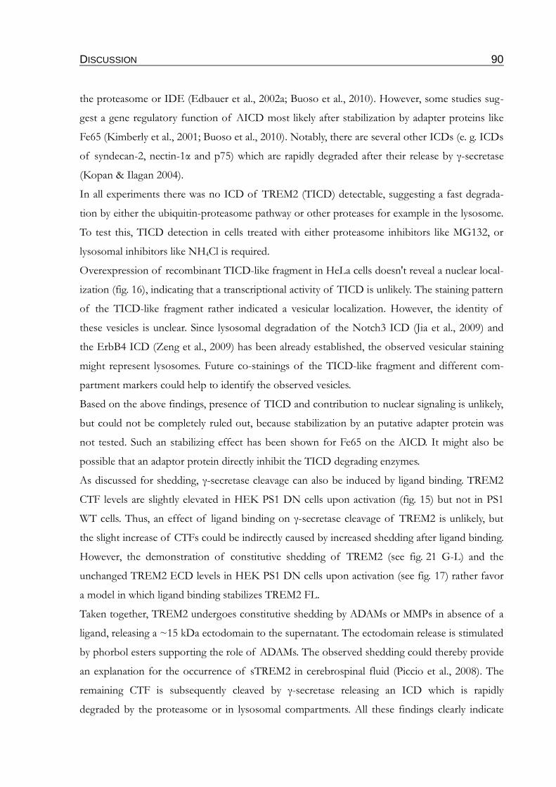
DISCUSSION 90
the proteasome or IDE (Edbauer et al., 2002a; Buoso et al., 2010). However, some studies sug-
gest a gene regulatory function of AICD most likely after stabilization by adapter proteins like
Fe65 (Kimberly et al., 2001; Buoso et al., 2010). Notably, there are several other ICDs (e. g. ICDs
of syndecan-2, nectin-1α and p75) which are rapidly degraded after their release by γ-secretase
(Kopan & Ilagan 2004).
In all experiments there was no ICD of TREM2 (TICD) detectable, suggesting a fast degrada-
tion by either the ubiquitin-proteasome pathway or other proteases for example in the lysosome.
To test this, TICD detection in cells treated with either proteasome inhibitors like MG132, or
lysosomal inhibitors like NH4Cl is required.
Overexpression of recombinant TICD-like fragment in HeLa cells doesn't reveal a nuclear local-
ization (fig. 16), indicating that a transcriptional activity of TICD is unlikely. The staining pattern
of the TICD-like fragment rather indicated a vesicular localization. However, the identity of
these vesicles is unclear. Since lysosomal degradation of the Notch3 ICD (Jia et al., 2009) and
the ErbB4 ICD (Zeng et al., 2009) has been already established, the observed vesicular staining
might represent lysosomes. Future co-stainings of the TICD-like fragment and different com-
partment markers could help to identify the observed vesicles.
Based on the above findings, presence of TICD and contribution to nuclear signaling is unlikely,
but could not be completely ruled out, because stabilization by an putative adapter protein was
not tested. Such an stabilizing effect has been shown for Fe65 on the AICD. It might also be
possible that an adaptor protein directly inhibit the TICD degrading enzymes.
As discussed for shedding, γ-secretase cleavage can also be induced by ligand binding. TREM2
CTF levels are slightly elevated in HEK PS1 DN cells upon activation (fig. 15) but not in PS1
WT cells. Thus, an effect of ligand binding on γ-secretase cleavage of TREM2 is unlikely, but
the slight increase of CTFs could be indirectly caused by increased shedding after ligand binding.
However, the demonstration of constitutive shedding of TREM2 (see fig. 21 G-L) and the
unchanged TREM2 ECD levels in HEK PS1 DN cells upon activation (see fig. 17) rather favor
a model in which ligand binding stabilizes TREM2 FL.
Taken together, TREM2 undergoes constitutive shedding by ADAMs or MMPs in absence of a
ligand, releasing a ~15 kDa ectodomain to the supernatant. The ectodomain release is stimulated
by phorbol esters supporting the role of ADAMs. The observed shedding could thereby provide
an explanation for the occurrence of sTREM2 in cerebrospinal fluid (Piccio et al., 2008). The
remaining CTF is subsequently cleaved by γ-secretase releasing an ICD which is rapidly
degraded by the proteasome or in lysosomal compartments. All these findings clearly indicate
DISCUSSION 91
that TREM2 undergoes RIP. However, presence of a ligand seems to stabilize TREM2 FL at the
cell surface and might induce endocytosis of the TREM2-DAP12-ligand complex.
4.2 γ-secretase dependent interaction of TREM2 and DAP12
4.2.1 Inhibition of γ-secretase cleavage alters interaction of TREM2 with DAP12
Since TICD seems to be rapidly degraded, it is unlikely that the main function of TREM2 RIP is
the release of a transcriptional active fragment. Furthermore, a function of RIP in the down-reg-
ulation of signaling is unlikely. Thus, the function of TREM2 RIP remains unclear. One putative
function of RIP could be the degradation of membrane localized TREM2 in absence of a ligand
(see fig. 26, right side). This would coincide with the hypothesis that γ-secretase has a general role
in degradation of membrane proteins (Kopan & Ilagan 2004).
Intracellularly, TREM2 is mainly localized in the Golgi complex and in cytoplasmic vesicles with
constitutive shuttling to the plasma membrane (Sessa et al., 2004; Prada et al., 2006). Absence of
a ligand or an interaction partner like plexin-A1 might therefore result in the accumulation of the
full length receptor inside the membrane. Under these conditions, RIP might be the mechanism
to remove TREM2 FL from the membrane (see fig. 26, right side). This hypothesis is supported
by the vesicular staining seen in TREM2 overexpressing cells without DAPT treatment
(see fig. 11). These stainings were achieved with an antibody against the C-terminal myc-tag,
which is part of the TICD liberated by γ-secretase from TREM2. Thus, the staining pattern
might represent vesicular structures, either containing myc-tagged TICD or uncleaved TREM2
CTFs. This idea is supported by the finding that overexpression of a TICD-like fragment results
in a comparable staining pattern (see fig. 16).
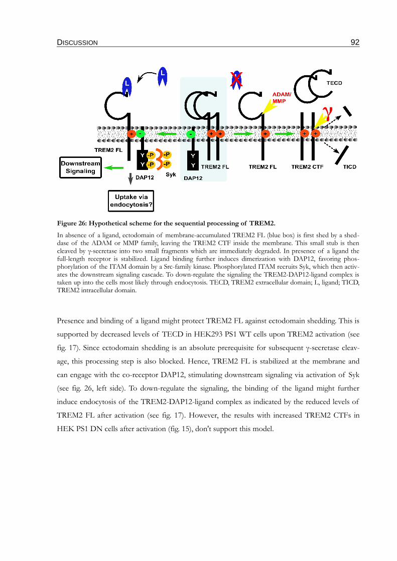
DISCUSSION 92
Presence and binding of a ligand might protect TREM2 FL against ectodomain shedding. This is
supported by decreased levels of TECD in HEK293 PS1 WT cells upon TREM2 activation (see
fig. 17). Since ectodomain shedding is an absolute prerequisite for subsequent γ-secretase cleav-
age, this processing step is also blocked. Hence, TREM2 FL is stabilized at the membrane and
can engage with the co-receptor DAP12, stimulating downstream signaling via activation of Syk
(see fig. 26, left side). To down-regulate the signaling, the binding of the ligand might further
induce endocytosis of the TREM2-DAP12-ligand complex as indicated by the reduced levels of
TREM2 FL after activation (see fig. 17). However, the results with increased TREM2 CTFs in
HEK PS1 DN cells after activation (fig. 15), don't support this model.
Figure 26: Hypothetical scheme for the sequential processing of TREM2.
In absence of a ligand, ectodomain of membrane-accumulated TREM2 FL (blue box) is first shed by a shed-dase of the ADAM or MMP family, leaving the TREM2 CTF inside the membrane. This small stub is then cleaved by γ-secretase into two small fragments which are immediately degraded. In presence of a ligand the full-length receptor is stabilized. Ligand binding further induces dimerization with DAP12, favoring phos-phorylation of the ITAM domain by a Src-family kinase. Phosphorylated ITAM recruits Syk, which then activ-ates the downstream signaling cascade. To down-regulate the signaling the TREM2-DAP12-ligand complex is taken up into the cells most likely through endocytosis. TECD, TREM2 extracellular domain; L, ligand; TICD, TREM2 intracellular domain.

DISCUSSION 93
Based on the model established above (fig. 26), inhibition of γ-secretase should result in the accu-
mulation of TREM2 CTF at the membrane. Indeed, this was shown in this work (fig. 9-13).
Interaction between TREM2 and DAP12 is mediated by the charged lysine residue inside the tm-
region (see 1.5 and 1.6). Since the tm-domain is part of the accumulating CTFs these fragments
can engage with DAP12. Accordingly, the CTFs might be able to trap DAP12. Thus, the amount
of co-receptor available to engage with TREM2 FL, or other DAP12 associated receptors, such
as signal regulatory protein-β1 (SIRPβ1) (Tomasello & Vivier 2005; Gaikwad et al., 2009) or com-
plement receptors (Mocsai et al., 2006) would be reduced. This in turn would prevent phos-
phorylation of DAP12 and blocks activation of the downstream located Syk. In consequence of
this, downstream signaling of TREM2/DAP12 or other DAP12 associated receptors would be
impaired (see fig. 27). Since TREM2-DAP12 signaling regulates phagocytosis processes, impaired
interaction of functional TREM2 FL with DAP12 through γ-secretase inhibition would result in
a phagocytosis defect.
4.2.2 Inhibition of γ-secretase impairs DAP12 phosphorylation
The accumulation of TREM2 at the plasma membrane (fig. 12 and 13) and the altered interaction
of TREM2 with DAP12 upon γ-secretase inhibition (fig. 22, 23), support the model established
in fig. 27. Moreover, the phosphorylation of DAP12 was found to be impaired. Notably, γ-
Figure 27: Hypothetical scheme of impaired TREM2 processing upon γ-secretase inhibition.
After ectodomain shedding of the full length TREM2 (blue box), TREM2 CTFs accumulate in the membrane, because subsequent degradation of the CTFs is blocked due to an inactive γ-secretase. Thus, CTFs trap DAP12 thereby preventing interaction with the FL receptor, witch is stabilized by ligand binding. This results in an incomplete phosphorylation of the ITAM domain, failed activation of Syk and impaired downstream sig-naling. TECD, TREM2 extracellular domain; L, ligand.
DISCUSSION 94
secretase inhibition doesn't result in full blocking of the DAP12 phosphorylation but in a mono-
phosphorylated ITAM domain (fig. 24).
It was previously shown that dependent on the interacting receptor, DAP12 mediated signaling
could either be activatory or inhibitory. For example signaling via DAP12 and MDL-1 (myeloid
DAP12-associated lectin 1) results in calcium mobilization and cellular activation (Bakker et al.,
1999). In contrast, as mentioned in the introduction (see 1.5.2), activation of DAP12 and
TREM2 results in inhibition of TLR responses (Hamerman et al., 2006). To explain this paradox
Turnbull & Colonna established a hypothesis in which the phosphorylation state of DAP12
(monophosphorylated or full phosphorylated) directs the downstream signaling either to activat-
ory or inhibitory cell response (Turnbull & Colonna 2007). Based on this hypothesis, high avidity
ligands are suggested to lead to a full phosphorylation of the DAP12 ITAM domain, whereas the
binding of a low avidity ligand is suggested to activate SH2-domain-containing protein tyrosine
phosphatase 1 (SHP1). Thereupon, SHP1 deactivates Src kinases in the signaling complex, pre-
venting full phosphorylation of the DAP12 ITAM motif, thereby stabilizing DAP12 in a mono-
phosphorylated state. This in turn blocks binding and activation of Syk and the downstream loc-
ated PLCγ (see fig. 28, left side). PLCγ catalyzes the degradation of phosphatidylinositol-4,5-
bisphosphate (PtdIns(4,5)P2) which is necessary to anchor two proteins involved in the TLR sig-
naling cascade, MyD88 (myeloid differentiation primary-response gene 88) and TIRAP (Toll/IL-
1-receptor-domain-containing adapter protein). Thus, inhibited PLCγ would stabilize
PtdIns(4,5)P2 in the membrane, allowing binding of MyD88 and TIRAP (Kagan & Medzhitov
2006). Finally, this cascade could stabilize the TLR response (fig. 28, left side). Moreover, the
inactive Syk kinase might fail to activate phosphatidylinositol-3 kinase (PI3K) (Vieira et al., 2001;
Huang et al., 2006). This inhibits inactivation of AKT which in turn amplifies activation of the
mitogen activated protein kinase kinase kinase (MAPKKK; fig. 29, left side ) (Turnbull &
Colonna 2007; Ivashkiv 2009). Recently, an additional indirect pathway was shown, coupling
DAP12 signaling via intergrins to the inhibition of TLRs and to the increased expression of the
anti-inflammatory cytokine IL-10 (Wang et al., 2010). This pathway would also be impaired by
reduced DAP12 phosphorylation, thereby facilitating TLR response resulting in higher levels of
proinflammatory cytokines.
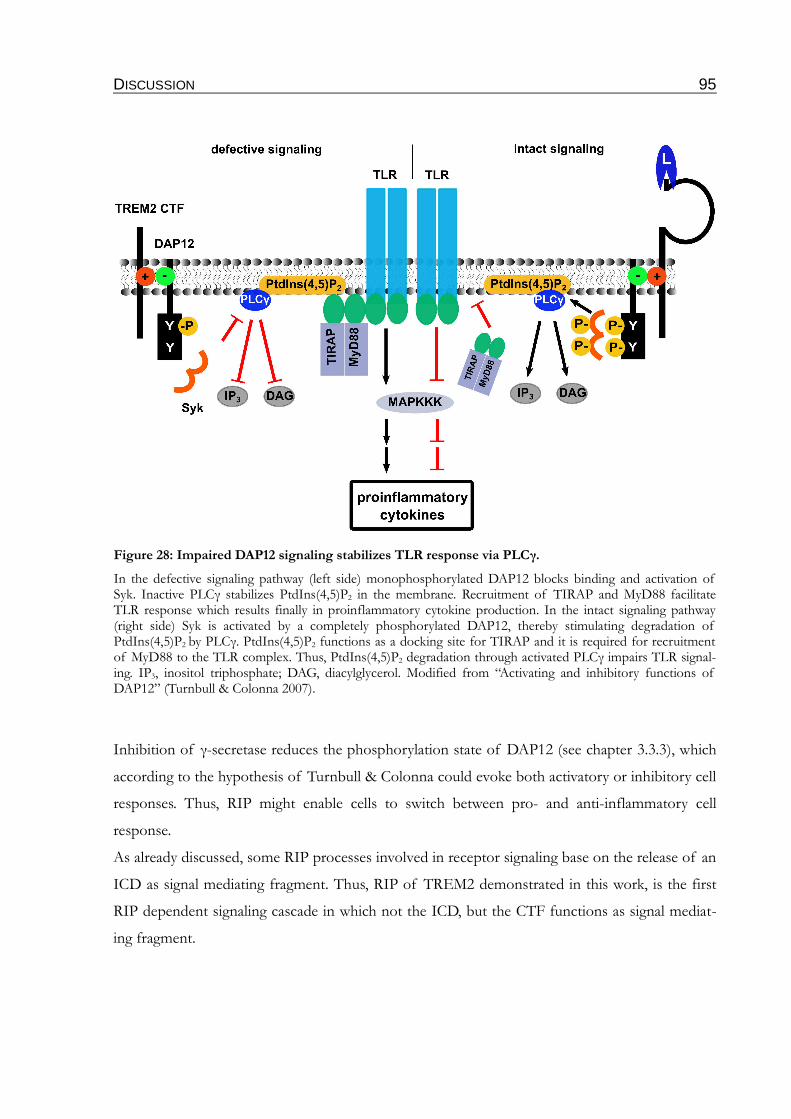
DISCUSSION 95
Inhibition of γ-secretase reduces the phosphorylation state of DAP12 (see chapter 3.3.3), which
according to the hypothesis of Turnbull & Colonna could evoke both activatory or inhibitory cell
responses. Thus, RIP might enable cells to switch between pro- and anti-inflammatory cell
response.
As already discussed, some RIP processes involved in receptor signaling base on the release of an
ICD as signal mediating fragment. Thus, RIP of TREM2 demonstrated in this work, is the first
RIP dependent signaling cascade in which not the ICD, but the CTF functions as signal mediat-
ing fragment.
Figure 28: Impaired DAP12 signaling stabilizes TLR response via PLCγ.
In the defective signaling pathway (left side) monophosphorylated DAP12 blocks binding and activation of Syk. Inactive PLCγ stabilizes PtdIns(4,5)P2 in the membrane. Recruitment of TIRAP and MyD88 facilitate TLR response which results finally in proinflammatory cytokine production. In the intact signaling pathway (right side) Syk is activated by a completely phosphorylated DAP12, thereby stimulating degradation of PtdIns(4,5)P2 by PLCγ. PtdIns(4,5)P2 functions as a docking site for TIRAP and it is required for recruitment of MyD88 to the TLR complex. Thus, PtdIns(4,5)P2 degradation through activated PLCγ impairs TLR signal-ing. IP3, inositol triphosphate; DAG, diacylglycerol. Modified from “Activating and inhibitory functions of DAP12” (Turnbull & Colonna 2007).
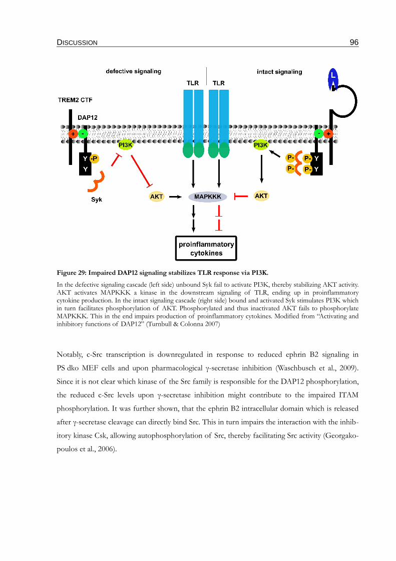
DISCUSSION 96
Notably, c-Src transcription is downregulated in response to reduced ephrin B2 signaling in
PS dko MEF cells and upon pharmacological γ-secretase inhibition (Waschbusch et al., 2009).
Since it is not clear which kinase of the Src family is responsible for the DAP12 phosphorylation,
the reduced c-Src levels upon γ-secretase inhibition might contribute to the impaired ITAM
phosphorylation. It was further shown, that the ephrin B2 intracellular domain which is released
after γ-secretase cleavage can directly bind Src. This in turn impairs the interaction with the inhib-
itory kinase Csk, allowing autophosphorylation of Src, thereby facilitating Src activity (Georgako-
poulos et al., 2006).
Figure 29: Impaired DAP12 signaling stabilizes TLR response via PI3K.
In the defective signaling cascade (left side) unbound Syk fail to activate PI3K, thereby stabilizing AKT activity. AKT activates MAPKKK a kinase in the downstream signaling of TLR, ending up in proinflammatory cytokine production. In the intact signaling cascade (right side) bound and activated Syk stimulates PI3K which in turn facilitates phosphorylation of AKT. Phosphorylated and thus inactivated AKT fails to phosphorylate MAPKKK. This in the end impairs production of proinflammatory cytokines. Modified from “Activating and inhibitory functions of DAP12” (Turnbull & Colonna 2007)
DISCUSSION 97
4.3 Potential effects of impaired TREM2/DAP12 signaling on phagocytosis and degradation of Aβ
Many studies have linked TREM2 to phagocytosis but the potential mechanism underlying this
effect is unclear. Regulation of PLCγ and PI3K activity by DAP12 signaling (Jiang et al., 2002;
Mao et al., 2006; Peng et al., 2010) might be involved. Impaired TREM2 signaling might thereby
result in changed turnover of phosphatidylinositols (PtdIns) in the membrane.
PtdIns(4,5)P2 is an important signaling lipid (~1-2 % of the whole content) of the inner leaflet
of the membrane (Tran et al., 1993; Raucher et al., 2000) which is well known to be involved in
actin filament reorganization and extension (Takenawa & Itoh 2001). It was also found to play an
important role during phagocytosis. While PtdIns(4,5)P2 accumulates in the forming phagosome
at the basement, it has been shown that its deprivation, accompanied with actin disassembling is
essential for phagosome sealing (Botelho et al., 2000). Impaired PtdIns(4,5)P2 metabolism or
overexpression of the PtdIns(4,5)P2 forming enzyme phosphatidylinositol phosphate kinase I
prevents actin disassembly, necessary for the completion of phagocytosis (Scott et al., 2005;
Cheeseman et al., 2006). Most likely the observed deprivation of PtdIns(4,5)P2 results from
hydrolysis by PLCγ which is recruited to the phagocytic cup through the SH2 domain. Hydrolysis
of PtdIns(4,5)P2 results in the release of IP3 and DAG. IP3 as a Ca2+-channel regulator, also plays
a crucial role in phagocytosis because the occurrence of Ca2+-transients during phagocytosis is
well established (Nunes & Demaurex 2010). The role of DAG is not that clear, but it can induce
negative curvatures of lipid membranes due to its small polar head group. Thus, it is suggested to
be involved in membrane bulging and fission (Carrasco & Merida 2007). PtdIns(4,5)P2 serves fur-
ther as source for phosphatidylinositol 3,4,5-trisphosphate (PtdIns(3,4,5)P3) another lipid found
to be accumulated in the phagocytic cup. It is also established to be responsible for recruitment
of myosin X, a motor protein important for pseudopod extension and closure of the phagosome
(Cox et al., 2002). Generation of PtdIns(3,4,5)P3 is catalyzed by PI3K which has to be activated
by binding of active Syk to the p85 regulatory subunit. It has been shown that inhibitors of PI3K
like Wortmannin affected the phagocytosis of large particles (< 3 µm in diameter), while smaller
particles remain unaffected (Yeung & Grinstein 2007).
As discussed above, both PLCγ and PI3K are important for the metabolism of PtdIns during
formation and closure of phagosomes. Decreased γ-secretase cleavage and impaired
DAP12/TREM2 signaling might therefore alter the activity of PLCγ and PI3K (see fig. 28) and
the lipid composition in and around the phagosome. This would be a putative mechanism by
which inhibited TREM2 processing can account for the blocked phagocytosis of fAβ in BV-2
DISCUSSION 98
cells stably expressing PS1 FAD variants (see fig. 25) or for the phagocytosis defect demonstrated
primary microglia of PS deficient mice and in the microglia cell line N9 after downregulation of
PS1 (Farfara et al., 2010).
TREM2 mediated phagocytosis is associated with decreased levels of proinflammatory cytokines
(Takahashi et al., 2005; Takahashi et al., 2007). Since PLCγ and PI3K regulate TLR signaling by
the pathways illustrated in fig. 28 and 29, this would support the involvement of these two pro-
teins in phagocytosis.
In the future, it would be interesting to measure the transition of PtdIns(4,5)P2 with the help of
the PH domain of PLCδ (Raucher et al., 2000) in life cell imaging experiments upon γ-secretase
inhibition. Moreover, measurements of cytokine profiles could help to assess the contribution of
γ-secretase inhibition to the regulation of pro-inflammatory cytokines. It would be further
important to analyze the triggering factor of TREM2 signaling. Two different variants could be
suggested which induce an ITAM mediated inflammatory cascade (see fig. 30). TREM2 might be
activated either directly through Aβ or might be cross activated through an Aβ binding scavenger
receptor. TREM2 has been found to be expressed in microglia surrounding Aβ plaques (Frank et
al., 2008; Melchior et al., 2010). Binding studies of TREM2 with different variants of Aβ are
necessary to check whether Aβ can serve as TREM2 ligand. In vivo phosphorylation assays can
then be used to monitor the DAP12 phosphorylation state upon Aβ treatment.
It has been reported that the uptake of fAβ in vitro by BV-2 cells was attenuated under proinflam-
matory conditions (Koenigsknecht-Talboo & Landreth 2005). The authors reported that fAβ
activated BV-2 cells which then responded by generating a proinflammatory environment. The
inflammatory mediated suppression of fAβ uptake was dependent on NFκB activation, linking
Aβ uptake to TLR response. Since ITAM signaling has been linked via the CARD9 directly to
NFκB activation (Ivashkiv 2009), the suppression might be regulated by DAP12 or other ITAM
containing co-receptors like FcRγ. Interestingly, the phagocytosis upon antibody mediated activa-
tion of FcR is unaffected by the proinflammatory milieu. Thus, FcRγ the ITAM-containing co-
receptor of FcRs might not be involved in the NFκB activation. The levels of cytokines,
chemokines and other proinflammatory molecules are elevated in brains of AD patients as well
as in animal disease models (Akiyama et al., 2000). This might explain why Aβ removal from the
brain is observed after passive immunization with anti-Aβ antibodies (Bard et al., 2000; Bacskai et
al., 2002; Wilcock et al., 2003) as well as active immunization with Aβ (Schenk et al., 1999), while
the removal of Aβ without vaccination is inefficient. Hickman and colleagues reported that the
DISCUSSION 99
levels of Aβ degrading enzymes like IDE and NEP are reduced in primary microglia from APP-
PS1 transgenic mice in an age-dependent manner. This reduction is accompanied by inreased
levels of proinflammatory cytokines and a reduction of Aβ scavenger receptors (Hickman et al.,
2008).
Taken together, the observed impaired γ-secretase cleavage of TREM2 leads to accumulation of
TREM2 CTFs in the plasma membrane, trapping DAP12. This in turn might block the interac-
tion with DAP12 associated receptors like TREM2 FL or SIRPβ1 involved in phagocytosis.
Moreover, the DAP12 phosphorylation state is impaired due to altered interaction with
TREM2 FL preventing activation of Syk. Thus, activation of PLCγ and PI3K might be
impaired, resulting in a facilitated TLR response and a stabilization of PtdIns(4,5)P2 in the
microglia membrane. This in combination with the elevated levels of pro-inflammatory
cytokines might then finally result in attenuated fAβ uptake and impaired degradation of sAβ by
degrading proteases (fig. 30).
This proposed cascade could thereby provide a mechanism by which γ-secretase inhibition can
be linked to defective phagocytosis of microglia, observed in PS deficient primary microglia
(Farfara et al., 2010). This could further underlie the inefficient clearance of Aβ in brains of PS-
linked AD patients.
Figure 30: Hypothetical link between impaired TREM2 cleavage and attenuated Aβ phagocytosis and degeneration.
OUTLOOK 100
5 OUTLOOK
So far the inefficient phagocytosis and clearance of Aβ by microglia in AD brains is not under-
stood. However, the impaired TREM2 processing demonstrated in this work, could provide an
possible explanation. Future experiments in transgenic mice would be important to test patho-
physiological relevance.
TREM2 deficient mice (Colonna et al., 2007) could be crossed with an AD model like APP trans-
genic mice which produce increased levels of Aβ. These mice could be then compared to control
mice concerning the age of plaque appearance.
The present findings indicate an involvement of γ-secretase in TREM2 mediated migration and
phagocytosis of microglia. Thus, microglia behavior could be monitored in these mice by in vivo
microscopy.
Alternatively, it would be feasible to inject different variants of Aβ into the brains of TREM2
deficient mice and to analyze the time-dependent Aβ clearance compared to control mice. The
different variants of Aβ would further allow to test whether TREM2 deficiency affects the
uptake of both sAβ and fAβ. However, the suggested in vivo experiments would only allow to
monitor the phagocytosis of Aβ in respect to TREM2 deficiency and not to an impaired TREM2
processing. For this it could be helpful to analyze mice with a conditional PS knockout in
microglia. In these mice the TREM2 processing could be proven by a C-terminal TREM2 anti-
body. Injection of Aβ in these mice would further allow to analyze phagocytosis with respect to
accumulation of TREM2 CTFs. Finally, a transgenic mouse could be generated that overexpress
the TREM2 CTF construct, used in this study, in microglia.
The cleavage of APP CTFs by γ-secretase is a very important step in the development of AD
pathology. Modulation of γ-secretase activity by artificial γ-secretase inhibitors is therefore a fre-
quently proposed therapeutic strategy against AD. However, based on the presented results, γ-
sceretase might also regulate microglia activity via TREM2 processing. An inhibition of γ-
secretase could have therefore also detrimental effects for the progress of AD. For this reason, it
would be necessary to develop γ-secretase modulators which impair γ-sceretase cleavage of APP
CTFs, but don't affect the processing of TREM2. Furthermore, it would be possible to develop
specific inhibitors for the TREM2 sheddase. Inhibition of the TREM2 sheddase stabilizes the
TREM2 FL receptor and would thereby stimulate the TREM2/DAP12 signaling pathway and
promote the uptake of Aβ.
Although the known clinical methods to diagnose AD in later stages are very efficient, these
OUTLOOK 101
methods fail to identify risk patients. For this purpose biomarkers are of very high interest.
sTREM2 has been previously detected in human blood and CSF (Gibot et al., 2004; Mahdy et
al., 2006; Piccio et al., 2008). Since the demonstrated TECD seems to be similar with these
sTREM2 variants, it should be feasible to detect TECD in CSF of AD patients. A strong
increase of TECD in CSF would indicate an accumulation of TREM2 CTFs which could result
in impaired Aβ clearance by microglia and might indicate a higher risk to develop AD. The
TREM2 Aβ-like fragment has not yet been detected in human CSF or blood. Thus, detection
methods for this particular fragment could be developed. Decreased levels of this fragment
would then directly indicate impaired γ-secretase activity. Accordingly, cleavage products of
TREM2 like TECD and the Aβ-like peptide might be used as biomarkers.
ABSTRACT 102
6 ABSTRACT
Alzheimer's disease (AD) is a progressive neurodegenerative disorder, affecting millions of
people worldwide. AD is histopathologically characterized by the appearance of neurofibrillary
tangles, which are intraneuronal accumulations of hyperphosphorylated tau protein, and extracel-
lular β-amyloid plaques. β-Amyloid plaques arise from progressive accumulation of Aβ, a small
hydrophobic peptide, in the brain. Aβ derives from sequential proteolytic processing of the amyl-
oid precursor protein (APP). The final processing step in the generation of Aβ is catalyzed by the
γ-secretase complex, consisting of presenilin-1 (PS1), the active subunit, nicastrin, Aph-1
(anterior pharynx defective-1) and PEN-2 (presenilin enhancer element-2). Besides APP, the γ-
secretase has several other substrates and is also involved in the endocytosis of membrane bound
lipoprotein receptors.
In addition to neurofibrillary tangles and amyloid plaques, activation of microglia and inflammat-
ory processes are also fundamental characteristics in the brain of AD patients. Activated
microglia appear to play a dual role in AD. On one hand they produce pro-inflammatory
cytokines, reactive oxygen species and nitric oxide, augmenting inflammatory processes and oxid-
ative stress which might promote neuronal damage. On the other hand microglia can phagocyt-
ose Aβ, thereby contributing to its clearance from the brain. However, the accumulation of Aβ in
AD brains indicates an insufficient clearance of Aβ in AD pathogenesis which is not yet under-
stood. Since γ-secretase was previously linked to endocytosis, it might also be implicated in pha-
gocytic processes by microglia and clearance of Aβ.
Here it is shown by biochemical experiments in cell culture models, that the triggering receptor
expressed on myeloid cells-2 (TREM2) represents a novel substrate for γ-secretase in microglia.
Pharmacological inhibition of γ-secretase resulted in accumulation of a TREM2 C-terminal frag-
ment. This fragment also accumulated upon expression of a dominant negative variant of PS1.
Immunofluorescence and biotinylation experiments further indicated that the processing of
TREM2 occurs at the plasma membrane. In addition, cell biological experiments demonstrated
shedding of the TREM2 ectodomain. Thus, TREM2 follows the canonical proteolytic processing
pathway of γ-secretase substrates which consists of an initial cleavage within the ectodomain fol-
lowed by intramembranous cleavage of the resulting membrane-tethered CTF by γ-secretase. The
usage of selective protease inhibitors also indicated the involvement of a metalloprotease, likely
of the ADAM family, in TREM2 ectodomain shedding. TREM2 dependent signaling required the
interaction with its co-receptor DAP12. Interestingly, co-immunoprecipitations revealed impaired

ABSTRACT 103
interaction of TREM2 and DAP12 upon γ-secretase inhibition Moreover, the impaired interac-
tion resulted in decreased phosphorylation of DAP12. Expression of different PS1 FAD
mutants, led to decreased phagocytosis of Aβ. Thus, a partial loss of γ-secretase activity might
decrease the capacity of microglia to clear Aβ.
Taken together, these results indicate a critical function of γ-secretase in microglia and might help
to understand molecular mechanisms underlying impaired Aβ clearance in the pathogenesis of
AD.

REFERENCES 104
7 REFERENCES
Acquati, F., M. Accarino, et al. (2000). "The gene encoding DRAP (BACE2), a glycosylated transmembrane protein of the aspartic protease family, maps to the down critical region." FEBS Lett 468(1): 59-64.
Ahmad, M., T. Takino, et al. (2006). "Cleavage of amyloid-beta precursor protein (APP) by membrane-type matrix metalloproteinases." J Biochem 139(3): 517-526.
Ahmed, R. R., C. J. Holler, et al. (2010). "BACE1 and BACE2 enzymatic activities in Alzheimer's disease." J Neurochem 112(4): 1045-1053.
Akiyama, H., S. Barger, et al. (2000). "Inflammation and Alzheimer's disease." Neurobiol Aging 21(3): 383-421.Akomolafe, A., A. Beiser, et al. (2006). "Diabetes mellitus and risk of developing Alzheimer disease: results from
the Framingham Study." Arch Neurol 63(11): 1551-1555.Alonso, A., T. Zaidi, et al. (2001). "Hyperphosphorylation induces self-assembly of tau into tangles of paired
helical filaments/straight filaments." Proc Natl Acad Sci U S A 98(12): 6923-6928.Alves da Costa, C., C. Sunyach, et al. (2006). "Presenilin-dependent gamma-secretase-mediated control of p53-
associated cell death in Alzheimer's disease." J Neurosci 26(23): 6377-6385.Andersen, K., L. J. Launer, et al. (1995). "Do nonsteroidal anti-inflammatory drugs decrease the risk for
Alzheimer's disease? The Rotterdam Study." Neurology 45(8): 1441-1445.Annaert, W. G., L. Levesque, et al. (1999). "Presenilin 1 controls gamma-secretase processing of amyloid
precursor protein in pre-golgi compartments of hippocampal neurons." J Cell Biol 147(2): 277-294.Aoki, N., A. Zganiacz, et al. (2004). "Differential regulation of DAP12 and molecules associated with DAP12
during host responses to mycobacterial infection." Infect Immun 72(5): 2477-2483.Apelt, J., M. Bigl, et al. (2004). "Aging-related increase in oxidative stress correlates with developmental pattern of
beta-secretase activity and beta-amyloid plaque formation in transgenic Tg2576 mice with Alzheimer-like pathology." Int J Dev Neurosci 22(7): 475-484.
Bacskai, B. J., S. T. Kajdasz, et al. (2002). "Non-Fc-mediated mechanisms are involved in clearance of amyloid-beta in vivo by immunotherapy." J Neurosci 22(18): 7873-7878.
Bakker, A. B., E. Baker, et al. (1999). "Myeloid DAP12-associating lectin (MDL)-1 is a cell surface receptor involved in the activation of myeloid cells." Proc Natl Acad Sci U S A 96(17): 9792-9796.
Ballatore, C., V. M. Lee, et al. (2007). "Tau-mediated neurodegeneration in Alzheimer's disease and related disorders." Nat Rev Neurosci 8(9): 663-672.
Bamberger, M. E., M. E. Harris, et al. (2003). "A cell surface receptor complex for fibrillar beta-amyloid mediates microglial activation." J Neurosci 23(7): 2665-2674.
Barbiero, L., L. Benussi, et al. (2003). "BACE-2 is overexpressed in Down's syndrome." Exp Neurol 182(2): 335-345.
Bard, F., C. Cannon, et al. (2000). "Peripherally administered antibodies against amyloid beta-peptide enter the central nervous system and reduce pathology in a mouse model of Alzheimer disease." Nat Med 6(8): 916-919.
Barrow, C. J. and M. G. Zagorski (1991). "Solution structures of beta peptide and its constituent fragments: relation to amyloid deposition." Science 253(5016): 179-182.
Bayer, T. A., R. Cappai, et al. (1999). "It all sticks together--the APP-related family of proteins and Alzheimer's disease." Mol Psychiatry 4(6): 524-528.
Bell, R. D., A. P. Sagare, et al. (2007). "Transport pathways for clearance of human Alzheimer's amyloid beta-peptide and apolipoproteins E and J in the mouse central nervous system." J Cereb Blood Flow Metab 27(5): 909-918.
Benjannet, S., A. Elagoz, et al. (2001). "Post-translational processing of beta-secretase (beta-amyloid-converting enzyme) and its ectodomain shedding. The pro- and transmembrane/cytosolic domains affect its cellular activity and amyloid-beta production." J Biol Chem 276(14): 10879-10887.
Bennett, B. D., S. Babu-Khan, et al. (2000a). "Expression analysis of BACE2 in brain and peripheral tissues." J Biol Chem 275(27): 20647-20651.
Bennett, B. D., P. Denis, et al. (2000b). "A furin-like convertase mediates propeptide cleavage of BACE, the Alzheimer's beta -secretase." J Biol Chem 275(48): 37712-37717.
Bentahir, M., O. Nyabi, et al. (2006). "Presenilin clinical mutations can affect gamma-secretase activity by different mechanisms." J Neurochem 96(3): 732-742.
Berezovska, O., C. Jack, et al. (2000). "Rapid Notch1 nuclear translocation after ligand binding depends on presenilin-associated gamma-secretase activity." Ann N Y Acad Sci 920: 223-226.
Bertram, L. and R. E. Tanzi (2008). "Thirty years of Alzheimer's disease genetics: the implications of systematic meta-analyses." Nat Rev Neurosci 9(10): 768-778.
Betancor, L., F. Schelotto, et al. (2005). "An attenuated Salmonella Enteritidis strain derivative of the main
REFERENCES 105
genotype circulating in Uruguay is an effective vaccine for chickens." Vet Microbiol 107(1-2): 81-89.Beutner, C., K. Roy, et al. (2010). "Generation of microglial cells from mouse embryonic stem cells." Nat Protoc
5(9): 1481-1494.Bickel, H. (2010). "Das Wichtigste 1 - Die Epidemiologie der Demenz." Deutsche Alzheimer Gesellschaft e.V.Birnboim, H. C. and J. Doly (1979). "A rapid alkaline extraction procedure for screening recombinant plasmid
DNA." Nucleic Acids Res 7(6): 1513-1523.Bleharski, J. R., V. Kiessler, et al. (2003). "A role for triggering receptor expressed on myeloid cells-1 in host
defense during the early-induced and adaptive phases of the immune response." J Immunol 170(7): 3812-3818.
Blumberg, P. M. (1988). "Protein kinase C as the receptor for the phorbol ester tumor promoters: sixth Rhoads memorial award lecture." Cancer Res 48(1): 1-8.
Bolmont, T., F. Haiss, et al. (2008). "Dynamics of the microglial/amyloid interaction indicate a role in plaque maintenance." J Neurosci 28(16): 4283-4292.
Bondareff, W., C. Harrington, et al. (1994). "Immunohistochemical staging of neurofibrillary degeneration in Alzheimer's disease." J Neuropathol Exp Neurol 53(2): 158-164.
Bonifacino, J. S. (2004). "The GGA proteins: adaptors on the move." Nat Rev Mol Cell Biol 5(1): 23-32.Borchelt, D. R., G. Thinakaran, et al. (1996). "Familial Alzheimer's disease-linked presenilin 1 variants elevate
Abeta1-42/1-40 ratio in vitro and in vivo." Neuron 17(5): 1005-1013.Botelho, R. J., M. Teruel, et al. (2000). "Localized biphasic changes in phosphatidylinositol-4,5-bisphosphate at
sites of phagocytosis." J Cell Biol 151(7): 1353-1368.Bouchon, A., J. Dietrich, et al. (2000). "Cutting edge: inflammatory responses can be triggered by TREM-1, a
novel receptor expressed on neutrophils and monocytes." J Immunol 164(10): 4991-4995.Bouchon, A., F. Facchetti, et al. (2001a). "TREM-1 amplifies inflammation and is a crucial mediator of septic
shock." Nature 410(6832): 1103-1107.Bouchon, A., C. Hernandez-Munain, et al. (2001b). "A DAP12-mediated pathway regulates expression of CC
chemokine receptor 7 and maturation of human dendritic cells." J Exp Med 194(8): 1111-1122.Bozkulak, E. C. and G. Weinmaster (2009). "Selective use of ADAM10 and ADAM17 in activation of Notch1
signaling." Mol Cell Biol 29(21): 5679-5695.Braak, H. and E. Braak (1991). "Neuropathological stageing of Alzheimer-related changes." Acta Neuropathol
82(4): 239-259.Braak, H. and E. Braak (1996). "Evolution of the neuropathology of Alzheimer's disease." Acta Neurol Scand
Suppl 165: 3-12.Bradford, M. M. (1976). "A rapid and sensitive method for the quantitation of microgram quantities of protein
utilizing the principle of protein-dye binding." Anal Biochem 72: 248-254.Brandt, R., J. Leger, et al. (1995). "Interaction of tau with the neural plasma membrane mediated by tau's amino-
terminal projection domain." J Cell Biol 131(5): 1327-1340.Breitner, J. C., K. A. Welsh, et al. (1995). "Delayed onset of Alzheimer's disease with nonsteroidal anti-
inflammatory and histamine H2 blocking drugs." Neurobiol Aging 16(4): 523-530.Bu, G. (2009). "Apolipoprotein E and its receptors in Alzheimer's disease: pathways, pathogenesis and therapy." Nat
Rev Neurosci 10(5): 333-344.Bulloj, A., M. C. Leal, et al. (2010). "Insulin-degrading enzyme sorting in exosomes: a secretory pathway for a key
brain amyloid-beta degrading protease." J Alzheimers Dis 19(1): 79-95.Buoso, E., C. Lanni, et al. (2010). "beta-Amyloid precursor protein metabolism: focus on the functions and
degradation of its intracellular domain." Pharmacol Res 62(4): 308-317.Butovsky, O., Y. Ziv, et al. (2006). "Microglia activated by IL-4 or IFN-gamma differentially induce neurogenesis
and oligodendrogenesis from adult stem/progenitor cells." Mol Cell Neurosci 31(1): 149-160.Buxbaum, J. D., K. N. Liu, et al. (1998). "Evidence that tumor necrosis factor alpha converting enzyme is
involved in regulated alpha-secretase cleavage of the Alzheimer amyloid protein precursor." J Biol Chem 273(43): 27765-27767.
Cai, X. D., T. E. Golde, et al. (1993). "Release of excess amyloid beta protein from a mutant amyloid beta protein precursor." Science 259(5094): 514-516.
Cameron, B. and G. E. Landreth (2010). "Inflammation, microglia, and Alzheimer's disease." Neurobiol Dis 37(3): 503-509.
Cao, X. and T. C. Sudhof (2001). "A transcriptionally [correction of transcriptively] active complex of APP with Fe65 and histone acetyltransferase Tip60." Science 293(5527): 115-120.
Capell, A., J. Grunberg, et al. (1998). "The proteolytic fragments of the Alzheimer's disease-associated presenilin-1 form heterodimers and occur as a 100-150-kDa molecular mass complex." J Biol Chem 273(6): 3205-3211.
Capell, A., H. Steiner, et al. (2000). "Maturation and pro-peptide cleavage of beta-secretase." J Biol Chem 275(40): 30849-30854.
Cardona, A. E., E. P. Pioro, et al. (2006). "Control of microglial neurotoxicity by the fractalkine receptor." Nat
REFERENCES 106
Neurosci 9(7): 917-924.Carrasco, S. and I. Merida (2007). "Diacylglycerol, when simplicity becomes complex." Trends Biochem Sci 32(1):
27-36.Cheeseman, K. L., T. Ueyama, et al. (2006). "Targeting of protein kinase C-epsilon during Fcgamma receptor-
dependent phagocytosis requires the epsilonC1B domain and phospholipase C-gamma1." Mol Biol Cell 17(2): 799-813.
Chen, F., H. Hasegawa, et al. (2006). "TMP21 is a presenilin complex component that modulates gamma-secretase but not epsilon-secretase activity." Nature 440(7088): 1208-1212.
Choi, S. H., K. Veeraraghavalu, et al. (2008). "Non-cell-autonomous effects of presenilin 1 variants on enrichment-mediated hippocampal progenitor cell proliferation and differentiation." Neuron 59(4): 568-580.
Chouery, E., V. Delague, et al. (2008). "Mutations in TREM2 lead to pure early-onset dementia without bone cysts." Hum Mutat 29(9): E194-204.
Chung, D. H., W. E. Seaman, et al. (2002). "Characterization of TREM-3, an activating receptor on mouse macrophages: definition of a family of single Ig domain receptors on mouse chromosome 17." Eur J Immunol 32(1): 59-66.
Chung, H., M. I. Brazil, et al. (1999). "Uptake, degradation, and release of fibrillar and soluble forms of Alzheimer's amyloid beta-peptide by microglial cells." J Biol Chem 274(45): 32301-32308.
Chung, S. H. (2009). "Aberrant phosphorylation in the pathogenesis of Alzheimer's disease." BMB Rep 42(8): 467-474.
Chyung, J. H. and D. J. Selkoe (2003). "Inhibition of receptor-mediated endocytosis demonstrates generation of amyloid beta-protein at the cell surface." J Biol Chem 278(51): 51035-51043.
Cirrito, J. R., R. Deane, et al. (2005). "P-glycoprotein deficiency at the blood-brain barrier increases amyloid-beta deposition in an Alzheimer disease mouse model." J Clin Invest 115(11): 3285-3290.
Citron, M., T. S. Diehl, et al. (1996). "Evidence that the 42- and 40-amino acid forms of amyloid beta protein are generated from the beta-amyloid precursor protein by different protease activities." Proc Natl Acad Sci U S A 93(23): 13170-13175.
Citron, M., T. Oltersdorf, et al. (1992). "Mutation of the beta-amyloid precursor protein in familial Alzheimer's disease increases beta-protein production." Nature 360(6405): 672-674.
Cleary, J. P., D. M. Walsh, et al. (2005). "Natural oligomers of the amyloid-beta protein specifically disrupt cognitive function." Nat Neurosci 8(1): 79-84.
Codony-Servat, J., J. Albanell, et al. (1999). "Cleavage of the HER2 ectodomain is a pervanadate-activable process that is inhibited by the tissue inhibitor of metalloproteases-1 in breast cancer cells." Cancer Res 59(6): 1196-1201.
Colonna, M. and F. Facchetti (2003). "TREM-1 (triggering receptor expressed on myeloid cells): a new player in acute inflammatory responses." J Infect Dis 187 Suppl 2: S397-401.
Colonna, M., I. Turnbull, et al. (2007). "The enigmatic function of TREM-2 in osteoclastogenesis." Adv Exp Med Biol 602: 97-105.
Corder, E. H., A. M. Saunders, et al. (1993). "Gene dose of apolipoprotein E type 4 allele and the risk of Alzheimer's disease in late onset families." Science 261(5123): 921-923.
Corneveaux, J. J., A. J. Myers, et al. (2010). "Association of CR1, CLU and PICALM with Alzheimer's disease in a cohort of clinically characterized and neuropathologically verified individuals." Hum Mol Genet 19(16): 3295-3301.
Cox, D., J. S. Berg, et al. (2002). "Myosin X is a downstream effector of PI(3)K during phagocytosis." Nat Cell Biol 4(7): 469-477.
Creemers, J. W., D. Ines Dominguez, et al. (2001). "Processing of beta-secretase by furin and other members of the proprotein convertase family." J Biol Chem 276(6): 4211-4217.
Daigle, I. and C. Li (1993). "apl-1, a Caenorhabditis elegans gene encoding a protein related to the human beta-amyloid protein precursor." Proc Natl Acad Sci U S A 90(24): 12045-12049.
Davalos, D., J. Grutzendler, et al. (2005). "ATP mediates rapid microglial response to local brain injury in vivo." Nat Neurosci 8(6): 752-758.
Daws, M. R., L. L. Lanier, et al. (2001). "Cloning and characterization of a novel mouse myeloid DAP12-associated receptor family." Eur J Immunol 31(3): 783-791.
Daws, M. R., P. M. Sullam, et al. (2003). "Pattern recognition by TREM-2: binding of anionic ligands." J Immunol 171(2): 594-599.
De Felice, F. G., M. N. Vieira, et al. (2009). "Protection of synapses against Alzheimer's-linked toxins: insulin signaling prevents the pathogenic binding of Abeta oligomers." Proc Natl Acad Sci U S A 106(6): 1971-1976.
De Strooper, B. (2007). "Loss-of-function presenilin mutations in Alzheimer disease. Talking Point on the role of presenilin mutations in Alzheimer disease." EMBO Rep 8(2): 141-146.
REFERENCES 107
De Strooper, B., W. Annaert, et al. (1999). "A presenilin-1-dependent gamma-secretase-like protease mediates release of Notch intracellular domain." Nature 398(6727): 518-522.
Deane, R., R. D. Bell, et al. (2009). "Clearance of amyloid-beta peptide across the blood-brain barrier: implication for therapies in Alzheimer's disease." CNS Neurol Disord Drug Targets 8(1): 16-30.
Deane, R., A. Sagare, et al. (2008a). "apoE isoform-specific disruption of amyloid beta peptide clearance from mouse brain." J Clin Invest 118(12): 4002-4013.
Deane, R., A. Sagare, et al. (2008b). "The role of the cell surface LRP and soluble LRP in blood-brain barrier Abeta clearance in Alzheimer's disease." Curr Pharm Des 14(16): 1601-1605.
Deane, R., Z. Wu, et al. (2004). "LRP/amyloid beta-peptide interaction mediates differential brain efflux of Abeta isoforms." Neuron 43(3): 333-344.
DeMattos, R. B., J. R. Cirrito, et al. (2004). "ApoE and clusterin cooperatively suppress Abeta levels and deposition: evidence that ApoE regulates extracellular Abeta metabolism in vivo." Neuron 41(2): 193-202.
Dickson, D. W. (2009). "Neuropathology of non-Alzheimer degenerative disorders." Int J Clin Exp Pathol 3(1): 1-23.
Diehlmann, A., N. Ida, et al. (1999). "Analysis of presenilin 1 and presenilin 2 expression and processing by newly developed monoclonal antibodies." J Neurosci Res 56(4): 405-419.
Dominguez, D., J. Tournoy, et al. (2005). "Phenotypic and biochemical analyses of BACE1- and BACE2-deficient mice." J Biol Chem 280(35): 30797-30806.
Dovey, H. F., V. John, et al. (2001). "Functional gamma-secretase inhibitors reduce beta-amyloid peptide levels in brain." J Neurochem 76(1): 173-181.
Drewes, G., B. Trinczek, et al. (1995). "Microtubule-associated protein/microtubule affinity-regulating kinase (p110mark). A novel protein kinase that regulates tau-microtubule interactions and dynamic instability by phosphorylation at the Alzheimer-specific site serine 262." J Biol Chem 270(13): 7679-7688.
Dries, D. R. and G. Yu (2008). "Assembly, maturation, and trafficking of the gamma-secretase complex in Alzheimer's disease." Curr Alzheimer Res 5(2): 132-146.
Duce, J. A., A. Tsatsanis, et al. (2010). "Iron-export ferroxidase activity of beta-amyloid precursor protein is inhibited by zinc in Alzheimer's disease." Cell 142(6): 857-867.
Duyckaerts, C., M. C. Potier, et al. (2008). "Alzheimer disease models and human neuropathology: similarities and differences." Acta Neuropathol 115(1): 5-38.
Edbauer, D., M. Willem, et al. (2002a). "Insulin-degrading enzyme rapidly removes the beta-amyloid precursor protein intracellular domain (AICD)." J Biol Chem 277(16): 13389-13393.
Edbauer, D., E. Winkler, et al. (2002b). "Presenilin and nicastrin regulate each other and determine amyloid beta-peptide production via complex formation." Proc Natl Acad Sci U S A 99(13): 8666-8671.
Edwards, D. R., M. M. Handsley, et al. (2008). "The ADAM metalloproteinases." Mol Aspects Med 29(5): 258-289.
Ekdahl, C. T., J. H. Claasen, et al. (2003). "Inflammation is detrimental for neurogenesis in adult brain." Proc Natl Acad Sci U S A 100(23): 13632-13637.
El Khoury, J., M. Toft, et al. (2007). "Ccr2 deficiency impairs microglial accumulation and accelerates progression of Alzheimer-like disease." Nat Med 13(4): 432-438.
Esch, F. S., P. S. Keim, et al. (1990). "Cleavage of amyloid beta peptide during constitutive processing of its precursor." Science 248(4959): 1122-1124.
Etcheberrigaray, R., M. Tan, et al. (2004). "Therapeutic effects of PKC activators in Alzheimer's disease transgenic mice." Proc Natl Acad Sci U S A 101(30): 11141-11146.
Farfara, D., D. Trudler, et al. (2010). "gamma-Secretase component presenilin is important for microglia beta-amyloid clearance." Ann Neurol.
Farris, W., S. Mansourian, et al. (2003). "Insulin-degrading enzyme regulates the levels of insulin, amyloid beta-protein, and the beta-amyloid precursor protein intracellular domain in vivo." Proc Natl Acad Sci U S A 100(7): 4162-4167.
Farzan, M., C. E. Schnitzler, et al. (2000). "BACE2, a beta -secretase homolog, cleaves at the beta site and within the amyloid-beta region of the amyloid-beta precursor protein." Proc Natl Acad Sci U S A 97(17): 9712-9717.
Feng, S. M., C. I. Sartor, et al. (2007). "The HER4 cytoplasmic domain, but not its C terminus, inhibits mammary cell proliferation." Mol Endocrinol 21(8): 1861-1876.
Fluhrer, R., A. Capell, et al. (2002). "A non-amyloidogenic function of BACE-2 in the secretory pathway." J Neurochem 81(5): 1011-1020.
Fluhrer, R., G. Grammer, et al. (2006). "A gamma-secretase-like intramembrane cleavage of TNFalpha by the GxGD aspartyl protease SPPL2b." Nat Cell Biol 8(8): 894-896.
Ford, J. W. and D. W. McVicar (2009). "TREM and TREM-like receptors in inflammation and disease." Curr Opin Immunol 21(1): 38-46.
Forstl, H. and A. Kurz (1999). "Clinical features of Alzheimer's disease." Eur Arch Psychiatry Clin Neurosci
REFERENCES 108
249(6): 288-290.Francis, R., G. McGrath, et al. (2002). "aph-1 and pen-2 are required for Notch pathway signaling, gamma-
secretase cleavage of betaAPP, and presenilin protein accumulation." Dev Cell 3(1): 85-97.Frank, S., G. J. Burbach, et al. (2008). "TREM2 is upregulated in amyloid plaque-associated microglia in aged
APP23 transgenic mice." Glia 56(13): 1438-1447.Frautschy, S. A., F. Yang, et al. (1998). "Microglial response to amyloid plaques in APPsw transgenic mice." Am J
Pathol 152(1): 307-317.Freeman, M. (2004). "Proteolysis within the membrane: rhomboids revealed." Nat Rev Mol Cell Biol 5(3): 188-197.Friedhoff, P., M. von Bergen, et al. (2000). "Structure of tau protein and assembly into paired helical filaments."
Biochim Biophys Acta 1502(1): 122-132.Friedmann, E., E. Hauben, et al. (2006). "SPPL2a and SPPL2b promote intramembrane proteolysis of TNFalpha
in activated dendritic cells to trigger IL-12 production." Nat Cell Biol 8(8): 843-848.Fulga, T. A., I. Elson-Schwab, et al. (2007). "Abnormal bundling and accumulation of F-actin mediates tau-
induced neuronal degeneration in vivo." Nat Cell Biol 9(2): 139-148.Gaikwad, S., S. Larionov, et al. (2009). "Signal regulatory protein-beta1: a microglial modulator of phagocytosis in
Alzheimer's disease." Am J Pathol 175(6): 2528-2539.Georgakopoulos, A., C. Litterst, et al. (2006). "Metalloproteinase/Presenilin1 processing of ephrinB regulates
EphB-induced Src phosphorylation and signaling." EMBO J 25(6): 1242-1252.Giaccone, G., F. Tagliavini, et al. (1989). "Down patients: extracellular preamyloid deposits precede neuritic
degeneration and senile plaques." Neurosci Lett 97(1-2): 232-238.Gibot, S., M. N. Kolopp-Sarda, et al. (2004). "A soluble form of the triggering receptor expressed on myeloid
cells-1 modulates the inflammatory response in murine sepsis." J Exp Med 200(11): 1419-1426.Gingras, M. C., H. Lapillonne, et al. (2002). "TREM-1, MDL-1, and DAP12 expression is associated with a
mature stage of myeloid development." Mol Immunol 38(11): 817-824.Goate, A., M. C. Chartier-Harlin, et al. (1991). "Segregation of a missense mutation in the amyloid precursor
protein gene with familial Alzheimer's disease." Nature 349(6311): 704-706.Goedert, M., C. M. Wischik, et al. (1988). "Cloning and sequencing of the cDNA encoding a core protein of the
paired helical filament of Alzheimer disease: identification as the microtubule-associated protein tau." Proc Natl Acad Sci U S A 85(11): 4051-4055.
Goldgaber, D., M. I. Lerman, et al. (1987). "Characterization and chromosomal localization of a cDNA encoding brain amyloid of Alzheimer's disease." Science 235(4791): 877-880.
Gomez-Pina, V., A. Soares-Schanoski, et al. (2007). "Metalloproteinases shed TREM-1 ectodomain from lipopolysaccharide-stimulated human monocytes." J Immunol 179(6): 4065-4073.
Gong, C. X., I. Grundke-Iqbal, et al. (1994). "Dephosphorylation of Alzheimer's disease abnormally phosphorylated tau by protein phosphatase-2A." Neuroscience 61(4): 765-772.
Goutte, C., M. Tsunozaki, et al. (2002). "APH-1 is a multipass membrane protein essential for the Notch signaling pathway in Caenorhabditis elegans embryos." Proc Natl Acad Sci U S A 99(2): 775-779.
Grathwohl, S. A., R. E. Kalin, et al. (2009). "Formation and maintenance of Alzheimer's disease beta-amyloid plaques in the absence of microglia." Nat Neurosci 12(11): 1361-1363.
Grigorenko, A. P., Y. K. Moliaka, et al. (2002). "Novel class of polytopic proteins with domains associated with putative protease activity." Biochemistry (Mosc) 67(7): 826-835.
Grundke-Iqbal, I., K. Iqbal, et al. (1986a). "Microtubule-associated protein tau. A component of Alzheimer paired helical filaments." J Biol Chem 261(13): 6084-6089.
Grundke-Iqbal, I., K. Iqbal, et al. (1986b). "Abnormal phosphorylation of the microtubule-associated protein tau (tau) in Alzheimer cytoskeletal pathology." Proc Natl Acad Sci U S A 83(13): 4913-4917.
Gupta-Rossi, N., O. Le Bail, et al. (2001). "Functional interaction between SEL-10, an F-box protein, and the nuclear form of activated Notch1 receptor." J Biol Chem 276(37): 34371-34378.
Gutwein, P., S. Mechtersheimer, et al. (2003). "ADAM10-mediated cleavage of L1 adhesion molecule at the cell surface and in released membrane vesicles." FASEB J 17(2): 292-294.
Haass, C., A. Y. Hung, et al. (1993). "beta-Amyloid peptide and a 3-kDa fragment are derived by distinct cellular mechanisms." J Biol Chem 268(5): 3021-3024.
Haass, C., E. H. Koo, et al. (1992a). "Targeting of cell-surface beta-amyloid precursor protein to lysosomes: alternative processing into amyloid-bearing fragments." Nature 357(6378): 500-503.
Haass, C., C. A. Lemere, et al. (1995). "The Swedish mutation causes early-onset Alzheimer's disease by beta-secretase cleavage within the secretory pathway." Nat Med 1(12): 1291-1296.
Haass, C., M. G. Schlossmacher, et al. (1992b). "Amyloid beta-peptide is produced by cultured cells during normal metabolism." Nature 359(6393): 322-325.
Haass, C. and D. J. Selkoe (1993). "Cellular processing of beta-amyloid precursor protein and the genesis of amyloid beta-peptide." Cell 75(6): 1039-1042.
Haass, C. and H. Steiner (2002). "Alzheimer disease gamma-secretase: a complex story of GxGD-type presenilin
REFERENCES 109
proteases." Trends Cell Biol 12(12): 556-562.Hamerman, J. A., J. R. Jarjoura, et al. (2006). "Cutting edge: inhibition of TLR and FcR responses in
macrophages by triggering receptor expressed on myeloid cells (TREM)-2 and DAP12." J Immunol 177(4): 2051-2055.
Hamerman, J. A., M. Ni, et al. (2009). "The expanding roles of ITAM adapters FcRgamma and DAP12 in myeloid cells." Immunol Rev 232(1): 42-58.
Hamerman, J. A., N. K. Tchao, et al. (2005). "Enhanced Toll-like receptor responses in the absence of signaling adaptor DAP12." Nat Immunol 6(6): 579-586.
Hanisch, U. K. and H. Kettenmann (2007). "Microglia: active sensor and versatile effector cells in the normal and pathologic brain." Nat Neurosci 10(11): 1387-1394.
Hanisch, U. K., M. Prinz, et al. (2001). "The protein tyrosine kinase inhibitor AG126 prevents the massive microglial cytokine induction by pneumococcal cell walls." Eur J Immunol 31(7): 2104-2115.
Hardy, J. and D. J. Selkoe (2002). "The amyloid hypothesis of Alzheimer's disease: progress and problems on the road to therapeutics." Science 297(5580): 353-356.
Harold, D., R. Abraham, et al. (2009). "Genome-wide association study identifies variants at CLU and PICALM associated with Alzheimer's disease." Nat Genet 41(10): 1088-1093.
Hartmann, D., B. de Strooper, et al. (2002). "The disintegrin/metalloprotease ADAM 10 is essential for Notch signalling but not for alpha-secretase activity in fibroblasts." Hum Mol Genet 11(21): 2615-2624.
Hass, M. R. and B. A. Yankner (2005). "A {gamma}-secretase-independent mechanism of signal transduction by the amyloid precursor protein." J Biol Chem 280(44): 36895-36904.
Hausler, K. G., M. Prinz, et al. (2002). "Interferon-gamma differentially modulates the release of cytokines and chemokines in lipopolysaccharide- and pneumococcal cell wall-stimulated mouse microglia and macrophages." Eur J Neurosci 16(11): 2113-2122.
Haynes, S. E., G. Hollopeter, et al. (2006). "The P2Y12 receptor regulates microglial activation by extracellular nucleotides." Nat Neurosci 9(12): 1512-1519.
He, G., W. Luo, et al. (2010). "Gamma-secretase activating protein is a therapeutic target for Alzheimer's disease." Nature 467(7311): 95-98.
He, X., W. P. Chang, et al. (2002). "Memapsin 2 (beta-secretase) cytosolic domain binds to the VHS domains of GGA1 and GGA2: implications on the endocytosis mechanism of memapsin 2." FEBS Lett 524(1-3): 183-187.
Heber, S., J. Herms, et al. (2000). "Mice with combined gene knock-outs reveal essential and partially redundant functions of amyloid precursor protein family members." J Neurosci 20(21): 7951-7963.
Hebert, S. S., L. Serneels, et al. (2006). "Regulated intramembrane proteolysis of amyloid precursor protein and regulation of expression of putative target genes." EMBO Rep 7(7): 739-745.
Helming, L., E. Tomasello, et al. (2008). "Essential role of DAP12 signaling in macrophage programming into a fusion-competent state." Sci Signal 1(43): ra11.
Hemming, M. L., J. E. Elias, et al. (2008). "Proteomic profiling of gamma-secretase substrates and mapping of substrate requirements." PLoS Biol 6(10): e257.
Herz, J. and H. H. Bock (2002). "Lipoprotein receptors in the nervous system." Annu Rev Biochem 71: 405-434.Herz, J. and Y. Chen (2006). "Reelin, lipoprotein receptors and synaptic plasticity." Nat Rev Neurosci 7(11): 850-
859.Hickman, S. E., E. K. Allison, et al. (2008). "Microglial dysfunction and defective beta-amyloid clearance
pathways in aging Alzheimer's disease mice." J Neurosci 28(33): 8354-8360.Higashi, S. and K. Miyazaki (2003). "Novel processing of beta-amyloid precursor protein catalyzed by membrane
type 1 matrix metalloproteinase releases a fragment lacking the inhibitor domain against gelatinase A." Biochemistry 42(21): 6514-6526.
Hjorth, E., D. Frenkel, et al. (2010). "Effects of immunomodulatory substances on phagocytosis of abeta(1-42) by human microglia." Int J Alzheimers Dis 2010.
Hoek, R. M., S. R. Ruuls, et al. (2000). "Down-regulation of the macrophage lineage through interaction with OX2 (CD200)." Science 290(5497): 1768-1771.
Hooper, C., R. Killick, et al. (2008). "The GSK3 hypothesis of Alzheimer's disease." J Neurochem 104(6): 1433-1439.
Horuk, R. and J. L. Gross (1990). "Protein kinase C-linked inactivation of the interleukin-1 receptor in a human transformed B-cell line." Biochim Biophys Acta 1052(1): 173-178.
Hsieh, C. L., M. Koike, et al. (2009). "A role for TREM2 ligands in the phagocytosis of apoptotic neuronal cells by microglia." J Neurochem 109(4): 1144-1156.
Hu, X., W. He, et al. (2008). "Genetic deletion of BACE1 in mice affects remyelination of sciatic nerves." FASEB J 22(8): 2970-2980.
Hu, X., C. W. Hicks, et al. (2006). "Bace1 modulates myelination in the central and peripheral nervous system." Nat Neurosci 9(12): 1520-1525.
REFERENCES 110
Hu, Y. and M. E. Fortini (2003). "Different cofactor activities in gamma-secretase assembly: evidence for a nicastrin-Aph-1 subcomplex." J Cell Biol 161(4): 685-690.
Huang, Z. Y., D. R. Barreda, et al. (2006). "Differential kinase requirements in human and mouse Fc-gamma receptor phagocytosis and endocytosis." J Leukoc Biol 80(6): 1553-1562.
Hung, A. Y. and D. J. Selkoe (1994). "Selective ectodomain phosphorylation and regulated cleavage of beta-amyloid precursor protein." EMBO J 13(3): 534-542.
Huse, J. T., D. S. Pijak, et al. (2000). "Maturation and endosomal targeting of beta-site amyloid precursor protein-cleaving enzyme. The Alzheimer's disease beta-secretase." J Biol Chem 275(43): 33729-33737.
Hussain, I., G. Christie, et al. (2001). "Prodomain processing of Asp1 (BACE2) is autocatalytic." J Biol Chem 276(26): 23322-23328.
Hussain, I., D. Powell, et al. (1999). "Identification of a novel aspartic protease (Asp 2) as beta-secretase." Mol Cell Neurosci 14(6): 419-427.
Hussain, I., D. J. Powell, et al. (2000). "ASP1 (BACE2) cleaves the amyloid precursor protein at the beta-secretase site." Mol Cell Neurosci 16(5): 609-619.
Ishiguro, K., A. Shiratsuchi, et al. (1993). "Glycogen synthase kinase 3 beta is identical to tau protein kinase I generating several epitopes of paired helical filaments." FEBS Lett 325(3): 167-172.
Ittner, L. M., Y. D. Ke, et al. (2010). "Dendritic function of tau mediates amyloid-beta toxicity in Alzheimer's disease mouse models." Cell 142(3): 387-397.
Ivashkiv, L. B. (2009). "Cross-regulation of signaling by ITAM-associated receptors." Nat Immunol 10(4): 340-347.Iwata, N., S. Tsubuki, et al. (2001). "Metabolic regulation of brain Abeta by neprilysin." Science 292(5521): 1550-
1552.Iwata, N., S. Tsubuki, et al. (2000). "Identification of the major Abeta1-42-degrading catabolic pathway in brain
parenchyma: suppression leads to biochemical and pathological deposition." Nat Med 6(2): 143-150.Jack, C., O. Berezovska, et al. (2001). "Effect of PS1 deficiency and an APP gamma-secretase inhibitor on Notch1
signaling in primary mammalian neurons." Brain Res Mol Brain Res 87(2): 166-174.Jacobsen, K. T., L. Adlerz, et al. (2010). "Insulin-like growth factor-1 (IGF-1)-induced processing of amyloid-beta
precursor protein (APP) and APP-like protein 2 is mediated by different metalloproteinases." J Biol Chem 285(14): 10223-10231.
Jalbert, J. J., L. A. Daiello, et al. (2008). "Dementia of the Alzheimer type." Epidemiol Rev 30: 15-34.Jarrett, J. T., E. P. Berger, et al. (1993). "The carboxy terminus of the beta amyloid protein is critical for the
seeding of amyloid formation: implications for the pathogenesis of Alzheimer's disease." Biochemistry 32(18): 4693-4697.
Jefferies, W. A., M. R. Food, et al. (1996). "Reactive microglia specifically associated with amyloid plaques in Alzheimer's disease brain tissue express melanotransferrin." Brain Res 712(1): 122-126.
Jia, L., G. Yu, et al. (2009). "Lysosome-dependent degradation of Notch3." Int J Biochem Cell Biol 41(12): 2594-2598.
Jiang, K., B. Zhong, et al. (2002). "Syk regulation of phosphoinositide 3-kinase-dependent NK cell function." J Immunol 168(7): 3155-3164.
Jiang, Q., C. Y. Lee, et al. (2008). "ApoE promotes the proteolytic degradation of Abeta." Neuron 58(5): 681-693.Joachim, C. L. and D. J. Selkoe (1992). "The seminal role of beta-amyloid in the pathogenesis of Alzheimer
disease." Alzheimer Dis Assoc Disord 6(1): 7-34.Jolly-Tornetta, C. and B. A. Wolf (2000). "Protein kinase C regulation of intracellular and cell surface amyloid
precursor protein (APP) cleavage in CHO695 cells." Biochemistry 39(49): 15282-15290.Kaether, C., C. Haass, et al. (2006a). "Assembly, trafficking and function of gamma-secretase." Neurodegener Dis
3(4-5): 275-283.Kaether, C., S. Schmitt, et al. (2006b). "Amyloid precursor protein and Notch intracellular domains are generated
after transport of their precursors to the cell surface." Traffic 7(4): 408-415.Kagan, J. C. and R. Medzhitov (2006). "Phosphoinositide-mediated adaptor recruitment controls Toll-like
receptor signaling." Cell 125(5): 943-955.Kamboh, M. I., R. L. Minster, et al. (2010). "Association of CLU and PICALM variants with Alzheimer's
disease." Neurobiol Aging.Kern, A., B. Roempp, et al. (2006). "Down-regulation of endogenous amyloid precursor protein processing due to
cellular aging." J Biol Chem 281(5): 2405-2413.Kim, J., J. M. Basak, et al. (2009). "The role of apolipoprotein E in Alzheimer's disease." Neuron 63(3): 287-303.Kim, S. H., J. Y. Leem, et al. (2001). "Multiple effects of aspartate mutant presenilin 1 on the processing and
trafficking of amyloid precursor protein." J Biol Chem 276(46): 43343-43350.Kimberly, W. T., W. Xia, et al. (2000). "The transmembrane aspartates in presenilin 1 and 2 are obligatory for
gamma-secretase activity and amyloid beta-protein generation." J Biol Chem 275(5): 3173-3178.Kimberly, W. T., J. B. Zheng, et al. (2001). "The intracellular domain of the beta-amyloid precursor protein is
stabilized by Fe65 and translocates to the nucleus in a notch-like manner." J Biol Chem 276(43): 40288-

REFERENCES 111
40292.Kitajima, I., M. Kuriyama, et al. (1989). "Nasu-Hakola disease (membranous lipodystrophy). Clinical,
histopathological and biochemical studies of three cases." J Neurol Sci 91(1-2): 35-52.Kitazume, S., Y. Tachida, et al. (2003). "Characterization of alpha 2,6-sialyltransferase cleavage by Alzheimer's
beta -secretase (BACE1)." J Biol Chem 278(17): 14865-14871.Klesney-Tait, J., I. R. Turnbull, et al. (2006). "The TREM receptor family and signal integration." Nat Immunol
7(12): 1266-1273.Koenigsknecht-Talboo, J. and G. E. Landreth (2005). "Microglial phagocytosis induced by fibrillar beta-amyloid
and IgGs are differentially regulated by proinflammatory cytokines." J Neurosci 25(36): 8240-8249.Koenigsknecht, J. and G. Landreth (2004). "Microglial phagocytosis of fibrillar beta-amyloid through a beta1
integrin-dependent mechanism." J Neurosci 24(44): 9838-9846.Koh, Y. H., C. A. von Arnim, et al. (2005). "BACE is degraded via the lysosomal pathway." J Biol Chem 280(37):
32499-32504.Kohutek, Z. A., C. G. diPierro, et al. (2009). "ADAM-10-mediated N-cadherin cleavage is protein kinase C-alpha
dependent and promotes glioblastoma cell migration." J Neurosci 29(14): 4605-4615.Koike, H., S. Tomioka, et al. (1999). "Membrane-anchored metalloprotease MDC9 has an alpha-secretase activity
responsible for processing the amyloid precursor protein." Biochem J 343 Pt 2: 371-375.Koizumi, S., Y. Shigemoto-Mogami, et al. (2007). "UDP acting at P2Y6 receptors is a mediator of microglial
phagocytosis." Nature 446(7139): 1091-1095.Kojro, E. and F. Fahrenholz (1995). "Ligand-induced cleavage of the V2 vasopressin receptor by a plasma
membrane metalloproteinase." J Biol Chem 270(12): 6476-6481.Koo, E. H. (2002). "The beta-amyloid precursor protein (APP) and Alzheimer's disease: does the tail wag the dog?"
Traffic 3(11): 763-770.Kopan, R. and A. Goate (2002). "Aph-2/Nicastrin: an essential component of gamma-secretase and regulator of
Notch signaling and Presenilin localization." Neuron 33(3): 321-324.Kopan, R. and M. X. Ilagan (2004). "Gamma-secretase: proteasome of the membrane?" Nat Rev Mol Cell Biol
5(6): 499-504.Kuhn, P. H., H. Wang, et al. (2010). "ADAM10 is the physiologically relevant, constitutive alpha-secretase of the
amyloid precursor protein in primary neurons." EMBO J 29(17): 3020-3032.Kuhnke, D., G. Jedlitschky, et al. (2007). "MDR1-P-Glycoprotein (ABCB1) Mediates Transport of Alzheimer's
amyloid-beta peptides--implications for the mechanisms of Abeta clearance at the blood-brain barrier." Brain Pathol 17(4): 347-353.
LaDu, M. J., M. T. Falduto, et al. (1994). "Isoform-specific binding of apolipoprotein E to beta-amyloid." J Biol Chem 269(38): 23403-23406.
LaFerla, F. M. and S. Oddo (2005). "Alzheimer's disease: Abeta, tau and synaptic dysfunction." Trends Mol Med 11(4): 170-176.
Lai, E. C. (2002). "Notch cleavage: Nicastrin helps Presenilin make the final cut." Curr Biol 12(6): R200-202.Lam, F. C., R. Liu, et al. (2001). "beta-Amyloid efflux mediated by p-glycoprotein." J Neurochem 76(4): 1121-
1128.Lambert, M. P., A. K. Barlow, et al. (1998). "Diffusible, nonfibrillar ligands derived from Abeta1-42 are potent
central nervous system neurotoxins." Proc Natl Acad Sci U S A 95(11): 6448-6453.Lammich, S., E. Kojro, et al. (1999). "Constitutive and regulated alpha-secretase cleavage of Alzheimer's amyloid
precursor protein by a disintegrin metalloprotease." Proc Natl Acad Sci U S A 96(7): 3922-3927.Lammich, S., M. Okochi, et al. (2002). "Presenilin-dependent intramembrane proteolysis of CD44 leads to the
liberation of its intracellular domain and the secretion of an Abeta-like peptide." J Biol Chem 277(47): 44754-44759.
Lanier, L. L. (2009). "DAP10- and DAP12-associated receptors in innate immunity." Immunol Rev 227(1): 150-160.Lanier, L. L., B. C. Corliss, et al. (1998). "Immunoreceptor DAP12 bearing a tyrosine-based activation motif is
involved in activating NK cells." Nature 391(6668): 703-707.Lazarov, O., J. Robinson, et al. (2005). "Environmental enrichment reduces Abeta levels and amyloid deposition in
transgenic mice." Cell 120(5): 701-713.Lee, H. K., P. Kumar, et al. (2009). "The insulin/Akt signaling pathway is targeted by intracellular beta-amyloid."
Mol Biol Cell 20(5): 1533-1544.Lee, S. F., S. Shah, et al. (2002). "Mammalian APH-1 interacts with presenilin and nicastrin and is required for
intramembrane proteolysis of amyloid-beta precursor protein and Notch." J Biol Chem 277(47): 45013-45019.
Legrue, S. J., T. L. Sheu, et al. (1991). "The role of receptor-ligand endocytosis and degradation in interleukin-2 signaling and T-lymphocyte proliferation." Lymphokine Cytokine Res 10(6): 431-436.
Leissring, M. A., W. Farris, et al. (2003). "Enhanced proteolysis of beta-amyloid in APP transgenic mice prevents plaque formation, secondary pathology, and premature death." Neuron 40(6): 1087-1093.

REFERENCES 112
Lesne, S., M. T. Koh, et al. (2006). "A specific amyloid-beta protein assembly in the brain impairs memory." Nature 440(7082): 352-357.
Levy-Lahad, E., E. M. Wijsman, et al. (1995). "A familial Alzheimer's disease locus on chromosome 1." Science 269(5226): 970-973.
Li, Q. and T. C. Sudhof (2004). "Cleavage of amyloid-beta precursor protein and amyloid-beta precursor-like protein by BACE 1." J Biol Chem 279(11): 10542-10550.
Li, T., C. Hawkes, et al. (2006). "Cyclin-dependent protein kinase 5 primes microtubule-associated protein tau site-specifically for glycogen synthase kinase 3beta." Biochemistry 45(10): 3134-3145.
Li, T. and H. K. Paudel (2006). "Glycogen synthase kinase 3beta phosphorylates Alzheimer's disease-specific Ser396 of microtubule-associated protein tau by a sequential mechanism." Biochemistry 45(10): 3125-3133.
Lichtenthaler, S. F., D. I. Dominguez, et al. (2003). "The cell adhesion protein P-selectin glycoprotein ligand-1 is a substrate for the aspartyl protease BACE1." J Biol Chem 278(49): 48713-48719.
Lin, X., G. Koelsch, et al. (2000). "Human aspartic protease memapsin 2 cleaves the beta-secretase site of beta-amyloid precursor protein." Proc Natl Acad Sci U S A 97(4): 1456-1460.
Liu, Y., W. Hao, et al. (2006). "Suppression of microglial inflammatory activity by myelin phagocytosis: role of p47-PHOX-mediated generation of reactive oxygen species." J Neurosci 26(50): 12904-12913.
Liu, Y., I. Soto, et al. (2005). "SIRPbeta1 is expressed as a disulfide-linked homodimer in leukocytes and positively regulates neutrophil transepithelial migration." J Biol Chem 280(43): 36132-36140.
Luo, L. Q., L. E. Martin-Morris, et al. (1990). "Identification, secretion, and neural expression of APPL, a Drosophila protein similar to human amyloid protein precursor." J Neurosci 10(12): 3849-3861.
Luo, W. J., H. Wang, et al. (2003). "PEN-2 and APH-1 coordinately regulate proteolytic processing of presenilin 1." J Biol Chem 278(10): 7850-7854.
Maas, T., J. Eidenmuller, et al. (2000). "Interaction of tau with the neural membrane cortex is regulated by phosphorylation at sites that are modified in paired helical filaments." J Biol Chem 275(21): 15733-15740.
Magdesian, M. H., M. M. Carvalho, et al. (2008). "Amyloid-beta binds to the extracellular cysteine-rich domain of Frizzled and inhibits Wnt/beta-catenin signaling." J Biol Chem 283(14): 9359-9368.
Magnus, T., A. Chan, et al. (2001). "Microglial phagocytosis of apoptotic inflammatory T cells leads to down-regulation of microglial immune activation." J Immunol 167(9): 5004-5010.
Mahdy, A. M., D. A. Lowes, et al. (2006). "Production of soluble triggering receptor expressed on myeloid cells by lipopolysaccharide-stimulated human neutrophils involves de novo protein synthesis." Clin Vaccine Immunol 13(4): 492-495.
Mahley, R. W. (1988). "Apolipoprotein E: cholesterol transport protein with expanding role in cell biology." Science 240(4852): 622-630.
Majumdar, A., D. Cruz, et al. (2007). "Activation of microglia acidifies lysosomes and leads to degradation of Alzheimer amyloid fibrils." Mol Biol Cell 18(4): 1490-1496.
Malito, E., R. E. Hulse, et al. (2008). "Amyloid beta-degrading cryptidases: insulin degrading enzyme, presequence peptidase, and neprilysin." Cell Mol Life Sci 65(16): 2574-2585.
Mandelkow, E. M. and E. Mandelkow (1998). "Tau in Alzheimer's disease." Trends Cell Biol 8(11): 425-427.Mandrekar, S., Q. Jiang, et al. (2009). "Microglia mediate the clearance of soluble Abeta through fluid phase
macropinocytosis." J Neurosci 29(13): 4252-4262.Mantovani, A., A. Sica, et al. (2004). "The chemokine system in diverse forms of macrophage activation and
polarization." Trends Immunol 25(12): 677-686.Mao, D., H. Epple, et al. (2006). "PLCgamma2 regulates osteoclastogenesis via its interaction with ITAM proteins
and GAB2." J Clin Invest 116(11): 2869-2879.Marambaud, P., J. Shioi, et al. (2002). "A presenilin-1/gamma-secretase cleavage releases the E-cadherin
intracellular domain and regulates disassembly of adherens junctions." EMBO J 21(8): 1948-1956.Marzolo, M. P., R. von Bernhardi, et al. (2000). "Expression of alpha(2)-macroglobulin receptor/low density
lipoprotein receptor-related protein (LRP) in rat microglial cells." J Neurosci Res 60(3): 401-411.Matsuda, S., Y. Matsuda, et al. (2009). "Maturation of BRI2 generates a specific inhibitor that reduces APP
processing at the plasma membrane and in endocytic vesicles." Neurobiol Aging.Mattiace, L. A., P. Davies, et al. (1990). "Microglia in cerebellar plaques in Alzheimer's disease." Acta Neuropathol
80(5): 493-498.May, P., Y. K. Reddy, et al. (2002). "Proteolytic processing of low density lipoprotein receptor-related protein
mediates regulated release of its intracellular domain." J Biol Chem 277(21): 18736-18743.McKercher, S. R., B. E. Torbett, et al. (1996). "Targeted disruption of the PU.1 gene results in multiple
hematopoietic abnormalities." EMBO J 15(20): 5647-5658.McVicar, D. W., L. S. Taylor, et al. (1998). "DAP12-mediated signal transduction in natural killer cells. A dominant
role for the Syk protein-tyrosine kinase." J Biol Chem 273(49): 32934-32942.Melchior, B., A. E. Garcia, et al. (2010). "Dual induction of TREM2 and tolerance-related transcript, Tmem176b,
in amyloid transgenic mice: implications for vaccine-based therapies for Alzheimer's disease." ASN Neuro
REFERENCES 113
2(3): e00037.Meyer-Luehmann, M., T. L. Spires-Jones, et al. (2008). "Rapid appearance and local toxicity of amyloid-beta
plaques in a mouse model of Alzheimer's disease." Nature 451(7179): 720-724.Milward, E. A., R. Papadopoulos, et al. (1992). "The amyloid protein precursor of Alzheimer's disease is a
mediator of the effects of nerve growth factor on neurite outgrowth." Neuron 9(1): 129-137.Miners, J. S., S. Baig, et al. (2008). "Abeta-degrading enzymes in Alzheimer's disease." Brain Pathol 18(2): 240-252.Mocsai, A., C. L. Abram, et al. (2006). "Integrin signaling in neutrophils and macrophages uses adaptors
containing immunoreceptor tyrosine-based activation motifs." Nat Immunol 7(12): 1326-1333.Mocsai, A., J. Ruland, et al. (2010). "The SYK tyrosine kinase: a crucial player in diverse biological functions." Nat
Rev Immunol 10(6): 387-402.Monje, M. L., H. Toda, et al. (2003). "Inflammatory blockade restores adult hippocampal neurogenesis." Science
302(5651): 1760-1765.Morris, H. R., M. N. Khan, et al. (2001). "The genetic and pathological classification of familial frontotemporal
dementia." Arch Neurol 58(11): 1813-1816.Motonaga, K., M. Itoh, et al. (2002). "Elevated expression of beta-site amyloid precursor protein cleaving enzyme
2 in brains of patients with Down syndrome." Neurosci Lett 326(1): 64-66.Mukherjee, A. and L. B. Hersh (2002). "Regulation of amyloid beta-peptide levels by enzymatic degradation." J
Alzheimers Dis 4(5): 341-348.Mullan, M., F. Crawford, et al. (1992). "A pathogenic mutation for probable Alzheimer's disease in the APP gene
at the N-terminus of beta-amyloid." Nat Genet 1(5): 345-347.Mumm, J. S. and R. Kopan (2000). "Notch signaling: from the outside in." Dev Biol 228(2): 151-165.Mumm, J. S., E. H. Schroeter, et al. (2000). "A ligand-induced extracellular cleavage regulates gamma-secretase-
like proteolytic activation of Notch1." Mol Cell 5(2): 197-206.Murakami, D., I. Okamoto, et al. (2003). "Presenilin-dependent gamma-secretase activity mediates the
intramembranous cleavage of CD44." Oncogene 22(10): 1511-1516.N'Diaye, E. N., C. S. Branda, et al. (2009). "TREM-2 (triggering receptor expressed on myeloid cells 2) is a
phagocytic receptor for bacteria." J Cell Biol 184(2): 215-223.Nadler, Y., A. Alexandrovich, et al. (2008). "Increased expression of the gamma-secretase components presenilin-
1 and nicastrin in activated astrocytes and microglia following traumatic brain injury." Glia 56(5): 552-567.Nagano, T., S. H. Kimura, et al. (2010). "Prostaglandin E2 reduces amyloid beta-induced phagocytosis in cultured
rat microglia." Brain Res 1323: 11-17.Najy, A. J., K. C. Day, et al. (2008). "The ectodomain shedding of E-cadherin by ADAM15 supports ErbB
receptor activation." J Biol Chem 283(26): 18393-18401.Napoli, I., K. Kierdorf, et al. (2009). "Microglial precursors derived from mouse embryonic stem cells." Glia
57(15): 1660-1671.Narita, M., D. M. Holtzman, et al. (1997). "Alpha2-macroglobulin complexes with and mediates the endocytosis
of beta-amyloid peptide via cell surface low-density lipoprotein receptor-related protein." J Neurochem 69(5): 1904-1911.
Naruse, S., G. Thinakaran, et al. (1998). "Effects of PS1 deficiency on membrane protein trafficking in neurons." Neuron 21(5): 1213-1221.
Neumann, H. and K. Takahashi (2007). "Essential role of the microglial triggering receptor expressed on myeloid cells-2 (TREM2) for central nervous tissue immune homeostasis." J Neuroimmunol 184(1-2): 92-99.
Nikolaev, A., T. McLaughlin, et al. (2009). "APP binds DR6 to trigger axon pruning and neuron death via distinct caspases." Nature 457(7232): 981-989.
Nimmerjahn, A., F. Kirchhoff, et al. (2005). "Resting microglial cells are highly dynamic surveillants of brain parenchyma in vivo." Science 308(5726): 1314-1318.
Nishimoto, I., T. Okamoto, et al. (1993). "Alzheimer amyloid protein precursor complexes with brain GTP-binding protein G(o)." Nature 362(6415): 75-79.
Nunes, P. and N. Demaurex (2010). "The role of calcium signaling in phagocytosis." J Leukoc Biol 88(1): 57-68.Oberg, C., J. Li, et al. (2001). "The Notch intracellular domain is ubiquitinated and negatively regulated by the
mammalian Sel-10 homolog." J Biol Chem 276(38): 35847-35853.Oddo, S., A. Caccamo, et al. (2003a). "Amyloid deposition precedes tangle formation in a triple transgenic model
of Alzheimer's disease." Neurobiol Aging 24(8): 1063-1070.Oddo, S., A. Caccamo, et al. (2003b). "Triple-transgenic model of Alzheimer's disease with plaques and tangles:
intracellular Abeta and synaptic dysfunction." Neuron 39(3): 409-421.Okamoto, I., Y. Kawano, et al. (2001). "Proteolytic release of CD44 intracellular domain and its role in the CD44
signaling pathway." J Cell Biol 155(5): 755-762.Oltersdorf, T., P. J. Ward, et al. (1990). "The Alzheimer amyloid precursor protein. Identification of a stable
intermediate in the biosynthetic/degradative pathway." J Biol Chem 265(8): 4492-4497.Paloneva, J., M. Kestila, et al. (2000). "Loss-of-function mutations in TYROBP (DAP12) result in a presenile
REFERENCES 114
dementia with bone cysts." Nat Genet 25(3): 357-361.Paloneva, J., J. Mandelin, et al. (2003). "DAP12/TREM2 deficiency results in impaired osteoclast differentiation
and osteoporotic features." J Exp Med 198(4): 669-675.Paloneva, J., T. Manninen, et al. (2002). "Mutations in two genes encoding different subunits of a receptor
signaling complex result in an identical disease phenotype." Am J Hum Genet 71(3): 656-662.Pardossi-Piquard, R., C. Bohm, et al. (2009). "TMP21 transmembrane domain regulates gamma-secretase
cleavage." J Biol Chem 284(42): 28634-28641.Paresce, D. M., H. Chung, et al. (1997). "Slow degradation of aggregates of the Alzheimer's disease amyloid beta-
protein by microglial cells." J Biol Chem 272(46): 29390-29397.Paresce, D. M., R. N. Ghosh, et al. (1996). "Microglial cells internalize aggregates of the Alzheimer's disease
amyloid beta-protein via a scavenger receptor." Neuron 17(3): 553-565.Pastorino, L., A. F. Ikin, et al. (2002). "The carboxyl-terminus of BACE contains a sorting signal that regulates
BACE trafficking but not the formation of total A(beta)." Mol Cell Neurosci 19(2): 175-185.Peng, Q., S. Malhotra, et al. (2010). "TREM2- and DAP12-dependent activation of PI3K requires DAP10 and is
inhibited by SHIP1." Sci Signal 3(122): ra38.Perlmutter, L. S., E. Barron, et al. (1990). "Morphologic association between microglia and senile plaque amyloid
in Alzheimer's disease." Neurosci Lett 119(1): 32-36.Perry, V. H., J. A. Nicoll, et al. (2010). "Microglia in neurodegenerative disease." Nat Rev Neurol 6(4): 193-201.Peschon, J. J., J. L. Slack, et al. (1998). "An essential role for ectodomain shedding in mammalian development."
Science 282(5392): 1281-1284.Piccio, L., C. Buonsanti, et al. (2008). "Identification of soluble TREM-2 in the cerebrospinal fluid and its
association with multiple sclerosis and CNS inflammation." Brain 131(Pt 11): 3081-3091.Piccio, L., C. Buonsanti, et al. (2007). "Blockade of TREM-2 exacerbates experimental autoimmune
encephalomyelitis." Eur J Immunol 37(5): 1290-1301.Pike, C. J., B. J. Cummings, et al. (1994). "Beta-amyloid-induced changes in cultured astrocytes parallel reactive
astrocytosis associated with senile plaques in Alzheimer's disease." Neuroscience 63(2): 517-531.Pitas, R. E., J. K. Boyles, et al. (1987). "Astrocytes synthesize apolipoprotein E and metabolize apolipoprotein E-
containing lipoproteins." Biochim Biophys Acta 917(1): 148-161.Ponting, C. P., M. Hutton, et al. (2002). "Identification of a novel family of presenilin homologues." Hum Mol
Genet 11(9): 1037-1044.Postina, R., A. Schroeder, et al. (2004). "A disintegrin-metalloproteinase prevents amyloid plaque formation and
hippocampal defects in an Alzheimer disease mouse model." J Clin Invest 113(10): 1456-1464.Prada, I., G. N. Ongania, et al. (2006). "Triggering receptor expressed in myeloid cells 2 (TREM2) trafficking in
microglial cells: continuous shuttling to and from the plasma membrane regulated by cell stimulation." Neuroscience 140(4): 1139-1148.
Prager, K., L. Wang-Eckhardt, et al. (2007). "A structural switch of presenilin 1 by glycogen synthase kinase 3beta-mediated phosphorylation regulates the interaction with beta-catenin and its nuclear signaling." J Biol Chem 282(19): 14083-14093.
Prince, M. and J. Jackson (2009). "World Alzheimer Report 2009." Alzheimer's Disease International.Prokop, S., K. Shirotani, et al. (2004). "Requirement of PEN-2 for stabilization of the presenilin N-/C-terminal
fragment heterodimer within the gamma-secretase complex." J Biol Chem 279(22): 23255-23261.Pul, R., T. Kopadze, et al. (2009). "Polyclonal immunoglobulins (IVIg) induce expression of MMP-9 in microglia."
J Neuroimmunol 217(1-2): 46-50.Qi-Takahara, Y., M. Morishima-Kawashima, et al. (2005). "Longer forms of amyloid beta protein: implications
for the mechanism of intramembrane cleavage by gamma-secretase." J Neurosci 25(2): 436-445.Qiu, Z., D. K. Strickland, et al. (1999). "Alpha2-macroglobulin enhances the clearance of endogenous soluble
beta-amyloid peptide via low-density lipoprotein receptor-related protein in cortical neurons." J Neurochem 73(4): 1393-1398.
Rahimi, N., T. E. Golde, et al. (2009). "Identification of ligand-induced proteolytic cleavage and ectodomain shedding of VEGFR-1/FLT1 in leukemic cancer cells." Cancer Res 69(6): 2607-2614.
Ransohoff, R. M. and V. H. Perry (2009). "Microglial physiology: unique stimuli, specialized responses." Annu Rev Immunol 27: 119-145.
Ratovitski, T., H. H. Slunt, et al. (1997). "Endoproteolytic processing and stabilization of wild-type and mutant presenilin." J Biol Chem 272(39): 24536-24541.
Raucher, D., T. Stauffer, et al. (2000). "Phosphatidylinositol 4,5-bisphosphate functions as a second messenger that regulates cytoskeleton-plasma membrane adhesion." Cell 100(2): 221-228.
Rawson, R. B., N. G. Zelenski, et al. (1997). "Complementation cloning of S2P, a gene encoding a putative metalloprotease required for intramembrane cleavage of SREBPs." Mol Cell 1(1): 47-57.
Reed-Geaghan, E. G., J. C. Savage, et al. (2009). "CD14 and toll-like receptors 2 and 4 are required for fibrillar A{beta}-stimulated microglial activation." J Neurosci 29(38): 11982-11992.
REFERENCES 115
Reth, M. (1989). "Antigen receptor tail clue." Nature 338(6214): 383-384.Roberson, E. D., K. Scearce-Levie, et al. (2007). "Reducing endogenous tau ameliorates amyloid beta-induced
deficits in an Alzheimer's disease mouse model." Science 316(5825): 750-754.Robinson, M. S. (2004). "Adaptable adaptors for coated vesicles." Trends Cell Biol 14(4): 167-174.Rogers, J., N. R. Cooper, et al. (1992). "Complement activation by beta-amyloid in Alzheimer disease." Proc Natl
Acad Sci U S A 89(21): 10016-10020.Rosen, D. R., L. Martin-Morris, et al. (1989). "A Drosophila gene encoding a protein resembling the human beta-
amyloid protein precursor." Proc Natl Acad Sci U S A 86(7): 2478-2482.Sastre, M., H. Steiner, et al. (2001). "Presenilin-dependent gamma-secretase processing of beta-amyloid precursor
protein at a site corresponding to the S3 cleavage of Notch." EMBO Rep 2(9): 835-841.Sato, T., T. S. Diehl, et al. (2007). "Active gamma-secretase complexes contain only one of each component." J
Biol Chem 282(47): 33985-33993.Satoh, J. and Y. Kuroda (2000). "Amyloid precursor protein beta-secretase (BACE) mRNA expression in human
neural cell lines following induction of neuronal differentiation and exposure to cytokines and growth factors." Neuropathology 20(4): 289-296.
Saxena, M. T., E. H. Schroeter, et al. (2001). "Murine notch homologs (N1-4) undergo presenilin-dependent proteolysis." J Biol Chem 276(43): 40268-40273.
Scaffidi, P., T. Misteli, et al. (2002). "Release of chromatin protein HMGB1 by necrotic cells triggers inflammation." Nature 418(6894): 191-195.
Schenk, D., R. Barbour, et al. (1999). "Immunization with amyloid-beta attenuates Alzheimer-disease-like pathology in the PDAPP mouse." Nature 400(6740): 173-177.
Schmid, C. D., L. N. Sautkulis, et al. (2002). "Heterogeneous expression of the triggering receptor expressed on myeloid cells-2 on adult murine microglia." J Neurochem 83(6): 1309-1320.
Schroeter, E. H., J. A. Kisslinger, et al. (1998). "Notch-1 signalling requires ligand-induced proteolytic release of intracellular domain." Nature 393(6683): 382-386.
Schweizer, A., S. Kornfeld, et al. (1996). "Cysteine34 of the cytoplasmic tail of the cation-dependent mannose 6-phosphate receptor is reversibly palmitoylated and required for normal trafficking and lysosomal enzyme sorting." J Cell Biol 132(4): 577-584.
Scott, C. C., W. Dobson, et al. (2005). "Phosphatidylinositol-4,5-bisphosphate hydrolysis directs actin remodeling during phagocytosis." J Cell Biol 169(1): 139-149.
Seals, D. F. and S. A. Courtneidge (2003). "The ADAMs family of metalloproteases: multidomain proteins with multiple functions." Genes Dev 17(1): 7-30.
Selkoe, D. J. (2001). "Alzheimer's disease: genes, proteins, and therapy." Physiol Rev 81(2): 741-766.Seno, H., H. Miyoshi, et al. (2009). "Efficient colonic mucosal wound repair requires Trem2 signaling." Proc Natl
Acad Sci U S A 106(1): 256-261.Sessa, G., P. Podini, et al. (2004). "Distribution and signaling of TREM2/DAP12, the receptor system mutated in
human polycystic lipomembraneous osteodysplasia with sclerosing leukoencephalopathy dementia." Eur J Neurosci 20(10): 2617-2628.
Seubert, P., T. Oltersdorf, et al. (1993). "Secretion of beta-amyloid precursor protein cleaved at the amino terminus of the beta-amyloid peptide." Nature 361(6409): 260-263.
Shah, S., S. F. Lee, et al. (2005). "Nicastrin functions as a gamma-secretase-substrate receptor." Cell 122(3): 435-447.
Shankar, G. M., S. Li, et al. (2008). "Amyloid-beta protein dimers isolated directly from Alzheimer's brains impair synaptic plasticity and memory." Nat Med 14(8): 837-842.
Shen, J., R. T. Bronson, et al. (1997). "Skeletal and CNS defects in Presenilin-1-deficient mice." Cell 89(4): 629-639.
Sherrington, R., E. I. Rogaev, et al. (1995). "Cloning of a gene bearing missense mutations in early-onset familial Alzheimer's disease." Nature 375(6534): 754-760.
Shiba, T., S. Kametaka, et al. (2004). "Insights into the phosphoregulation of beta-secretase sorting signal by the VHS domain of GGA1." Traffic 5(6): 437-448.
Shibata, M., S. Yamada, et al. (2000). "Clearance of Alzheimer's amyloid-ss(1-40) peptide from brain by LDL receptor-related protein-1 at the blood-brain barrier." J Clin Invest 106(12): 1489-1499.
Shoji, M., T. E. Golde, et al. (1992). "Production of the Alzheimer amyloid beta protein by normal proteolytic processing." Science 258(5079): 126-129.
Sinha, S., J. P. Anderson, et al. (1999). "Purification and cloning of amyloid precursor protein beta-secretase from human brain." Nature 402(6761): 537-540.
Sisodia, S. S. (1992). "Beta-amyloid precursor protein cleavage by a membrane-bound protease." Proc Natl Acad Sci U S A 89(13): 6075-6079.
Sisodia, S. S. (1999). "Alzheimer's disease: perspectives for the new millennium." J Clin Invest 104(9): 1169-1170.Sisodia, S. S., E. H. Koo, et al. (1990). "Evidence that beta-amyloid protein in Alzheimer's disease is not derived

REFERENCES 116
by normal processing." Science 248(4954): 492-495.Skovronsky, D. M., D. B. Moore, et al. (2000). "Protein kinase C-dependent alpha-secretase competes with beta-
secretase for cleavage of amyloid-beta precursor protein in the trans-golgi network." J Biol Chem 275(4): 2568-2575.
Slack, B. E., L. K. Ma, et al. (2001). "Constitutive shedding of the amyloid precursor protein ectodomain is up-regulated by tumour necrosis factor-alpha converting enzyme." Biochem J 357(Pt 3): 787-794.
Small, S. A. and K. Duff (2008). "Linking Abeta and tau in late-onset Alzheimer's disease: a dual pathway hypothesis." Neuron 60(4): 534-542.
Smith, A. M., H. M. Gibbons, et al. (2010). "Valproic acid enhances microglial phagocytosis of amyloid-beta(1-42)." Neuroscience 169(1): 505-515.
Smith, P. K., R. I. Krohn, et al. (1985). "Measurement of protein using bicinchoninic acid." Anal Biochem 150(1): 76-85.
Soba, P., S. Eggert, et al. (2005). "Homo- and heterodimerization of APP family members promotes intercellular adhesion." EMBO J 24(20): 3624-3634.
Solans, A., X. Estivill, et al. (2000). "A new aspartyl protease on 21q22.3, BACE2, is highly similar to Alzheimer's amyloid precursor protein beta-secretase." Cytogenet Cell Genet 89(3-4): 177-184.
Song, W., P. Nadeau, et al. (1999). "Proteolytic release and nuclear translocation of Notch-1 are induced by presenilin-1 and impaired by pathogenic presenilin-1 mutations." Proc Natl Acad Sci U S A 96(12): 6959-6963.
Sorensen, E. B. and S. D. Conner (2010). "gamma-secretase-dependent cleavage initiates notch signaling from the plasma membrane." Traffic 11(9): 1234-1245.
Sorkin, A. and J. E. Duex (2010). "Quantitative analysis of endocytosis and turnover of epidermal growth factor (EGF) and EGF receptor." Curr Protoc Cell Biol Chapter 15: Unit 15 14.
Spasic, D., A. Tolia, et al. (2006). "Presenilin-1 maintains a nine-transmembrane topology throughout the secretory pathway." J Biol Chem 281(36): 26569-26577.
Stalder, M., A. Phinney, et al. (1999). "Association of microglia with amyloid plaques in brains of APP23 transgenic mice." Am J Pathol 154(6): 1673-1684.
Stefano, L., G. Racchetti, et al. (2009). "The surface-exposed chaperone, Hsp60, is an agonist of the microglial TREM2 receptor." J Neurochem 110(1): 284-294.
Steiner, H., H. Romig, et al. (1999). "Amyloidogenic function of the Alzheimer's disease-associated presenilin 1 in the absence of endoproteolysis." Biochemistry 38(44): 14600-14605.
Steiner, H., E. Winkler, et al. (2002). "PEN-2 is an integral component of the gamma-secretase complex required for coordinated expression of presenilin and nicastrin." J Biol Chem 277(42): 39062-39065.
Stewart, C. R., L. M. Stuart, et al. (2010). "CD36 ligands promote sterile inflammation through assembly of a Toll-like receptor 4 and 6 heterodimer." Nat Immunol 11(2): 155-161.
Stockley, J. H., R. Ravid, et al. (2006). "Altered beta-secretase enzyme kinetics and levels of both BACE1 and BACE2 in the Alzheimer's disease brain." FEBS Lett 580(28-29): 6550-6560.
Strittmatter, W. J., A. M. Saunders, et al. (1993). "Apolipoprotein E: high-avidity binding to beta-amyloid and increased frequency of type 4 allele in late-onset familial Alzheimer disease." Proc Natl Acad Sci U S A 90(5): 1977-1981.
Tagliavini, F., G. Giaccone, et al. (1988). "Preamyloid deposits in the cerebral cortex of patients with Alzheimer's disease and nondemented individuals." Neurosci Lett 93(2-3): 191-196.
Takahashi, K., M. Prinz, et al. (2007). "TREM2-transduced myeloid precursors mediate nervous tissue debris clearance and facilitate recovery in an animal model of multiple sclerosis." PLoS Med 4(4): e124.
Takahashi, K., C. D. Rochford, et al. (2005). "Clearance of apoptotic neurons without inflammation by microglial triggering receptor expressed on myeloid cells-2." J Exp Med 201(4): 647-657.
Takaki, R., S. R. Watson, et al. (2006). "DAP12: an adapter protein with dual functionality." Immunol Rev 214: 118-129.
Takasugi, N., T. Tomita, et al. (2003). "The role of presenilin cofactors in the gamma-secretase complex." Nature 422(6930): 438-441.
Takata, K., Y. Kitamura, et al. (2003). "Heat shock protein-90-induced microglial clearance of exogenous amyloid-beta1-42 in rat hippocampus in vivo." Neurosci Lett 344(2): 87-90.
Takegahara, N., H. Takamatsu, et al. (2006). "Plexin-A1 and its interaction with DAP12 in immune responses and bone homeostasis." Nat Cell Biol 8(6): 615-622.
Takenawa, T. and T. Itoh (2001). "Phosphoinositides, key molecules for regulation of actin cytoskeletal organization and membrane traffic from the plasma membrane." Biochim Biophys Acta 1533(3): 190-206.
Talamagas, A. A., S. Efthimiopoulos, et al. (2007). "Abeta(1-40)-induced secretion of matrix metalloproteinase-9 results in sAPPalpha release by association with cell surface APP." Neurobiol Dis 28(3): 304-315.
Tamboli, I. Y., E. Barth, et al. (2010). "Statins promote the degradation of extracellular amyloid {beta}-peptide by microglia via stimulation of exosome-associated IDE secretion." J Biol Chem.
REFERENCES 117
Tamboli, I. Y., K. Prager, et al. (2008). "Loss of gamma-secretase function impairs endocytosis of lipoprotein particles and membrane cholesterol homeostasis." J Neurosci 28(46): 12097-12106.
Tan, Z. S., S. Seshadri, et al. (2003). "Plasma total cholesterol level as a risk factor for Alzheimer disease: the Framingham Study." Arch Intern Med 163(9): 1053-1057.
Tanaka, J. (2000). "Nasu-Hakola disease: a review of its leukoencephalopathic and membranolipodystrophic features." Neuropathology 20 Suppl: S25-29.
Tatsch, M. F., C. M. Bottino, et al. (2006). "Neuropsychiatric symptoms in Alzheimer disease and cognitively impaired, nondemented elderly from a community-based sample in Brazil: prevalence and relationship with dementia severity." Am J Geriatr Psychiatry 14(5): 438-445.
Thinakaran, G., D. B. Teplow, et al. (1996). "Metabolism of the "Swedish" amyloid precursor protein variant in neuro2a (N2a) cells. Evidence that cleavage at the "beta-secretase" site occurs in the golgi apparatus." J Biol Chem 271(16): 9390-9397.
Tian, Q. and J. Wang (2002). "Role of serine/threonine protein phosphatase in Alzheimer's disease." Neurosignals 11(5): 262-269.
Tomasello, E. and E. Vivier (2005). "KARAP/DAP12/TYROBP: three names and a multiplicity of biological functions." Eur J Immunol 35(6): 1670-1677.
Tomita, S., Y. Kirino, et al. (1998). "Cleavage of Alzheimer's amyloid precursor protein (APP) by secretases occurs after O-glycosylation of APP in the protein secretory pathway. Identification of intracellular compartments in which APP cleavage occurs without using toxic agents that interfere with protein metabolism." J Biol Chem 273(11): 6277-6284.
Tran, D., P. Gascard, et al. (1993). "Cellular distribution of polyphosphoinositides in rat hepatocytes." Cell Signal 5(5): 565-581.
Turnbull, I. R. and M. Colonna (2007). "Activating and inhibitory functions of DAP12." Nat Rev Immunol 7(2): 155-161.
Turnbull, I. R., S. Gilfillan, et al. (2006). "Cutting edge: TREM-2 attenuates macrophage activation." J Immunol 177(6): 3520-3524.
Tyler, S. J., D. Dawbarn, et al. (2002). "alpha- and beta-secretase: profound changes in Alzheimer's disease." Biochem Biophys Res Commun 299(3): 373-376.
Uchihara, T., C. Duyckaerts, et al. (1995). "ApoE immunoreactivity and microglial cells in Alzheimer's disease brain." Neurosci Lett 195(1): 5-8.
Urban, S., J. R. Lee, et al. (2001). "Drosophila rhomboid-1 defines a family of putative intramembrane serine proteases." Cell 107(2): 173-182.
Vassar, R., B. D. Bennett, et al. (1999). "Beta-secretase cleavage of Alzheimer's amyloid precursor protein by the transmembrane aspartic protease BACE." Science 286(5440): 735-741.
Vassar, R. and M. Citron (2000). "Abeta-generating enzymes: recent advances in beta- and gamma-secretase research." Neuron 27(3): 419-422.
Veeraraghavalu, K., S. H. Choi, et al. (2010). "Expression of familial Alzheimer's disease-linked human presenilin 1 variants impair enrichment-induced adult hippocampal neurogenesis." Neurodegener Dis 7(1-3): 46-49.
Verloes, A., P. Maquet, et al. (1997). "Nasu-Hakola syndrome: polycystic lipomembranous osteodysplasia with sclerosing leucoencephalopathy and presenile dementia." J Med Genet 34(9): 753-757.
Vernon-Wilson, E. F., W. J. Kee, et al. (2000). "CD47 is a ligand for rat macrophage membrane signal regulatory protein SIRP (OX41) and human SIRPalpha 1." Eur J Immunol 30(8): 2130-2137.
Vetrivel, K. S., X. Meckler, et al. (2009). "Alzheimer disease Abeta production in the absence of S-palmitoylation-dependent targeting of BACE1 to lipid rafts." J Biol Chem 284(6): 3793-3803.
Vieira, O. V., R. J. Botelho, et al. (2001). "Distinct roles of class I and class III phosphatidylinositol 3-kinases in phagosome formation and maturation." J Cell Biol 155(1): 19-25.
Voehringer, D., D. B. Rosen, et al. (2004). "CD200 receptor family members represent novel DAP12-associated activating receptors on basophils and mast cells." J Biol Chem 279(52): 54117-54123.
von Arnim, C. A., A. Kinoshita, et al. (2005). "The low density lipoprotein receptor-related protein (LRP) is a novel beta-secretase (BACE1) substrate." J Biol Chem 280(18): 17777-17785.
von Koch, C. S., H. Zheng, et al. (1997). "Generation of APLP2 KO mice and early postnatal lethality in APLP2/APP double KO mice." Neurobiol Aging 18(6): 661-669.
Vossel, K. A., K. Zhang, et al. (2010). "Tau reduction prevents Abeta-induced defects in axonal transport." Science 330(6001): 198.
Wahle, T., K. Prager, et al. (2005). "GGA proteins regulate retrograde transport of BACE1 from endosomes to the trans-Golgi network." Mol Cell Neurosci 29(3): 453-461.
Walsh, D. M., J. V. Fadeeva, et al. (2003). "gamma-Secretase cleavage and binding to FE65 regulate the nuclear translocation of the intracellular C-terminal domain (ICD) of the APP family of proteins." Biochemistry 42(22): 6664-6673.
Walsh, D. M., I. Klyubin, et al. (2002). "Naturally secreted oligomers of amyloid beta protein potently inhibit
REFERENCES 118
hippocampal long-term potentiation in vivo." Nature 416(6880): 535-539.Walter, J., A. Capell, et al. (1997). "Ectodomain phosphorylation of beta-amyloid precursor protein at two distinct
cellular locations." J Biol Chem 272(3): 1896-1903.Walter, J., R. Fluhrer, et al. (2001a). "Phosphorylation regulates intracellular trafficking of beta-secretase." J Biol
Chem 276(18): 14634-14641.Walter, J., J. Grunberg, et al. (1998). "Proteolytic fragments of the Alzheimer's disease associated presenilins-1 and
-2 are phosphorylated in vivo by distinct cellular mechanisms." Biochemistry 37(17): 5961-5967.Walter, J., C. Kaether, et al. (2001b). "The cell biology of Alzheimer's disease: uncovering the secrets of
secretases." Curr Opin Neurobiol 11(5): 585-590.Walter, J., A. Schindzielorz, et al. (2000). "Phosphorylation of the beta-amyloid precursor protein at the cell
surface by ectocasein kinases 1 and 2." J Biol Chem 275(31): 23523-23529.Wang, D. S., D. W. Dickson, et al. (2006). "beta-Amyloid degradation and Alzheimer's disease." J Biomed
Biotechnol 2006(3): 58406.Wang, L., R. A. Gordon, et al. (2010). "Indirect inhibition of Toll-like receptor and type I interferon responses by
ITAM-coupled receptors and integrins." Immunity 32(4): 518-530.Waschbusch, D., S. Born, et al. (2009). "Presenilin 1 affects focal adhesion site formation and cell force generation
via c-Src transcriptional and posttranslational regulation." J Biol Chem 284(15): 10138-10149.Webster, S., B. Bradt, et al. (1997a). "Aggregation state-dependent activation of the classical complement pathway
by the amyloid beta peptide." J Neurochem 69(1): 388-398.Webster, S., L. F. Lue, et al. (1997b). "Molecular and cellular characterization of the membrane attack complex,
C5b-9, in Alzheimer's disease." Neurobiol Aging 18(4): 415-421.Weggen, S., A. Diehlmann, et al. (1998). "Prominent expression of presenilin-1 in senile plaques and reactive
astrocytes in Alzheimer's disease brain." Neuroreport 9(14): 3279-3283.Weggen, S., J. L. Eriksen, et al. (2001). "A subset of NSAIDs lower amyloidogenic Abeta42 independently of
cyclooxygenase activity." Nature 414(6860): 212-216.Weidemann, A., G. Konig, et al. (1989). "Identification, biogenesis, and localization of precursors of Alzheimer's
disease A4 amyloid protein." Cell 57(1): 115-126.Weihofen, A., K. Binns, et al. (2002). "Identification of signal peptide peptidase, a presenilin-type aspartic
protease." Science 296(5576): 2215-2218.Weldon, D. T., S. D. Rogers, et al. (1998). "Fibrillar beta-amyloid induces microglial phagocytosis, expression of
inducible nitric oxide synthase, and loss of a select population of neurons in the rat CNS in vivo." J Neurosci 18(6): 2161-2173.
Wenk, G. L. (2003). "Neuropathologic changes in Alzheimer's disease." J Clin Psychiatry 64 Suppl 9: 7-10.Wertkin, A. M., R. S. Turner, et al. (1993). "Human neurons derived from a teratocarcinoma cell line express
solely the 695-amino acid amyloid precursor protein and produce intracellular beta-amyloid or A4 peptides." Proc Natl Acad Sci U S A 90(20): 9513-9517.
Wilcock, D. M., G. DiCarlo, et al. (2003). "Intracranially administered anti-Abeta antibodies reduce beta-amyloid deposition by mechanisms both independent of and associated with microglial activation." J Neurosci 23(9): 3745-3751.
Willem, M., A. N. Garratt, et al. (2006). "Control of peripheral nerve myelination by the beta-secretase BACE1." Science 314(5799): 664-666.
Wiltfang, J., H. Esselmann, et al. (2002). "Highly conserved and disease-specific patterns of carboxyterminally truncated Abeta peptides 1-37/38/39 in addition to 1-40/42 in Alzheimer's disease and in patients with chronic neuroinflammation." J Neurochem 81(3): 481-496.
Wimo, A. and M. Prince (2010). "World Alzheimer Report 2010: The global economic impact of dementia." Alzheimer's Disease International.
Winkler, E., S. Hobson, et al. (2009). "Purification, pharmacological modulation, and biochemical characterization of interactors of endogenous human gamma-secretase." Biochemistry 48(6): 1183-1197.
Wolfe, M. S. (2007). "When loss is gain: reduced presenilin proteolytic function leads to increased Abeta42/Abeta40. Talking Point on the role of presenilin mutations in Alzheimer disease." EMBO Rep 8(2): 136-140.
Wolfe, M. S., J. De Los Angeles, et al. (1999a). "Are presenilins intramembrane-cleaving proteases? Implications for the molecular mechanism of Alzheimer's disease." Biochemistry 38(35): 11223-11230.
Wolfe, M. S., W. Xia, et al. (1999b). "Two transmembrane aspartates in presenilin-1 required for presenilin endoproteolysis and gamma-secretase activity." Nature 398(6727): 513-517.
Wong, P. C., H. Cai, et al. (2002). "Genetically engineered mouse models of neurodegenerative diseases." Nat Neurosci 5(7): 633-639.
Woo, M. S., J. S. Park, et al. (2008). "Inhibition of MMP-3 or -9 suppresses lipopolysaccharide-induced expression of proinflammatory cytokines and iNOS in microglia." J Neurochem 106(2): 770-780.
Wright, G. J., M. J. Puklavec, et al. (2000). "Lymphoid/neuronal cell surface OX2 glycoprotein recognizes a novel
REFERENCES 119
receptor on macrophages implicated in the control of their function." Immunity 13(2): 233-242.Wyss-Coray, T. and L. Mucke (2002). "Inflammation in neurodegenerative disease--a double-edged sword."
Neuron 35(3): 419-432.Xu, H., P. Greengard, et al. (1995). "Regulated formation of Golgi secretory vesicles containing Alzheimer beta-
amyloid precursor protein." J Biol Chem 270(40): 23243-23245.Yamaguchi, H., S. Hirai, et al. (1988). "Diffuse type of senile plaques in the brains of Alzheimer-type dementia."
Acta Neuropathol 77(2): 113-119.Yamaguchi, H., S. Hirai, et al. (1989). "Diffuse type of senile plaques in the cerebellum of Alzheimer-type
dementia demonstrated by beta protein immunostain." Acta Neuropathol 77(3): 314-319.Yan, P., A. W. Bero, et al. (2009). "Characterizing the appearance and growth of amyloid plaques in APP/PS1
mice." J Neurosci 29(34): 10706-10714.Yan, R., M. J. Bienkowski, et al. (1999). "Membrane-anchored aspartyl protease with Alzheimer's disease beta-
secretase activity." Nature 402(6761): 533-537.Yan, R., J. B. Munzner, et al. (2001). "BACE2 functions as an alternative alpha-secretase in cells." J Biol Chem
276(36): 34019-34027.Yan, S. D., X. Chen, et al. (1996). "RAGE and amyloid-beta peptide neurotoxicity in Alzheimer's disease." Nature
382(6593): 685-691.Yang, H. Q., J. Pan, et al. (2007). "New protein kinase C activator regulates amyloid precursor protein processing
in vitro by increasing alpha-secretase activity." Eur J Neurosci 26(2): 381-391.Yeung, T. and S. Grinstein (2007). "Lipid signaling and the modulation of surface charge during phagocytosis."
Immunol Rev 219: 17-36.Yin, K. J., J. R. Cirrito, et al. (2006). "Matrix metalloproteinases expressed by astrocytes mediate extracellular
amyloid-beta peptide catabolism." J Neurosci 26(43): 10939-10948.Yu, G., M. Nishimura, et al. (2000). "Nicastrin modulates presenilin-mediated notch/glp-1 signal transduction and
betaAPP processing." Nature 407(6800): 48-54.Zeng, F., J. Xu, et al. (2009). "Nedd4 mediates ErbB4 JM-a/CYT-1 ICD ubiquitination and degradation in MDCK
II cells." FASEB J 23(6): 1935-1945.Zhang, Y., R. Guan, et al. (2001). "Growth hormone (GH)-induced dimerization inhibits phorbol ester-stimulated
GH receptor proteolysis." J Biol Chem 276(27): 24565-24573.Zheng, H., M. Jiang, et al. (1995). "beta-Amyloid precursor protein-deficient mice show reactive gliosis and
decreased locomotor activity." Cell 81(4): 525-531.Zheng, H. and E. H. Koo (2006). "The amyloid precursor protein: beyond amyloid." Mol Neurodegener 1: 5.Zhou, S., H. Zhou, et al. (2005). "CD147 is a regulatory subunit of the gamma-secretase complex in Alzheimer's
disease amyloid beta-peptide production." Proc Natl Acad Sci U S A 102(21): 7499-7504.Zlokovic, B. V. (2008). "The blood-brain barrier in health and chronic neurodegenerative disorders." Neuron 57(2):
178-201.
ACKNOWLEDGMENT 120
8 ACKNOWLEDGMENT
I express my deep gratitude to my supervisor Prof. Dr. Jochen Walter for giving me the oppor-
tunity to carry out this study in his lab. His encouragement, continued guidance and perpetual
support from the initial to the final level made this thesis possible.
I am grateful to Prof. Dr. Sven Burgdorf that he kindly agreed to act as the second referee for
this dissertation. I am also thankful to Prof. Dr. Thorsten Lang and Prof. Dr. Harald Neumann
for their valuable time.
I thank Prof. Harald Neumann for his support. The meetings with him brought up always new
ideas. Furthermore; I thank him and his colleagues for providing me the ES derived microglia
cells as well as the initial TREM2 and DAP12 constructs which serve as basis for all constructs
cloned during this thesis. I thank Dr. Markus Kummer for helping me to carry out the Aβ phago-
cytosis assay.
I deeply appreciate the help of all my former and present lab colleagues Dr. Kai Prager, Dr.
Natasa Kukoc, Dr. Tina Wahle, Dr. Martin Siepmann, Dr. Irfan Tamboli, Dr. Sathish Kumar, Dr.
Konstantin Glebov, Jessica, Heike, Esther, Sonia, Manish, Tien, Sebastian, Leonie and Mareike
and the members of the neurobiology lab. I sincerely thank all of you for the discussions, advice,
critical comments, friendship and time and for offering a very conducive environment in the lab.
I enjoyed all the fun, laughs and lab events we had together.
I thank all people, who critically read this thesis and gave constructive comments. I also thank all
my friends for understanding me and giving a helping hand whenever needed.
Last but not least, I owe my deepest gratitude to my family. Without your support and believe in
me, I would have never reach this milestone.
In no words I can express my gratitude to Heike. Your everlasting encouragement and under-
standing, faith in me and love, gave me the energy to overcome all adversities.
CURRICULUM VITAE 121
9 CURRICULUM VITAE
PATRICK WUNDERLICH
PUBLIKATIONEN
Tamboli IY, Barth E, Christian L, Siepmann M, Kumar S, Singh S, Tolksdorf K, Heneka MT, Lütjohann D, Wunderlich P, Walter J (2010): Statins promote the degradation of extracellular amyloid {beta}-peptide by microglia via stimulation of exosome-associated insulin-degrading enzyme (IDE) secretion. J Biol Chem. 2010 Nov 26;285(48):37405-14
Stirnberg M, Maurer E, Horstmeyer A, Kolp S, Frank S, Bald T, Arenz K, Janzer A, Prager K, Wunderlich P, Walter J, Gütschow M (2010): Proteolytic processing of the serine protease matriptase-2: identification of the cleavage sites required for its autocatalytic release from the cell surface. Biochem J. 2010 Aug 15;430(1):87-95
Apelt J, Bigl M, Wunderlich P, Schliebs R (2004): Aging-related increase in oxidative stress correlates with developmental pattern of beta-secretase activity and beta-amyloid plaque formation in transgenic Tg2576 mice with Alzheimer-like pathology. Int J Dev Neurosci. 2004 Nov;22(7):475-84

![arXiv:1509.05833v2 [astro-ph.HE] 26 Feb 2016hub.hku.hk/bitstream/10722/225660/1/content.pdf · arXiv:1509.05833v2 [astro-ph.HE] 26 Feb 2016 The tale ofthe twotails of theoldish PSRJ2055+2539](https://static.fdocument.org/doc/165x107/5e7432a685846778ce626d1a/arxiv150905833v2-astro-phhe-26-feb-arxiv150905833v2-astro-phhe-26-feb.jpg)
![Index []Beer foam carbonation, 314 fatty acids, 71 hops, 46 proteolytic enzymes, 264 total soluble nitrogen, 29 Beer industry advertising, 15 beer sales, 7 beer segments, 12 classification](https://static.fdocument.org/doc/165x107/5f1733991d40922a93492bd1/index-beer-foam-carbonation-314-fatty-acids-71-hops-46-proteolytic-enzymes.jpg)


![The blockage of the Nogo/NgR signal pathway in microglia ... · In thioflavin S (ThioS, Sigma) staining [21], the brain sections were incubated with a 1 % ThioS solution dis-solved](https://static.fdocument.org/doc/165x107/603afd3783b6396ead2e39a5/the-blockage-of-the-nogongr-signal-pathway-in-microglia-in-thioflavin-s-thios.jpg)