Full Text (PDF) -...
10
Brennan K. Smith, 1 Rebecca J. Ford, 1 Eric M. Desjardins, 1 Alex E. Green, 1 Meghan C. Hughes, 2 Vanessa P. Houde, 1 Emily A. Day, 1 Katarina Marcinko, 1 Justin D. Crane, 1 Emilio P. Mottillo, 1 Christopher G.R. Perry, 2 Bruce E. Kemp, 3,4 Mark A. Tarnopolsky, 5 and Gregory R. Steinberg 1,6 Salsalate (Salicylate) Uncouples Mitochondria, Improves Glucose Homeostasis, and Reduces Liver Lipids Independent of AMPK-b1 Diabetes 2016;65:3352–3361 | DOI: 10.2337/db16-0564 Salsalate is a prodrug of salicylate that lowers blood glucose in patients with type 2 diabetes (T2D) and reduces nonalcoholic fatty liver disease (NAFLD) in animal models; however, the mechanism mediating these effects is unclear. Salicylate directly activates AMPK via the b1 sub- unit, but whether salsalate requires AMPK-b1 to improve T2D and NAFLD has not been examined. Therefore, wild-type (WT) and AMPK-b1–knockout (AMPK-b1KO) mice were treated with a salsalate dose resulting in clinically relevant serum salicylate concentrations ( ∼1 mmol/L). Salsalate treat- ment increased VO 2 , lowered fasting glucose, improved glu- cose tolerance, and led to an ∼55% reduction in liver lipid content. These effects were observed in both WT and AMPK- b1KO mice. To explain these AMPK-independent effects, we found that salicylate increases oligomycin-insensitive respi- ration (state 4o) and directly increases mitochondrial proton conductance at clinical concentrations. This uncoupling ef- fect is tightly correlated with the suppression of de novo lipogenesis. Salicylate is also able to stimulate brown adipose tissue respiration independent of uncoupling protein 1. These data indicate that the primary mechanism by which salsalate improves glucose homeostasis and NAFLD is via salicylate- driven mitochondrial uncoupling. Nonalcoholic fatty liver disease (NAFLD) is considered an important contributing factor to the development of insulin resistance and type 2 diabetes (T2D) (1). Despite the rising prevalence of NAFLD and importance for the development of T2D, there are currently no pharmacolog- ical approaches for the treatment of this disease (2). Salsalate is a prodrug of salicylate and is hydrolyzed in the small intestine to produce two molecules of salic- ylate (3,4). The circulating concentration of salicylate in humans administered salsalate in T2D clinical trials is ;1 mmol/L (5–8). Salsalate has also been shown to im- prove symptoms of NAFLD (9) and nonalcoholic steato- hepatitis in mice (10). The mechanism by which salsalate improves T2D and NAFLD is currently unclear, although multiple mechanisms have been proposed (10–16). The mechanism of action most commonly associated with sal- salate is the direct repressing effect of salicylate on in- hibitor of nuclear factor k-B kinase subunit b (IKK-b) to reduce inflammation (11–13). However, the concentra- tion of salicylate used in these studies nonspecifically in- hibits many protein kinases through direct competition with their ATP binding sites (16–18). In contrast to ki- nase inhibition, salicylate has also been shown to directly activate AMPK, a metabolic-sensing enzyme important for regulating inflammation (19), liver lipid metabolism (20), and brown fat thermogenesis (21,22). The effect of salicylate on AMPK occurs via a direct interaction with the Ser108 residue of the b1 subunit (16,23). The most 1 Division of Endocrinology and Metabolism, Department of Medicine, McMaster University, Hamilton, Ontario, Canada 2 Muscle Health Research Centre, School of Kinesiology and Health Science, York University, Toronto, Ontario, Canada 3 Protein Chemistry and Metabolism, St Vincent’s Institute and Department of Medicine, University of Melbourne, Melbourne, Victoria, Australia 4 Mary MacKillop Institute for Health Research, Australian Catholic University, Fitzroy, Victoria, Australia 5 Department of Pediatrics, McMaster University, Hamilton, Ontario, Canada 6 Department of Biochemistry and Biomedical Sciences, McMaster University, Hamilton, Ontario, Canada Corresponding author: Gregory R. Steinberg, [email protected]. Received 2 May 2016 and accepted 16 August 2016. This article contains Supplementary Data online at http://diabetes .diabetesjournals.org/lookup/suppl/doi:10.2337/db16-0564/-/DC1. © 2016 by the American Diabetes Association. Readers may use this article as long as the work is properly cited, the use is educational and not for profit, and the work is not altered. More information is available at http://www.diabetesjournals .org/content/license. 3352 Diabetes Volume 65, November 2016 METABOLISM
Transcript of Full Text (PDF) -...
Brennan K. Smith,1 Rebecca J. Ford,1 Eric M. Desjardins,1 Alex E. Green,1
Meghan C. Hughes,2 Vanessa P. Houde,1 Emily A. Day,1 Katarina Marcinko,1
Justin D. Crane,1 Emilio P. Mottillo,1 Christopher G.R. Perry,2 Bruce E. Kemp,3,4
Mark A. Tarnopolsky,5 and Gregory R. Steinberg1,6
Salsalate (Salicylate) UncouplesMitochondria, Improves GlucoseHomeostasis, and Reduces Liver LipidsIndependent of AMPK-b1Diabetes 2016;65:3352–3361 | DOI: 10.2337/db16-0564
Salsalate is a prodrug of salicylate that lowers bloodglucose in patients with type 2 diabetes (T2D) andreduces nonalcoholic fatty liver disease (NAFLD) in animalmodels; however, the mechanism mediating these effects isunclear. Salicylate directly activates AMPK via the b1 sub-unit, but whether salsalate requires AMPK-b1 to improveT2D andNAFLDhas not been examined. Therefore,wild-type(WT) and AMPK-b1–knockout (AMPK-b1KO) mice weretreated with a salsalate dose resulting in clinically relevantserum salicylate concentrations (∼1mmol/L). Salsalate treat-ment increased VO2, lowered fasting glucose, improved glu-cose tolerance, and led to an ∼55% reduction in liver lipidcontent. These effectswere observed in bothWT andAMPK-b1KOmice. To explain these AMPK-independent effects, wefound that salicylate increases oligomycin-insensitive respi-ration (state 4o) and directly increases mitochondrial protonconductance at clinical concentrations. This uncoupling ef-fect is tightly correlated with the suppression of de novolipogenesis. Salicylate is also able to stimulate brown adiposetissue respiration independent of uncoupling protein 1. Thesedata indicate that the primary mechanism by which salsalateimproves glucose homeostasis and NAFLD is via salicylate-driven mitochondrial uncoupling.
Nonalcoholic fatty liver disease (NAFLD) is consideredan important contributing factor to the development of
insulin resistance and type 2 diabetes (T2D) (1). Despitethe rising prevalence of NAFLD and importance for thedevelopment of T2D, there are currently no pharmacolog-ical approaches for the treatment of this disease (2).
Salsalate is a prodrug of salicylate and is hydrolyzed inthe small intestine to produce two molecules of salic-ylate (3,4). The circulating concentration of salicylate inhumans administered salsalate in T2D clinical trials is;1 mmol/L (5–8). Salsalate has also been shown to im-prove symptoms of NAFLD (9) and nonalcoholic steato-hepatitis in mice (10). The mechanism by which salsalateimproves T2D and NAFLD is currently unclear, althoughmultiple mechanisms have been proposed (10–16). Themechanism of action most commonly associated with sal-salate is the direct repressing effect of salicylate on in-hibitor of nuclear factor k-B kinase subunit b (IKK-b) toreduce inflammation (11–13). However, the concentra-tion of salicylate used in these studies nonspecifically in-hibits many protein kinases through direct competitionwith their ATP binding sites (16–18). In contrast to ki-nase inhibition, salicylate has also been shown to directlyactivate AMPK, a metabolic-sensing enzyme importantfor regulating inflammation (19), liver lipid metabolism(20), and brown fat thermogenesis (21,22). The effect ofsalicylate on AMPK occurs via a direct interaction withthe Ser108 residue of the b1 subunit (16,23). The most
1Division of Endocrinology and Metabolism, Department of Medicine, McMasterUniversity, Hamilton, Ontario, Canada2Muscle Health Research Centre, School of Kinesiology and Health Science, YorkUniversity, Toronto, Ontario, Canada3Protein Chemistry and Metabolism, St Vincent’s Institute and Department ofMedicine, University of Melbourne, Melbourne, Victoria, Australia4Mary MacKillop Institute for Health Research, Australian Catholic University,Fitzroy, Victoria, Australia5Department of Pediatrics, McMaster University, Hamilton, Ontario, Canada6Department of Biochemistry and Biomedical Sciences, McMaster University,Hamilton, Ontario, Canada
Corresponding author: Gregory R. Steinberg, [email protected].
Received 2 May 2016 and accepted 16 August 2016.
This article contains Supplementary Data online at http://diabetes.diabetesjournals.org/lookup/suppl/doi:10.2337/db16-0564/-/DC1.
© 2016 by the American Diabetes Association. Readers may use this article aslong as the work is properly cited, the use is educational and not for profit, and thework is not altered. More information is available at http://www.diabetesjournals.org/content/license.
3352 Diabetes Volume 65, November 2016
METABOLISM
recent proposal to explain the mechanism of salicylatesuggests that salsalate can activate brown adipose tissue(BAT) through activation of cAMP-dependent proteinkinase (15).
Although salicylate directly activates AMPK via theb1 subunit, daily intraperitoneal injections of salicylate(250 mg/kg) improved a marker of HOMA insulin resis-tance in both wild-type (WT) and AMPK-b1–knockout(KO) mice fed a high-fat diet (HFD) (16). Because thedose of salicylate used in this study results in serum con-centrations of salicylate more than double the clinicallevels after the oral intake of salsalate (;2.4 mmol/Lcompared with ;1.0 mmol/L, respectively) we hypothe-sized that the AMPK-b1–independent effects may havebeen a result of off-target kinase inhibition (16–18).
The purpose of this study was to investigate whetheroral delivery of clinically relevant concentrations of salsa-late improves glucose homeostasis and reduces NAFLDthrough an AMPK-b1–dependent pathway. Salsalate wasobserved to improve whole-body glucose homeostasis,reduce liver lipid content, and improve adipose tissueinflammation independently of AMPK-b1. These diversemetabolic effects of salsalate are associated with the pro-tonophoric effects of salicylate and subsequent mitochon-drial uncoupling and increased energy expenditure. Thesedata suggest that salicylate-driven mitochondrial uncou-pling is the primary mechanism mediating the beneficialeffects of salsalate therapy on NAFLD and T2D.
RESEARCH DESIGN AND METHODS
Study ApprovalAll animal procedures were approved by the McMasterUniversity Animal Ethics Research Board (AUP #: 12-12-44; Hamilton, Ontario, Canada) and conform to the Guidefor the Care and Use of Laboratory Animals published by theU.S. National Institutes of Health.
AnimalsWT and AMPK-b1KO mice were started on an HFD (60%calories from fat) at 8 weeks of age. At 4 weeks after theinitiation of the HFD, half of the mice continued on theHFD and the other half were switched to an HFD supple-mented with 2.5 g/kg salsalate. These diets were main-tained for 8 weeks until sacrifice (Supplementary Fig. 1A).The glucose tolerance test was performed in 6-h fastedmice after an injection of glucose (0.8 g/kg i.p.). The ala-nine tolerance test was performed in 16-h fasted miceafter an injection of alanine (2 g/kg i.p.) (24). Blood glu-cose levels were determined from a small tail vein nickusing a One Touch Ultra Glucometer (LifeScan Canada).Metabolic monitoring was performed in a ComprehensiveLab Animal Monitoring System (CLAMS), an indirect cal-orimetry system (Columbus Instruments, Columbus, OH)at week 10. Nonmoving VO2 measurements were takenunder light anesthetic to remove activity level confound-ing (25,26). To assess uncoupling protein 1 (UCP1)–mediated thermogenesis, CL-316,243 (0.033 nmol/g
body weight) administration was performed as previouslydescribed (26). In a subset of animals, insulin (1 unit/kg)was administered before sacrifice to examine insulin sig-naling in the liver and 2-deoxy-D-glucose uptake into skeletalmuscle and adipose tissue (27).
Analytical MeasurementsSerum salicylate concentrations were determined from acommercially available kit (Neogen Corporation) follow-ing the manufacturer’s instructions. Liver sections werestained with hematoxylin and eosin. Liver and tibialisanterior samples were extracted by the Folch method tomeasure tissue triglyceride levels (28). Primary hepato-cytes were freshly isolated by collagenase perfusion forthe lipogenesis and respiration measurements. Mitochon-drial membrane potential (Δcm) was measured usingthe tetramethyl rhodamine methyl ester (TMRM) stain(20 nmol/L, nonquenching) (29). Primary hepatocyte denovo lipogenesis was measured similar to previous re-ports using 3H acetate (PerkinElmer) (20). Quantitativereal-time PCR was performed as previously described to de-termine mRNA expression levels (19). Briefly, epididymaladipose tissue was lysed in TRIzol reagent (Invitrogen,Carlsbad, CA) to remove lipid, and the aqueous phasewas applied to an RNeasy kit (Qiagen, Valencia, CA) col-umn for subsequent purification. Relative gene expressionwas calculated using the comparative Ct (22DDCt) method,where values were normalized to the housekeeping genePpia. TaqMan primers F4/80 (Emr1, Mm00802529_m1),cluster of differentiation 68 (Cd68, Mm00839636_g1),tumor necrosis factor-a (Tnf-a, Mm00443258_m1), che-mokine (C-C motif) ligand 2 (CCL2, Mm00441242_m1),and interleukin-1b (Il-1b, Mm00434228_m1), were pur-chased from Invitrogen. Western blotting was performedsimilar to the description by Ford et al. (9), and all anti-bodies were purchased from Cell Signaling. ATP concen-tration was determined in freeze-clamped liver tissueaccording to the manufacturer’s instruction (ab113849;Abcam) (30).
Respiration MethodsMitochondrial respiration was measured by high-resolutionrespirometry (Oxygraph-2k; Oroboros, Innsbruck, Austria)at 37°C and room air saturated oxygen tension. Per-meabilized primary hepatocyte respiration was per-formed in MIRO5 buffer containing EGTA (0.5 mmol/L),MgCl2*6H2O (3 mmol/L), K-lactobionate (60 mmol/L),KH2PO4 (10 mmol/L), HEPES (20 mmol/L), sucrose(110 mmol/L), and fatty acid–free BSA (1 g/L). Primaryhepatocytes were scraped into 2 mL of respiration buffer,and 800 mL of the suspension was quickly added to therespiration chambers. Digitonin (8.1 mmol/L) was addedto the chambers to permeabilize the cells, and the assaywas initiated after a 5-min incubation period. Permeabi-lized skeletal muscle fibers and epididymal adipose tissuewere prepared as previously described (31,32). BAT mito-chondria were isolated, and respiration was performedsimilar to previous reports (33,34).
diabetes.diabetesjournals.org Smith and Associates 3353
Mitochondrial Proton ConductanceIsolated liver mitochondrial VO2 rates and Δcm were mea-sured simultaneously in the Oroboros system at 37°C (35,36).Mitochondria were isolated similar to previous descriptions(37), and experiments were run in Buffer Z containing K-2-(N-morpholino)ethanesulfonic acid (110 mmol/L), KCl(35 mmol/L), EGTA (1 mmol/L), K2HPO4 (5 mmol/L),MgCl2*6H2O (3 mmol/L), and BSA (0.5 mg/mL), pH 7.1,295 mOsm. Buffer Z was supplemented with carboxy atrac-tyloside (1.5 mmol/L), oligomycin (1.25 mg/mL), guanosinediphosphate (GDP) (0.5 mmol/L), nigericin (0.1 mmol/L), ro-tenone (5 mmol/L), and succinate (6 mmol/L). Δcm wasmeasured using electrodes sensitive to tetraphenylphospo-nium (TPP+), and the TPP+ electrode was calibrated by a5-point titration (0.9–1.7 mmol/L, every 2 mmol/L) at thebeginning of each experiment. Δcm was lowered by titrationof the complex II inhibitor malonate (0.1 to 5 mmol/L) in theabsence or presence of 1 mmol/L salicylate. Salicylate was alsotitrated directly into respiration chambers.
Acute Effects of Salsalate In VivoTo assess the acute effects of salsalate on in vivo lipo-genesis, energy expenditure, VO2, and VCO2, mice were
intraperitoneally injected with salsalate (Cayman Chemicals)or vehicle. Salsalate was initially dissolved in 100% DMSOand then further suspended in 20% 2-hydroxypropyl-b-cyclodextrin (Sigma-Aldrich) in saline down to a finalconcentration of 5% DMSO. The vehicle control for theseexperiments was the same stock of 20% 2-hydroxypropyl-b-cyclodextrin (in saline) with 5% DMSO. In vivo lipogenesiswas performed similar to previous reports (20,38). Micewere fasted for 15 h, refed for 2 h, and then 20 mCi of3H acetate (PerkinElmer) was intraperitoneally injectedinto the mouse. The mice were intraperitoneally injected15 min later with salsalate or vehicle made up as above.The mice were sacrificed 1 h later (Supplementary Fig. 1B).
The lipids were extracted by the Folch method, andthe entire chloroform layer was counted for radioactivity.The liver was also examined for measurements of AMPKactivation and ATP concentration. For acute changes inenergy expenditure, 1 h after the administration of salsalateor vehicle, mice were lightly anesthetized with an intraper-itoneal injection of 0.5 mg/g body weight Avertin (2,2,2-tribromoethanol dissolved in 2-methyl-2-butanol; Sigma-Aldrich)to obtain nonmoving measurements. This protocol wasundertaken to ensure that energy expenditure associated
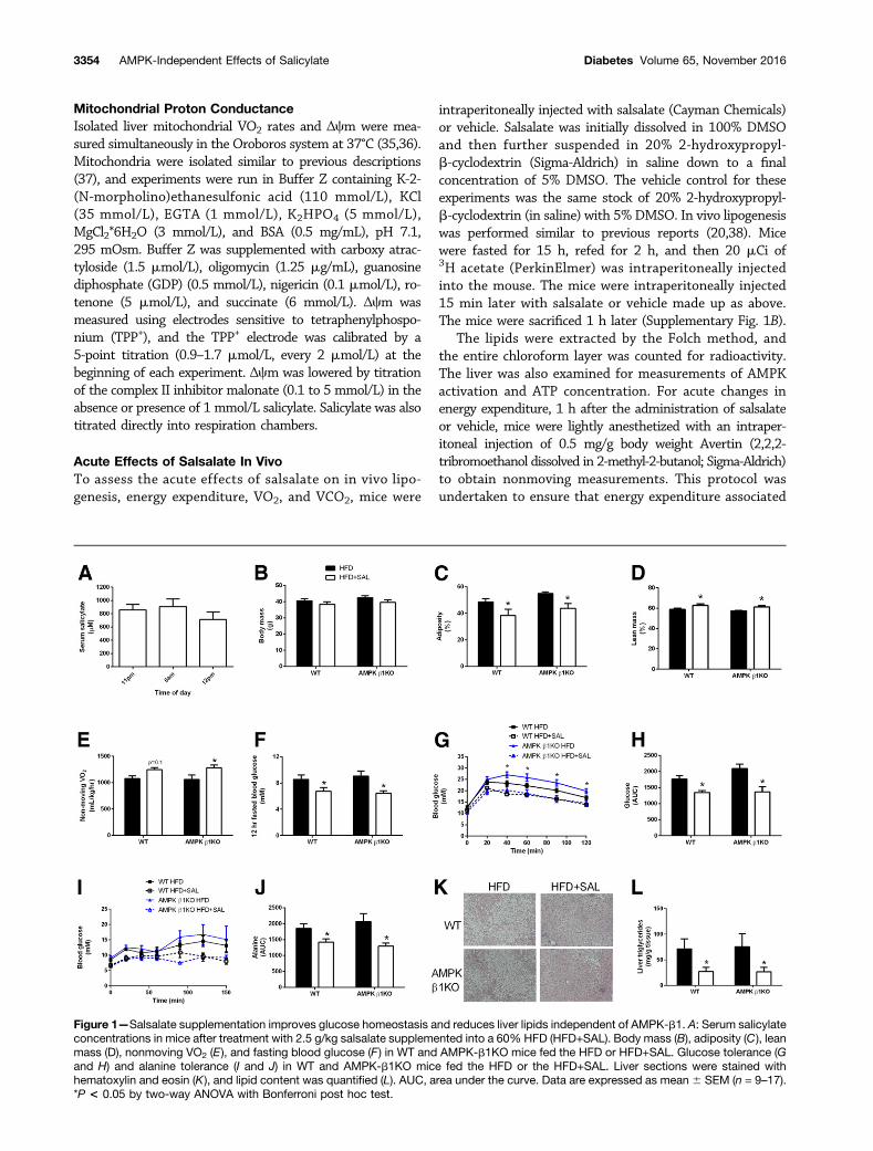
Figure 1—Salsalate supplementation improves glucose homeostasis and reduces liver lipids independent of AMPK-b1. A: Serum salicylateconcentrations in mice after treatment with 2.5 g/kg salsalate supplemented into a 60% HFD (HFD+SAL). Body mass (B), adiposity (C), leanmass (D), nonmoving VO2 (E), and fasting blood glucose (F ) in WT and AMPK-b1KO mice fed the HFD or HFD+SAL. Glucose tolerance (Gand H) and alanine tolerance (I and J) in WT and AMPK-b1KO mice fed the HFD or the HFD+SAL. Liver sections were stained withhematoxylin and eosin (K), and lipid content was quantified (L). AUC, area under the curve. Data are expressed as mean 6 SEM (n = 9–17).*P < 0.05 by two-way ANOVA with Bonferroni post hoc test.
3354 AMPK-Independent Effects of Salicylate Diabetes Volume 65, November 2016
with activity (skeletal muscle contraction) would not con-found our energy expenditure data. The mice were placeddorsal side up onto an enclosed stationary treadmill andmetabolic measurements were monitored for 12 min usingCLAMS (Supplementary Fig. 1C).
Statistical AnalysesValues are reported as mean 6 SEM. Data were analyzedusing two-way or one-way ANOVA with Bonferroni posthoc test or Student t test where indicated. Differenceswere considered significant when P , 0.05.
RESULTS
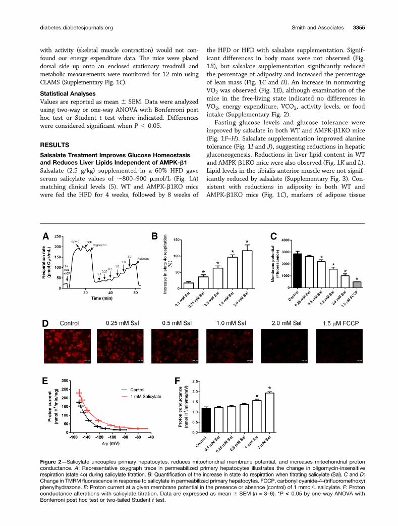
Salsalate Treatment Improves Glucose Homeostasisand Reduces Liver Lipids Independent of AMPK-b1Salsalate (2.5 g/kg) supplemented in a 60% HFD gaveserum salicylate values of ;800–900 mmol/L (Fig. 1A)matching clinical levels (5). WT and AMPK-b1KO micewere fed the HFD for 4 weeks, followed by 8 weeks of
the HFD or HFD with salsalate supplementation. Signif-icant differences in body mass were not observed (Fig.1B), but salsalate supplementation significantly reducedthe percentage of adiposity and increased the percentageof lean mass (Fig. 1C and D). An increase in nonmovingVO2 was observed (Fig. 1E), although examination of themice in the free-living state indicated no differences inVO2, energy expenditure, VCO2, activity levels, or foodintake (Supplementary Fig. 2).
Fasting glucose levels and glucose tolerance wereimproved by salsalate in both WT and AMPK-b1KO mice(Fig. 1F–H). Salsalate supplementation improved alaninetolerance (Fig. 1I and J), suggesting reductions in hepaticgluconeogenesis. Reductions in liver lipid content in WTand AMPK-b1KO mice were also observed (Fig. 1K and L).Lipid levels in the tibialis anterior muscle were not signif-icantly reduced by salsalate (Supplementary Fig. 3). Con-sistent with reductions in adiposity in both WT andAMPK-b1KO mice (Fig. 1C), markers of adipose tissue
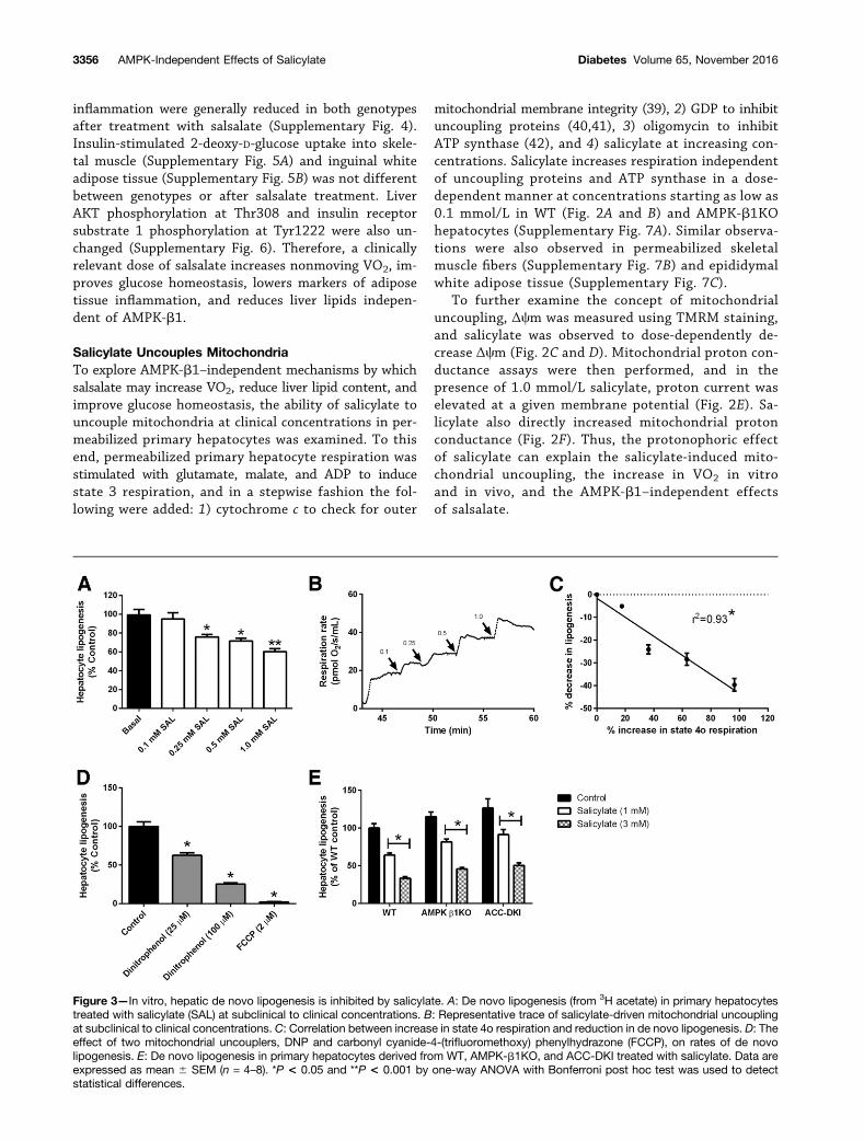
Figure 2—Salicylate uncouples primary hepatocytes, reduces mitochondrial membrane potential, and increases mitochondrial protonconductance. A: Representative oxygraph trace in permeabilized primary hepatocytes illustrates the change in oligomycin-insensitiverespiration (state 4o) during salicylate titration. B: Quantification of the increase in state 4o respiration when titrating salicylate (Sal). C and D:Change in TMRM fluorescence in response to salicylate in permeabilized primary hepatocytes. FCCP, carbonyl cyanide-4-(trifluoromethoxy)phenylhydrazone. E: Proton current at a given membrane potential in the presence or absence (control) of 1 mmol/L salicylate. F: Protonconductance alterations with salicylate titration. Data are expressed as mean 6 SEM (n = 3–6). *P < 0.05 by one-way ANOVA withBonferroni post hoc test or two-tailed Student t test.
diabetes.diabetesjournals.org Smith and Associates 3355
inflammation were generally reduced in both genotypesafter treatment with salsalate (Supplementary Fig. 4).Insulin-stimulated 2-deoxy-D-glucose uptake into skele-tal muscle (Supplementary Fig. 5A) and inguinal whiteadipose tissue (Supplementary Fig. 5B) was not differentbetween genotypes or after salsalate treatment. LiverAKT phosphorylation at Thr308 and insulin receptorsubstrate 1 phosphorylation at Tyr1222 were also un-changed (Supplementary Fig. 6). Therefore, a clinicallyrelevant dose of salsalate increases nonmoving VO2, im-proves glucose homeostasis, lowers markers of adiposetissue inflammation, and reduces liver lipids indepen-dent of AMPK-b1.
Salicylate Uncouples MitochondriaTo explore AMPK-b1–independent mechanisms by whichsalsalate may increase VO2, reduce liver lipid content, andimprove glucose homeostasis, the ability of salicylate touncouple mitochondria at clinical concentrations in per-meabilized primary hepatocytes was examined. To thisend, permeabilized primary hepatocyte respiration wasstimulated with glutamate, malate, and ADP to inducestate 3 respiration, and in a stepwise fashion the fol-lowing were added: 1) cytochrome c to check for outer
mitochondrial membrane integrity (39), 2) GDP to inhibituncoupling proteins (40,41), 3) oligomycin to inhibitATP synthase (42), and 4) salicylate at increasing con-centrations. Salicylate increases respiration independentof uncoupling proteins and ATP synthase in a dose-dependent manner at concentrations starting as low as0.1 mmol/L in WT (Fig. 2A and B) and AMPK-b1KOhepatocytes (Supplementary Fig. 7A). Similar observa-tions were also observed in permeabilized skeletalmuscle fibers (Supplementary Fig. 7B) and epididymalwhite adipose tissue (Supplementary Fig. 7C).
To further examine the concept of mitochondrialuncoupling, Δcm was measured using TMRM staining,and salicylate was observed to dose-dependently de-crease Δcm (Fig. 2C and D). Mitochondrial proton con-ductance assays were then performed, and in thepresence of 1.0 mmol/L salicylate, proton current waselevated at a given membrane potential (Fig. 2E). Sa-licylate also directly increased mitochondrial protonconductance (Fig. 2F). Thus, the protonophoric effectof salicylate can explain the salicylate-induced mito-chondrial uncoupling, the increase in VO2 in vitroand in vivo, and the AMPK-b1–independent effectsof salsalate.
Figure 3—In vitro, hepatic de novo lipogenesis is inhibited by salicylate. A: De novo lipogenesis (from 3H acetate) in primary hepatocytestreated with salicylate (SAL) at subclinical to clinical concentrations. B: Representative trace of salicylate-driven mitochondrial uncouplingat subclinical to clinical concentrations. C: Correlation between increase in state 4o respiration and reduction in de novo lipogenesis. D: Theeffect of two mitochondrial uncouplers, DNP and carbonyl cyanide-4-(trifluoromethoxy) phenylhydrazone (FCCP), on rates of de novolipogenesis. E: De novo lipogenesis in primary hepatocytes derived from WT, AMPK-b1KO, and ACC-DKI treated with salicylate. Data areexpressed as mean 6 SEM (n = 4–8). *P < 0.05 and **P < 0.001 by one-way ANOVA with Bonferroni post hoc test was used to detectstatistical differences.
3356 AMPK-Independent Effects of Salicylate Diabetes Volume 65, November 2016
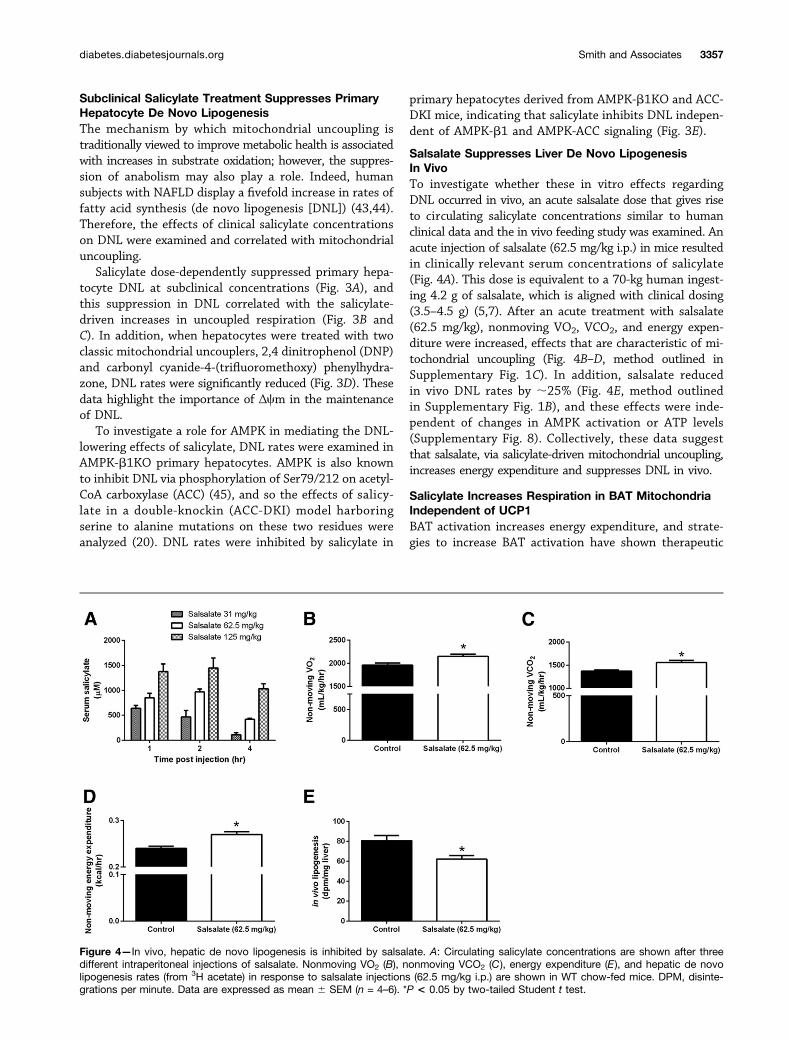
Subclinical Salicylate Treatment Suppresses PrimaryHepatocyte De Novo LipogenesisThe mechanism by which mitochondrial uncoupling istraditionally viewed to improve metabolic health is associatedwith increases in substrate oxidation; however, the suppres-sion of anabolism may also play a role. Indeed, humansubjects with NAFLD display a fivefold increase in rates offatty acid synthesis (de novo lipogenesis [DNL]) (43,44).Therefore, the effects of clinical salicylate concentrationson DNL were examined and correlated with mitochondrialuncoupling.
Salicylate dose-dependently suppressed primary hepa-tocyte DNL at subclinical concentrations (Fig. 3A), andthis suppression in DNL correlated with the salicylate-driven increases in uncoupled respiration (Fig. 3B andC). In addition, when hepatocytes were treated with twoclassic mitochondrial uncouplers, 2,4 dinitrophenol (DNP)and carbonyl cyanide-4-(trifluoromethoxy) phenylhydra-zone, DNL rates were significantly reduced (Fig. 3D). Thesedata highlight the importance of Δcm in the maintenanceof DNL.
To investigate a role for AMPK in mediating the DNL-lowering effects of salicylate, DNL rates were examined inAMPK-b1KO primary hepatocytes. AMPK is also knownto inhibit DNL via phosphorylation of Ser79/212 on acetyl-CoA carboxylase (ACC) (45), and so the effects of salicy-late in a double-knockin (ACC-DKI) model harboringserine to alanine mutations on these two residues wereanalyzed (20). DNL rates were inhibited by salicylate in
primary hepatocytes derived from AMPK-b1KO and ACC-DKI mice, indicating that salicylate inhibits DNL indepen-dent of AMPK-b1 and AMPK-ACC signaling (Fig. 3E).
Salsalate Suppresses Liver De Novo LipogenesisIn VivoTo investigate whether these in vitro effects regardingDNL occurred in vivo, an acute salsalate dose that gives riseto circulating salicylate concentrations similar to humanclinical data and the in vivo feeding study was examined. Anacute injection of salsalate (62.5 mg/kg i.p.) in mice resultedin clinically relevant serum concentrations of salicylate(Fig. 4A). This dose is equivalent to a 70-kg human ingest-ing 4.2 g of salsalate, which is aligned with clinical dosing(3.5–4.5 g) (5,7). After an acute treatment with salsalate(62.5 mg/kg), nonmoving VO2, VCO2, and energy expen-diture were increased, effects that are characteristic of mi-tochondrial uncoupling (Fig. 4B–D, method outlined inSupplementary Fig. 1C). In addition, salsalate reducedin vivo DNL rates by ;25% (Fig. 4E, method outlinedin Supplementary Fig. 1B), and these effects were inde-pendent of changes in AMPK activation or ATP levels(Supplementary Fig. 8). Collectively, these data suggestthat salsalate, via salicylate-driven mitochondrial uncoupling,increases energy expenditure and suppresses DNL in vivo.
Salicylate Increases Respiration in BAT MitochondriaIndependent of UCP1BAT activation increases energy expenditure, and strate-gies to increase BAT activation have shown therapeutic
Figure 4—In vivo, hepatic de novo lipogenesis is inhibited by salsalate. A: Circulating salicylate concentrations are shown after threedifferent intraperitoneal injections of salsalate. Nonmoving VO2 (B), nonmoving VCO2 (C), energy expenditure (E), and hepatic de novolipogenesis rates (from 3H acetate) in response to salsalate injections (62.5 mg/kg i.p.) are shown in WT chow-fed mice. DPM, disinte-grations per minute. Data are expressed as mean 6 SEM (n = 4–6). *P < 0.05 by two-tailed Student t test.
diabetes.diabetesjournals.org Smith and Associates 3357
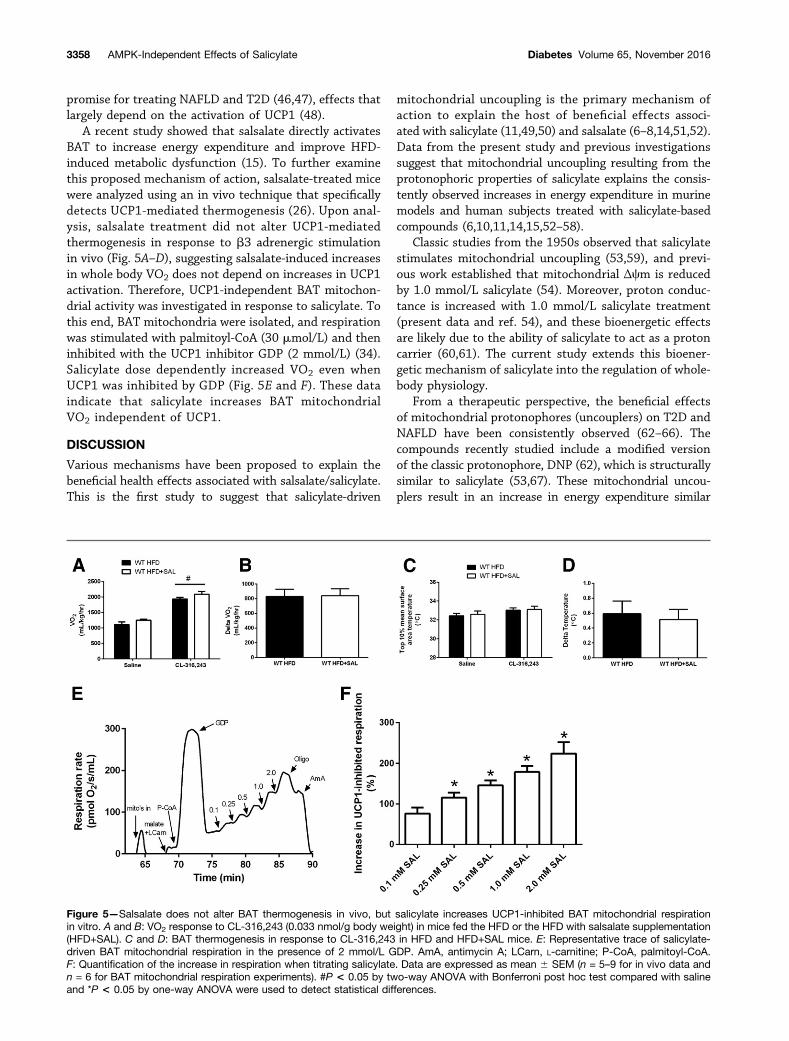
promise for treating NAFLD and T2D (46,47), effects thatlargely depend on the activation of UCP1 (48).
A recent study showed that salsalate directly activatesBAT to increase energy expenditure and improve HFD-induced metabolic dysfunction (15). To further examinethis proposed mechanism of action, salsalate-treated micewere analyzed using an in vivo technique that specificallydetects UCP1-mediated thermogenesis (26). Upon anal-ysis, salsalate treatment did not alter UCP1-mediatedthermogenesis in response to b3 adrenergic stimulationin vivo (Fig. 5A–D), suggesting salsalate-induced increasesin whole body VO2 does not depend on increases in UCP1activation. Therefore, UCP1-independent BAT mitochon-drial activity was investigated in response to salicylate. Tothis end, BAT mitochondria were isolated, and respirationwas stimulated with palmitoyl-CoA (30 mmol/L) and theninhibited with the UCP1 inhibitor GDP (2 mmol/L) (34).Salicylate dose dependently increased VO2 even whenUCP1 was inhibited by GDP (Fig. 5E and F). These dataindicate that salicylate increases BAT mitochondrialVO2 independent of UCP1.
DISCUSSION
Various mechanisms have been proposed to explain thebeneficial health effects associated with salsalate/salicylate.This is the first study to suggest that salicylate-driven
mitochondrial uncoupling is the primary mechanism ofaction to explain the host of beneficial effects associ-ated with salicylate (11,49,50) and salsalate (6–8,14,51,52).Data from the present study and previous investigationssuggest that mitochondrial uncoupling resulting from theprotonophoric properties of salicylate explains the consis-tently observed increases in energy expenditure in murinemodels and human subjects treated with salicylate-basedcompounds (6,10,11,14,15,52–58).
Classic studies from the 1950s observed that salicylatestimulates mitochondrial uncoupling (53,59), and previ-ous work established that mitochondrial Δcm is reducedby 1.0 mmol/L salicylate (54). Moreover, proton conduc-tance is increased with 1.0 mmol/L salicylate treatment(present data and ref. 54), and these bioenergetic effectsare likely due to the ability of salicylate to act as a protoncarrier (60,61). The current study extends this bioener-getic mechanism of salicylate into the regulation of whole-body physiology.
From a therapeutic perspective, the beneficial effectsof mitochondrial protonophores (uncouplers) on T2D andNAFLD have been consistently observed (62–66). Thecompounds recently studied include a modified versionof the classic protonophore, DNP (62), which is structurallysimilar to salicylate (53,67). These mitochondrial uncou-plers result in an increase in energy expenditure similar
Figure 5—Salsalate does not alter BAT thermogenesis in vivo, but salicylate increases UCP1-inhibited BAT mitochondrial respirationin vitro. A and B: VO2 response to CL-316,243 (0.033 nmol/g body weight) in mice fed the HFD or the HFD with salsalate supplementation(HFD+SAL). C and D: BAT thermogenesis in response to CL-316,243 in HFD and HFD+SAL mice. E: Representative trace of salicylate-driven BAT mitochondrial respiration in the presence of 2 mmol/L GDP. AmA, antimycin A; LCarn, L-carnitine; P-CoA, palmitoyl-CoA.F: Quantification of the increase in respiration when titrating salicylate. Data are expressed as mean 6 SEM (n = 5–9 for in vivo data andn = 6 for BAT mitochondrial respiration experiments). #P < 0.05 by two-way ANOVA with Bonferroni post hoc test compared with salineand *P < 0.05 by one-way ANOVA were used to detect statistical differences.
3358 AMPK-Independent Effects of Salicylate Diabetes Volume 65, November 2016
to that obtained when treating with salsalate/salicylate (pre-sent work and previous studies [6,10,11,14,15,52–58]) andalso improve markers of NAFLD and T2D (62–66). Therefore,considering that mitochondrial uncouplers improve T2D andNAFLD, and increasing energy expenditure is a potent mech-anism to improve T2D and NAFLD (62,63,68–70), the pre-sent work, together with previous reports in mice andhumans, is highly suggestive that salicylate-driven mitochon-drial uncoupling is the primary mechanism of action explain-ing the improvement in T2D and NAFLD associated withsalsalate treatment.
As a potential mechanism to explain why mitochon-drial uncouplers improve T2D and NAFLD, salsalate wasfound to potently suppress hepatic DNL in vitro and in vivo.NAFLD is associated with increased rates of hepatic DNL(43,44), and this report is the first to show that DNL can besuppressed in vivo with an acute, therapeutically relevantdose of salsalate. DNL requires three basic constituents; sub-strate (acetyl-CoA), reductive power (NADPH), and energysupply (ATP). By reducing Δcm, salicylate and other uncou-pling agents compromise the availability of all three factorsas the cell shifts to a catabolic state and downregulatesanabolic activity (67,71). Considering the potential impor-tance of DNL in human NAFLD, future work investigatingwhether salsalate can suppress DNL in humans is warranted.
From a general mechanism standpoint, previous workin humans supplemented with high-dose aspirin (57) orsalsalate (14) observed increases in carbohydrate and fatoxidation. The only way carbohydrate and fat oxidationcan simultaneously increase in vivo is if the overall de-mand of the system (i.e., uncoupling or increased ATPturnover) changes. In the case of salsalate, by providinga protonophore (salicylate), Δcm is reduced, an effect thatallows fat and carbohydrate catabolism to increase. Inenvironments typified by overnutrition, this uncouplingmechanism works to blunt the toxic excess of substrate bysiphoning fat and/or carbohydrate away from anabolicpathways (DNL) in favor of oxidation. In other words,mitochondrial uncouplers suppress the toxic effects ofsubstrate oversupply by removing the substrate.
A limitation of this work is that definitive geneticevidence is unavailable because removing mitochondriaand, therefore, the uncoupling effects of salicylate, is notpossible. A further limitation is the difficulty in discern-ing which cell type or organ system is playing the mostprominent role in mediating the beneficial effects ofsalsalate. Inhibition of hepatic DNL has shown therapeuticpromise for NAFLD (72), as has promoting hepatic sub-strate consumption (62), indicating that the liver may beimportant. However, increasing BAT mitochondrial activityhas been shown to spare the liver from substrate oversup-ply (46), and skeletal muscle is responsible for the majorityof glucose clearance (73). Further work is thus required toinvestigate which organ system is playing the most prom-inent role.
In addition to the positive metabolic effects associatedwith mitochondrial uncouplers in the presence of nutrient
excess, significant negative adverse effects have beenassociated with uncoupling agents, including DNP (74,75).The present report is an important message from not onlya therapeutic perspective but also from a potential adverseeffects perspective, and future work examining salicylate-based compounds should consider the fact that salicylateuncouples mitochondria at clinically relevant concentrations.
In summary, a therapeutically relevant dose of salsa-late improves glucose homeostasis, lowers adipose tissueinflammation, and reduces liver lipid content, indepen-dent of AMPK-b1. Instead of AMPK, it seems likely thatthe protonophoric effect of salicylate is the primary mech-anism underlying the metabolic improvements associatedwith salicylate (11,49) and salsalate (6–8,14,51,52) underconditions of nutrient excess. Future studies should con-sider this mechanism of action when examining effects ofsalsalate on T2D and NAFLD.
Funding. B.K.S. is a recipient of a Canadian Institutes of Health Research(CIHR) Postdoctoral Fellowship and Michael G. DeGroote Postdoctoral Fellowship.E.M.D. is a recipient of an Ontario Graduate Scholarship and Queen Elizabeth IIGraduate Scholarship in Science and Technology. A.E.G. is a recipient of a CanadianGraduate Scholarship from CIHR/MitoCanada. E.P.M. is a Canadian DiabetesAssociation postdoctoral fellow. B.E.K. is supported by grants and a fellowship fromthe Australian Research Council and the National Health and Medical ResearchCouncil, supported in part by the Victorian Government’s Operational Infrastructure.These studies were supported by grants from the Canadian Diabetes Association(G.R.S.), the CIHR (G.R.S.), and the Natural Sciences and Engineering ResearchCouncil of Canada (G.R.S.). G.R.S. is a Canada Research Chair in Metabolism andObesity and the J. Bruce Duncan Chair in Metabolic Diseases.Duality of Interest. No potential conflicts of interest relevant to this articlewere reported.Author Contributions. B.K.S. and G.R.S. designed research studies,analyzed data, and wrote the manuscript. B.K.S., R.J.F., E.M.D., A.E.G., M.C.H.,V.P.H., E.A.D., K.M., J.D.C., E.P.M., and C.G.R.P. conducted experiments andanalyzed data. B.K.S., R.J.F., E.M.D., A.E.G., V.P.H., E.A.D., K.M., J.D.C., E.P.M.,B.E.K., M.A.T., and G.R.S. edited the manuscript. G.R.S. is the guarantor of thiswork and, as such, had full access to all the data in the study and takesresponsibility for the integrity of the data and the accuracy of the data analysis.
References1. Rinella ME. Nonalcoholic fatty liver disease: a systematic review. JAMA2015;313:2263–22732. Perry RJ, Samuel VT, Petersen KF, Shulman GI. The role of hepatic lipids inhepatic insulin resistance and type 2 diabetes. Nature 2014;510:84–913. Dromgoole SH, Cassell S, Furst DE, Paulus HE. Availability of salicylate fromsalsalate and aspirin. Clin Pharmacol Ther 1983;34:539–5454. Dromgoole SH, Furst DE, Paulus HE. Metabolism of salsalate in normalsubjects. J Pharm Sci 1984;73:1657–16595. Fleischman A, Shoelson SE, Bernier R, Goldfine AB. Salsalate improvesglycemia and inflammatory parameters in obese young adults. Diabetes Care2008;31:289–2946. Goldfine AB, Silver R, Aldhahi W, et al. Use of salsalate to target in-flammation in the treatment of insulin resistance and type 2 diabetes. Clin TranslSci 2008;1:36–437. Goldfine AB, Fonseca V, Jablonski KA, Pyle L, Staten MA, Shoelson SE;TINSAL-T2D (Targeting Inflammation Using Salsalate in Type 2 Diabetes) StudyTeam. The effects of salsalate on glycemic control in patients with type 2 di-abetes: a randomized trial. Ann Intern Med 2010;152:346–357
diabetes.diabetesjournals.org Smith and Associates 3359
8. Barzilay JI, Jablonski KA, Fonseca V, et al.; TINSAL-T2D Research Con-sortium. The impact of salsalate treatment on serum levels of advanced glycationend products in type 2 diabetes. Diabetes Care 2014;37:1083–10919. Ford RJ, Fullerton MD, Pinkosky SL, et al. Metformin and salicylate syn-ergistically activate liver AMPK, inhibit lipogenesis and improve insulin sensitivity.Biochem J 2015;468:125–13210. Liang W, Verschuren L, Mulder P, et al. Salsalate attenuates diet inducednon-alcoholic steatohepatitis in mice by decreasing lipogenic and inflammatoryprocesses. Br J Pharmacol 2015;172:5293–530511. Kim JK, Kim YJ, Fillmore JJ, et al. Prevention of fat-induced insulin re-sistance by salicylate. J Clin Invest 2001;108:437–44612. Yuan M, Konstantopoulos N, Lee J, et al. Reversal of obesity- and diet-induced insulin resistance with salicylates or targeted disruption of Ikkbeta.Science 2001;293:1673–167713. Yin MJ, Yamamoto Y, Gaynor RB. The anti-inflammatory agents aspirin andsalicylate inhibit the activity of I(kappa)B kinase-beta. Nature 1998;396:77–8014. Meex RC, Phielix E, Moonen-Kornips E, Schrauwen P, Hesselink MK.Stimulation of human whole-body energy expenditure by salsalate is fueled byhigher lipid oxidation under fasting conditions and by higher oxidative glucosedisposal under insulin-stimulated conditions. J Clin Endocrinol Metab 2011;96:1415–142315. van Dam AD, Nahon KJ, Kooijman S, et al. Salsalate activates brown adi-pose tissue in mice. Diabetes 2015;64:1544–155416. Hawley SA, Fullerton MD, Ross FA, et al. The ancient drug salicylate directlyactivates AMP-activated protein kinase. Science 2012;336:918–92217. Alpert D, Vilcek J. Inhibition of IkappaB kinase activity by sodium salicylatein vitro does not reflect its inhibitory mechanism in intact cells. J Biol Chem 2000;275:10925–1092918. Steinberg GR, Dandapani M, Hardie DG. AMPK: mediating the metaboliceffects of salicylate-based drugs? Trends Endocrinol Metab 2013;24:481–48719. Galic S, Fullerton MD, Schertzer JD, et al. Hematopoietic AMPK b1 reducesmouse adipose tissue macrophage inflammation and insulin resistance in obe-sity. J Clin Invest 2011;121:4903–491520. Fullerton MD, Galic S, Marcinko K, et al. Single phosphorylation sites inAcc1 and Acc2 regulate lipid homeostasis and the insulin-sensitizing effects ofmetformin. Nat Med 2013;19:1649–165421. Mottillo EP, Desjardins EM, Crane JD, et al. Lack of adipocyte AMPK ex-acerbates insulin resistance and hepatic steatosis through brown and beigeadipose tissue function. Cell Metab 2016;24:118–12922. Zhang H, Guan M, Townsend KL, et al. MicroRNA-455 regulates brownadipogenesis via a novel HIF1an-AMPK-PGC1a signaling network. EMBO Rep2015;16:1378–139323. Calabrese MF, Rajamohan F, Harris MS, et al. Structural basis for AMPKactivation: natural and synthetic ligands regulate kinase activity from oppositepoles by different molecular mechanisms. Structure 2014;22:1161–117224. Bujak AL, Crane JD, Lally JS, et al. AMPK activation of muscle autophagyprevents fasting-induced hypoglycemia and myopathy during aging. Cell Metab2015;21:883–89025. O’Neill HM, Maarbjerg SJ, Crane JD, et al. AMP-activated protein kinase(AMPK) beta1beta2 muscle null mice reveal an essential role for AMPK inmaintaining mitochondrial content and glucose uptake during exercise. Proc NatlAcad Sci U S A 2011;108:16092–1609726. Crane JD, Mottillo EP, Farncombe TH, Morrison KM, Steinberg GR. Astandardized infrared imaging technique that specifically detects UCP1-mediatedthermogenesis in vivo. Mol Metab 2014;3:490–49427. Howlett KF, Andrikopoulos S, Proietto J, Hargreaves M. Exercise-inducedmuscle glucose uptake in mice with graded, muscle-specific GLUT-4 deletion.Physiol Rep 2013;1:e0006528. Folch J, Lees M, Sloane Stanley GH. A simple method for the isolation andpurification of total lipides from animal tissues. J Biol Chem 1957;226:497–50929. Scaduto RC Jr, Grotyohann LW. Measurement of mitochondrial membranepotential using fluorescent rhodamine derivatives. Biophys J 1999;76:469–477
30. Borowski LS, Dziembowski A, Hejnowicz MS, Stepien PP, Szczesny RJ.Human mitochondrial RNA decay mediated by PNPase-hSuv3 complex takesplace in distinct foci. Nucleic Acids Res 2013;41:1223–124031. Smith BK, Perry CG, Herbst EA, et al. Submaximal ADP-stimulated respi-ration is impaired in ZDF rats and recovered by resveratrol. J Physiol 2013;591:6089–610132. Paglialunga S, Ludzki A, Root-McCaig J, Holloway GP. In adipose tissue,increased mitochondrial emission of reactive oxygen species is important forshort-term high-fat diet-induced insulin resistance in mice. Diabetologia 2015;58:1071–108033. Cannon B, Nedergaard J. Studies of thermogenesis and mitochondrialfunction in adipose tissues. Methods Mol Biol 2008;456:109–12134. Shabalina IG, Petrovic N, de Jong JM, Kalinovich AV, Cannon B, NedergaardJ. UCP1 in brite/beige adipose tissue mitochondria is functionally thermogenic.Cell Reports 2013;5:1196–120335. Fisher-Wellman KH, Lin CT, Ryan TE, et al. Pyruvate dehydrogenasecomplex and nicotinamide nucleotide transhydrogenase constitute an energy-consuming redox circuit. Biochem J 2015;467:271–28036. Brand MD, Nicholls DG. Assessing mitochondrial dysfunction in cells. Bio-chem J 2011;435:297–31237. Smith BK, Perry CG, Koves TR, et al. Identification of a novel malonyl-CoA IC(50) for CPT-I: implications for predicting in vivo fatty acid oxidation rates. Bio-chem J 2012;448:13–2038. Dupont J, Mathias MM, Cabacungan NB. Dietary lipid, fatty acid synthesisand cholesterol metabolism in aging rats. Lipids 1972;7:576–58939. Wikström M, Casey R. The oxidation of exogenous cytochrome c by mito-chondria. Resolution of a long-standing controversy. FEBS Lett 1985;183:293–29840. Echtay KS, Esteves TC, Pakay JL, et al. A signalling role for 4-hydroxy-2-nonenal in regulation of mitochondrial uncoupling. EMBO J 2003;22:4103–411041. Nègre-Salvayre A, Hirtz C, Carrera G, et al. A role for uncoupling protein-2as a regulator of mitochondrial hydrogen peroxide generation. FASEB J 1997;11:809–81542. Zheng J, Ramirez VD. Inhibition of mitochondrial proton F0F1-ATPase/ATPsynthase by polyphenolic phytochemicals. Br J Pharmacol 2000;130:1115–112343. Lambert JE, Ramos-Roman MA, Browning JD, Parks EJ. Increased de novolipogenesis is a distinct characteristic of individuals with nonalcoholic fatty liverdisease. Gastroenterology 2014;146:726–73544. Donnelly KL, Smith CI, Schwarzenberg SJ, Jessurun J, Boldt MD, Parks EJ.Sources of fatty acids stored in liver and secreted via lipoproteins in patients withnonalcoholic fatty liver disease. J Clin Invest 2005;115:1343–135145. Carling D, Zammit VA, Hardie DG. A common bicyclic protein kinase cas-cade inactivates the regulatory enzymes of fatty acid and cholesterol bio-synthesis. FEBS Lett 1987;223:217–22246. Crane JD, Palanivel R, Mottillo EP, et al. Inhibiting peripheral serotoninsynthesis reduces obesity and metabolic dysfunction by promoting brown adi-pose tissue thermogenesis. Nat Med 2015;21:166–17247. Sidossis L, Kajimura S. Brown and beige fat in humans: thermogenic ad-ipocytes that control energy and glucose homeostasis. J Clin Invest 2015;125:478–48648. Enerbäck S, Jacobsson A, Simpson EM, et al. Mice lacking mitochondrialuncoupling protein are cold-sensitive but not obese. Nature 1997;387:90–9449. Williamson RT. On the treatment of glycosuria and diabetes mellitus withsodium salicylate. BMJ 1901;1:760–76250. Cameron AR, Logie L, Patel K, et al. Investigation of salicylate hepatic re-sponses in comparison with chemical analogues of the drug. Biochim BiophysActa 2016;1862:1412–142251. Ariel D, Kim SH, Liu A, et al. Salsalate-induced changes in lipid, lipoprotein,and apoprotein concentrations in overweight or obese, insulin-resistant, non-diabetic individuals. J Clin Lipidol 2015;9:658–663
3360 AMPK-Independent Effects of Salicylate Diabetes Volume 65, November 2016
52. Cao Y, Dubois DC, Sun H, Almon RR, Jusko WJ. Modeling diabetes diseaseprogression and salsalate intervention in Goto-Kakizaki rats. J Pharmacol ExpTher 2011;339:896–90453. Sproull DH. A peripheral action of sodium salicylate. Br Pharmacol Che-mother 1954;9:262–26454. Haas R, Parker WD Jr, Stumpf D, Eguren LA. Salicylate-induced loosecoupling: protonmotive force measurements. Biochem Pharmacol 1985;34:900–90255. Sproull DH. A comparison of sodium salicylate and 2:4-dinitrophenol asmetabolic stimulants in vitro. Biochem J 1957;66:527–53256. Denis W, Means JH. The influence of salicylate on metabolism in man.J Pharmacol Exp Ther 1916;8:273–28357. Hundal RS, Petersen KF, Mayerson AB, et al. Mechanism by which high-dose aspirin improves glucose metabolism in type 2 diabetes. J Clin Invest 2002;109:1321–132658. Cochran JB. The respiratory effects of salicylate. BMJ 1952;2:964–96759. Brody TM. Action of sodium salicylate and related compounds on tissuemetabolism in vitro. J Pharmacol Exp Ther 1956;117:39–5160. Gutknecht J. Salicylates and proton transport through lipid bilayer mem-branes: a model for salicylate-induced uncoupling and swelling in mitochondria.J Membr Biol 1990;115:253–26061. Gutknecht J. Aspirin, acetaminophen and proton transport throughphospholipid bilayers and mitochondrial membranes. Mol Cell Biochem1992;114:3–862. Perry RJ, Zhang D, Zhang XM, Boyer JL, Shulman GI. Controlled-releasemitochondrial protonophore reverses diabetes and steatohepatitis in rats. Science2015;347:1253–125663. Tao H, Zhang Y, Zeng X, Shulman GI, Jin S. Niclosamide ethanolamine-induced mild mitochondrial uncoupling improves diabetic symptoms in mice. NatMed 2014;20:1263–126964. Samuel VT, Liu ZX, Qu X, et al. Mechanism of hepatic insulin resistance innon-alcoholic fatty liver disease. J Biol Chem 2004;279:32345–32353
65. Figarola JL, Singhal P, Rahbar S, Gugiu BG, Awasthi S, Singhal SS. COH-SR4 reduces body weight, improves glycemic control and prevents hepaticsteatosis in high fat diet-induced obese mice. PLoS One 2013;8:e8380166. Kalinovich AV, Shabalina IG. Novel Mitochondrial Cationic Uncoupler C4R1 Isan Effective Treatment for Combating Obesity in Mice. Biochemistry (Mosc) 2015;80:620–62867. Smith MJ, Moses V. Uncoupling reagents and metabolism. 1. Effects ofsalicylate and 2:4-dinitrophenol on the incorporation of C from labelled glucoseand acetate into the soluble intermediates of isolated rat tissues. Biochem J1960;76:579–58568. Keating SE, Hackett DA, Parker HM, et al. Effect of aerobic exercise trainingdose on liver fat and visceral adiposity. J Hepatol 2015;63:174–18269. Colberg SR, Sigal RJ, Fernhall B, et al.; American College of Sports Medi-cine; American Diabetes Association. Exercise and type 2 diabetes: the AmericanCollege of Sports Medicine and the American Diabetes Association: joint positionstatement. Diabetes Care 2010;33:e147–e16770. Stanford KI, Middelbeek RJ, Townsend KL, et al. Brown adipose tissueregulates glucose homeostasis and insulin sensitivity. J Clin Invest 2013;123:215–22371. Blacker TS, Mann ZF, Gale JE, et al. Separating NADH and NADPH fluo-rescence in live cells and tissues using FLIM. Nat Commun 2014;5:393672. Harriman G, Greenwood J, Bhat S, et al. Acetyl-CoA carboxylase in-hibition by ND-630 reduces hepatic steatosis, improves insulin sensitivity, andmodulates dyslipidemia in rats. Proc Natl Acad Sci U S A 2016;113:E1796–E180573. DeFronzo RA, Gunnarsson R, Björkman O, Olsson M, Wahren J. Effects ofinsulin on peripheral and splanchnic glucose metabolism in noninsulin-dependent(type II) diabetes mellitus. J Clin Invest 1985;76:149–15574. Fawaz G, Tutunji B. The mechanism of dinitrophenol heart failure. BrPharmacol Chemother 1957;12:273–27875. Tainter ML, Cutting WC, Stockton AB. Use of dinitrophenol in nutritionaldisorders: a critical survey of clinical results. Am J Public Health Nations Health1934;24:1045–1053
diabetes.diabetesjournals.org Smith and Associates 3361