
Formation of hydroxyl radicals by -Fe O dinitrophenol and ...
18
Formation of hydroxyl radicals by a-Fe 2 O 3 microcrystals and its role in photodegradation of 2,4- dinitrophenol and lipid peroxidation Gilma Granados-Oliveros 1 • Erika Torres 2 • Marcela Zambrano 2 • Antonio Nieto-Camacho 3 • Virginia Go ´mez-Vidales 3 Received: 9 October 2017 / Accepted: 30 January 2018 Ó Springer Science+Business Media B.V., part of Springer Nature 2018 Abstract a-Fe 2 O 3 microcrystals were produced for application as catalyst in dif- ferent oxidation processes in both chemical and biological matrices. Hematite was produced by sol–gel method in situ with silica matrix and characterized by X-ray diffraction analysis, scanning electron microscopy with energy-dispersive X-ray spectrometry, and transmission electron microscopy. The ability of the catalyst to produce hydroxyl radicals ( OH) was evaluated by electron paramagnetic resonance measurements using 5,5-dimethyl- 1-pyrroline-N-oxide (DMPO) as spin trap. Characterization of the resulting DMPO-OH adduct established that a-Fe 2 O 3 microcrystals could generate OH when Fenton chemistry was present. Additionally, the catalyst exhibited semiconducting properties, as the DMPO-OH signal was produced under visible-light irradiation in presence of O 2 but without requiring H 2 O 2 . In a pollution control context, 2,4-dinitrophenol (2,4-DNP) degradation was used as probe reaction, with [ 99 % of this pollutant being removed in presence of H 2 O 2 under visible light. NO 2 - , NO 3 - , hydroxylated compounds, and a carboxylic acid were identified as photoproducts, suggesting a degradation pathway. Finally, catalyst reactivity in biological matrices was evaluated by oxidative degradation of Electronic supplementary material The online version of this article (https://doi.org/10.1007/s11164- 018-3315-2) contains supplementary material, which is available to authorized users. & Gilma Granados-Oliveros [email protected] 1 Nuevos Materiales Nano y Supramoleculares, Departamento de Quı ´mica, Facultad de Ciencias, Universidad Nacional de Colombia, Bogota ´, D.C., Colombia 2 Facultad de Quı ´mica Ambiental, Universidad Santo Toma ´s de Aquino, Bucaramanga, Colombia 3 Instituto de Quı ´mica, Universidad Nacional Auto ´noma de Me ´xico, Circuito exterior, Ciudad Universitaria, 04510 Coyoaca ´n, D.F., Mexico 123 Res Chem Intermed https://doi.org/10.1007/s11164-018-3315-2
Transcript of Formation of hydroxyl radicals by -Fe O dinitrophenol and ...
Formation of hydroxyl radicals by a-Fe2O3
microcrystals and its role in photodegradation of 2,4-dinitrophenol and lipid peroxidation
Gilma Granados-Oliveros1 • Erika Torres2 • Marcela Zambrano2 •
Antonio Nieto-Camacho3 • Virginia Gomez-Vidales3
Received: 9 October 2017 / Accepted: 30 January 2018
� Springer Science+Business Media B.V., part of Springer Nature 2018
Abstract a-Fe2O3 microcrystals were produced for application as catalyst in dif-
ferent oxidation processes in both chemical and biological matrices. Hematite was
produced by sol–gel method in situ with silica matrix and characterized by X-ray
diffraction analysis, scanning electron microscopy with energy-dispersive X-ray
spectrometry, and transmission electron microscopy. The ability of the catalyst to
produce hydroxyl radicals (�OH) was evaluated by electron paramagnetic resonance
measurements using 5,5-dimethyl- 1-pyrroline-N-oxide (DMPO) as spin trap.
Characterization of the resulting DMPO-OH adduct established that a-Fe2O3
microcrystals could generate �OH when Fenton chemistry was present. Additionally,
the catalyst exhibited semiconducting properties, as the DMPO-OH signal was
produced under visible-light irradiation in presence of O2 but without requiring
H2O2. In a pollution control context, 2,4-dinitrophenol (2,4-DNP) degradation was
used as probe reaction, with[99 % of this pollutant being removed in presence of
H2O2 under visible light. NO2-, NO3
-, hydroxylated compounds, and a carboxylic
acid were identified as photoproducts, suggesting a degradation pathway. Finally,
catalyst reactivity in biological matrices was evaluated by oxidative degradation of
Electronic supplementary material The online version of this article (https://doi.org/10.1007/s11164-
018-3315-2) contains supplementary material, which is available to authorized users.
& Gilma Granados-Oliveros
1 Nuevos Materiales Nano y Supramoleculares, Departamento de Quımica, Facultad de Ciencias,
Universidad Nacional de Colombia, Bogota, D.C., Colombia
2 Facultad de Quımica Ambiental, Universidad Santo Tomas de Aquino, Bucaramanga,
Colombia
3 Instituto de Quımica, Universidad Nacional Autonoma de Mexico, Circuito exterior, Ciudad
Universitaria, 04510 Coyoacan, D.F., Mexico
123
Res Chem Intermed
https://doi.org/10.1007/s11164-018-3315-2
lipids, revealing that a-Fe2O3 is a good oxidative stress inducer, representing a new
application for materials based on iron oxides.
Keywords Hematite � Hydroxyl radicals � 2,4-DNP degradation � Lipid
peroxidation � Visible light
Introduction
New catalytic methods involving environmentally friendly oxidants are needed
to perform advanced oxidation of toxic and refractory aromatic pollutants.
Although oxidation of pollutants can be realized by several homogeneous
methodologies, for example, ultraviolet (UV)/H2O2, UV/H2O2/O3, and UV/
H2O2/Fe(II) or Fe(III) (photoassisted Fenton reaction) [1], the heterogeneous
photo–Fenton process is an important alternative, offering various advantages
such as low toxicity, good reuse capabilities, and stability [2]. In addition,
unlimited solar energy can be efficiently harnessed. This process is based on
formation of hydroxyl radicals (�OH), which are powerful oxidizing species in
aqueous media with notable reactivity toward a wide variety of aromatic
pollutants [3]. �OH are produced from solid Fe species, H2O2, and UV
irradiation, according to reactions 1 and 2, where =Fe2? and =Fe3? are iron
species in solid phase or at the solid–liquid interface [4].
¼Fe3þ � OH þ hm ! ¼Fe2þ þ �OH ð1Þ
¼Fe2þ þ H2O2 ! ¼Fe3þ þ OH�� �e:g:¼Fe3þ � OH� �
þ �OH ð2Þ
Hematite (a-Fe2O3) is the most thermodynamically stable iron oxide phase, being
an n-type semiconductor (bandgap energy, Eg & 2.2 eV); its absorption range
includes a considerable portion of the solar spectrum [5], making it an interesting
option for use in photocatalytic processes [4, 5]. This kind of process initiates when
the surface of a semiconductor (in this case, hematite) is irradiated with energy
greater than or equal to Eg, releasing electrons from the valence band (reaction 3) to
conduction band. Photogenerated holes (h?) react with water (reaction 4) and
electrons (e-) with oxygen (reaction 5) to form �OH and superoxide radical anions,
O2�-, respectively [6]. However, hematite has a very short excited-state lifetime
(*1 ps) [7] and short hole diffusion length (*2–4 nm) [8], resulting in undesirable
electron–hole recombination [7] that affects the photocatalytic performance.
a� Fe2O3 þ hm ! hþ þ e� ð3Þ
hþ þ H2O ! �OH þ Hþ ð4Þ
e� þ O2 ! O�2� ð5Þ
It has been shown that the efficiency of hematite depends on its crystalline
structure [9], morphology, particle size, and surface area, which are related to the
G. Granados-Oliveros et al.
123
preparation method and starting materials [10–14]. a-Fe2O3 with high crystallinity
can be easily synthesized using different techniques such as spray pyrolysis,
hydrothermal, electrodeposition, and atmospheric-pressure chemical vapor deposi-
tion [15–24]. In situ preparation of a-Fe2O3 crystals in silica matrix is an
inexpensive alternative, where the porous nature of the matrix provides sites for
nucleation of the iron oxide particles, minimizes their aggregation, and suppresses
interparticle interactions [7, 19–26].
The aim of this work is to evaluate the oxidizing properties of well-formed a-
Fe2O3 particles in silica matrix under visible light. We used EPR measurements to
evaluate the ability of a-Fe2O3 to produce �OH. In a pollution control context, the
oxidant properties of this catalyst were studied for 2,4-DNP degradation, a toxic
refractory chemical and carcinogenic environmental pollutant [27, 28]. This
molecule is widely used in pesticide production, paints, and explosive materials, and
has been detected not only in industrial wastewater but also in freshwater and
marine environments [29]. Degradation of 2,4-DNP can be carried out by chemical
oxidation [30], heterogeneous ozonation [31], electrochemical oxidation [32, 33],
sonochemical methods [34, 35], and photocatalysis induced by UV [29] or visible
light [36, 37]. In the case of hematite-based systems, removal of 2,4-DNP has been
induced with UV light [38], but the mechanism involved in oxidation processes of
this pollutant under visible light has yet not been reported.
Finally, to generate new applications of iron-based catalysts, specifically in the
clinical field, we demonstrated that a-Fe2O3 microcrystals can induce lipid
peroxidation. This kind of reaction is found as an intermediate process in many
pathologies, such as cancers and neurodegenerative diseases [39].
Materials and methods
Reagents
Iron(III) chloride hexahydrate (FeCl3�6H2O, [99.8 %), tetraethylorthosilicate
(TEOS, 99.9 %), Griess reagent, oxalic acid dihydrate (99 %), ammonium
metavanadate, vanadium(III) chloride, sodium nitrite (99 %), and sodium nitrate
(99 %) were purchased from Sigma Chemical Co.; acetone, dichloromethane
(99 %), and ethyl alcohol absolute (99.95 %) from J.T. Baker; hydroquinone (99 %)
from Carlo Erba; glacial acetic acid (C99.5 %), p-benzoquinone (99 %), 2,4-
dinitrophenol (2,4-DNP, C97 %), potassium nitrate, and methanol (C99.8 %) from
Merck; 5,5-dimethyl-1-pyrroline-N-oxide (DMPO) of ultrahigh purity acquired
from Dojindo; and water from a Millipore Waters Milli-Q water purification system
were used.
Synthesis of a-Fe2O3 particles
a-Fe2O3 microcrystals were obtained by sol–gel method as follows [40]: 20 mmol
FeCl3�6H2O and 42 mmol oxalic acid were dissolved in 40 mL ethanol by magnetic
stirring at room temperature for 24 h, then the mixture was heated at 70 �C for 2 h.
Formation of hydroxyl radicals by a-Fe2O3 microcrystals…
123
A second solution was prepared by mixing 41.2 mmol TEOS [Si(OC2H5)4] and
2.5 mmol potassium nitrate. This mix was subsequently added dropwise to solution
containing iron chloride and oxalic acid with vigorous stirring at 80 �C. When
homogeneous transparent solution was obtained, 100 mL water/ethanol (60/40 V/
V) solution was added with vigorous stirring. Then, the reaction was heated at
100 �C for 16 h to give clear gel. The resulting gel was dried at 150 �C in vacuum
for 24 h and calcined at 600 �C for 6 h in static air.
Catalyst characterization
Powder X-ray diffraction (XRD) analysis was performed using a Siemens D500
X-ray diffractometer with Cu Ka radiation source (140 V, 40 mA). The obtained
pattern was compared with Joint Committee on Powder Diffraction Standards
(JCPDS) data cards.
For scanning electron microscopy (SEM), a JEOL JSM-7600F was employed,
and the energy-dispersive X-ray spectroscopy (EDX) spectrum was recorded using
an Oxford Instruments INCA X-ACT microanalysis system. For transmission
electron microscopy (TEM), a JEOL JEM-1200EX TEM operating at 200 kV was
used. Average grain size was determined by computer-aided image analysis [ImageJ
software, National Institutes of Health (NIH), Bethesda, MD, USA].
EPR measurements
Electron paramagnetic resonance (EPR) spectroscopy to detect hydroxyl radicals
was carried out using a JEOL JES-TE300 EPR spectrometer, operated in X-band
mode at modulation frequency of 100 kHz with a cylindrical cavity (TE011 mode),
using parameters and experimental conditions similar to those previously reported
[41] and described in detail in the Electronic Supporting Information.
Hydroxyl radicals generated by a-Fe2O3 were determined by using DMPO as
spin trap. In a characteristic experiment, 20 mg catalyst was dispersed into 1 mL
30 mM DMPO aqueous solution by magnetic stirring for 1 h in the dark. O2 was
bubbled into the suspension for 30 min, then an aliquot of 200 lL was collected and
placed into an EPR cell. The EPR signal was measured when 50 lmol H2O2 was
added to the catalyst suspension. DMPO-OH was evaluated using the peak-height
intensity of the second peak at room temperature. To confirm formation of �OH by
a-Fe2O3, 200 lL 1 M mannitol solution (a �OH scavenger [42]) was added to
catalyst suspension.
In photocatalytic experiments, suspensions of sample were irradiated for 5 min,
directly in the EPR spectrometer microwave cavity, and EPR spectra were recorded
in situ. A 1000-W Hg lamp (ES-USH10) equipped with a GG395 (Schott) optical
filter (to eliminate UV wavelengths) was employed as irradiation system. The
incident light flux (I0) was measured by actinometry of potassium ferrioxalate
[K3Fe(C2O4)3�3H2O] (I0 = 1.5 9 10-5 mol L-1 s-1) [43].
To determine the effect of dissolved oxygen on formation of �OH, a-Fe2O3 was
added to DMPO solution in flowing nitrogen with magnetic stirring for 1 h, before
G. Granados-Oliveros et al.
123
initiating EPR measurements. In all experiments, pH was 7. Control experiments
were also carried out, i.e., without/with catalyst, H2O2, or DMPO.
Determination of H2O2
Production of H2O2 from a-Fe2O3 suspensions was monitored by formation of a
colored complex which can be measured by UV–Vis spectrophotometry at 450 nm
(vanadate method [44]). Vanadate solution (0.060 M) was prepared by dissolving
ammonium metavanadate (NH4VO3) in sulfuric acid solution (0.06 M) under
magnetic stirring at 50 �C. The resulting solution was cooled and diluted with
deionized water to obtain the desired concentration.
H2O2 concentration (mmol L-1) was determined using a standard calibration;
0.2 mL vanadate solution was added to 0.2 mL H2O2 solutions (standards and
aliquots from reaction). Spectrophotometric determinations were performed using
an Agilent Technologies UV–Vis spectrophotometer with a 1-cm cell.
A typical procedure to produce H2O2 was as follows: a-Fe2O3 suspensions were
prepared by adding 20 mg catalyst to 20 mL water. The mixture was magnetically
stirred for 1 h before taking samples. O2 was bubbled into the suspension, and the
reactions were performed at room temperature. The irradiation conditions were the
same as those employed in the EPR experiments. Aliquots of 0.2 mL were taken at
regular times, then filtered through 0.45-lm nylon filters (Millipore) to remove
catalyst particles and quantified using the vanadate solution as described above.
Degradation of 2,4-DNP
Degradation of pollutant was realized in a Pyrex cylindrical flask (80 mL capacity)
equipped with a magnetic stirring bar and a water-circulating jacket. A 100-W
halogen lamp (OSRAM, k C 390 nm) was employed, and I0 was 5.2 9 10-6
mol L-1 s-1 as determined by potassium ferrioxalate actinometry [43]. The
emission spectrum of the light source is shown in the Electronic Supporting
Information.
Degradation was achieved by adding 0.08 g catalyst to 80 mL 2,4-DNP (50 ppm)
in aqueous solution with O2 bubbled into the suspension. Then, the suspension was
magnetically stirred in the dark for 1 h before irradiating to ensure adsorption/
desorption equilibrium. All experiments were realized at pH *7.0 (not controlled in
the reactor) and room temperature. Some reactions were carried out in presence of
H2O2 (0.01, 0.05, and 0.08 M). Reactions were monitored by collecting aliquots of
the aqueous suspensions at regular times, which were filtered through 0.45-lm
nylon filters (Millipore) and analyzed by high-performance liquid chromatography
with diode array detector (HPLC–DAD).
Analytical determinations
HPLC analyses were carried out using an Agilent Technologies 1200 HPLC–DAD
system and a ZORBAX SB C18 column (particle size 5 lm, 250 mm
length 9 4.6 mm i.d.) with a mixture of water (39 vol%), methanol (60 vol%),
Formation of hydroxyl radicals by a-Fe2O3 microcrystals…
123
and acetic acid (1 vol%) as mobile phase at flow rate of 1 mL min-1. The detection
wavelength was 280 nm. These conditions were also employed to separate
hydroquinone and benzoquinone as possible reaction intermediates; however,
benzoquinone was not evidenced at the end of the reaction.
Gas chromatography (GC)–mass spectrometry (MS) analyses were performed on
a Thermo Scientific ITQ 900 GC–MS system, with a capillary MEGA 5MS column
(length 30 m, internal diameter 0.25 mm, film thickness 0.25 mm). Injection was
carried out at 280 �C with split ratio of 9:1. The column temperature was held at
50 �C during 2 min, then raised at 10 �C min-1 to 280 �C, and finally held at
280 �C during 40 min. Electron ionization mass spectra were identified using the
NIST 2002 library program with fit higher than 90 %.
Nitrite quantification
The amount of nitrite formed from 2,4-DNP degradation was quantified by Griess
colorimetric method, which converts nitrite into a purple-colored azo compound
that can be measured by UV–Vis spectrophotometry at 540 nm [45], using an
Agilent Technologies UV–Vis spectrophotometer and a 1-cm cell. To determine
nitrite, 1 g Griess reagent was dissolved in 25 mL deionized water. The nitrite
(NO2)- concentration (mmol L-1) was determined using a standard calibration
curve of NaNO2. Griess solution (1 mL) was added to 1 mL solutions containing
NO2- (standards and aliquots from 2,4-DNP degradation), and the absorbance was
measured after 15 min.
Nitrate quantification
The amount of nitrate produced was also determined by Griess method, but nitrate
was reduced to nitrite by acid solution of vanadium(III) according to the following
procedure [46]: 0.5 g vanadium(III) chloride was dissolved in 200 mL HCl solution
(0.5 M). Griess reagent (0.2 g) was added to VCl3 solution. The nitrate (NO3)-
concentration (mmol L-1) was determined using a standard calibration curve of
NaNO3. Vanadium(III) solution (1 mL) was added to 1 mL NO3- solutions
(standards and aliquots from 2,4-DNP degradation), and the absorbance was
measured at 540 nm after 24 h. In the case of aliquots, the nitrate concentration was
calculated as the difference from the amount of formed nitrite.
Lipid peroxidation experiments
Lipid peroxidation by a-Fe2O3 microcrystals was studied by production of
thiobarbituric acid reactive substances (TBARS). The subsequent procedures were
previously described [41], and only minor modifications were made (see Electronic
Supporting Information for more details).
G. Granados-Oliveros et al.
123
Results and discussion
Characterization of catalyst
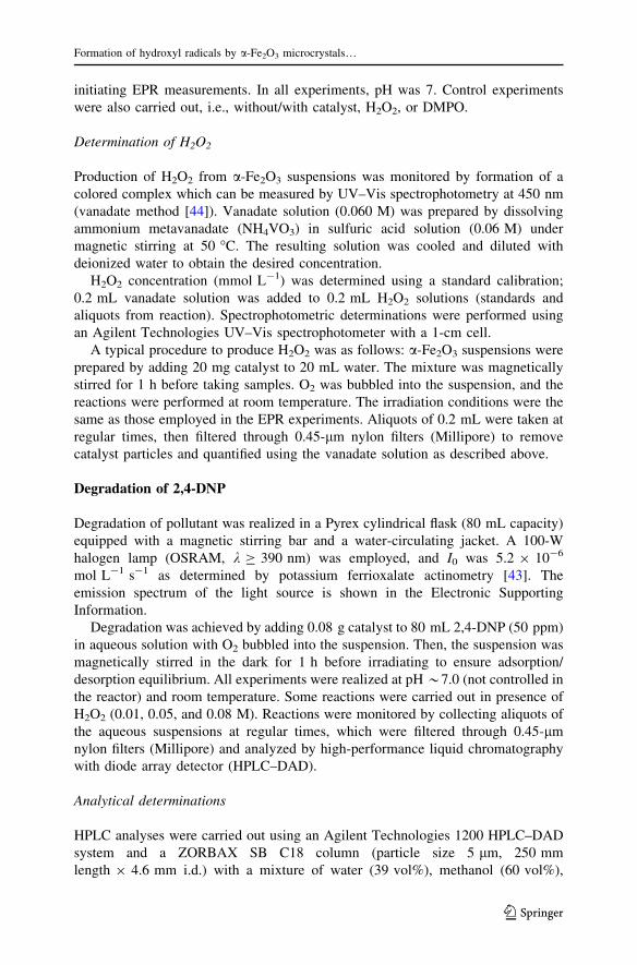
Figure 1a shows the XRD pattern of a-Fe2O3 sample, which can be assigned to
rhombohedral phase (JCPDS no. 33-664). The strong and sharp diffraction peaks
indicate good crystallinity of the synthesized a-Fe2O3 [47]. No other diffraction
peaks were observed, indicating that SiO2 was in amorphous state, as also observed
in a previous report [48].
Figure 1b shows a SEM image of the sample. Hematite single crystals with
elongated shape and average length of 1.15 ± 0.39 lm were obtained upon
annealing at 600 �C. Since the a-Fe2O3 microcrystals were developed in situ with
silica matrix by hydrolysis and condensation of TEOS, the porous silica
nanostructure before the calcination process can be appreciated by SEM (see
Electronic Supporting Information). The chemical composition determined by EDX
analysis confirmed that Si, O, and Fe were the only elements contained within the
catalyst (Fig. 1c). The sample was further examined by TEM (Fig. 1d); the
difference in contrast between darker and lighter areas indicates silica dispersed on
a-Fe2O3 microcrystals.
1 µm
B
0 1 2 3 4 5 6 7Energy keV
C1 µmD
α-Fe2O3
JCPDS 33-664
A
10 20 30 40 50 60 70 80
2-Theta (°)
Inte
nsity
[a.u
.]
Fig. 1 a XRD pattern of a-Fe2O3 prepared in presence of SiO2; red lines indicate positions of standardpeaks of hematite a-Fe2O3 (JCPDS 33-664). b SEM image, c EDX analysis, and d TEM image of a-Fe2O3 microcrystals
Formation of hydroxyl radicals by a-Fe2O3 microcrystals…
123
EPR analysis
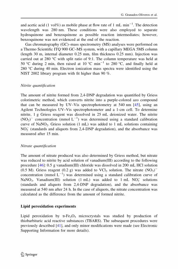
We employed DMPO as a free-radical trap to provide evidence of the formation of
hydroxyl radicals from a-Fe2O3 in oxygenated aqueous suspension, at room
temperature under visible-light irradiation. In dark conditions, no signal was
observed for the catalyst in presence of DMPO (Fig. 2a); however, when H2O2 was
added to the suspension, the DMPO-OH adduct signal was observed, indicating
generation of �OH (Fig. 2b). This signal exhibits four strong splitting lines with
intensity ratio of 1:2:2:1 ratio and hyperfine splitting constant (hfsc) of
aN = aH = 14.9 G and g = 2.0057 [49, 50]. Control experiments showed that
catalyst and H2O2 were indispensable for production of the DMPO-OH adduct
signal. The role of dissolved O2 was evaluated by bubbling nitrogen through the
suspension. In this condition, signal intensity slightly decreased (Fig. 2c), in
comparison with Fig. 2b, suggesting that O2 is not essential to produce �OH.
To confirm �OH formation from catalyst suspensions in presence of H2O2, the
effect of mannitol as �OH scavenger [51] was determined. In this circumstance, the
intensity of DMPO-OH adduct signal was clearly inhibited by mannitol (Fig. 2d),
demonstrating that �OH was effectively formed. These results suggest that H2O2 is
necessarily required to produce �OH by a-Fe2O3 microcrystals, via reactions 6
followed by reaction 2, in accordance with a heterogeneous Fenton reaction [52].
¼Fe3þ þ H2O2 ! ¼Fe2þ þ Hþ þ HO2� ð6Þ
Figure 3 shows the photoinduced formation of �OH by the catalyst. After
irradiation with visible light (k[ 400 nm) for 5 min, oxygenated aqueous
suspension containing a-Fe2O3 particles could produce the DMPO-OH adduct
without H2O2 as prerequisite (Fig. 3a). When N2 was bubbled through the
suspension to remove dissolved oxygen from solution, the DMPO-OH signal was
affected (Fig. 3b). In addition, we evaluated the effect of H2O2 (0.05 M) on
formation of hydroxyl radicals by catalyst, finding that the adduct signal was
dramatically increased (Fig. 3c). No DMPO-OH adduct was produced in presence
of H2O2 but without hematite under irradiation (Fig. 3d).
Fig. 2 EPR spectra of DMPO-OH adduct from a-Fe2O3 inoxygenated aqueous suspensions(1 g L-1) in dark condition andunder several experimentalconditions: a without H2O2,b with H2O2, c with H2O2 andN2, and d in presence ofmannitol
G. Granados-Oliveros et al.
123

Based on all these observations, we hypothesized that photoproduction of �OH by
visible light could occur via two main paths: (1) via heterogeneous photo-Fenton
reaction, which would require H2O2 to produce �OH (reactions 1 and 2), and (2)
formation of O2�- after hematite excitation (reaction 5). These species are very
reactive, and through a disproportionation process produce H2O2 and then �OH
(reactions 7–9) [53]. The strong production of �OH by a-Fe2O3 microcrystals under
visible-light irradiation results by combining these paths.
2O�2� þ 2Hþ ! H2O2 þ O2 ð7Þ
H2O2 þ O�2� ! �OH þ OH� þ O2 ð8Þ
H2O2 þ e� ! �OH þ OH� ð9Þ
Because H2O2 may be produced as an intermediate (reaction 7), we determined
the amount of H2O2 formed in the reaction. Figure 4 shows the formation of
hydrogen peroxide by a-Fe2O3 in dark condition and upon visible-light irradiation
(k[ 400 nm). No formation of H2O2 was detected in dark conditions, but when a-
Fe2O3 dispersions were irradiated, production of H2O2 increased to 28 lM. After
this, the amount of H2O2 decreased, possibly due to reactions 8–9. We emphasize
that the photogenerated H2O2 was sufficient to produce a detectable amount of
Fig. 3 DMPO spin-trappingEPR spectra from a-Fe2O3
microcrystals in oxygenatedaqueous suspensions (1 g L-1)under visible-light irradiation:a without H2O2, b with N2 andwithout H2O2, c with H2O2, andd without catalyst: DMPOalone, DMPO ? H2O2
Formation of hydroxyl radicals by a-Fe2O3 microcrystals…
123
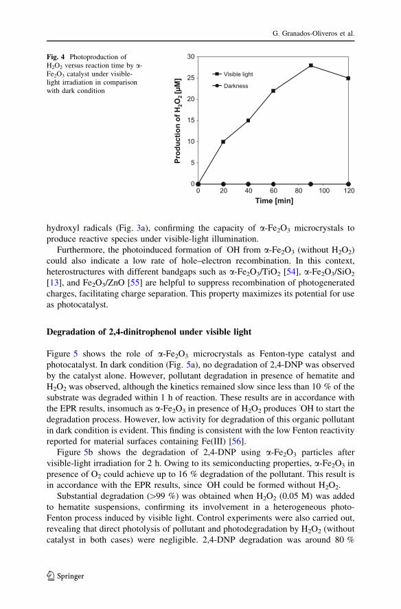
hydroxyl radicals (Fig. 3a), confirming the capacity of a-Fe2O3 microcrystals to
produce reactive species under visible-light illumination.
Furthermore, the photoinduced formation of �OH from a-Fe2O3 (without H2O2)
could also indicate a low rate of hole–electron recombination. In this context,
heterostructures with different bandgaps such as a-Fe2O3/TiO2 [54], a-Fe2O3/SiO2
[13], and Fe2O3/ZnO [55] are helpful to suppress recombination of photogenerated
charges, facilitating charge separation. This property maximizes its potential for use
as photocatalyst.
Degradation of 2,4-dinitrophenol under visible light
Figure 5 shows the role of a-Fe2O3 microcrystals as Fenton-type catalyst and
photocatalyst. In dark condition (Fig. 5a), no degradation of 2,4-DNP was observed
by the catalyst alone. However, pollutant degradation in presence of hematite and
H2O2 was observed, although the kinetics remained slow since less than 10 % of the
substrate was degraded within 1 h of reaction. These results are in accordance with
the EPR results, insomuch as a-Fe2O3 in presence of H2O2 produces �OH to start the
degradation process. However, low activity for degradation of this organic pollutant
in dark condition is evident. This finding is consistent with the low Fenton reactivity
reported for material surfaces containing Fe(III) [56].
Figure 5b shows the degradation of 2,4-DNP using a-Fe2O3 particles after
visible-light irradiation for 2 h. Owing to its semiconducting properties, a-Fe2O3 in
presence of O2 could achieve up to 16 % degradation of the pollutant. This result is
in accordance with the EPR results, since �OH could be formed without H2O2.
Substantial degradation ([99 %) was obtained when H2O2 (0.05 M) was added
to hematite suspensions, confirming its involvement in a heterogeneous photo-
Fenton process induced by visible light. Control experiments were also carried out,
revealing that direct photolysis of pollutant and photodegradation by H2O2 (without
catalyst in both cases) were negligible. 2,4-DNP degradation was around 80 %
0
5
10
15
20
25
30
0 20 40 60 80 100 120
Prod
uctio
n of
H2O
2 [µ
M]
Time [min]
Visible light
Darkness
Fig. 4 Photoproduction ofH2O2 versus reaction time by a-Fe2O3 catalyst under visible-light irradiation in comparisonwith dark condition
G. Granados-Oliveros et al.
123
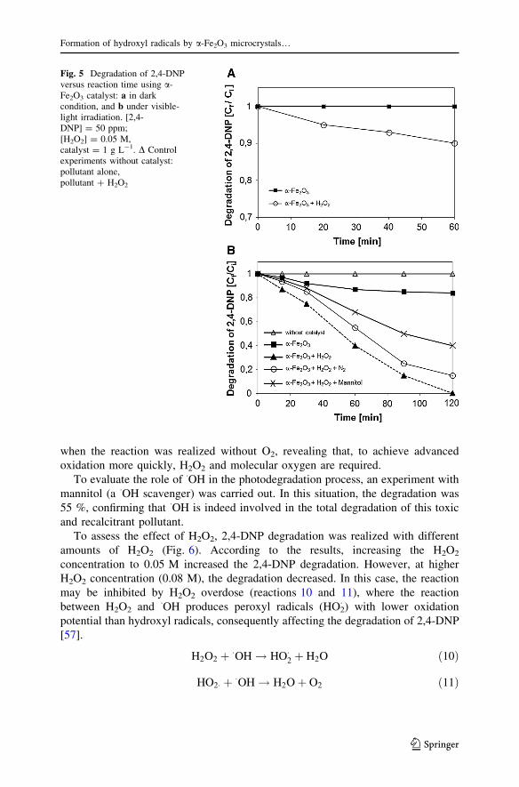
when the reaction was realized without O2, revealing that, to achieve advanced
oxidation more quickly, H2O2 and molecular oxygen are required.
To evaluate the role of �OH in the photodegradation process, an experiment with
mannitol (a �OH scavenger) was carried out. In this situation, the degradation was
55 %, confirming that �OH is indeed involved in the total degradation of this toxic
and recalcitrant pollutant.
To assess the effect of H2O2, 2,4-DNP degradation was realized with different
amounts of H2O2 (Fig. 6). According to the results, increasing the H2O2
concentration to 0.05 M increased the 2,4-DNP degradation. However, at higher
H2O2 concentration (0.08 M), the degradation decreased. In this case, the reaction
may be inhibited by H2O2 overdose (reactions 10 and 11), where the reaction
between H2O2 and �OH produces peroxyl radicals (HO2� ) with lower oxidation
potential than hydroxyl radicals, consequently affecting the degradation of 2,4-DNP
[57].
H2O2 þ �OH ! HO�2 þ H2O ð10Þ
HO2� þ �OH ! H2O þ O2 ð11Þ
Fig. 5 Degradation of 2,4-DNPversus reaction time using a-Fe2O3 catalyst: a in darkcondition, and b under visible-light irradiation. [2,4-DNP] = 50 ppm;[H2O2] = 0.05 M,catalyst = 1 g L-1. D Controlexperiments without catalyst:pollutant alone,pollutant ? H2O2
Formation of hydroxyl radicals by a-Fe2O3 microcrystals…
123
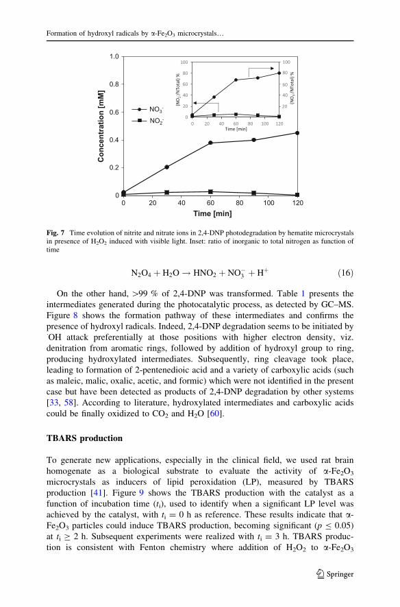
Photoproducts
Hydroxyl radicals can attack 2,4-DNP, leading to release of the nitro group [58].
This was evaluated by the formation of nitrite (NO2-) and nitrate (NO3
-) anions,
which were estimated as the percentage of the initial amount of nitrogen, given
initial concentration of 50 ppm (0.27 mM), i.e., [N]0 = 0.54 mM (Eqs. 12 and 13):
NO�2 in%N ¼ NO�
2
N0
� 100 ð12Þ
NO�3 in%N ¼ NO�
3
N0
� 100 ð13Þ
Figure 7 shows the evolution of NO2- and NO3
- as photoproducts of 2-4-DNP
degradation. According to these results, in dark condition, less than 10 % of 2,4-
DNP was degraded by a-Fe2O3, in presence of H2O2 (0.05 M), so an amount of
NO2- (10 lM) and NO3
- (25 lM) could be detected, evidencing that the 2,4-DNP
degradation process took place. During irradiation, the nitrite concentration
increased to 28 lM, equivalent to 5 % considering the ratio of inorganic to total
nitrogen. Meanwhile, the nitrate concentration reached a maximum of 450 lM,
corresponding to 80.4 % (see inset in Fig. 7) of the nitro group detached. Nitrite
could be transformed into NO3- by secondary reactions, which could explain the
small amount of NO2- formed in the course of the reaction. It is known that nitrites
can act as efficient scavengers of �OH to produce �NO2 species (reaction 14) [59].
Furthermore, �NO2 is not a stable intermediate in aqueous solution, as it undergoes
fast dimerization (reaction 15) and hydrolysis (reaction 16), producing NO3-
[47, 49].
NO�2 þ �OH ! �NO2 þ OH� ð14Þ
2�NO2 �N2O4 ð15Þ
0
20
40
60
80
100
0 0,02 0,04 0,06 0,084,2fo
noitadargeD
-]
%[P
ND
Concentration of H2O2 [M]
Fig. 6 Effect of H2O2
concentration on 2,4-DNPphotodegradation
G. Granados-Oliveros et al.
123
N2O4 þ H2O ! HNO2 þ NO�3 þ Hþ ð16Þ
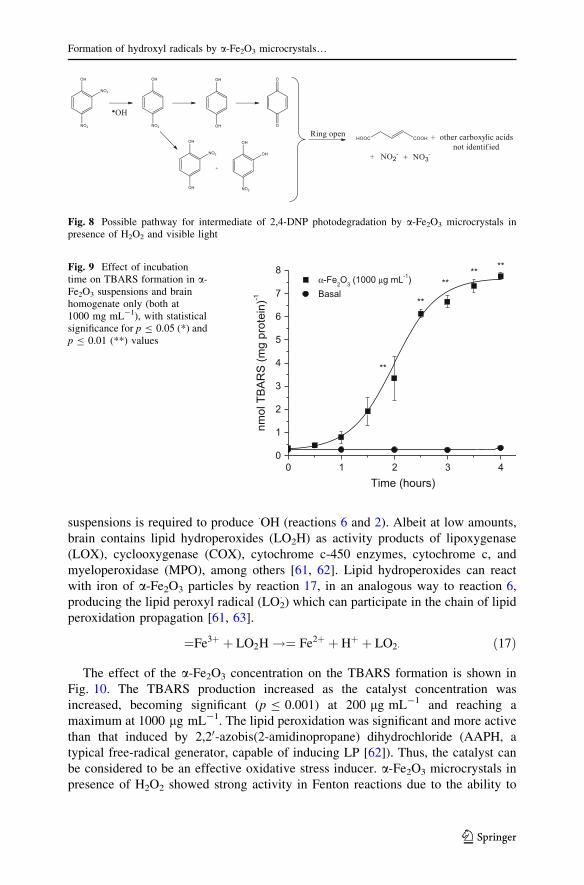
On the other hand, [99 % of 2,4-DNP was transformed. Table 1 presents the
intermediates generated during the photocatalytic process, as detected by GC–MS.
Figure 8 shows the formation pathway of these intermediates and confirms the
presence of hydroxyl radicals. Indeed, 2,4-DNP degradation seems to be initiated by�OH attack preferentially at those positions with higher electron density, viz.
denitration from aromatic rings, followed by addition of hydroxyl group to ring,
producing hydroxylated intermediates. Subsequently, ring cleavage took place,
leading to formation of 2-pentenedioic acid and a variety of carboxylic acids (such
as maleic, malic, oxalic, acetic, and formic) which were not identified in the present
case but have been detected as products of 2,4-DNP degradation by other systems
[33, 58]. According to literature, hydroxylated intermediates and carboxylic acids
could be finally oxidized to CO2 and H2O [60].
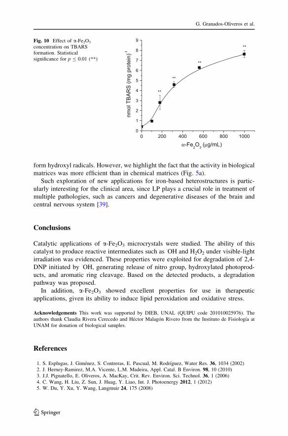
TBARS production
To generate new applications, especially in the clinical field, we used rat brain
homogenate as a biological substrate to evaluate the activity of a-Fe2O3
microcrystals as inducers of lipid peroxidation (LP), measured by TBARS
production [41]. Figure 9 shows the TBARS production with the catalyst as a
function of incubation time (ti), used to identify when a significant LP level was
achieved by the catalyst, with ti = 0 h as reference. These results indicate that a-
Fe2O3 particles could induce TBARS production, becoming significant (p B 0.05)
at ti C 2 h. Subsequent experiments were realized with ti = 3 h. TBARS produc-
tion is consistent with Fenton chemistry where addition of H2O2 to a-Fe2O3
00 20 40 60 80 100 120
noit artnec noC
[ mM
]
Time [min]
0.2
0.4
0.6
0.8
1.0
0
20
40
60
80
100
0 20 40 60 80 100 120
(NO
2- /NTo
tal)
%
Time [min]
(NO
3- /NTo
tal)
%
20
40
60
80
100
NO3-
NO2-
Fig. 7 Time evolution of nitrite and nitrate ions in 2,4-DNP photodegradation by hematite microcrystalsin presence of H2O2 induced with visible light. Inset: ratio of inorganic to total nitrogen as function oftime
Formation of hydroxyl radicals by a-Fe2O3 microcrystals…
123
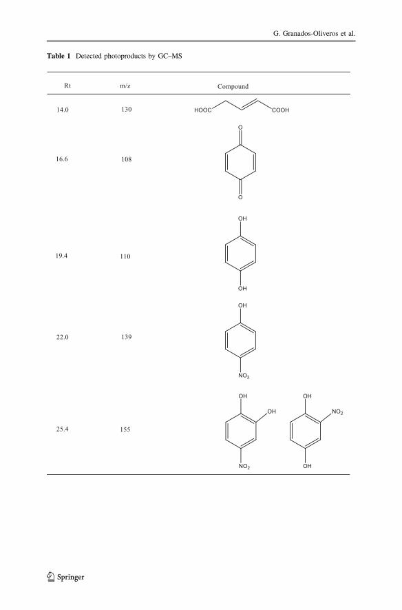
Table 1 Detected photoproducts by GC–MS
G. Granados-Oliveros et al.
123
suspensions is required to produce �OH (reactions 6 and 2). Albeit at low amounts,
brain contains lipid hydroperoxides (LO2H) as activity products of lipoxygenase
(LOX), cyclooxygenase (COX), cytochrome c-450 enzymes, cytochrome c, and
myeloperoxidase (MPO), among others [61, 62]. Lipid hydroperoxides can react
with iron of a-Fe2O3 particles by reaction 17, in an analogous way to reaction 6,
producing the lipid peroxyl radical (LO2� ) which can participate in the chain of lipid
peroxidation propagation [61, 63].
¼Fe3þ þ LO2H !¼ Fe2þ þ Hþ þ LO2� ð17Þ
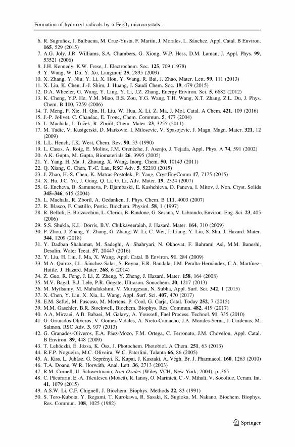
The effect of the a-Fe2O3 concentration on the TBARS formation is shown in
Fig. 10. The TBARS production increased as the catalyst concentration was
increased, becoming significant (p B 0.001) at 200 lg mL-1 and reaching a
maximum at 1000 lg mL-1. The lipid peroxidation was significant and more active
than that induced by 2,20-azobis(2-amidinopropane) dihydrochloride (AAPH, a
typical free-radical generator, capable of inducing LP [62]). Thus, the catalyst can
be considered to be an effective oxidative stress inducer. a-Fe2O3 microcrystals in
presence of H2O2 showed strong activity in Fenton reactions due to the ability to
Fig. 8 Possible pathway for intermediate of 2,4-DNP photodegradation by a-Fe2O3 microcrystals inpresence of H2O2 and visible light
0 1 2 3 40
1
2
3
4
5
6
7
8 ******
**)nietorpg
m(S
RA
BTlomn
-1
Time (hours)
α-Fe2O3 (1000 μg mL-1) Basal
**
Fig. 9 Effect of incubationtime on TBARS formation in a-Fe2O3 suspensions and brainhomogenate only (both at1000 mg mL-1), with statisticalsignificance for p B 0.05 (*) andp B 0.01 (**) values
Formation of hydroxyl radicals by a-Fe2O3 microcrystals…
123
form hydroxyl radicals. However, we highlight the fact that the activity in biological
matrices was more efficient than in chemical matrices (Fig. 5a).
Such exploration of new applications for iron-based heterostructures is partic-
ularly interesting for the clinical area, since LP plays a crucial role in treatment of
multiple pathologies, such as cancers and degenerative diseases of the brain and
central nervous system [39].
Conclusions
Catalytic applications of a-Fe2O3 microcrystals were studied. The ability of this
catalyst to produce reactive intermediates such as �OH and H2O2 under visible-light
irradiation was evidenced. These properties were exploited for degradation of 2,4-
DNP initiated by �OH, generating release of nitro group, hydroxylated photoprod-
ucts, and aromatic ring cleavage. Based on the detected products, a degradation
pathway was proposed.
In addition, a-Fe2O3 showed excellent properties for use in therapeutic
applications, given its ability to induce lipid peroxidation and oxidative stress.
Acknowledgements This work was supported by DIEB, UNAL (QUIPU code 201010025976). The
authors thank Claudia Rivera Cerecedo and Hector Malagon Rivero from the Instituto de Fisiologıa at
UNAM for donation of biological samples.
References
1. S. Esplugas, J. Gimenez, S. Contreras, E. Pascual, M. Rodrıguez, Water Res. 36, 1034 (2002)
2. J. Herney-Ramirez, M.A. Vicente, L.M. Madeira, Appl. Catal. B Environ. 98, 10 (2010)
3. J.J. Pignatello, E. Oliveros, A. MacKay, Crit. Rev. Environ. Sci. Technol. 36, 1 (2006)
4. C. Wang, H. Liu, Z. Sun, J. Huag, Y. Liao, Int. J. Photoenergy 2012, 1 (2012)
5. W. Du, Y. Xu, Y. Wang, Langmuir 24, 175 (2008)
0 200 400 600 800 10000
1
2
3
4
5
6
7
8
9**
**
**
)nietorpg
m(S
RABTlo
mn-1
α-Fe2O3 (μg/mL)
**
Fig. 10 Effect of a-Fe2O3
concentration on TBARSformation. Statisticalsignificance for p B 0.01 (**)
G. Granados-Oliveros et al.
123
6. R. Sugranez, J. Balbuena, M. Cruz-Yusta, F. Martın, J. Morales, L. Sanchez, Appl. Catal. B Environ.
165, 529 (2015)
7. A.G. Joly, J.R. Williams, S.A. Chambers, G. Xiong, W.P. Hess, D.M. Laman, J. Appl. Phys. 99,
53521 (2006)
8. J.H. Kennedy, K.W. Frese, J. Electrochem. Soc. 125, 709 (1978)
9. Y. Wang, W. Du, Y. Xu, Langmuir 25, 2895 (2009)
10. X. Zhang, Y. Niu, Y. Li, X. Hou, Y. Wang, R. Bai, J. Zhao, Mater. Lett. 99, 111 (2013)
11. X. Liu, K. Chen, J.-J. Shim, J. Huang, J. Saudi Chem. Soc. 19, 479 (2015)
12. D.A. Wheeler, G. Wang, Y. Ling, Y. Li, J.Z. Zhang, Energy Environ. Sci. 5, 6682 (2012)
13. K. Cheng, Y.P. He, Y.M. Miao, B.S. Zou, Y.G. Wang, T.H. Wang, X.T. Zhang, Z.L. Du, J. Phys.
Chem. B 110, 7259 (2006)
14. T. Meng, P. Xie, H. Qin, H. Liu, W. Hua, X. Li, Z. Ma, J. Mol. Catal. A Chem. 421, 109 (2016)
15. J.-P. Jolivet, C. Chaneac, E. Tronc, Chem. Commun. 5, 477 (2004)
16. L. Machala, J. Tucek, R. Zboril, Chem. Mater. 23, 3255 (2011)
17. M. Tadic, V. Kusigerski, D. Markovic, I. Milosevic, V. Spasojevic, J. Magn. Magn. Mater. 321, 12
(2009)
18. L.L. Hench, J.K. West, Chem. Rev. 90, 33 (1990)
19. L. Casas, A. Roig, E. Molins, J.M. Greneche, J. Asenjo, J. Tejada, Appl. Phys. A 74, 591 (2002)
20. A.K. Gupta, M. Gupta, Biomaterials 26, 3995 (2005)
21. Y. Yang, H. Ma, J. Zhuang, X. Wang, Inorg. Chem. 50, 10143 (2011)
22. Q. Xiang, G. Chen, T.-C. Lau, RSC Adv. 5, 52210 (2015)
23. J. Zhao, H.-S. Chen, K. Matras-Postolek, P. Yang, CrystEngComm 17, 7175 (2015)
24. X. Hu, J.C. Yu, J. Gong, Q. Li, G. Li, Adv. Mater. 19, 2324 (2007)
25. G. Encheva, B. Samuneva, P. Djambaski, E. Kashchieva, D. Paneva, I. Mitov, J. Non. Cryst. Solids
345–346, 615 (2004)
26. L. Machala, R. Zboril, A. Gedanken, J. Phys. Chem. B 111, 4003 (2007)
27. R. Blasco, F. Castillo, Pestic. Biochem. Physiol. 58, 1 (1997)
28. R. Belloli, E. Bolzacchini, L. Clerici, B. Rindone, G. Sesana, V. Librando, Environ. Eng. Sci. 23, 405
(2006)
29. S.S. Shukla, K.L. Dorris, B.V. Chikkaveeraiah, J. Hazard. Mater. 164, 310 (2009)
30. P. Zhou, J. Zhang, Y. Zhang, G. Zhang, W. Li, C. Wei, J. Liang, Y. Liu, S. Shu, J. Hazard. Mater.
344, 1209 (2018)
31. Y. Dadban Shahamat, M. Sadeghi, A. Shahryari, N. Okhovat, F. Bahrami Asl, M.M. Baneshi,
Desalin. Water Treat. 57, 20447 (2016)
32. Y. Liu, H. Liu, J. Ma, X. Wang, Appl. Catal. B Environ. 91, 284 (2009)
33. M.A. Quiroz, J.L. Sanchez-Salas, S. Reyna, E.R. Bandala, J.M. Peralta-Hernandez, C.A. Martınez-
Huitle, J. Hazard. Mater. 268, 6 (2014)
34. Z. Guo, R. Feng, J. Li, Z. Zheng, Y. Zheng, J. Hazard. Mater. 158, 164 (2008)
35. M.V. Bagal, B.J. Lele, P.R. Gogate, Ultrason. Sonochem. 20, 1217 (2013)
36. M. Myilsamy, M. Mahalakshmi, V. Murugesan, N. Subha, Appl. Surf. Sci. 342, 1 (2015)
37. X. Chen, Y. Liu, X. Xia, L. Wang, Appl. Surf. Sci. 407, 470 (2017)
38. E.M. Seftel, M. Puscasu, M. Mertens, P. Cool, G. Carja, Catal. Today 252, 7 (2015)
39. M.M. Gaschler, B.R. Stockwell, Biochem. Biophys. Res. Commun. 482, 419 (2017)
40. A.A. Mirzaei, A.B. Babaei, M. Galavy, A. Youssefi, Fuel Process. Technol. 91, 335 (2010)
41. G. Granados-Oliveros, V. Gomez-Vidales, A. Nieto-Camacho, J.A. Morales-Serna, J. Cardenas, M.
Salmon, RSC Adv. 3, 937 (2013)
42. G. Granados-Oliveros, E.A. Paez-Mozo, F.M. Ortega, C. Ferronato, J.M. Chovelon, Appl. Catal.
B Environ. 89, 448 (2009)
43. T. Lehoczki, E. Jozsa, K. Osz, J. Photochem. Photobiol. A Chem. 251, 63 (2013)
44. R.F.P. Nogueira, M.C. Oliveira, W.C. Paterlini, Talanta 66, 86 (2005)
45. A. Kiss, L. Juhasz, G. Seprenyi, K. Kupai, J. Kaszaki, A. Vegh, Br. J. Pharmacol. 160, 1263 (2010)
46. T.A. Doane, W.R. Horwath, Anal. Lett. 36, 2713 (2003)
47. R.M. Cornell, U. Schwertmann, Iron Oxides (Wiley-VCH, New York, 2004), p. 365
48. C. Pacurariu, E.-A. Taculescu (Moaca), R. Ianos, O. Marinica, C.-V. Mihali, V. Socoliuc, Ceram. Int.
41, 1079 (2015)
49. A.S.W. Li, C.F. Chignell, J. Biochem. Biophys. Methods 22, 83 (1991)
50. S. Tero-Kubota, Y. Ikegami, T. Kurokawa, R. Sasaki, K. Sugioka, M. Nakano, Biochem. Biophys.
Res. Commun. 108, 1025 (1982)
Formation of hydroxyl radicals by a-Fe2O3 microcrystals…
123
51. K.K. Mothilal, J. Johnson Inbaraj, R. Gandhidasan, R. Murugesan, J. Photochem. Photobiol. A Chem.
162, 9 (2004)
52. C. Hammond, M.M. Forde, M.H. Ab Rahim, A. Thetford, Q. He, R.L. Jenkins, N. Dimitratos, J.A.
Lopez-Sanchez, N.F. Dummer, D.M. Murphy, A.F. Carley, S.H. Taylor, D.J. Willock, E.E. Stang-
land, J. Kang, H. Hagen, C.J. Kiely, G.J. Hutchings, Angew. Chem. Int. Ed. 51, 5129 (2012)
53. P. Pichat, C. Guillard, L. Amalric, A.-C. Renard, O. Plaidy, Sol. Energy Mater. Sol. Cells 38, 391
(1995)
54. X. Zhang, L. Lei, Appl. Surf. Sci. 254, 2406 (2008)
55. S. Si, C. Li, X. Wang, Q. Peng, Y. Li, Sensors Actuators B Chem. 119, 52 (2006)
56. W. Huang, M. Brigante, F. Wu, K. Hanna, G. Mailhot, Environ. Sci. Pollut. Res. 20, 39 (2013)
57. I. Muthuvel, M. Swaminathan, Sol. Energy Mater. Sol. Cells 92, 857 (2008)
58. Y. Liu, H. Liu, J. Ma, X. Wang, Appl. Catal. B Environ. 91, 284 (2009)
59. J.A. Herrera-Melian, A.J. Martın-Rodrıguez, A. Ortega-Mendez, J. Arana, J.M. Dona-Rodrıguez, J.
Perez-Pena, J. Environ. Manag. 105, 53 (2012)
60. L. Demarchis, M. Minella, R. Nistico, V. Maurino, C. Minero, D. Vione, J. Photochem. Photobiol.
A Chem. 307–308, 99 (2015)
61. T.S. Anthonymuthu, E.M. Kenny, H. Bayır, Brain Res. 1640, 57 (2016)
62. E. Niki, Free Radic. Biol. Med. 47, 469 (2009)
63. Z. Cheng, Y. Li, Chem. Rev. 107, 2165 (2007)
G. Granados-Oliveros et al.
123