Food protein-stabilized nanoemulsions as potential ... · ... exhibited better stability and...
13
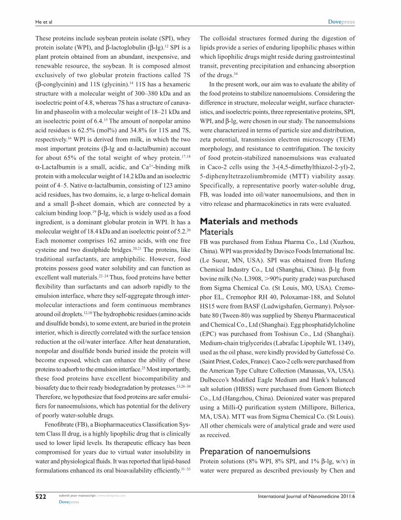
© 2011 He et al, publisher and licensee Dove Medical Press Ltd. This is an Open Access article which permits unrestricted noncommercial use, provided the original work is properly cited. International Journal of Nanomedicine 2011:6 521–533 International Journal of Nanomedicine Dovepress submit your manuscript | www.dovepress.com Dovepress 521 ORIGINAL RESEARCH open access to scientific and medical research Open Access Full Text Article DOI: 10.2147/IJN.S17282 Food protein-stabilized nanoemulsions as potential delivery systems for poorly water-soluble drugs: preparation, in vitro characterization, and pharmacokinetics in rats Wei He 1 Yanan Tan 1 Zhiqiang Tian 1 Lingyun Chen 2 Fuqiang Hu 3 Wei Wu 1 1 Department of Pharmaceutics, School of Pharmacy, Fudan University, Shanghai, People’s Republic of China; 2 Department of Agricultural, Food and Nutritional Sciences, University of Alberta, Alberta, Canada; 3 Department of Pharmaceutics, School of Pharmacy, Zhejiang University, Hangzhou, Zhejiang, People’s Republic of China Correspondence: Wei Wu Department of Pharmaceutics, School of Pharmacy, Fudan University, Shanghai 201203, People’s Republic of China Tel/Fax +86 21 5198 0002 Email [email protected] Abstract: Nanoemulsions stabilized by traditional emulsifiers raise toxicological concerns for long-term treatment. The present work investigates the potential of food proteins as safer sta- bilizers for nanoemulsions to deliver hydrophobic drugs. Nanoemulsions stabilized by food proteins (soybean protein isolate, whey protein isolate, β-lactoglobulin) were prepared by high- pressure homogenization. The toxicity of the nanoemulsions was tested in Caco-2 cells using the 3-(4,5-dimethylthiazol-2-yl)-2,5-diphenyltetrazoliumbromide viability assay. In vivo absorp- tion in rats was also evaluated. Food protein-stabilized nanoemulsions, with small particle size and good size distribution, exhibited better stability and biocompatibility compared with nano- emulsions stabilized by traditional emulsifiers. Moreover, β-lactoglobulin had a better emulsify- ing capacity and biocompatibility than the other two food proteins. The pancreatic degradation of the proteins accelerated drug release. It is concluded that an oil/water nanoemulsion system with good biocompatibility can be prepared by using food proteins as emulsifiers, allowing better and more rapid absorption of lipophilic drugs. Keywords: oil in water nanoemulsions, food proteins, poorly water-soluble drugs, biocompatibility, in vivo absorption Introduction Nanoemulsions are nonequilibrium, heterogeneous systems consisting of two immis- cible liquids in which one liquid is dispersed in another liquid as droplets with diameters of tens to a few hundred nanometers. Oil/water nanoemulsions have great potential for the delivery of poorly water-soluble drugs. 1–3 The major advantages of nanoemul- sions as drug delivery carriers include ease of fabrication, increased drug loading, enhanced drug solubility and bioavailability, reduced patient variability, controlled drug release, and protection from enzymatic degradation. 1,4 To stabilize nanoemulsions, a large amount of surfactant (20%–30% based on the oil phase, wt%) must be used in the formulations, which hinders the therapeutic application of nanoemulsions due to toxicological concerns during long-term treatment. 5–8 Another main problem with nanoemulsions is their thermodynamic instability, resulting in aggregation and floc- culation; furthermore, loading a drug into a nanoemulsion system can cause droplet coalescence and even phase separation. 9–11 Therefore, it is necessary to develop stable nanoemulsions using alternative safer surfactants. Food biopolymers, especially food proteins, are widely used in formulated foods because they have high nutritional value and are generally recognized as safe. 12,13
Transcript of Food protein-stabilized nanoemulsions as potential ... · ... exhibited better stability and...
© 2011 He et al, publisher and licensee Dove Medical Press Ltd. This is an Open Access article which permits unrestricted noncommercial use, provided the original work is properly cited.
International Journal of Nanomedicine 2011:6 521–533
International Journal of Nanomedicine Dovepress
submit your manuscript | www.dovepress.com
Dovepress 521
O r I g I N A L r e s e A r c H
open access to scientific and medical research
Open Access Full Text Article
DOI: 10.2147/IJN.S17282
Food protein-stabilized nanoemulsions as potential delivery systems for poorly water-soluble drugs: preparation, in vitro characterization, and pharmacokinetics in rats
Wei He1
Yanan Tan1
Zhiqiang Tian1
Lingyun chen2
Fuqiang Hu3
Wei Wu1
1Department of Pharmaceutics, school of Pharmacy, Fudan University, shanghai, People’s republic of china; 2Department of Agricultural, Food and Nutritional sciences, University of Alberta, Alberta, canada; 3Department of Pharmaceutics, school of Pharmacy, Zhejiang University, Hangzhou, Zhejiang, People’s republic of china
correspondence: Wei WuDepartment of Pharmaceutics, school of Pharmacy, Fudan University, shanghai 201203, People’s republic of chinaTel/Fax +86 21 5198 0002email [email protected]
Abstract: Nanoemulsions stabilized by traditional emulsifiers raise toxicological concerns for
long-term treatment. The present work investigates the potential of food proteins as safer sta-
bilizers for nanoemulsions to deliver hydrophobic drugs. Nanoemulsions stabilized by food
proteins (soybean protein isolate, whey protein isolate, β-lactoglobulin) were prepared by high-
pressure homogenization. The toxicity of the nanoemulsions was tested in Caco-2 cells using
the 3-(4,5-dimethylthiazol-2-yl)-2,5-diphenyltetrazoliumbromide viability assay. In vivo absorp-
tion in rats was also evaluated. Food protein-stabilized nanoemulsions, with small particle size
and good size distribution, exhibited better stability and biocompatibility compared with nano-
emulsions stabilized by traditional emulsifiers. Moreover, β-lactoglobulin had a better emulsify-
ing capacity and biocompatibility than the other two food proteins. The pancreatic degradation
of the proteins accelerated drug release. It is concluded that an oil/water nanoemulsion system
with good biocompatibility can be prepared by using food proteins as emulsifiers, allowing
better and more rapid absorption of lipophilic drugs.
Keywords: oil in water nanoemulsions, food proteins, poorly water-soluble drugs,
biocompatibility, in vivo absorption
IntroductionNanoemulsions are nonequilibrium, heterogeneous systems consisting of two immis-
cible liquids in which one liquid is dispersed in another liquid as droplets with diameters
of tens to a few hundred nanometers. Oil/water nanoemulsions have great potential
for the delivery of poorly water-soluble drugs.1–3 The major advantages of nanoemul-
sions as drug delivery carriers include ease of fabrication, increased drug loading,
enhanced drug solubility and bioavailability, reduced patient variability, controlled
drug release, and protection from enzymatic degradation.1,4 To stabilize nanoemulsions,
a large amount of surfactant (20%–30% based on the oil phase, wt%) must be used
in the formulations, which hinders the therapeutic application of nanoemulsions due
to toxicological concerns during long-term treatment.5–8 Another main problem with
nanoemulsions is their thermodynamic instability, resulting in aggregation and floc-
culation; furthermore, loading a drug into a nanoemulsion system can cause droplet
coalescence and even phase separation.9–11 Therefore, it is necessary to develop stable
nanoemulsions using alternative safer surfactants.
Food biopolymers, especially food proteins, are widely used in formulated foods
because they have high nutritional value and are generally recognized as safe.12,13
International Journal of Nanomedicine 2011:6submit your manuscript | www.dovepress.com
Dovepress
Dovepress
522
He et al
These proteins include soybean protein isolate (SPI), whey
protein isolate (WPI), and β-lactoglobulin (β-lg).12 SPI is a
plant protein obtained from an abundant, inexpensive, and
renewable resource, the soybean. It is composed almost
exclusively of two globular protein fractions called 7S
(β-conglycinin) and 11S (glycinin).14 11S has a hexameric
structure with a molecular weight of 300–380 kDa and an
isoelectric point of 4.8, whereas 7S has a structure of canava-
lin and phaseolin with a molecular weight of 18–21 kDa and
an isoelectric point of 6.4.15 The amount of nonpolar amino
acid residues is 62.5% (mol%) and 34.8% for 11S and 7S,
respectively.16 WPI is derived from milk, in which the two
most important proteins (β-lg and α-lactalbumin) account
for about 65% of the total weight of whey protein.17,18
α-Lactalbumin is a small, acidic, and Ca2+-binding milk
protein with a molecular weight of 14.2 kDa and an isoelectric
point of 4–5. Native α-lactalbumin, consisting of 123 amino
acid residues, has two domains, ie, a large α-helical domain
and a small β-sheet domain, which are connected by a
calcium binding loop.19 β-lg, which is widely used as a food
ingredient, is a dominant globular protein in WPI. It has a
molecular weight of 18.4 kDa and an isoelectric point of 5.2.20
Each monomer comprises 162 amino acids, with one free
cysteine and two disulphide bridges.20,21 The proteins, like
traditional surfactants, are amphiphilic. However, food
proteins possess good water solubility and can function as
excellent wall materials.22–24 Thus, food proteins have better
flexibility than surfactants and can adsorb rapidly to the
emulsion interface, where they self-aggregate through inter-
molecular interactions and form continuous membranes
around oil droplets.12,18 The hydrophobic residues (amino acids
and disulfide bonds), to some extent, are buried in the protein
interior, which is directly correlated with the surface tension
reduction at the oil/water interface. After heat denaturation,
nonpolar and disulfide bonds buried inside the protein will
become exposed, which can enhance the ability of these
proteins to adsorb to the emulsion interface.25 Most importantly,
these food proteins have excellent biocompatibility and
biosafety due to their ready biodegradation by proteases.13,26–30
Therefore, we hypothesize that food proteins are safer emulsi-
fiers for nanoemulsions, which has potential for the delivery
of poorly water-soluble drugs.
Fenofibrate (FB), a Biopharmaceutics Classification Sys-
tem Class II drug, is a highly lipophilic drug that is clinically
used to lower lipid levels. Its therapeutic efficacy has been
compromised for years due to virtual water insolubility in
water and physiological fluids. It was reported that lipid-based
formulations enhanced its oral bioavailability efficiently.31–33
The colloidal structures formed during the digestion of
lipids provide a series of enduring lipophilic phases within
which lipophilic drugs might reside during gastrointestinal
transit, preventing precipitation and enhancing absorption
of the drugs.34
In the present work, our aim was to evaluate the ability of
the food proteins to stabilize nanoemulsions. Considering the
difference in structure, molecular weight, surface character-
istics, and isoelectric points, three representative proteins, SPI,
WPI, and β-lg, were chosen in our study. The nanoemulsions
were characterized in terms of particle size and distribution,
zeta potential, transmission electron microscopy (TEM)
morphology, and resistance to centrifugation. The toxicity
of food protein-stabilized nanoemulsions was evaluated
in Caco-2 cells using the 3-(4,5-dimethylthiazol-2-yl)-2,
5-diphenyltetrazoliumbromide (MTT) viability assay.
Specifically, a representative poorly water-soluble drug,
FB, was loaded into oil/water nanoemulsions, and then in
vitro release and pharmacokinetics in rats were evaluated.
Materials and methodsMaterialsFB was purchased from Enhua Pharma Co., Ltd (Xuzhou,
China). WPI was provided by Davisco Foods International Inc.
(Le Sueur, MN, USA). SPI was obtained from Hufeng
Chemical Industry Co., Ltd (Shanghai, China). β-lg from
bovine milk (No. L3908, .90% purity grade) was purchased
from Sigma Chemical Co. (St Louis, MO, USA). Cremo-
phor EL, Cremophor RH 40, Poloxamar-188, and Solutol
HS15 were from BASF (Ludwigshafen, Germany). Polysor-
bate 80 (Tween-80) was supplied by Shenyu Pharmaceutical
and Chemical Co., Ltd (Shanghai). Egg phosphatidylcholine
(EPC) was purchased from Toshisun Co., Ltd (Shanghai).
Medium-chain triglycerides (Labrafac Lipophile WL 1349),
used as the oil phase, were kindly provided by Gattefossé Co.
(Saint Priest, Cedex, France). Caco-2 cells were purchased from
the American Type Culture Collection (Manassas, VA, USA).
Dulbecco’s Modified Eagle Medium and Hank’s balanced
salt solution (HBSS) were purchased from Genom Biotech
Co., Ltd (Hangzhou, China). Deionized water was prepared
using a Milli-Q purification system (Millipore, Billerica,
MA, USA). MTT was from Sigma Chemical Co. (St Louis).
All other chemicals were of analytical grade and were used
as received.
Preparation of nanoemulsionsProtein solutions (8% WPI, 8% SPI, and 1% β-lg, w/v) in
water were prepared as described previously by Chen and
International Journal of Nanomedicine 2011:6 submit your manuscript | www.dovepress.com
Dovepress
Dovepress
523
Protein-stabilized nanoemulsions for drug delivery
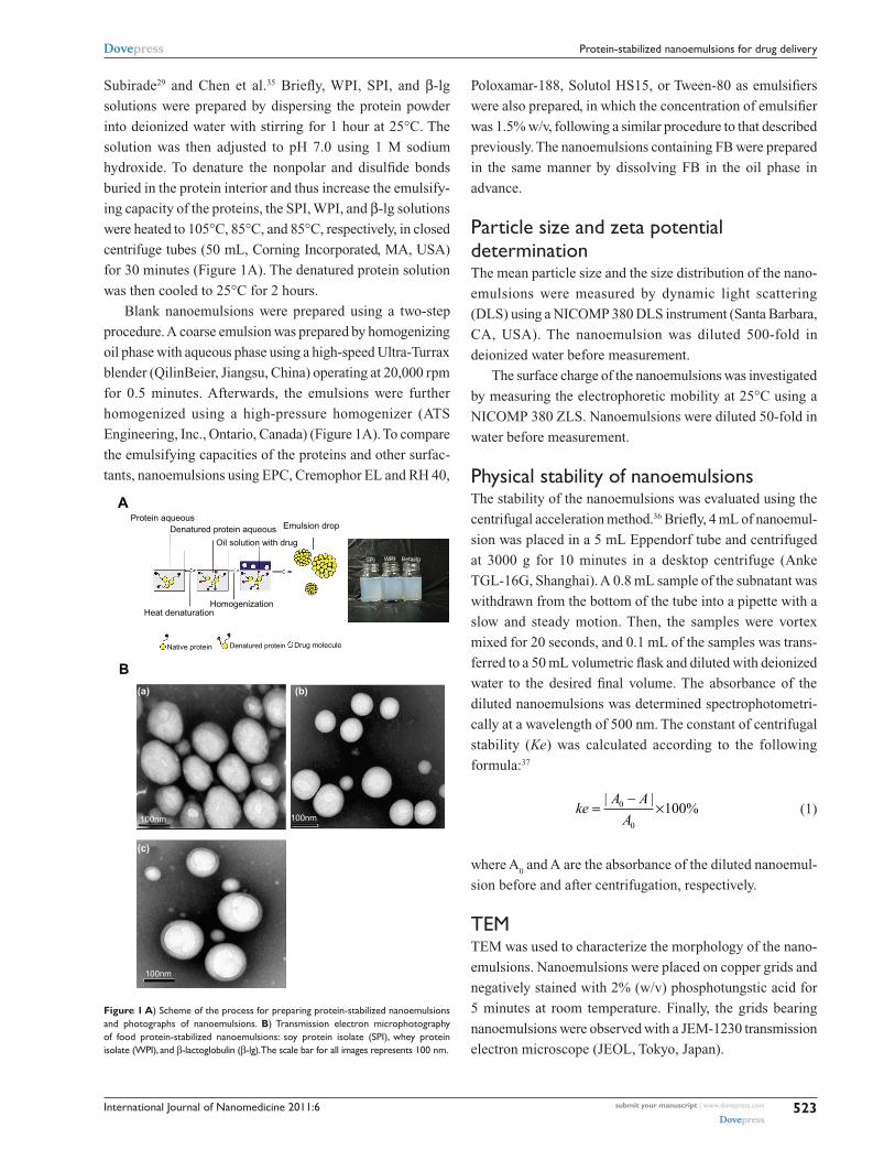
Subirade29 and Chen et al.35 Briefly, WPI, SPI, and β-lg
solutions were prepared by dispersing the protein powder
into deionized water with stirring for 1 hour at 25°C. The
solution was then adjusted to pH 7.0 using 1 M sodium
hydroxide. To denature the nonpolar and disulfide bonds
buried in the protein interior and thus increase the emulsify-
ing capacity of the proteins, the SPI, WPI, and β-lg solutions
were heated to 105°C, 85°C, and 85°C, respectively, in closed
centrifuge tubes (50 mL, Corning Incorporated, MA, USA)
for 30 minutes (Figure 1A). The denatured protein solution
was then cooled to 25°C for 2 hours.
Blank nanoemulsions were prepared using a two-step
procedure. A coarse emulsion was prepared by homogenizing
oil phase with aqueous phase using a high-speed Ultra-Turrax
blender (QilinBeier, Jiangsu, China) operating at 20,000 rpm
for 0.5 minutes. Afterwards, the emulsions were further
homogenized using a high-pressure homogenizer (ATS
Engineering, Inc., Ontario, Canada) (Figure 1A). To compare
the emulsifying capacities of the proteins and other surfac-
tants, nanoemulsions using EPC, Cremophor EL and RH 40,
Poloxamar-188, Solutol HS15, or Tween-80 as emulsifiers
were also prepared, in which the concentration of emulsifier
was 1.5% w/v, following a similar procedure to that described
previously. The nanoemulsions containing FB were prepared
in the same manner by dissolving FB in the oil phase in
advance.
Particle size and zeta potential determinationThe mean particle size and the size distribution of the nano-
emulsions were measured by dynamic light scattering
(DLS) using a NICOMP 380 DLS instrument (Santa Barbara,
CA, USA). The nanoemulsion was diluted 500-fold in
deionized water before measurement.
The surface charge of the nanoemulsions was investigated
by measuring the electrophoretic mobility at 25°C using a
NICOMP 380 ZLS. Nanoemulsions were diluted 50-fold in
water before measurement.
Physical stability of nanoemulsionsThe stability of the nanoemulsions was evaluated using the
centrifugal acceleration method.36 Briefly, 4 mL of nanoemul-
sion was placed in a 5 mL Eppendorf tube and centrifuged
at 3000 g for 10 minutes in a desktop centrifuge (Anke
TGL-16G, Shanghai). A 0.8 mL sample of the subnatant was
withdrawn from the bottom of the tube into a pipette with a
slow and steady motion. Then, the samples were vortex
mixed for 20 seconds, and 0.1 mL of the samples was trans-
ferred to a 50 mL volumetric flask and diluted with deionized
water to the desired final volume. The absorbance of the
diluted nanoemulsions was determined spectrophotometri-
cally at a wavelength of 500 nm. The constant of centrifugal
stability (Ke) was calculated according to the following
formula:37
keA A
A=
-×
| |%0
0
100 (1)
where A0 and A are the absorbance of the diluted nanoemul-
sion before and after centrifugation, respectively.
TeMTEM was used to characterize the morphology of the nano-
emulsions. Nanoemulsions were placed on copper grids and
negatively stained with 2% (w/v) phosphotungstic acid for
5 minutes at room temperature. Finally, the grids bearing
nanoemulsions were observed with a JEM-1230 transmission
electron microscope (JEOL, Tokyo, Japan).
Protein aqueous
A
Heat denaturation
Native protein Denatured protein Drug molecule
Homogenization
Emulsion drop
SPI WPI Beta-Ig
Denatured protein aqueous
Oil solution with drug
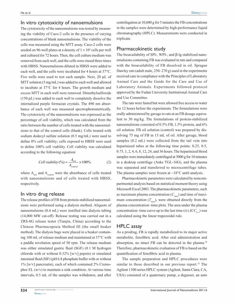
Figure 1 A) scheme of the process for preparing protein-stabilized nanoemulsions and photographs of nanoemulsions. B) Transmission electron microphotography of food protein-stabilized nanoemulsions: soy protein isolate (sPI), whey protein isolate (WPI), and β-lactoglobulin (β-lg). The scale bar for all images represents 100 nm.
(a) (b)
(c)
100nm
100nm
100nm
B
International Journal of Nanomedicine 2011:6submit your manuscript | www.dovepress.com
Dovepress
Dovepress
524
He et al
In vitro cytotoxicity of nanoemulsionsThe cytotoxicity of the nanoemulsions was tested by measur-
ing the viability of Caco-2 cells in the presence of varying
concentrations of blank nanoemulsions. The viability of the
cells was measured using the MTT assay. Caco-2 cells were
seeded on 96-well plates at a density of 1 × 10⁴ cells per well
and cultured for 72 hours. Then, the cell culture medium was
removed from each well, and the cells were rinsed three times
with HBSS. Nanoemulsions diluted in HBSS were added to
each well, and the cells were incubated for 4 hours at 37°C.
Five wells were used to test each sample. Next, 20 µL of
MTT solution (5 mg/mL) was added to each well and allowed
to incubate at 37°C for 4 hours. The growth medium and
excess MTT in each well were removed. Dimethylsulfoxide
(150 µL) was added to each well to completely dissolve the
internalized purple formazan crystals. The 490 nm absor-
bance of each well was measured spectrophotometrically.
The cytotoxicity of the nanoemulsions was expressed as the
percentage of cell viability, which was calculated from the
ratio between the number of cells treated with the nanoemul-
sions to that of the control cells (blank). Cells treated with
sodium dodecyl sulfate solution (0.5 mg/mL) were used to
define 0% cell viability; cells exposed to HBSS were used
to define 100% cell viability. Cell viability was calculated
according to the following equation:
Cell viabilityA
Atest
control
(%) %= ×100 (2)
where Atest
and Acontrol
were the absorbance of cells treated
with nanoemulsions and of cells treated with HBSS,
respectively.
In vitro drug releaseThe release profiles of FB from protein-stabilized nanoemul-
sions were performed using a dialysis method. Aliquots of
nanoemulsions (0.4 mL) were instilled into dialysis tubing
(14,000 MW cut-off). Release testing was carried out in a
ZRS-8G release tester (Tianjin, China) according to the
Chinese Pharmacopoeia Method III (the small beaker
method). The dialysis bags were placed in a beaker contain-
ing 100 mL of release medium and maintained at 37°C with
a paddle revolution speed of 50 rpm. The release medium
was either simulated gastric fluid (SGF) (0.1 M hydrogen
chloride with or without 0.32% [w/v] pepsin) or simulated
intestinal fluid (SIF) (pH 6.8 phosphate buffer with or without
1% [w/v] pancreatin), each of which contained 2% Cremo-
phor EL (w/v) to maintain a sink condition. At various time
intervals, 0.5 mL of the samples was withdrawn, and after
centrifugation at 10,600 g for 5 minutes the FB concentrations
in the samples were determined by high-performance liquid
chromatography (HPLC). Measurements were conducted in
triplicate.
Pharmacokinetic studyThe bioavailability of SPI-, WPI-, and β-lg-stabilized nano-
emulsions containing FB was evaluated in rats and compared
with the bioavailability of FB dissolved in oil. Sprague
Dawley rats (adult male, 250–270 g) used in the experiments
received care in compliance with the Principles of Laboratory
Animal Care and the Guide for the Care and Use of
Laboratory Animals. Experiments followed protocol
approved by the Fudan University Institutional Animal Care
and Use Committee.
The rats were fasted but were allowed free access to water
for 12 hours before the experiments. The formulations were
orally administered by gavage to rats at an FB dosage equiva-
lent to 30 mg/kg. The formulations of protein-stabilized
nanoemulsions consisted of 0.5% FB, 1.5% protein, and 6%
oil solution. FB oil solution (control) was prepared by dis-
solving 75 mg of FB in 15 mL of oil. After gavage, blood
samples (0.2 mL) were collected from the tail vein into
heparinized tubes at the following time points: 0.25, 0.5,
0.75, 1, 2, 4, 6, 8, 12, 24, and 36 hours. The heparinized blood
samples were immediately centrifuged at 3000 g for 10 minutes
in a desktop centrifuge (Anke TGL-16G), and the plasma
was separated and transferred to microcentrifuge tubes.
The plasma samples were frozen at -18°C until analysis.
Pharmacokinetic parameters were calculated by noncom-
partmental analysis based on statistical moment theory using
Microsoft Excel 2003. The pharmacokinetic parameters, such
as maximum plasma concentration (Cmax
) and time of maxi-
mum concentration (Tmax
), were obtained directly from the
plasma concentration–time plots. The area under the plasma
concentration–time curve up to the last time (t) (AUC0–t
) was
calculated using the linear trapezoidal rule.
HPLc assayAs a prodrug, FB is rapidly metabolized to its major active
metabolite, fenofibric acid. After oral administration and
absorption, no intact FB can be detected in the plasma.38
Therefore, pharmacokinetic evaluation of FB is based on the
quantification of fenofibric acid in plasma.
The sample preparation and HPLC procedures were
similar to those described in our previous report.32 The
Agilent 1100 series HPLC system (Agilent, Santa Clara, CA,
USA) consisted of a quaternary pump, a degasser, an auto

International Journal of Nanomedicine 2011:6 submit your manuscript | www.dovepress.com
Dovepress
Dovepress
525
Protein-stabilized nanoemulsions for drug delivery
sampler, a column heater, and a tunable ultraviolet detector.
Fenofibric acid was separated at 30°C using a C18 column
(Diamonsil, 5 µm, 4.6 mm × 250 mm, Dikma, China) with
a refillable C18 precolumn (2.0 mm × 20 mm, Alltech,
Lexington, KT, USA) and detected by measuring the 287 nm
absorbance of the eluate. Indomethacin (100 µg/mL) was
used as an internal standard. The mobile phase was a mixture
of methanol/water/phosphoric acid (70:30:0.1, v/v/v) pumped
at a flow rate of 1 mL/min.
statistical analysisThe results were expressed as mean ± standard deviation.
One-way analysis of variance was performed to assess the
statistical significance of differences among samples. Results
with P , 0.05 were considered statistically significant.
Results and discussionPreparation and characterization of nanoemulsionsWPI and β-lg dissolved rapidly in water yielding homoge-
nous and clear solutions, whereas SPI dissolved partially in
water yielding a turbid suspension. After heat denaturation,
WPI and β-lg solutions remained clear, whereas the viscosity
of the SPI dispersion decreased significantly. The heating
procedure was intended to improve the emulsifying capacity
of the proteins by breaking the disulfide bonds buried inside
the protein.25 The food protein-stabilized nanoemulsions were
prepared using a combination of mechanical mixing and
high-pressure homogenization (Figure 1A). The coarse
emulsions prior to homogenization were typically a few
microns in size, yet they exhibited a wide distribution under
photomicroscopy. After homogenization, milky white nano-
emulsions were obtained (Figure 1A) and the particle size
was reduced dramatically. After homogenization at 800 bars
for 10 cycles, the particle size of the nanoemulsions was
further reduced to between 200 nm and 250 nm with a log-
normal distribution. No drug precipitation was observed
during the preparation of protein-stabilized nanoemulsions
containing FB. Furthermore, an increase of drug in formula-
tions would not result in drug precipitation. TEM photo-
graphs of the food protein-stabilized nanoemulsions revealed
a spherical morphology with particle diameters ranging from
150 nm to 250 nm (Figure 1B), closely corresponding to the
results obtained by DLS.
It is well known that particle size and distribution, as
well as physical stability, exert significant influence on the
bioavailability of nanoemulsions. In this study, we investi-
gated the effects of homogenization pressure and number
of homogenization cycles, protein concentration, oil-to-water
ratio, and pH on particle size, polydispersity index (PI),
and physical stability of the nanoemulsions. Furthermore,
a comparison of the emulsifying capacity of the three
food proteins with that of several surfactants was also
performed.
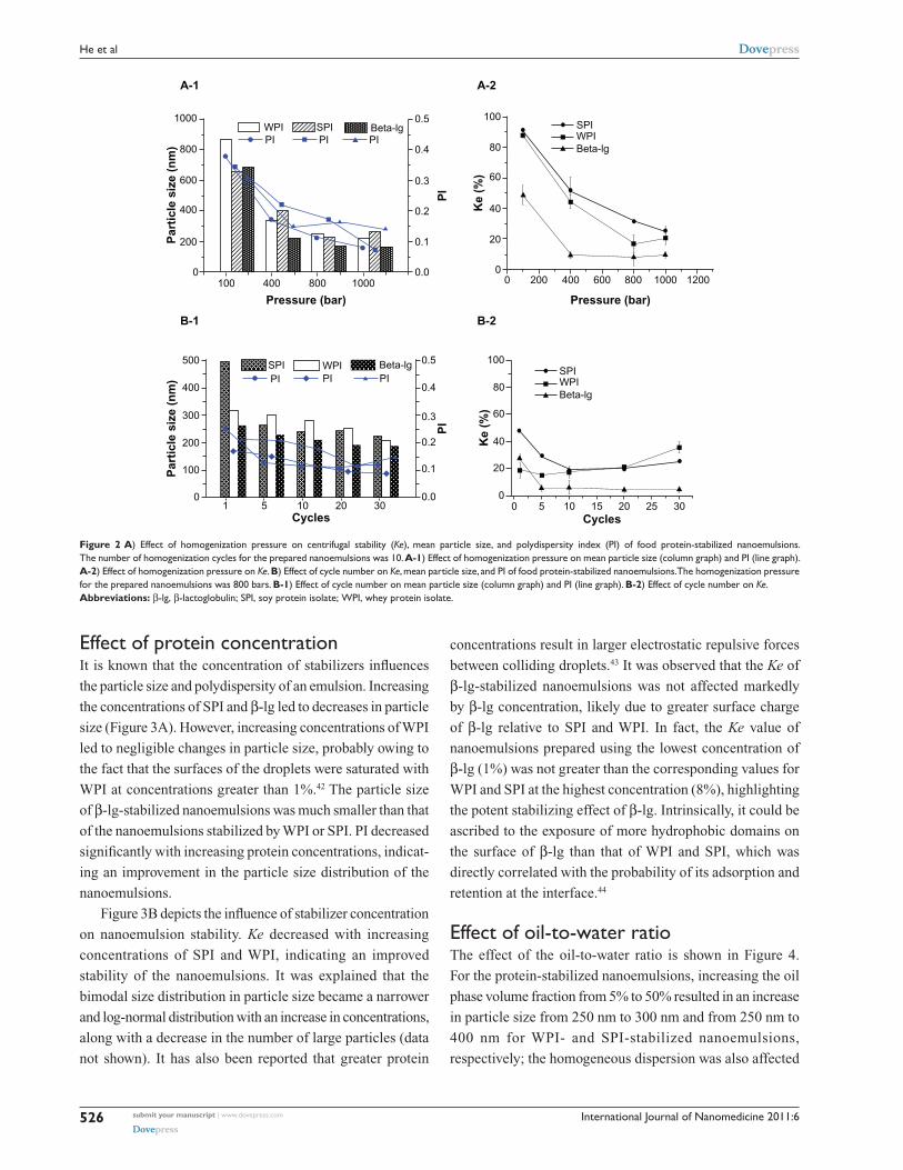
effect of homogenization pressure and number of homogenization cyclesNanoemulsions with good dispersion can be obtained using
a high-pressure homogenizer. Figure 2(A-1/B-1) shows
the effect of homogenization pressure and cycles on Ke,
particle size, and polydispersity. A considerable decrease in
particle size and PI was achieved with an increase in pressure
homogenization and/or the number of cycles. Increasing the
pressure from 100 bars to 800 bars and the number of cycles
from 1 to 10 led to a significant reduction in particle size and
PI, with the β-lg-stabilized nanoemulsions having the smallest
particle size. However, further increasing the homogenization
pressure to 1000 bars and the number of cycles to 30 did not
result in significant smaller particle sizes due to the increased
surface area and interfacial tension caused by the high homog-
enization energy input.39 PI is a measure of dispersion homo-
geneity with values ranging from 0 to 1. PI values lower than
0.3 suggest a homogeneous dispersion.40 The nanoemulsions
stabilized by food proteins revealed a relatively small par-
ticle size ranging from about 200 nm to 250 nm and a very
narrow size distribution (PI , 0.2) when a homogenization
pressure of 800 bars was applied for 10 cycles.
Figure 2(A-2/B-2) depicts the effect of homogenization
pressure and number of cycles on the physical stability of
nanoemulsions. It was observed that the Ke decreased
markedly with an increase in pressure and number of cycles
(1 to 10), indicating an enhanced stability of the nanoemulsions.
The nanoemulsion stabilized by β-lg was the most stable.
It has been reported that an increase in homogenization
pressure and number of cycles can improve surfactant
adsorption to the surface of emulsion drops, which plays
an important role in the stabilization of nanoemulsions.41
However, increasing the number of homogenization cycles
beyond 10 would not further improve the stability of
nanoemulsions. When the number of homogenization cycles
increased to more than 10, the stability of nanoemulsion
stabilized by WPI and SPI decreased. The log-normal
size distribution in particle size became bimodal distri-
bution with an increased fraction of larger particles
(data not shown), owing to too much energy input to the
nanoemulsions.
International Journal of Nanomedicine 2011:6submit your manuscript | www.dovepress.com
Dovepress
Dovepress
526
He et al
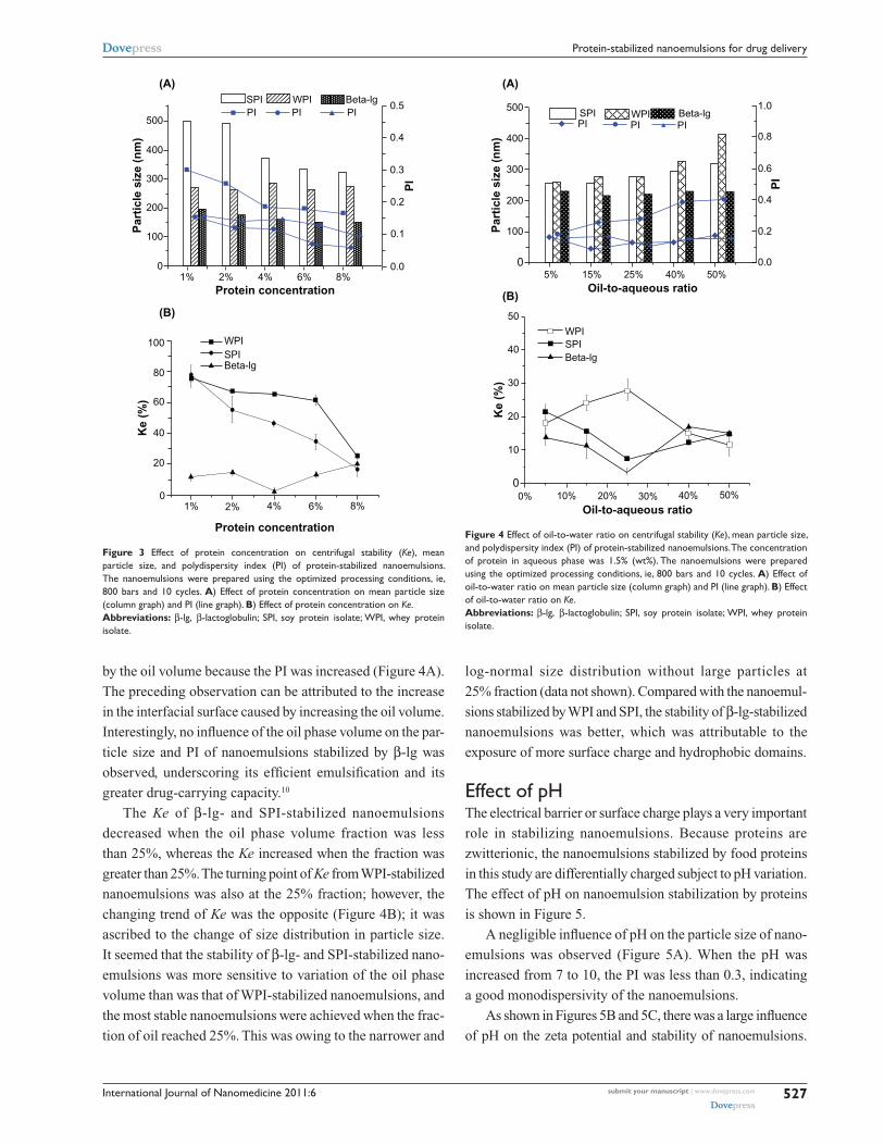
effect of protein concentrationIt is known that the concentration of stabilizers influences
the particle size and polydispersity of an emulsion. Increasing
the concentrations of SPI and β-lg led to decreases in particle
size (Figure 3A). However, increasing concentrations of WPI
led to negligible changes in particle size, probably owing to
the fact that the surfaces of the droplets were saturated with
WPI at concentrations greater than 1%.42 The particle size
of β-lg-stabilized nanoemulsions was much smaller than that
of the nanoemulsions stabilized by WPI or SPI. PI decreased
significantly with increasing protein concentrations, indicat-
ing an improvement in the particle size distribution of the
nanoemulsions.
Figure 3B depicts the influence of stabilizer concentration
on nanoemulsion stability. Ke decreased with increasing
concentrations of SPI and WPI, indicating an improved
stability of the nanoemulsions. It was explained that the
bimodal size distribution in particle size became a narrower
and log-normal distribution with an increase in concentrations,
along with a decrease in the number of large particles (data
not shown). It has also been reported that greater protein
concentrations result in larger electrostatic repulsive forces
between colliding droplets.43 It was observed that the Ke of
β-lg-stabilized nanoemulsions was not affected markedly
by β-lg concentration, likely due to greater surface charge
of β-lg relative to SPI and WPI. In fact, the Ke value of
nanoemulsions prepared using the lowest concentration of
β-lg (1%) was not greater than the corresponding values for
WPI and SPI at the highest concentration (8%), highlighting
the potent stabilizing effect of β-lg. Intrinsically, it could be
ascribed to the exposure of more hydrophobic domains on
the surface of β-lg than that of WPI and SPI, which was
directly correlated with the probability of its adsorption and
retention at the interface.44
effect of oil-to-water ratioThe effect of the oil-to-water ratio is shown in Figure 4.
For the protein-stabilized nanoemulsions, increasing the oil
phase volume fraction from 5% to 50% resulted in an increase
in particle size from 250 nm to 300 nm and from 250 nm to
400 nm for WPI- and SPI-stabilized nanoemulsions,
respectively; the homogeneous dispersion was also affected
1000 100
80
60
40
20
00 200 400 600 800 1000 1200
800
600
500
400
300
200
100
0
400
200
0 0.0
0.1
0.2
0.3
0.4
0.5
0.0
0.1
0.2
0.3
0.4
0.5
100
1 5 10 20 30
400 800 1000
WPI SPI Beta-lgWPISPI
Beta-lgPI PI PI
WPISPI Beta-lgPI PI PI
Pressure (bar)
Cycles Cycles
Pressure (bar)
Par
ticl
e si
ze (
nm
)P
arti
cle
size
(n
m)
PI
PI
Ke
(%)
100
80
60
40
20
00 5 10 15 20 25 30
Ke
(%)
A-1 A-2
B-1 B-2
WPISPI
Beta-lg
Figure 2 A) effect of homogenization pressure on centrifugal stability (Ke), mean particle size, and polydispersity index (PI) of food protein-stabilized nanoemulsions. The number of homogenization cycles for the prepared nanoemulsions was 10. A-1) effect of homogenization pressure on mean particle size (column graph) and PI (line graph). A-2) effect of homogenization pressure on Ke. B) effect of cycle number on Ke, mean particle size, and PI of food protein-stabilized nanoemulsions. The homogenization pressure for the prepared nanoemulsions was 800 bars. B-1) effect of cycle number on mean particle size (column graph) and PI (line graph). B-2) effect of cycle number on Ke.Abbreviations: β-lg, β-lactoglobulin; sPI, soy protein isolate; WPI, whey protein isolate.
International Journal of Nanomedicine 2011:6 submit your manuscript | www.dovepress.com
Dovepress
Dovepress
527
Protein-stabilized nanoemulsions for drug delivery
by the oil volume because the PI was increased (Figure 4A).
The preceding observation can be attributed to the increase
in the interfacial surface caused by increasing the oil volume.
Interestingly, no influence of the oil phase volume on the par-
ticle size and PI of nanoemulsions stabilized by β-lg was
observed, underscoring its efficient emulsification and its
greater drug-carrying capacity.10
The Ke of β-lg- and SPI-stabilized nanoemulsions
decreased when the oil phase volume fraction was less
than 25%, whereas the Ke increased when the fraction was
greater than 25%. The turning point of Ke from WPI- stabilized
nanoemulsions was also at the 25% fraction; however, the
changing trend of Ke was the opposite (Figure 4B); it was
ascribed to the change of size distribution in particle size.
It seemed that the stability of β-lg- and SPI-stabilized nano-
emulsions was more sensitive to variation of the oil phase
volume than was that of WPI-stabilized nanoemulsions, and
the most stable nanoemulsions were achieved when the frac-
tion of oil reached 25%. This was owing to the narrower and
log-normal size distribution without large particles at
25% fraction (data not shown). Compared with the nanoemul-
sions stabilized by WPI and SPI, the stability of β-lg-stabilized
nanoemulsions was better, which was attributable to the
exposure of more surface charge and hydrophobic domains.
effect of pHThe electrical barrier or surface charge plays a very important
role in stabilizing nanoemulsions. Because proteins are
zwitterionic, the nanoemulsions stabilized by food proteins
in this study are differentially charged subject to pH variation.
The effect of pH on nanoemulsion stabilization by proteins
is shown in Figure 5.
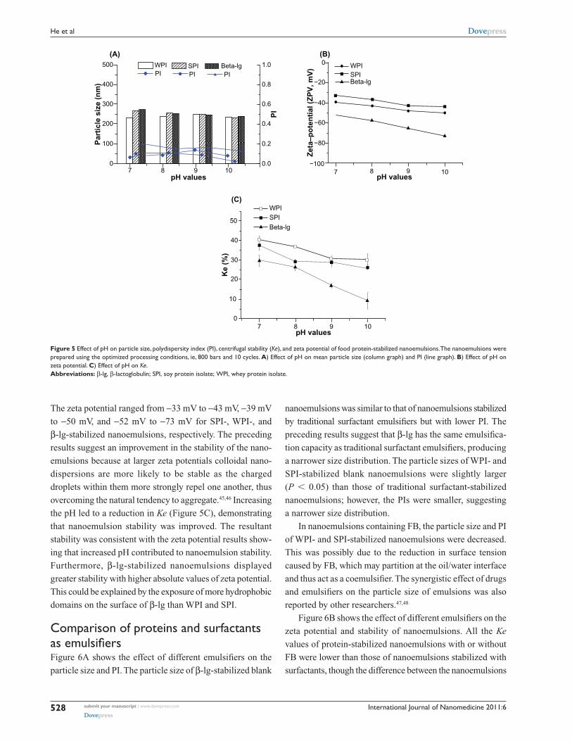
A negligible influence of pH on the particle size of nano-
emulsions was observed (Figure 5A). When the pH was
increased from 7 to 10, the PI was less than 0.3, indicating
a good monodispersivity of the nanoemulsions.
As shown in Figures 5B and 5C, there was a large influence
of pH on the zeta potential and stability of nanoemulsions.
01% 2% 4% 6% 8%
1% 2% 4% 6% 8%
500
400
300
200
100
0.0
0.1
0.2
0.3
0.4
0.5WPISPI Beta-lg
PI PI PI
Protein concentration
Protein concentration
Par
ticl
e si
ze (
nm
)
PI
(A)
(B)
100
80
60
40
20
0
WPISPIBeta-lg
Ke
(%)
Figure 3 effect of protein concentration on centrifugal stability (Ke), mean particle size, and polydispersity index (PI) of protein-stabilized nanoemulsions. The nanoemulsions were prepared using the optimized processing conditions, ie, 800 bars and 10 cycles. A) effect of protein concentration on mean particle size (column graph) and PI (line graph). B) effect of protein concentration on Ke.Abbreviations: β-lg, β-lactoglobulin; sPI, soy protein isolate; WPI, whey protein isolate.
500
400
300
200
100
0
50
40
30
20
10
0
5% 15% 25% 40% 50%0.0
0.2
0.6
1.0
0.8
0.4
WPISPI Beta-lg
WPISPIBeta-lg
PIPI PI
Par
ticl
e si
ze (
nm
)
Oil-to-aqueous ratio
0% 10% 20% 30% 40% 50%
Oil-to-aqueous ratioK
e (%
)
PI
(A)
(B)
Figure 4 effect of oil-to-water ratio on centrifugal stability (Ke), mean particle size, and polydispersity index (PI) of protein-stabilized nanoemulsions. The concentration of protein in aqueous phase was 1.5% (wt%). The nanoemulsions were prepared using the optimized processing conditions, ie, 800 bars and 10 cycles. A) effect of oil-to-water ratio on mean particle size (column graph) and PI (line graph). B) effect of oil-to-water ratio on Ke.Abbreviations: β-lg, β-lactoglobulin; sPI, soy protein isolate; WPI, whey protein isolate.
International Journal of Nanomedicine 2011:6submit your manuscript | www.dovepress.com
Dovepress
Dovepress
528
He et al
The zeta potential ranged from -33 mV to -43 mV, -39 mV
to -50 mV, and -52 mV to -73 mV for SPI-, WPI-, and
β-lg-stabilized nanoemulsions, respectively. The preceding
results suggest an improvement in the stability of the nano-
emulsions because at larger zeta potentials colloidal nano-
dispersions are more likely to be stable as the charged
droplets within them more strongly repel one another, thus
overcoming the natural tendency to aggregate.45,46 Increasing
the pH led to a reduction in Ke (Figure 5C), demonstrating
that nanoemulsion stability was improved. The resultant
stability was consistent with the zeta potential results show-
ing that increased pH contributed to nanoemulsion stability.
Furthermore, β-lg-stabilized nanoemulsions displayed
greater stability with higher absolute values of zeta potential.
This could be explained by the exposure of more hydrophobic
domains on the surface of β-lg than WPI and SPI.
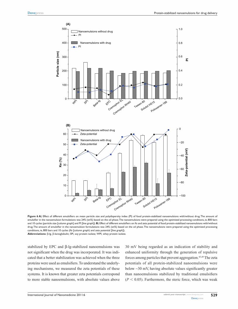
comparison of proteins and surfactants as emulsifiersFigure 6A shows the effect of different emulsifiers on the
particle size and PI. The particle size of β-lg-stabilized blank
nanoemulsions was similar to that of nanoemulsions stabilized
by traditional surfactant emulsifiers but with lower PI. The
preceding results suggest that β-lg has the same emulsifica-
tion capacity as traditional surfactant emulsifiers, producing
a narrower size distribution. The particle sizes of WPI- and
SPI-stabilized blank nanoemulsions were slightly larger
(P , 0.05) than those of traditional surfactant- stabilized
nanoemulsions; however, the PIs were smaller, suggesting
a narrower size distribution.
In nanoemulsions containing FB, the particle size and PI
of WPI- and SPI-stabilized nanoemulsions were decreased.
This was possibly due to the reduction in surface tension
caused by FB, which may partition at the oil/water interface
and thus act as a coemulsifier. The synergistic effect of drugs
and emulsifiers on the particle size of emulsions was also
reported by other researchers.47,48
Figure 6B shows the effect of different emulsifiers on the
zeta potential and stability of nanoemulsions. All the Ke
values of protein-stabilized nanoemulsions with or without
FB were lower than those of nanoemulsions stabilized with
surfactants, though the difference between the nanoemulsions
500
400
300
200
100
0
50
40
30
20
10
0
7 8 9 100.0
0.2
0.4
0.6
0.8
1.0
pH values
7 8 9 10pH values
Ke
(%)
7 8 9 10pH values
PI
Zet
a–p
ote
nti
al (
ZP
V, m
V)
Par
ticl
e si
ze (
nm
)
WPI SPI Beta-lgPIPI PI
(A)
(C)
(B)
WPISPIBeta-lg
0
−20
−40
−60
−80
−100
WPISPIBeta-lg
Figure 5 effect of pH on particle size, polydispersity index (PI), centrifugal stability (Ke), and zeta potential of food protein-stabilized nanoemulsions. The nanoemulsions were prepared using the optimized processing conditions, ie, 800 bars and 10 cycles. A) effect of pH on mean particle size (column graph) and PI (line graph). B) effect of pH on zeta potential. C) effect of pH on Ke.Abbreviations: β-lg, β-lactoglobulin; sPI, soy protein isolate; WPI, whey protein isolate.
International Journal of Nanomedicine 2011:6 submit your manuscript | www.dovepress.com
Dovepress
Dovepress
529
Protein-stabilized nanoemulsions for drug delivery
stabilized by EPC and β-lg-stabilized nanoemulsions was
not significant when the drug was incorporated. It was indi-
cated that a better stabilization was achieved when the three
proteins were used as emulsifiers. To understand the underly-
ing mechanisms, we measured the zeta potentials of these
systems. It is known that greater zeta potentials correspond
to more stable nanoemulsions, with absolute values above
30 mV being regarded as an indication of stability and
enhanced uniformity through the generation of repulsive
forces among particles that prevent aggregation.45,49 The zeta
potentials of all protein-stabilized nanoemulsions were
below -30 mV, having absolute values significantly greater
than nanoemulsions stabilized by traditional emulsifiers
(P , 0.05). Furthermore, the steric force, which was weak
500
400
300
200
100
0 0.0
0.2
0.4
0.6
0.8
1.0
WPI
SPI
Beta-
lgEPC
Cremophor EL
Cremophor RH40
Tween-80
Solutol HS15
Poloxamer-188
WPI
SPI
Beta-lgEPC
Cremophor EL
Cremophor RH40
Tween-80
Solutol HS15
Poloxamer-188
Par
ticl
e si
ze (
nm
)
PI
(A)
(B)
Ke
(%)
Nanoemulsions without drug
Nanoemulsions with drug
PI
PI
Nanoemulsions without drug
Nanoemulsions with drugZeta-potential
Zeta-potential
Zet
a-p
ote
nti
al (
mV
)
60
50
40
30
20
10
0
0
−20
−40
−60
−80
−100
Figure 6 A) Effect of different emulsifiers on mean particle size and polydispersity index (PI) of food protein-stabilized nanoemulsions with/without drug. The amount of emulsifier in the nanoemulsion formulations was 24% (wt%) based on the oil phase. The nanoemulsions were prepared using the optimized processing conditions, ie, 800 bars and 10 cycles (particle size [column graph] and PI [line graph]). B) Effect of different emulsifiers on Ke and zeta potential of food protein-stabilized nanoemulsions with/without drug. The amount of emulsifier in the nanoemulsion formulations was 24% (wt%) based on the oil phase. The nanoemulsions were prepared using the optimized processing conditions, ie, 800 bars and 10 cycles (Ke [column graph) and zeta potential [line graph]).Abbreviations: β-lg, β-lactoglobulin; sPI, soy protein isolate; WPI, whey protein isolate.
International Journal of Nanomedicine 2011:6submit your manuscript | www.dovepress.com
Dovepress
Dovepress
530
He et al
in small molecular surfactant-stabilized nanoemulsions,
was also beneficial to the improvement in stability. Notably,
that additional improvement in stability observed for WPI-
and SPI-stabilized nanoemulsions containing FB was similar
to that of nanoemulsions stabilized with traditional emulsifiers.
The preceding observation was likely due to the synergistic
effect of drug and emulsifier.47,48
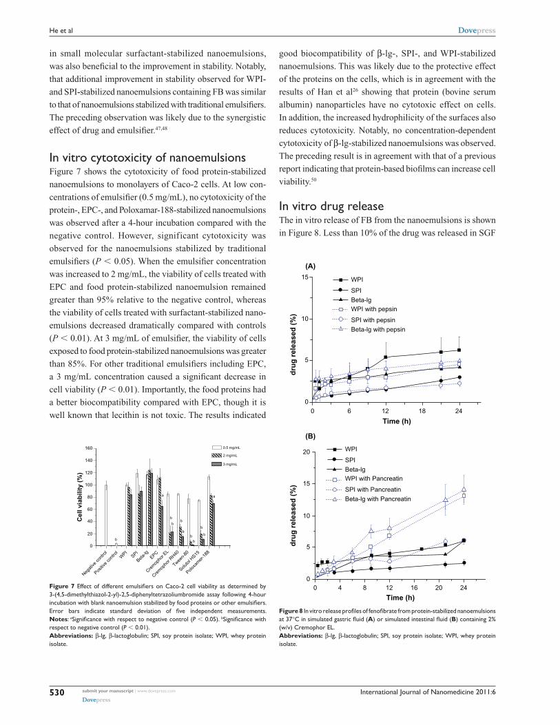
In vitro cytotoxicity of nanoemulsionsFigure 7 shows the cytotoxicity of food protein-stabilized
nanoemulsions to monolayers of Caco-2 cells. At low con-
centrations of emulsifier (0.5 mg/mL), no cytotoxicity of the
protein-, EPC-, and Poloxamar-188-stabilized nanoemulsions
was observed after a 4-hour incubation compared with the
negative control. However, significant cytotoxicity was
observed for the nanoemulsions stabilized by traditional
emulsifiers (P , 0.05). When the emulsifier concentration
was increased to 2 mg/mL, the viability of cells treated with
EPC and food protein-stabilized nanoemulsion remained
greater than 95% relative to the negative control, whereas
the viability of cells treated with surfactant-stabilized nano-
emulsions decreased dramatically compared with controls
(P , 0.01). At 3 mg/mL of emulsifier, the viability of cells
exposed to food protein-stabilized nanoemulsions was greater
than 85%. For other traditional emulsifiers including EPC,
a 3 mg/mL concentration caused a significant decrease in
cell viability (P , 0.01). Importantly, the food proteins had
a better biocompatibility compared with EPC, though it is
well known that lecithin is not toxic. The results indicated
good biocompatibility of β-lg-, SPI-, and WPI-stabilized
nanoemulsions. This was likely due to the protective effect
of the proteins on the cells, which is in agreement with the
results of Han et al26 showing that protein (bovine serum
albumin) nanoparticles have no cytotoxic effect on cells.
In addition, the increased hydrophilicity of the surfaces also
reduces cytotoxicity. Notably, no concentration-dependent
cytotoxicity of β-lg-stabilized nanoemulsions was observed.
The preceding result is in agreement with that of a previous
report indicating that protein-based biofilms can increase cell
viability.50
In vitro drug releaseThe in vitro release of FB from the nanoemulsions is shown
in Figure 8. Less than 10% of the drug was released in SGF
160
140
120
100
80
60
40
20
0
WPI
Negat
ive co
ntro
l
Positiv
e co
ntro
lSPI
Beta-
lgEPC
Crem
opho
r EL
Crem
opho
r RH40
Tween-
80
Soluto
l HS15
Poloxa
mer
-188
Cel
l via
bili
ty (
%)
a a
b
b
bb
bb
b
bb
0.5 mg/mL
2 mg/mL
3 mg/mL
Figure 7 Effect of different emulsifiers on Caco-2 cell viability as determined by 3-(4,5-dimethylthiazol-2-yl)-2,5-diphenyltetrazoliumbromide assay following 4-hour incubation with blank nanoemulsion stabilized by food proteins or other emulsifiers. Error bars indicate standard deviation of five independent measurements. Notes: aSignificance with respect to negative control (P , 0.05). bSignificance with respect to negative control (P , 0.01).Abbreviations: β-lg, β-lactoglobulin; sPI, soy protein isolate; WPI, whey protein isolate.
WPI
SPI
Beta-lgWPI with pepsin
SPI with pepsin
Beta-lg with pepsin
WPI
SPI
Beta-lgWPI with Pancreatin
SPI with Pancreatin
Beta-lg with Pancreatin
15
10
5
00 6 12 18 24
dru
g r
elea
sed
(%
)
Time (h)
0
5
10
15
20
0 4 8 12 16 20 24
dru
g r
elea
sed
(%
)
Time (h)
(A)
(B)
Figure 8 In vitro release profiles of fenofibrate from protein-stabilized nanoemulsions at 37°C in simulated gastric fluid (A) or simulated intestinal fluid (B) containing 2% (w/v) cremophor eL.Abbreviations: β-lg, β-lactoglobulin; sPI, soy protein isolate; WPI, whey protein isolate.
International Journal of Nanomedicine 2011:6 submit your manuscript | www.dovepress.com
Dovepress
Dovepress
531
Protein-stabilized nanoemulsions for drug delivery
or SIF without digestive enzymes due to the poor water
solubility of the drug. To test the effect of digestive enzymes
on drug release, we added the digestive enzymes pepsin and
pancreatin to the SGF and SIF, respectively. The percentage
drug release in SGF was not increased in the presence of
pepsin, indicating that the proteins resist pepsin degradation
in the gastrointestinal tract (Figure 8A). Pepsin is known to
preferentially cleave peptide bonds between hydrophobic
aromatic amino acids; however, the hydrophobic amino acids
that are adsorbed to the surface of the oil droplets are trapped
inside the protein during the preparation of nanoemulsions.51
The hydrophobic amino acids are thus hidden, protecting
them from pepsin degradation. A significant increase in drug
release was observed on addition of pancreatin to SIF
(Figure 8B), owing to pancreatic degradation of the proteins.27
A similar report by Chen and Subirade52 also demonstrated
that pancreatic digestion promoted drug release from
microparticles based on the proteins they contained. It seems
that, to some extent, desirable enteric properties of protein-
stabilized nanoemulsions can be achieved, which is useful
for delivering a hydrophobic and acid-labile drug to the
intestine.
120
100
80
60
40
20
00 4 8 12 16 20 24 28 32 36
Time (h)
Pla
sma
fen
ofi
bri
c ac
id
con
cen
trat
ion
(u
g/ m
L)
FB oill solution
SPI–stabilized nanoemulsions
WPI–stabilized nanoemulsionsBeta–lg–stabilized nanoemulsions
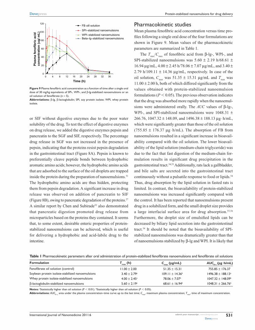
Figure 9 Plasma fenofibric acid concentration as a function of time after a single oral dose of 30 mg/kg equivalents of sPI-, WPI-, and β-lg-stabilized nanoemulsions or an oil solution of fenofibrate (n = 5).Abbreviations: β-lg, β-lactoglobulin; sPI, soy protein isolate; WPI, whey protein isolate.
Pharmacokinetic studiesMean plasma fenofibric acid concentration versus time pro-
files following a single oral dose of the four formulations are
shown in Figure 9. Mean values of the pharmacokinetic
parameters are summarized in Table 1.
The Tmax
/Cmax
of fenofibric acid from β-lg-, WPI-, and
SPI-stabilized nanoemulsions was 5.60 ± 2.19 h/68.61 ±
16.94 µg/mL, 4.00 ± 2.45 h/78.06 ± 7.07 µg/mL, and 3.40 ±
2.79 h/109.11 ± 14.36 µg/mL, respectively. In case of the
oil solution, Cmax
was 51.35 ± 15.31 µg/mL and Tmax
was
11.00 ± 2.00 h, both of which differed significantly from the
values obtained with protein-stabilized nanoemulsion
formulations (P , 0.05). The previous observation indicates
that the drug was absorbed more rapidly when the nanoemul-
sions were administered orally. The AUC values of β-lg-,
WPI-, and SPI-stabilized nanoemulsions were 1048.31 ±
266.76, 1047.32 ± 148.09, and 1496.38 ± 188.13 µg ⋅ h/mL,
which were significantly greater than those of the oil solution
(755.85 ± 176.37 µg ⋅ h/mL). The absorption of FB from
nanoemulsions resulted in a significant increase in bioavail-
ability compared with the oil solution. The lower bioavail-
ability of the lipid solution (medium-chain triglyceride) was
due to the fact that fast digestion of the medium-chain for-
mulation results in significant drug precipitation in the
gastrointestinal tract.34,53 Additionally, rats lack a gallbladder,
and bile salts are secreted into the gastrointestinal tract
continuously without a pulsatile response to food or lipids.54
Thus, drug absorption by the lipid solution in fasted rats is
limited. In contrast, the bioavailability of protein-stabilized
nanoemulsions was increased significantly compared with
the control. It has been reported that nanoemulsions present
drug in a solubilized form, and the small droplet size provides
a large interfacial surface area for drug absorption.55,56
Furthermore, the droplet size of emulsified lipids can be
decreased by biliary lipid secretion into the gastrointestinal
tract.54 It should be noted that the bioavailability of SPI-
stabilized nanoemulsions was dramatically greater than that
of nanoemulsions stabilized by β-lg and WPI. It is likely that
Table 1 Pharmacokinetic parameters after oral administration of protein-stabilized fenofibrate nanoemulsions and fenofibrate oil solutions
Formulation Tmax (h) Cmax (μg/mL) AUC0-t (μg ⋅ h/mL)
Fenofibrate oil solution (control) 11.00 ± 2.00 51.35 ± 15.31 755.85 ± 176.37soybean protein isolate-stabilized nanoemulsions 3.40 ± 2.79a 109.11 ± 14.36b 1496.38 ± 188.13a
Whey protein isolate-stabilized nanoemulsions 4.00 ± 2.45a 78.06 ± 7.07b 1047.32 ± 148.09b
β-lactoglobulin-stabilized nanoemulsions 5.60 ± 2.19a 68.61 ± 16.94b 1048.31 ± 266.76b
Notes: astatistically higher than oil solution (P , 0.01). bstatistically higher than oil solution (P , 0.05).Abbreviations: AUC0-t, area under the plasma concentration–time curve up to the last time; Cmax, maximum plasma concentration; Tmax, time of maximum concentration.
International Journal of Nanomedicine 2011:6submit your manuscript | www.dovepress.com
Dovepress
Dovepress
532
He et al
SPI is more sensitive to digestion by pancreatin, leading to
a further reduction in the droplet size of emulsified lipids.
The results clearly demonstrate that the droplets can be
stabilized by proteins, and the stabilized lipids enable a
lipophilic drug to be absorbed more rapidly and better com-
pared with oil solution.
ConclusionBiocompatible nanoemulsions stabilized by food proteins
can be produced successfully and deliver a poorly water-
soluble drug in vivo. The nanoemulsions enable the lipophilic
drug to be absorbed more rapidly and better when compared
with the oil solution. As emulsifiers, the proteins WPI, SPI,
and β-lg have better emulsifying capacity and biocompatibil-
ity than do traditional emulsifiers. A much better stability
was observed in protein-stabilized nanoemulsions relative
to nanoemulsions stabilized with surfactants. The preceding
observation was likely due to the greater surface potential of
proteins. Furthermore, β-lg-stabilized nanoemulsions exhib-
ited greater resistance to gravitational separation and better
biocompatibility compared with nanoemulsions stabilized
by the other two proteins. The particle size, stability, and
zeta potential were affected dramatically by protein concen-
tration, pH, homogenization pressure, and number of cycles.
Therefore, we conclude that by using the proteins as a sur-
factant, the development of biocompatible and biodegradable
nanoemulsion systems can be achieved, and the proteins are
viable replacements for traditional surfactants.
AcknowledgmentsThis study was supported by the National Key Basic Research
Program of China (2009CB930300, 2007CB935800) and
partly by the Shanghai Commission of Education (10SG05)
and the Shanghai Commission of Science and Technology
(10430709200).
DisclosureThe authors report no conflicts of interest in this work.
References1. Chen H, Khemtong C, Yang X, Chang X, Gao J. Nanonization strategies
for poorly water-soluble drugs. Drug Discov Today. 2010;doi:10.1016/j.drudis.2010.02.009.
2. Amani A, York P, Chrystyn H, Clark BJ. Factors affecting the stability of nanoemulsions: use of artif icial neural networks. Pharm Res. 2010;27(1):37–45.
3. Mathur AM, Drescher B, Scranton AB, Klier J. Polymeric emulsifiers based on reversible formation of hydrophobic units. Nature. 1998; 392(6674):367–370.
4. Karasulu HY. Microemulsions as novel drug carriers: the formation, stability, applications and toxicity. Exp Opin Drug Deliv. 2008;5(1): 119–135.
5. Lawrence MJ, Rees GD. Microemulsion-based media as novel drug delivery systems. Adv Drug Deliv Rev. 2000;45(1):89–121.
6. Yan A, Von Dem Bussche A, Kane AB, Hurt RH. Tocopheryl polyeth-ylene glycol succinate as a safe, antioxidant surfactant for processing carbon nanotubes and fullerenes. Carbon. 2007;45(13):2463–2470.
7. Jiao J. Polyoxyethylated nonionic surfactants and their applications in topical ocular drug delivery. Adv Drug Deliv Rev. 2008;60(15): 1663–1673.
8. Sivakumar S, Bansal V, Cortez C, et al. Degradable, surfactant-free, monodisperse polymer-encapsulated emulsions as anticancer drug carriers. Adv Mater. 2009;21(18):1820–1824.
9. Gutiérrez JM, González C, Maestro A, et al. Nano-emulsions: new applications and optimization of their preparation. Curr Opin Colloid Inter Sci. 2008;13:245–251.
10. Trotta M, Pattarino F, Ignoni T. Stability of drug-carrier emulsions containing phosphatidylcholine mixtures. Eur J Pharm Biopharm. 2002; 53(2):203–208.
11. Sznitowska M, Janicki S, Dabrowska E, Zurowska-Pryczkowska K. Submicron emulsions as drug carriers: studies on destabilization poten-tial of various drugs. Eur J Pharm Biopharm. 2001;12(3):175–179.
12. Chen L, Remondetto GE, Subirade M. Food protein-based materials as nutraceutical delivery systems. Trends Food Sci Tech. 2006;17(5): 272–283.
13. MaHam A, Tang ZW, Wu H, et al. Protein-based nanomedicine plat-forms for drug delivery. Small. 2009;5(15):1706–1721.
14. Chen LY, Subirade M. Elaboration and characterization of soy/zein protein microspheres for controlled nutraceutical delivery. Biomacromolecules. 2009;10(12):3327–3334.
15. Adachi M, Takenaka Y, Gidamis AB, et al. Crystal structure of soybean proglycinin alaB1b homotrimer. J Mol Biol. 2001;305(2):291–305.
16. Riblett AL, Herald TJ, Schmidt KA, Tilley KA. Characterization of beta-conglycinin and glycinin soy protein fractions from four selected soybean genotypes. J Agric Food Chem. 2001;49(10):4983–4989.
17. Perez OE, Wargon V, M.R. Pilosof A. Gelation and structural charac-teristics of incompatible whey proteins/hydroxypropylmethylcellulose mixtures. Food Hydrocolloids. 2006;20(7):966–974.
18. Davis JP, Foegeding EA. Foaming and interfacial properties of polymer-ized whey protein isolate. J Food Sci. 2004;69(5):C404–C410.
19. Permyakov EA, Berliner LJ. [alpha]-Lactalbumin: structure and function. FEBS Let. 2000;473(3):269–274.
20. Sawyer L. Beta-lactoglobulin. Advanced Dairy Chemistry. Vol 1. Fox PF, McSweeney LH, editors. New York: Kluwer Academic; 2003: 319–386.
21. Brownlow S, Cabral JHM, Cooper R, et al. Bovine [beta]-lactoglobulin at 1.8 resolution: still an enigmatic lipocalin. Structure. 1997;5(4): 481–495.
22. Semenova MG, Antipova AS, Belyakova LE. Food protein interactions in sugar solutions. Cur Opin Coll Int Sci. 2002;7(5–6):438–444.
23. Lee SJ, Rosenberg M. Microencapsulation of theophylline in whey proteins: effects of core-to-wall ratio. Int J Pharm. 2000;205(1–2): 147–158.
24. Lee SJ, Rosenberg M. Preparation and properties of glutaraldehyde cross-linked whey protein-based microcapsules containing theophylline. J Control Release. 1999;61(1–2):123–136.
25. Augustin MA, Hemar Y. Nano- and micro-structured assemblies for encapsulation of food ingredients. Chem Soc Rev. 2009;38(4): 902–912.
26. Han YS, Shchukin D, Yang J, et al. Biocompatible protein nanocontainers for controlled drugs release. Acs Nano. 2010;4(5):2838–2844.
27. Chen LY, Subirade M. Alginate-whey protein granular microspheres as oral delivery vehicles for bioactive compounds. Biomaterials. 2006;27(26):4646–4654.
28. Chen LY, Remondetto G, Rouabhia M, Subirade M. Kinetics of the breakdown of cross-linked soy protein f ilms for drug delivery. Biomaterials. 2008;29(27):3750–3756.
29. Chen L, Subirade M. Chitosan/[beta]-lactoglobulin core-shell nano-particles as nutraceutical carriers. Biomaterials. 2005;26(30): 6041–6053.
International Journal of Nanomedicine
Publish your work in this journal
Submit your manuscript here: http://www.dovepress.com/international-journal-of-nanomedicine-journal
The International Journal of Nanomedicine is an international, peer-reviewed journal focusing on the application of nanotechnology in diagnostics, therapeutics, and drug delivery systems throughout the biomedical field. This journal is indexed on PubMed Central, MedLine, CAS, SciSearch®, Current Contents®/Clinical Medicine, Journal
Citation Reports/Science Edition, EMBase, Scopus and the Elsevier Bibliographic databases. The manuscript management system is completely online and includes a very quick and fair peer-review system, which is all easy to use. Visit http://www.dovepress.com/ testimonials.php to read real quotes from published authors.
International Journal of Nanomedicine 2011:6 submit your manuscript | www.dovepress.com
Dovepress
Dovepress
Dovepress
533
Protein-stabilized nanoemulsions for drug delivery
30. Chan OCM, So KF, Chan BP. Fabrication of nano-fibrous collagen microspheres for protein delivery and effects of photochemical cross-linking on release kinetics. J Control Release. 2008;129(2):135–143.
31. Patel AR, Vavia PR. Preparation and in vivo evaluation of SMEDDS (Self-Microemulsifying Drug Delivery System) containing fenofibrate. AAPS J. 2007;9(3):E344–E352.
32. Chen Y, Lu Y, Chen J, et al. Enhanced bioavailability of the poorly water-soluble drug fenofibrate by using liposomes containing a bile salt. Int J Pharm. 2009;376(1–2):153–160.
33. Hanafy A, Spahn-Langguth H, Vergnault G, et al. Pharmacokinetic evaluation of oral fenofibrate nanosuspensions and SLN in comparison to conventional suspensions of micronized drug. Adv Drug Deliv Rev. 2007;59(6):419–426.
34. Porter CJH, Trevaskis NL, Charman WN. Lipids and lipid-based formulations: optimizing the oral delivery of lipophilic drugs. Nat Rev Drug Discov. 2007;6(3):231–248.
35. Chen L, Remondetto G, Rouabhia M, Subirade M. Kinetics of the breakdown of cross-linked soy protein f ilms for drug delivery. Biomaterials. 2008;29(27):3750–3756.
36. He SN, Wang DK, Li LS, et al. Preparation and characterization of docetaxel phospholipid complex submicron emulsion. Chin J Pharm. 2010;1:9–16.
37. Yang X, Wang DK, Kong LW, et al. Preparation and characteriztion of cisplatin emulsions for injection. Chin J New Drugs. 2007;16: 1376–1379.
38. Streel B, Hubert P, Ceccato A. Determination of fenofibric acid in human plasma using automated solid-phase extraction coupled to liquid chromatography. J Chromatogr B. 2000;742(2):391–400.
39. Yilmaz E, Borchert H-H. Design of a phytosphingosine-containing, positively-charged nanoemulsion as a colloidal carrier system for dermal application of ceramides. Eur J Pharm Biopharm. 2005;60(1):91–98.
40. Chu B, Wang ZL, Yu JQ. Dynamic light-scattering study of internal motions of polymer coils in dilute-solution. Macromolecules. 1991; 24(26):6832–6838.
41. Tcholakova S, Denkov ND, Sidzhakova D, et al. Effects of electrolyte concentration and pH on the coalescence stability of beta-lactoglobulin emulsions: experiment and interpretation. Langmuir. 2005;21(11): 4842–4855.
42. Chu BS, Ichikawa S, Kanafusa S, Nakajima M. Preparation and characterization of beta-carotene nanodispersions prepared by sol-vent displacement technique. J Agric Food Chem. 2007;55(16): 6754–6760.
43. Mohan S, Narsimhan G. Coalescence of protein-stabilized emulsions in a high-pressure homogenizer. J Colloid Inter Sci. 1997;192(1): 1–15.
44. Adams JJ, Anderson BF, Norris GE, et al. Structure of bovine [beta]-lactoglobulin (variant A) at very low ionic strength. J Struct Biol. 2006; 154(3):246–254.
45. Tagne JB, Kakurnanu S, Nicolosi RJ. Nanoemulsion preparations of the anticancer drug dacarbazine significantly increase its efficacy in a xenograft mouse melanoma model. Mol Pharmaceut. 2008;5(6): 1055–1063.
46. Grosse C, Pedrosa S, Shilov VN. On the influence of size, [zeta] poten-tial, and state of motion of dispersed particles on the conductivity of a colloidal suspension. J Colloid Inter Sci. 2002;251(2):304–310.
47. Buyukozturk F, Benneyan JC, Carrier RL. Impact of emulsion-based drug delivery systems on intestinal permeability and drug release kinetics. J Control Release. 2010;142(1):22–30.
48. Akkar A, Muller RH. Formulation of intravenous carbamazepine emul-sions by SolEmuls technology. Eu J Pharm Biopharm. 2003;55(3): 305–312.
49. Ye SQ, Wang CY, Liu XX, Tong Z. Multilayer nanocapsules of poly-saccharide chitosan and alginate through layer-by-layer assembly directly on PS nanoparticles for release. J Biomat Sci-Polym E. 2005; 16(7):909–923.
50. Gilbert V, Rouabhia M, Wang H, et al. Characterization and evaluation of whey protein-based biofilms as substrates for in vitro cell cultures. Biomaterials. 2005;26(35):7471–7480.
51. Beaulieu L, Savoie L, Paquin P, Subirade M. Elaboration and charac-terization of whey protein beads by an emulsification/cold gelation process: application for the protection of retinol. Biomacromolecules. 2002;3(2):239–248.
52. Chen LY, Subirade M. Effect of preparation conditions on the nutrient release properties of alginate-whey protein granular minicrospheres. EurJ Pharm Biopharm. 2007;65(3):354–362.
53. Porter CJH, Kaukonen AM, Boyd BJ, et al. Susceptibility to lipase- mediated digestion reduces the oral bioavailability of danazol after administration as a medium-chain lipid-based microemulsion formulation. Pharm Res. 2004;21(8):1405–1412.
54. O’Driscoll CM. Lipid-based formulations for intestinal lymphatic delivery. Eur J Pharm Biopharm. 2002;15(5):405–415.
55. Tan A, Simovic S, Davey AK, et al. Silica-lipid hybrid (SLH) microcapsules: a novel oral delivery system for poorly soluble drugs. J Control Release. 2009;134(1):62–70.
56. Shah NH, Carvajal MT, Patel CI, et al. Self-emulsifying drug delivery systems (SEDDS) with polyglycolyzed glycerides for improving in vitro dissolution and oral absorption of lipophilic drugs. Int J Pharm. 1994;106(1):15–23.