Fibroadenoma and Phyllodes Tumor - medtu.tv · 94 Fibroadenoma and Phyllodes Tumor...
23
Fibroadenoma and Phyllodes Tumor Õ“®“√¬å·æ∑¬åÀ≠‘ß «‘‰≈√—μπå ª√–‡ √‘∞ ● ● ● ● ● ● ● ● ● ● ● ● ● ● ● ● ● ● ● ● ● 93 93 °âÕπ∑’ˇμâ“π¡„πºŸâÀ≠‘ß«—¬√ÿàπÀ√◊Õ«—¬ºŸâ„À≠àμÕπμâππ—Èπ·¡â®–‰¡à¡’Õ“°“√·≈– Õÿ∫—μ‘°“√≥å¢Õß¡–‡√Á߇μâ“π¡„πºŸâÀ≠‘ß°≈ÿà¡π’Èæ∫‰¥âπâÕ¬ ·μຟâªÉ«¬·≈–≠“μ‘¡—°¡’ §«“¡«‘μ°°—ß«≈«à“®–‡ªìπ¡–‡√Áß √«¡∑—Èß°—ß«≈«à“°âÕπ®–‚μ¢÷Èπ®π∑”„Àâ‡μâ“π¡ ‡ ’¬√Ÿª√à“ß ·æ∑¬å®÷ßμâÕß„Àâ°“√¥Ÿ·≈∑’Ë∂Ÿ°μâÕ߇À¡“– ¡ ∑’Ë ”§—≠§◊Õ„Àâ°“√§«“¡ ‡™◊ËÕ¡—Ëπ·°àºŸâªÉ«¬·≈–≠“쑇æ◊ËÕ„Àâ§≈“¬°—ß«≈„π°√≥’∑’ˉ¡à„™à‡π◊ÈÕ√⓬ ´÷Ëß “¡“√∂ π—¥μ√«®μ‘¥μ“¡Õ“°“√‰¥âÕ¬à“ߪ≈Õ¥¿—¬‚¥¬‰¡à®”‡ªìπμâÕߺà“μ—¥‡Õ“°âÕπÕÕ° ∑ÿ°√“¬ ·≈–‡≈◊Õ°ºà“μ—¥‡©æ“–„π√“¬∑’ˇÀ¡“– ¡ ∫∑§«“¡π’È¡’‡π◊ÈÕÀ“‡°’ˬ«°—∫ °âÕπ‡μâ“π¡∑’Ëæ∫∫àÕ¬„πºŸâÀ≠‘ßÕ“¬ÿπâÕ¬§◊Õ fibroadenoma ·≈–°≈à“«∂÷ß‚√§∑’Ë ¡—°¡’≈—°…≥–§≈⓬ fibroadenoma §◊Õ phyllodes tumor ´÷Ëß¡’欓°√≥å‚√§·≈– √“¬≈–‡Õ’¬¥°“√√—°…“∑’Ë·μ°μà“ß°—𠧫“¡ ”§—≠§◊ÕμâÕß«‘π‘®©—¬·¬°‚√§„À≥⠰àÕπ«“ß·ºπ°“√√—°…“À√◊Õºà“μ—¥ Fibroadenoma Fibroadenoma ‡ªìπ benign fibroepithelial lesion ∑’Ëæ∫∫àÕ¬ ¡—°æ∫„π ºŸâÀ≠‘ß™à«ßÕ“¬ÿ 20-30 ªï ¡’§«“¡™ÿ°¢Õß‚√§„π™à«ßÕ“¬ÿπ’Ȫ√–¡“≥ 2.2% 1 ®“° °“√»÷°…“™‘Èπ‡π◊ÈÕæ∫«à“ fibroadenoma ‡ªìπ°âÕπ¢Õ߇μâ“π¡∑’Ëæ∫∫àÕ¬∑’Ë ÿ¥„πºŸâ À≠‘ß«—¬√ÿàπ ‚¥¬æ∫ 44-94% ¢Õß°âÕπ„π‡μâ“π¡∑—ÈßÀ¡¥∑’ˉ¥â√—∫°“√ºà“μ—¥ 2 欓∏‘«‘∑¬“·≈–°“√®”·π°™π‘¥¢Õß fibroadenoma 3 Fibroadenoma ¡’§«“¡º‘¥ª°μ‘∑—Èß„π à«π¢Õß epithelium ·≈– stroma ¿“§«‘™“»—≈¬»“ μ√å §≥–·æ∑¬»“ μ√å¡À“«‘∑¬“≈—¬∏√√¡»“ μ√å ∫∑∑’Ë 7
-
Upload
nguyenphuc -
Category
Documents
-
view
247 -
download
0
Transcript of Fibroadenoma and Phyllodes Tumor - medtu.tv · 94 Fibroadenoma and Phyllodes Tumor...
Fibroadenoma and Phyllodes TumorÕ“®“√¬å·æ∑¬åÀ≠‘ß «‘‰≈√—μπå ª√–‡ √‘∞
● ● ● ● ● ● ● ● ● ● ● ● ● ● ● ● ● ● ● ● ●
9393
°âÕπ∑’ˇμâ“π¡„πºŸâÀ≠‘ß«—¬√ÿàπÀ√◊Õ«—¬ºŸâ„À≠àμÕπμâππ—Èπ·¡â®–‰¡à¡’Õ“°“√·≈–
Õÿ∫—μ‘°“√≥å¢Õß¡–‡√Á߇μâ“π¡„πºŸâÀ≠‘ß°≈ÿà¡π’Èæ∫‰¥âπâÕ¬ ·μຟâªÉ«¬·≈–≠“μ‘¡—°¡’
§«“¡«‘μ°°—ß«≈«à“®–‡ªìπ¡–‡√Áß √«¡∑—Èß°—ß«≈«à“°âÕπ®–‚μ¢÷Èπ®π∑”„Àâ‡μâ“π¡
‡ ’¬√Ÿª√à“ß ·æ∑¬å®÷ßμâÕß„Àâ°“√¥Ÿ·≈∑’Ë∂Ÿ°μâÕ߇À¡“– ¡ ∑’Ë ”§—≠§◊Õ„Àâ°“√§«“¡
‡™◊ËÕ¡—Ëπ·°àºŸâªÉ«¬·≈–≠“쑇æ◊ËÕ„Àâ§≈“¬°—ß«≈„π°√≥’∑’ˉ¡à„™à‡π◊ÈÕ√⓬ ÷Ëß “¡“√∂
π—¥μ√«®μ‘¥μ“¡Õ“°“√‰¥âÕ¬à“ߪ≈Õ¥¿—¬‚¥¬‰¡à®”‡ªìπμâÕߺà“μ—¥‡Õ“°âÕπÕÕ°
∑ÿ°√“¬ ·≈–‡≈◊Õ°ºà“μ—¥‡©æ“–„π√“¬∑’ˇÀ¡“– ¡ ∫∑§«“¡π’È¡’‡π◊ÈÕÀ“‡°’ˬ«°—∫
°âÕπ‡μâ“π¡∑’Ëæ∫∫àÕ¬„πºŸâÀ≠‘ßÕ“¬ÿπâÕ¬§◊Õ fibroadenoma ·≈–°≈à“«∂÷ß‚√§∑’Ë
¡—°¡’≈—°…≥–§≈⓬ fibroadenoma §◊Õ phyllodes tumor ´÷Ëß¡’欓°√≥å‚√§·≈–
√“¬≈–‡Õ’¬¥°“√√—°…“∑’Ë·μ°μà“ß°—𠧫“¡ ”§—≠§◊ÕμâÕß«‘π‘®©—¬·¬°‚√§„À≥â
°àÕπ«“ß·ºπ°“√√—°…“À√◊Õºà“μ—¥
Fibroadenoma
Fibroadenoma ‡ªìπ benign fibroepithelial lesion ∑’Ëæ∫∫àÕ¬ ¡—°æ∫„π
ºŸâÀ≠‘ß™à«ßÕ“¬ÿ 20-30 ªï ¡’§«“¡™ÿ°¢Õß‚√§„π™à«ßÕ“¬ÿπ’Ȫ√–¡“≥ 2.2%1 ®“°
°“√»÷°…“™‘Èπ‡π◊ÈÕæ∫«à“ fibroadenoma ‡ªìπ°âÕπ¢Õ߇μâ“π¡∑’Ëæ∫∫àÕ¬∑’Ë ÿ¥„πºŸâ
À≠‘ß«—¬√ÿàπ ‚¥¬æ∫ 44-94% ¢Õß°âÕπ„π‡μâ“π¡∑—ÈßÀ¡¥∑’ˉ¥â√—∫°“√ºà“μ—¥2
欓∏‘«‘∑¬“·≈–°“√®”·π°™π‘¥¢Õß fibroadenoma3
Fibroadenoma ¡’§«“¡º‘¥ª°μ‘∑—Èß„π à«π¢Õß epithelium ·≈– stroma
¿“§«‘™“»—≈¬»“ μ√å §≥–·æ∑¬»“ μ√å¡À“«‘∑¬“≈—¬∏√√¡»“ μ√å
∫∑∑’Ë 7
Fibroadenoma and Phyllodes Tumor94
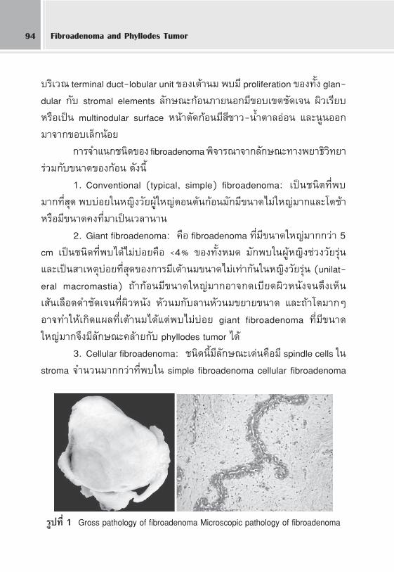
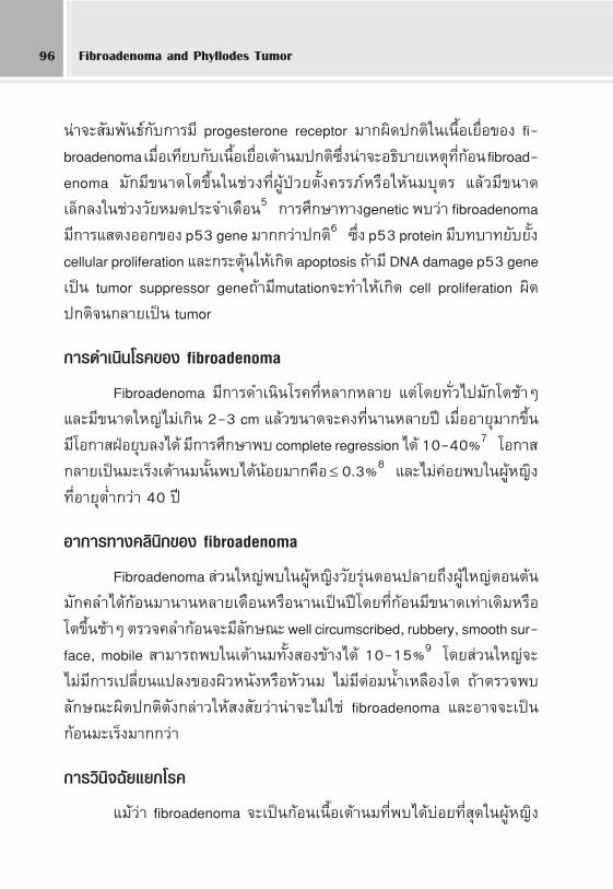
∫√‘‡«≥ terminal duct-lobular unit ¢Õ߇μâ“π¡ æ∫¡’ proliferation ¢Õß∑—Èß glan-
dular °—∫ stromal elements ≈—°…≥–°âÕπ¿“¬πÕ°¡’¢Õ∫‡¢μ™—¥‡®π º‘«‡√’¬∫
À√◊Õ‡ªìπ multinodular surface Àπâ“μ—¥°âÕπ¡’ ’¢“«-πÈ”μ“≈ÕàÕπ ·≈–πŸπÕÕ°
¡“®“°¢Õ∫‡≈Á°πâÕ¬
°“√®”·π°™π‘¥¢Õß fibroadenoma æ‘®“√≥“®“°≈—°…≥–∑“ß欓∏‘«‘∑¬“
√à«¡°—∫¢π“¥¢Õß°âÕπ ¥—ßπ’È
1. Conventional (typical, simple) fibroadenoma: ‡ªìπ™π‘¥∑’Ëæ∫
¡“°∑’Ë ÿ¥ æ∫∫àÕ¬„πÀ≠‘ß«—¬ºŸâ„À≠àμÕπμâπ°âÕπ¡—°¡’¢π“¥‰¡à„À≠à¡“°·≈–‚μ™â“
À√◊Õ¡’¢π“¥§ß∑’Ë¡“‡ªìπ‡«≈“π“π
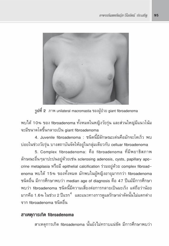
2. Giant fibroadenoma: §◊Õ fibroadenoma ∑’Ë¡’¢π“¥„À≠à¡“°°«à“ 5
cm ‡ªìπ™π‘¥∑’Ëæ∫‰¥â‰¡à∫àÕ¬§◊Õ <4% ¢Õß∑—ÈßÀ¡¥ ¡—°æ∫„πºŸâÀ≠‘ß™à«ß«—¬√ÿàπ
·≈–‡ªì𠓇Àμÿ∫àÕ¬∑’Ë ÿ¥¢Õß°“√¡’‡μâ“π¡¢π“¥‰¡à‡∑à“°—π„πÀ≠‘ß«—¬√ÿàπ (unilat-
eral macromastia) ∂â“°âÕπ¡’¢π“¥„À≠à¡“°Õ“®°¥‡∫’¬¥º‘«Àπ—ß®πμ÷߇ÀÁπ
‡ âπ‡≈◊Õ¥¥”™—¥‡®π∑’˺‘«Àπ—ß À—«π¡°—∫≈“πÀ—«π¡¢¬“¬¢π“¥ ·≈–∂â“‚μ¡“°Ê
Õ“®∑”„À⇰‘¥·º≈∑’ˇμâ“π¡‰¥â·μàæ∫‰¡à∫àÕ¬ giant fibroadenoma ∑’Ë¡’¢π“¥
„À≠à¡“°®÷ß¡’≈—°…≥–§≈⓬°—∫ phyllodes tumor ‰¥â
3. Cellular fibroadenoma: ™π‘¥π’È¡’≈—°…≥–‡¥àπ§◊Õ¡’ spindle cells „π
stroma ®”π«π¡“°°«à“∑’Ëæ∫„π simple fibroadenoma cellular fibroadenoma
√Ÿª∑’Ë 1 Gross pathology of fibroadenoma Microscopic pathology of fibroadenoma
Õ“®“√¬å·æ∑¬åÀ≠‘ß «‘‰≈√—μπå ª√–‡ √‘∞ 95
æ∫‰¥â 10% ¢Õß fibroadenoma ∑—ÈßÀ¡¥„πÀ≠‘ß«—¬√ÿàπ ·≈– à«π„À≠à¡’·π«‚πâ¡
®–¡’¢π“¥‚μ¢÷Èπ°≈“¬‡ªìπ giant fibroadenoma
4. Juvenile fibroadenoma : ™π‘¥π’È¡’≈—°…≥–‡¥àπ§◊Õ¡—°®–‚μ‡√Á« æ∫
∫àÕ¬„π™à«ß«—¬√ÿàπ ∫“ß ∂“∫—π®—¥„ÀâÕ¬Ÿà„π°≈ÿࡇ¥’¬«°—∫ celluar fibroadenoma
5. Complex fibroadenoma: §◊Õ fibroadenoma ∑’Ë¡’欓∏‘ ¿“æ
≈—°…≥–Õ◊ËπÊ¡“ª–ªπÕ¬Ÿà¥â«¬‡™àπ sclerosing adenosis, cysts, papillary apo-
crine metaplasia À√◊Õ¡’ epithelial calcification √à«¡Õ¬Ÿà¥â«¬ complex fibroad-
enoma æ∫‰¥â 15% ¢Õß∑—ÈßÀ¡¥ ¡—°æ∫„πºŸâÀ≠‘ßÕ“¬ÿ¡“°°«à“ fibroadenoma
™π‘¥Õ◊Ëπ ¡’°“√»÷°…“æ∫«à“ median age of diagnosis §◊Õ 47 ªï·¡â¡’°“√»÷°…“
æ∫«à“ fibroadenoma ™π‘¥π’È¡’§«“¡‡ ’ˬßμàÕ°“√°≈“¬‡ªìπ¡–‡√Áß ·μà∂◊Õ«à“πâÕ¬
¡“°§◊Õ 1.6% „π™à«ß 2 ªï·√°4 ·≈–·π«∑“ß°“√¥Ÿ·≈√—°…“ºà“μ—¥π—Èπ‰¡à·μ°μà“ß
®“° fibroadenoma ™π‘¥Õ◊Ëπ
“‡Àμÿ°“√‡°‘¥ fibroadenoma
“‡Àμÿ°“√‡°‘¥ fibroadenoma π—Èπ¬—߉¡à∑√“∫·πà™—¥ ¡’°“√»÷°…“æ∫«à“
√Ÿª∑’Ë 2 ¿“æ unilateral macromastia ¢ÕߺŸâªÉ«¬ giant fibroadenoma
Fibroadenoma and Phyllodes Tumor96
πà“®– —¡æ—π∏å°—∫°“√¡’ progesterone receptor ¡“°º‘¥ª°μ‘„π‡π◊ÈÕ‡¬◊ËÕ¢Õß fi-
broadenoma ‡¡◊ËÕ‡∑’¬∫°—∫‡π◊ÈÕ‡¬◊ËÕ‡μâ“π¡ª°μ‘ ÷Ëßπà“®–Õ∏‘∫“¬‡Àμÿ∑’Ë°âÕπ fibroad-
enoma ¡—°¡’¢π“¥‚μ¢÷Èπ„π™à«ß∑’˺ŸâªÉ«¬μ—Èߧ√√¿åÀ√◊Õ„Àâπ¡∫ÿμ√ ·≈â«¡’¢π“¥
‡≈Á°≈ß„π™à«ß«—¬À¡¥ª√–®”‡¥◊Õπ5 °“√»÷°…“∑“ßgenetic æ∫«à“ fibroadenoma
¡’°“√· ¥ßÕÕ°¢Õß p53 gene ¡“°°«à“ª°μ‘6 ´÷Ëß p53 protein ¡’∫∑∫“∑¬—∫¬—Èß
cellular proliferation ·≈–°√–μÿâπ„À⇰‘¥ apoptosis ∂â“¡’ DNA damage p53 gene
‡ªìπ tumor suppressor gene∂â“¡’mutation®–∑”„À⇰‘¥ cell proliferation º‘¥
ª°μ‘®π°≈“¬‡ªìπ tumor
°“√¥”‡π‘π‚√§¢Õß fibroadenoma
Fibroadenoma ¡’°“√¥”‡π‘π‚√§∑’ËÀ≈“°À≈“¬ ·μà‚¥¬∑—Ë«‰ª¡—°‚μ™â“Ê
·≈–¡’¢π“¥„À≠à‰¡à‡°‘π 2-3 cm ·≈â«¢π“¥®–§ß∑’Ëπ“πÀ≈“¬ªï ‡¡◊ËÕÕ“¬ÿ¡“°¢÷Èπ
¡’‚Õ°“ ΩÉÕ¬ÿ∫≈߉¥â ¡’°“√»÷°…“æ∫ complete regression ‰¥â 10-40%7 ‚Õ°“
°≈“¬‡ªìπ¡–‡√Á߇μâ“π¡π—Èπæ∫‰¥âπâÕ¬¡“°§◊Õ ≤ 0.3%8 ·≈–‰¡à§àÕ¬æ∫„πºŸâÀ≠‘ß
∑’ËÕ“¬ÿμË”°«à“ 40 ªï
Õ“°“√∑“ߧ≈‘π‘°¢Õß fibroadenoma
Fibroadenoma à«π„À≠àæ∫„πºŸâÀ≠‘ß«—¬√ÿàπμÕπª≈“¬∂÷ߺŸâ„À≠àμÕπμâπ
¡—°§≈”‰¥â°âÕπ¡“π“πÀ≈“¬‡¥◊ÕπÀ√◊Õπ“π‡ªìπªï‚¥¬∑’Ë°âÕπ¡’¢π“¥‡∑à“‡¥‘¡À√◊Õ
‚μ¢÷Èπ™â“Ê μ√«®§≈”°âÕπ®–¡’≈—°…≥– well circumscribed, rubbery, smooth sur-
face, mobile “¡“√∂æ∫„π‡μâ“π¡∑—Èß Õߢâ“߉¥â 10-15%9 ‚¥¬ à«π„À≠à®–
‰¡à¡’°“√‡ª≈’ˬπ·ª≈ߢÕߺ‘«Àπ—ßÀ√◊ÕÀ—«π¡ ‰¡à¡’μàÕ¡πÈ”‡À≈◊Õß‚μ ∂â“μ√«®æ∫
≈—°…≥–º‘¥ª°μ‘¥—ß°≈à“«„Àâ ß —¬«à“πà“®–‰¡à„™à fibroadenoma ·≈–Õ“®®–‡ªìπ
°âÕπ¡–‡√Áß¡“°°«à“
°“√«‘π‘®©—¬·¬°‚√§
·¡â«à“ fibroadenoma ®–‡ªìπ°âÕπ‡π◊ÈÕ‡μâ“π¡∑’Ëæ∫‰¥â∫àÕ¬∑’Ë ÿ¥„πºŸâÀ≠‘ß
Õ“®“√¬å·æ∑¬åÀ≠‘ß «‘‰≈√—μπå ª√–‡ √‘∞ 97
Õ“¬ÿπâÕ¬ Õ¬à“߉√°Áμ“¡·æ∑¬å§«√´—°ª√–«—μ‘ μ√«®√à“ß°“¬Õ¬à“ß≈–‡Õ’¬¥ ·≈–
æ‘®“√≥“ àßμ√«®‡æ‘Ë¡‡μ‘¡‡æ◊Ëՙ૬„π°“√«‘π‘®©—¬·¬°‚√§Õ◊Ëπ∑’Ëæ∫‰¥â ‡™àπ
- Phyllodes tumor - Inflammatory mass
- Fibrocystic change - Tubular adenoma
- Mammary harmatoma - Malignancies
°“√μ√«®‡æ‘Ë¡‡μ‘¡
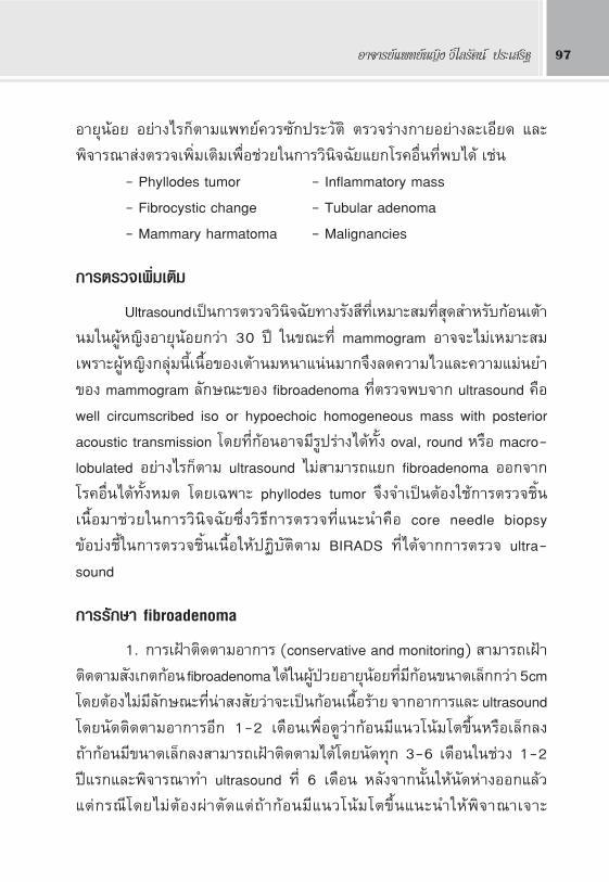
Ultrasound ‡ªìπ°“√μ√«®«‘π‘®©—¬∑“ß√—ß ’∑’ˇÀ¡“– ¡∑’Ë ÿ¥ ”À√—∫°âÕπ‡μâ“
π¡„πºŸâÀ≠‘ßÕ“¬ÿπâÕ¬°«à“ 30 ªï „π¢≥–∑’Ë mammogram Õ“®®–‰¡à‡À¡“– ¡
‡æ√“–ºŸâÀ≠‘ß°≈ÿà¡π’ȇπ◊ÈÕ¢Õ߇μâ“π¡Àπ“·πàπ¡“°®÷ß≈¥§«“¡‰«·≈–§«“¡·¡àπ¬”
¢Õß mammogram ≈—°…≥–¢Õß fibroadenoma ∑’Ëμ√«®æ∫®“° ultrasound §◊Õ
well circumscribed iso or hypoechoic homogeneous mass with posterior
acoustic transmission ‚¥¬∑’Ë°âÕπÕ“®¡’√Ÿª√à“߉¥â∑—Èß oval, round À√◊Õ macro-
lobulated Õ¬à“߉√°Áμ“¡ ultrasound ‰¡à “¡“√∂·¬° fibroadenoma ÕÕ°®“°
‚√§Õ◊Ëπ‰¥â∑—ÈßÀ¡¥ ‚¥¬‡©æ“– phyllodes tumor ®÷ß®”‡ªìπμâÕß„™â°“√μ√«®™‘Èπ
‡π◊ÈÕ¡“™à«¬„π°“√«‘π‘®©—¬´÷Ëß«‘∏’°“√μ√«®∑’Ë·π–π”§◊Õ core needle biopsy
¢âÕ∫àß™’È„π°“√μ√«®™‘Èπ‡π◊ÈÕ„ÀâªØ‘∫—μ‘μ“¡ BIRADS ∑’ˉ¥â®“°°“√μ√«® ultra-
sound
°“√√—°…“ fibroadenoma
1. °“√‡ΩÑ“μ‘¥μ“¡Õ“°“√ (conservative and monitoring) “¡“√∂‡ΩÑ“
μ‘¥μ“¡ —߇°μ°âÕπ fibroadenoma ‰¥â„πºŸâªÉ«¬Õ“¬ÿπâÕ¬∑’Ë¡’°âÕπ¢π“¥‡≈Á°°«à“ 5cm
‚¥¬μâÕ߉¡à¡’≈—°…≥–∑’Ëπà“ ß —¬«à“®–‡ªìπ°âÕπ‡π◊ÈÕ√⓬ ®“°Õ“°“√·≈– ultrasound
‚¥¬π—¥μ‘¥μ“¡Õ“°“√Õ’° 1-2 ‡¥◊Õπ‡æ◊ËÕ¥Ÿ«à“°âÕπ¡’·π«‚πâ¡‚μ¢÷ÈπÀ√◊Õ‡≈Á°≈ß
∂â“°âÕπ¡’¢π“¥‡≈Á°≈ß “¡“√∂‡ΩÑ“μ‘¥μ“¡‰¥â‚¥¬π—¥∑ÿ° 3-6 ‡¥◊Õπ„π™à«ß 1-2
ªï·√°·≈–æ‘®“√≥“∑” ultrasound ∑’Ë 6 ‡¥◊Õπ À≈—ß®“°π—Èπ„Àâπ—¥Àà“ßÕÕ°·≈â«
·μà°√≥’‚¥¬‰¡àμâÕߺà“μ—¥·μà∂â“°âÕπ¡’·π«‚πâ¡‚μ¢÷Èπ·π–π”„Àâæ‘®“≥“‡®“–
Fibroadenoma and Phyllodes Tumor98
μ√«®™‘Èπ‡π◊ÈÕ10,11
2. °“√ºà“μ—¥°âÕπÕÕ°∑—ÈßÀ¡¥ (excision) ¡’·π«∑“ßæ‘®“√≥“ºà“μ—¥°âÕπ
∑’Ë ß —¬ fibroadenoma ¥—ßπ’È12
- Rapidly growing mass
- Mass diameter > 5 cm (giant fibroadenoma)
- Mass causing distortion of breast architecture or overlying skin
changes
- Persistent mass without regression
- Multiple and bilateral breast masses
- Stromal hypercellularity or cystic change on ultrasound
- Symptoms and signs worrisome for malignancy
- Presence of a high-risk genetic mutation
- Histologically complex fibroadenoma
´÷Ëß°√≥’∑’ËÕ“°“√∑“ߧ≈‘π‘° ß —¬ phyllodes tumor À√◊Õ°âÕπ¡–‡√Áߧ«√
∑” CNB °àÕπ«“ß·ºπºà“μ—¥ À≈—ßμ—¥°âÕπÕÕ°·≈â« à«π„À≠à‰¡à®”‡ªìπμâÕ߇ √‘¡
‡μâ“π¡ (augmentation) ·¡â°√–∑—Ëß°√≥’ giant fibroadenoma ‡æ√“–‡π◊ÈÕ‡¬◊ËÕ
‡μâ“π¡∑’ˇ§¬∂Ÿ°°¥‡∫’¬¥®–¢¬“¬μ—«¡“‡μ‘¡‡μÁ¡™àÕß«à“ß∑’ˇ°‘¥¢÷ÈπÀ≈—ߺà“μ—¥
3. Vacuum-assisted ultrasound-guided percutaneous excision
‡ªìπ«‘∏’√—°…“∑’Ë∑”„À⇰‘¥∫“¥·º≈πâÕ¬ (minimally invasive technique)
‡À¡“–°—∫°âÕπ fibroadenoma ∑’Ë¡’¢π“¥‡≈Á° ‚¥¬„™â‡¢Á¡ vacuum biopsy ∑’Ë¡’
¢π“¥„À≠à·≈â«„™â ultrasound „π°“√π”∑“ß ¡’√“¬ß“πº≈ ”‡√Á® 75-100%13
·μà¡’°“√°≈—∫‡ªìπ´È”‰¥â ‡™◊ËÕ«à“‡°‘¥®“°‡»…°âÕπ‡π◊ÈÕ∑’Ë¡’¢π“¥‡≈Á°¡“°®π ultra-
sound ¡Õ߉¡à‡ÀÁπ ®÷ߥŸ¥ÕÕ°¡“‰¡àÀ¡¥ ‚¥¬æ∫«à“°“√°≈—∫‡ªìπ´È”¡—°‡°‘¥°—∫
°âÕπ∑’Ë¡’¢π“¥„À≠à°«à“ 2cm æ∫°≈—∫‡ªìπ´È”‰¥â 33% ‡¡◊ËÕμ‘¥μ“¡ºŸâªÉ«¬π“π 59
‡¥◊Õπ¥—ßπ—Èπ§«√‡≈◊Õ°„™â«‘∏’π’È„π°√≥’°âÕπ∑’Ë¡’¢π“¥‡≈Á°°«à“ 2cm ·≈–ºŸâªÉ«¬‰¡à
Õ¬“°‡ΩÑ“μ‘¥μ“¡Õ“°“√ ‚¥¬‡©æ“–Õ¬à“߬‘Ëß°√≥’∑’Ë¡’À≈“¬°âÕπ√à«¡°—∫ºŸâªÉ«¬¡’
§«“¡‡ ’Ë¬ß Ÿß„π°“√‡°‘¥·º≈‡ªìπ™π‘¥ keloid
Õ“®“√¬å·æ∑¬åÀ≠‘ß «‘‰≈√—μπå ª√–‡ √‘∞ 99
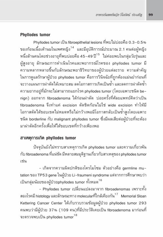
Phyllodes tumorPhyllodes tumor ‡ªìπ fibroepithelial lesions ∑’Ëæ∫‰¡à∫àÕ¬§◊Õ 0.3-0.5%
¢Õß°âÕπ‡π◊ÈÕ‡μâ“π¡„π‡æ»À≠‘ß14 ·≈–¡’Õÿ∫—μ‘°“√≥åª√–¡“≥ 2.1 §πμàÕºŸâÀ≠‘ß
Àπ÷Ëß≈â“π§π‚¥¬™à«ßÕ“¬ÿ∑’Ëæ∫∫àÕ¬§◊Õ 45-49 ªï15 ‰¡à§àÕ¬æ∫„π°≈ÿà¡«—¬√ÿàπ·≈–
ºŸâ ŸßÕ“¬ÿ ≈—°…≥–°“√¥”‡π‘π‚√§·≈–欓°√≥å‚√§¢Õß phyllodes tumor ¡’
§«“¡À≈“°À≈“¬¢÷Èπ°—∫≈—°…≥–欓∏‘«‘∑¬“¢ÕߺŸâªÉ«¬·μà≈–√“¬ §«“¡ ”§—≠
„π°“√¥Ÿ·≈√—°…“ºŸâªÉ«¬ phyllodes tumor §◊Õ°“√«‘π‘®©—¬∑’Ë∂Ÿ°μâÕß·¡à𬔰àÕπ∑’Ë
®–«“ß·ºπ°“√ºà“μ—¥‰¥â‡À¡“– ¡ ≈¥‚Õ°“ °“√‡°‘¥‡ªìπ È” ·≈–≈¥°“√ºà“μ—¥ È”
§«“¡¬“°Õ¬Ÿà∑’Ë¡—°®–‰¡à “¡“√∂·¬°‚√§ phyllodes tumor (‚¥¬‡©æ“–™π‘¥ be-
nign) ÕÕ°®“° fibroadenoma ‰¥â°àÕπºà“μ—¥ ∫àÕ¬§√—Èß∑’Ë»—≈¬·æ∑¬å§‘¥«à“‡ªìπ
fibroadenoma ®÷ß∑”·§à excision μ—¥™‘¥°âÕπ‰¡à„™à wide excision ∑”„Àâ¡’
‚Õ°“ μ—¥‰¥â¢Õ∫‡¢μ‰¡àÀ¡¥À√◊Õ‰¡à°«â“ßæÕ¡’‚Õ°“ °≈—∫‡ªìπ È” Ÿß‚¥¬‡©æ“–
™π‘¥ borderline °—∫ malignant phyllodes tumor ÷Ëß¡’º≈‡ ’¬μàÕºŸâªÉ«¬∑’Ë®–μâÕß
¡“ºà“μ—¥Õ’°§√—È߇æ◊ËÕ„À≥â¢Õ∫‡¢μ∑’Ë°«â“߇撬ßæÕ
“‡Àμÿ°“√‡°‘¥ phyllodes tumor
ªí®®ÿ∫—π¬—߉¡à∑√“∫ “‡Àμÿ°“√‡°‘¥ phyllodes tumor ·≈–§«“¡‡°’ˬ«æ—π
°—∫ fibroadenoma ∑’Ë·πà™—¥ ¡’À≈“¬ ¡¡ÿμ‘∞“π‡°’Ë¬«°—∫ “‡Àμÿ¢Õß phyllodes tumor
‡™àπ
- ‡°‘¥®“°§«“¡º‘¥ª°μ‘¢Õß‚§√‚¡‚´¡ μ—«Õ¬à“ߧ◊Õ germline mu-
tation ¢Õß TP53 gene „πºŸâªÉ«¬ Li-fraumeni syndrome ·μஓ°°“√»÷°…“æ∫«à“
‡ªìπ°≈ÿà¡πâÕ¬¢ÕߺŸâªÉ«¬phyllodes tumor ∑—ÈßÀ¡¥16
- Phyllodes tumor ‡ª≈’ˬπ·ª≈ß¡“®“° fibroadenomas ‡æ√“–∑—Èß
Õß‚√§¡’ histology ·≈–≈—°…≥–∑“ß molecular∑’Ë„°≈⇧’¬ß°—π17 Memorial Sloan
Kettering Cancer Center ‰¥â‡°Á∫√«∫√«¡¢âÕ¡Ÿ≈ºŸâªÉ«¬ phyllodes tumor 293
§πæ∫«à“¡’ºŸâªÉ«¬ 37% (109 §π)∑’Ë¡’ª√–«—쑇§¬‡ªìπ fibroadenoma ¡“°àÕπ∑’Ë
®–μ√«®æ∫‡ªìπ phyllodes tumor18
Fibroadenoma and Phyllodes Tumor100
- ‡™◊ËÕ«à“¿“«–ª°μ‘ epithelium °—∫ stroma ®–¡’°“√§«∫§ÿ¡´÷Ëß°—π·≈–
°—π‰¡à„À⇮√‘≠º‘¥ª°μ‘ (interactions between the epithelium and stroma)
·μà‡¡◊ËÕ¡’§«“¡º‘¥ª°μ‘ ‡ ’¬°“√§«∫§ÿ¡π’È (loss of the stromal-epithelial inter-
dependency) ®–∑”„Àâ stromal cells ¡’°“√·∫àßμ—«¡“°º‘¥ª°μ‘‰¥â19
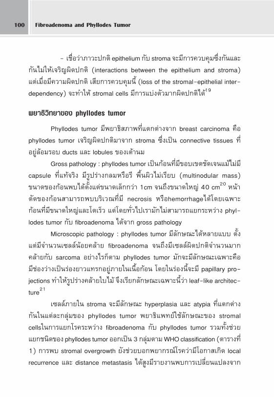
欓∏‘«‘∑¬“¢Õß phyllodes tumor
Phyllodes tumor ¡’欓∏‘ ¿“æ∑’Ë·μ°μà“ß®“° breast carcinoma §◊Õ
phyllodes tumor ‡®√‘≠º‘¥ª°μ‘¡“®“° stroma ´÷Ë߇ªìπ connective tissues ∑’Ë
Õ¬Ÿà≈âÕ¡√Õ∫ ducts ·≈– lobules ¢Õ߇μâ“π¡
Gross pathology : phyllodes tumor ‡ªìπ°âÕπ∑’Ë¡’¢Õ∫‡¢μ™—¥‡®π·¡â‰¡à¡’
capsule ∑’Ë·∑â®√‘ß ¡’√Ÿª√à“ß°≈¡À√◊Õ√’ æ◊Èπº‘«‰¡à‡√’¬∫ (multinodular mass)
¢π“¥¢Õß°âÕπæ∫‰¥âμ—Èß·μà¢π“¥‡≈Á°°«à“ 1cm ®π∂÷ß¢π“¥„À≠à 40 cm20 Àπâ“
μ—¥¢Õß°âÕπ “¡“√∂æ∫∫√‘‡«≥∑’Ë¡’ necrosis À√◊Õhemorrhage‰¥â‚¥¬‡©æ“–
°âÕπ∑’Ë¡’¢π“¥„À≠à·≈–‚μ‡√Á« ·μà‚¥¬∑—Ë«‰ª‡√“¡—°‰¡à “¡“√∂·¬°√–À«à“ß phyl-
lodes tumor °—∫ fibroadenoma ‰¥â®“° gross pathology
Microscopic pathology : phyllodes tumor ¡’≈—°…≥–‰¥âÀ≈“¬·∫∫ μ—Èß
·μà¡’®”π«π‡´≈≈åπâÕ¬§≈⓬ fibroadenoma ®π∂÷ß¡’‡´≈≈庑¥ª°μ‘®”π«π¡“°
§≈⓬°—∫ sarcoma Õ¬à“߉√°Áμ“¡ phyllodes tumor ¡—°®–¡’≈—°…≥–‡©æ“–§◊Õ
¡’™àÕß«à“߇ªìπ√àÕ߬“«·∑√°Õ¬Ÿà¿“¬„π‡π◊ÈÕ°âÕπ ‚¥¬„π√àÕßπ’È®–¡’ papillary pro-
jections ∑”„Àâ√Ÿª√à“ߧ≈⓬„∫‰¡â ®÷߇√’¬°≈—°…≥–‡©æ“–π’È«à“ leaf-like architec-
ture21
‡´≈≈å¿“¬„π stroma ®–¡’≈—°…≥– hyperplasia ·≈– atypia ∑’Ë·μ°μà“ß
°—π„π·μà≈–°≈ÿà¡¢Õß phyllodes tumor 欓∏‘·æ∑¬å„™â≈—°…≥–¢Õß stromal
cells„π°“√·¬°‚√§√–À«à“ß fibroadenoma °—∫ phyllodes tumor √«¡∑—Èߙ૬
·¬°™π‘¥¢Õß phyllodes tumor ÕÕ°‡ªìπ 3 °≈ÿà¡μ“¡ WHO classification (μ“√“ß∑’Ë
1) °“√æ∫ stromal overgrowth ¬—ߙ૬∫հ欓°√≥å‚√§«à“¡’‚Õ°“ ‡°‘¥ local
recurrence ·≈– distance metastasis ‰¥â Ÿß¡’√“¬ß“πæ∫°“√‡ª≈’ˬπ·ª≈ß®“°
Õ“®“√¬å·æ∑¬åÀ≠‘ß «‘‰≈√—μπå ª√–‡ √‘∞ 101
phyllodes tumor °≈“¬‡ªìπ sarcomas (sarcomatous differentiation) ‰¥â·μà
πâÕ¬§◊Õ< 5% ¢Õß phyllodes tumor ∑—ÈßÀ¡¥22
°“√®”·π°™π‘¥¢Õß phyllodes tumor
„πªï §.». 1981 Õߧ尓√Õπ“¡—¬‚≈°‰¥â·∫àß phyllodes tumor ÕÕ°‡ªìπ
3 °≈ÿࡧ◊Õ benign, borderline ·≈– malignant phyllodes tumor ‚¥¬Õ“»—¬
‡°≥±å∑“ß欓∏‘«‘∑¬“23 benign phyllodes tumor æ∫‡ªìπ —¥ à«π¡“°∑’Ë ÿ¥§◊Õ
35-64% ∑’ˇÀ≈◊ÕÕ¬à“ß≈–§√÷Ëß®–‡ªìπ™π‘¥ borderline °—∫ malignant24
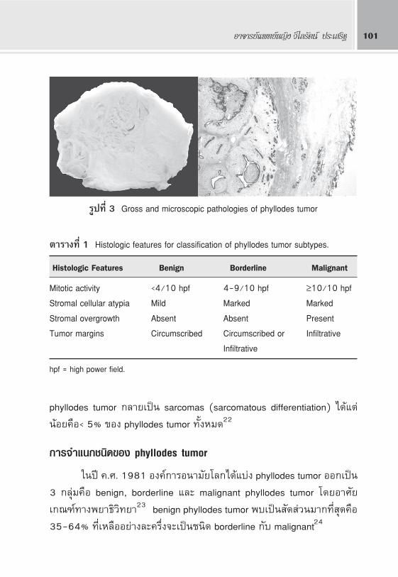
μ“√“ß∑’Ë 1 Histologic features for classification of phyllodes tumor subtypes.
Histologic Features Benign Borderline Malignant
Mitotic activity <4/10 hpf 4-9/10 hpf ≥10/10 hpfStromal cellular atypia Mild Marked MarkedStromal overgrowth Absent Absent PresentTumor margins Circumscribed Circumscribed or Infiltrative
Infiltrative
hpf = high power field.
√Ÿª∑’Ë 3 Gross and microscopic pathologies of phyllodes tumor
Fibroadenoma and Phyllodes Tumor102
Õ“°“√∑“ߧ≈‘π‘°¢Õß phyllodes tumor25
- ¡—°æ∫„πºŸâÀ≠‘ß™à«ßÕ“¬ÿ 35-55 ªï (¡“°°«à“°≈ÿà¡ fibroadenoma
ª√–¡“≥ 20 ªï)
- °âÕπ‚μ‡√Á«¿“¬„π√–¬–‡«≈“‡ªìπ‡¥◊Õπ À√◊Õ‡ªìπ°âÕπ∑’˧≈”æ∫¡“
À≈“¬ªï°àÕπ∑’Ë®–‚μ‡√Á«¢÷Èπ„π™à«ß√–¬–À≈—ß
- °âÕπ¡—°§≈”‰¥â¢Õ∫‡¢μ™—¥‡®π‚¬°°âÕπ‰¥âßà“¬ ‰¡àμ‘¥°—∫‡π◊ÈÕ‡¬◊ËÕ¢â“ß
‡§’¬ß ´÷Ëߧ≈⓬°—∫ fibroadenoma
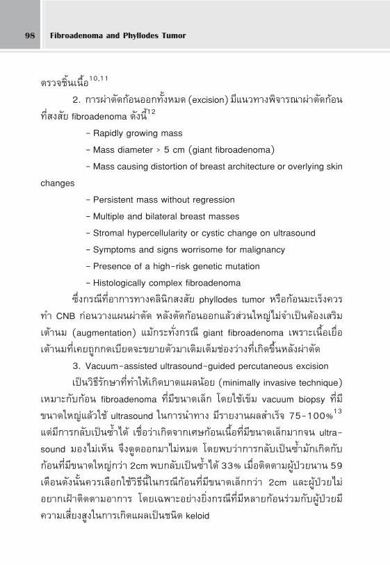
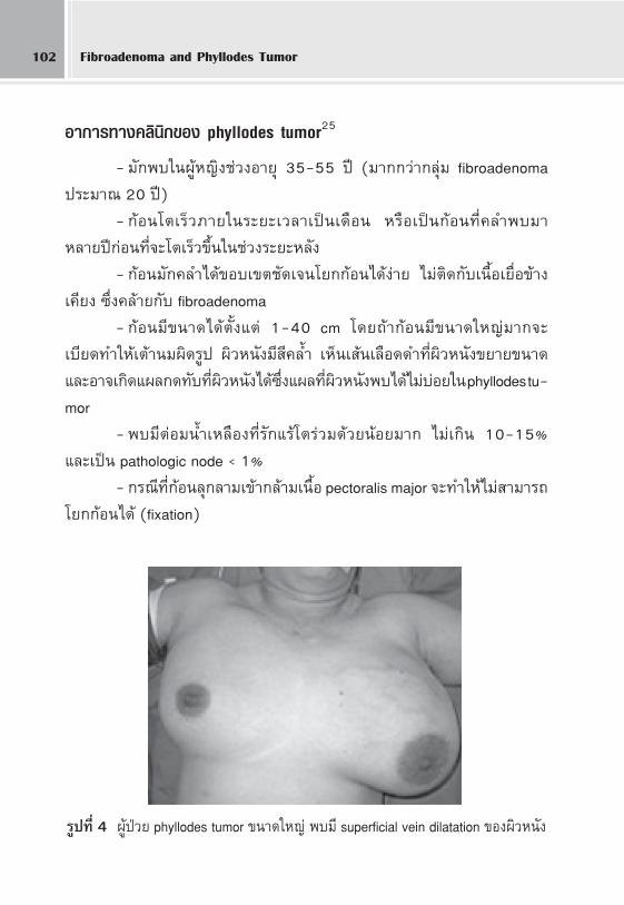
- °âÕπ¡’¢π“¥‰¥âμ—Èß·μà 1-40 cm ‚¥¬∂â“°âÕπ¡’¢π“¥„À≠à¡“°®–
‡∫’¬¥∑”„Àâ‡μâ“π¡º‘¥√Ÿª º‘«Àπ—ß¡’ ’§≈È” ‡ÀÁπ‡ âπ‡≈◊Õ¥¥”∑’˺‘«Àπ—ߢ¬“¬¢π“¥
·≈–Õ“®‡°‘¥·º≈°¥∑—∫∑’˺‘«Àπ—߉¥â ÷Ëß·º≈∑’˺‘«Àπ—ßæ∫‰¥â‰¡à∫àÕ¬„π phyllodes tu-
mor
- æ∫¡’μàÕ¡πÈ”‡À≈◊Õß∑’Ë√—°·√â‚μ√à«¡¥â«¬πâÕ¬¡“° ‰¡à‡°‘π 10-15%
·≈–‡ªìπ pathologic node < 1%
- °√≥’∑’Ë°âÕπ≈ÿ°≈“¡‡¢â“°≈â“¡‡π◊ÈÕ pectoralis major ®–∑”„Àâ‰¡à “¡“√∂
‚¬°°âÕπ‰¥â (fixation)
√Ÿª∑’Ë 4 ºŸâªÉ«¬ phyllodes tumor ¢π“¥„À≠à æ∫¡’ superficial vein dilatation ¢Õߺ‘«Àπ—ß
Õ“®“√¬å·æ∑¬åÀ≠‘ß «‘‰≈√—μπå ª√–‡ √‘∞ 103
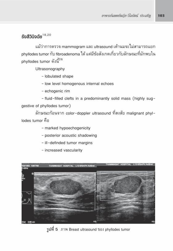
√—ß ’«‘π‘®©—¬18,20
·¡â«à“°“√μ√«® mammogram ·≈– ultrasound ‡μâ“π¡®–‰¡à “¡“√∂·¬°
phyllodes tumor °—∫ fibroadenoma ‰¥â ·μà¡’¢âÕ —߇°μ‡°’ˬ«°—∫≈—°…≥–∑’Ë¡—°æ∫„π
phyllodes tumor ¥—ßπ’È26
Ultrasonography
- lobulated shape
- low level homogenous internal echoes
- echogenic rim
- fluid-filled clefts in a predominantly solid mass (highly sug-
gestive of phyllodes tumor)
≈—°…≥–°âÕπ®“° color-doppler ultrasound ∑’Ë ß —¬ malignant phyl-
lodes tumor §◊Õ
- marked hypoechogenicity
- posterior acoustic shadowing
- ill-definded tumor margins
- increased vascularity
√Ÿª∑’Ë 5 ¿“æ Breast ultrasound ¢Õß phyllodes tumor
Fibroadenoma and Phyllodes Tumor104
Mammography
- well circumscribed oval or lobulated mass with rounded bor-
ders
- radiolucent halo may be seen
- coarse calcification may be present (malignant microcalcification
is rare)
°“√μ√«®«‘π‘®©—¬∑“ß欓∏‘«‘∑¬“
FNA : ‡π◊ËÕß®“°∑—Èß phyllodes tumor ·≈– fibroadenoma ‡ªìπ‡π◊ËÕßÕ°
„π°≈ÿà¡ fibroepithellial lesions μâÕßÕ“»—¬°“√¥Ÿ√“¬≈–‡Õ’¬¥¢Õß stromal cells
®÷ß®– “¡“√∂«‘π‘®©—¬·¬°‚√§‰¥â·¡à𬔠·μà FNA „Àâ¢âÕ¡Ÿ≈‡ªìπ cytology ®÷߉¡à
‡À¡“– ¡„π°“√„™â«‘π‘®©—¬·¬°‚√§√–À«à“ß phyllodes tumor °—∫ fibroadenoma
¡’°“√»÷°…“æ∫«à“ FNA ¡’§«“¡·¡à𬔄π°“√«‘π‘®©—¬ phyllodes tumor ‡æ’¬ß·§à
63%27
Core needle biopsy (CNB) : ¡’§«“¡πà“‡™◊ËÕ∂◊Õ¡“°°«à“ FNA ‡æ√“–‰¥â
™‘Èπ‡π◊ÈÕ‡ªìπ histology “¡“√∂„Àâ√“¬≈–‡Õ’¬¥¢Õß stromal cells ‰¥â¥’°«à“FNA
¡’°“√»÷°…“æ∫«à“ CNB ¡’ sensitivity 99%, negative predictive value 93%
·≈– positive predictive value 83% „π°“√«‘π‘®©—¬ phyllodes tumor28
Phyllodes tumor ¡’≈—°…≥– histology ∑’Ë·μ°μà“ß®“° fibroadenoma ¥—ßπ’È
- increased stromal cellularity
- increased mitotic activity
- stromal cell atypia
Õ¬à“߉√°Áμ“¡∫“ߧ√—Èß CNB °Á‰¡à “¡“√∂·¬° phyllodes tumor °—∫fi-
broadenoma ‰¥â·¡à𬔇æ√“– „π benign phyllodes tumor Õ“®‰¡à¡’ cellular
atypia ·≈–‰¡àæ∫ mitotic activity ∑’ˇæ‘Ë¡º‘¥ª°μ‘‰¥â„π¢≥–‡¥’¬«°—π juvenile fi-
broadenoma °Á “¡“√∂¡’®”π«π stromal cells ‡æ‘Ë¡¢÷Èπ‰¥â ®÷ß®”‡ªìπμâÕß∑” ex-
cision ‡Õ“°âÕπÕÕ°¡“μ√«®∑—Èß°âÕπ‡æ◊ËÕ°“√«‘π‘®©—¬∑’Ë∂Ÿ°μâÕß
Õ“®“√¬å·æ∑¬åÀ≠‘ß «‘‰≈√—μπå ª√–‡ √‘∞ 105
Excisional biopy : ¡’∫∑∫“∑ ”§—≠„π°√≥’∑’˺≈®“° CNB „À⧔μÕ∫
‰¡à™—¥‡®π ∑—È߬—߇ªìπ°“√√—°…“¥â«¬ ”À√—∫ fibroadenoma ¡’‡°≥±å∑’Ëπà“ —߇°μ
‡æ◊Ëՙ૬„π°“√μ—¥ ‘π„®«à“ºŸâªÉ«¬√“¬„¥¡’§«“¡‡ªìπ‰ª‰¥â Ÿß∑’Ë®–‡ªìπ phyllodes tu-
mor ´÷Ëߧ«√∑” CNB ‡æ◊ËÕ„À≥Ⱃ√«‘π‘®©—¬∑’Ë∂Ÿ°μâÕß°àÕπ«“ß·ºπºà“μ—¥∑’ˇÀ¡“–
¡∂â“æ∫≈—°…≥–‡¢â“‡°≥±å¥â“π≈à“ßπ’ÈÕ¬à“ßπâÕ¬ ÕߢâÕ ·π–π”„Àâ∑” CNB °àÕπ
excision
Paddington Clinicopathological Suspicious Score29
Clinical findings :
1) Sudden increase in size in a longstanding breast lesion
2) Apparent fibroadenoma > 3 cm diameter or in patient > 35
years
Imaging findings :
1) Rounded borders/ lobulated appearance at mammography
2) Attenuation or cystic areas within a solid mass on ultrasonog-
raphy
FNA findings :
1) Presence of hypercellularity stromal fragments
2) Indeterminate features
ANY 2 features mandate core biopsy
Tumor markers in phyllodes tumor
ªí®®ÿ∫—π∫∑∫“∑¢Õß tumor markers „π phyllodes tumor ¬—߉¡à™—¥‡®π
·≈–¬—߉¡à‰¥âπ”¡“„™âª√–‚¬™πå∑“ߧ≈‘π‘° μ—«Õ¬à“ß°“√»÷°…“‡°’ˬ«°—∫ tumor mark-
ers „π phyllodes tumor ‡™àπæ∫ p53 ·≈– CD117 ‰¥â∫àÕ¬„π malignant phyl-
lodes ‚¥¬¡—°®– —¡æ—π∏å°—∫ high histologic grade ·≈– tumor ∑’Ë¡’ CD117 pro-
tein expression ®–æ∫ recurrence ‰¥â∫àÕ¬30
°“√√—°…“ phyllodes tumor
Fibroadenoma and Phyllodes Tumor106
°“√ºà“μ—¥ (surgical management)
°“√ºà“μ—¥‡ªìπ°“√√—°…“À≈—°¢Õß phyllodes tumor ¡ÿàßÀ¡“¬‡æ◊ËÕ‡Õ“°âÕπ
ÕÕ°„ÀâÀ¡¥ ·≈– ≈¥‚Õ°“ °≈—∫‡ªìπ´È” ·π–π”„Àâ«“ß·ºπ°“√√—°…“·¬°μ“¡
·μà≈– ∂“π°“√≥套ßπ’È
● °√≥’∑√“∫°àÕπºà“μ—¥«à“‡ªìπ phyllodes tumor
·π–π”„Àâ∑” wide excision ‚¥¬æ¬“¬“¡μ—¥„Àâ°«â“ßÕ¬à“ßπâÕ¬ 1 cm
‚¥¬√Õ∫°âÕπ ‚¥¬‡©æ–Õ¬à“߬‘ËߺŸâªÉ«¬°≈ÿà¡ borderline ·≈– malignant phyllodes
tumors ´÷Ëß¡’‚Õ°“ °≈—∫‡ªìπ´È” Ÿß°“√æ‘®“√≥“‡≈◊Õ°√–À«à“ß°“√∑” wide exci-
sion À√◊Õ mastectomy π—Èπ¢÷ÈπÕ¬Ÿà°—∫¢π“¥¢Õß°âÕπ‡∑’¬∫°—∫¢π“¥‡μâ“π¡¢Õß
ºŸâªÉ«¬°“√‡∫’¬¥∫—߇π◊ÈÕ‡μâ“π¡·≈–°“√≈ÿ°≈“¡ Ÿà‡π◊ÈÕ‡¬◊ËÕ¢â“߇§’¬ß °√≥’∑’Ë°âÕπ¡’
¢π“¥„À≠àÀ≈—ß∑” wide excision ·≈â«¡—°®–¡’‡μâ“π¡º‘¥√Ÿª ®÷ß®”‡ªìπμâÕߺà“μ—¥
μ°·μà߇ √‘¡‡μâ“π¡ (breast reconstruction) ¥â«¬„π§√“«‡¥’¬«°—π‡æ◊ËÕ§«“¡
«¬ß“¡‚¥¬‡©æ“–°√≥’ Giant phyllodes tumor (phyllodes tumor ∑’Ë¡’¢π“¥
>10 cm æ∫‰¥â 20% ¢Õß phyllodes tumor ∑—ÈßÀ¡¥25 ºŸâªÉ«¬°≈ÿà¡π’È¡—°®–μâÕß
∑” mastectomy ‚¥¬®–‡≈◊Õ° total mastectomy À√◊Õ subcutaneous mastec-
tomy π—Èπ¢÷Èπ°—∫°“√≈ÿ°≈“¡¢Õß°âÕπ«à“®– “¡“√∂√—°…“º‘«Àπ—ßÀ√◊ÕÀ—«π¡‰«â
‰¥âÀ√◊Õ‰¡à
● °√≥’‰¡à∑√“∫°àÕπºà“μ—¥«à“‡ªìπ phyllodes tumors ·≈–‰¥â√—∫°“√ºà“μ—¥
excision ¡“·≈â«
ºŸâªÉ«¬∑’ˉ¥â√—∫°“√ºà“μ—¥°âÕπ‡π◊ÈÕ·∫∫ local excision ·≈⫺≈™‘Èπ‡π◊ÈÕ
ÕÕ°¡“‡ªìπ phyllodes tumor §«√æ‘®“√≥“·π«∑“ß°“√√—°…“®“°º≈™‘Èπ‡π◊ÈÕ∑’Ë
‰¥â¥—ßπ’È
1. °√≥’ benign phyllodes tumor
- ∂⓺≈™‘Èπ‡π◊ÈÕæ∫«à“‰¥â negative margin ·π–π”„Àâ‡ΩÑ“μ‘¥μ“¡
Õ“°“√ºŸâªÉ«¬‰¥â (wait and watch policy) ‰¡à®”‡ªìπμâÕߺà“μ—¥ È” ¡’√“¬ß“πæ∫
«à“‡¡◊ËÕμ‘¥μ“¡ºŸâªÉ«¬°≈ÿà¡π’ȉª 5 ªï®–æ∫°“√‡°‘¥‡ªìπ´È”ª√–¡“≥ 4% ·≈–¡’
Õ—μ√“°“√Õ¬Ÿà√Õ¥ 96%25
Õ“®“√¬å·æ∑¬åÀ≠‘ß «‘‰≈√—μπå ª√–‡ √‘∞ 107
- ∂⓺≈™‘Èπ‡π◊ÈÕæ∫«à“‰¥â positive margin ¬—ß¡’¢âÕ∂°‡∂’¬ß°—π‡°’Ë¬«°—∫
·π«∑“ߪؑ∫—μ‘«à“§«√®–°≈—∫‰ªºà“μ—¥Õ’°§√—Èß∑—π∑’‡æ◊ËÕ‡Õ“°âÕπ‡π◊ÈÕÕÕ°„ÀâÀ¡¥
‡æ◊ËÕ≈¥°“√‡°‘¥‡ªìπ´È” À√◊Õ®–‡ΩÑ“μ‘¥μ“¡ºŸâªÉ«¬Õ¬à“ß„°≈♑¥∂â“æ∫«à“¡’°“√°≈—∫
‡ªìπ´È”§àÕ¬¡“ºà“μ—¥°≈ÿà¡»—≈¬·æ∑¬å∑’ˬա√—∫«à“°“√‡ΩÑ“μ‘¥μ“¡ºŸâªÉ«¬‡ªìπ∑“ß
‡≈◊Õ°Àπ÷Ëß∑’ˬա√—∫‰¥â‚¥¬Õâ“ßÕ‘ß°“√»÷°…“∑’Ëæ∫«à“¡’·§à 8% ¢ÕߺŸâªÉ«¬benign
phyllodes tumor ∑’Ë®–‡°‘¥‚√§ È”´÷ËßμË”°«à“ºŸâªÉ«¬°≈ÿà¡ borderline °—∫malignant
phyllodes tumor ∑’Ë¡’‚Õ°“ °≈—∫‡ªìπ´È” 20-23%31
2. °√≥’ borderline À√◊Õ malignant phyllodes tumor
·π–π”„Àâ∑”ºà“μ—¥Õ’°§√—È߇æ◊ËÕ„À≥â¢Õ∫‡¢μ∑’Ë°«â“ߢ÷Èπ (re-excision)
‡æ√“–ºŸâªÉ«¬°≈ÿà¡π’È¡’‚Õ°“ °≈—∫‡ªìπ´È” Ÿß
Breast reconstruction
°√≥’∑’˺ŸâªÉ«¬‰¥â√—∫°“√ºà“μ—¥ wide excision ‡μâ“π¡¡’‚Õ°“ º‘¥√Ÿª À√◊Õ
‰¥â√—∫°“√ºà“μ—¥ mastectomy »—≈¬·æ∑¬å “¡“√∂∑”ºà“μ—¥μ°·μà߇ √‘¡‡μâ“π¡
„À⺟âªÉ«¬‰¥â„π§√“«‡¥’¬«°—π (immediate breast reconstruction) ‡æ◊ËÕ§«“¡
«¬ß“¡´÷Ëß¡’º≈μàÕ§ÿ≥¿“æ™’«‘μ¢ÕߺŸâªÉ«¬ ‚¥¬∑’Ë°“√ºà“μ—¥ breast reconstruc-
tion π’ȉ¡à¡’º≈‡ ’¬μàÕ°“√‡ΩÑ“μ‘¥μ“¡°“√°≈—∫‡ªìπ È”¢Õß‚√§ ¥—ßπ—Èπ®÷߉¡à¡’¢âÕ
Àâ“¡„π°“√ºà“μ—¥ immediate breast reconstruction À≈—ß mastectomy „πºŸâªÉ«¬
giant phyllodes tumor32
Axillary lymph node management
°“√ºà“μ—¥μàÕ¡πÈ”‡À≈◊Õß∑’Ë√—°·√â„πºŸâªÉ«¬ phyllodes tumor π—Èπ‰¡à·π–π”
„Àâ∑”∑ÿ°√“¬ ‰¡à«à“®–‡ªìπ sentinel lymph node biopsy À√◊Õ axillary lymph
node dissection ‡æ√“– malignant phyllodes tumor ¡—°·æ√à°√–®“¬∑“ß
À≈Õ¥‡≈◊Õ¥ (hematogenous spread) ·≈–¡’√“¬ß“πæ∫°“√°√–®“¬¡“μàÕ¡πÈ”
‡À≈◊Õß∑’Ë√—°·√âπâÕ¬¡“°§◊Õ < 1% ·π–π”„Àâºà“μ—¥‡≈“–μàÕ¡πÈ”‡À≈◊Õß∑’Ë√—°·√⇩擖
°√≥’∑’Ë°àÕπºà“μ—¥æ∫«à“¡’μàÕ¡πÈ”‡À≈◊Õß‚μ·≈–‰¥âæ‘ Ÿ®πå·≈â««à“¡’¡–‡√Áß°√–®“¬
¡“®√‘ß (histologically positive for malignant cells)33,34 °√≥’°àÕπºà“μ—¥æ∫
«à“¡’μàÕ¡πÈ”‡À≈◊Õß√—°·√â‚μ ∑—Èß®“° clinical À√◊Õ imaging ·π–π”„À⇮“–μ√«®
Fibroadenoma and Phyllodes Tumor108
‚¥¬„™â ultrasound guided core needle biopsy
Chest wall invasion25
°“√≈ÿ°≈“¡∂÷ß chest wall ¢Õß phyllodes tumor π—Èπæ∫‰¥â‰¡à∫àÕ¬ ‚¥¬
¡—°®–æ∫„π√“¬∑’Ë°âÕπ„À≠à¡“°Ê·≈–‡ªìπ™π‘¥ malignant phyllodes ‡¡◊ËÕ ß —¬
«à“¡’ chest wall invasion ®“°ª√–«—μ‘·≈–μ√«®√à“ß°“¬§«√ àßμ√«® CT chest
‡æ◊ËÕª√–‡¡‘π°“√≈ÿ°≈“¡¢Õß°âÕπ°àÕπ«“ß·ºπºà“μ—¥‡æ◊ËÕ„À≥â¢Õ∫‡¢μ∑’Ë°«â“ßæÕ
Õ“®μâÕßμ—¥°≈â“¡‡π◊ÈÕ pectoralis major ÕÕ°‰ª°—∫‡μâ“π¡·≈–∫“ß√“¬®”‡ªìπ
μâÕßμ—¥°√–¥Ÿ°´’Ë‚§√ßÕÕ°‰ª¥â«¬ ÷ËßμâÕß«“ß·ºπ°“√ àÕ¡ºπ—ßÀπâ“Õ°∑’Ë∂Ÿ°μ—¥
ÕÕ°‰ª (chest wall reconstruction) ‚¥¬¡’À≈“¬«‘∏’·≈â«·μà°√≥’‡™àπ „™â latissi-
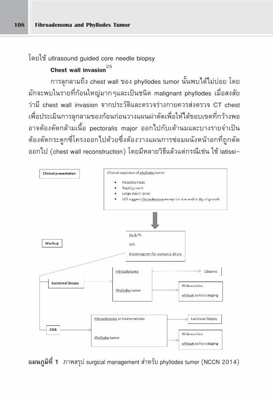
·ºπ¿Ÿ¡‘∑’Ë 1 ¿“æ √ÿª surgical management ”À√—∫ phyllodes tumor (NCCN 2014)
Õ“®“√¬å·æ∑¬åÀ≠‘ß «‘‰≈√—μπå ª√–‡ √‘∞ 109
mus dorsi myocutaneous flap ·≈– marlex mesh ‡ªìπμâπ À≈—ߺà“μ—¥§«√
æ‘®“√≥“„Àâ√—ß ’√—°…“‡æ◊Ëՙ૬≈¥‚Õ°“ °“√°≈—∫‡ªìπ È” ‡æ√“–°“√ºà“μ—¥§√—Èß
μàÕ‰ª®–¡’§«“¡¬ÿà߬“°·≈–‡ ’ˬߡ“° À√◊ÕÕ“®®–‰¡à “¡“√∂ºà“μ—¥‰¥âÕ’°
°“√√—°…“‡ √‘¡À≈—ߺà“μ—¥ (Adjuvant treatment)
ª√–‚¬™πå·≈–∫∑∫“∑¢Õß√—ß ’√—°…“°—∫‡§¡’∫”∫—¥„π°“√√—°…“ phyllodes
tumor π—Èπ¬—߉¡à™—¥‡®π ¡’μ—«Õ¬à“ß·π«∑“ߪؑ∫—μ‘¥—ßπ’È
MD Anderson Cancer Center giudeline 2012
·π–𔇰’ˬ«°—∫°“√„Àâ√—ß ’√—°…“‡ √‘¡ ‚¥¬æ‘®“√≥“„À⇩擖malignant
phyllodes tumor ¥—ßπ’È
- ∂â“∑” mastectomy ·≈⫉¥â negative margins ‰¡à®”‡ªìπμâÕß„Àâ
√—ß ’√—°…“À≈—ߺà“μ—¥
- ∂â“∑” mastectomy ·≈⫉¥â positive/ close/ concerning mar-
gins À√◊Õ‡π◊ÈÕßÕ°¡’°“√≈ÿ°≈“¡∂÷ß fascia, chest wall À√◊Õ °âÕπ phyllodes tumor
¡’¢π“¥„À≠à > 5 cm
- ∂â“∑” partial mastectomy ·≈â«„Àâæ‘®“√≥“„Àâ√—ß ’√—°…“À≈—ߺà“μ—¥
Chaney ·≈–§≥–35 æ∫«à“°“√„Àâ√—ß ’√—°…“‡ √‘¡Õ“®¡’ª√–‚¬™πå„πºŸâªÉ«¬
¥—ßμàÕ‰ªπ’È
- Bulky mass - High mitotic rate
- Close or positive margins - Presence of necrosis
- Hypercellular stroma - Increased vascularity within the tumor
- High nuclear pleomorphism - Tumor recurrence
NCCN guideline 2014
·π–π”„Àâ√—ß ’√—°…“‡ √‘¡„π°√≥’∑’ˇ°‘¥ local recurrent ¢Õß phyllodes
tumor ´÷Ë߇¡◊ËÕºà“μ—¥·≈â«„πÕπ“§μ∂â“°“√°≈—∫‡ªìπ´È”Õ’°®–∑”„Àâ°“√ºà“μ—¥
∑”‰¥â¬“° ¡’ morbodity Ÿß‡™àπ chest wall recurrence À≈—ß salvage mastec-
Fibroadenoma and Phyllodes Tumor110
tomy ®÷ß·π–π”„Àâ√—ß ’√—°…“À≈—ߺà“μ—¥‡æ◊ËÕÀ«—ß≈¥‚Õ°“ °“√°≈—∫‡ªìπ´È”„Àâ
‡°‘¥πâÕ¬∑’Ë ÿ¥
欓°√≥å‚√§
Local recurrence : ·¡â«à“®– “¡“√∂ºà“μ—¥°âÕπ phyllodes tumor ÕÕ°
¡“‰¥â∑—ÈßÀ¡¥°Á¡’√“¬ß“πæ∫ local recurrence ‰¥â¡“°∂÷ß 30%‚¥¬°≈ÿà¡ benign
phyllodes tumor ‡°‘¥ local recurrence ‰¥â 5-15% à«π°≈ÿà¡ borderline °—∫
malignant phyllodes tumors æ∫local recurrence ‰¥â20-30%36
Distant metastases : ºŸâªÉ«¬ phyllodes tumors ∑—ÈßÀ¡¥æ∫¡’ 10% ∑’Ë
‡°‘¥°“√·æ√à°√–®“¬‰ª¬—ßÕ«—¬«–Õ◊Ëπ (distant metastases) „π∫“ß√“¬ß“πÕ“®
Ÿß∂÷ß 22% ÷Ë߇ªìπºŸâªÉ«¬ borderline °—∫ malignant phyllodes tumors ‚¥¬
μ”·Àπàß∑’Ëæ∫¡’°“√°√–®“¬‰ª‰¥â∫àÕ¬§◊Õ lungs, soft tissue, bone ·≈– pleura37
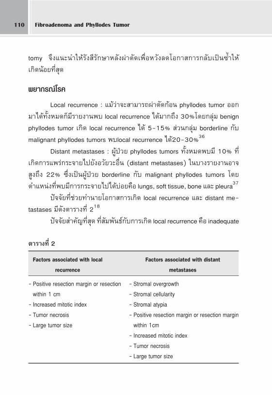
ªí®®—¬∑’˙૬∑”π“¬‚Õ°“ °“√‡°‘¥ local recurrence ·≈– distant me-
tastases ¡’¥—ßμ“√“ß∑’Ë 218
ªí®®—¬ ”§—≠∑’Ë ÿ¥ ∑’Ë —¡æ—π∏å°—∫°“√‡°‘¥ local recurrence §◊Õ inadequate
μ“√“ß∑’Ë 2
Factors associated with local Factors associated with distant
recurrence metastases
- Positive resection margin or resection - Stromal overgrowthwithin 1 cm - Stromal cellularity
- Increased mitotic index - Stromal atypia- Tumor necrosis - Positive resection margin or resection margin- Large tumor size within 1cm
- Increased mitotic index- Tumor necrosis- Large tumor size
Õ“®“√¬å·æ∑¬åÀ≠‘ß «‘‰≈√—μπå ª√–‡ √‘∞ 111
surgery ‚¥¬æ∫«à“°“√ºà“μ—¥°âÕπÕÕ°¡“‰¥â¢Õ∫‡¢μ‚¥¬√Õ∫πâÕ¬°«à“ 1 cm
‡ªìπªí®®—¬‡ ’Ë¬ß Ÿß∑’Ë ÿ¥∑’Ë®–‡°‘¥ local recurrence à«πªí®®—¬‡ ’ˬß∑’Ë¡’º≈¡“°
∑’Ë ÿ¥μàÕ°“√‡°‘¥ distant metastases §◊Õ stromal overgrowth
Õ—μ√“°“√Õ¬Ÿà√Õ¥∑—ÈßÀ¡¥ (overall survival) ¢ÕߺŸâªÉ«¬ phyllodes tu-
mor ∑’Ë 15 ªï§◊Õ 62% ‚¥¬°≈ÿà¡ nonmalignant (benign ·≈– borderline) phyl-
lodes tumor ®–¡’Õ—μ√“°“√Õ¬Ÿà√Õ¥∑’Ë¥’°«à“§◊Õ 79% ∑’Ë 10 ªï „π¢≥–∑’Ë°≈ÿà¡ ma-
lignant phyllodes tumor ®–¡’Õ—μ√“°“√Õ¬Ÿà√Õ¥·¬à°«à“§◊Õ 42% ∑’Ë 10 ªï38 ∂â“¡’
distant metastases ·≈⫺ŸâªÉ«¬®–¡’欓°“√≥å‚√§·¬à¡“° ¡’√“¬ß“π«à“ºŸâ
ªÉ«¬°≈ÿà¡π’È®–¡’Õ—μ√“Õ¬Ÿà√Õ¥‡©≈’ˬ 30 ‡¥◊Õππ—∫®“°«—π∑’Ë«‘π‘®©—¬‰¥â«à“¡’°“√·æ√à
°√–®“¬39
°“√μ√«®μ‘¥μ“¡ºŸâªÉ«¬ phyllodes tumors À≈—ߺà“μ—¥
®”‡ªìπμâÕßπ—¥μ√«®μ‘¥μ“¡Õ“°“√ºŸâªÉ«¬À≈—ߺà“μ—¥Õ¬à“ß„°≈♑¥‡æ√“–
phyllodes tumor ¡’‚Õ°“ ‡°‘¥‡ªìπ´È”‡©æ“–∑’ˉ¥â “¡“√∂·æ√à°√–®“¬‰ª
Õ«—¬«–Õ◊Ëπ‰¥â‚¥¬‡©æ“–Õ¬à“߬‘Ëß°≈ÿà¡ malignant phyllodes
·π«∑“ߪؑ∫—μ‘„π°“√μ√«®μ‘¥μ“¡ºŸâªÉ«¬¡’¥—ßπ’È
- π—¥μ√«®μ‘¥μ“¡Õ“°“√∑ÿ° 6 ‡¥◊Õπ„π™à«ß 2 ªï·√°À≈—ߺà“μ—¥
‡æ√“–‚Õ°“ °≈—∫‡ªìπ´È”®–‡°‘¥ Ÿß ÿ¥„π™à«ß 2 ªï·√°
- À≈—ß®“° 2 ªï‰ª·≈â«„Àâπ—¥μ√«®∑ÿ° 1ªï
- ·π–π”„À⺟âªÉ«¬μ√«®§≈”‡μâ“π¡¥â«¬μπ‡Õß ¡Ë”‡ ¡Õ ∂ⓧ≈”æ∫
‘Ëߺ‘¥ª°μ‘„Àâ¡“ª√÷°…“·æ∑¬å
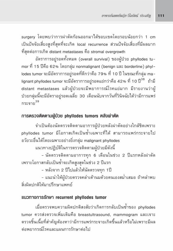
·π«∑“ß°“√√—°…“ recurrent phyllodes tumor
‡¡◊ËÕμ√«®æ∫§«“¡º‘¥ª°μ‘ ß —¬«à“‡°‘¥°“√°≈—∫‡ªìπ´È”¢Õß phyllodes
tumor §«√ àßμ√«®‡æ‘Ë¡‡μ‘¡§◊Õ breastultrasound, mammogram ·≈–‡®“–
μ√«®™‘Èπ‡π◊ÈÕ∑’Ë ”§—≠μâÕßÀ“«à“¡’°“√·æ√à°√–®“¬‡°‘¥¢÷Èπ·≈â«À√◊Õ‰¡à‡æ√“–¡’º≈
μàÕ欓°√≥å‚√§·≈–·ºπ°“√√—°…“μàÕ‰ª
Fibroadenoma and Phyllodes Tumor112
√ÿª
Phyllodes tumor ‡ªìπ‚√§¢Õ߇μâ“π¡∑’Ëæ∫‰¥â‰¡à∫àÕ¬·μ৫√§‘¥∂÷ß‚√§π’È
¥â«¬‡ ¡Õ„πºŸâªÉ«¬∑’Ë¡“¥â«¬°âÕπ∫√‘‡«≥‡μâ“π¡‚¥¬‡©æ“–°âÕπ¢Õ∫‡¢μ™—¥‡®π∑’Ë
‚μ‡√Á« „πºŸâªÉ«¬™à«ßÕ“¬ÿ 35-55 ªï ´÷ËßÕ“¬ÿ¡“°°«à“°≈ÿà¡ fibroadenoma
‘Ëß ”§—≠§◊Õ°“√«‘π‘®©—¬„À≥â°àÕπºà“μ—¥«à“‡ªìπ phyllodes tumor À√◊Õ
fibroadenoma ‡æ◊ËÕ«“ß·ºπ°“√ºà“μ—¥∑’ˇÀ¡“– ¡ ‚¥¬·π–π”„À⇮“–™‘Èπ‡π◊ÈÕ‚¥¬«‘∏’
CNB ‡¡◊ËÕº≈‡ªìπ phyllodes tumor ·π–π”„Àâ∑” wide excision ®–™à«¬≈¥
‚Õ°“ °“√°≈—∫‡ªìπ´È”°“√√—°…“‡ √‘¡Õ◊ËπÊ ‡™àπ √—ß ’√—°…“ À√◊Õ‡§¡’∫”∫—¥π—Èπ
ªí®®ÿ∫—π∫∑∫“∑¬—߉¡à™—¥‡®π‚¥¬∑—Ë«‰ª·π–π”„Àâ √—ß ’√—°…“À≈—ߺà“μ—¥°√≥’∑’Ë∂â“
¡’°“√‡°‘¥‡ªìπ´È”·≈â«®–‰¡à “¡“√∂ºà“μ—¥ÕÕ°‰¥âÀ√◊Õ°“√ºà“μ—¥®–¡’§«“¡‡ ’ˬß
Ÿß‡™àπ chest wall invasion à«π‡§¡’∫”∫—¥¡—°„Àâ„π°√≥’∑’ˇªìπ palliative treat-
ment ”À√—∫ºŸâªÉ«¬∑’Ë¡’°“√·æ√à°√–®“¬·≈â«
·ºπ¿Ÿ¡‘∑’Ë 2 ·π«∑“ß°“√ªØ‘∫—쑇¡◊ËÕæ∫ local recurrence ¢Õß phyllodes tumor (NCCN2014)
Õ“®“√¬å·æ∑¬åÀ≠‘ß «‘‰≈√—μπå ª√–‡ √‘∞ 113
¢Õ¢Õ∫§ÿ≥ ¿“æ∂à“¬æ¬“∏‘ ¿“æ fibroadenoma ·≈– phyllodes tumor
®“°
Õ“®“√¬å·æ∑¬åÀ≠‘ß«—π«‘ “¢å À‘¡–§ÿ≥ Õ“®“√¬åª√–®”¿“§«‘™“欓∏‘«‘∑¬“
§≥–·æ∑¬»“ μ√å ¡À“«‘∑¬“≈—¬∏√√¡»“ μ√å
‡Õ° “√Õâ“ßÕ‘ß
1. Santen R, Mansel R. Benign breast disorders. N Engl J Med 2005;353:275-85.2. Simmons P, Wold L. Surgically treated breast disease in adolescent females:a retrospective
review of 185 cases. Adolesc Pediatr Gynecol 1989;2:95-8.3. Dehner L, Hill A, Deschryver K. Pathology of the breast in children, adolescents, and young
adults. Sem Diag Pathol 1999;16:235-47.4. Sklair-Levy M, Sella M, Alweiss T, et al. Incidence and management of complex fibroad-
enomas. AJR Am J Roentgenol 2008;190:214-8.5. Branchini G, Schneider L, Cericatto R, et al. Progesterone receptors A and B and estrogen
receptor alpha expression in normal breast tissue and fibroadenomas. Endocrine 2009.6. Schneider L, Branchini G, Cericatto R, et al. Gene and protein expression of p53 and p21 in
fibroadenomas and adjacent normal mammary tissue. Endocrine 2009;35:118-22.7. Neinstein L, Atkinson J, Diament M. Prevalence and longitudinal study of breast masses in
adolescents. J Adolesc Health 1993;13:277-81.8. Ozello L, Gump F. The management of patients with carcinomas in fibroadenomatous tumors
of the breast. Surg Gynecol Obstet 1985;160:99-104.9. Onuigbo W. Breast fibroadenoma in teenage females. Turkish J Pediatrics 2003;45:326-8.
10. Greenberg R, Skornick Y, Kaplan O. Management of breast fibroadenomas. J Gen Intern Med1998;13:640-5.
11. NCCN clinical practice guidelines in oncology (NCCN guidelines). Breast cancer screening anddiagnosis. Version2.2013. Available at www.NCCN.org/patients.
12. Jayasinghe Y, Simmons P. Breast disorders in the female. In Fisher M, Alderman E, Kreipe R,Rosenfeld W, editors. Textbook of adolescent healthcare. American Academy of Pediatrics(chapter66).
13. Grady I, Gorsuch H, Wilburn-Bailey S. Long-term outcome of benign fibroadenomas treated byultrasound-guided percutaneous excision. Breast J 2008;14:275-8.
14. Rowell MD, Perry RR, Hsiu JG, Barranco SC. Phyllodes tumors. Am J Surg 1993;165:376-9.
Fibroadenoma and Phyllodes Tumor114
15. Bernstein L, Deapen D, Ross RK. The descriptive epidemiology of malignant cystosarcomaphyllodes tumors of the breast. Cancer 1993;71:3020-4.
16. Birch JM, Alston RD, McNally RJ, Evans DG, Kelsey AM, Harris M, et al. Relative frequencyand morphology of cancers in carriers of germline TP53 mutations. Oncogene 2001;20:4621-8.
17. Kuijper A, Buerger H, Simon R, Schaefer KL, Croonen A, Boecker W, et al. Analysis of theprogression of fibroepithelial tumours of the breast by PCR based clonality assay. J Pathol2002;197:575-81.
18. Barrio AV, Clark BD, Goldberg JI, Hoque LW, Bernik SF, Flynn LW, et al. Clinicopathologicfeatures and long-term outcomes of 293 phyllodes tumors of the breast. Ann Surg Oncol2007;14:2961-70.
19. Karim RZ, Scolyer RA, Tse GM, Tan PH, Putti TC, Lee CS. Pathogenic mechanisms in theinitiation and progression of mammary phyllodes tumours. Pathology 2009;41:105-17.
20. Hawkins RE, Schofield JB, Fischer C, Wiltshaw E, McKinna JA. The clinical and histologiccriteria that predict metastases from cystosarcoma phyllodes. Cancer 1992;69:141-7.
21. Tavassoli FA, Devilee P. Pathology and genetics of tumors of the breast and female genitalorgans. In: World health organization classification of tumours. Lyons: IARC Press; 2003. p. 99-103.
22. Tomas D, Bujas T, Stajduhar E. Malignant phyllodes tumor with associated osteosarcomatous,chondrosarcomatous, and liposarcomatous overgrowth. APMIS 2007;115:367-70.
23. World Health Organization, Histology Typing of Breast Tumors, WHO, Geneva, Switzerland,2nd edition; 1981; vol 2.
24. Rosen PP. Rosenûs Breast Pathology. 2nd edition. New York, NY USA: Lippincott WilliamWikins; 2001.
25. Reinfuss M, Mitus J, Duda K, Stelmach A, Rys J, Smolak K. The treatment and prognosis ofpatients with phyllodes tumor of the breast: an analysis of 170 cases. Cancer 1996;77:910-6.
26. Feder JM, de Paredes ES, Hogg JP, Wilken JJ. Unusual breast lesions: radiologic-pathologiccorrelation. Radiographics 1999;19:S11-S26.
27. Simi U, Moretti D, Iacconi P, et al. Fine needle aspiration cytopathology of phyllodes tumor.Differential diagnosis with fibroadenoma. Acta Cytologica 1988;32:63-6.
28. Komenaka IK, El-Tamer M, Pile-Spellman E, Hibshoosh H. Core needle biopsy as a diagnostictool to differentiate phyllodes tumor from fibroadenoma. Archives of surgery 2003;138:987-90.
29. JacklinRK, Ridgway PF, Ziprin P, Healy V, Hadjiminas D, Darzi A. Optimising preoperativediagnosis in phyllodes tumour of the breast. J Clin Pathol 2006;59:454-9.
30. Puay-Hoon T, Thiyagarajan J, George Y, et al. p53 and c-kit (CD117) protein expression as
Õ“®“√¬å·æ∑¬åÀ≠‘ß «‘‰≈√—μπå ª√–‡ √‘∞ 115
prognostic indicators in breast phyllodes tumors : a tissue microarray study. Modern Pathology2005;18:1525-34.
31. Zurrida S, Bartoli C, Galimberti V, et al. Which therapy for unexpected phyllode tumour of thebreast? Eur J Cancer 1992;28:654-7.
32. Orenstein A, Tsur H. Cystosarcoma phylloides treated by excision and immediate reconstruc-tion with silicon implant. Annals of Plastic Surgery 1987;18:520-3.
33. Salvadori B, Cusumano F, Del Bo RI. Surgical treatment of phyllodes tumors of the breast.Cancer 1989;63:2532-6.
34. Mangi AA, Smith BL, Gadd MA, et al. Surgical management of phyllodes tumors. Arch Surg1999;134:487-92.
35. Chaney AW, Pollack A, McNeese MD, Zagars GK. Adjuvant radiotherapy for phyllodes tumorof breast. Radiat Oncol Invest 1998;6:264-7.
36. Ciatto S, Bonardi R, Cataliotti L, Cardona GP. Phyllodes tumor of the breast: a multicenterseries of 59 cases. Eur J Surg Oncol 1992;18:5454-9.
37. Silver SA, Tavassoli, FA.Osteosarcomatous differentiation in phyllodes tumors. Am J SurgPathol 1999;23:815-21.
38. Chaney AW, Pollack A, McNeese MD, Zagars GK, Pisters PW, Pollock RE, et al. Primarytreatment of cystosarcoma phyllodes of the breast. Cancer 2000;89:1502-11.
39. Kessinger A, Foley JF, Lemon HM, et al. Metastatic cystosarcoma phyllodes: a case reportand review of the literature. J Surg Oncol 1972;4:131-47.