FGL2 A new biomarker for cancer in a simple blood test
31
FGL2 – A new biomarker for cancer in a simple blood test
Transcript of FGL2 A new biomarker for cancer in a simple blood test
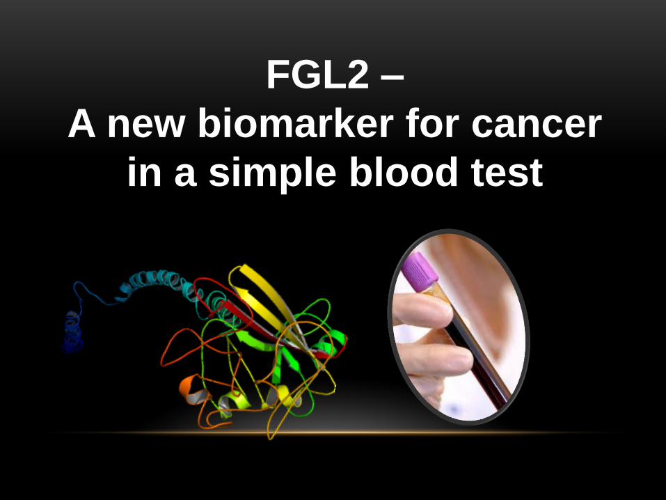
FGL2 –
A new biomarker for cancer
in a simple blood test
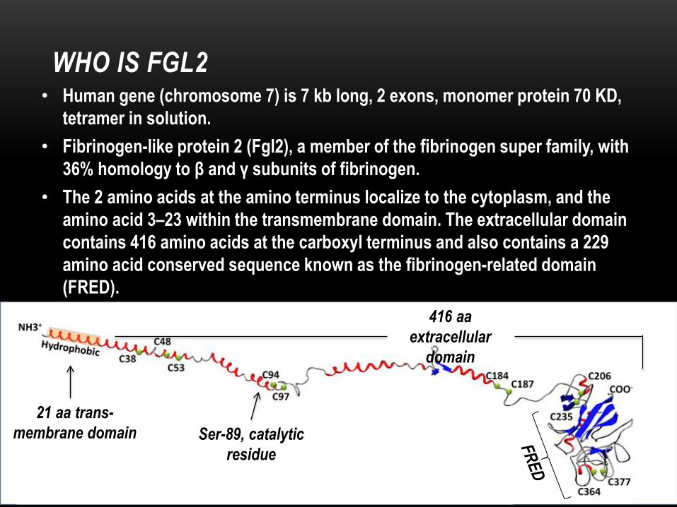
WHO IS FGL2 • Human gene (chromosome 7) is 7 kb long, 2 exons, monomer protein 70 KD,
tetramer in solution.
• Fibrinogen-like protein 2 (Fgl2), a member of the fibrinogen super family, with
36% homology to β and γ subunits of fibrinogen.
• The 2 amino acids at the amino terminus localize to the cytoplasm, and the
amino acid 3–23 within the transmembrane domain. The extracellular domain
contains 416 amino acids at the carboxyl terminus and also contains a 229
amino acid conserved sequence known as the fibrinogen-related domain
(FRED).
21 aa trans-
membrane domain
416 aa
extracellular
domain
Ser-89, catalytic
residue
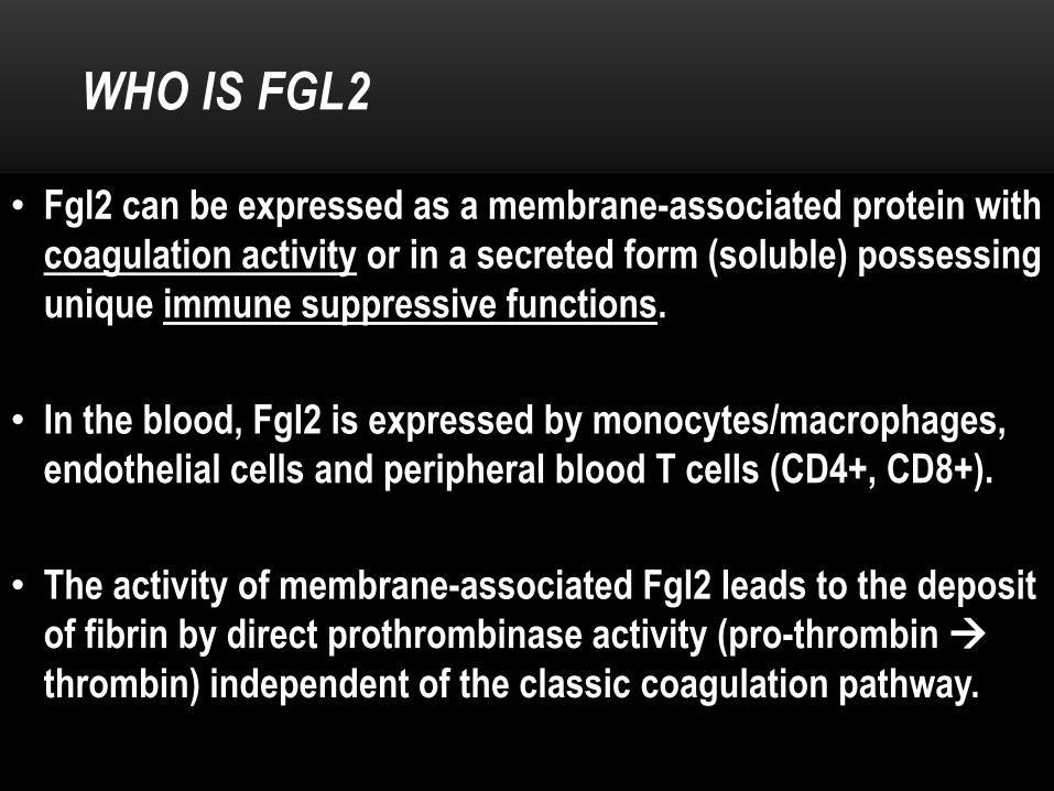
• Fgl2 can be expressed as a membrane-associated protein with
coagulation activity or in a secreted form (soluble) possessing
unique immune suppressive functions.
• In the blood, Fgl2 is expressed by monocytes/macrophages,
endothelial cells and peripheral blood T cells (CD4+, CD8+).
• The activity of membrane-associated Fgl2 leads to the deposit
of fibrin by direct prothrombinase activity (pro-thrombin
thrombin) independent of the classic coagulation pathway.
WHO IS FGL2
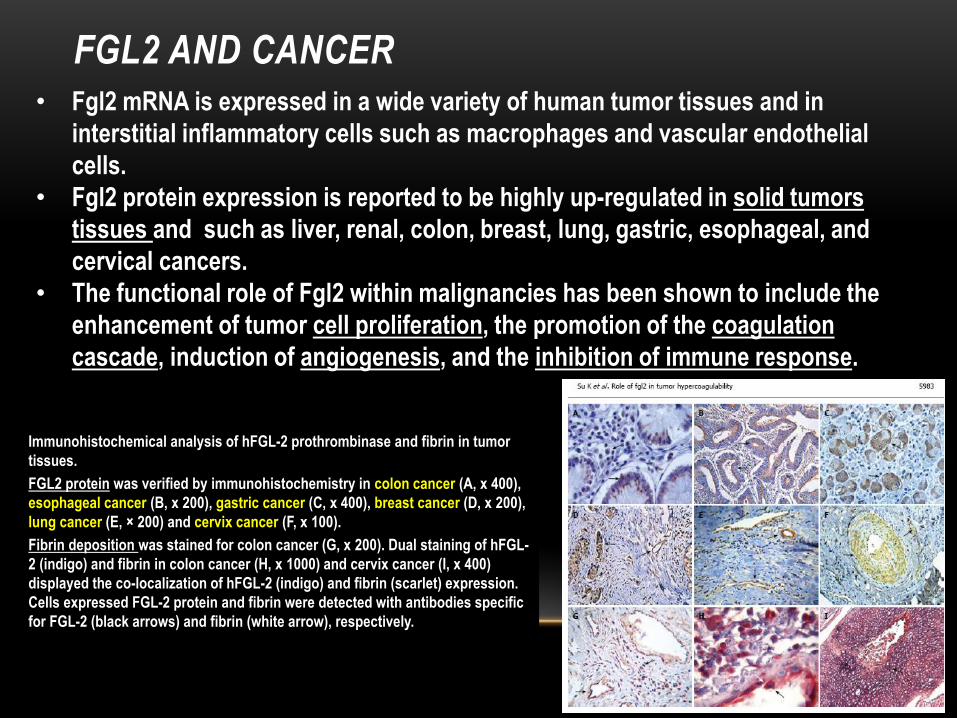
FGL2 AND CANCER • Fgl2 mRNA is expressed in a wide variety of human tumor tissues and in
interstitial inflammatory cells such as macrophages and vascular endothelial
cells.
• Fgl2 protein expression is reported to be highly up-regulated in solid tumors
tissues and such as liver, renal, colon, breast, lung, gastric, esophageal, and
cervical cancers.
• The functional role of Fgl2 within malignancies has been shown to include the
enhancement of tumor cell proliferation, the promotion of the coagulation
cascade, induction of angiogenesis, and the inhibition of immune response.
Immunohistochemical analysis of hFGL-2 prothrombinase and fibrin in tumor
tissues.
FGL2 protein was verified by immunohistochemistry in colon cancer (A, x 400),
esophageal cancer (B, x 200), gastric cancer (C, x 400), breast cancer (D, x 200),
lung cancer (E, × 200) and cervix cancer (F, x 100).
Fibrin deposition was stained for colon cancer (G, x 200). Dual staining of hFGL-
2 (indigo) and fibrin in colon cancer (H, x 1000) and cervix cancer (I, x 400)
displayed the co-localization of hFGL-2 (indigo) and fibrin (scarlet) expression.
Cells expressed FGL-2 protein and fibrin were detected with antibodies specific
for FGL-2 (black arrows) and fibrin (white arrow), respectively.

HYPOTHESIS
• Based on the observed upregulation of Fgl2 in
tumor tissues:
Fgl2 activity in peripheral blood mononuclear
cells (PBMC) is increased in cancer patients.
AIM OF STUDY
• To measure the level of Fgl2 activity in PBMC of
different cancer patients as compared to normal
controls.

ACTIVITY ASSAY:
• The method of measurement is based on thrombin generation assay, which reflects the
prothrombinase activity of FGL2.
• We were the first to monitor and show the elevation of FGL2
activity in PBMC of cancer patients.
Thrombin Generation activity of FGL2 in PBMC
PBMC are isolated from blood samples
↓
Prothrombin (FGL2 substrate) is added to cells
lysate for 30 minutes
↓
The concentration of generated thrombin is
determined by a fluorogenic assay
↓
The activity of FGL2 is determined according to
thrombin generation calibration curve
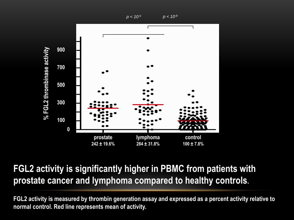
prostate control
900
700
500
300
100
0
% F
GL
2 th
rom
bin
ase
acti
vity
p < 10-8
242 ± 19.6% 100 ± 7.8%
lymphoma 284 ± 31.8%
p < 10-5
FGL2 activity is significantly higher in PBMC from patients with
prostate cancer and lymphoma compared to healthy controls.
FGL2 activity is measured by thrombin generation assay and expressed as a percent activity relative to
normal control. Red line represents mean of activity.
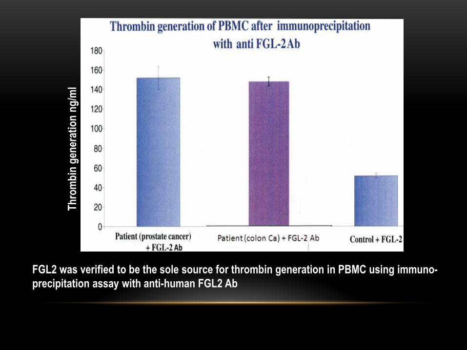
FGL2 was verified to be the sole source for thrombin generation in PBMC using immuno-
precipitation assay with anti-human FGL2 Ab
Th
rom
bin
gen
erat
ion
ng
/ml

LYMPHOMA
• Despite being the most common blood cancer, the diagnosis of
(B-cell) lymphoma is largely based on the pathologic workup of
patients with suspicious clinical presentation.
• The response to therapy is based mainly on clinical assessment
tools such as PET scan.
• No molecular Biomarkers are currently available for all types of
Lymphoma
• There is a deficiency of simple, non-invasive biomarkers
that may assist in diagnosis and follow up of patients
with lymphoma.
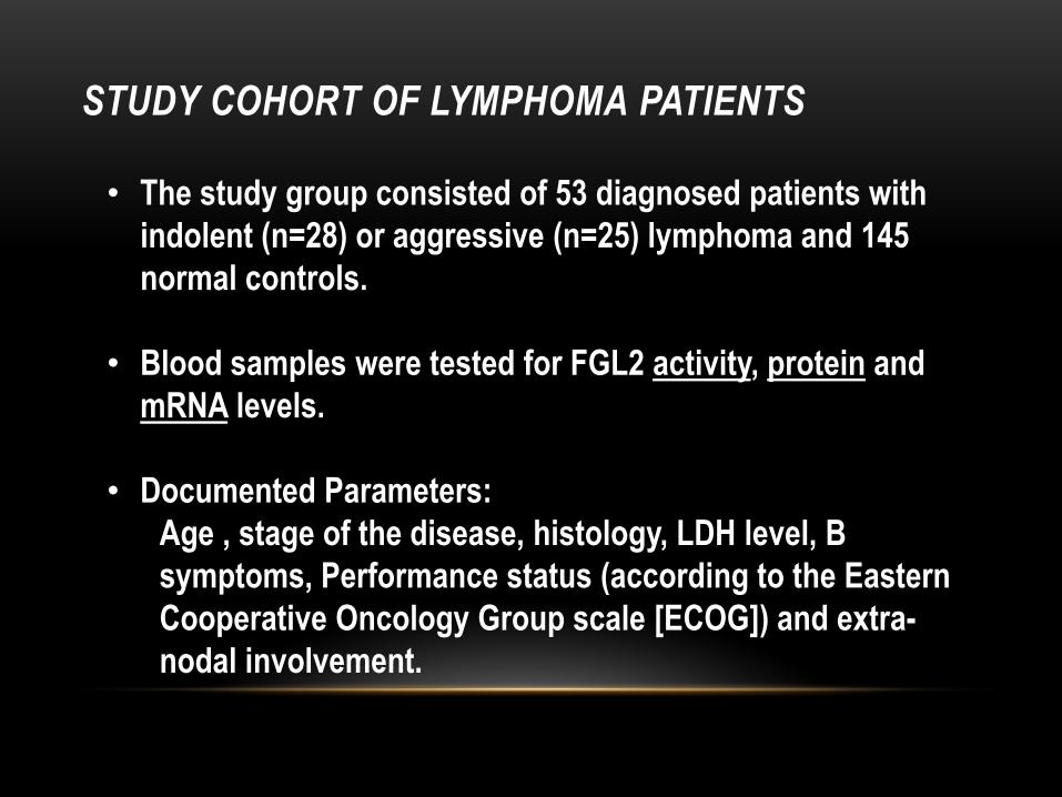
STUDY COHORT OF LYMPHOMA PATIENTS
• The study group consisted of 53 diagnosed patients with
indolent (n=28) or aggressive (n=25) lymphoma and 145
normal controls.
• Blood samples were tested for FGL2 activity, protein and
mRNA levels.
• Documented Parameters:
Age , stage of the disease, histology, LDH level, B
symptoms, Performance status (according to the Eastern
Cooperative Oncology Group scale [ECOG]) and extra-
nodal involvement.

Characteristics
Aggressive lymphoma
(n=25)
Indolent lymphoma
(n=28)
All patients (n=53)
Age, median (range) 69 (29-83) 65 (39-85) 66 (29-85)
Female, n (%) 12 (48%) 11 (39%) 23 (44%)
Stage, n (%)a
1
2
3
4
Extra-nodal disease
2 (8%)
5 (20%)
1 (4%)
17 (68%)
11 (44%)
3 (10%)
3 (10%)
8 (29%)
14 (50%)
8 (29%)
5 (10%)
8 (15%)
9 (17%)
31 (58%)
19 (36%)b
LDH (IU/L), median 447 388 413
a According to the Ann Arbor staging system.
No correlation between
FGL-2 activity and age or
gender in either patients
or control groups was
observed.
STUDY COHORT OF LYMPHOMA PATIENTS

FGL2 ACTIVITY IN PBMC OF LYMPHOMA
PATIENTS AT DIAGNOSIS
1000
800
600
400
200
0
Control Lymphoma
FG
L-2
act
ivit
y (%
)
A
×
×
1.0
0.8
0.6
0.4
0.2
0.0
1.0 0.8 0.6 0.4 0.2 0.0
Sen
siti
vity
1-Specificity
B
Sensitivity = 73.6%
Specificity = 80.7%
Activity was increased by 3±0.3-fold inactivity in patients (n=53) as compared to control
(n=145). p<0.001
The increase in FGL2 activity over a cutoff value of 150% appeared to exhibit a sensitivity of
73.6% and specificity of 80.7% for the diagnosis of lymphoma,
Sensitivity and specificity
ROC analysis FGL2 activity analysis
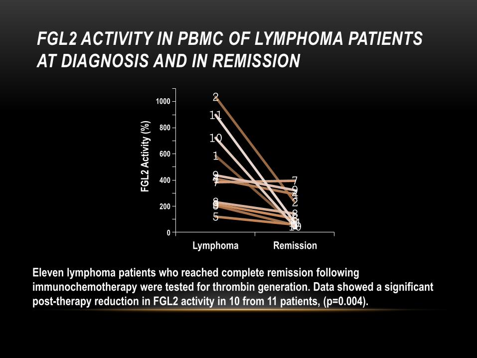
FGL2 ACTIVITY IN PBMC OF LYMPHOMA PATIENTS
AT DIAGNOSIS AND IN REMISSION
1
1
2
2 3 3
4 4
5 5 6
6
7 7
8 8
9 9
10
10
11
11
1000
800
600
400
200
0
Lymphoma Remission
FG
L2
Act
ivit
y (%
)
Eleven lymphoma patients who reached complete remission following
immunochemotherapy were tested for thrombin generation. Data showed a significant
post-therapy reduction in FGL2 activity in 10 from 11 patients, (p=0.004).
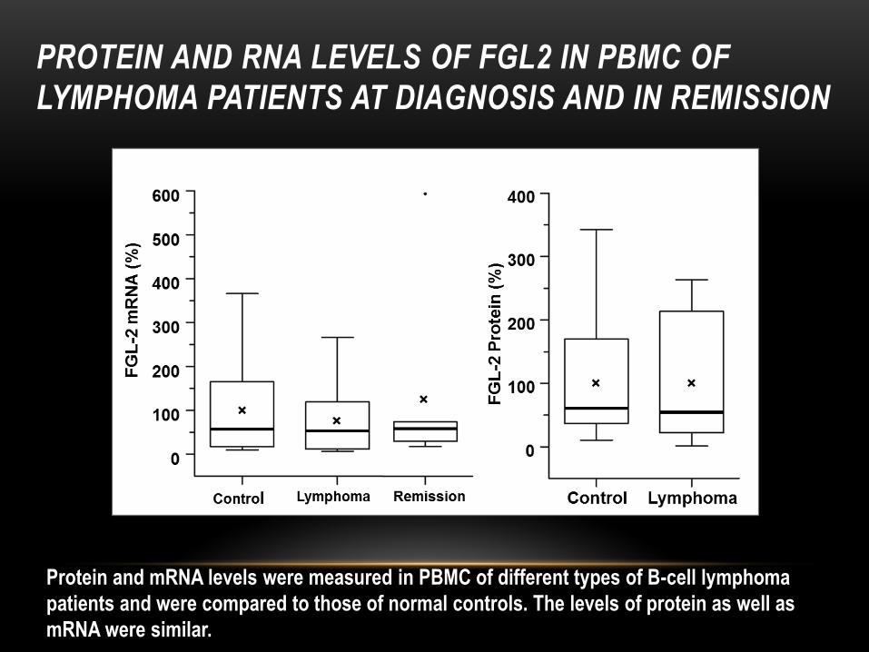
PROTEIN AND RNA LEVELS OF FGL2 IN PBMC OF
LYMPHOMA PATIENTS AT DIAGNOSIS AND IN REMISSION
Protein and mRNA levels were measured in PBMC of different types of B-cell lymphoma
patients and were compared to those of normal controls. The levels of protein as well as
mRNA were similar.
MYCOSIS FUNGOIDES (MF)
• Mycosis fungoides (and the Sézary syndrome) are diseases in
which lymphocytes become malignant and affect the skin.
• Cancer cells of mycosis fungoides and the Sézary syndrome are
able to spread from the skin to other parts of the body (either
through tissue, the lymph system, or the blood).
• Recurrent mycosis fungoides and the Sézary syndrome may
come back in the skin or in other parts of the body.
• Despite being the two most common types of cutaneous T-cell
lymphoma, the diagnosis of MF or the Sézary syndrome is
entirely based on the pathologic workup of patients with
suspicious clinical presentation.
• No molecular Biomarkers are currently available.
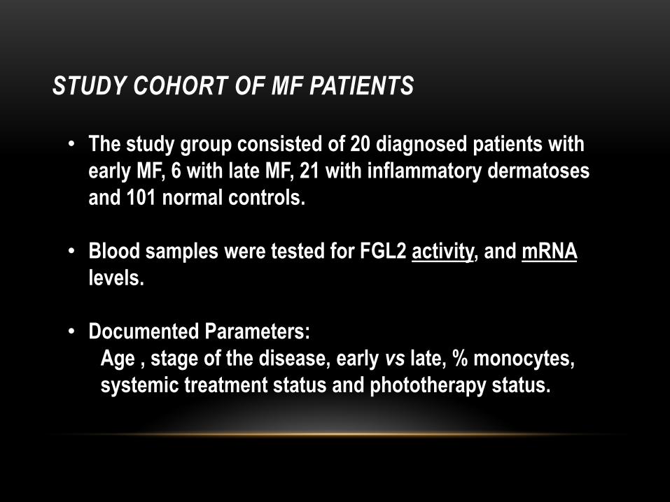
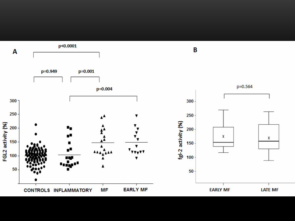
STUDY COHORT OF MF PATIENTS
• The study group consisted of 20 diagnosed patients with
early MF, 6 with late MF, 21 with inflammatory dermatoses
and 101 normal controls.
• Blood samples were tested for FGL2 activity, and mRNA
levels.
• Documented Parameters:
Age , stage of the disease, early vs late, % monocytes,
systemic treatment status and phototherapy status.
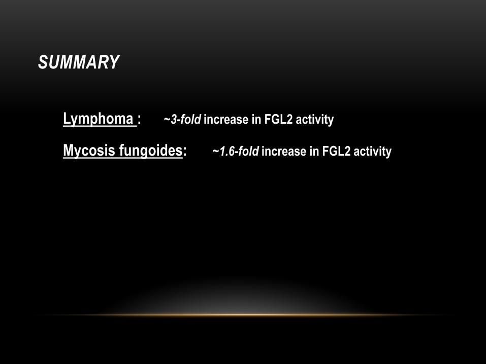
SUMMARY
Lymphoma : ~3-fold increase in FGL2 activity
Mycosis fungoides: ~1.6-fold increase in FGL2 activity
CONCLUSIONS (1)
• FGL2 activity is significantly increased in PBMC of patients
with lymphoma or MF, while decreased in remission.
• FGL2 provides a compelling candidate to act as a biomarker
for lymphoma and MF diseases diagnosis and follow up as
well as a therapeutic target.
• It is intriguing to assume that increased activity of FGL2 in
PBMC may have a role in these malignancies.
ROLE OF FGL2 IN MALIGNANCIES
OUR AIMS:
• To substantiate the role of FGL2 in angiogenesis
and tumor development.
• To uncover the mechanism underlying these
activities.
Silencing of Fgl2 gene in tumor cells
0%
20%
40%
60%
80%
100%
120%
fgl-
2 m
RN
A
WT Non specific
silenced clone fgl-2 silenced
clone
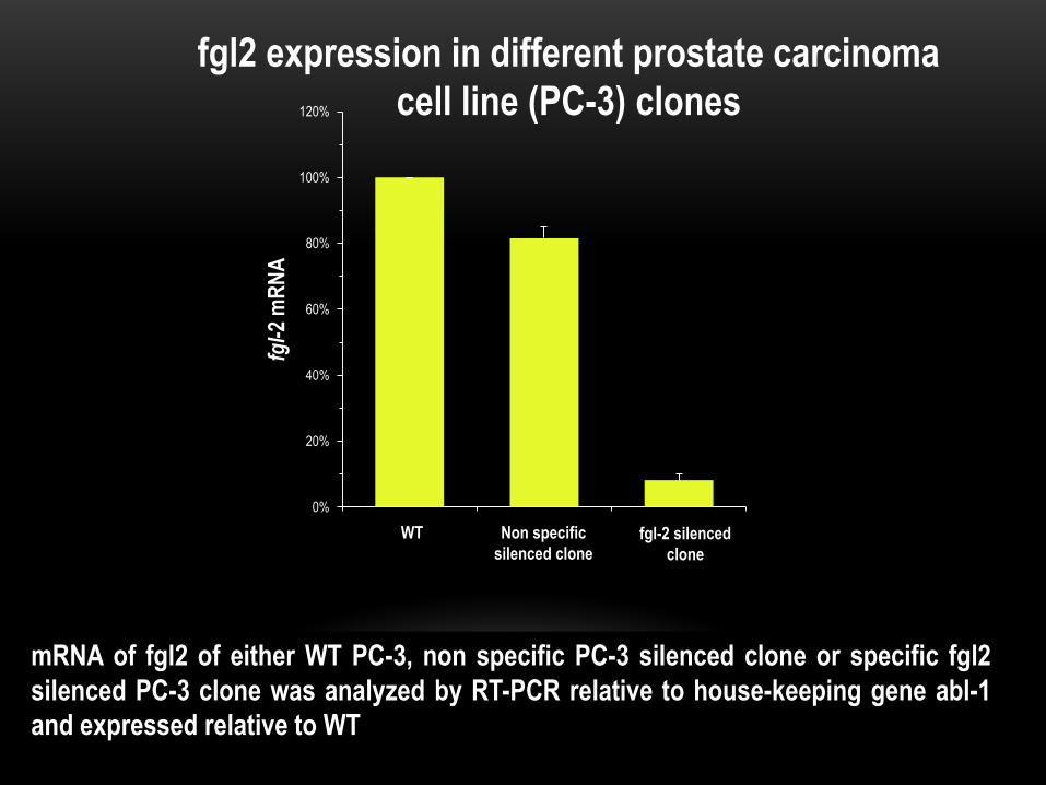
fgl2 expression in different prostate carcinoma
cell line (PC-3) clones
mRNA of fgl2 of either WT PC-3, non specific PC-3 silenced clone or specific fgl2
silenced PC-3 clone was analyzed by RT-PCR relative to house-keeping gene abl-1
and expressed relative to WT
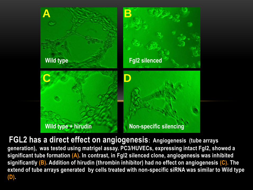
D C
B A
FGL2 has a direct effect on angiogenesis: Angiogenesis (tube arrays
generation), was tested using matrigel assay. PC3/HUVECs, expressing intact Fgl2, showed a
significant tube formation (A). In contrast, in Fgl2 silenced clone, angiogenesis was inhibited
significantly (B). Addition of hirudin (thrombin inhibitor) had no effect on angiogenesis (C). The
extend of tube arrays generated by cells treated with non-specific siRNA was similar to Wild type
(D).
Wild type Fgl2 silenced
Wild type + hirudin Non-specific silencing
• FGL-2 role in angiogenesis is not thrombin-mediated.
CONCLUSION (2)
• The effect of fgl-2 silencing on angiogenesis-related
proteins was analyzed using Proteome Profiler Human
Angiogenesis Antibody Array kits
• Protein expression profile of PC-3 cells was compared
to that of PC-3 cells transfected with non specific or
specific siRNA for fgl-2 silencing
• Fgl-2 silencing was associated with a significant
decrease in FGF-2 protein and ERK1/2 phosphorylation.
The effect of Fgl2 on angiogenesis-related
proteins
0%
20%
40%
60%
80%
100%m
RN
A o
f fg
f-2
WT fgl2 siRNA Non specific
siRNA
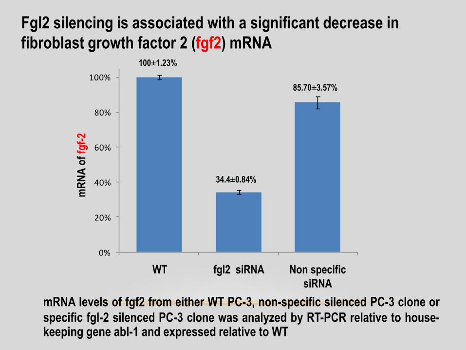
Fgl2 silencing is associated with a significant decrease in
fibroblast growth factor 2 (fgf2) mRNA
mRNA levels of fgf2 from either WT PC-3, non-specific silenced PC-3 clone or
specific fgl-2 silenced PC-3 clone was analyzed by RT-PCR relative to house-keeping gene abl-1 and expressed relative to WT
34.4±0.84%
85.70±3.57%
100±1.23%
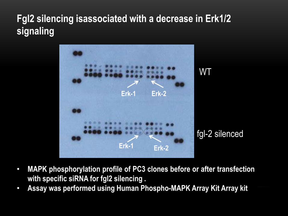
• MAPK phosphorylation profile of PC3 clones before or after transfection
with specific siRNA for fgl2 silencing .
• Assay was performed using Human Phospho-MAPK Array Kit Array kit
Fgl2 silencing isassociated with a decrease in Erk1/2
signaling
WT
fgl-2 silenced
Erk-1
Erk-1
Erk-2
Erk-2
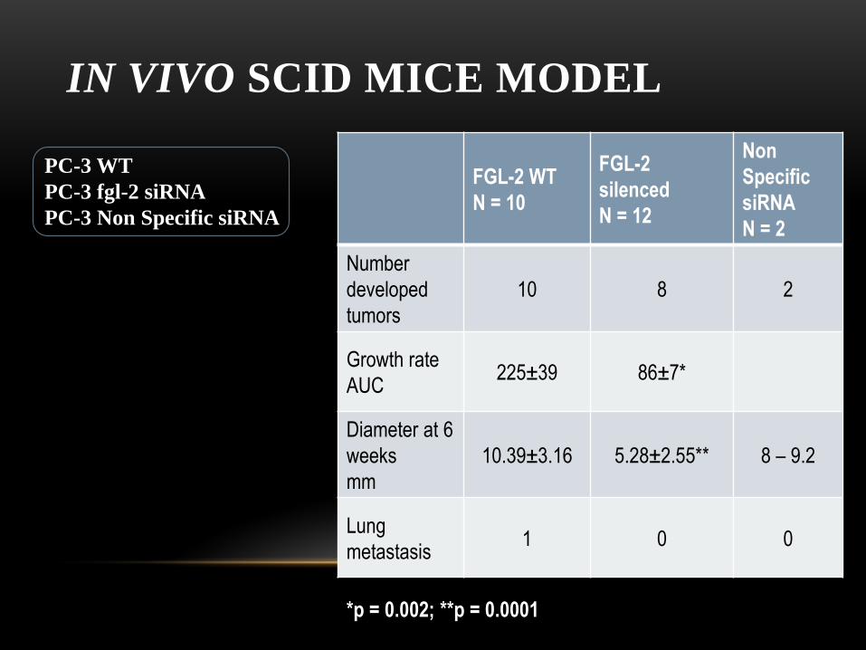
IN VIVO SCID MICE MODEL
PC-3 WT
PC-3 fgl-2 siRNA
PC-3 Non Specific siRNA
FGL-2 WT
N = 10
FGL-2
silenced
N = 12
Non
Specific
siRNA
N = 2
Number
developed
tumors
10 8 2
Growth rate
AUC 225±39 86±7*
Diameter at 6
weeks
mm
10.39±3.16 5.28±2.55** 8 – 9.2
Lung
metastasis 1 0 0
*p = 0.002; **p = 0.0001
A
5mm 5mm
Fgl-2 silenced PC-3 WT
% f
gl-
2 ex
pre
ssio
n l
evel
120
60
40
20
0
100
80
Tumor growth rate
PC-3 WT fgl-2
fgl-2 silenced
mRNA of fgl-2 in tumors
HO stain
FGL-2 stain
0
2
4
6
8
10
12
14
0 1 2 3 4 5 6
Tum
or
dia
met
er (
mm
)
Time (weeks(
WT
Fgl-2 silenced
Conclusions
• Fgl2 mediates tumor development: Fgl2 silencing induced
smaller and less aggressive tumors
• The pro-angiogenic/pro-tumorigenic activity of FGL2 can be
mediated by EGF or FGF-2 via ERK1/2 signaling pathways
• FGL2 inhibition may have therapeutic potential in cancer

Study conducted by:
Izhack Cherny, Ph.D.
Shany Sherman, M.D.
Doron Lederfein, Ph.D.
Natalia Binkovski, M.Sc.
Esther Ziv, Ph.D.
Collaborators:
Aida Inbal, M.D.
Ofir Wolach, M.D.
Alon Peretz, M.D.
Thank you for your attention!
Funding Agent:
Nofar grant number 44776, Chief Scientist, Ministry of Health, Israel.