Fabrication of paper-based enzyme immobilized microarray ...
11
Guo et al. Chin Med (2019) 14:13 https://doi.org/10.1186/s13020-019-0236-y RESEARCH Fabrication of paper-based enzyme immobilized microarray by 3D-printing technique for screening α-glucosidase inhibitors in mulberry leaves and lotus leaves Shangxin Guo † , Xiaotong Lin † , Yi Wang and Xingchu Gong * Abstract Background: The discovery of bioactive compounds in traditional Chinese medicine (TCM) has become an impor- tant field in TCM modernization. Ligand fishing is a suitable method for discovery of bioactive compounds in complex mixtures such as TCM with high selectivity. Because of unique advantage of low cost and convenience, paper-based microdevices can be good carriers for enzyme immobilized ligand fishing. Methods: As an important enzyme for glucose metabolism, α-glucosidase was immobilized on polycaprolactone– chitosan-modified paper to prepare the microdevice with unique microfluid structure generated by 3D printing technology, which can be easily applied to screen active compounds in herbal extracts. The preparation conditions of the paper microarray were optimized. The activity of immobilized α-glucosidase was verified by colorimetric reac- tions which can be easily monitored by cellphone. The paper microarray with α-glucosidase immobilized was used to screen active compounds in the water extracts of mulberry leaves and lotus leaves. Results: Several key parameters including Na 2 CO 3 solution concentration, Na 2 CO 3 solution volume, glutaraldehyde concentration, crosslinking time of glutaraldehyde and time of α-glucosidase immobilization were optimized. The proposed paper-based microarray was successfully applied in screening active compounds in two herbal extracts. Four compounds including chlorogenic acid, quercetin-3-O-glucuronide, isoquercetin, and quercetin were identi- fied as α-glucosidase inhibitors. The compounds with significant non-specific adsorption caused by chitosan, such as isoquercitrin, astragalin, quercetin, were also found to be active compounds. Conclusions: An enzyme immobilized paper microarray was designed and fabricated in this work. Polycaprolactone and chitosan were used to modify filter paper to prepare paper microarrays. Parameters of paper device preparation were optimized. Our findings suggested that 3D-printing paper-based microarrays can be a simple and low-cost approach for discovery of active compounds of TCM. Keywords: Paper-based microarray, Ligand fishing, α-Glucosidase, 3D printing © The Author(s) 2019. This article is distributed under the terms of the Creative Commons Attribution 4.0 International License (http://creativecommons.org/licenses/by/4.0/), which permits unrestricted use, distribution, and reproduction in any medium, provided you give appropriate credit to the original author(s) and the source, provide a link to the Creative Commons license, and indicate if changes were made. The Creative Commons Public Domain Dedication waiver (http://creativecommons.org/ publicdomain/zero/1.0/) applies to the data made available in this article, unless otherwise stated. Open Access Chinese Medicine *Correspondence: [email protected] † Shangxin Guo and Xiaotong Lin contributed equally to this work Pharmaceutical Informatics Institute, College of Pharmaceutical Sciences, Zhejiang University, Hangzhou 310058, China
Transcript of Fabrication of paper-based enzyme immobilized microarray ...
Guo et al. Chin Med (2019) 14:13 https://doi.org/10.1186/s13020-019-0236-y
RESEARCH
Fabrication of paper-based enzyme immobilized microarray by 3D-printing technique for screening α-glucosidase inhibitors in mulberry leaves and lotus leavesShangxin Guo†, Xiaotong Lin†, Yi Wang and Xingchu Gong*
Abstract
Background: The discovery of bioactive compounds in traditional Chinese medicine (TCM) has become an impor-tant field in TCM modernization. Ligand fishing is a suitable method for discovery of bioactive compounds in complex mixtures such as TCM with high selectivity. Because of unique advantage of low cost and convenience, paper-based microdevices can be good carriers for enzyme immobilized ligand fishing.
Methods: As an important enzyme for glucose metabolism, α-glucosidase was immobilized on polycaprolactone–chitosan-modified paper to prepare the microdevice with unique microfluid structure generated by 3D printing technology, which can be easily applied to screen active compounds in herbal extracts. The preparation conditions of the paper microarray were optimized. The activity of immobilized α-glucosidase was verified by colorimetric reac-tions which can be easily monitored by cellphone. The paper microarray with α-glucosidase immobilized was used to screen active compounds in the water extracts of mulberry leaves and lotus leaves.
Results: Several key parameters including Na2CO3 solution concentration, Na2CO3 solution volume, glutaraldehyde concentration, crosslinking time of glutaraldehyde and time of α-glucosidase immobilization were optimized. The proposed paper-based microarray was successfully applied in screening active compounds in two herbal extracts. Four compounds including chlorogenic acid, quercetin-3-O-glucuronide, isoquercetin, and quercetin were identi-fied as α-glucosidase inhibitors. The compounds with significant non-specific adsorption caused by chitosan, such as isoquercitrin, astragalin, quercetin, were also found to be active compounds.
Conclusions: An enzyme immobilized paper microarray was designed and fabricated in this work. Polycaprolactone and chitosan were used to modify filter paper to prepare paper microarrays. Parameters of paper device preparation were optimized. Our findings suggested that 3D-printing paper-based microarrays can be a simple and low-cost approach for discovery of active compounds of TCM.
Keywords: Paper-based microarray, Ligand fishing, α-Glucosidase, 3D printing
© The Author(s) 2019. This article is distributed under the terms of the Creative Commons Attribution 4.0 International License (http://creat iveco mmons .org/licen ses/by/4.0/), which permits unrestricted use, distribution, and reproduction in any medium, provided you give appropriate credit to the original author(s) and the source, provide a link to the Creative Commons license, and indicate if changes were made. The Creative Commons Public Domain Dedication waiver (http://creat iveco mmons .org/publi cdoma in/zero/1.0/) applies to the data made available in this article, unless otherwise stated.
Open Access
Chinese Medicine
*Correspondence: [email protected] †Shangxin Guo and Xiaotong Lin contributed equally to this workPharmaceutical Informatics Institute, College of Pharmaceutical Sciences, Zhejiang University, Hangzhou 310058, China
Page 2 of 11Guo et al. Chin Med (2019) 14:13
BackgroundNovel omics techniques including transcriptomics, prot-eomics, metabolomics as well as microarrays have greatly contributed to the modernization of traditional Chi-nese medicine (TCM) in chemical and pharmacological researches. Microarray assays have been widely applied in screening and characterization of TCM [1]. Microar-rays can be used to identify bioactive components from TCM and target biologically active molecules in TCM [2]. Zhang et al. [3] incorporated the ITS1-5.8S-ITS2 sequences of 16 Dendrobium species on a glass slide to fabricate a DNA microarray. It was used to detect the presence of D. nobile in a Chinese medicinal formula-tion containing nine herbal components. Wang et al. [4] identified the potential targets of the ingredients of Xue-saitong by integrating microarray data, text mining and pharmacophore model-based prediction. Notoginseno-side R1, ginsenoside Rg1, Rb1, Rd and Re were found to be the major bioactive compound and were validated. Bioactive compound discovery is an important part of the TCM modernization [5]. The discovered active com-pounds can be used as the quality markers of the TCM [6]. They can also be used for developing new drugs. However, the discovery of bioactive compounds in TCM is usually difficult because of the very complex composi-tion of TCM systems. In most cases, the compounds in TCM systems are extracted with different solvents, and then separated by preparative chromatography. After that, chemical structure and biological activity will be further determined. However, it is time-consuming and labor-intensive. Thus, it’s urgent to develop effective methods for the discovery of bioactive compounds in TCM.
Ligand fishing is a technical method to separate bio-active compounds from complex mixtures with high selectivity. The method is achieved by interacting receptors from ligands on the basis of intermolecular affinity [7–9]. Ligand fishing can be carried out with or without the ligand immobilized on a carrier. The captured receptors can be separated by washing and detected by mass spectrometry or other detectors. Due to high selectivity of the ligand fishing method, it’s possible to obtain bioactive compounds for a spe-cific target from complex systems directly. At present, ligand fishing is widely applied to separate active com-pounds from complex products including plant extracts and cell extracts [10]. It has been widely reported that magnetic beads, hollow fiber membranes, etc. were applied as carries for ligand fishing to discover active compounds from the TCM. Liu et al. [11] prepared nano-magnetic beads bonding with bovine serum albu-min and used chromatography-mass spectrometry to
screen active ingredients in the Pueraria lobate. Thir-teen active ingredients were screened. Deng et al. [12] performed a new screening assay based on ligand fish-ing using magnetic Fe3O4@SiO2-COX-2 combined with high-performance liquid chromatography-diode array detector-mass spectrometry to screen and iden-tify COX-2 inhibitors from green tea. It was a simple, robust and reproducible approach to discover COX-2 inhibitors from complex matrix. Tao [5] established a hollow fiber adsorption screening method for triglyc-eride inhibitors to discover active compounds in lotus leaf extract. Three active compounds were found hav-ing inhibitory activity on triglyceride. Overall, ligand fishing is quicker, more effective, and more sensitive for discovery of active compounds in TCM compared with traditional methods [13].
The paper device, a kind of analytical device made of paper, is developed rapidly in recent years. Since Martinez et al. [14] introduced paper as microfluidic device, paper devices have been widely used as ana-lytical devices in chemical reactions and environmen-tal monitoring. It showed that it’s low-cost, easy to use, and capable of multiplexed chemical analysis [15–18]. The paper devices own high specific surface area which makes it easy to combine molecules to adsorb pro-teins. Used paper devices can be handled easily through burning them, reducing pollution caused by experi-mental consumables. White paper provided ideal back-ground signals for observing colorimetric reactions. Kaewarse et al. [19] performed a reduction reaction of nitro blue tetrazolium on the paper-based chip to detect enzymatic activity and the results of the reaction showed purple on the paper. Qualitative and quantita-tive results can be obtained combining some portable detectors such as cellphones and cameras. Therefore, it is possible to judge the immobilization effects of enzymes on paper easily. There are also some examples of paper devices with commercial successes, such as pregnancy test strips and pH test paper.
In this work, an effective omics technology based on paper device was proposed. Paper-based micro-arrays were prepared and applied to discover active compounds in TCM extracts. Polycaprolactone–chi-tosan-modified paper was prepared with the assistance of 3D printing technology. α-Glucosidase was immobi-lized on the modified paper. The preparation conditions of paper microarrays were optimized. α-Glucosidase was one of the targets of diabetes. It’s reported that mulberry leaves and lotus leaves are effective on treat-ment of hyperglycemia in type 2 diabetes [20, 21]. Therefore, the paper microarray was then used to dis-cover active compounds from mulberry leaf extracts and lotus leaf extracts in this work.
Page 3 of 11Guo et al. Chin Med (2019) 14:13
MethodsThe Minimum Standards of Reporting Checklist con-tains details of the experimental design, and statistics, and resources used in this study (Additional file 1).
Materials and chemicalsEthanol (≥ 99.7%) and glacial acetic acid (≥ 99.5%) were purchased from Shanghai Lingfeng Chemical Reagent Co., Ltd. (Shanghai, China). Chitosan and α-glucosidase were purchased from Merck (Darm-stadt, Germany). Glutaraldehyde solution (50%) was purchased from Sangon Biotech Co., Ltd. (Shanghai, China). 4-Nitrophenyl-α-d-glucopyranoside (PNPG) was purchased from Shanghai Aladdin Bio-Chem Tech-nology Co., Ltd. (Shanghai, China). Sodium chloride (≥ 99.5%), disodium hydrogen phosphate dodecahy-drate (≥ 99.0%), potassium chloride (≥ 99.5%), potas-sium dihydrogen phosphate (≥ 99.5%) and sodium carbonate (≥ 99.8%) were purchased from Sinopharm Chemical Reagent Co., Ltd. (Shanghai, China). Mul-berry leaves and lotus leaves were purchased from Huadong Herbal Slice Co., Ltd. (Hangzhou, China). Polycaprolactone was purchased from Dongguan Top Cool Electronics Technology Co., Ltd. (Dongguan, China). Quantitative filter paper was purchased from Hangzhou Wohua Filter Paper Co., Ltd. (Hangzhou, China). Ultrahigh-purity water was produced using a water purification system (Milli-Q, Millipore, US).
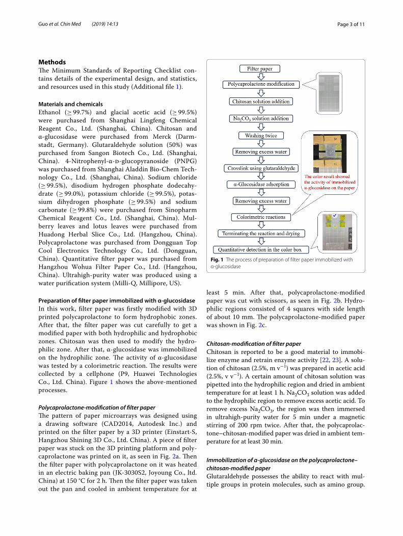
Preparation of filter paper immobilized with α‑glucosidaseIn this work, filter paper was firstly modified with 3D printed polycaprolactone to form hydrophobic zones. After that, the filter paper was cut carefully to get a modified paper with both hydrophilic and hydrophobic zones. Chitosan was then used to modify the hydro-philic zone. After that, α-glucosidase was immobilized on the hydrophilic zone. The activity of α-glucosidase was tested by a colorimetric reaction. The results were collected by a cellphone (P9, Huawei Technologies Co., Ltd. China). Figure 1 shows the above-mentioned processes.
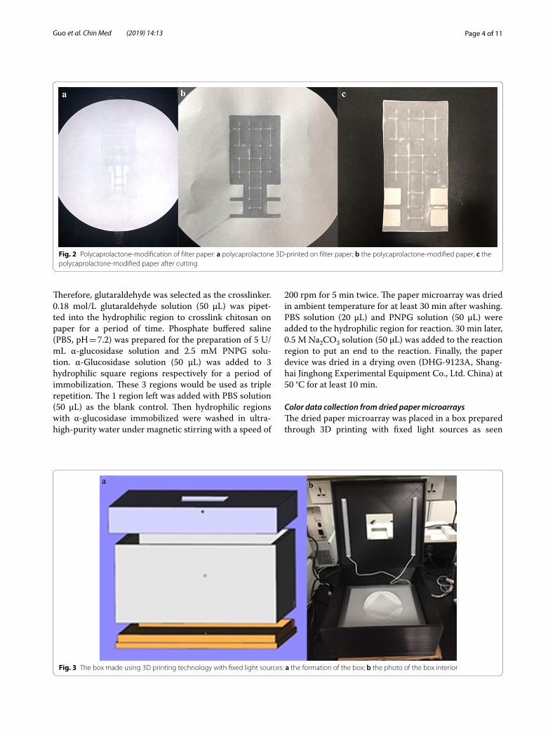
Polycaprolactone‑modification of filter paperThe pattern of paper microarrays was designed using a drawing software (CAD2014, Autodesk Inc.) and printed on the filter paper by a 3D printer (Einstart-S, Hangzhou Shining 3D Co., Ltd. China). A piece of filter paper was stuck on the 3D printing platform and poly-caprolactone was printed on it, as seen in Fig. 2a. Then the filter paper with polycaprolactone on it was heated in an electric baking pan (JK-3030S2, Joyoung Co., ltd. China) at 150 °C for 2 h. Then the filter paper was taken out the pan and cooled in ambient temperature for at
least 5 min. After that, polycaprolactone-modified paper was cut with scissors, as seen in Fig. 2b. Hydro-philic regions consisted of 4 squares with side length of about 10 mm. The polycaprolactone-modified paper was shown in Fig. 2c.
Chitosan‑modification of filter paperChitosan is reported to be a good material to immobi-lize enzyme and retrain enzyme activity [22, 23]. A solu-tion of chitosan (2.5%, m v−1) was prepared in acetic acid (2.5%, v v−1). A certain amount of chitosan solution was pipetted into the hydrophilic region and dried in ambient temperature for at least 1 h. Na2CO3 solution was added to the hydrophilic region to remove excess acetic acid. To remove excess Na2CO3, the region was then immersed in ultrahigh-purity water for 5 min under a magnetic stirring of 200 rpm twice. After that, the polycaprolac-tone–chitosan-modified paper was dried in ambient tem-perature for at least 30 min.
Immobilization of α‑glucosidase on the polycaprolactone–chitosan‑modified paperGlutaraldehyde possesses the ability to react with mul-tiple groups in protein molecules, such as amino group.
Fig. 1 The process of preparation of filter paper immobilized with α-glucosidase
Page 4 of 11Guo et al. Chin Med (2019) 14:13
Therefore, glutaraldehyde was selected as the crosslinker. 0.18 mol/L glutaraldehyde solution (50 μL) was pipet-ted into the hydrophilic region to crosslink chitosan on paper for a period of time. Phosphate buffered saline (PBS, pH = 7.2) was prepared for the preparation of 5 U/mL α-glucosidase solution and 2.5 mM PNPG solu-tion. α-Glucosidase solution (50 μL) was added to 3 hydrophilic square regions respectively for a period of immobilization. These 3 regions would be used as triple repetition. The 1 region left was added with PBS solution (50 μL) as the blank control. Then hydrophilic regions with α-glucosidase immobilized were washed in ultra-high-purity water under magnetic stirring with a speed of
200 rpm for 5 min twice. The paper microarray was dried in ambient temperature for at least 30 min after washing. PBS solution (20 μL) and PNPG solution (50 μL) were added to the hydrophilic region for reaction. 30 min later, 0.5 M Na2CO3 solution (50 μL) was added to the reaction region to put an end to the reaction. Finally, the paper device was dried in a drying oven (DHG-9123A, Shang-hai Jinghong Experimental Equipment Co., Ltd. China) at 50 °C for at least 10 min.
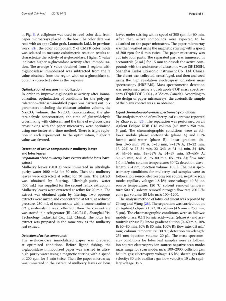
Color data collection from dried paper microarraysThe dried paper microarray was placed in a box prepared through 3D printing with fixed light sources as seen
Fig. 2 Polycaprolactone-modification of filter paper: a polycaprolactone 3D-printed on filter paper; b the polycaprolactone-modified paper; c the polycaprolactone-modified paper after cutting
Fig. 3 The box made using 3D printing technology with fixed light sources: a the formation of the box; b the photo of the box interior
Page 5 of 11Guo et al. Chin Med (2019) 14:13
in Fig. 3. A cellphone was used to read color data from paper microarrays placed in the box. The color data was read with an app (Color grab, Loomatix Ltd.). In previous work [24], the color component Y of CMYK color mode was selected to measure colorimetric reaction results to characterize the activity of α-glucosidase. Higher Y value indicates higher α-glucosidase activity after immobiliza-tion. The average Y value obtained from 3 regions with α-glucosidase immobilized was subtracted from the Y value obtained from the region with no α-glucosidase to obtain a corrected value as the response.
Optimization of enzyme immobilizationIn order to improve α-glucosidase activity after immo-bilization, optimization of conditions for the polycap-rolactone–chitosan-modified paper was carried out. Six parameters including the chitosan solution volume, the Na2CO3 volume, the Na2CO3 concentration, the glu-taraldehyde concentration, the time of glutaraldehyde crosslinking with chitosan, and the time of α-glucosidase crosslinking with the paper microarray were optimized using one-factor-at-a-time method. There is triple reple-tion in each experiment. In the optimization, higher Y value was favored.
Detection of active compounds in mulberry leaves and lotus leavesPreparation of the mulberry leave extract and the lotus leave extractMulberry leaves (50.0 g) were immersed in ultrahigh-purity water (600 mL) for 30 min. Then the mulberry leaves were extracted at reflux for 30 min. The extract was obtained by filtering. Ultrahigh-purity water (500 mL) was supplied for the second reflux extraction. Mulberry leaves were extracted at reflux for 20 min. The extract was obtained again by filtering. Two aqueous extracts were mixed and concentrated at 60 °C at reduced pressure. 250 mL of concentrate with a concentration of 0.2 g material/mL was collected. Then the concentrate was stored in a refrigerator (BL-240/241L, Shanghai Yisi Technology Industrial Co., Ltd. China). The lotus leaf extract was prepared in the same way as the mulberry leaf extract.
Detection of active compoundsThe α-glucosidase immobilized paper was prepared at optimized conditions. Before ligand fishing, the α-glucosidase immobilized paper was washed in ultra-high-purity water using a magnetic stirring with a speed of 200 rpm for 5 min twice. Then the paper microarray was immersed in the extract of mulberry leaves or lotus
leaves under stirring with a speed of 200 rpm for 60 min. After that, active compounds were expected to be adsorbed on the paper microarray. The paper microarray was then washed using the magnetic stirring with a speed of 200 rpm for 5 min twice. The paper microarray was cut into four parts. The separated part was immersed in acetonitrile (2 mL) for 15 min to desorb the active com-pounds with the assistance of ultrasonic wave (SK1200H, Shanghai Kudos ultrasonic instrument Co., Ltd. China). The eluent was collected, centrifuged, and then analyzed using the high resolution electrospray ionization mass spectroscopy (HREIMS). Mass spectrometric detection was performed using a quadrupole-TOF mass spectros-copy (TripleTOF 5600+, ABSciex, Canada). According to the design of paper microarrays, the acetonitrile sample of the blank control was also obtained.
Liquid chromatography–mass spectrometry conditionsThe analysis method of mulberry leaf eluent was reported by Zhao et al. [25]. The separation was performed on an Agilent Eclipse XDB C18 column (4.6 mm × 250 mm, 5 μm). The chromatographic conditions were as fol-lows: mobile phase: acetonitrile (phase A) and 0.1% formic acid–water (phase B); linear gradient elu-tion (0–5 min, 9% A; 5–13 min, 9–13% A; 13–22 min, 13–22% A; 22–31 min, 22–34% A; 31–44 min, 34–48% A; 44–54 min, 48–53% A; 54–59 min, 53–65% A; 59–75 min, 65% A; 75–80 min, 65–79% A); flow rate: 1.0 mL/min; column temperature: 30 °C; detection wave-length: 254 nm; injection volume: 10 μL. The mass spec-trometry conditions for mulberry leaf samples were as follows: ion source: electrospray ion source; negative scan mode; capillary voltage: 1.8 kV; cone voltage: 40 V; ion source temperature: 120 °C; solvent removal tempera-ture: 500 °C; solvent removal nitrogen flow rate 700 L/h; cone gas volume: 50 L/h; m/z: 100–1200.
The analysis method of lotus leaf eluent was reported by Cheng and Wang [26]. The separation was carried out on an Agilent Eclipse XDB C18 column (4.6 mm × 250 mm, 5 μm). The chromatographic conditions were as follows: mobile phase: 0.1% formic acid–water (phase A) and ace-tonitrile (phase B); linear gradient elution (0–60 min, 10% B; 60–80 min, 50% B; 80 min, 100% B); flow rate: 0.5 mL/min; column temperature: 30 °C; detection wavelength: 254 nm; injection volume: 20 μL. The mass spectrom-etry conditions for lotus leaf samples were as follows: ion source: electrospray ion source; negative scan mode; mass range for scan mode: m/z: 100–2000; collision gas: helium gas; electrospray voltage: 4.5 kV; sheath gas flow velocity: 30 arb; auxiliary gas flow velocity: 10 arb; capil-lary voltage: 15 V.
Page 6 of 11Guo et al. Chin Med (2019) 14:13
ResultsOptimization of the preparation parameters of paper microarraysOptimization experiments were carried out with the one-factor-at-a-time method. Results of colorimetric reactions on paper microarrays were characterized by corrected Y value. According to the results in previous work [24], higher Y value indicates better immobilization of α-glucosidase.
The influence of chitosan solution volumeChitosan solution of 100, 200, 300, and 400 μL was added to the hydrophilic region of polycaprolactone-modified paper. After adding glutaraldehyde solution, overflow was observed in the hydrophilic regions with 200, 300, and 400 μL of chitosan solution added. Because of the small area of hydrophilic region, it is difficult to load large vol-ume of aqueous solution. Therefore, 100 μL of chitosan solution was used in following research works.
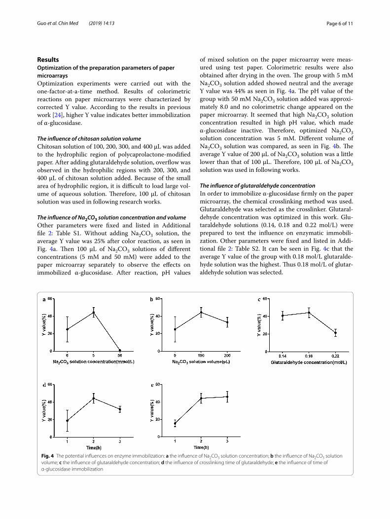
The influence of Na2CO3 solution concentration and volumeOther parameters were fixed and listed in Additional file 2: Table S1. Without adding Na2CO3 solution, the average Y value was 25% after color reaction, as seen in Fig. 4a. Then 100 μL of Na2CO3 solutions of different concentrations (5 mM and 50 mM) were added to the paper microarray separately to observe the effects on immobilized α-glucosidase. After reaction, pH values
of mixed solution on the paper microarray were meas-ured using test paper. Colorimetric results were also obtained after drying in the oven. The group with 5 mM Na2CO3 solution added showed neutral and the average Y value was 44% as seen in Fig. 4a. The pH value of the group with 50 mM Na2CO3 solution added was approxi-mately 8.0 and no colorimetric change appeared on the paper microarray. It seemed that high Na2CO3 solution concentration resulted in high pH value, which made α-glucosidase inactive. Therefore, optimized Na2CO3 solution concentration was 5 mM. Different volume of Na2CO3 solution was compared, as seen in Fig. 4b. The average Y value of 200 μL of Na2CO3 solution was a little lower than that of 100 μL. Therefore, 100 μL of Na2CO3 solution was used in following works.
The influence of glutaraldehyde concentrationIn order to immobilize α-glucosidase firmly on the paper microarray, the chemical crosslinking method was used. Glutaraldehyde was selected as the crosslinker. Glutaral-dehyde concentration was optimized in this work. Glu-taraldehyde solutions (0.14, 0.18 and 0.22 mol/L) were prepared to test the influence on enzymatic immobili-zation. Other parameters were fixed and listed in Addi-tional file 2: Table S2. It can be seen in Fig. 4c that the average Y value of the group with 0.18 mol/L glutaralde-hyde solution was the highest. Thus 0.18 mol/L of glutar-aldehyde solution was selected.
Fig. 4 The potential influences on enzyme immobilization: a the influence of Na2CO3 solution concentration; b the influence of Na2CO3 solution volume; c the influence of glutaraldehyde concentration; d the influence of crosslinking time of glutaraldehyde; e the influence of time of α-glucosidase immobilization
Page 7 of 11Guo et al. Chin Med (2019) 14:13
The influence of time of glutaraldehyde crosslinkingA period of time is required for the reactions between aldehyde groups of glutaraldehyde and amino groups of chitosan. The effect of crosslinking time between glutaraldehyde and chitosan was compared for 1–3 h. Other parameters were fixed and listed in Additional file 2: Table S3. As seen in Fig. 4d, the largest average Y value was obtained at 2 h crosslinking. Therefore, 2 h of crosslinking time between glutaraldehyde and chitosan was applied for the following works.
The influence of time of α‑glucosidase immobilizationα-Glucosidase cannot be fully immobilized on the paper microarray if immobilization time is not enough. There-fore, the influence of time of α-glucosidase immobili-zation was optimized with other parameters fixed and listed in Additional file 2: Table S4. The results can be seen in Fig. 4e. The average Y value became higher with the increase of time. Finally, 3 h of α-glucosidase immo-bilization was selected to prepare the paper microarray for active compound discovery.
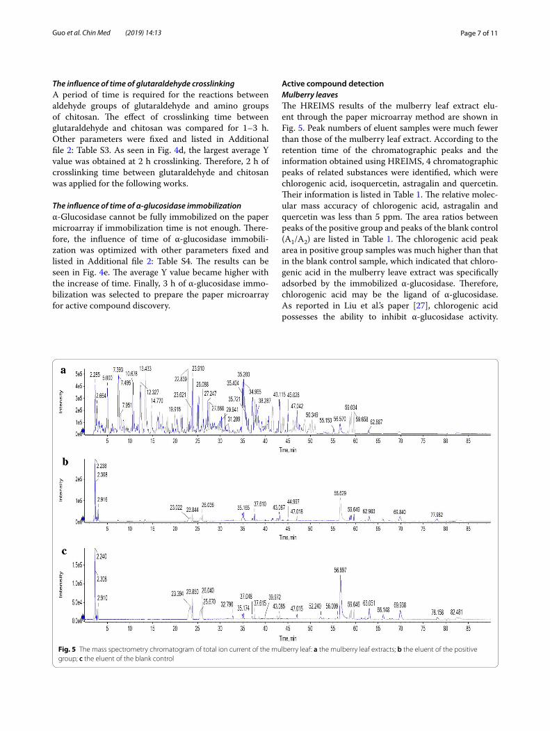
Active compound detectionMulberry leavesThe HREIMS results of the mulberry leaf extract elu-ent through the paper microarray method are shown in Fig. 5. Peak numbers of eluent samples were much fewer than those of the mulberry leaf extract. According to the retention time of the chromatographic peaks and the information obtained using HREIMS, 4 chromatographic peaks of related substances were identified, which were chlorogenic acid, isoquercetin, astragalin and quercetin. Their information is listed in Table 1. The relative molec-ular mass accuracy of chlorogenic acid, astragalin and quercetin was less than 5 ppm. The area ratios between peaks of the positive group and peaks of the blank control (A1/A2) are listed in Table 1. The chlorogenic acid peak area in positive group samples was much higher than that in the blank control sample, which indicated that chloro-genic acid in the mulberry leave extract was specifically adsorbed by the immobilized α-glucosidase. Therefore, chlorogenic acid may be the ligand of α-glucosidase. As reported in Liu et al.’s paper [27], chlorogenic acid possesses the ability to inhibit α-glucosidase activity.
Fig. 5 The mass spectrometry chromatogram of total ion current of the mulberry leaf: a the mulberry leaf extracts; b the eluent of the positive group; c the eluent of the blank control
Page 8 of 11Guo et al. Chin Med (2019) 14:13
Therefore, it was confirmed that active compounds in mulberry leaves included chlorogenic acid.
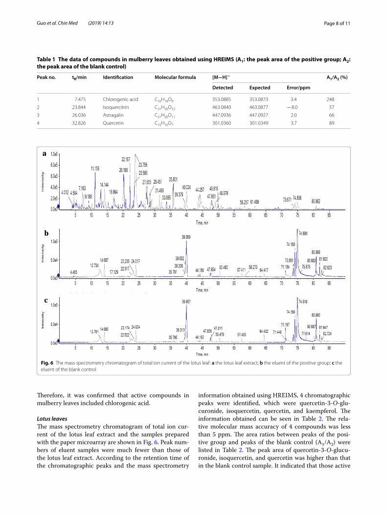
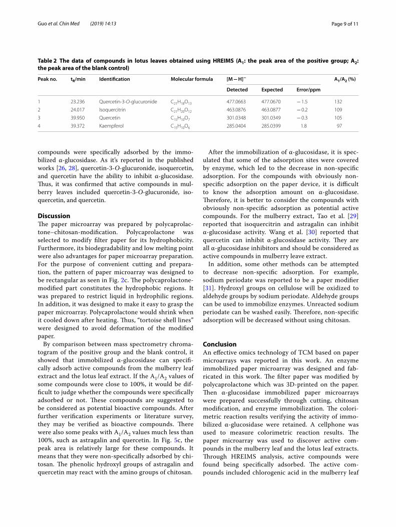
Lotus leavesThe mass spectrometry chromatogram of total ion cur-rent of the lotus leaf extract and the samples prepared with the paper microarray are shown in Fig. 6. Peak num-bers of eluent samples were much fewer than those of the lotus leaf extract. According to the retention time of the chromatographic peaks and the mass spectrometry
information obtained using HREIMS, 4 chromatographic peaks were identified, which were quercetin-3-O-glu-curonide, isoquercetin, quercetin, and kaempferol. The information obtained can be seen in Table 2. The rela-tive molecular mass accuracy of 4 compounds was less than 5 ppm. The area ratios between peaks of the posi-tive group and peaks of the blank control (A1/A2) were listed in Table 2. The peak area of quercetin-3-O-glucu-ronide, isoquercetin, and quercetin was higher than that in the blank control sample. It indicated that those active
Table 1 The data of compounds in mulberry leaves obtained using HREIMS (A1: the peak area of the positive group; A2: the peak area of the blank control)
Peak no. tR/min Identification Molecular formula [M−H]− A1/A2 (%)
Detected Expected Error/ppm
1 7.475 Chlorogenic acid C16H18O9 353.0885 353.0873 3.4 248
2 23.844 Isoquercitrin C21H20O12 463.0840 463.0877 − 8.0 57
3 26.036 Astragalin C21H20O11 447.0936 447.0927 2.0 66
4 32.826 Quercetin C15H10O7 301.0360 301.0349 3.7 89
Fig. 6 The mass spectrometry chromatogram of total ion current of the lotus leaf: a the lotus leaf extract; b the eluent of the positive group; c the eluent of the blank control
Page 9 of 11Guo et al. Chin Med (2019) 14:13
compounds were specifically adsorbed by the immo-bilized α-glucosidase. As it’s reported in the published works [26, 28], quercetin-3-O-glucuronide, isoquercetin, and quercetin have the ability to inhibit α-glucosidase. Thus, it was confirmed that active compounds in mul-berry leaves included quercetin-3-O-glucuronide, iso-quercetin, and quercetin.
DiscussionThe paper microarray was prepared by polycaprolac-tone–chitosan-modification. Polycaprolactone was selected to modify filter paper for its hydrophobicity. Furthermore, its biodegradability and low melting point were also advantages for paper microarray preparation. For the purpose of convenient cutting and prepara-tion, the pattern of paper microarray was designed to be rectangular as seen in Fig. 2c. The polycaprolactone-modified part constitutes the hydrophobic regions. It was prepared to restrict liquid in hydrophilic regions. In addition, it was designed to make it easy to grasp the paper microarray. Polycaprolactone would shrink when it cooled down after heating. Thus, “tortoise shell lines” were designed to avoid deformation of the modified paper.
By comparison between mass spectrometry chroma-togram of the positive group and the blank control, it showed that immobilized α-glucosidase can specifi-cally adsorb active compounds from the mulberry leaf extract and the lotus leaf extract. If the A1/A2 values of some compounds were close to 100%, it would be dif-ficult to judge whether the compounds were specifically adsorbed or not. These compounds are suggested to be considered as potential bioactive compounds. After further verification experiments or literature survey, they may be verified as bioactive compounds. There were also some peaks with A1/A2 values much less than 100%, such as astragalin and quercetin. In Fig. 5c, the peak area is relatively large for these compounds. It means that they were non-specifically adsorbed by chi-tosan. The phenolic hydroxyl groups of astragalin and quercetin may react with the amino groups of chitosan.
After the immobilization of α-glucosidase, it is spec-ulated that some of the adsorption sites were covered by enzyme, which led to the decrease in non-specific adsorption. For the compounds with obviously non-specific adsorption on the paper device, it is difficult to know the adsorption amount on α-glucosidase. Therefore, it is better to consider the compounds with obviously non-specific adsorption as potential active compounds. For the mulberry extract, Tao et al. [29] reported that isoquercitrin and astragalin can inhibit α-glucosidase activity. Wang et al. [30] reported that quercetin can inhibit α-glucosidase activity. They are all α-glucosidase inhibitors and should be considered as active compounds in mulberry leave extract.
In addition, some other methods can be attempted to decrease non-specific adsorption. For example, sodium periodate was reported to be a paper modifier [31]. Hydroxyl groups on cellulose will be oxidized to aldehyde groups by sodium periodate. Aldehyde groups can be used to immobilize enzymes. Unreacted sodium periodate can be washed easily. Therefore, non-specific adsorption will be decreased without using chitosan.
ConclusionAn effective omics technology of TCM based on paper microarrays was reported in this work. An enzyme immobilized paper microarray was designed and fab-ricated in this work. The filter paper was modified by polycaprolactone which was 3D-printed on the paper. Then α-glucosidase immobilized paper microarrays were prepared successfully through cutting, chitosan modification, and enzyme immobilization. The colori-metric reaction results verifying the activity of immo-bilized α-glucosidase were retained. A cellphone was used to measure colorimetric reaction results. The paper microarray was used to discover active com-pounds in the mulberry leaf and the lotus leaf extracts. Through HREIMS analysis, active compounds were found being specifically adsorbed. The active com-pounds included chlorogenic acid in the mulberry leaf
Table 2 The data of compounds in lotus leaves obtained using HREIMS (A1: the peak area of the positive group; A2: the peak area of the blank control)
Peak no. tR/min Identification Molecular formula [M − H]− A1/A2 (%)
Detected Expected Error/ppm
1 23.236 Quercetin-3-O-glucuronide C21H18O13 477.0663 477.0670 − 1.5 132
2 24.017 Isoquercitrin C21H20O12 463.0876 463.0877 − 0.2 109
3 39.950 Quercetin C15H10O7 301.0348 301.0349 − 0.3 105
4 39.372 Kaempferol C15H10O6 285.0404 285.0399 1.8 97
Page 10 of 11Guo et al. Chin Med (2019) 14:13
extract, and quercetin-3-O-glucuronide, isoquerce-tin, and quercetin in the lotus leaf extract. Some com-pounds with remarkable non-specific adsorption, such as astragalin and quercetin were also found to be active compounds. All of these compounds should be consid-ered in the quality control of anti-diabetic drugs that are made from mulberry leaves and lotus leaves.
Additional files
Additional file 1. Minimum standards of reporting checklist.
Additional file 2. The fixed parameters in the experiments for optimiza-tion of enzyme immobilization.
Authors’ contributionsData curation, XL and SG; methodology, XG and YW; software, SG and XL; for-mal analysis, XL and XG; resources, XG and YW; writing—original draft prepara-tion, SG and XL; writing—review and editing, SG, XG, and YW; supervision, XG. All authors read and approved the final manuscript.
AcknowledgementsNot applicable.
Competing interestsThe authors declare that they have no competing interests.
Availability of data and materialsAll data generated or analyzed during this study are included in this published article and its additional files.
Consent for publicationNot applicable.
Ethics approval and consent to participateNot applicable.
FundingThe authors would like to acknowledge the support of the National Natural Science Foundation of China (No. 81503242) and the Fundamental Research Funds for the Central Universities (No. 2018FZA7018).
Publisher’s NoteSpringer Nature remains neutral with regard to jurisdictional claims in pub-lished maps and institutional affiliations.
Received: 8 February 2019 Accepted: 21 March 2019
References 1. Kiyama R. DNA microarray-based screening and characterization of
traditional Chinese medicine. Microarrays. 2017;6(1):4. 2. Zhang Q, Yang M. DNA microarray technology and traditional Chinese
medicines. Prog Nutr. 2010;12(1):6–12. 3. Zhang YB, Wang J, Wang ZT, But PPH, Shaw PC. DNA microarray for
identification of the herb of dendrobium species from Chinese medici-nal formulations. Planta Med. 2003;69(12):1172–4.
4. Wang LL, Li Z, Shao Q, Li X, Ai N, Zhao XP, et al. Dissecting active ingre-dients of Chinese medicine by content-weighted ingredient-target network. Mol BioSyst. 2014;10(7):1905–11.
5. Tao Y. Studies on chemical biology approaches for rapid discovery of bioactive compounds from traditional Chinese medicine. Hangzhou: Zhejiang University; 2014.
6. Liu CX, Chen SL, Xiao XH, Zhang TJ, Hou WB, Liao ML. A new con-cept on quality marker of Chinese materia medica: quality con-trol for Chinese medicinal products. Chinese Tradit Herbal Drugs. 2016;47(09):1443–57.
7. Lourenço VK, Jiang ZJ, Zhang XQ, Curcino Vieira LC, Gonçalvez Corrêa A, Lucia Cardoso C, et al. Acetylcholinesterase immobilized capillary reactors coupled to protein coated magnetic beads: a new tool for plant extract ligand screening. Talanta. 2013;116:647–52.
8. Wiekhorst F, Steinhoff U, Eberbeck D, Trahms L. Magnetorelaxometry assisting biomedical applications of magnetic nanoparticles. Pharm Res. 2012;29(5):1189–202.
9. Wubshet SG, Brighente IMC, Moaddel R, Staerk D. Magnetic ligand fish-ing as a targeting tool for HPLC-HRMS-SPE-NMR: α-glucosidase inhibi-tory ligands and alkylresorcinol glycosides from Eugenia catharinae. J Nat Prod. 2015;78(11):2657–65.
10. Zhuo RJ, Liu H, Liu NN, Wang Y. Ligand fishing: a remarkable strategy for discovering bioactive compounds from complex mixture of natural products. Molecules. 2016;21:11.
11. Liu LL, Ma YJ, Chen XQ, Xiong X, Shi SY. Screening and identification of BSA bound ligands from Puerariae lobata flower by BSA functionalized Fe3O4 magnetic nanoparticles coupled with HPLC–MS/MS. J Chroma-togr B. 2012;887–888:55–60.
12. Deng X, Shi SY, Li SM, Yang TL. Magnetic ligand fishing combination with high-performance liquid chromatography-diode array detector-mass spectrometry to screen and characterize cyclooxygenase-2 inhibitors from green tea. J Chromatogr B. 2014;973:55–60.
13. Qing LS, Xue Y, Zheng Y, Xiong J, Liao X, Ding LS, et al. Ligand fishing from Dioscorea nipponica extract using human serum albumin functionalized magnetic nanoparticles. J Chromatogr A. 2010;1217(28):4663–8.
14. Martinez AW, Phillips ST, Butte MJ, Whitesides GM. Patterned paper as a platform for inexpensive, low-volume, portable bioassays. Angew Chem Int Ed. 2007;46(8):1318–20.
15. He Y, Wu WB, Fu JZ. Rapid fabrication of paper-based microfluidic analytical devices with desktop stereolithography 3D printer. RSC Adv. 2015;5(4):2694–701.
16. Ahmed S, Bui M-PN, Abbas A. Paper-based chemical and biological sensors: engineering aspects. Biosens Bioelect. 2016;77:249–63.
17. Gu Z, Zhao MX, Sheng YW, Bentolila LA, Tang Y. Detection of mercury ion by infrared fluorescent protein and its hydrogel-based paper assay. Anal Chem. 2011;83(6):2324–9.
18. Mitchell HT, Noxon IC, Chaplan CA, Carlton SJ, Liu CH, Ganaja KA, et al. Reagent pencils: a new technique for solvent-free deposi-tion of reagents onto paper-based microfluidic devices. Lab Chip. 2015;15(10):2213–20.
19. Kaewarsa P, Laiwattanapaisal W, Palasuwan A, Palasuwan D. A new paper-based analytical device for detection of glucose-6-phosphate dehydrogenase deficiency. Talanta. 2017;164:534–9.
20. Chen JG, Bu WL, Lai WQ, Liu DY, Mei S, Liu Z, et al. Hypoglycemic effects and mechanism of mulberry leaves polysaccharide. Chin Tradit Herbal Drugs. 2011;42(03):515–20.
21. Zhao J, Gao L, Qi ZP. Extraction of folium nelumbinis total alkaloid and its salt and comparison in lowering blood lipid between them. Tianjin J Tradit Chin Med. 2005;02(02):161–2.
22. Sheldon RA. Enzyme immobilization: the quest for optimum perfor-mance. Adv Synth Catal. 2007;349(8–9):1289–307.
23. Nery EW, Kubota LT. Evaluation of enzyme immobilization methods for paper-based devices—a glucose oxidase study. J Pharm Biomed Anal. 2016;117:551–9.
24. Guo SX, Shao JY, Gong XC. Paper-based analytical devices pre-pared with polycaprolactone printing and their application in the activity determination of mulberry extracts. J Pharm Biomed Anal. 2018;161:28–34.
25. Zhao TT, Wei H, Chen LM, Wang ZM, Zhang QW, Zhao ZB, et al. HPLC fingerprints of different medicinal parts of morus alba L. Chin Pharm J. 2017;52(07):560–6.
Page 11 of 11Guo et al. Chin Med (2019) 14:13
• fast, convenient online submission
•
thorough peer review by experienced researchers in your field
• rapid publication on acceptance
• support for research data, including large and complex data types
•
gold Open Access which fosters wider collaboration and increased citations
maximum visibility for your research: over 100M website views per year •
At BMC, research is always in progress.
Learn more biomedcentral.com/submissions
Ready to submit your research ? Choose BMC and benefit from:
26. Cheng YY, Wang Y. The compounds with inhibitory activity on α-glucosidase in lotus leaves and its use. China, CN 102827221 A. 2012.
27. Liu XH, Li ML, Tan B, Chen HH, Lu Y. Inhibitory effects of chlorogenic acid and isochlorogenic acid from purple sweet potato leaves on α-glucosidase. Modern Food Sci Technol. 2014;30(03):103–7.
28. Cheng JY. Screening study on the α-glucosidase inhibitors from dio-spyros kaki leaves. Changsha: Hunan Agricultural University; 2013.
29. Tao Y, Zhang YF, Cheng YY, Wang Y. Rapid screening and identi-fication of α-glucosidase inhibitors from mulberry leaves using
enzyme-immobilized magnetic beads coupled with HPLC/MS and NMR. Biomed Chromatogr. 2013;27(2):148–55.
30. Wang SH, Huang WL, Chen QS, Zeng L, Zeng FJ. Inhibition of rutin and quercetin on α-glycosidase. China Brewing. 2012;31(1):133–5.
31. Mu C, Guo J, Li X, Lin W, Li D. Preparation and properties of dialdehyde carboxymethyl cellulose crosslinked gelatin edible films. Food Hydrocol-loids. 2012;27(1):22–9.