Expression of μ-protocadherin is negatively regulated by ... · by the activation of the...
9
OPEN Expression of μ-protocadherin is negatively regulated by the activation of the β-catenin signaling pathway in normal and cancer colorectal enterocytes L Montorsi 1 , S Parenti 1 , L Losi 1 , F Ferrarini 2 , C Gemelli 1 , A Rossi 3 , G Manco 3 , S Ferrari 1 , B Calabretta 4,5 , E Tagliafico 6,7 , T Zanocco-Marani 1,8 and A Grande* ,1,8 Mu-protocadherin (MUCDHL) is an adhesion molecule predominantly expressed by colorectal epithelial cells which is markedly downregulated upon malignant transformation. Notably, treatment of colorectal cancer (CRC) cells with mesalazine lead to increased expression of MUCDHL, and is associated with sequestration of β-catenin on the plasma membrane and inhibition of its transcriptional activity. To better characterize the causal relationship between β-catenin and MUCDHL expression, we performed various experiments in which CRC cell lines and normal colonic organoids were subjected to culture conditions inhibiting (FH535 treatment, transcription factor 7-like 2 siRNA inactivation, Wnt withdrawal) or stimulating (LiCl treatment) β-catenin activity. We show here that expression of MUCDHL is negatively regulated by functional activation of the β-catenin signaling pathway. This finding was observed in cell culture systems representing conditions of physiological stimulation and upon constitutive activation of β-catenin in CRC. The ability of MUCDHL to sequester and inhibit β-catenin appears to provide a positive feedback enforcing the effect of β-catenin inhibitors rather than serving as the primary mechanism responsible for β-catenin inhibition. Moreover, MUCDHL might have a role as biomarker in the development of CRC chemoprevention drugs endowed with β-catenin inhibitory activity. Cell Death and Disease (2016) 7, e2263; doi:10.1038/cddis.2016.163; published online 16 June 2016 Cadherins are integral membrane proteins constituting a complex superfamily, which consists of 4100 members and are divided into classical cadherins and protocadherins. The latter are characterized by the presence of one or more unconventional domains in addition to the typical cadherin-like domains, which are responsible for the canonical function of these proteins. 1 From a structural point of view, cadherins are composed of an extracellular portion, harboring its cadherin- like domains through which they promote intercellular adhe- sion, and an intra-cellular portion, through which they inhibit cell proliferation. This effect is achieved by sequestering β-catenin in a submembrane location, thereby preventing its translocation to the nucleus where, in normal conditions, it activates the transcription of growth-promoting target genes. 2 Not surprisingly, as a result of these activities, cadherins exert a recognized onco-suppressor effect that can be observed in different cell types, depending on their expression pattern. 3 In the last decade, a novel cadherin, named μ-protocadherin (MUCDHL) to underline the hybrid nature of its extracellular portion, which contains four cadherin-like and three mucin-like domains, has been isolated and characterized. 4 This protein maintains all the general features of cadherins and carries, in its intra-cellular portion, four prolin-rich domains together with a single PDZ domain, previously shown to bind β-catenin. A survey of MUCDHL distribution revealed that significant levels of its expression were exclusively detected in a couple of anatomical sites, including colorectal mucosa. 5 This observation suggested a possible role in colorectal carcino- genesis that was subsequently supported by experimental results. Indeed, interaction of MUCDHL with β-catenin, which is constitutively activated in colorectal cancer (CRC), was confirmed and MUCDHL expression exhibited a remarkable downregulation in 470% of CRC cases, in association with higher proliferation indexes. 6 Notably, data obtained in our laboratory showed that treatment of CRC cells with an anti- inflammatory drug named mesalazine (5-ASA), that is able to inhibit β-catenin signaling in vitro and to exert anti-CRC chemoprevention activity in vivo, 7 markedly induces MUCDHL 1 Department of Life Sciences, University of Modena and Reggio Emilia, Modena, Italy; 2 SOFAR S.p.A., Milano, Italy; 3 Department of Surgical, Medical, Dental and Morphological Sciences with Interest in Transplant, Oncological and Regenerative Medicine, University of Modena and Reggio Emilia, Modena, Italy; 4 Department of Clinical and Diagnostic Medicine and Public Health, University of Modena and Reggio Emilia, Modena, Italy; 5 Department of Cancer Biology and SKKC, Thomas Jefferson University, Philadelphia, PA, USA; 6 Department of Medical and Surgical Sciences, University of Modena and Reggio Emilia, Modena, Italy and 7 Center for Genome Research, University of Modena and Reggio Emilia, Modena, Italy *Corresponding author: A Grande, Department of Life Sciences, University of Modena and Reggio Emilia, Via Campi 287, Modena 41125, Italy. Tel: +39 59 2055409; Fax +39 59 2055410; E-mail: [email protected] 8 These authors shared the last authorship of the paper. Received 04.12.15; revised 05.5.16; accepted 05.5.16; Edited by A Stephanou Abbreviations: MUCDHL, μ-protocadherin; CRC, colorectal cancer; 5-ASA, mesalazine; TCF4, transcription factor 7-like 2; CDX2, caudal type homeobox transcription factor 2; QRT-PCR, quantitative real-time PCR; GAPDH, glyceraldehyde-3-phosphate dehydrogenase; BBM, Bio-Bank of Modena; RSPO1, R-spondin 1; EGF, epidermal growth factor; WNT3a, wingless-Type MMTV Integration Site Family, Member 3A; PGE2, prostaglandin E2; APC, adenomatous polyposis coli; Snail, snail family zinc finger 1; ITF2, transcription factor 4; Twist, twist family bHLH transcription factor 1; Slug, snail family zinc finger 2; GSK3β, glycogen synthase kinase 3 beta; Zeb1, zinc finger E-box binding homeobox 1 Citation: Cell Death and Disease (2016) 7, e2263; doi:10.1038/cddis.2016.163 & 2016 Macmillan Publishers Limited All rights reserved 2041-4889/16 www.nature.com/cddis
-
Upload
duongthien -
Category
Documents
-
view
215 -
download
0
Transcript of Expression of μ-protocadherin is negatively regulated by ... · by the activation of the...
OPEN
Expression of μ-protocadherin is negatively regulatedby the activation of the β-catenin signaling pathway innormal and cancer colorectal enterocytes
L Montorsi1, S Parenti1, L Losi1, F Ferrarini2, C Gemelli1, A Rossi3, G Manco3, S Ferrari1, B Calabretta4,5, E Tagliafico6,7,T Zanocco-Marani1,8 and A Grande*,1,8
Mu-protocadherin (MUCDHL) is an adhesion molecule predominantly expressed by colorectal epithelial cells which is markedlydownregulated upon malignant transformation. Notably, treatment of colorectal cancer (CRC) cells with mesalazine lead toincreased expression of MUCDHL, and is associated with sequestration of β-catenin on the plasma membrane and inhibition of itstranscriptional activity. To better characterize the causal relationship between β-catenin and MUCDHL expression, we performedvarious experiments in which CRC cell lines and normal colonic organoids were subjected to culture conditions inhibiting (FH535treatment, transcription factor 7-like 2 siRNA inactivation, Wnt withdrawal) or stimulating (LiCl treatment) β-catenin activity. Weshow here that expression of MUCDHL is negatively regulated by functional activation of the β-catenin signaling pathway. Thisfinding was observed in cell culture systems representing conditions of physiological stimulation and upon constitutive activationof β-catenin in CRC. The ability of MUCDHL to sequester and inhibit β-catenin appears to provide a positive feedback enforcing theeffect of β-catenin inhibitors rather than serving as the primary mechanism responsible for β-catenin inhibition. Moreover,MUCDHL might have a role as biomarker in the development of CRC chemoprevention drugs endowed with β-catenin inhibitoryactivity.Cell Death and Disease (2016) 7, e2263; doi:10.1038/cddis.2016.163; published online 16 June 2016
Cadherins are integral membrane proteins constituting acomplex superfamily, which consists of 4100 members andare divided into classical cadherins and protocadherins. Thelatter are characterized by the presence of one or moreunconventional domains in addition to the typical cadherin-likedomains, which are responsible for the canonical function ofthese proteins.1 From a structural point of view, cadherins arecomposed of an extracellular portion, harboring its cadherin-like domains through which they promote intercellular adhe-sion, and an intra-cellular portion, through which they inhibitcell proliferation. This effect is achieved by sequesteringβ-catenin in a submembrane location, thereby preventing itstranslocation to the nucleus where, in normal conditions, itactivates the transcription of growth-promoting target genes.2
Not surprisingly, as a result of these activities, cadherins exerta recognized onco-suppressor effect that can be observed indifferent cell types, depending on their expression pattern.3 Inthe last decade, a novel cadherin, named μ-protocadherin(MUCDHL) to underline the hybrid nature of its extracellular
portion, which contains four cadherin-like and three mucin-likedomains, has been isolated and characterized.4 This proteinmaintains all the general features of cadherins and carries, inits intra-cellular portion, four prolin-rich domains together witha single PDZ domain, previously shown to bind β-catenin.A survey of MUCDHL distribution revealed that significantlevels of its expression were exclusively detected in a coupleof anatomical sites, including colorectal mucosa.5 Thisobservation suggested a possible role in colorectal carcino-genesis that was subsequently supported by experimentalresults. Indeed, interaction of MUCDHL with β-catenin, whichis constitutively activated in colorectal cancer (CRC), wasconfirmed and MUCDHL expression exhibited a remarkabledownregulation in 470% of CRC cases, in association withhigher proliferation indexes.6 Notably, data obtained in ourlaboratory showed that treatment of CRC cells with an anti-inflammatory drug named mesalazine (5-ASA), that is able toinhibit β-catenin signaling in vitro and to exert anti-CRCchemoprevention activity in vivo,7 markedly inducesMUCDHL
1Department of Life Sciences, University of Modena and Reggio Emilia, Modena, Italy; 2SOFAR S.p.A., Milano, Italy; 3Department of Surgical, Medical, Dental andMorphological Sciences with Interest in Transplant, Oncological and Regenerative Medicine, University of Modena and Reggio Emilia, Modena, Italy; 4Department ofClinical and Diagnostic Medicine and Public Health, University of Modena and Reggio Emilia, Modena, Italy; 5Department of Cancer Biology and SKKC, Thomas JeffersonUniversity, Philadelphia, PA, USA; 6Department of Medical and Surgical Sciences, University of Modena and Reggio Emilia, Modena, Italy and 7Center for GenomeResearch, University of Modena and Reggio Emilia, Modena, Italy*Corresponding author: A Grande, Department of Life Sciences, University of Modena and Reggio Emilia, Via Campi 287, Modena 41125, Italy. Tel: +39 59 2055409;Fax +39 59 2055410; E-mail: [email protected] authors shared the last authorship of the paper.
Received 04.12.15; revised 05.5.16; accepted 05.5.16; Edited by A Stephanou
Abbreviations: MUCDHL, μ-protocadherin; CRC, colorectal cancer; 5-ASA, mesalazine; TCF4, transcription factor 7-like 2; CDX2, caudal type homeobox transcriptionfactor 2; QRT-PCR, quantitative real-time PCR; GAPDH, glyceraldehyde-3-phosphate dehydrogenase; BBM, Bio-Bank of Modena; RSPO1, R-spondin 1; EGF, epidermalgrowth factor; WNT3a, wingless-Type MMTV Integration Site Family, Member 3A; PGE2, prostaglandin E2; APC, adenomatous polyposis coli; Snail, snail family zincfinger 1; ITF2, transcription factor 4; Twist, twist family bHLH transcription factor 1; Slug, snail family zinc finger 2; GSK3β, glycogen synthase kinase 3 beta; Zeb1, zincfinger E-box binding homeobox 1
Citation: Cell Death and Disease (2016) 7, e2263; doi:10.1038/cddis.2016.163& 2016 Macmillan Publishers Limited All rights reserved 2041-4889/16
www.nature.com/cddis
expression.8 This finding might, in our opinion, reflect twodistinct explanations: (i) 5-ASA could induce MUCDHLexpression, sequester β-catenin on the cell membrane andinhibit its signaling pathway; or (ii) 5-ASA could inhibit β-catenin, leading to an upregulation of MUCDHL, dependent onthe fact that expression of this protein is negatively regulatedby β-catenin signaling (Supplementary Figure 1). In otherterms, upregulation of MUCDHL may represent the cause orthe effect of β-catenin inhibition, respectively. In both casesMUCDHL would, once expressed, sequester and inhibit β-catenin on the plasma membrane, although with differentbiological consequences. To better understand the correlationbetween β-catenin activity and MUCDHL expression, weperformed experiments in which the β-catenin signalingpathway was inhibited in CRC cell lines using a chemicalinhibitor or a siRNA-based approach and showed thatMUCDHL expression is upregulated by both treatments. Bycontrast, the opposite effect was observed in human colonicorganoids treated with a compound able to activate the β-catenin signaling pathway.
Results
Downregulation of MUCDHL expression correlates withAPC mutation in sporadic CRC. An important cluesupporting the possibility that regulation of MUCDHLexpression is a downstream event of the β-catenin signalingpathway (second hypothesis of Introduction) came from theobservation that MUCDHL downregulation always accom-panies adenomatous polyposis coli (APC) inactivation inCRC. In fact, immuno-histochemical data obtained in ourlaboratory showed that 11 out of 21 analyzed CRC caseswere negative for MUCDHL and APC expression, whereasthe remaining exhibited a double positivity for the sameproteins (Figure 1). This finding suggests that the constitutiveactivation of the β-catenin signaling pathway, determined bymutation/inactivation of APC, is responsible for a reducedexpression of MUCDHL, implying that the latter is negativelyregulated by the former.
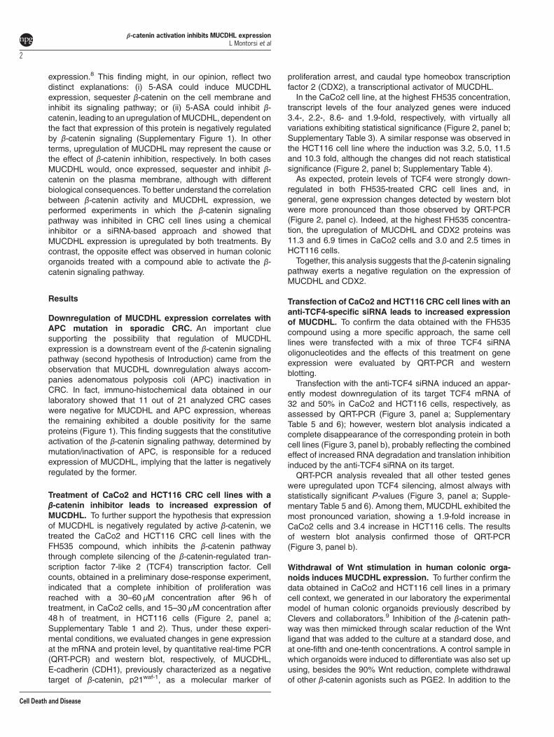
Treatment of CaCo2 and HCT116 CRC cell lines with aβ-catenin inhibitor leads to increased expression ofMUCDHL. To further support the hypothesis that expressionof MUCDHL is negatively regulated by active β-catenin, wetreated the CaCo2 and HCT116 CRC cell lines with theFH535 compound, which inhibits the β-catenin pathwaythrough complete silencing of the β-catenin-regulated tran-scription factor 7-like 2 (TCF4) transcription factor. Cellcounts, obtained in a preliminary dose-response experiment,indicated that a complete inhibition of proliferation wasreached with a 30–60 μM concentration after 96 h oftreatment, in CaCo2 cells, and 15–30 μM concentration after48 h of treatment, in HCT116 cells (Figure 2, panel a;Supplementary Table 1 and 2). Thus, under these experi-mental conditions, we evaluated changes in gene expressionat the mRNA and protein level, by quantitative real-time PCR(QRT-PCR) and western blot, respectively, of MUCDHL,E-cadherin (CDH1), previously characterized as a negativetarget of β-catenin, p21waf-1, as a molecular marker of
proliferation arrest, and caudal type homeobox transcriptionfactor 2 (CDX2), a transcriptional activator of MUCDHL.In the CaCo2 cell line, at the highest FH535 concentration,
transcript levels of the four analyzed genes were induced3.4-, 2.2-, 8.6- and 1.9-fold, respectively, with virtually allvariations exhibiting statistical significance (Figure 2, panel b;Supplementary Table 3). A similar response was observed inthe HCT116 cell line where the induction was 3.2, 5.0, 11.5and 10.3 fold, although the changes did not reach statisticalsignificance (Figure 2, panel b; Supplementary Table 4).As expected, protein levels of TCF4 were strongly down-
regulated in both FH535-treated CRC cell lines and, ingeneral, gene expression changes detected by western blotwere more pronounced than those observed by QRT-PCR(Figure 2, panel c). Indeed, at the highest FH535 concentra-tion, the upregulation of MUCDHL and CDX2 proteins was11.3 and 6.9 times in CaCo2 cells and 3.0 and 2.5 times inHCT116 cells.Together, this analysis suggests that the β-catenin signaling
pathway exerts a negative regulation on the expression ofMUCDHL and CDX2.
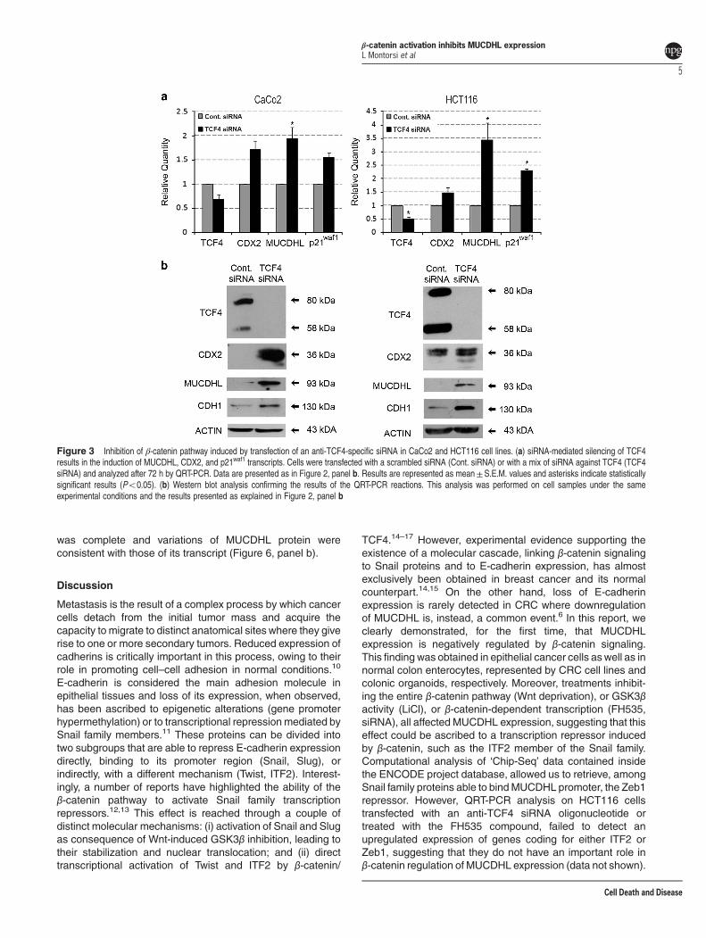
Transfection of CaCo2 and HCT116 CRC cell lines with ananti-TCF4-specific siRNA leads to increased expressionof MUCDHL. To confirm the data obtained with the FH535compound using a more specific approach, the same celllines were transfected with a mix of three TCF4 siRNAoligonucleotides and the effects of this treatment on geneexpression were evaluated by QRT-PCR and westernblotting.Transfection with the anti-TCF4 siRNA induced an appar-
ently modest downregulation of its target TCF4 mRNA of32 and 50% in CaCo2 and HCT116 cells, respectively, asassessed by QRT-PCR (Figure 3, panel a; SupplementaryTable 5 and 6); however, western blot analysis indicated acomplete disappearance of the corresponding protein in bothcell lines (Figure 3, panel b), probably reflecting the combinedeffect of increased RNA degradation and translation inhibitioninduced by the anti-TCF4 siRNA on its target.QRT-PCR analysis revealed that all other tested genes
were upregulated upon TCF4 silencing, almost always withstatistically significant P-values (Figure 3, panel a; Supple-mentary Table 5 and 6). Among them, MUCDHL exhibited themost pronounced variation, showing a 1.9-fold increase inCaCo2 cells and 3.4 increase in HCT116 cells. The resultsof western blot analysis confirmed those of QRT-PCR(Figure 3, panel b).
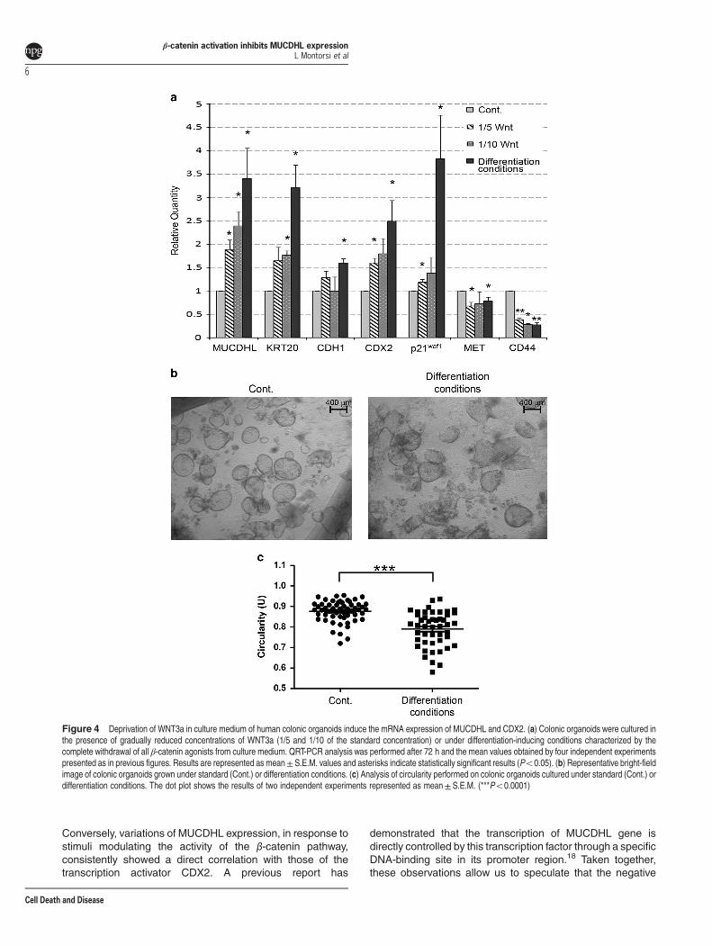
Withdrawal of Wnt stimulation in human colonic orga-noids induces MUCDHL expression. To further confirm thedata obtained in CaCo2 and HCT116 cell lines in a primarycell context, we generated in our laboratory the experimentalmodel of human colonic organoids previously described byClevers and collaborators.9 Inhibition of the β-catenin path-way was then mimicked through scalar reduction of the Wntligand that was added to the culture at a standard dose, andat one-fifth and one-tenth concentrations. A control sample inwhich organoids were induced to differentiate was also set upusing, besides the 90% Wnt reduction, complete withdrawalof other β-catenin agonists such as PGE2. In addition to the
β-catenin activation inhibits MUCDHL expressionL Montorsi et al
2
Cell Death and Disease

genes tested above, QRT-PCR analysis included the Keratin-20 gene, as a marker of enterocyte differentiation, and theMet and CD44 genes, as direct targets of the β-cateninsignaling pathway. As shown in Figure 4a and SupplementaryTable 7, mRNA expression of MUCDHL, Keratin-20, E-cad-herin, CDX2 and p21waf1 genes, showed a gradual increasefrom standard culture to differentiation conditions, reachingmean values of 3.4, 3.2, 1.6, 2.5, 3.8, respectively. The Metand CD44 genes showed an exactly inverted trend, exhibitinga 21 and 73% decrease, respectively, of their mRNAexpression. Most comparisons were statistically significant(Figure 4, panel a; Supplementary Table 7). Inhibitionof the β-catenin signaling also affected the morphology oforganoids, revealing a collapsed shape evidenced bylight-inverted microscope examination followed by theassessment of circularity values with the ImageJ software(0.88±0.007 U of control versus 0.79±0.012 U of differen-tiated samples, Po0.0001) (Figure 4, panels b and c,Supplementary Table 10).
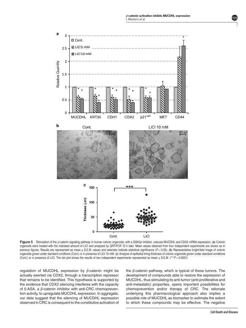
Treatment of human colonic organoids with LiCl inhibitsMUCDHL expression. The experimental advantage ofhuman colonic organoids is also represented by thepossibility to analyze the effects of inducible stimulation ofβ-catenin signaling, unlike the CRC cell lines where thispathway is constitutively activated as consequence of APCmutation. This condition was obtained upon treatment with 5or 10 mM LiCl, which is known to inhibit the GSK3β enzyme,leading to stabilization and activation of β-catenin. Then,QRT-PCR analysis was carried out as described in theprevious experiment. With the only exception of the Met gene,
the results observed were, as expected, opposite to thoseelicited by withdrawal of the β-catenin agonists. In fact, at thehighest LiCl concentration, there was a 46% decrease forMUCDHL, 61% for Keratin-20, 48% for E-cadherin, 60% forCDX2 and 42% for p21waf1, whereas, under the sameconditions, the CD44 gene showed a 2.6-fold increase of itstranscript levels (Figure 5, panel a; Supplementary Table 8).All variations were statistically significant. In agreementwith its activity on the β-catenin pathway, treatment withLiCl determined a positive effect on the proliferation rate oforganoids demonstrated by a more pronounced thickness oftheir peripheral lining, observed again through a light-invertedmicroscope and measured with the ImageJ software(50.2± 2.7 μm of LiCl treated versus 23.7±1.5 μm ofcontrol samples, Po0.0001) (Figure 5, panels b and c;Supplementary Table 11).
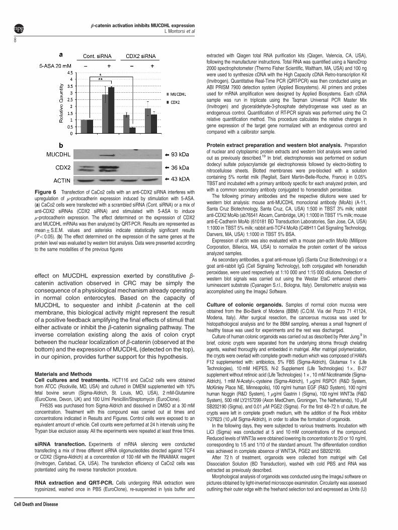
CDX2 silencing abolishes MUCDHL upregulationinduced by 5-ASA treatment. In the studies presentedhere, variations of MUCDHL expression in response tomodulators of β-catenin activity, correlate with those of theCDX2 transcription factor, suggesting its involvement in theregulation of MUCDHL levels. To verify this possibility, theCaCo2 cells were transfected with a scrambled control siRNAor with a mix of three anti-CDX2 siRNAs for 12 h, and thencultured for an additional 4-day period in the absence orpresence of 20 mM 5-ASA. The decision to use thiscompound was based on its anti-CRC chemopreventioncapacity, previously related to its ability to inhibit β-cateninactivity and induce MUCDHL expression.
Figure 1 (a) Table summarizing the results of the immuno-histochemical analysis of APC and μ-protocadherin (MUCDHL) protein levels in 21 CRC samples; + and −indicate presence or absence of the analyzed protein, respectively. (b) Immuno-histochemical analysis of two representative CRC cases exhibiting a double positive (APC+/MUCDHL+) (CRC +/+) or a double negative (APC− /MUCDHL− ) (CRC − /− ) expression pattern. Positivity appears as a dark staining; cell nuclei were counterstained withhematoxylin
β-catenin activation inhibits MUCDHL expressionL Montorsi et al
3
Cell Death and Disease
As expected, CaCo2 cells transfected with control siRNAand exposed to 5-ASA, exhibited a 2.8-and 3.4-fold increaseof CDX2 and MUCDHL mRNA, respectively, detected byQRT-PCR (Figure 6, panel a, Supplementary Table 9).
Instead, transfection of the anti-CDX2 siRNAs resulted in~ 50% reduction of these values returning them approximatelyto their basal level.Western blot analysis, carried out under thesame experimental conditions, showed that CDX2 silencing
Figure 2 Effects of FH535 treatment in the CaCo2 and HCT116 CRC cell lines. (a) FH5353 causes a dose- and time-dependent inhibition of cell proliferation. Cells weretreated with the indicated concentrations of FH535 and counted after 24, 48, 72 and 96 h (x axis). Number of cell expansions are reported on y axis as mean values obtained fromthree independent experiments. (b) FH535 induces upregulation of μ-protocadherin (MUCDHL), E-cadherin (CDH1), p21waf1 and CDX2 transcripts. Cells were treated with theindicated doses of FH5353 and analyzed by QRT-PCR after 96 h for CaCo2 and 48 h for HCT116 cells. Mean variations of mRNA expression levels, deriving from threeindependent experiments, are reported in the y axis as relative fold-change (relative quantity). Results are represented as mean±S.E.M. values and asterisks indicatestatistically significant results (Po0.05). (c) Western blot analysis following treatment with FH535 shows a decreased expression of TCF4 protein and a concomitant increase ofMUCDHL and CDX2 protein expression. Cells were treated with the indicated doses of FH535 and analyzed after 96 h for CaCo2 and 48 h for HCT116 cells. This analysis wasperformed on nuclear and cytoplasmic extracts and normalized with actin. Analyzed proteins and their molecular weights are indicated on the left and on the right, respectively
β-catenin activation inhibits MUCDHL expressionL Montorsi et al
4
Cell Death and Disease
was complete and variations of MUCDHL protein wereconsistent with those of its transcript (Figure 6, panel b).
Discussion
Metastasis is the result of a complex process by which cancercells detach from the initial tumor mass and acquire thecapacity to migrate to distinct anatomical sites where they giverise to one or more secondary tumors. Reduced expression ofcadherins is critically important in this process, owing to theirrole in promoting cell–cell adhesion in normal conditions.10
E-cadherin is considered the main adhesion molecule inepithelial tissues and loss of its expression, when observed,has been ascribed to epigenetic alterations (gene promoterhypermethylation) or to transcriptional repression mediated bySnail family members.11 These proteins can be divided intotwo subgroups that are able to repress E-cadherin expressiondirectly, binding to its promoter region (Snail, Slug), orindirectly, with a different mechanism (Twist, ITF2). Interest-ingly, a number of reports have highlighted the ability of theβ-catenin pathway to activate Snail family transcriptionrepressors.12,13 This effect is reached through a couple ofdistinct molecular mechanisms: (i) activation of Snail and Slugas consequence of Wnt-induced GSK3β inhibition, leading totheir stabilization and nuclear translocation; and (ii) directtranscriptional activation of Twist and ITF2 by β-catenin/
TCF4.14–17 However, experimental evidence supporting theexistence of a molecular cascade, linking β-catenin signalingto Snail proteins and to E-cadherin expression, has almostexclusively been obtained in breast cancer and its normalcounterpart.14,15 On the other hand, loss of E-cadherinexpression is rarely detected in CRC where downregulationof MUCDHL is, instead, a common event.6 In this report, weclearly demonstrated, for the first time, that MUCDHLexpression is negatively regulated by β-catenin signaling.This finding was obtained in epithelial cancer cells aswell as innormal colon enterocytes, represented by CRC cell lines andcolonic organoids, respectively. Moreover, treatments inhibit-ing the entire β-catenin pathway (Wnt deprivation), or GSK3βactivity (LiCl), or β-catenin-dependent transcription (FH535,siRNA), all affected MUCDHL expression, suggesting that thiseffect could be ascribed to a transcription repressor inducedby β-catenin, such as the ITF2 member of the Snail family.Computational analysis of ‘Chip-Seq’ data contained insidethe ENCODE project database, allowed us to retrieve, amongSnail family proteins able to bindMUCDHL promoter, the Zeb1repressor. However, QRT-PCR analysis on HCT116 cellstransfected with an anti-TCF4 siRNA oligonucleotide ortreated with the FH535 compound, failed to detect anupregulated expression of genes coding for either ITF2 orZeb1, suggesting that they do not have an important role inβ-catenin regulation of MUCDHL expression (data not shown).
Figure 3 Inhibition of β-catenin pathway induced by transfection of an anti-TCF4-specific siRNA in CaCo2 and HCT116 cell lines. (a) siRNA-mediated silencing of TCF4results in the induction of MUCDHL, CDX2, and p21waf1 transcripts. Cells were transfected with a scrambled siRNA (Cont. siRNA) or with a mix of siRNA against TCF4 (TCF4siRNA) and analyzed after 72 h by QRT-PCR. Data are presented as in Figure 2, panel b. Results are represented as mean±S.E.M. values and asterisks indicate statisticallysignificant results (Po0.05). (b) Western blot analysis confirming the results of the QRT-PCR reactions. This analysis was performed on cell samples under the sameexperimental conditions and the results presented as explained in Figure 2, panel b
β-catenin activation inhibits MUCDHL expressionL Montorsi et al
5
Cell Death and Disease
Conversely, variations of MUCDHL expression, in response tostimuli modulating the activity of the β-catenin pathway,consistently showed a direct correlation with those of thetranscription activator CDX2. A previous report has
demonstrated that the transcription of MUCDHL gene isdirectly controlled by this transcription factor through a specificDNA-binding site in its promoter region.18 Taken together,these observations allow us to speculate that the negative
Figure 4 Deprivation of WNT3a in culture medium of human colonic organoids induce the mRNA expression of MUCDHL and CDX2. (a) Colonic organoids were cultured inthe presence of gradually reduced concentrations of WNT3a (1/5 and 1/10 of the standard concentration) or under differentiation-inducing conditions characterized by thecomplete withdrawal of all β-catenin agonists from culture medium. QRT-PCR analysis was performed after 72 h and the mean values obtained by four independent experimentspresented as in previous figures. Results are represented as mean± S.E.M. values and asterisks indicate statistically significant results (Po0.05). (b) Representative bright-fieldimage of colonic organoids grown under standard (Cont.) or differentiation conditions. (c) Analysis of circularity performed on colonic organoids cultured under standard (Cont.) ordifferentiation conditions. The dot plot shows the results of two independent experiments represented as mean± S.E.M. (***Po0.0001)
β-catenin activation inhibits MUCDHL expressionL Montorsi et al
6
Cell Death and Disease
regulation of MUCDHL expression by β-catenin might beactually exerted via CDX2, through a transcription repressorthat remains to be identified. This hypothesis is supported bythe evidence that CDX2 silencing interferes with the capacityof 5-ASA, a β-catenin inhibitor with anti-CRC chemopreven-tion activity, to upregulate MUCDHL expression. In aggregate,our data suggest that the silencing of MUCDHL expressionobserved in CRC is consequent to the constitutive activation of
the β-catenin pathway, which is typical of these tumors. Thedevelopment of compounds able to restore the expression ofMUCDHL, thus stimulating its anti-tumor (anti-proliferative andanti-metastatic) properties, opens important possibilities forchemoprevention and/or therapy of CRC. The rationaleunderlying this pharmacological approach also implies apossible role of MUCDHL as biomarker to estimate the extentto which these compounds may be effective. The negative
Figure 5 Stimulation of the β-catenin signaling pathway in human colonic organoids, with a GSK3β inhibitor, reduces MUCDHL and CDX2 mRNA expression. (a) Colonicorganoids were treated with the indicated amount of LiCl and analyzed by QRT-PCR 72 h later. Mean values obtained from four independent experiments are shown as inprevious figures. Results are represented as mean±S.E.M. values and asterisks indicate statistical significance (Po0.05). (b) Representative bright-field image of colonicorganoids grown under standard conditions (Cont.) or in presence of LiCl 10 mM. (c) Analysis of epithelial lining thickness of colonic organoids grown under standard conditions(Cont.) or in presence of LiCl. The dot plot shows the results of two independent experiments represented as mean± S.E.M. (***Po0.0001)
β-catenin activation inhibits MUCDHL expressionL Montorsi et al
7
Cell Death and Disease
effect on MUCDHL expression exerted by constitutive β-catenin activation observed in CRC may be simply theconsequence of a physiological mechanism already operatingin normal colon enterocytes. Based on the capacity ofMUCDHL to sequester and inhibit β-catenin at the cellmembrane, this biological activity might represent the resultof a positive feedback amplifying the final effects of stimuli thateither activate or inhibit the β-catenin signaling pathway. Theinverse correlation existing along the axis of colon cryptbetween the nuclear localization of β-catenin (observed at thebottom) and the expression of MUCDHL (detected on the top),in our opinion, provides further support for this hypothesis.
Materials and MethodsCell cultures and treatments. HCT116 and CaCo2 cells were obtainedfrom ATCC (Rockville, MD, USA) and cultured in DMEM supplemented with 10%fetal bovine serum (Sigma-Aldrich, St. Louis, MO, USA), 2 mM-Glutamine(EuroClone, Devon, UK) and 100 U/ml Penicillin/Streptomycin (EuroClone).FH535 was purchased from Sigma-Aldrich and dissolved in DMSO at a 30 mM
concentration. Treatment with this compound was carried out at times andconcentrations indicated in Results and Figures. Control cells were exposed to anequivalent amount of vehicle. Cell counts were performed at 24 h intervals using theTrypan blue exclusion assay. All the experiments were repeated at least three times.
siRNA transfection. Experiments of mRNA silencing were conductedtransfecting a mix of three different siRNA oligonucleotides directed against TCF4or CDX2 (Sigma-Aldrich) at a concentration of 100 nM with the RNAiMAX reagent(Invitrogen, Carlsbad, CA, USA). The transfection efficiency of CaCo2 cells waspotentiated using the reverse transfection procedure.
RNA extraction and QRT-PCR. Cells undergoing RNA extraction weretrypsinized, washed once in PBS (EuroClone), re-suspended in lysis buffer and
extracted with Qiagen total RNA purification kits (Qiagen, Valencia, CA, USA),following the manufacturer instructions. Total RNA was quantified using a NanoDrop2000 spectrophotometer (Thermo Fisher Scientific, Waltham, MA, USA) and 100 ngwere used to synthesize cDNA with the High Capacity cDNA Retro-transcription Kit(Invitrogen). Quantitative Real-Time PCR (QRT-PCR) was then conducted using anABI PRISM 7900 detection system (Applied Biosystems). All primers and probesused for mRNA amplification were designed by Applied Biosystems. Each cDNAsample was run in triplicate using the Taqman Universal PCR Master Mix(Invitrogen) and glyceraldehyde-3-phosphate dehydrogenase was used as anendogenous control. Quantification of RT-PCR signals was performed using the Ctrelative quantification method. This procedure calculates the relative changes ingene expression of the target gene normalized with an endogenous control andcompared with a calibrator sample.
Protein extract preparation and western blot analysis. Preparationof nuclear and cytoplasmic protein extracts and western blot analysis were carriedout as previously described.19 In brief, electrophoresis was performed on sodiumdodecyl sulfate polyacrylamide gel electrophoresis followed by electro-blotting tonitrocellulose sheets. Blotted membranes were pre-blocked with a solutioncontaining 5% nonfat milk (Regilait, Saint Martin-Belle-Roche, France) in 0.05%TBSTand incubated with a primary antibody specific for each analyzed protein, andwith a common secondary antibody conjugated to horseradish peroxidase.The following primary antibodies and the respective dilutions were used for
western blot analysis: mouse anti-MUCDHL monoclonal antibody (MoAb) (A-11,Santa Cruz Biotechnology, Santa Cruz, CA, USA) 1:500 in TBST 3% milk; rabbitanti-CDX2 MoAb (ab76541 Abcam, Cambridge, UK) 1:1000 in TBST 1% milk; mouseanti-E-Cadherin MoAb (610181 BD Transduction Laboratories, San Jose, CA, USA)1:1000 in TBST 5% milk; rabbit anti-TCF4 MoAb (C48H11 Cell Signaling Technology,Danvers, MA, USA) 1:1000 in TBST 5% BSA.Expression of actin was also evaluated with a mouse pan-actin MoAb (Millipore
Corporation, Billerica, MA, USA) to normalize the protein content of the variousanalyzed samples.As secondary antibodies, a goat anti-mouse IgG (Santa Cruz Biotechnology) or a
goat anti-rabbit IgG (Cell Signaling Technology), both conjugated with horseradishperoxidase, were used respectively at 1:10 000 and 1:15 000 dilutions. Detection ofwestern blot signals was carried out using the Westar EtaC enhanced chemi-luminescent substrate (Cyanagen S.r.l., Bologna, Italy). Densitometric analysis wasaccomplished using the ImageJ Software.
Culture of colonic organoids. Samples of normal colon mucosa wereobtained from the Bio-Bank of Modena (BBM) (C.O.M. Via del Pozzo 71 41124,Modena, Italy). After surgical resection, the cancerous mucosa was used forhistopathological analysis and for the BBM sampling, whereas a small fragment ofhealthy tissue was used for experiments and the rest was discharged.Culture of human colonic organoids was carried out as described by Peter Jung.9 In
brief, colonic crypts were separated from the underlying stroma through chelatingagents, washed thoroughly and embedded in matrigel. After matrigel polymerization,the crypts were overlaid with complete growth medium which was composed of HAM’sF12 supplemented with: antibiotics, 5% FBS (Sigma-Aldrich), Glutamax 1 × (LifeTechnologies), 10 mM HEPES, N-2 Supplement (Life Technologies) 1 × , B-27supplement without retinoic acid (Life Technologies) 1 × , 10 mM Nicotinamide (Sigma-Aldrich), 1 mM N-Acetyl-L-cysteine (Sigma-Aldrich), 1 μg/ml RSPO1 (R&D System,McKinley Place NE, Minneapolis), 100 ng/ml human EGF (R&D System), 100 ng/mlhuman Noggin (R&D System), 1 μg/ml Gastrin I (Sigma), 100 ng/ml WNT3a (R&DSystem), 500 nM LY2157299 (Axon MedChem, Groningen, The Netherlands), 10 μMSB202190 (Sigma), and 0.01 μM PGE2 (Sigma). For the first 48–72 h of culture, thecrypts were left in complete growth medium, with the addition of the Rock inhibitorY-27623 (10 μM Sigma-Aldrich), in order to allow the formation of organoids.In the following days, they were subjected to various treatments. Incubation with
LiCl (Sigma) was conducted at 5 and 10 mM concentrations of the compound.Reduced levels of WNT3a were obtained lowering its concentration to 20 or 10 ng/ml,corresponding to 1/5 and 1/10 of the standard amount. The differentiation conditionwas achieved in complete absence of WNT3A, PGE2 and SB202190.After 72 h of treatment, organoids were collected from matrigel with Cell
Dissociation Solution (BD Transduction), washed with cold PBS and RNA wasextracted as previously described.Morphological analysis of organoids was conducted using the ImageJ software on
pictures obtained by light-inverted microscope examination. Circularity was assessedoutlining their outer edge with the freehand selection tool and expressed as Units (U)
Figure 6 Transfection of CaCo2 cells with an anti-CDX2 siRNA interferes withupregulation of μ-protocadherin expression induced by stimulation with 5-ASA.(a) CaCo2 cells were transfected with a scrambled siRNA (Cont. siRNA) or a mix ofanti-CDX2 siRNAs (CDX2 siRNA) and stimulated with 5-ASA to induceμ-protocadherin expression. The effect determined on the expression of CDX2and MUCDHL mRNAs was then analyzed by QRT-PCR. Results are represented asmean± S.E.M. values and asterisks indicate statistically significant results(Po0.05). (b) The effect determined on the expression of the same genes at theprotein level was evaluated by western blot analysis. Data were presented accordingto the same modalities of the previous figures
β-catenin activation inhibits MUCDHL expressionL Montorsi et al
8
Cell Death and Disease

(1 U representing the value generated by a perfect circle), whereas the thickness oftheir epithelial lining was measured using the straight line selection tool, andexpressed in micrometers (μm).
Statistical analysis. All experiments were repeated at least three times,unless otherwise stated, and the results presented in terms of mean± S.E.M.values. Pairwise comparisons were carried out using the Student’s t-test procedure.Results of statistical analysis were considered significant at P-values o0.05(*o0.05, **o0.001, ***o0.0001).
Conflict of InterestThe authors declare no conflict of interest.
Acknowledgements. This work was supported by grants from Sofar S.p.A.,Milano, Italy and Fondazione di Vignola, Vignola (Mo), Italy. We thanks Paola Mannifor technical assistance and Sebastian Fantini together with Vincenzo Zappavigna forthe kind gift of anti-CDX2 siRNA oligonucleotides.
1. Nollet F, Kools P, van Roy F. Phylogenetic analysis of the cadherin superfamily allowsidentification of six major subfamilies besides several solitary members. J Mol Biol 2000;299: 551–572.
2. Ose R, Yanagawa T, Ikeda S, Ohara O, Koga H. PCDH24-induced contact inhibition involvesdownregulation of beta-catenin signaling. Mol Oncol 2009; 3: 54–66.
3. Okazaki N, Takahashi N, Kojima S, Masuho Y, Koga H. Protocadherin LKC, a new candidatefor a tumor suppressor of colon and liver cancers, its association with contact inhibition of cellproliferation. Carcinogenesis 2002; 23: 1139–1148.
4. Goldberg M, Peshkovsky C, Shifteh A, Al-Awqati Q. mu-Protocadherin, a noveldevelopmentally regulated protocadherin with mucin-like domains. J Biol Chem 2000; 275:24622–24629.
5. Goldberg M, Wei M, Tycko B, Falikovich I, Warburton D. Identification and expressionanalysis of the human mu-protocadherin gene in fetal and adult kidneys. Am J Physiol RenalPhysiol 2002; 283: F454–F463.
6. Losi L, Parenti S, Ferrarini F, Rivasi F, Gavioli M, Natalini G et al. downregulation ofmu-protocadherin expression is a common event in colorectal carcinogenesis. Hum Pathol2011; 42: 960–971.
7. Lyakhovich A, Gasche C. Systematic review: molecular chemoprevention of colorectalmalignancy by mesalazine. Aliment Pharmacol Ther 2010; 31: 202–209.
8. Parenti S, Ferrarini F, Zini R, Montanari M, Losi L, Canovi B et al. Mesalazine inhibits thebeta-catenin signalling pathway acting through the upregulation of mu-protocadherin gene incolorectal cancer cells. Aliment Pharmacol Ther 2010; 31: 108–119.
9. Jung P, Sato T, Merlos-Suarez A, Barriga FM, Iglesias M, Rossell D et al. Isolation andin vitro expansion of human colonic stem cells. Nat Med 2011; 17: 1225–1227.
10. Christofori G. New signals from the invasive front. Nature 2006; 441: 444–450.11. Wang Y, Shi J, Chai K, Ying X, Zhou BP. The Role of Snail in EMTand Tumorigenesis. Curr
Cancer Drug Targets 2013; 13: 963–972.12. Yook JI, Li XY, Ota I, Fearon ER, Weiss SJ. Wnt-dependent regulation of the E-cadherin
repressor snail. J Biol Chem 2005; 280: 11740–11748.13. Yook JI, Li XY, Ota I, Hu C, Kim HS, Kim NH et al. AWnt-Axin2-GSK3beta cascade regulates
Snail1 activity in breast cancer cells. Nat Cell Biol 2006; 8: 1398–1406.14. Bachelder RE, Yoon SO, Franci C, de Herreros AG, Mercurio AM. Glycogen synthase
kinase-3 is an endogenous inhibitor of Snail transcription: implications for the epithelial-mesenchymal transition. J Cell Biol 2005; 168: 29–33.
15. Howe LR, Watanabe O, Leonard J, Brown AM. Twist is upregulated in responseto Wnt1 and inhibits mouse mammary cell differentiation. Cancer Res 2003; 63:1906–1913.
16. Kolligs FT, Nieman MT, Winer I, Hu G, Van Mater D, Feng Y et al. ITF-2, a downstream targetof the Wnt/TCF pathway, is activated in human cancers with beta-catenin defects andpromotes neoplastic transformation. Cancer Cell 2002; 1: 145–155.
17. Zhou BP, Deng J, Xia W, Xu J, Li YM, Gunduz M et al. Dual regulation of Snail byGSK-3beta-mediated phosphorylation in control of epithelial-mesenchymal transition. NatCell Biol 2004; 6: 931–940.
18. Hinkel I, Duluc I, Martin E, Guenot D, Freund JN, Gross I. Cdx2 controls expression of theprotocadherin Mucdhl, an inhibitor of growth and beta-catenin activity in colon cancer cells.Gastroenterology 2012; 142: 875–85 e3.
19. Grande A, Manfredini R, Pizzanelli M, Tagliafico E, Balestri R, Trevisan F et al. Presence of afunctional vitamin D receptor does not correlate with vitamin D3 phenotypic effects in myeloiddifferentiation. Cell Death Differ 1997; 4: 497–505.
Cell Death and Disease is an open-access journalpublished by Nature Publishing Group. This work is
licensed under a Creative Commons Attribution 4.0 InternationalLicense. The images or other third party material in this article areincluded in the article’s Creative Commons license, unless indicatedotherwise in the credit line; if the material is not included under theCreative Commons license, users will need to obtain permission fromthe license holder to reproduce the material. To view a copy of thislicense, visit http://creativecommons.org/licenses/by/4.0/
Supplementary Information accompanies this paper on Cell Death and Disease website (http://www.nature.com/cddis)
β-catenin activation inhibits MUCDHL expressionL Montorsi et al
9
Cell Death and Disease