
Expression and localization of α-adaptin isoforms · 2001-05-03 · Clathrin-coated vesicles are...
11
INTRODUCTION Clathrin-coated vesicles are responsible for the selective endo- cytosis of a number of different membrane proteins, including receptors for various extracellular ligands, and proteins that need to cycle between the plasma membrane and an intracel- lular compartment. This cycling pathway is particularly important in neuronal cells, which must retrieve synaptic vesicle membrane components within seconds after exocyto- sis (Heuser and Reese, 1973). Coated vesicles are also thought to play a major role in the removal of cell adhesion molecules from the plasma membrane, which in neurons would allow new cell contacts to be made and new synapses formed (Hu et al., 1993). In addition to endocytic coated vesicles associated with the plasma membrane, the cell also contains clathrin- coated vesicles associated with the trans-Golgi network (TGN), which facilitate the delivery of newly synthesised lysosomal enzymes to a pre-lysosomal compartment. The major component of the coat is clathrin, which in its native form consists of a triskelion containing three copies of clathrin heavy chain and three copies of clathrin light chain (Kirchhausen and Harrison, 1981). In mammalian cells there is only one type of clathrin heavy chain so far identified, but there are two types of clathrin light chains in most cells, called LC a and LC b , which are encoded by separate genes. In brain there are two additional isoforms of LC a and one additional isoform of LC b , all of which are somewhat larger than those expressed in other tissues. The complete conservation of the flanking sequences between the brain and non-brain forms of the light chains indicates that the different isoforms are produced by alternative splicing of the mRNAs (Jackson et al., 1987; Kirchhausen et al., 1987). The brain-specific sequences are exposed and are thought possibly to interact with a specific factor in brain cytoplasm (Jackson et al., 1987). The inner layer of the coat is made up of adaptors, protein complexes that are in close proximity to the membrane. The adaptors are believed to interact with the cytoplasmic domains of selected membrane proteins, causing these proteins to become concentrated in coated vesicles (Pearse, 1988; Glickman et al., 1989). Different adaptors are associated with the plasma membrane and the TGN (Robinson, 1987; Ahle et al., 1988). Both types of adaptors are composed of four distinct subunits: two of M r ~100×10 3 , called adaptins (α- and β- adaptin at the plasma membrane, γ- and β′-adaptin at the TGN), one of M r ~50×10 3 , and one of M r ~20×10 3 (Ahle et al., 1988). Structural studies show that the adaptors consist of a central brick-like mass (the ‘head’) flanked by two symmetrically placed appendages (the ‘ears’), which are connected to the head by protease-sensitive hinges (Heuser and Keen, 1988). The ears have been found to correspond to the C-terminal domains of the adaptins, while the hinges are proline- and glycine-rich stretches of sequence found in all three classes of 2865 There are two α-adaptin genes, αA and α C , which in brain encode proteins of of M r 108×10 3 and 104×10 3 , respectively. Although both mRNAs can be detected on northern blots of brain and liver, the higher molecular mass polypeptide can only be detected on western blots of brain. Here we explain these observations by showing that α A is alterna- tively spliced and that the protein product in most tissues is different from the one expressed in brain in that it is missing 21 amino acids within the hinge region, giving it a similar mobility to that of α C . Monospecific antibodies were raised against the various α-adaptin isoforms and used to compare their distribution in cells and tissues. Both α A and α C are co-assembled into the same coated pits, and the larger isoform of α A is co-assembled with the smaller isoforms of α-adaptin, both in cells that naturally express it and in transfected cells. Examination of brain and spinal cord sections, labelled either for the larger isoform of α A or for α C , reveals that that the two are to some extent dif- ferentially distributed, consistent with previous in situ hybridisation studies. This finding, combined with the observation that there is considerable variability in the relative expression of the two isoforms in different tissues, indicates that the two genes are switched on in response to different stimuli. Moreover, the larger isoform of α A appears to be more efficiently concentrated in the nerve terminals than α C , which is found not only at the terminals but also diffusely distributed in the cell bodies and dendrites. This suggests that α C may play more of a role in the recycling of membrane components throughout the cell. Key words: α-adaptin, brain, endocytosis SUMMARY Expression and localization of α-adaptin isoforms Catriona L. Ball 1 , Stephen P. Hunt 2 and Margaret S. Robinson 1, * 1 Department of Clinical Biochemistry and 2 MRC Laboratory of Molecular Biology, Hills Road, Cambridge CB2 2QR, UK *Author for correspondence Journal of Cell Science 108, 2865-2875 (1995) Printed in Great Britain © The Company of Biologists Limited 1995
Transcript of Expression and localization of α-adaptin isoforms · 2001-05-03 · Clathrin-coated vesicles are...
2865Journal of Cell Science 108, 2865-2875 (1995)Printed in Great Britain © The Company of Biologists Limited 1995
Expression and localization of α-adaptin isoforms
Catriona L. Ball1, Stephen P. Hunt2 and Margaret S. Robinson1,*1Department of Clinical Biochemistry and 2MRC Laboratory of Molecular Biology, Hills Road, Cambridge CB2 2QR, UK
*Author for correspondence
There are two α-adaptin genes, αA and αC, which in brainencode proteins of of Mr 108×103 and 104×103, respectively.Although both mRNAs can be detected on northern blotsof brain and liver, the higher molecular mass polypeptidecan only be detected on western blots of brain. Here weexplain these observations by showing that αA is alterna-tively spliced and that the protein product in most tissuesis different from the one expressed in brain in that it ismissing 21 amino acids within the hinge region, giving it asimilar mobility to that of αC. Monospecific antibodies wereraised against the various α-adaptin isoforms and used tocompare their distribution in cells and tissues. Both αA andαC are co-assembled into the same coated pits, and thelarger isoform of αA is co-assembled with the smallerisoforms of α-adaptin, both in cells that naturally expressit and in transfected cells. Examination of brain and spinal
cord sections, labelled either for the larger isoform of αAor for αC, reveals that that the two are to some extent dif-ferentially distributed, consistent with previous in situhybridisation studies. This finding, combined with theobservation that there is considerable variability in therelative expression of the two isoforms in different tissues,indicates that the two genes are switched on in response todifferent stimuli. Moreover, the larger isoform of αAappears to be more efficiently concentrated in the nerveterminals than αC, which is found not only at the terminalsbut also diffusely distributed in the cell bodies anddendrites. This suggests that αC may play more of a role inthe recycling of membrane components throughout the cell.
Key words: α-adaptin, brain, endocytosis
SUMMARY
INTRODUCTION
Clathrin-coated vesicles are responsible for the selective endo-cytosis of a number of different membrane proteins, includingreceptors for various extracellular ligands, and proteins thatneed to cycle between the plasma membrane and an intracel-lular compartment. This cycling pathway is particularlyimportant in neuronal cells, which must retrieve synapticvesicle membrane components within seconds after exocyto-sis (Heuser and Reese, 1973). Coated vesicles are also thoughtto play a major role in the removal of cell adhesion moleculesfrom the plasma membrane, which in neurons would allownew cell contacts to be made and new synapses formed (Hu etal., 1993). In addition to endocytic coated vesicles associatedwith the plasma membrane, the cell also contains clathrin-coated vesicles associated with the trans-Golgi network(TGN), which facilitate the delivery of newly synthesisedlysosomal enzymes to a pre-lysosomal compartment.
The major component of the coat is clathrin, which in itsnative form consists of a triskelion containing three copies ofclathrin heavy chain and three copies of clathrin light chain(Kirchhausen and Harrison, 1981). In mammalian cells thereis only one type of clathrin heavy chain so far identified, butthere are two types of clathrin light chains in most cells, calledLCa and LCb, which are encoded by separate genes. In brainthere are two additional isoforms of LCa and one additional
isoform of LCb, all of which are somewhat larger than thoseexpressed in other tissues. The complete conservation of theflanking sequences between the brain and non-brain forms ofthe light chains indicates that the different isoforms areproduced by alternative splicing of the mRNAs (Jackson et al.,1987; Kirchhausen et al., 1987). The brain-specific sequencesare exposed and are thought possibly to interact with a specificfactor in brain cytoplasm (Jackson et al., 1987).
The inner layer of the coat is made up of adaptors, proteincomplexes that are in close proximity to the membrane. Theadaptors are believed to interact with the cytoplasmic domainsof selected membrane proteins, causing these proteins tobecome concentrated in coated vesicles (Pearse, 1988;Glickman et al., 1989). Different adaptors are associated withthe plasma membrane and the TGN (Robinson, 1987; Ahle etal., 1988). Both types of adaptors are composed of four distinctsubunits: two of Mr ~100×103, called adaptins (α- and β-adaptin at the plasma membrane, γ- and β′-adaptin at the TGN),one of Mr ~50×103, and one of Mr ~20×103 (Ahle et al., 1988).Structural studies show that the adaptors consist of a centralbrick-like mass (the ‘head’) flanked by two symmetricallyplaced appendages (the ‘ears’), which are connected to thehead by protease-sensitive hinges (Heuser and Keen, 1988).The ears have been found to correspond to the C-terminaldomains of the adaptins, while the hinges are proline- andglycine-rich stretches of sequence found in all three classes of
2866 C. L. Ball, S. P. Hunt and M. S. Robinson
adaptins between amino acids ~600 and ~700-750 (Robinson,1989, 1990; Kirchhausen et al., 1989).
Cloning and sequencing of the adaptins has revealed theexistence of different isoforms which could potentially havedifferent functions. Although so far only a single γ-adaptingene has been cloned, Southern blotting suggests that theremay be at least one other gene (Robinson, 1990; Ball andRobinson, unpublished data). Three β-type adaptins have beenidentified: two from rat brain (Kirchhausen et al., 1989), whichare encoded by separate genes and have been shown to corre-spond to β- and β′-adaptins (Galluser and Kirchhausen, 1993),and the third from rat lymphocytes (Ponnambalam et al.,1990). This is identical to rat brain β-adaptin except that itlacks a 42 bp insert in the hinge, indicating that β-adaptin, likethe clathrin light chains, is alternatively spliced, giving rise toboth brain and non-brain isoforms. Two α-adaptins have beencloned from brain, and are called αA and αC. They are encodedby different genes and show 84% identity at the amino acidlevel (Robinson, 1989). From their sequences, αA-adaptin hasa deduced size of 108×103 while αC-adaptin has a deduced sizeof 104×103. Western blots of various tissues probed with amonoclonal antibody that reacts with both isoforms of α-adaptin show two bands in brain and spinal cord with apparentmolecular masses of ~112×103 and ~105×103, presumably αA-and αC-adaptins, but only a single band of ~105×103 in othertissues (Robinson, 1987). These observations suggest that αCis universally expressed while αA is only expressed in neuronaltissues. However, northern blots of RNA from brain and liverprobed for the two α-adaptin messages show that a small butsignificant amount of αA-adaptin mRNA is present in liver aswell as in brain, even though the protein cannot be detected onwestern blots (Robinson, 1989).
The experiments described in this paper were designed tofind out why αA-adaptin can be detected in liver on northernblots but not western blots; to investigate the patterns ofexpression of both α-adaptin genes in different cells andtissues; and to localise the protein products of the two genesto find out whether the distribution of the different α-adaptinisoforms might be correlated with possible differences infunction.
MATERIALS AND METHODS
DNA manipulationsMost DNA manipulations were carried out using techniques describedby Sambrook et al. (1989). αA-Adaptin sequences were amplified byPCR from rat liver cDNA (generously provided by Paul Guest) andfrom mouse genomic DNA prepared from liver. The forward primercorresponded to bases 1895 to 1920, while the reverse primer corre-sponded to bases 2374 to 2399. The amplified αA-adaptin DNA wasreamplified using similar primers but with BamHI and EcoRI sitesadded to the 5′ ends of the forward and reverse primers, respectively.These sites were then used to ligate the reamplified DNA into pBlue-script, and the DNA was sequenced using the Sequenase Version 2.0kit (United States Biochemicals).
For northern blotting, poly(A)-containing RNA was purified from3T3 tissue culture cells by the lithium/urea method (Auffray andRougeon, 1980), subjected to electrophoresis (9 µg/lane), and blottedonto Hybond paper. An additional northern blot containing mRNAfrom mouse tissues was bought from Clontech. Three antisenseoligonucleotides were used to probe the blots. Probe 1 was taken from
nucleotides 2115 to 2180 of αA-adaptin, probe 2 was taken fromnucleotides 2175 to 2240 of αA-adaptin, and probe 3 was taken fromnucleotides 2818 to 2883 of αC-adaptin (part of the 3′ untranslatedregion). The three oligonucleotides were end-labelled and the blotswere probed following the instructions supplied by Clontech, thenwashed at room temperature in four changes of 2× SSC, 0.1% SDS,1 mM EDTA, followed by a wash at 65°C for 30 minutes in the samesolution.
Production of antibodiesFusion proteins were made in pGEX-3X and expressed in MC1061cells. The proteins were purified as soluble fusion proteins asdescribed by Smith and Johnson (1988). Regions to be expressed wereamplified from plasmid DNA, using primers that would allow themto be inserted in frame into the BamHI and EcoRI sites of the vector.Fusion protein Α706-727 contains the sequence of αA-adaptin that isremoved in the splicing event; fusion protein A620-655 contains asequence from αA-adaptin that is not removed in the splicing event;and fusion protein C619-656 contains a sequence from αC-adaptincorresponding to the sequence of αA-adaptin used for fusion proteinA620-655 (see Figs 1 and 3).
Rabbits were injected subcutaneously along the flank with 0.5 mgfusion protein made up in complete Freund’s adjuvant, and boostedwith the same amount of protein, in incomplete Freund’s adjuvant,after 2 weeks and then after a further 6 weeks. One week after thefinal boost, a test bleed was performed, and the rabbits were bled outafter an additional week.
The antibodies were absorbed with glutathione S-transferase (GST)fusion proteins to ensure that they would be specific for the proteinfor which they had been designed. Antiserum C619-656 was absorbedwith fusion protein A620-655, antiserum A620-655 with fusionprotein C619-656, and antiserum Α706-727 with GST alone. Theantisera were then affinity purified (Robinson and Pearse, 1986) usingthe fusion proteins against which they had been made. In every case1 mg of protein was used per ml of serum. (In a previous study,antiserum A706-727 was used as a probe for brain α-adaptin and wascalled C4; Seaman et al., 1993.)
Immunoprecipitation and western blottingA 20 µl sample of pig brain plasma membrane adaptors at a concen-tration of 1 mg/ml (prepared as described by Pearse and Robinson,1984) was boiled in 1% SDS for five minutes, then diluted into 180µl RIPA buffer (50 mM Tris-HCl, pH 8, 150 mM NaCl, 0.1% SDS,0.5% deoxycholate, 1% NP40) made without the SDS to give a finalconcentration of 0.1% SDS. The solution was pre-cleared by rotatingwith 50 µl of a 50% slurry of Protein A-Sepharose (Pharmacia) inPBS for one hour, after which the Sepharose was removed by cen-trifugation. The antibodies were added to the supernatants and thetubes rotated for a further hour. A 50 µl sample of Protein ASepharose slurry was added and the tubes rotated again for one hour,then the Sepharose was collected by centrifugation, washed six timeswith 500 µl RIPA buffer, resuspended in 40 µl sample buffer (0.2 MCHES, pH 9.5, 40% glycerol, 8% SDS, 5% mercaptoethanol, 0.25%Bromophenol Blue), and boiled for two minutes. SDS-PAGE, westernblotting, and antibody labelling were carried out as described(Robinson and Pearse, 1986). Antibodies included the three antiseraagainst fusion proteins described above, and the mouse monoclonalanti-α-adaptin antibodies AC1-M11 and AC2-M15. 125I-Protein Awas used to visualise all the antibodies directly except AC2-M15,which needed to be incubated with rabbit anti-mouse Ig (Sigma)before the 125I-Protein A incubation step. Western blotting of totalplasma membrane adaptors was performed in the same manner.
Cell cultureCos cells were maintained in DMEM supplemented with 10% foetalcalf serum, 4 mM L-glutamine, 50 i.u./ml penicillin, 50 µg/ml strep-tomycin. Transfection and immunofluorescence were carried out as
2867α-Adaptin isoforms
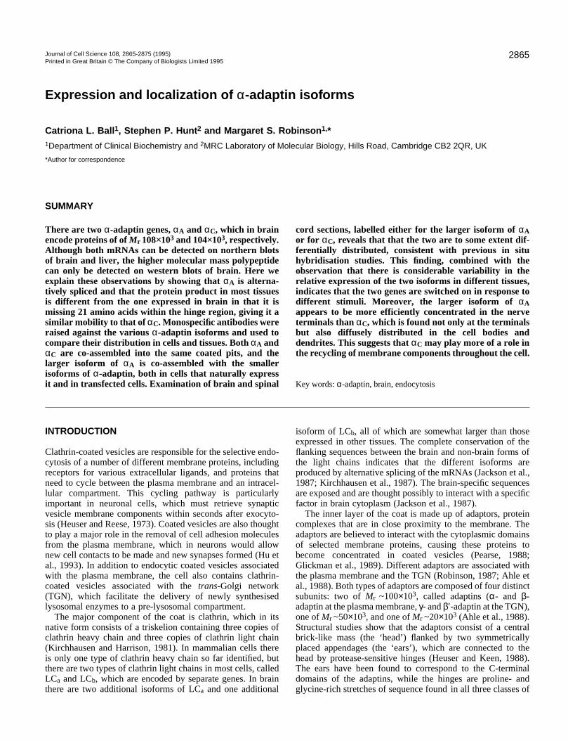
Fig. 1. Comparison of part of the sequences of αA-adaptin (above)and αC-adaptin (below). The amino acids in italics are those used todesign PCR primers to amplify αA-adaptin from rat liver cDNA andfrom mouse genomic DNA. The boxed-in sequence is the region thatwas found to be present in brain but absent in liver and other tissuesbecause of alternative splicing. This sequence was expressed as afusion protein and used to raise antibody A706-A727 (the numbersrefer to the amino acids). The underlined sequences further upstreamwere also expressed as fusion proteins and used to raise antibodiesA620-655 and C619-656. The arrowheads indicate the positions ofintrons.
described previously for Rat1 cells (Robinson, 1990). The plasmidused for transfection contained cDNA encoding the longer form ofαA-adaptin (Robinson, 1989), ligated into the EcoRI site of the vectorpHYKS3 (Robinson, 1990), which contains the SV40 early promoter.The cells were examined two days after transfection.
Primary neuronal cells were cultured on sterile coated coverslips,prepared by incubating the coverslips in 100 µg/ml polyornithine in0.15 M sodium borate buffer, pH 8.4, overnight, washing in PBS,incubating in 5 µg/ml laminin in PBS for four hours, and washingagain in PBS. The cells were prepared from the brains of 17-day ratembryos. The brains were washed in EBSS (Gibco), the two lobesseparated, and the meninges peeled away. The lobes were gentlypushed through a 100 µm nylon mesh into EBSS to separate the cells,and the cells were centrifuged at 1000 rpm for five minutes and resus-pended in 2 ml medium (DMEM supplemented with 10% foetal calfserum, 10% horse serum, 20 i.u./ml penicillin, 20 µg/ml streptomycin,and 4 mM glutamine). The cells were then passed through a 20 µmnylon mesh and plated at 0.6×10⁶ cells/ml onto the coated coverslips.After 24 hours, the medium was changed to defined medium (DMEMsupplemented with, for each litre, 5 mg insulin, 100 mg transferrin,16.1 mg putrescine, 6.3 µg progesterone, 5.36 µg selenium, 18 mMHEPES, pH 7.4) and the cells were left to grow for a further 5-8 daysbefore the coverslips were fixed and labelled.
For some experiments, the cells were double labelled with mousemonoclonal antibodies including the α-adaptin antibodies AC2-M15(Robinson, 1987) and AP6 (Chin et al., 1989) (generously providedby Frances Brodsky), and commercially available antibodies againstMAP5 and GFAP (Sigma).
Labelling of rat brain sectionsRats were terminally anaesthetized with saturated aqueous chloralhydrate (1.5 ml i.p.) and perfused intracardially with 200 ml of normalsaline followed by 400 ml of freshly prepared paraformaldehyde, 4%in 0.05 M sodium phosphate buffer (PB), pH 7.4. Brains wereremoved, post-fixed in the latter solution for 2 hours, and washedovernight in 0.1 M PB containing 30% sucrose and 0.01% sodiumazide. Sections 40 µm thick were cut and incubated free-floating inantibody Α706-727 or C619-656 diluted in TTBS (0.1 M Tris-HCl,pH 7.4, 0.9% NaCl, 0.3% Triton) at 4°C for 48 hours, then washedin four changes of 0.1 M PB. The sections were then incubated inbiotinylated anti-rabbit IgG (Vector Laboratories) diluted into TTBSfor two hours at room temperature, washed in PB, and then incubatedin fluorescein/avidin D (Vector Laboratories), diluted into TTBS fortwo hours at room temperature and washed again. The sections weremounted onto microscope slides and viewed with an MRC 600confocal microscope.
RESULTS
A novel isoform of αA-adaptinAlthough αA-adaptin mRNA can be detected in liver bynorthern blotting, an immunoreactive protein of the expectedsize is only found in brain and spinal cord. This suggests eitherthat αA-adaptin is not translated in liver in sufficiently highamounts to be detected, or that it is made as a smaller isoformwhich co-migrates with αC-adaptin and is therefore effectivelymasked. Fig. 1 shows partial amino acid sequences of αA(upper sequence) and αC (lower sequence), including the hingeregions, where the two proteins are most divergent (the hingeis defined as amino acids 621-746 in αA and 620-707 in αC;see Robinson, 1993). In particular, there is an extra stretch of41 amino acids in αA. To investigate whether all or part of thisregion might be missing in liver, PCR primers correspondingto amino acids 632-640 and 792-800 of αA (see italics in Fig.
1) were used to amplify cDNA from rat liver. The major PCRproduct migrated with an apparent size of ~440 bp, smallerthan the expected size for αA-adaptin of 504 bp. Sequencingrevealed that 66 bp of the DNA was missing from the extrastretch, encoding the boxed-in region in Fig. 1. This indicatesthat αA-adaptin, like clathrin light chains and β-adaptin, isalternatively spliced, with a smaller isoform expressed in liverthan in brain.
To confirm that the difference between liver and brain αA-adaptin is due to alternative splicing, the genomic structure ofthe region around the putative splice site was investigated,using the same PCR primers to amplify αA-adaptin frommouse genomic DNA. Sequencing the PCR product showedthat the extra 66 bp in brain is a single exon with introns oneither side of it. Three additional introns were also found in theamplified DNA (indicated with arrowheads in Fig. 1), with allintron/exon boundaries conforming to the consensus sequencesfor splice sites (Padgett et al., 1986).
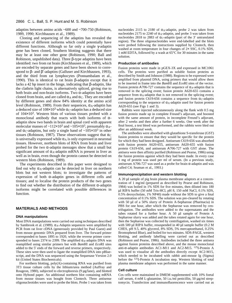
Tissue-specific expression of the different α-adaptinsIs the smaller isoform of αA-adaptin expressed in other tissuesas well as liver? To investigate the patterns of expression of thedifferent α-adaptin isoforms, three oligonucleotide probes wereused to label northern blots of different tissues and also of 3T3cells (Fig. 2). Probe 1 was designed to hybridize to the DNAremoved in the splicing event, probe 2 to the DNA just down-stream from the spliced sequence, and probe 3 to the 3′ untrans-lated region of αC-adaptin. Probe 1 (Fig. 2a) labelled only brain(lane 2) and skeletal muscle (lane 6). In contrast, probe 2 (Fig.2b), like probe 3 (Fig. 2d), labelled all the tissues on the blot aswell as the 3T3 cells (Fig. 2c). These results show that, far from
2868 C. L. Ball, S. P. Hunt and M. S. Robinson
Fig. 2. Northern blots of mRNA from a variety of tissues, labelled with three different probes: (a) probe 1, which recognises the larger isoformof αA-adaptin; (b and c) probe 2, which recognises both isoforms of αA-adaptin; (d) probe 3, which recognises αC-adaptin. Lane 1, heart; 2,brain; 3, spleen; 4, lung; 5, liver; 6, skeletal muscle; 7, kidney; 8, testis; 9, 3T3 cells. The two bands in the blot probed for αC-adaptin (d)appear to be the result of differences in the 3′ untranslated region (Robinson, 1989).
being brain-specific, αA-adaptin is ubiquitously expressed alongwith αC-adaptin, although the relative amounts of the two vary(e.g. compare kidney (lane 7) and testis (lane 8)). However, thelarger isoform of αA-adaptin only appears to be expressed inneuronal tissue and, unexpectedly, in skeletal muscle.
Production of monospecific antibodiesTo study the subcellular distribution of the different α-adaptins, it is necessary to have antibody probes that can dis-tinguish between the various isoforms. A number of mono-clonal anti-α-adaptin antibodies have been raised, includingtwo that appear to be specific for αC-adaptin (Robinson, 1987).However, these are of limited use, since they only recognisethe SDS-denatured protein. All other α-adaptin antibodies thathave so far been raised appear by western blotting to cross-react with all isoforms of the protein. Thus, we set out to raisemonospecific antibodies against fragments of the different α-adaptins expressed as fusion proteins which could be used forimmunolocalisation and immunoprecipitation experiments.The hinge region was chosen for these studies for two reasons:first, because it is here that there is least homology between αAand αC; and second, because the hinge is known to be exposedin the native adaptor complex. Three fusion proteins were con-structed, using the expression vector pGEX-3X: Α706-727,A620-655, and C619-656 (see Figs 1 and 3). Α706-727 wasdesigned to elicit antibodies that would be specific for thelarger isoform of αA-adaptin; A620-655 was designed to elicitantibodies that would be specific for both αA-adaptins but notαC-adaptin; and C619-656 was designed to elicit antibodiesthat would be specific for αC-adaptin.
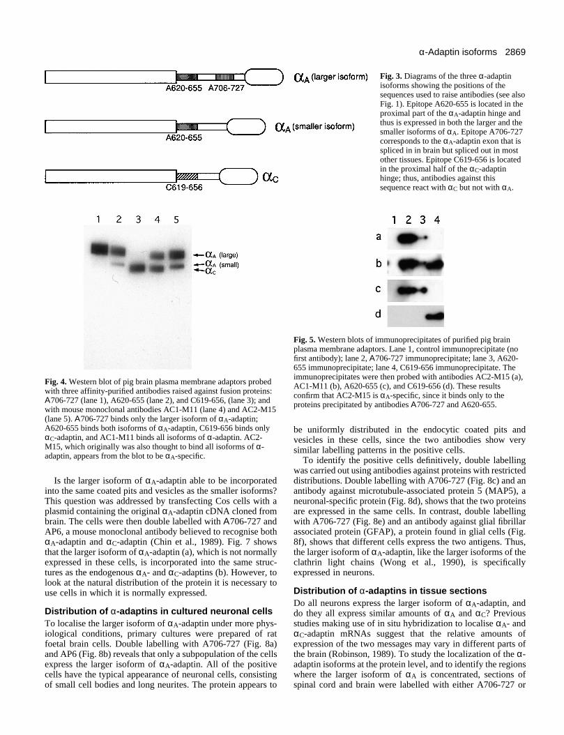
Affinity-purified and cross-absorbed antibodies against allthree fusion proteins were tested by labelling western blots ofpurified pig brain plasma membrane adaptors (Fig. 4).Antibody Α706-727 binds only to the upper α-adaptin band(lane 1), while C619-656 binds only to the lower α-adaptinband (lane 3). However, A620-655, which should recogniseboth forms of αA-adaptin, binds to the upper band and a lowerband, although the upper band is much more heavily labelledthan the lower band (lane 2). In addition, the mobility of thelower band is not identical to that of αC-adaptin, although it is
very close to it. This indicates that in brain, a small amount ofthe smaller, ‘non-brain’ form of αA-adaptin is expressed aswell as the larger isoform, and it confirms that the smallerisoform of αA-adaptin runs on SDS-polyacrylamide gels witha very similar mobility to that of αC-adaptin.
Two mouse monoclonal anti-α-adaptin antibodies were alsoincluded on the blot as positive controls: AC1-M11 and AC2-M15. AC1-M11 is known to recognise both αA-adaptin andαC-adaptin, since it reacts with both recombinant proteins(Robinson, 1989, and unpublished results). On the blot, itlabels both bands with approximately equal intensity (lane 4).However, AC2-M15 (lane 5), which was also thought to bindboth αA-adaptin and αC-adaptin (Robinson, 1987), produces apattern more similar to that of antibody A620-655, suggestingthat AC2-M15 may in fact be specific for αA-adaptin.
To test whether AC2-M15 is an αA-specific antibody,purified pig brain adaptors were immunoprecipitated andwestern blotted. The four lanes in Fig. 5 contain immunopre-cipitates carried out with no first antibody (lane 1), Α706-727(lane 2), A620-655 (lane 3), and C619-656 (lane 4) (A620-655precipitated less efficiently than the other antibodies). Whenthe blot was probed with AC2-M15 (a), the αA-adaptin bandsin lanes 2 and 3 were labelled but not the αC-adaptin band inlane 4. A similar pattern was seen when the blot was probedwith A620-655 (c) or with Α706-727 (not shown). In contrast,the universal anti-α-adaptin antibody AC1-M11 was found tolabel bands in all three lanes (b), while C619-656 labelled theαC-adaptin band in lane 4 only (d). These results confirm thatAC2-M15 is specific for αA-adaptin and does not cross-reactwith αC-adaptin.
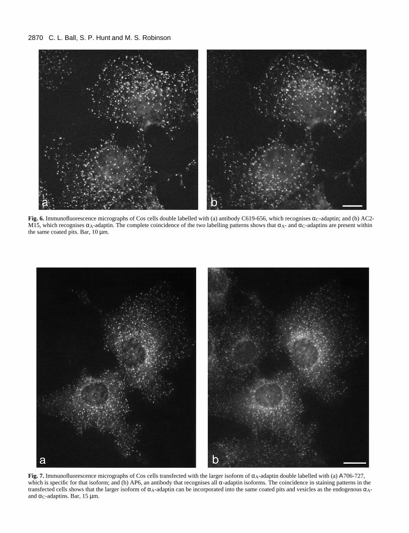
Colocalization of the different α-adaptinsAre the different α-adaptin isoforms incorporated into thesame coated pits? To compare the distribution of αC-adaptinand αA-adaptin, Cos cells were double labelled with the rabbitαC-specific antibody C619-656 (Fig. 6a) and the mouse αA-specific antibody AC2-M15 (Fig. 6b). The staining patternsshow almost complete coincidence, indicating that, at least inthese cells, all of the plasma membrane coated pits appear tocontain both α-adaptins.
2869α-Adaptin isoforms
Fig. 3. Diagrams of the three α-adaptinisoforms showing the positions of thesequences used to raise antibodies (see alsoFig. 1). Epitope A620-655 is located in theproximal part of the αA-adaptin hinge andthus is expressed in both the larger and thesmaller isoforms of αA. Epitope A706-727corresponds to the αA-adaptin exon that isspliced in in brain but spliced out in mostother tissues. Epitope C619-656 is locatedin the proximal half of the αC-adaptinhinge; thus, antibodies against thissequence react with αC but not with αA.
Fig. 4. Western blot of pig brain plasma membrane adaptors probedwith three affinity-purified antibodies raised against fusion proteins:Α706-727 (lane 1), A620-655 (lane 2), and C619-656, (lane 3); andwith mouse monoclonal antibodies AC1-M11 (lane 4) and AC2-M15(lane 5). Α706-727 binds only the larger isoform of αA-adaptin;A620-655 binds both isoforms of αA-adaptin, C619-656 binds onlyαC-adaptin, and AC1-M11 binds all isoforms of α-adaptin. AC2-M15, which originally was also thought to bind all isoforms of α-adaptin, appears from the blot to be αA-specific.
Fig. 5. Western blots of immunoprecipitates of purified pig brainplasma membrane adaptors. Lane 1, control immunoprecipitate (nofirst antibody); lane 2, Α706-727 immunoprecipitate; lane 3, A620-655 immunoprecipitate; lane 4, C619-656 immunoprecipitate. Theimmunoprecipitates were then probed with antibodies AC2-M15 (a),AC1-M11 (b), A620-655 (c), and C619-656 (d). These resultsconfirm that AC2-M15 is αA-specific, since it binds only to theproteins precipitated by antibodies Α706-727 and A620-655.
Is the larger isoform of αA-adaptin able to be incorporatedinto the same coated pits and vesicles as the smaller isoforms?This question was addressed by transfecting Cos cells with aplasmid containing the original αA-adaptin cDNA cloned frombrain. The cells were then double labelled with Α706-727 andAP6, a mouse monoclonal antibody believed to recognise bothαA-adaptin and αC-adaptin (Chin et al., 1989). Fig. 7 showsthat the larger isoform of αA-adaptin (a), which is not normallyexpressed in these cells, is incorporated into the same struc-tures as the endogenous αA- and αC-adaptins (b). However, tolook at the natural distribution of the protein it is necessary touse cells in which it is normally expressed.
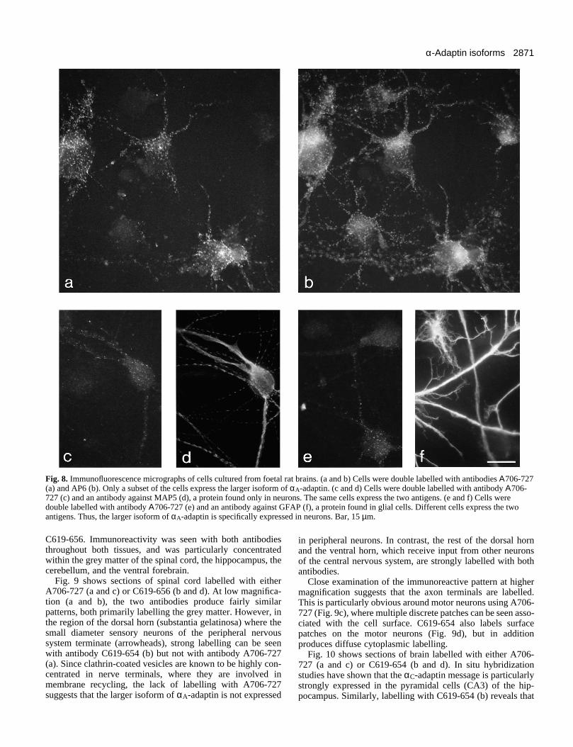
Distribution of α-adaptins in cultured neuronal cellsTo localise the larger isoform of αA-adaptin under more phys-iological conditions, primary cultures were prepared of ratfoetal brain cells. Double labelling with A706-727 (Fig. 8a)and AP6 (Fig. 8b) reveals that only a subpopulation of the cellsexpress the larger isoform of αA-adaptin. All of the positivecells have the typical appearance of neuronal cells, consistingof small cell bodies and long neurites. The protein appears to
be uniformly distributed in the endocytic coated pits andvesicles in these cells, since the two antibodies show verysimilar labelling patterns in the positive cells.
To identify the positive cells definitively, double labellingwas carried out using antibodies against proteins with restricteddistributions. Double labelling with A706-727 (Fig. 8c) and anantibody against microtubule-associated protein 5 (MAP5), aneuronal-specific protein (Fig. 8d), shows that the two proteinsare expressed in the same cells. In contrast, double labellingwith A706-727 (Fig. 8e) and an antibody against glial fibrillarassociated protein (GFAP), a protein found in glial cells (Fig.8f), shows that different cells express the two antigens. Thus,the larger isoform of αA-adaptin, like the larger isoforms of theclathrin light chains (Wong et al., 1990), is specificallyexpressed in neurons.
Distribution of α-adaptins in tissue sectionsDo all neurons express the larger isoform of αA-adaptin, anddo they all express similar amounts of αA and αC? Previousstudies making use of in situ hybridization to localise αA- andαC-adaptin mRNAs suggest that the relative amounts ofexpression of the two messages may vary in different parts ofthe brain (Robinson, 1989). To study the localization of the α-adaptin isoforms at the protein level, and to identify the regionswhere the larger isoform of αA is concentrated, sections ofspinal cord and brain were labelled with either A706-727 or
2870 C. L. Ball, S. P. Hunt and M. S. Robinson
Fig. 6. Immunofluorescence micrographs of Cos cells double labelled with (a) antibody C619-656, which recognises αC-adaptin; and (b) AC2-M15, which recognises αA-adaptin. The complete coincidence of the two labelling patterns shows that αA- and αC-adaptins are present withinthe same coated pits. Bar, 10 µm.
Fig. 7. Immunofluorescence micrographs of Cos cells transfected with the larger isoform of αA-adaptin double labelled with (a) Α706-727,which is specific for that isoform; and (b) AP6, an antibody that recognises all α-adaptin isoforms. The coincidence in staining patterns in thetransfected cells shows that the larger isoform of αA-adaptin can be incorporated into the same coated pits and vesicles as the endogenous αA-and αC-adaptins. Bar, 15 µm.
2871α-Adaptin isoforms
Fig. 8. Immunofluorescence micrographs of cells cultured from foetal rat brains. (a and b) Cells were double labelled with antibodies Α706-727(a) and AP6 (b). Only a subset of the cells express the larger isoform of αA-adaptin. (c and d) Cells were double labelled with antibody Α706-727 (c) and an antibody against MAP5 (d), a protein found only in neurons. The same cells express the two antigens. (e and f) Cells weredouble labelled with antibody Α706-727 (e) and an antibody against GFAP (f), a protein found in glial cells. Different cells express the twoantigens. Thus, the larger isoform of αA-adaptin is specifically expressed in neurons. Bar, 15 µm.
C619-656. Immunoreactivity was seen with both antibodiesthroughout both tissues, and was particularly concentratedwithin the grey matter of the spinal cord, the hippocampus, thecerebellum, and the ventral forebrain.
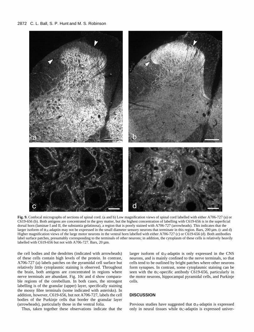
Fig. 9 shows sections of spinal cord labelled with eitherA706-727 (a and c) or C619-656 (b and d). At low magnifica-tion (a and b), the two antibodies produce fairly similarpatterns, both primarily labelling the grey matter. However, inthe region of the dorsal horn (substantia gelatinosa) where thesmall diameter sensory neurons of the peripheral nervoussystem terminate (arrowheads), strong labelling can be seenwith antibody C619-654 (b) but not with antibody A706-727(a). Since clathrin-coated vesicles are known to be highly con-centrated in nerve terminals, where they are involved inmembrane recycling, the lack of labelling with A706-727suggests that the larger isoform of αA-adaptin is not expressed
in peripheral neurons. In contrast, the rest of the dorsal hornand the ventral horn, which receive input from other neuronsof the central nervous system, are strongly labelled with bothantibodies.
Close examination of the immunoreactive pattern at highermagnification suggests that the axon terminals are labelled.This is particularly obvious around motor neurons using A706-727 (Fig. 9c), where multiple discrete patches can be seen asso-ciated with the cell surface. C619-654 also labels surfacepatches on the motor neurons (Fig. 9d), but in additionproduces diffuse cytoplasmic labelling.
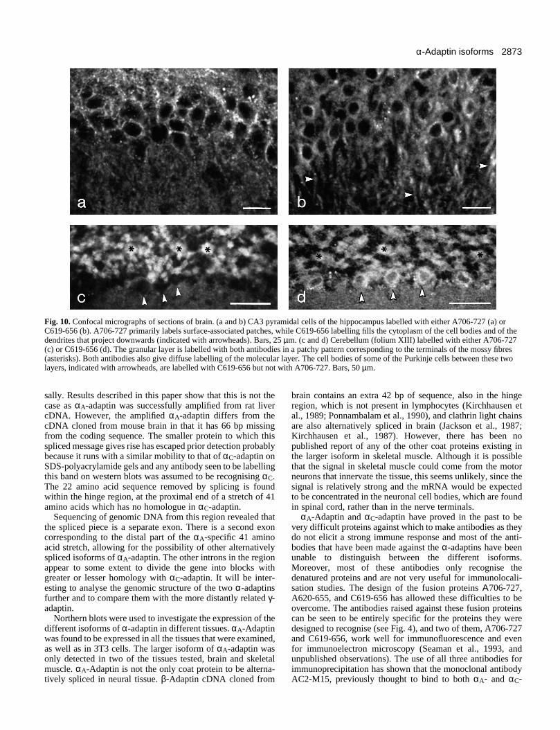
Fig. 10 shows sections of brain labelled with either A706-727 (a and c) or C619-654 (b and d). In situ hybridizationstudies have shown that the αC-adaptin message is particularlystrongly expressed in the pyramidal cells (CA3) of the hip-pocampus. Similarly, labelling with C619-654 (b) reveals that
2872 C. L. Ball, S. P. Hunt and M. S. Robinson
Fig. 9. Confocal micrographs of sections of spinal cord. (a and b) Low magnification views of spinal cord labelled with either A706-727 (a) orC619-656 (b). Both antigens are concentrated in the grey matter, but the highest concentration of labelling with C619-656 is in the superficialdorsal horn (laminae I and II, the substantia gelatinosa), a region that is poorly stained with A706-727 (arrowheads). This indicates that thelarger isoform of αA-adaptin may not be expressed in the small diameter sensory neurons that terminate in this region. Bars, 200 µm. (c and d)Higher magnification views of the large motor neurons in the ventral horn labelled with either A706-727 (c) or C619-656 (d). Both antibodieslabel surface patches, presumably corresponding to the terminals of other neurons; in addition, the cytoplasm of these cells is relatively heavilylabelled with C619-656 but not with A706-727. Bars, 20 µm.
the cell bodies and the dendrites (indicated with arrowheads)of these cells contain high levels of the protein. In contrast,A706-727 (a) labels patches on the pyramidal cell surface butrelatively little cytoplasmic staining is observed. Throughoutthe brain, both antigens are concentrated in regions wherenerve terminals are abundant. Fig. 10c and d show compara-ble regions of the cerebellum. In both cases, the strongestlabelling is of the granular (upper) layer, specifically stainingthe mossy fibre terminals (some indicated with asterisks). Inaddition, however, C619-656, but not A706-727, labels the cellbodies of the Purkinje cells that border the granular layer(arrowheads), particularly those in the ventral folia.
Thus, taken together these observations indicate that the
larger isoform of αA-adaptin is only expressed in the CNSneurons, and is mainly confined to the nerve terminals, so thatcells tend to be outlined by bright patches where other neuronsform synapses. In contrast, some cytoplasmic staining can beseen with the αC-specific antibody C619-656, particularly inthe motor neurons, hippocampal pyramidal cells, and Purkinjecells.
DISCUSSION
Previous studies have suggested that αA-adaptin is expressedonly in neural tissues while αC-adaptin is expressed univer-
2873α-Adaptin isoforms
Fig. 10. Confocal micrographs of sections of brain. (a and b) CA3 pyramidal cells of the hippocampus labelled with either A706-727 (a) orC619-656 (b). A706-727 primarily labels surface-associated patches, while C619-656 labelling fills the cytoplasm of the cell bodies and of thedendrites that project downwards (indicated with arrowheads). Bars, 25 µm. (c and d) Cerebellum (folium XIII) labelled with either A706-727(c) or C619-656 (d). The granular layer is labelled with both antibodies in a patchy pattern corresponding to the terminals of the mossy fibres(asterisks). Both antibodies also give diffuse labelling of the molecular layer. The cell bodies of some of the Purkinje cells between these twolayers, indicated with arrowheads, are labelled with C619-656 but not with A706-727. Bars, 50 µm.
sally. Results described in this paper show that this is not thecase as αA-adaptin was successfully amplified from rat livercDNA. However, the amplified αA-adaptin differs from thecDNA cloned from mouse brain in that it has 66 bp missingfrom the coding sequence. The smaller protein to which thisspliced message gives rise has escaped prior detection probablybecause it runs with a similar mobility to that of αC-adaptin onSDS-polyacrylamide gels and any antibody seen to be labellingthis band on western blots was assumed to be recognising αC.The 22 amino acid sequence removed by splicing is foundwithin the hinge region, at the proximal end of a stretch of 41amino acids which has no homologue in αC-adaptin.
Sequencing of genomic DNA from this region revealed thatthe spliced piece is a separate exon. There is a second exoncorresponding to the distal part of the αA-specific 41 aminoacid stretch, allowing for the possibility of other alternativelyspliced isoforms of αA-adaptin. The other introns in the regionappear to some extent to divide the gene into blocks withgreater or lesser homology with αC-adaptin. It will be inter-esting to analyse the genomic structure of the two α-adaptinsfurther and to compare them with the more distantly related γ-adaptin.
Northern blots were used to investigate the expression of thedifferent isoforms of α-adaptin in different tissues. αA-Adaptinwas found to be expressed in all the tissues that were examined,as well as in 3T3 cells. The larger isoform of αA-adaptin wasonly detected in two of the tissues tested, brain and skeletalmuscle. αA-Adaptin is not the only coat protein to be alterna-tively spliced in neural tissue. β-Adaptin cDNA cloned from
brain contains an extra 42 bp of sequence, also in the hingeregion, which is not present in lymphocytes (Kirchhausen etal., 1989; Ponnambalam et al., 1990), and clathrin light chainsare also alternatively spliced in brain (Jackson et al., 1987;Kirchhausen et al., 1987). However, there has been nopublished report of any of the other coat proteins existing inthe larger isoform in skeletal muscle. Although it is possiblethat the signal in skeletal muscle could come from the motorneurons that innervate the tissue, this seems unlikely, since thesignal is relatively strong and the mRNA would be expectedto be concentrated in the neuronal cell bodies, which are foundin spinal cord, rather than in the nerve terminals.
αA-Adaptin and αC-adaptin have proved in the past to bevery difficult proteins against which to make antibodies as theydo not elicit a strong immune response and most of the anti-bodies that have been made against the α-adaptins have beenunable to distinguish between the different isoforms.Moreover, most of these antibodies only recognise thedenatured proteins and are not very useful for immunolocali-sation studies. The design of the fusion proteins Α706-727,A620-655, and C619-656 has allowed these difficulties to beovercome. The antibodies raised against these fusion proteinscan be seen to be entirely specific for the proteins they weredesigned to recognise (see Fig. 4), and two of them, A706-727and C619-656, work well for immunofluorescence and evenfor immunoelectron microscopy (Seaman et al., 1993, andunpublished observations). The use of all three antibodies forimmunoprecipitation has shown that the monoclonal antibodyAC2-M15, previously thought to bind to both αA- and αC-
2874 C. L. Ball, S. P. Hunt and M. S. Robinson
adaptin, in fact binds only to αA-adaptin, the shorter form ofαA having previously been mistaken for αC. This finding helpsto explain the species specificity of AC2-M15 (Robinson,1987), since it seemed unlikely a monoclonal antibody againstthe products of two distinct genes would recognise both ofthem in some animals and neither of them in others. Moreover,because AC2-M15 is a mouse antibody specific for αA-adaptin,it can be used in conjunction with the rabbit antiserum C619-656 to compare the distribution of αA-adaptin and αC-adaptinin the same cells by double labelling.
Cos cells double labelled with these two antibodies showvirtually identical staining patterns, indicating that, at leastwithin these cells, αA-adaptin and αC-adaptin are being usedwithin the same coated pits as each other. Similarly, Cos cellstransfected with the larger isoform of αA incorporate it into thesame coated pits as their endogenous, shorter α-adaptins; andin cultured neuronal cells, which naturally express the largerisoform, the protein appears to be uniformly equilibratedamongst all the plasma membrane coated pits and not concen-trated in any particular part of the cell.
So do these observations indicate that the three α-adaptinisoforms are functionally equivalent? It is possible that thereare more subtle differences between the different isoformswhich are not reflected in their subcellular localisation. Forinstance, αA and αC could be preferentially binding to differentsubsets of membrane proteins in coated pits. However, all ofthe studies to date indicate that plasma membrane adaptorscontaining αA and those containing αC bind to the samemembrane proteins equally well. Thus, when purified adaptorsare passed over affinity columns bearing the cytoplasmicdomains of the LDL receptor (Pearse, 1988) or the mannose 6-phosphate receptor (Glickman et al., 1989), the adaptors thatbind and the adaptors in the starting material look similar whencompared by SDS-PAGE. Similarly, studies on interactionsbetween adaptors and the EGF receptor, which involve treatingcells with EGF, immunoprecipitating receptors plus associatedproteins, and probing western blots with the anti-α-adaptinantibodies AC1-M11 and AC2-M15, indicate that the relativeamounts of αA and αC that co-immunoprecipitate with thereceptor are directly in proportion to the relative amounts ofαA and αC in the cell as a whole (Sorkin and Carpenter, 1993).
Is the larger isoform of αA-adaptin performing a specialisedrole? The finding that αA-adaptin, β-adaptin, and the clathrinlight chains all have brain-specific inserts suggests that the coatproteins in neurons might need to be different from the coatproteins in other cells: for instance, to allow them to be trans-ported along the axon or to facilitate the very rapid endocyto-sis that must occur in the nerve terminal. It may be significantin this regard that the brain-specific isoform of αA-adaptin isvery highly concentrated in nerve terminals, while in at leastsome neurons much of the αC-adaptin has a cytoplasmic dis-tribution in the cell body and dendrites, suggesting that it isless efficiently transported. It is also worth noting that the extrasequences in both αA-adaptin and β-adaptin are found in thehinge region, where they might help to increase the flexibilityof the brain adaptor complexes, and where they would beexposed and accessible to cytoplasmic factors.
One observation that tends to argue against a specialised rolefor alternatively spliced coat in the axon or nerve terminal isour finding that β′-adaptin is also alternatively spliced: β′cDNA amplified by PCR from rat liver was found to have 21
bp of the brain sequence missing, again within the hinge region(Ball and Robinson, unpublished observations). As β′-adaptinis a component of the TGN adaptor complex, it would beexpected to reside in the cell body. Thus, it is possible that thepresence of brain-specific coat proteins may simply be a reflec-tion of differences in splice site selection between brain andother tissues. On the other hand, β-adaptin and β′-adaptinappear to be somewhat promiscuous, with a fraction of theplasma membrane adaptor complexes containing β′ instead ofβ (Page and Robinson, unpublished data); thus, it might beimportant for those complexes also to have a brain-specificinsert in their β subunits.
The finding that different tissues express different amountsof αA-adaptin and αC-adaptin relative to each other might beseen as evidence that the two proteins fulfil different roles.However, there are precedents for tissue-specific isoforms ofproteins being functionally equivalent. In particular, there arenumerous isoforms of both α- and β-tubulin, encoded bydifferent genes, with much more restricted distributions thanthe α-adaptins, yet in most cases no functional differences havebeen detected between the proteins. Instead, it has beenproposed that differences in the upstream elements of thevarious tubulin genes allow them to be switched on in responseto different stimuli (Sullivan, 1988). The same may be true forthe α-adaptins. For instance, the strikingly strong expressionof αA-adaptin in testis relative to αC-adaptin might be due toa particular transcription factor in testis which activates theexpression of αA for a testis-specific role. It is not clear whatthat role might be, but a clathrin heavy chain mutation has beendescribed in Drosophila which specifically causes sterility inmales (Bazinet et al., 1993). Similarly, in neurons αA and αCmay be switched on in response to different factors; e.g.although both might be switched on in response to increasedsynaptic activity, αC might be preferentially switched on inresponse to a need to break pre-existing cell-cell interactions.Hu et al. (1993) have shown that clathrin light chain is one ofthe main proteins to be expressed at increased levels in sensoryneurons during long-term facilitation in Aplysia. They proposethat clathrin light chain (and presumably coat proteins ingeneral) are switched on to enable the cells to internalise celladhesion molecules, a first step in the process of new synapseformation. It is interesting to note that in mammalian brain, αC-adaptin is concentrated in the cell bodies and dendrites of manyof the neurons, and is particularly strongly expressed in thepyramidal cells of the hippocampus (Robinson, 1989), whereit may play a similar role in the generation of long-termmemory.
We thank Carmen Le Filipe for help and advice with the neuronalcell cultures; Paul Guest for the rat liver cDNA; Matthew Seaman forthe plasma membrane adaptors; and Paul Luzio, Howard Davidson,John Kilmartin, and members of the Robinson lab for helpfulcomments on the manuscript. This work was supported by grants fromthe Wellcome Trust and the Medical Research Council.
REFERENCES
Ahle, S., Mann, A., Eichelsbacher, U. and Ungewickell, E. (1988). Structuralrelationships between clathrin assembly proteins from the Golgi and theplasma membrane. EMBO J. 7, 919-929.
Auffray, C. and Rougeon, F. (1980). Purification of mouse immunoglobulin
2875α-Adaptin isoforms
heavy-chain messenger RNAs from total myeloma tumour RNA. Eur. J.Biochem. 107, 303-314.
Bazinet, C., Katzen, A. L., Morgan, M., Mahowald, A. P. and Lemmon, S.K. (1993). The Drosophila clathrin heavy chain gene: clathrin function isessential in a multicellular organism. Genetics 134, 1119-1134.
Chin, D. J., Straubinger, R. M., Acton, S., Näthke, I. and Brodsky, F. M.(1989). 100kDa polypeptides in peripheral clathrin-coated vesicles arerequired for receptor-mediated endocytosis. Proc. Nat. Acad. Sci. USA 86,9289-9293.
Galluser, A. and Kirchhausen, T. (1993). The β1 and β2 subunits of the APcomplexes are the clathrin coat assembly components. EMBO J. 12, 5237-5244.
Glickman, J. N., Conibear, E. and Pearse, B. M. F. (1989). Specificity ofbinding of clathrin adaptors to signals on the mannose-6-phosphate/insulin-like growth factor II receptor. EMBO J. 8, 1041-1047.
Heuser, J. E. and Reese, T. S. (1973). Evidence for recycling of synapticvesicle membrane during transmitter release at the frog neuromuscularjunction. J. Cell Biol. 57, 315-344.
Heuser, J. E. and Keen, J. (1988). Deep-etch visualisation of proteinsinvolved in clathrin assembly. J. Cell Biol. 107, 877-886.
Hu, Y., Barzilai, A., Chen, M., Bailey, C. H. and Kandel, E. R. (1993). 5-HTand cAMP induce the formation of coated pits and vesicles and increase theexpression of clathrin light chain in sensory neurons of Aplysia. Neuron 10,921-929.
Jackson, A. P., Seow, H-F., Holmes, N., Drickmer, K. and Parham, P.(1987). Clathrin light chains contain brain-specific insertion sequences and aregion of homology with intermediate filaments. Nature 326, 154-159.
Kirchhausen, T. and Harrison, S. C. (1981). Protein organisation in clathrintrimers. Cell 23, 755-761.
Kirchhausen, T., Scarmato, P., Harrison, S. C., Monroe, J. J., Chow, E. P.,Mattaliano, R. J., Ramachandran, K. L., Smart, J. E., Ahn, A. H. andBrosius, J. (1987). Clathrin light chains LCA and LCB are similar,polymorphic, and share repeated heptad motifs. Science 236, 320-324.
Kirchhausen, T., Nathanson, K. L., Matsui, W., Vaisberg, A., Chow, E. P.,Burne, C., Keen, J. H. and Davis, A. E. (1989). Structural and functionaldivision in the two domains of the large (100- to 115-kDa) chains of theclathrin-associated protein complex AP-2. Proc. Nat. Acad. Sci. USA 86,2612-2616.
Padgett, R. A., Grabowski, P. J., Konarska, M. M., Seiler, S. and Sharp, P.A. (1986). Splicing of messenger RNA precursors. Annu. Rev. Biochem. 55,1119-1150.
Pearse, B. M. F. and Robinson, M. S. (1984). Purification and properties of100 kd proteins from coated vesicles and their reconstitution with clathrin.EMBO J. 4, 1951-1957.
Pearse, B. M. F. (1988). Receptors compete for adaptors found in plasmamembrane coated pits. EMBO J. 7, 3331-3336.
Ponnambalam, S., Robinson, M. S., Jackson, A. P., Peiperl, L. andParham, P. (1990). Conservation and diversity in families of coated vesicleadaptins. J. Biol. Chem. 265, 4814-4820.
Robinson, M. S. and Pearse, B. M. F. (1986). Immunofluorescent localisationof 100K coated vesicle proteins. J. Cell Biol. 102, 48-54.
Robinson, M. S. (1987). 100kD coated vesicle proteins: molecularheterogeneity and intracellular distribution studied with monoclonalantibodies. J. Cell Biol. 104, 887-895.
Robinson, M. S. (1989). Cloning of cDNAs encoding two related 100kDcoated vesicle proteins (α-adaptins). J. Cell Biol. 108, 833-842.
Robinson, M. S. (1990). Cloning and expression of γ-adaptin, a component ofclathrin-coated vesicles associated with the Golgi apparatus. J. Cell Biol.111, 2319-2326.
Sambrook, J., Fritsch, E. F. and Maniatis, T. (1989). Molecular Cloning: aLaboratory Guide, second edition. Cold Spring Harbor Laboratory Press, NY.
Seaman, M. N. J., Ball, C. L. and Robinson, M. S. (1993). Targeting andmistargeting of plasma membrane adaptors in vitro. J. Cell Biol. 123, 1093-1105.
Smith, D. B. and Johnson, K. S. (1988). Single-step purification ofpolypeptides expressed in Escherichia coli as fusions with glutathione S-transferase. Gene 67, 31-40.
Sorkin, A. and Carpenter, G. (1993). Interaction of activated EGF receptorswith coated pit adaptins. Science 261, 612-615.
Sullivan, K. F. (1988). Structure and utilization of tubulin isotypes. Annu. Rev.Cell Biol. 4, 687-716.
Wong, D. H., Ignatius, M. J., Parosky, G., Parham, P., Trojanowski, J. Q.and Brodsky, F. M. (1990). Neuron-specific expression of high-molecular-weight clathrin light chain. J. Neurosci. 10, 3025-3031.
(Received 10 April 1995 - Accepted 18 May 1995)


![NEAT1 regulates microtubule stabilization via FZD3/GSK3β/P ...€¦ · to control gene expression and epigenetic events [10, 11]. The NEAT1 gene has two isoforms, NEAT1v1 (3.7 kb](https://static.fdocument.org/doc/165x107/60e1a9861d33103c6f3754f5/neat1-regulates-microtubule-stabilization-via-fzd3gsk3p-to-control-gene.jpg)


![OPEN ACCESS International Journal of Molecular Sciences...hair growth [2].Platelet-derived growth factor (PDGF) isoforms reportedlyinduce and maintain theanagen phase of the murine](https://static.fdocument.org/doc/165x107/60f85444d7faee31306fdb0e/open-access-international-journal-of-molecular-sciences-hair-growth-2platelet-derived.jpg)