Alpha-helical Topology and Tertiary Structure Prediction ...
Upload
vuongxuyenCategory
view
247download
3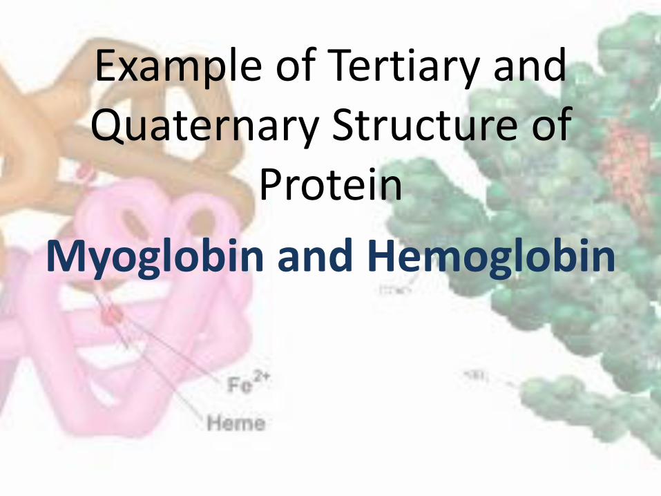
Example of Tertiary and Quaternary Structure of
Protein
Myoglobin and Hemoglobin
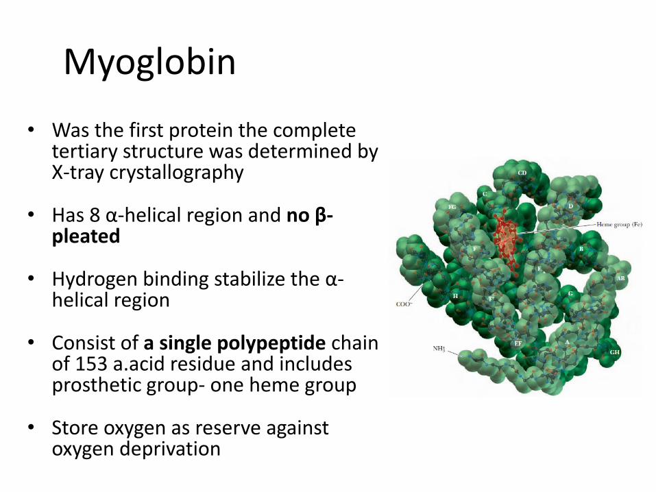
Myoglobin
• Was the first protein the complete tertiary structure was determined by X-tray crystallography
• Has 8 α-helical region and no β-pleated
• Hydrogen binding stabilize the α-helical region
• Consist of a single polypeptide chain of 153 a.acid residue and includes prosthetic group- one heme group
• Store oxygen as reserve against oxygen deprivation
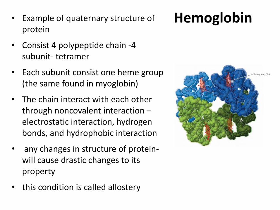
Hemoglobin • Example of quaternary structure of protein
• Consist 4 polypeptide chain -4 subunit- tetramer
• Each subunit consist one heme group (the same found in myoglobin)
• The chain interact with each other through noncovalent interaction – electrostatic interaction, hydrogen bonds, and hydrophobic interaction
• any changes in structure of protein- will cause drastic changes to its property
• this condition is called allostery
Hemoglobin
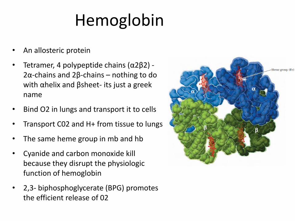
• An allosteric protein
• Tetramer, 4 polypeptide chains (α2β2) - 2α-chains and 2β-chains – nothing to do with αhelix and βsheet- its just a greek name
• Bind O2 in lungs and transport it to cells
• Transport C02 and H+ from tissue to lungs
• The same heme group in mb and hb
• Cyanide and carbon monoxide kill because they disrupt the physiologic function of hemoglobin
• 2,3- biphosphoglycerate (BPG) promotes the efficient release of 02
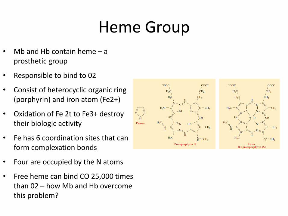
Heme Group • Mb and Hb contain heme – a
prosthetic group
• Responsible to bind to 02
• Consist of heterocyclic organic ring (porphyrin) and iron atom (Fe2+)
• Oxidation of Fe 2t to Fe3+ destroy their biologic activity
• Fe has 6 coordination sites that can form complexation bonds
• Four are occupied by the N atoms
• Free heme can bind CO 25,000 times than 02 – how Mb and Hb overcome this problem?
Structure of heme group in Mb and HB
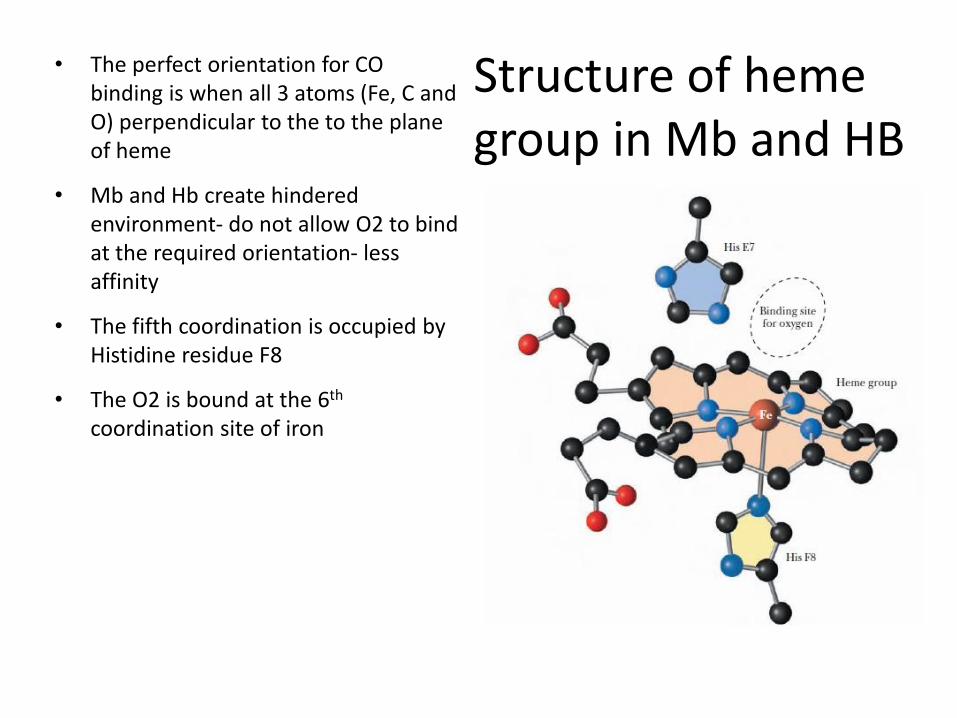
• The perfect orientation for CO binding is when all 3 atoms (Fe, C and O) perpendicular to the to the plane of heme
• Mb and Hb create hindered environment- do not allow O2 to bind at the required orientation- less affinity
• The fifth coordination is occupied by Histidine residue F8
• The O2 is bound at the 6th coordination site of iron
heme group
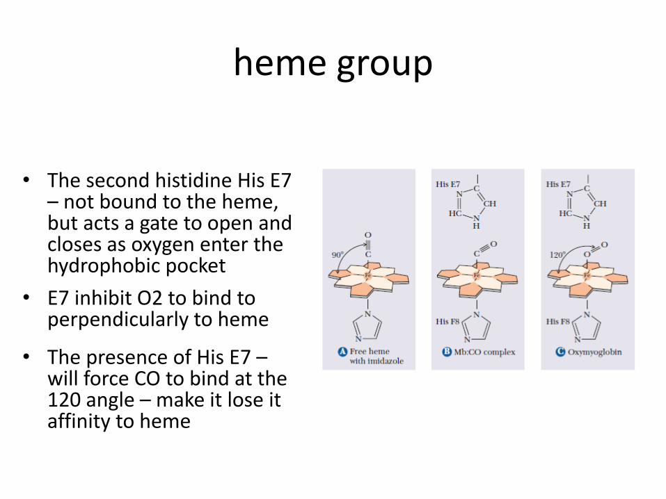
• The second histidine His E7 – not bound to the heme, but acts a gate to open and closes as oxygen enter the hydrophobic pocket
• E7 inhibit O2 to bind to perpendicularly to heme
• The presence of His E7 – will force CO to bind at the 120 angle – make it lose it affinity to heme
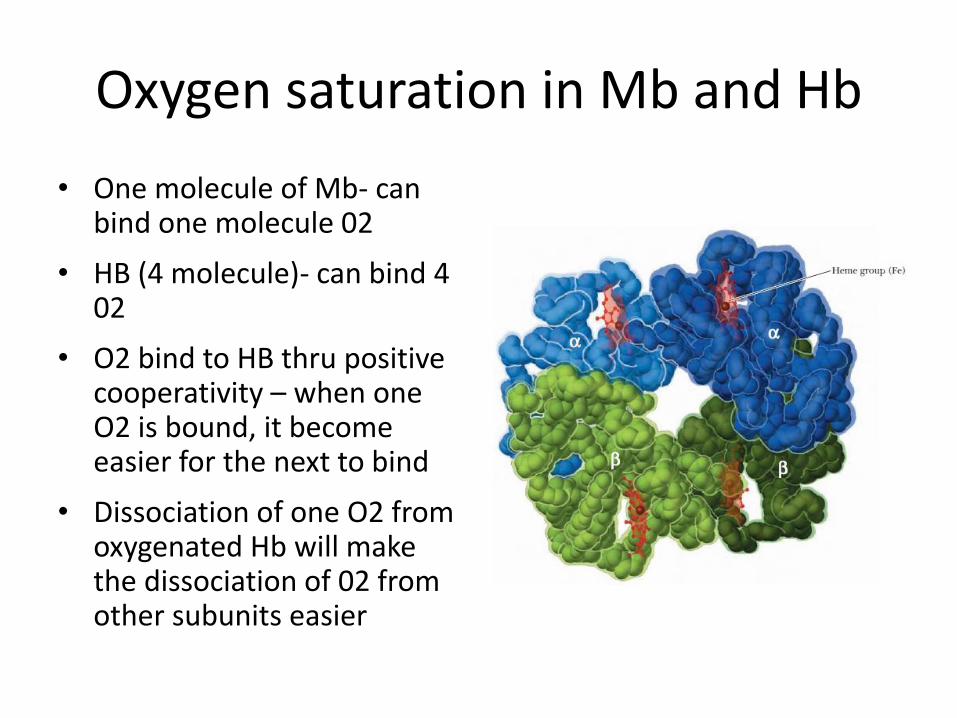
Oxygen saturation in Mb and Hb
• One molecule of Mb- can bind one molecule 02
• HB (4 molecule)- can bind 4 02
• O2 bind to HB thru positive cooperativity – when one O2 is bound, it become easier for the next to bind
• Dissociation of one O2 from oxygenated Hb will make the dissociation of 02 from other subunits easier
Different form of HB
• Hb is bound to 02- oxyhemoglobin – relaxed (R state)
• Without 02 – deoxyhb – tense (T) state
• If Fe2+ is oxidized to Fe3+ - unable to bind 02- methemoglobin
• C0 and NO have higher affinity for heme FE2+ than 02- toxicity
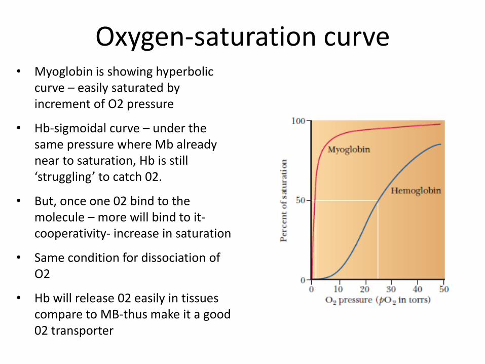
Oxygen-saturation curve • Myoglobin is showing hyperbolic
curve – easily saturated by increment of O2 pressure
• Hb-sigmoidal curve – under the same pressure where Mb already near to saturation, Hb is still ‘struggling’ to catch 02.
• But, once one 02 bind to the molecule – more will bind to it-cooperativity- increase in saturation
• Same condition for dissociation of O2
• Hb will release 02 easily in tissues compare to MB-thus make it a good 02 transporter
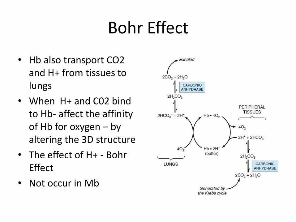
Bohr Effect
• Hb also transport CO2 and H+ from tissues to lungs
• When H+ and C02 bind to Hb- affect the affinity of Hb for oxygen – by altering the 3D structure
• The effect of H+ - Bohr Effect
• Not occur in Mb
Bohr effect
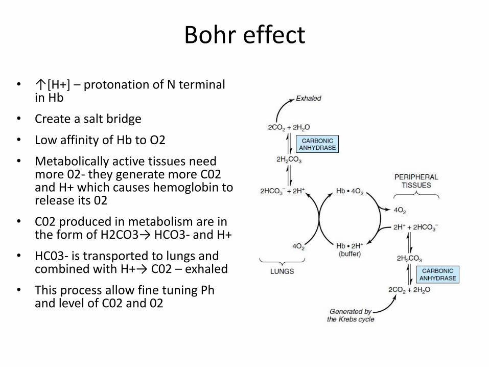
• ↑[H+] – protonation of N terminal in Hb
• Create a salt bridge
• Low affinity of Hb to O2
• Metabolically active tissues need more 02- they generate more C02 and H+ which causes hemoglobin to release its 02
• C02 produced in metabolism are in the form of H2CO3→ HCO3- and H+
• HC03- is transported to lungs and combined with H+→ C02 – exhaled
• This process allow fine tuning Ph and level of C02 and 02
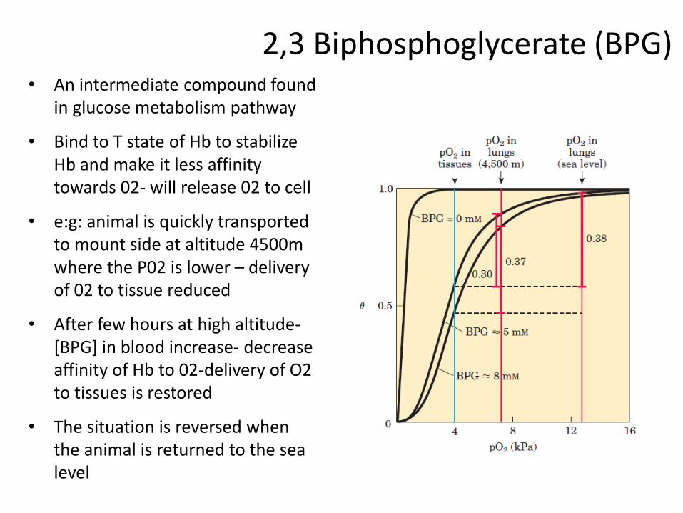
2,3 Biphosphoglycerate (BPG) • An intermediate compound found
in glucose metabolism pathway
• Bind to T state of Hb to stabilize Hb and make it less affinity towards 02- will release 02 to cell
• e:g: animal is quickly transported to mount side at altitude 4500m where the P02 is lower – delivery of 02 to tissue reduced
• After few hours at high altitude-[BPG] in blood increase- decrease affinity of Hb to 02-delivery of O2 to tissues is restored
• The situation is reversed when the animal is returned to the sea level
2,3 Biphosphoglycerate (BPG)
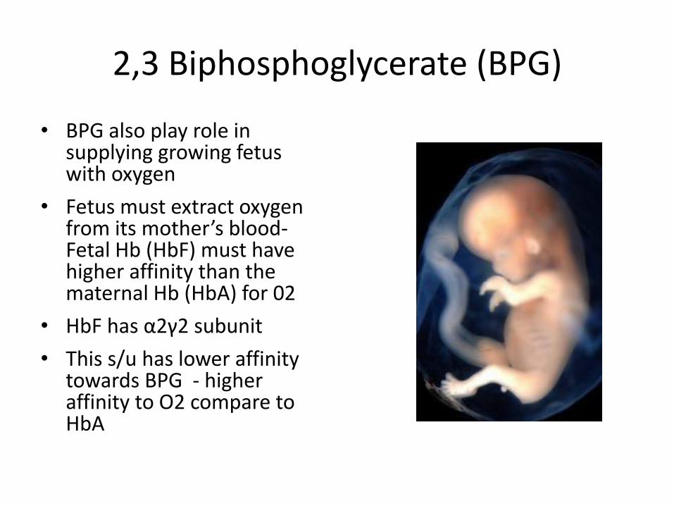
• BPG also play role in supplying growing fetus with oxygen
• Fetus must extract oxygen from its mother’s blood- Fetal Hb (HbF) must have higher affinity than the maternal Hb (HbA) for 02
• HbF has α2γ2 subunit
• This s/u has lower affinity towards BPG - higher affinity to O2 compare to HbA