Evidence for Abnormal Protein Processing in AD Carl W. Cotman Institute for Brain Aging and Dementia...
34
Evidence for Abnormal Protein Processing in AD Carl W. Cotman Institute for Brain Aging and Dementia UC Irvine
-
date post
20-Dec-2015 -
Category
Documents
-
view
214 -
download
0
Transcript of Evidence for Abnormal Protein Processing in AD Carl W. Cotman Institute for Brain Aging and Dementia...
Evidence for Abnormal Protein Processing in AD
Carl W. Cotman
Institute for Brain Aging and Dementia
UC Irvine
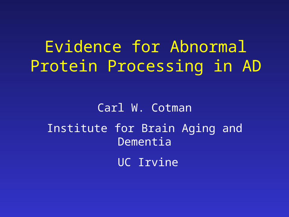
Examples of Abnormal Protein Processing
• APP generating β-amyloid
• Cytoskeletal proteins: hyperphosphorylation and proteolysis
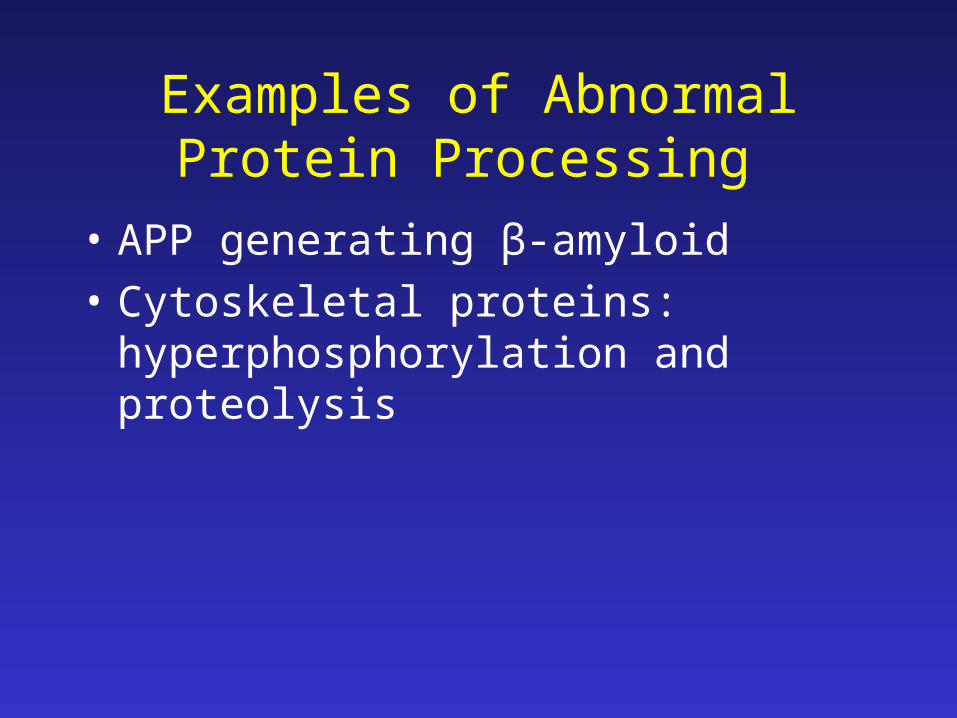
Proteins implicated in AD are targets of caspases
Amyloid precursor protein (APP)
Gervais et al. (1999) Cell 97: 395-406
Lu et al. (2003) J Neurochem 87: 733-41
Presenilin-1 (PS1)
van de Craen et al. (1999) FEBS Lett 445: 149-54
Fluhrer et al. (2004) J Biol Chem 279: 1585-93
Tau
Canu et al. (1998) J Neurosci 18:7061-74
Fasulo et al. (2000) J Neurochem 75: 624-33
Gamblin et al. (2003) PNAS 100: 10032-7
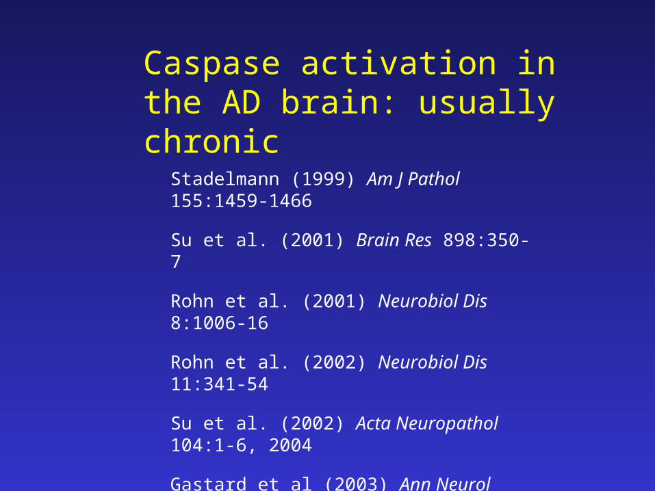
Caspase activation in the AD brain: usually chronic
Stadelmann (1999) Am J Pathol 155:1459-1466
Su et al. (2001) Brain Res 898:350-7
Rohn et al. (2001) Neurobiol Dis 8:1006-16
Rohn et al. (2002) Neurobiol Dis 11:341-54
Su et al. (2002) Acta Neuropathol 104:1-6, 2004
Gastard et al (2003) Ann Neurol 54:393-8
Pompl et al. (2003) Arch Neurol 60: 369-76.
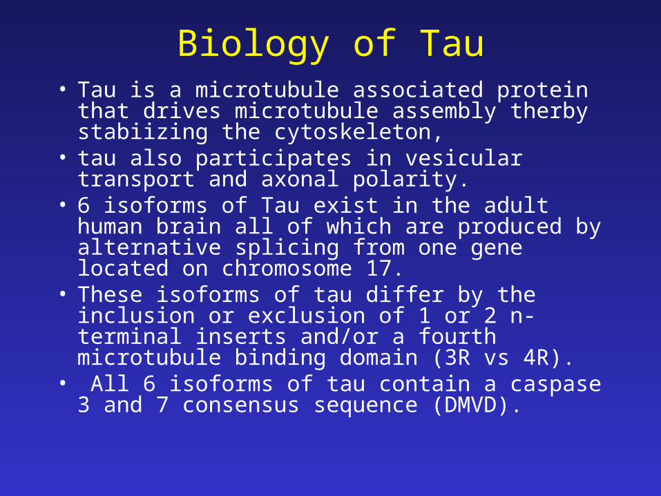
Biology of Tau• Tau is a microtubule associated protein that drives
microtubule assembly therby stabiizing the cytoskeleton,
• tau also participates in vesicular transport and axonal polarity.
• 6 isoforms of Tau exist in the adult human brain all of which are produced by alternative splicing from one gene located on chromosome 17.
• These isoforms of tau differ by the inclusion or exclusion of 1 or 2 n-terminal inserts and/or a fourth microtubule binding domain (3R vs 4R).
• All 6 isoforms of tau contain a caspase 3 and 7 consensus sequence (DMVD).

Spillantini et al., (1998) Trends in Neuroscience 21: 428-33
Tau is a microtubule associated protein
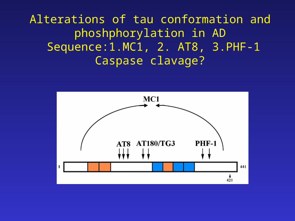
Alterations of tau conformation and phoshphorylation in AD
Sequence:1.MC1, 2. AT8, 3.PHF-1Caspase clavage?
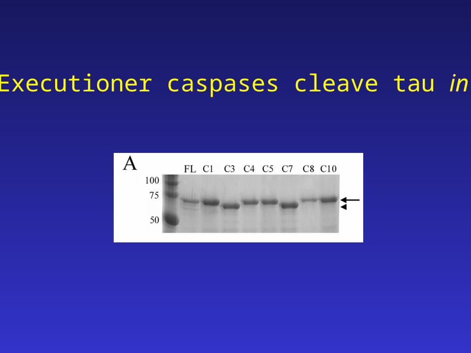
Executioner caspases cleave tau in vitro
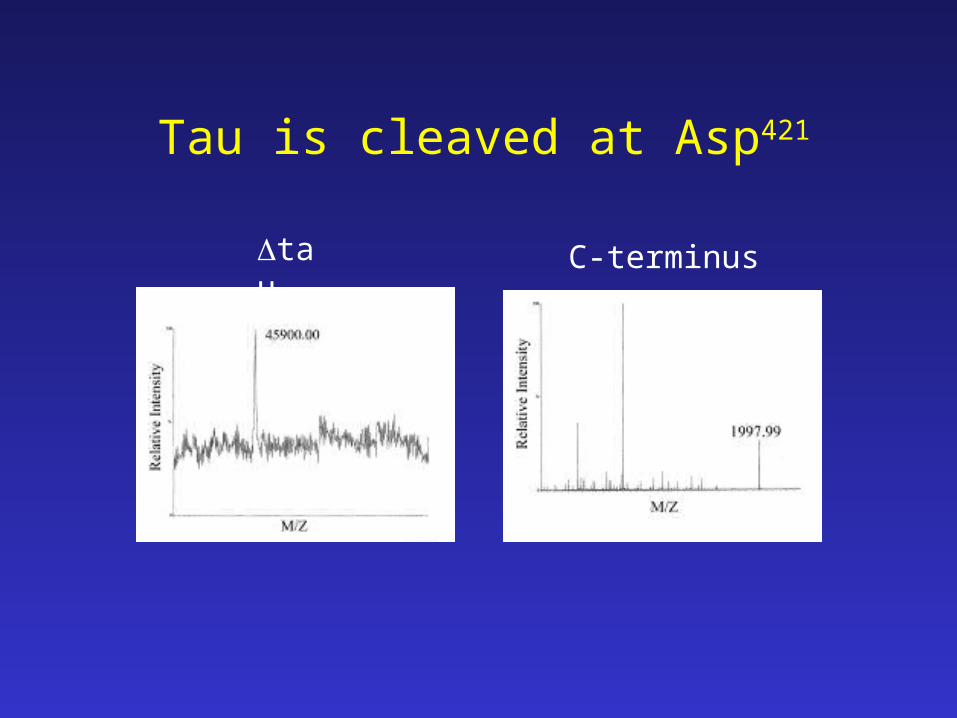
tau C-terminus
Tau is cleaved at Asp421
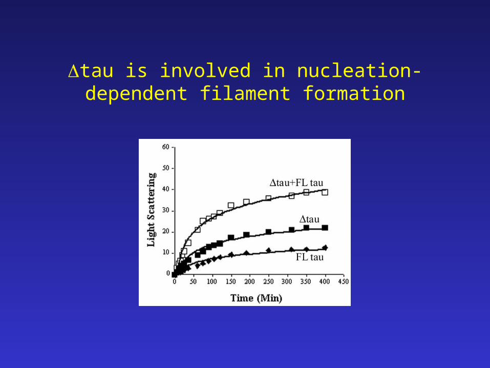
tau is involved in nucleation-dependent filament formation
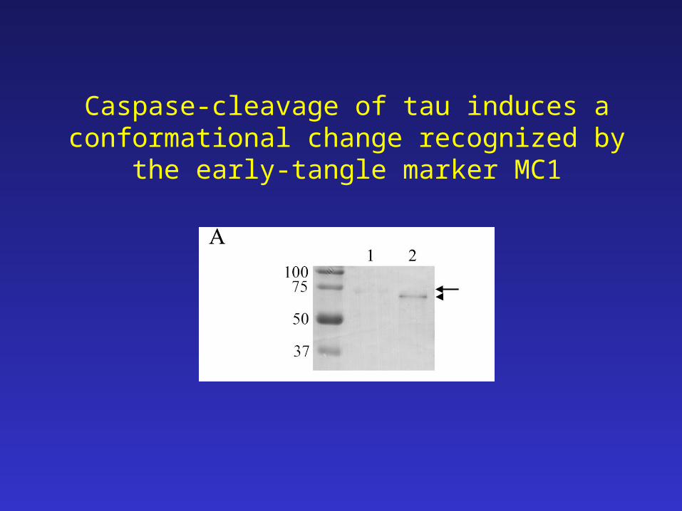
Caspase-cleavage of tau induces a conformational change recognized by the early-tangle marker MC1
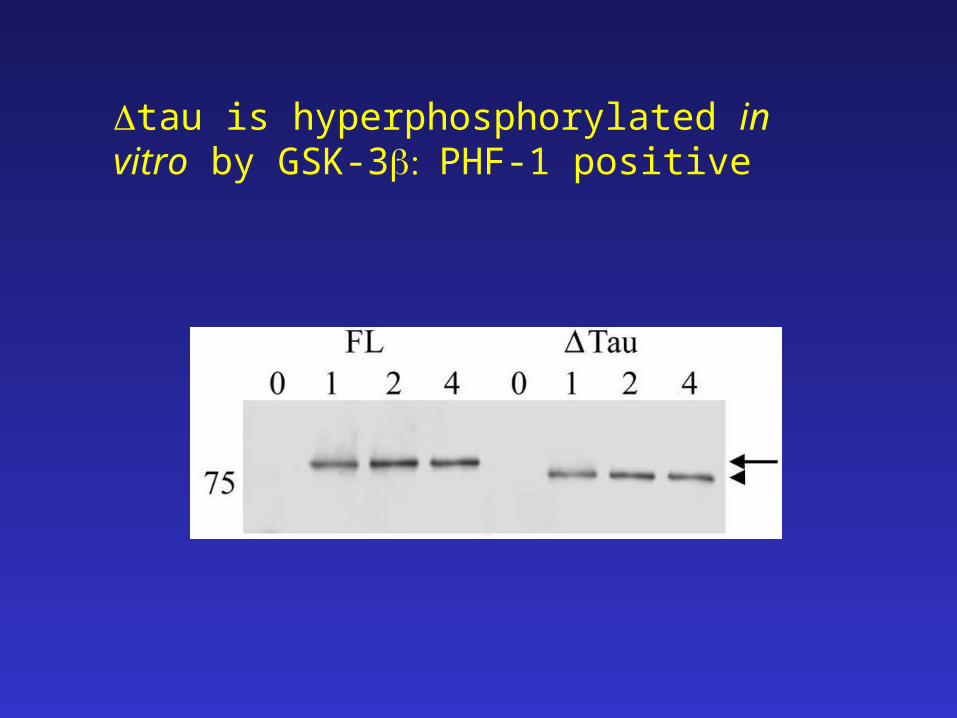
tau is hyperphosphorylated in vitro by GSK-3PHF-1 positive
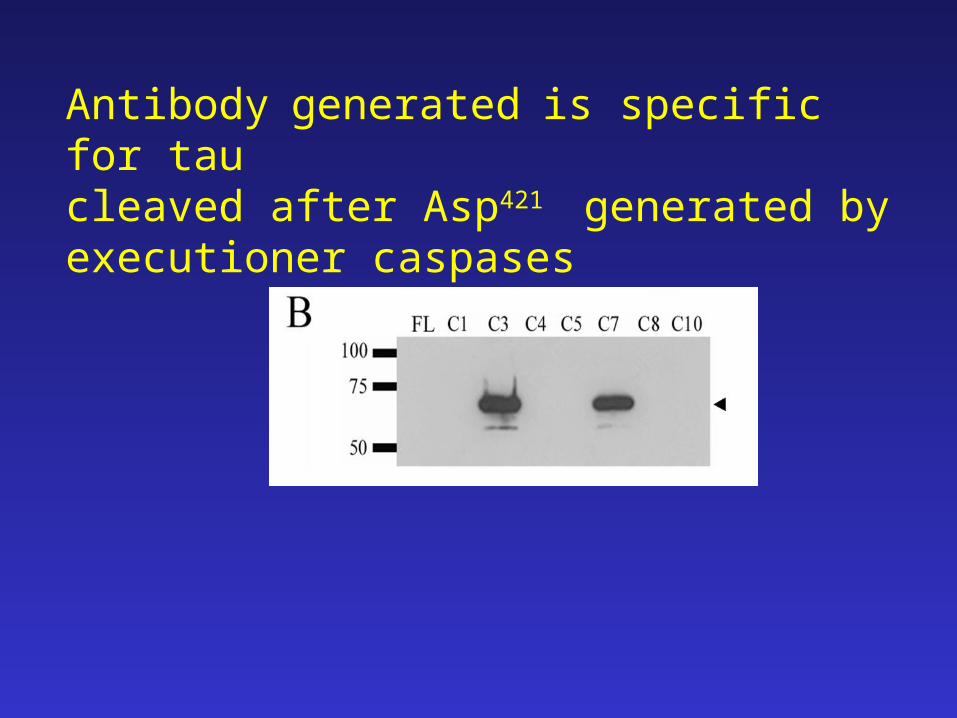
Antibodygeneratedis specific for tau cleaved after Asp421 generated by executioner caspases
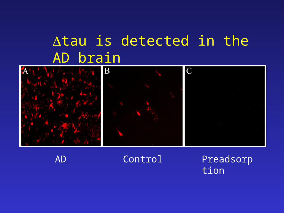
tau is detected in the AD brain
AD Control Preadsorption
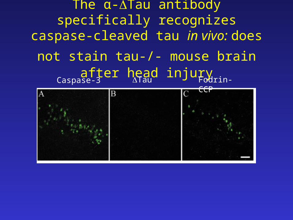
The α-Tau antibody specifically recognizes caspase-cleaved tau in vivo: does not stain
tau-/- mouse brain after head injury
Caspase-3 Tau Fodrin-CCP
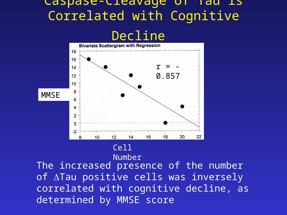
Caspase-Cleavage of Tau is Correlated with
Cognitive Decline
r = -0.857
The increased presence of the number of Tau positive cells was inversely correlated with cognitive decline, as determined by MMSE score
Cell Number
MMSE
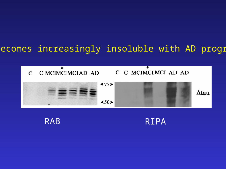
RAB RIPA
tau becomes increasingly insoluble with AD progression
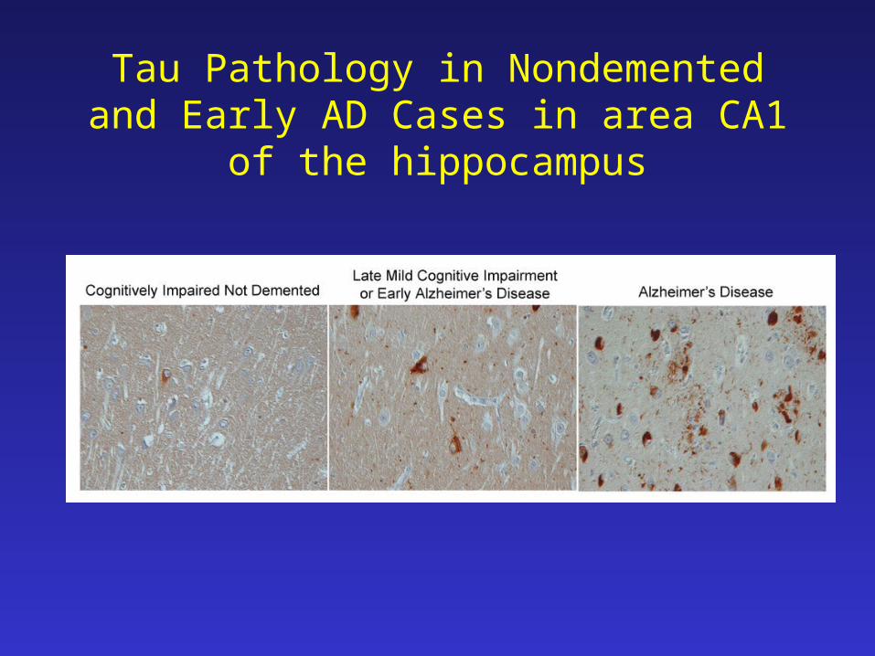
Tau Pathology in Nondemented and Early AD Cases in area CA1 of the hippocampus
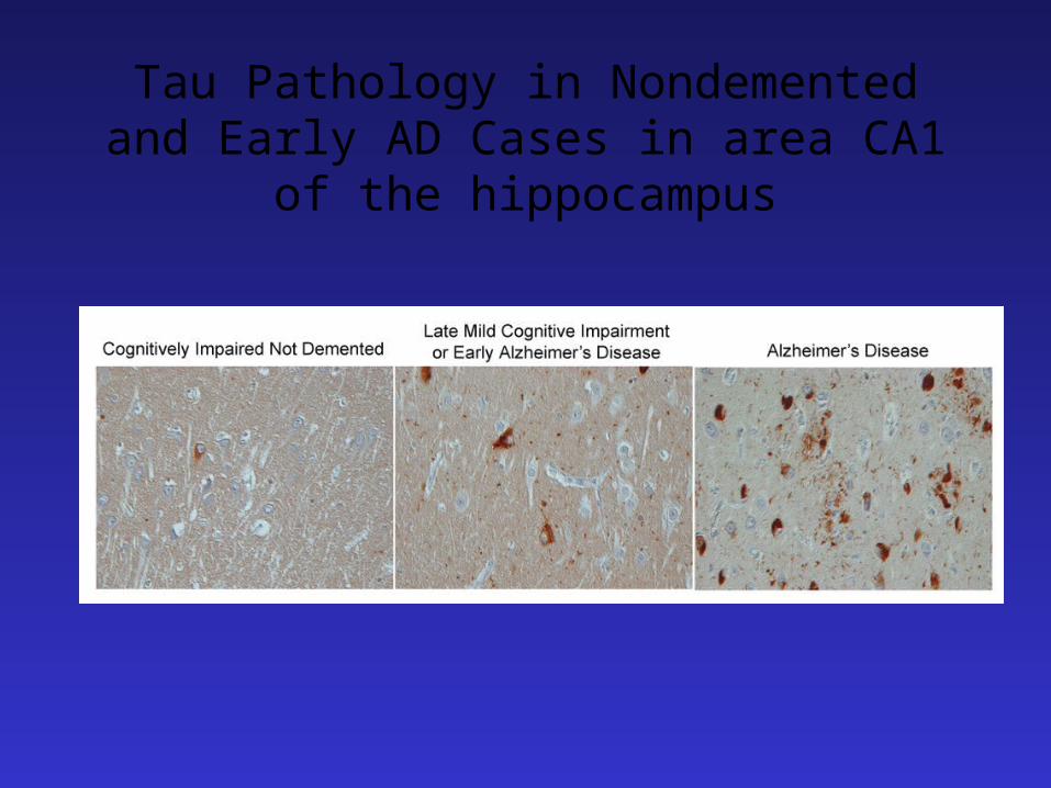
Tau Pathology in Nondemented and Early AD Cases in area CA1 of the hippocampus
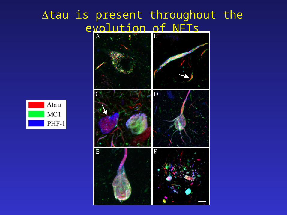
tau is present throughout the evolution of NFTs
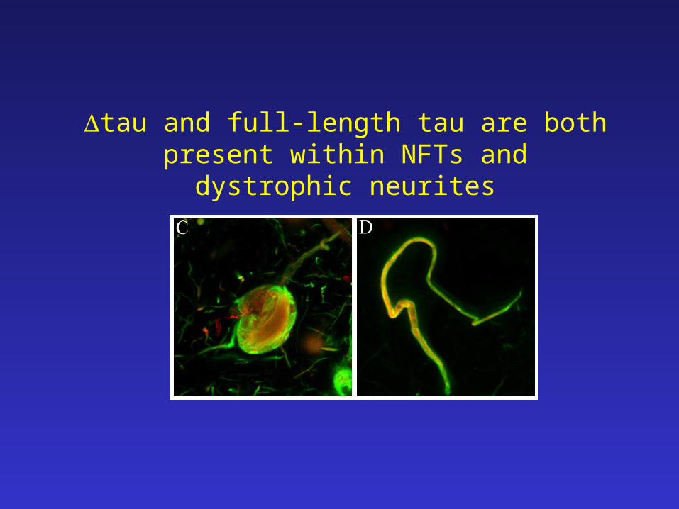
tau and full-length tau are both present within NFTs and dystrophic neurites
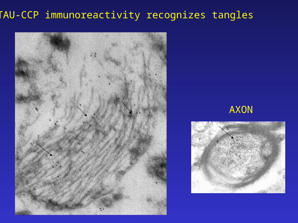
AXON
TAU-CCP immunoreactivity recognizes tangles
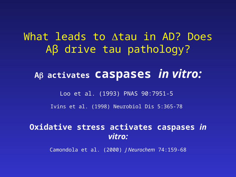
What leads to tau in AD? Does Aβ drive tau pathology?
Aactivates caspases in vitro:
Loo et al. (1993) PNAS 90:7951-5
Ivins et al. (1998) Neurobiol Dis 5:365-78
Oxidative stress activates caspases in vitro:
Camondola et al. (2000) J Neurochem 74:159-68
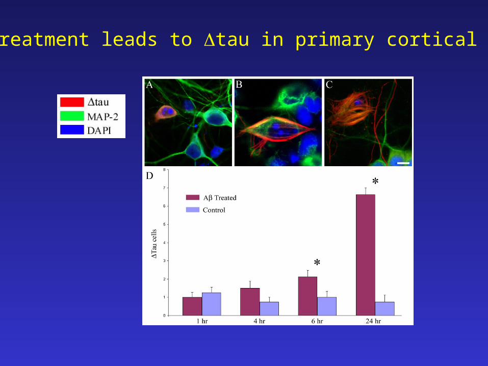
A1-42 treatment leads to tau in primary cortical neurons
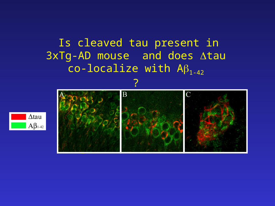
Is cleaved tau present in 3xTg-AD mouse and does tau co-localize with
A1-42
?
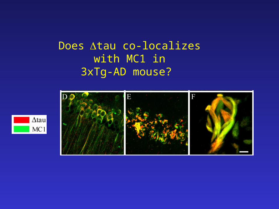
Doestau co-localizes with MC1 in3xTg-AD mouse?
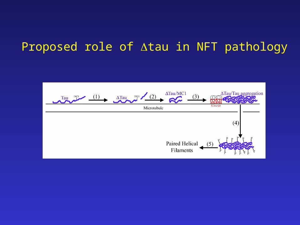
Proposed role of tau in NFT pathology
Summary• Several proteins get cleaved by caspases and appear
to be present in neurons for prolonged periods of time, e.g., fodrin, actin and tau as well as APP
• Tau is cleaved by executioner caspases initiated by β-amyloid, oxidative damage
• Cleaved tau seeds (nucleates) the assembly of tau into PHF-1 like assemblies and assumes an MC-1 conformation.
• Cleaved tau is present in pre-tangle and tangle neurons
• Cleaved tau neurons inversely correlate with cognitive function
• Chronic abnormal protein processing may be a new mechanism catalyzing AD pathology
Acknowledgements
Mass spectroscopyNemone Muster
Laser Light ScatteringDr. Wytze van der Veer
Cotman LabDr. Carl CotmanDr. David CribbsDr. Elizabeth HeadChristina TuDr. Reidun TorpDr. Paul AdlardDr. Pat Kesslak
LaFerla LabDr. Frank LaFerlaSalvatore Oddo
Boise State U.Dr. Troy Rohn

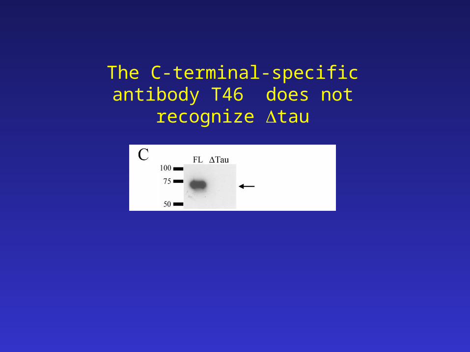
The C-terminal-specific antibody T46 does not recognize tau
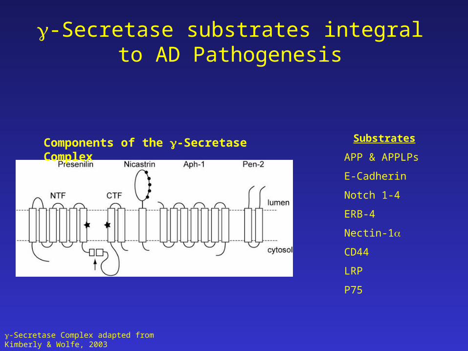
-Secretase substrates integral to AD Pathogenesis
-Secretase Complex adapted from Kimberly & Wolfe, 2003
Components of the -Secretase Complex Substrates
APP & APPLPs
E-Cadherin
Notch 1-4
ERB-4
Nectin-1
CD44
LRP
P75
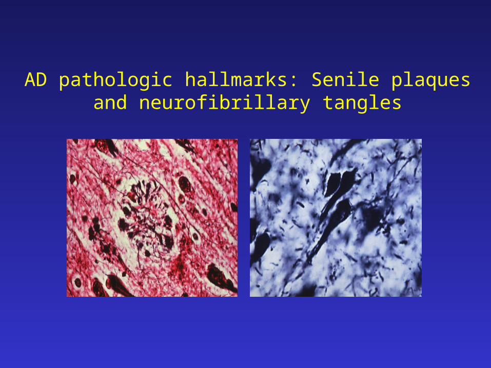
AD pathologic hallmarks: Senile plaques and neurofibrillary tangles
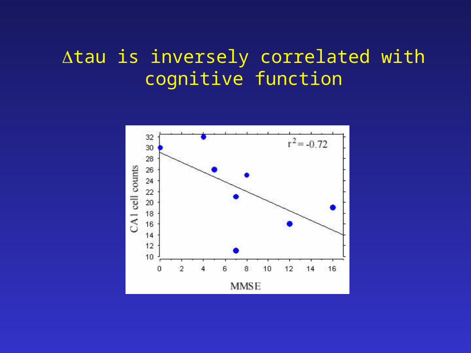
tau is inversely correlated with cognitive function
![sin2βin the BaBar Experiment - Vanderbilt University...BABAR Collaboration 9 Countries 72 Institutions 554 Physicists USA [35/276] California Institute of Technology UC, Irvine UC,](https://static.fdocument.org/doc/165x107/610f211a5dcad3628b41722d/sin2in-the-babar-experiment-vanderbilt-university-babar-collaboration-9.jpg)


![HOTAIR Knockdown Decreased the Activity Wnt/β-Catenin ... · of this pathway are frequently altered in human cancer mainly by genetic and epigenetic mechanisms [25-27]. The abnormal](https://static.fdocument.org/doc/165x107/5e638e505ba2f7369635202e/hotair-knockdown-decreased-the-activity-wnt-catenin-of-this-pathway-are-frequently.jpg)