Enhanced H3K4 Trimethylation in TNF-α Promoter Gene …
10
Research Article Enhanced H3K4 Trimethylation in TNF-α Promoter Gene Locus with Cell Apoptosis in the Ventral-Medial Striatum following Opioid Withdrawal of Neonatal Rat Offspring from Morphine-Addicted Mothers Pei-Ling Wu , 1,2,3 Jau-Ling Suen, 3 Chun-Hwa Yang , 2 Kuang-Che Kuo , 4 Yu-Chen S. H. Yang , 5 and San-Nan Yang 1,2 1 School of Medicine, College of Medicine, I-Shou University, Kaohsiung, Taiwan 2 Department of Pediatrics, E-Da Hospital, Kaohsiung, Taiwan 3 Graduate Institute of Medicine, College of Medicine, Kaohsiung Medical University, Kaohsiung, Taiwan 4 Department of Pediatrics, Kaohsiung Chang Gung Memorial Hospital and Chang Gung University College of Medicine, Kaohsiung, Taiwan 5 Joint Biobank, Office of Human Research, Taipei Medical University, Taipei, Taiwan Correspondence should be addressed to Yu-Chen S. H. Yang; [email protected] and San-Nan Yang; [email protected] Received 8 April 2021; Revised 12 May 2021; Accepted 31 May 2021; Published 16 June 2021 Academic Editor: Rômulo Dias Novaes Copyright © 2021 Pei-Ling Wu et al. This is an open access article distributed under the Creative Commons Attribution License, which permits unrestricted use, distribution, and reproduction in any medium, provided the original work is properly cited. Prenatal opioid exposure might disturb epigenetic programming in the brain of neonatal offspring with various consequences for gene expressions and behaviors. This study determined whether altered trimethylation of histone 3 at lysine 4 (H3K4me3) in the promoter of the tumor necrosis factor-α (tnf-α) gene with neural cell apoptosis was involved in the ventral-medial striatum, an important brain region for withdrawal symptoms, of neonatal rat offspring from morphine-addicted mothers. Female adult rats were injected with morphine before gestation and until 14 days after giving birth. On postnatal day 14 (P14), rat offspring from morphine-addicted mothers were subjected to an opioid-withdrawal protocol and were analyzed 2 or 8 h after administration of that protocol. Expressions of the TNF-α protein, H3K4me3 in the tnf-α promoter gene, and neural cell apoptosis within the ventral-medial striatum of neonatal rat offspring were evaluated. In the absence of significant opioid withdrawal (2 h after initiation of the opioid-withdrawal protocol on P14), prenatal morphine exposure led to increased levels of H3K4me3 in the tnf- α promoter gene, of the TNF-α protein, and of neural cell apoptosis within the ventral-medial striatum of neonatal rat offspring. Following opioid withdrawal (8 h after initiation of the opioid-withdrawal protocol on P14), differential expression of H3K4me3 in the tnf-α promoter gene locus and upregulation of the level of TNF-α protein expression were further enhanced in these offspring. In addition, increased levels of caspase-3 and neural cell apoptosis were also observed. Taken together, this study revealed that prenatal opioid exposure can activate an epigenetic histone mechanism which regulates proinflammatory factor generation, which hence, led to cell apoptotic damage within the ventral-medial striatum of neonatal rat offspring from morphine-addicted mothers. More importantly, the opioid-withdrawal episode may provide augmented effects for the abovementioned alterations and could lead to deleterious effects in the neonatal brain of such offspring. 1. Introduction Early life adversity is related to increased risks for developing many behavioral and psychiatric disorders [1]. A spectrum of prenatal, intrapartum, and postnatal episodes is likely to contribute to neurodevelopment via interactions with the individual’s genotype to disrupt specific neural and psycho- physiological system-related cognitive functions, thereby enhancing the risk for psychopathologies later in life [2, 3]. Indeed, children exposed to perinatal opioid drugs appear Hindawi Mediators of Inflammation Volume 2021, Article ID 9828995, 10 pages https://doi.org/10.1155/2021/9828995
Transcript of Enhanced H3K4 Trimethylation in TNF-α Promoter Gene …

Research ArticleEnhanced H3K4 Trimethylation in TNF-α Promoter GeneLocus with Cell Apoptosis in the Ventral-Medial Striatumfollowing Opioid Withdrawal of Neonatal Rat Offspring fromMorphine-Addicted Mothers
Pei-Ling Wu ,1,2,3 Jau-Ling Suen,3 Chun-Hwa Yang ,2 Kuang-Che Kuo ,4
Yu-Chen S. H. Yang ,5 and San-Nan Yang 1,2
1School of Medicine, College of Medicine, I-Shou University, Kaohsiung, Taiwan2Department of Pediatrics, E-Da Hospital, Kaohsiung, Taiwan3Graduate Institute of Medicine, College of Medicine, Kaohsiung Medical University, Kaohsiung, Taiwan4Department of Pediatrics, Kaohsiung Chang Gung Memorial Hospital and Chang Gung University College of Medicine,Kaohsiung, Taiwan5Joint Biobank, Office of Human Research, Taipei Medical University, Taipei, Taiwan
Correspondence should be addressed to Yu-Chen S. H. Yang; [email protected] and San-Nan Yang; [email protected]
Received 8 April 2021; Revised 12 May 2021; Accepted 31 May 2021; Published 16 June 2021
Academic Editor: Rômulo Dias Novaes
Copyright © 2021 Pei-Ling Wu et al. This is an open access article distributed under the Creative Commons Attribution License,which permits unrestricted use, distribution, and reproduction in any medium, provided the original work is properly cited.
Prenatal opioid exposure might disturb epigenetic programming in the brain of neonatal offspring with various consequences forgene expressions and behaviors. This study determined whether altered trimethylation of histone 3 at lysine 4 (H3K4me3) in thepromoter of the tumor necrosis factor-α (tnf-α) gene with neural cell apoptosis was involved in the ventral-medial striatum, animportant brain region for withdrawal symptoms, of neonatal rat offspring from morphine-addicted mothers. Female adult ratswere injected with morphine before gestation and until 14 days after giving birth. On postnatal day 14 (P14), rat offspring frommorphine-addicted mothers were subjected to an opioid-withdrawal protocol and were analyzed 2 or 8 h after administration ofthat protocol. Expressions of the TNF-α protein, H3K4me3 in the tnf-α promoter gene, and neural cell apoptosis within theventral-medial striatum of neonatal rat offspring were evaluated. In the absence of significant opioid withdrawal (2 h afterinitiation of the opioid-withdrawal protocol on P14), prenatal morphine exposure led to increased levels of H3K4me3 in the tnf-α promoter gene, of the TNF-α protein, and of neural cell apoptosis within the ventral-medial striatum of neonatal rat offspring.Following opioid withdrawal (8 h after initiation of the opioid-withdrawal protocol on P14), differential expression of H3K4me3in the tnf-α promoter gene locus and upregulation of the level of TNF-α protein expression were further enhanced in theseoffspring. In addition, increased levels of caspase-3 and neural cell apoptosis were also observed. Taken together, this studyrevealed that prenatal opioid exposure can activate an epigenetic histone mechanism which regulates proinflammatory factorgeneration, which hence, led to cell apoptotic damage within the ventral-medial striatum of neonatal rat offspring frommorphine-addicted mothers. More importantly, the opioid-withdrawal episode may provide augmented effects for theabovementioned alterations and could lead to deleterious effects in the neonatal brain of such offspring.
1. Introduction
Early life adversity is related to increased risks for developingmany behavioral and psychiatric disorders [1]. A spectrumof prenatal, intrapartum, and postnatal episodes is likely to
contribute to neurodevelopment via interactions with theindividual’s genotype to disrupt specific neural and psycho-physiological system-related cognitive functions, therebyenhancing the risk for psychopathologies later in life [2, 3].Indeed, children exposed to perinatal opioid drugs appear
HindawiMediators of InflammationVolume 2021, Article ID 9828995, 10 pageshttps://doi.org/10.1155/2021/9828995
to have a higher mortality rate after birth and sufferfrom neurodevelopmental consequences and behavioralproblems [4, 5].
During the fetal and neonatal periods, the centralnervous system (CNS) displays highly significant plasticityand is vulnerable to changes by prenatal influences such asprenatal opioid exposure [6–8]. The mechanisms throughwhich early life exposures play important roles for later-lifepsychopathologies remain undetermined, but an epigeneticmechanism represents a reasonable candidate [9, 10]. Theventral-medial striatum of the mammalian brain serves asan integration center to mediate goal-directed behaviors(i.e., rewards of drug craving and drug addiction) [11].Previous studies demonstrated that morphine exposure cantrigger the production of proinflammatory cytokines, includ-ing tumor necrosis factor- (TNF-) α, interleukin- (IL-) 1, andIL-6 [12–14]. Chronic opioid administration leads toincreased messenger (m)RNA levels of IL-1 and IL-6 in theventral-medial striatum of rats in vivo [15]. In addition, mor-phine withdrawal neuro-behaviors can induce TNF-α releasefrom neural cells [13]. Recently, TNF-α expression levelswere found to increase in the amygdala during morphine-withdrawal episodes [14]. Indeed, inflammation in the brainnot merely occurs with direct opioid exposure and with-drawal but has transplacental effects on newborn offspringas well; for instance, offspring suffering from prenatal opioidexposure exhibited increased TNF-α production in the hip-pocampus [16]. Prenatal stresses, such as chronic illicit drugabuse, can dysregulate the maternal immune function andcause elevated proinflammatory cytokine levels in the brainof neonatal offspring [17]. Taken together, those studiesrevealed that prenatal opioid exposure can generate transpla-cental inflammation cascades during early life.
The epigenetic histone regulation in a target promotergene locus might play a role in the molecular basis of mam-malian drug-craving and addiction [18, 19]. Recently, tri-methylation of histone H3 at lysine 4 (H3K4me3), one typeof histone regulation, was suggested to act as a transcriptionbooster for the tnf-α gene expression within the promoterregion [18, 20]. In addition, early life stress or drug exposurecan also induce histone modifications within the ventral teg-mental area resulting in dysregulated signal conduction [21].However, little is known about histone methylation as aresult of perinatal opioid exposure with apoptosis in the off-spring brain. Thus, the aim of this study was to examine thehypothesis that prenatal opioid exposure activates the tri-methylation of H3K4me3 in the tnf-α promoter gene and isassociated with neural cell apoptosis in the ventral-medialstriatum of neonatal rat offspring (on postnatal day 14; P14).
2. Materials and Methods
2.1. Experimental Animal Protocol. Female adult Sprague-Dawley (SD) rats which underwent the opioid-withdrawalprotocol were housed under a 12h light/dark cycle withhumidity maintained around 60%~70% and were initiallyinjected with morphine hydrochloride (2mg/kg body weight(BW), subcutaneously twice daily with the dosage progres-sively increased at a rate of 1mg/kg BW every 7 days with a
maximum dose of 7mg/kg BW) [6–8, 22]. These female ratswere mated on about day 7 or 8. During pregnancy and afterthe rat offspring were born, morphine was still routinelyadministered as scheduled until the offspring were 14 daysold. The last opioid injection was on postnatal day 13(P13). Throughout the experimental course, there were nosignificant opioid withdrawal symptoms between the mater-nal opioid injections. The vehicle-control group consisted ofmaternal SD rats injected with NaCl at the same dose andinterval as the experimental group.
We evaluated the growth of BW of rat offspring on post-natal day 7 (P7) and P14 (an age regarded as a neonate in thehuman lifespan) [23]. On P14, rat offspring from maternalrats were transferred to an observational chamber main-tained at around 33°C with warm light and were observedfor 2 h for cumulative episodes of and latency in exhibitingopioid-withdrawal behaviors, such as rearing, teeth chatter-ing, backward locomotion, tremors, and wet-dog shaking.Rat offspring from morphine-addicted mothers were dividedinto two groups: an early group represented rat offspringobserved for opioid withdrawal behaviors for 2 h immedi-ately after transfer, after which they were sacrificed for bio-chemical experiments (the early group, n = 48 rats); and thelate group represented rat offspring for observation ofopioid-withdrawal behaviors for 2 h starting from the 6thhour after transfer. These rat offspring were then sacrificedfor biochemical experiments (the late group, n = 48 rats).Rat offspring from the vehicle-control group were transferredto an observational chamber on P14 and were observed foropioid-withdrawal behaviors for 2 h starting from the 6thhour after transfer. These rat offspring were then sacrificedfor biochemical experiments (the control group, n = 48 rats).There were no differences in sex among the three groups.
All experimental protocols were approved by the AnimalCare and Use Committee at the National Defense MedicalCenter, Taiwan (NDMC-04-088 and IACUC-04-085) withall actions undertaken being designed to lessen sufferingand decrease the number of animals used.
2.2. Preparation of Slices of the Ventral-Medial StriatumBrain Region.After the opioid-withdrawal protocol was com-pleted on P14, the pups were sacrificed, and brain slices (400μm) containing the ventral-medial striatum brain regionwere immediately harvested with a vibro-slicer (CampdenInstruments, Sileby, Loughboroug, UK). Brain slices wereselectively transferred to an incubation chamber perfusedwith oxygenated artificial cerebrospinal fluid (95% O2/5%CO2 gas, at 30:0 ± 0:5°C) for 1 h. Artificial cerebrospinal fluidconsisted of (in mM) NaCl (124), MgCl2 (1), CaCl2 (2), KCl(3.5), NaH2PO4 (1.25), NaHCO3 (26), and D-glucose (10) atpH7.4 and with an osmolarity of 305 ± 5mOsm.
2.3. Immunoblotting. Cells of the ventral-medial striatumwere lysed in buffer (10mM Tris at pH7.4, 150mM NaCl,1mM PMSF, 0.2% Triton X-100, 2mM EDTA, and 1×protease inhibitor mixture). The protein concentration wasclarified with a BCA assay (Thermo Scientific, Rockford, IL,USA). Cell lysates were sorted on sodium dodecylsulfatepolyacrylamide gel electrophoresis (SDS-PAGE), later
2 Mediators of Inflammation
transferred to nitrocellulose membranes, and then probedwith antibodies against TNF-α (SC-1351; Santa Cruz) andcleaved caspase-3 (no. 9662; Cell Signaling). Depending onthe different types of primary antibody, either rabbit anti-mouse immunoglobulin G (IgG) or goat anti-rabbit IgG (1:3000) was chosen as the secondary antibody. Immunoreac-tive proteins were detected using the BioSpectrum 810 Imag-ing System (UVP).
2.4. Real-Time Polymerase Chain Reaction (PCR). Isolation ofRNA, including synthesis of complementary (c)DNA, andDNase treatment was accomplished using the TranscriptorFirst Strand cDNA Synthesis Kit RT-PCR System™ (Roche,Indianapolis, IN, USA) following the manufacturer’s instruc-tions. The two-step real-time PCR with the LightCycler™System (Roche) and Sybr Green I dye was used for monitor-ing the PCR. FastStart DNA Master SYBR Green I (Roche)was performed according to the producer’s instructions andused for the real-time PCR. This study utilized the followingset of primers: TNF-α: forward (5′-CCCTACGGGTCATTGAGAGA-3′) and reverse (5′-GGTTGTGGACTGCCTTTTGT-3′); and 18s ribosomal (r)RNA: forward (5′-CCAGTAAGTGCGGGTCATAA-3′) and reverse (5′-TAGTCAAGTTCGACCGTCTTC-3′). The thermal protocol of thePCR was set to 95°C (for 4min), followed by 30 amplificationcycles of 94°C (for 30 s), 62°C (for 1min), and 72°C (for 1min), with a final melting curve analysis. Amplicons wereanalyzed by the melting curve (Light Cycler software) andagarose gel electrophoresis; the LightCycler analytical sys-tem was applied to decide cycle threshold (CT) values, andthe relative messenger (m)RNA level of each experimentwas assessed by a real-time PCR followed by measurementby the relative CT method (ΔΔCT) (the expression of theobjective was normalized to an endogenous standard (18SrRNA) and compared to a calibrator (cDNA from a pooledsample)).
2.5. Chromatin Immunoprecipitation (ChIP) Assay. A ChIPassay was performed as previously described [24, 25]. Briefly,5 × 105 cells were treated with 1% formaldehyde at roomtemperature (for 10min) accompanied by sonication ofDNA and immunoprecipitation of chromatin overnight withantibodies of trimethylated H3K4 (2μg for 25μg of chroma-tin; antihistone H3K4me3 antibody, ChIP Grade; ab8580;Abcam) and later purification with a ChIP kit (no. 17-295;Upstate Biotechnology, Lake Placid, NY, USA). Probes andprimers were planned by exploring the proximal promoterand intronic enhancer areas of the tnf-α gene [24, 25], includ-ing the following subregions as described below relative tothe transcription start site: tnf-α 1 (-2686 to -2667); forward,5′-CCCTAGTCCTCCTGGGATGT-3′ and reverse, 5′-GCCTGCTGCAACAGAGAGA-3′; tnf-α 2 (-2202 to -2183); for-ward, 5′-CGTCTCACTATGCCTGGGTCT-3′ and reverse,5′-AAGCAAAGCACTTCTACCAAAT-3′; tnf-α 3 (-1672to -1653); forward, 5′-AAACTCAGACCAGGCTGCAT-3′and reverse, 5′-CAGGTCATCTCTTGACGTGGT-3′; tnf-α4 (-502 to -480); forward, 5′-GAGTTCTGCATGTATT
GGATAGG-3′ and reverse, 5′-TGCTACCAAGCCTAAAGACC-3′; and tnf-α 5 (-230 to -213); forward, 5′-GGTTCAGTTCCCAGCACCTA-3′ and reverse, 5′-ATGGGCATATCTGCACAGCA-3′. PCRs were conducted on theABI Gene Amp ⓐ PCR System (Applied Biosystems). Thequantity of immunoprecipitated DNA was calculated andcompared to the quantity of total input DNA.
2.6. Apoptosis Evaluation. A double immunofluorescenceinvestigation by laser-scanning confocal microscopy wasapplied to examine apoptosis in neurons of brain sectionsof the ventral-medial striatum, which were cut at a coronalplane and measured 30μm and then stained with an anti-neuronal nuclei (NeuN) antibody (clone A60, MAB377; Che-micon, Temecula, CA, USA) to identify neuronal cells in thepresence or absence of terminal uridine nick-end labeling(TUNEL; 11684795910, Sigma-Aldrich, St. Louis, MO,USA), and immunostaining was used to detect DNA break-down. A Cy3-conjugated anti-mouse antibody for stainingNeuN and a fluorescein-conjugated antibody for stainingTUNEL were used for a secondary amplification that wasvisualized with a Zeiss LSM510 laser microscope (Thorn-wood, NY, USA). Quantitative analyses of the NeuN-sensitive cell density, TUNEL-sensitive cell density, andNeuN/TUNEL-sensitive cell density were performed in theventral-medial striatum according to the brain atlas [26].Average results for each group were taken from six slicesper animal.
2.7. Statistical Analysis. Data are shown as mean ± standarderror of themean (SEM). Data were analyzed using Sigma-Plot 10.0 and Sigmastat 3.5. A one-way analysis of variance(ANOVA) with Bonferroni’s test for post hoc comparisonswas applied with the level of significance set to p < 0:05.
0
10
P7 P14
20
Body
wei
ght (
gm) 30
40
ControlMorphine
⁎
⁎
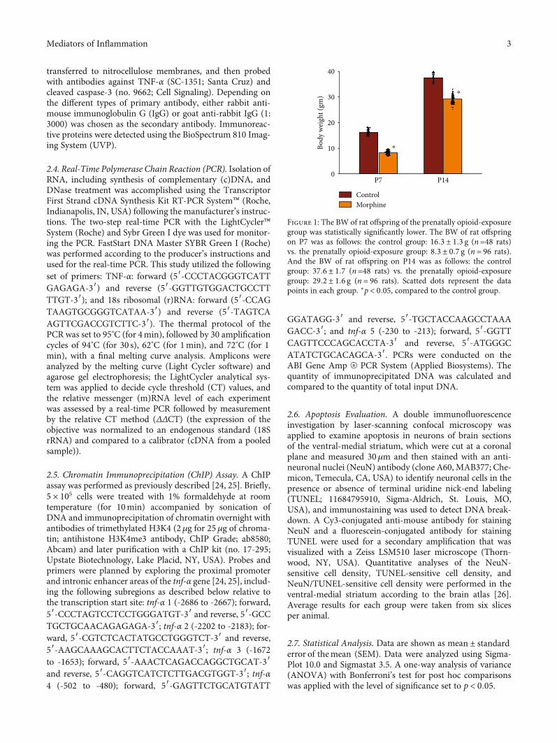
Figure 1: The BW of rat offspring of the prenatally opioid-exposuregroup was statistically significantly lower. The BW of rat offspringon P7 was as follows: the control group: 16:3 ± 1:3 g (n=48 rats)vs. the prenatally opioid-exposure group: 8:3 ± 0:7 g (n = 96 rats).And the BW of rat offspring on P14 was as follows: the controlgroup: 37:6 ± 1:7 (n=48 rats) vs. the prenatally opioid-exposuregroup: 29:2 ± 1:6 g (n = 96 rats). Scatted dots represent the datapoints in each group. ∗p < 0:05, compared to the control group.
3Mediators of Inflammation
3. Results
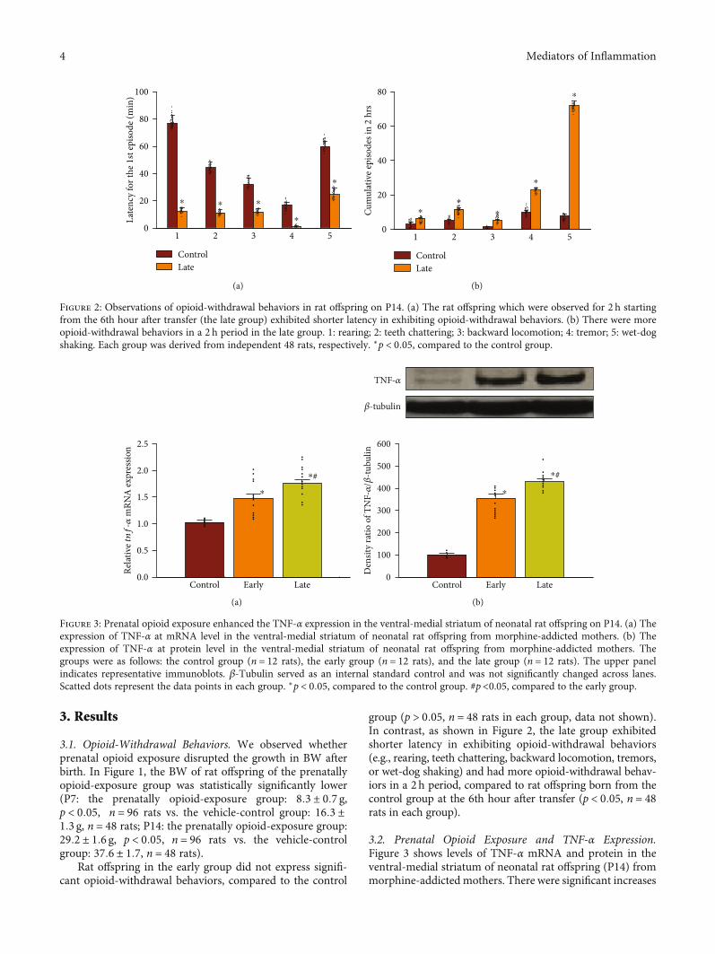
3.1. Opioid-Withdrawal Behaviors. We observed whetherprenatal opioid exposure disrupted the growth in BW afterbirth. In Figure 1, the BW of rat offspring of the prenatallyopioid-exposure group was statistically significantly lower(P7: the prenatally opioid-exposure group: 8:3 ± 0:7 g,p < 0:05, n = 96 rats vs. the vehicle-control group: 16:3 ±1:3 g, n = 48 rats; P14: the prenatally opioid-exposure group:29:2 ± 1:6 g, p < 0:05, n = 96 rats vs. the vehicle-controlgroup: 37:6 ± 1:7, n = 48 rats).
Rat offspring in the early group did not express signifi-cant opioid-withdrawal behaviors, compared to the control
group (p > 0:05, n = 48 rats in each group, data not shown).In contrast, as shown in Figure 2, the late group exhibitedshorter latency in exhibiting opioid-withdrawal behaviors(e.g., rearing, teeth chattering, backward locomotion, tremors,or wet-dog shaking) and had more opioid-withdrawal behav-iors in a 2h period, compared to rat offspring born from thecontrol group at the 6th hour after transfer (p < 0:05, n = 48rats in each group).
3.2. Prenatal Opioid Exposure and TNF-α Expression.Figure 3 shows levels of TNF-α mRNA and protein in theventral-medial striatum of neonatal rat offspring (P14) frommorphine-addicted mothers. There were significant increases
0
20
40
60
80
100
1 2 3 4 5
Late
ncy
for t
he 1
st ep
isode
(min
)
⁎ ⁎ ⁎
⁎
⁎
ControlLate
(a)
Cum
ulat
ive e
piso
des i
n 2
hrs
0
20
40
60
80
1 2 3 4 5
⁎⁎
⁎
⁎
⁎
ControlLate
(b)
Figure 2: Observations of opioid-withdrawal behaviors in rat offspring on P14. (a) The rat offspring which were observed for 2 h startingfrom the 6th hour after transfer (the late group) exhibited shorter latency in exhibiting opioid-withdrawal behaviors. (b) There were moreopioid-withdrawal behaviors in a 2 h period in the late group. 1: rearing; 2: teeth chattering; 3: backward locomotion; 4: tremor; 5: wet-dogshaking. Each group was derived from independent 48 rats, respectively. ∗p < 0:05, compared to the control group.
0.0
0.5
1.0
1.5
2.0
2.5
Control Early Late
Rela
tive tnf
-𝛼 m
RNA
expr
essio
n
⁎
⁎#
(a)
0
100
200
300
400
600
500
Control Early Late
Den
sity
ratio
of T
NF-𝛼
/𝛽-tu
bulin
⁎
⁎#
𝛽-tubulin
TNF-𝛼
(b)
Figure 3: Prenatal opioid exposure enhanced the TNF-α expression in the ventral-medial striatum of neonatal rat offspring on P14. (a) Theexpression of TNF-α at mRNA level in the ventral-medial striatum of neonatal rat offspring from morphine-addicted mothers. (b) Theexpression of TNF-α at protein level in the ventral-medial striatum of neonatal rat offspring from morphine-addicted mothers. Thegroups were as follows: the control group (n = 12 rats), the early group (n = 12 rats), and the late group (n = 12 rats). The upper panelindicates representative immunoblots. β-Tubulin served as an internal standard control and was not significantly changed across lanes.Scatted dots represent the data points in each group. ∗p < 0:05, compared to the control group. #p<0.05, compared to the early group.
4 Mediators of Inflammation
at the mRNA and protein levels of the TNF-α expression inthe early group compared to the control group (p < 0:05, n= 12 rats in each group). In addition, mRNA and proteinlevels of the TNF-α expression were further enhanced inthe late group compared to the early group (p < 0:05, n = 12rats in each group).
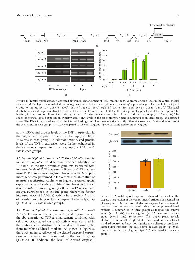
3.3. Prenatal Opioid Exposure and H3K4me3Modifications inthe tnf-α Promoter. To determine whether activation ofH3K4me3 in the tnf-α promoter gene was associated withincreased levels of TNF-α as seen in Figure 3, ChIP analysesusing PCR primers matching five subregions of the tnf-α pro-moter gene were performed in the ventral-medial striatum ofneonatal rat offspring. As shown in Figure 4, prenatal opioidexposure increased levels of H3K4me3 in subregions 2, 3, and4 of the tnf-α promoter gene (p < 0:05, n = 12 rats in eachgroup). Furthermore, in the late group, there were furtherincreased levels of H3K4me3 activity in subregions 3 and 4of the tnf-α promoter gene locus compared to the early group(p < 0:05, n = 12 rats in each group).
3.4. Prenatal Opioid Exposure and Apoptotic Caspase-3Activity. To observe whether prenatal opioid exposure causedthe abovementioned TNF-α enhancement combined withcell apoptosis, cleaved caspase-3 activity was evaluated inthe ventral-medial striatum of neonatal rat offspring (P14)from morphine-addicted mothers. As shown in Figure 5,there was an increased level of the cleaved caspase-3 expres-sion in the early group compared to the control group(p < 0:05). In addition, the level of cleaved caspase-3
–2686 –2667 –2202 –2183 –1672 –1653 –502 –480 –230
TATA
+1 transcription start site
–213
tnf-𝛼 1 tnf-𝛼 2 tnf-𝛼 3 tnf-𝛼 4 tnf-𝛼 5
TNF-𝛼
(a)
tnf-𝛼 (1)
tnf-𝛼 (2)
tnf-𝛼 (3)
tnf-𝛼 (4)
tnf-𝛼 (5)
H3K4me3 outputa b c
Inputa b c
(b)
0
5
10
15
20
30
25
ChIP fragment
Den
sity
ratio
(out
put/i
nput
)
⁎⁎
⁎⁎
⁎#
⁎#
1a b c
2a b c
3a b c
4a b c
5a b c
tnf-𝛼
(c)
Figure 4: Prenatal opioid exposure activated differential enhancement of H3K4me3 in the tnf-α promoter gene locus in the ventral-medialstriatum. (a) The figure demonstrated the subregions relative to the transcription start site of tnf-α promoter gene locus as follows: tnf-α 1(−2667 to −2686), tnf-α 2 (−2183 to −2202), tnf-α 3 (−1653 to −1672), tnf-α 4 (−174 to −496), and tnf-α 5 (−205 to −224). (b) The panelillustrations indicate representative ChIP assay of the levels of trimethylated H3K4 in the tnf-α promoter gene locus at the subregions. Theinsets a, b, and c are as follows: the control group (n = 12 rats), the early group (n = 12 rats), and the late group (n = 12 rats). (c) Theeffects of prenatal opioid exposure in trimethylated H3K4 levels in the tnf-α promoter gene is summarized in three groups as describedabove. The DNA input signal served as the internal loading control and was not significantly different across lanes. Scatted dots representthe data points in each group. ∗p < 0:05, compared to the control group. #p < 0:05, compared to the early group.
0
200
400
600
800
1400
1000
1200
Control Early Late
Den
sity
ratio
of c
aspa
se-3
/𝛽-tu
bulin
⁎⁎
𝛽-tubulin
Caspase-3
Figure 5: Prenatal opioid exposure enhanced the level of thecaspase-3 expression in the ventral-medial striatum of neonatal ratoffspring on P14. The level of cleaved caspase-3 in the ventral-medial striatum of neonatal rat offspring from morphine-addictedmothers is summarized in three groups as follows: the controlgroup (n = 12 rats), the early group (n = 12 rats), and the lategroup (n = 12 rats), respectively. The upper panel revealsillustrative immunoblots. β-Tubulin was used as an internalstandard control and was not significantly different across lanes.Scatted dots represent the data points in each group. ∗p < 0:05,compared to the control group. #p < 0:05, compared to the earlygroup.
5Mediators of Inflammation
expression in the late group did not statistically differ com-pared to the early group (p > 0:05, n = 12 rats in each group).
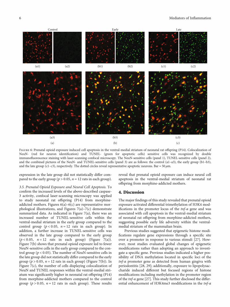
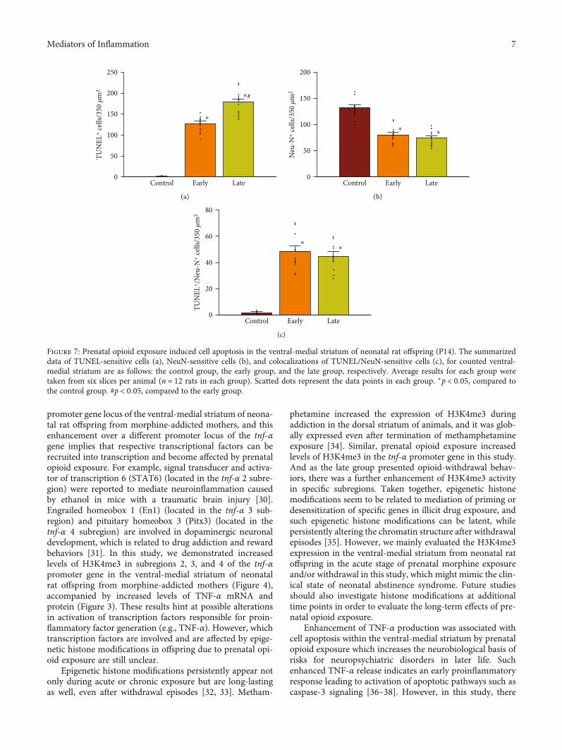
3.5. Prenatal Opioid Exposure and Neural Cell Apoptosis. Toconfirm the increased levels of the above-described caspase-3 activity, confocal laser-scanning microscopy was appliedto study neonatal rat offspring (P14) from morphine-addicted mothers. Figures 6(a)–6(c) are representative mor-phological illustrations, and Figures 7(a)–7(c) demonstratesummarized data. As indicated in Figure 7(a), there was anincreased number of TUNEL-sensitive cells within theventral-medial striatum in the early group compared to thecontrol group (p < 0:05, n = 12 rats in each group). Inaddition, a further increase in TUNEL-sensitive cells wasobserved in the late group compared to the early group(p < 0:05, n = 12 rats in each group) (Figure 7(a)).Figure 7(b) shows that prenatal opioid exposure led to fewerNeuN-sensitive cells in the early group compared to the con-trol group (p < 0:05). The number of NeuN-sensitive cells inthe late group did not statistically differ compared to the earlygroup (p > 0:05, n = 12 rats in each group) (Figure 7(b)). InFigure 7(c), the number of cells displaying colocalization ofNeuN and TUNEL responses within the ventral-medial stri-atum was significantly higher in neonatal rat offspring (P14)from morphine-addicted mothers compared to the controlgroup (p > 0:05, n = 12 rats in each group). These results
reveal that prenatal opioid exposure can induce neural cellapoptosis in the ventral-medial striatum of neonatal ratoffspring from morphine-addicted mothers.
4. Discussion
The major findings of this study revealed that prenatal opioidexposure activated differential trimethylation of H3K4 mod-ifications in the promoter locus of the tnf-α gene and wasassociated with cell apoptosis in the ventral-medial striatumof neonatal rat offspring from morphine-addicted mothers,suggesting possible early life adversity within the ventral-medial striatum of the mammalian brain.
Previous studies suggested that epigenetic histone modi-fications regulate gene expressions through a specific siteover a promoter in response to various stimuli [27]. How-ever, most studies evaluated global changes of epigeneticmodifications rather than adopting an approach to investi-gate a specific gene. Previous studies indicated a higher pos-sibility of DNA methylation located in specific loci of thetnf-α promoter gene as detected from human gingiva withperiodontitis [28, 29]; additionally, exposure to lipopolysac-charide induced different but focused regions of histonemodifications including methylation in the promoter regionof the tnf-α gene [27]. This study further disclosed the differ-ential enhancement of H3K4me3 modifications in the tnf-α
(a1)
(a3)
(a2)
Control
(a)
(b3)
(b1) (b2)
Early
(b)
(c3)
(c1) (c2)
Late
(c)
Figure 6: Prenatal opioid exposure induced cell apoptosis in the ventral-medial striatum of neonatal rat offspring (P14). Colocalization ofNeuN- (red for neuron identification) and TUNEL- (green for apoptotic cells) sensitive cells was recognized by doubleimmunofluorescence staining with laser-scanning confocal microscopy. The NeuN-sensitive cells (panel 1), TUNEL-sensitive cells (panel 2),and the combined pictures of the NeuN- and TUNEL-sensitive cells (panel 3) are as follows: the control (a1–a3), the early group (b1–b3),and the late group (c1–c3), respectively. The dotted circles reveal representative apoptotic neurons. Bar = 50μm.
6 Mediators of Inflammation
promoter gene locus of the ventral-medial striatum of neona-tal rat offspring from morphine-addicted mothers, and thisenhancement over a different promoter locus of the tnf-αgene implies that respective transcriptional factors can berecruited into transcription and become affected by prenatalopioid exposure. For example, signal transducer and activa-tor of transcription 6 (STAT6) (located in the tnf-α 2 subre-gion) were reported to mediate neuroinflammation causedby ethanol in mice with a traumatic brain injury [30].Engrailed homeobox 1 (En1) (located in the tnf-α 3 sub-region) and pituitary homeobox 3 (Pitx3) (located in thetnf-α 4 subregion) are involved in dopaminergic neuronaldevelopment, which is related to drug addiction and rewardbehaviors [31]. In this study, we demonstrated increasedlevels of H3K4me3 in subregions 2, 3, and 4 of the tnf-αpromoter gene in the ventral-medial striatum of neonatalrat offspring from morphine-addicted mothers (Figure 4),accompanied by increased levels of TNF-α mRNA andprotein (Figure 3). These results hint at possible alterationsin activation of transcription factors responsible for proin-flammatory factor generation (e.g., TNF-α). However, whichtranscription factors are involved and are affected by epige-netic histone modifications in offspring due to prenatal opi-oid exposure are still unclear.
Epigenetic histone modifications persistently appear notonly during acute or chronic exposure but are long-lastingas well, even after withdrawal episodes [32, 33]. Metham-
phetamine increased the expression of H3K4me3 duringaddiction in the dorsal striatum of animals, and it was glob-ally expressed even after termination of methamphetamineexposure [34]. Similar, prenatal opioid exposure increasedlevels of H3K4me3 in the tnf-α promoter gene in this study.And as the late group presented opioid-withdrawal behav-iors, there was a further enhancement of H3K4me3 activityin specific subregions. Taken together, epigenetic histonemodifications seem to be related to mediation of priming ordesensitization of specific genes in illicit drug exposure, andsuch epigenetic histone modifications can be latent, whilepersistently altering the chromatin structure after withdrawalepisodes [35]. However, we mainly evaluated the H3K4me3expression in the ventral-medial striatum from neonatal ratoffspring in the acute stage of prenatal morphine exposureand/or withdrawal in this study, which might mimic the clin-ical state of neonatal abstinence syndrome. Future studiesshould also investigate histone modifications at additionaltime points in order to evaluate the long-term effects of pre-natal opioid exposure.
Enhancement of TNF-α production was associated withcell apoptosis within the ventral-medial striatum by prenatalopioid exposure which increases the neurobiological basis ofrisks for neuropsychiatric disorders in later life. Suchenhanced TNF-α release indicates an early proinflammatoryresponse leading to activation of apoptotic pathways such ascaspase-3 signaling [36–38]. However, in this study, there
0
50
100
150
250
200
Control Early Late
TUN
EL+ c
ells/
350 𝜇
m2
⁎
⁎#
(a)
0
50
100
150
200
Control Early Late
Neu
-N+ c
ells/
350 𝜇
m2
⁎ ⁎
(b)
0
20
40
60
80
Control Early Late
TUN
EL+ /
Neu
-N+ c
ells/
350 𝜇
m2
⁎⁎
(c)
Figure 7: Prenatal opioid exposure induced cell apoptosis in the ventral-medial striatum of neonatal rat offspring (P14). The summarizeddata of TUNEL-sensitive cells (a), NeuN-sensitive cells (b), and colocalizations of TUNEL/NeuN-sensitive cells (c), for counted ventral-medial striatum are as follows: the control group, the early group, and the late group, respectively. Average results for each group weretaken from six slices per animal (n = 12 rats in each group). Scatted dots represent the data points in each group. ∗p < 0:05, compared tothe control group. #p < 0:05, compared to the early group.
7Mediators of Inflammation
was no significant decrease in apoptotic neurons in the lategroup (Figure 7). Indeed, morphine can directly induce apo-ptosis of microglia and neurons in vitro, but it seemed thatchronic morphine exposure might not affect glial cells at anearly age in vivo from animal models [36, 37]. Therefore,apoptosis did play a role in prenatal opioid exposure, butthe detailed involvement of specific cell types remains to beinvestigated. In this study, prenatal opioid exposure andwithdrawal induced TNF-α production might lead to neuralcell apoptosis in the ventral-medial striatum. In addition,opioid exposure might also enhance apoptosis in selected,specific brain areas, such as the amygdala, ventral-medialstriatum, or prefrontal cortex in vivo [36, 38]. Prenatal heroinexposure enhances apoptosis in the hippocampus and resultsin impairments to learning and memory [39]. Those resultsindicate that prenatal opioid exposure and withdrawal mightinduce apoptosis in other specific brain areas and result inneurological impairments.
Recently, a closely interactive and related relationshipbetween inflammation and neuropsychiatric disorders wasproposed in the mammalian brain [40]. For instance, proin-flammatory cytokines can affect cognition and mood behav-iors by promoting neuroexcitatory reactions and impairingneuron plasticity [41]. Furthermore, as shown in this study,the epigenetic H3K4me3 mechanism for the upregulationof the TNF-α expression was associated with increased neu-ral cell apoptosis in the ventral-medial striatum, which mightcontribute to neurological impairments. Such neural cellapoptotic damage within the ventral-medial striatum duringearly life might lead to increased dose requirements for opi-oid drugs to satisfy the need to increase the dosage and to ful-fill drug-craving and addictive behaviors later in life. Inaddition, prenatal opioid exposure may initiate long-lastingalterations in the gene expressions of the synapse structurein the ventral-medial striatum in rat offspring [8, 22]. Sucheffects of prenatal opioid exposure seem to exist longer thanexpected. Our current results showed that prenatal opioidexposure induced neural cell apoptosis within the ventral-medial striatum in early life. However, whether neural cellapoptosis persistently appears in later life or the next genera-tion is still unknown, and this has motivated us to conductfurther experiments on the long-term effect of prenatal opi-oid exposure and withdrawal.
5. Conclusions
In summary, this study revealed that prenatal opioid exposurecan activate an epigenetic histone mechanism for regulatingproinflammatory factor generation and hence lead to cell apo-ptotic damage within the ventral-medial striatum of neonatalrat offspring from morphine-addicted mothers. More impor-tantly, the opioid-withdrawal episode may provide augmentedeffects for such abovementioned alterations and may lead toadverse effects in the neonatal brain of these offspring.
Data Availability
The data used to support the findings of this study are avail-able from the corresponding author upon request.
Conflicts of Interest
The authors declare that there is no conflict of interestregarding the publication of this paper.
Authors’ Contributions
PLW contributed to the data curation, writing—originaldraft, and funding acquisition. JLS contributed to theresources, writing—review and editing, and visualization.CHY contributed to the investigation and data curation.KCK contributed to the methodology and visualization.YCY and SNY contributed to the conceptualization, method-ology, writing—review and editing, supervision, and fundingacquisition. All authors contributed to the manuscript revi-sion and read and approved the submitted version.
Funding
This study was supported in part by research grants toSan-Nan Yang (EDPJ107068, EDPJ108057, EDPJ109059)and Pei Ling Wu (EDAHI 107004, EDAHP108015,EDAHP105030, and EDCHP106007).
References
[1] A. Agorastos, P. Pervanidou, G. P. Chrousos, and D. G. Baker,“Developmental trajectories of early life stress and trauma: anarrative review on neurobiological aspects beyond stresssystem dysregulation,” Frontiers in Psychiatry, vol. 10, p. 118,2019.
[2] S. Lisonkova, L. L. Richter, J. Ting et al., “Neonatal abstinencesyndrome and associated neonatal and maternal mortality andmorbidity,” Pediatrics, vol. 144, no. 2, p. e20183664, 2019.
[3] K. Ramphul, S. G. Mejias, and J. Joynauth, “An update on theburden of neonatal abstinence syndrome in the United States,”Hospital Pediatrcs, vol. 10, no. 2, pp. 181–184, 2020.
[4] R. W. S. Coulter, J. E. Egan, S. Kinsky et al., “Mental health,drug, and violence interventions for sexual/gender minorities:a systematic review,” Pediatrics, vol. 144, no. 3, p. e20183367,2019.
[5] A. J. Czynski, J. M. Davis, L. M. Dansereau et al., “Neurodeve-lopmental outcomes of neonates randomized to morphine ormethadone for treatment of neonatal abstinence syndrome,”The Journal of pediatrics, vol. 219, 2020.
[6] S. N. Yang, L. T. Huang, C. L. Wang et al., “Prenatal adminis-tration of morphine decreases CREBSerine-133 phosphoryla-tion and synaptic plasticity range mediated by glutamatergictransmission in the hippocampal CA1 area of cognitive-deficient rat offspring,” Hippocampus, vol. 13, no. 8, pp. 915–921, 2003.
[7] S. N. Yang, C. A. Liu, M. Y. Chung et al., “Alterations of post-synaptic density proteins in the hippocampus of rat offspringfrom the morphine-addicted mother: beneficial effect of dex-tromethorphan,” Hippocampus, vol. 16, no. 6, pp. 521–530,2006.
[8] C. S. Lin, P. L. Tao, Y. J. Jong et al., “Prenatal morphine altersthe synaptic complex of postsynaptic density 95 with N-methyl-D-aspartate receptor subunit in hippocampal CA1subregion of rat offspring leading to long-term cognitive defi-cits,” Neuroscience, vol. 158, no. 4, pp. 1326–1337, 2009.
8 Mediators of Inflammation
[9] H. J. Harder and A. Z. Murphy, “Early life opioid exposure andpotential long-term effects,” Neurobiology of Stress, vol. 10,p. 100156, 2019.
[10] E. J. Ross, D. L. Graham, K. M. Money, and G. D. Stanwood,“Developmental consequences of fetal exposure to drugs: whatwe know and what we still must learn,” Neuropsychopharma-cology, vol. 40, no. 1, pp. 61–87, 2015.
[11] M. D. Scofield, J. A. Heinsbroek, C. D. Gipson et al., “Thenucleus accumbens: mechanisms of addiction across drug clas-ses reflect the importance of glutamate homeostasis,” Pharma-cological Reviews, vol. 68, no. 3, pp. 816–871, 2016.
[12] L. C. Loram, P. M. Grace, K. A. Strand et al., “Prior exposure torepeated morphine potentiates mechanical allodynia inducedby peripheral inflammation and neuropathy,” Brain Behavior,and Immunity, vol. 26, no. 8, pp. 1256–1264, 2012.
[13] S. Hao, S. Liu, X. Zheng et al., “The role of TNFα in the peria-queductal gray during naloxone-precipitated morphine with-drawal in rats,” Neuropsychopharmacology, vol. 36, no. 3,pp. 664–676, 2011.
[14] S. J. O'Sullivan, E. Malahias, J. Park et al., “Single-cell glia andneuron gene expression in the central amygdala in opioidwithdrawal suggests inflammation with correlated gut dysbio-sis,” Frontiers in Neuroscience, vol. 13, p. 665, 2019.
[15] S. L. Chen, P. L. Tao, C. H. Chu et al., “Low-dose memantineattenuated morphine addictive behavior through its anti-inflammation and neurotrophic effects in rats,” Journal ofNeuroimmune Pharmacology, vol. 7, no. 2, pp. 444–453, 2012.
[16] J. Amri, M. Sadegh, N. Moulaei, and M. R. Palizvan, “Transge-nerational modification of hippocampus TNF-α and S100Blevels in the offspring of rats chronically exposed to morphineduring adolescence,” The American Journal of Drug and Alco-hol Abuse, vol. 44, no. 1, pp. 95–102, 2018.
[17] L. Hantsoo, S. Kornfield, M. C. Anguera, and C. N. Epperson,“Inflammation: a proposed intermediary between maternalstress and offspring neuropsychiatric risk,” Biological Psychia-try, vol. 85, no. 2, pp. 97–106, 2019.
[18] K. Hyun, J. Jeon, K. Park, and J. Kim, “Writing, erasing andreading histone lysine methylations,” Experimental & molecu-lar medicine, vol. 49, no. 4, p. e324, 2017.
[19] E. J. Nestler, “Epigenetic mechanisms of drug addiction,” Neu-ropharmacology, vol. 76, pp. 259–268, 2014.
[20] T. Kusch, “Histone H3 lysine 4 methylation revisited,” Tran-scription, vol. 3, no. 6, pp. 310–314, 2012.
[21] R. D. Shepard and F. S. Nugent, “Early life stress- and drug-induced histone modifications within the ventral tegmentalarea,” Frontiers in cell and developmental biology, vol. 8,p. 588476, 2020.
[22] P. L. Wu, Y. N. Yang, J. L. Suen, Y. C. S. Yang, C. H. Yang, andS. N. Yang, “Long-lasting alterations in gene expression ofpostsynaptic density 95 and inotropic glutamatergic receptorsubunit in the mesocorticolimbic system of rat offspring bornto morphine-addicted mothers,” BioMed Research Interna-tional, vol. 2018, Article ID 5437092, 9 pages, 2018.
[23] P. Sengupta, “The laboratory rat relating its age with human’s,”International Journal of Preventive Medicine, vol. 4, no. 6,pp. 624–630, 2013.
[24] Y. N. Yang, Y. T. Su, P. L. Wu et al., “Granulocyte colony-stimulating factor alleviates bacterial-induced neuronalapoptotic damage in the neonatal rat brain through epigenetichistone modification,” Oxidative Medicine and Cellular Lon-gevity, vol. 2018, Article ID 9797146, 10 pages, 2018.
[25] Y. N. Yang, Y. C. S. H. Yang, P. L. Wu, C. H. Yang, K. C.Kuo, and S. N. Yang, “Dextromethorphan suppresseslipopolysaccharide-induced epigenetic histone regulation inthe tumor necrosis factor-α expression in primary ratmicroglia,” Mediators of Inflammation, vol. 2020, ArticleID 9694012, 8 pages, 2020.
[26] S. R. Jones, S. J. O'Dell, J. F. Marshall, and R. M. Wightman,“Functional and anatomical evidence for different dopaminedynamics in the core and shell of the nucleus accumbens inslices of rat brain,” Synapse, vol. 23, no. 3, pp. 224–231,1996.
[27] K. E. Sullivan, A. B. M. Reddy, K. Dietzmann et al., “Epigeneticregulation of tumor necrosis factor alpha,” Molecular andCellular Biology, vol. 27, no. 14, pp. 5147–5160, 2007.
[28] S. Zhang, S. P. Barros, A. J. Moretti et al., “Epigenetic regula-tion of TNFA expression in periodontal disease,” Journal ofPeriodontology, vol. 84, no. 11, pp. 1606–1616, 2013.
[29] A. Kojima, T. Kobayashi, S. Ito, A. Murasawa, K. Nakazono,and H. Yoshie, “Tumor necrosis factor-alpha gene promotermethylation in Japanese adults with chronic periodontitisand rheumatoid arthritis,” Journal of Periodontal Research,vol. 51, no. 3, pp. 350–358, 2016.
[30] F. O. Heuvel, S. Holl, A. Chandrasekar et al., “STAT6 medi-ates the effect of ethanol on neuroinflammatory response inTBI,” Brain, Behavior, and Immunity, vol. 81, pp. 228–246,2019.
[31] M. T. Alves dos Santos and M. P. Smidt, “En1 andWnt signal-ing in midbrain dopaminergic neuronal development,” NeuralDevelopment, vol. 6, no. 1, p. 23, 2011.
[32] Y. D. Black, F. R. Maclaren, A. V. Naydenov, Carlezon WA Jr,M. G. Baxter, and C. Konradi, “Altered attention and prefron-tal cortex gene expression in rats after binge-like exposure tococaine during adolescence,” The Journal of Neuroscience:The Official Journal of the Society for Neuroscience, vol. 26,no. 38, pp. 9656–9665, 2006.
[33] A. Kumar, K. H. Choi, W. Renthal et al., “Chromatin remodel-ing is a key mechanism underlying cocaine-induced plasticityin striatum,” Neuron, vol. 48, no. 2, pp. 303–314, 2005.
[34] I. N. Krasnova, M. Chiflikyan, Z. Justinova et al., “CREB phos-phorylation regulates striatal transcriptional responses in theself- administration model of methamphetamine addiction inthe rat,” Neurobiology of Disease, vol. 58, pp. 132–143, 2013.
[35] A. J. Robison and E. J. Nestler, “Transcriptional and epigeneticmechanisms of addiction,” Nature Reviews Neuroscience,vol. 12, no. 11, pp. 623–637, 2011.
[36] D. Bajic, K. G. Commons, and S. G. Soriano, “Morphine-enhanced apoptosis in selective brain regions of neonatal rats,”International journal of developmental neuroscience: the offi-cial journal of the International Society for Developmental Neu-roscience, vol. 31, no. 4, pp. 258–266, 2013.
[37] S. Hu, W. S. Sheng, J. R. Lokensgard, and P. K. Peterson,“Morphine induces apoptosis of human microglia and neu-rons,” Neuropharmacology, vol. 42, no. 6, pp. 829–836,2002.
[38] S. N. Katebi, Y. Razavi, S. Z. Alamdary, F. Khodagholi, andA. Haghparast, “Morphine could increase apoptotic factorsin the nucleus accumbens and prefrontal cortex of rat brain'sreward circuitry,” Brain Research, vol. 1540, pp. 1–8, 2013.
[39] Y. Wang and T. Z. Han, “Prenatal exposure to heroin in miceelicits memory deficits that can be attributed to neuronal apo-ptosis,” Neuroscience, vol. 160, no. 2, pp. 330–338, 2009.
9Mediators of Inflammation

[40] G. Z. Réus, G. R. Fries, L. Stertz et al., “The role of inflam-mation and microglial activation in the pathophysiology ofpsychiatric disorders,” Neuroscience, vol. 300, pp. 141–154,2015.
[41] M. E. Bauer and A. L. Teixeira, “Inflammation in psychiatricdisorders: what comes first?,”Annals of the New York Academyof Sciences, vol. 1437, no. 1, pp. 57–67, 2019.
10 Mediators of Inflammation