eld* ToF-SIMS imaging reveals that p-hydroxybenzoate ...
11
Original article Robyn E. Goacher, Yaseen Mottiar and Shawn D. Mansfield* ToF-SIMS imaging reveals that p-hydroxybenzoate groups specifically decorate the lignin of fibres in the xylem of poplar and willow https://doi.org/10.1515/hf-2020-0130 Received May 29, 2020; accepted August 11, 2020; published online September 18, 2020 Abstract: Esterified phenolics occur as γ-linked lignin ac- ylations in many plant taxa but little is known about the distribution or function of such groups. In the family Sali- caceae, p-hydroxybenzoate groups are present in the lignin of poplars (Populus spp.) and willows (Salix spp.). In this study, the distribution of cell wall-bound p-hydrox- ybenzoate was examined in different tissue and cell types of poplar and willow trees. This analysis showed that p-hydroxybenzoate groups were most prevalent in juvenile stems, and were elevated in tension wood and following nitrogen fertilisation. Closer examination of stem cross- sections using time-of-flight secondary-ion mass spectrom- etry (ToF-SIMS) directly showed that p-hydroxybenzoate groups occur predominantly in the cell walls of fibres and are largely absent from vessel elements. These results point to a role for ester-linked groups in modifying the syringyl- rich lignin of fibres, perhaps to strengthen the cell walls of these structural elements or to increase lignification rates. Keywords: cell wall-bound phenolics; ester-linked pendant groups; lignin acylations; mass spectrometry imaging; p-hydroxybenzoic acid. 1 Introduction Lignin is a primary component of the secondary cell walls of vascular plants. It contributes greatly to the mechanical strength of stems and branches, and it plays important roles in water conduction and plant defence (Novo-Uzal et al. 2012). Although lignin is indispensable for plant vi- tality, it poses a major challenge for the industrial pro- cessing of plant biomass. As such, lignin chemistry and biosynthesis have received considerable research atten- tion, and great strides have been made in modifying lignin structures through plant biotechnology (Mottiar et al. 2016). Lignin is a complex phenolic polymer that is largely resistant to chemical degradation (Figure 1a). While the recalcitrance of lignin presents a challenge for efficient biomass deconstruction, it also provides opportunities for the development of biochemicals and biomaterials (Glasser 2019). Traditionally, lignin was seen as a low- value byproduct of industrial processing and was typically burned for energy. Today, new processes are being devel- oped to preserve phenolic structures during lignocellulose deconstruction in a strategy termed ‘lignin-first bio- refining’ (Schutyser et al. 2018). Various biomaterials are envisioned from recovered polymers and oligomers, and lignin-derived phenolics could be fed to microbes for fermentation into diverse secondary products just as cel- lulose and hemicellulose can be converted into biofuels (Beckham et al. 2016; Kai et al. 2016). Lignin is assembled primarily from three cinnamic acid derivatives known as monolignols: p-coumaryl, con- iferyl and sinapyl alcohol (Boerjan et al. 2003). Once incorporated into a lignin polymer, these precursors give Robyn E. Goacher and Yaseen Mottiar, These authors contributed equally to this work. *Corresponding author: Shawn D. Mansfield, Department of Wood Science, University of British Columbia, 2424 Main Mall, Vancouver, BC, V6T 1Z4, Canada, e-mail: shawn.mansfi[email protected]. https:// orcid.org/0000-0002-0175-554X Robyn E. Goacher, Department of Biochemistry, Chemistry and Physics, Niagara University, 5795 Lewiston Road, Lewiston, NY, 14109, USA, e-mail: [email protected]. https://orcid.org/0000-0001- 7863-4888 Yaseen Mottiar, Department of Wood Science, University of British Columbia, 2424 Main Mall, Vancouver, BC, V6T 1Z4, Canada, e-mail: [email protected]. https://orcid.org/0000-0002-4106- 6159 Holzforschung 2021; 75(5): 452–462
Transcript of eld* ToF-SIMS imaging reveals that p-hydroxybenzoate ...
Original article
Robyn E. Goacher, Yaseen Mottiar and Shawn D. Mansfield*
ToF-SIMS imaging reveals thatp-hydroxybenzoate groups specifically decoratethe lignin of fibres in the xylem of poplar andwillowhttps://doi.org/10.1515/hf-2020-0130Received May 29, 2020; accepted August 11, 2020; published onlineSeptember 18, 2020
Abstract: Esterified phenolics occur as γ-linked lignin ac-ylations in many plant taxa but little is known about thedistribution or function of such groups. In the family Sali-caceae, p-hydroxybenzoate groups are present in the ligninof poplars (Populus spp.) and willows (Salix spp.). In thisstudy, the distribution of cell wall-bound p-hydrox-ybenzoate was examined in different tissue and cell types ofpoplar and willow trees. This analysis showed thatp-hydroxybenzoate groups were most prevalent in juvenilestems, and were elevated in tension wood and followingnitrogen fertilisation. Closer examination of stem cross-sections using time-of-flight secondary-ion mass spectrom-etry (ToF-SIMS) directly showed that p-hydroxybenzoategroups occur predominantly in the cell walls of fibres andare largely absent from vessel elements. These results pointto a role for ester-linked groups in modifying the syringyl-rich lignin of fibres, perhaps to strengthen the cell walls ofthese structural elements or to increase lignification rates.
Keywords: cell wall-bound phenolics; ester-linked pendantgroups; lignin acylations; mass spectrometry imaging;p-hydroxybenzoic acid.
1 Introduction
Lignin is a primary component of the secondary cell wallsof vascular plants. It contributes greatly to the mechanicalstrength of stems and branches, and it plays importantroles in water conduction and plant defence (Novo-Uzalet al. 2012). Although lignin is indispensable for plant vi-tality, it poses a major challenge for the industrial pro-cessing of plant biomass. As such, lignin chemistry andbiosynthesis have received considerable research atten-tion, and great strides have been made in modifying ligninstructures through plant biotechnology (Mottiar et al.2016).
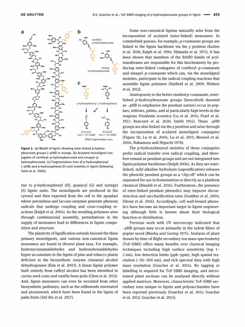
Lignin is a complex phenolic polymer that is largelyresistant to chemical degradation (Figure 1a). While therecalcitrance of lignin presents a challenge for efficientbiomass deconstruction, it also provides opportunities forthe development of biochemicals and biomaterials(Glasser 2019). Traditionally, lignin was seen as a low-value byproduct of industrial processing and was typicallyburned for energy. Today, new processes are being devel-oped to preserve phenolic structures during lignocellulosedeconstruction in a strategy termed ‘lignin-first bio-refining’ (Schutyser et al. 2018). Various biomaterials areenvisioned from recovered polymers and oligomers, andlignin-derived phenolics could be fed to microbes forfermentation into diverse secondary products just as cel-lulose and hemicellulose can be converted into biofuels(Beckham et al. 2016; Kai et al. 2016).
Lignin is assembled primarily from three cinnamicacid derivatives known as monolignols: p-coumaryl, con-iferyl and sinapyl alcohol (Boerjan et al. 2003). Onceincorporated into a lignin polymer, these precursors give
Robyn E. Goacher and Yaseen Mottiar, These authors contributedequally to this work.
*Corresponding author: Shawn D. Mansfield, Department of WoodScience, University of British Columbia, 2424 Main Mall, Vancouver,BC, V6T 1Z4, Canada, e-mail: [email protected]. https://orcid.org/0000-0002-0175-554XRobyn E. Goacher, Department of Biochemistry, Chemistry andPhysics, NiagaraUniversity, 5795 Lewiston Road, Lewiston, NY, 14109,USA, e-mail: [email protected]. https://orcid.org/0000-0001-7863-4888Yaseen Mottiar, Department of Wood Science, University of BritishColumbia, 2424 Main Mall, Vancouver, BC, V6T 1Z4, Canada,e-mail: [email protected]. https://orcid.org/0000-0002-4106-6159
Holzforschung 2021; 75(5): 452–462
rise to p-hydroxyphenyl (H), guaiacyl (G) and syringyl(S) lignin units. The monolignols are produced in thecytosol and then exported from the cell to the apoplastwhere peroxidase and laccase enzymes generate phenoxyradicals that undergo coupling and cross-coupling re-actions (Ralph et al. 2004). As the resulting polymers arisethrough combinatorial assembly, perturbations in thesupply of monomers lead to differences in lignin compo-sition and structure.
The plasticity of lignification extends beyond the threeprimary monolignols, and various non-canonical ligninmonomers are found in diverse plant taxa. For example,hydroxycinnamaldehydes and hydroxybenzaldehydeshyper-accumulate in the lignin of pine and tobacco plantsdeficient in the biosynthetic enzyme cinnamyl alcoholdehydrogenase (Kim et al. 2003). A linear lignin polymerbuilt entirely from caffeyl alcohol has been identified incactus seed coats and vanilla bean pods (Chen et al. 2012).And, lignin monomers can even be recruited from otherbiosynthetic pathways, such as the stilbenoids resveratroland piceatannol, which have been found in the lignin ofpalm fruits (Del Río et al. 2017).
Some non-canonical lignins naturally arise from theincorporation of acylated (ester-linked) monomers. Incommelinid grasses, for example, p-coumarate groups arelinked to the lignin backbone via the γ position (Karlenet al. 2018; Ralph et al. 1994; Shimada et al. 1971). It hasbeen shown that members of the BAHD family of acyl-transferases are responsible for this biochemistry by pro-ducing ester-linked conjugates of coniferyl–p-coumarateand sinapyl–p-coumarate which can, via the monolignolmoieties, participate in the radical-coupling reactions thatassemble lignin polymers (Hatfield et al. 2009; Witherset al. 2012).
Analogously to the better-studied p-coumarate, ester-linked p-hydroxybenzoate groups (henceforth denotedas –pHB to emphasise the pendant nature) occur in pop-lars, willows, palms, and at particularly high levels in theseagrass Posidonia oceanica (Lu et al. 2015; Pearl et al.1957; Rencoret et al. 2020; Smith 1955). These –pHBgroups are also linked via the γ position and arise throughthe incorporation of acylated monolignol conjugates(Figure 1b; Lu et al. 2004; Lu et al. 2015; Morreel et al.2004; Nakamura and Higuchi 1978).
The p-hydroxybenzoyl moieties of these conjugatesprefer radical transfer over radical coupling, and there-fore remain as pendant groups and are not integrated intolignin polymer backbones (Ralph 2006). As they are ester-linked, mild alkaline hydrolysis (saponification) releasesthe phenolic pendant groups as a ‘clip-off’ which can beseparated for use in fermentation or directly as a platformchemical (Rinaldi et al. 2016). Furthermore, the presenceof ester-linked pendant phenolics may improve decon-struction and saccharification rates (Grabber et al. 2004;Sibout et al. 2016). Accordingly, cell wall-bound pheno-lics have become an important target in lignin engineer-ing although little is known about their biologicalfunction or distribution.
Previous work with UV microscopy indicated that–pHB groups may occur primarily in the xylem fibres ofpoplar wood (Musha and Goring 1975). Analysis of planttissues by time-of-flight secondary-ionmass spectrometry(ToF-SIMS) offers many benefits over classical imagingtechniques including high surface sensitivity (top 1–2 nm), low detection limits (ppb–ppm), high spatial res-olution (∼50–300 nm), and rich spectral data with highmass resolution (Goacher et al. 2014). No tagging orlabelling is required for ToF-SIMS imaging, and micro-tomed plant sections can be analysed directly withoutapplied matrices. Moreover, characteristic ToF-SIMS sec-ondary ions unique to lignin and polysaccharides havebeen reported previously (Goacher et al. 2011; Goacheret al. 2012; Goacher et al. 2013).
a bconiferylp-hydroxybenzoate
sinapylp-hydroxybenzoate
p-hydroxybenzoyl p-hydroxyphenyl
SIMS fragmentation ions
[C7H5O2]+
m/z = 121.0289[C8H9O]+
m/z = 121.0652
c
OH
OO
HOOMe
MeOOH
OO
HOOMe
OH
C
O
OH
CH2
H2C
OH
OO
OH
OHHO
O
O
HO
HOOMe
O
MeO OMe
OH
OOMe
HO
MeO
O
HO
O
OOMe
OH
O
HO
OMe
OHHO
O
OMe
MeO
OHHO
O
O
OOMeMeO
HO
MeO
OMe
OMeO
OMe
OH
HO OH
O
MeO
HO
O
OO
OH
HO OH
O OH
HO
O
OMe
MeO
MeO
OH
O
HO
MeO
OMe
MeO
OMe
OHO
OMeO
OMe
MeO
OHO
HO
OOH
OMe
MeO
OMeMeO
HO
OH
HO
MeO
MeO
O
OH
HO
OHO
OH
OMeMeO
HO
Figure 1: (a) Model of lignin showing ester-linked p-hydrox-ybenzoate groups (–pHB) in orange. (b) Acylated monolignol con-jugates of coniferyl–p-hydroxybenzoate and sinapyl–p-hydroxybenzoate. (c) Fragmentation ions of p-hydroxybenzoyl(–pHB) and p-hydroxyphenyl (H unit) moieties in lignin (followingSaito et al. 2006).
R.E. Goacher et al.: ToF-SIMS imaging of p-hydroxybenzoate groups in lignin 453
In this study, the occurrence of –pHB groups was sur-veyed in various tissues of poplar and willow by chemicalanalysis, and ToF-SIMS was then used to examine the dis-tribution of –pHB in wood. To our knowledge, this reportrepresents the first use of ToF-SIMS to examine the distribu-tion of non-canonical lignin monomers.
2 Materials and methods
2.1 Plant samples
Samples for chemical analysis were harvested from trees grown in aglasshouse. Dormant cuttings of Populus nigra cv. ‘Italica’ (Lombardypoplar), Populus trichocarpa cv. ‘Nisqually-1’ (black cottonwood) andSalix eriocephala DUK-E5 (heart-leaf willow) were harvested frommature trees, dipped in rooting powder (0.8% indole-3-butyric acid),planted in two-gallon pots with peat-based potting soil and irrigatedas needed with water containing Hoagland’s solution (Hoagland andArnon 1950). After six months of growth, the main stems were dividedinto 50 cm sections (i.e., young, juvenile, and mature) while the rootswere separated into young andmature roots. Youngandmature leaveswere also collected, aswere branches and barkwhichwas peeled fromthe main stems. Stem samples were also harvested from glasshouse-grown Populus tremula × alba INRA 717-IB4 (hybrid poplar) fed withlow nitrogen (0.1 mM ammonium nitrate) and high nitrogen (10 mMammonium nitrate) fertilisers. Samples of tension wood, oppositewood, and normal wood from the same genotype were describedpreviously (Gerttula et al. 2015). All samples were comminuted using aWiley mill to pass a 40-mesh screen and then Soxhlet-extracted for24 hwith hot acetoneprior to analysis. Three biological replicateswereanalysed for each genotype.
Wood samples for ToF-SIMS were harvested from the sameLombardy poplar and heart-leaf willow trees described above.Transversal xylem cross-sections were cut from the main stems to athickness of roughly 100 µmusing a sliding-blockmicrotome and thenallowed to air dry. Prior to ToF-SIMS analysis, several cross-sectionswere solvent-extracted to remove small-molecule extractives (Goacheret al. 2013). Sections were placed in cellulose thimbles and Soxhlet-extracted for 8 h with 95% ethanol, 8 h with 70% ethanol/30%toluene, and 4 hwith distilledwater, followingASTMstandardD1105–96 (2007) but with a doubled ethanol extraction time. Subsequently,some of the untreated and extracted microtomed sections underwentmild alkaline hydrolysis to cleave ester-linked –pHB by soaking in 2 Msodium hydroxide for 24 h at 30 °C with shaking at 60 rpm (Goldstein1984). After extraction and/or alkaline hydrolysis, the cross-sectionswere rinsed with 20 mL aliquots: once with distilled water, four timeswith 1 M acetic acid, and finally 10 times with distilled water (Brahamand Goacher 2015). Samples of wood powder from Betula allegha-niensis (yellow birch), an angiosperm genus which is devoid of –pHBgroups (Bardet et al. 1985a), were used as a negative control for peakidentification purposes.
2.2 Chemical analysis of –pHB groups and lignin
The amount of ester-linked cell wall-bound –pHB groups wasdetermined by mild alkaline hydrolysis of extractive-free powdered
samples. Duplicate 20 mg samples of oven-dried powder wereweighed into 2 mL screw-cap vials and 1 mL of 2 M sodium hy-droxide was added along with 100 μL of 1 mg mL−1 o-anisic acid asan internal standard. Tubes were incubated at 30 °C for 24 h. Thesaponification reactions were terminated by the addition of 100 μLof 72% sulphuric acid. After incubation on ice for 5 min, the su-pernatants were collected by centrifugation and filtered through0.45 μm nylon syringe filters prior to analysis using reversed phaseHPLC.
Using a Dionex Summit HPLC apparatus equipped with a diodearray detector (Thermo Scientific, Waltham, Massachusetts, USA),20 μL samples were injected onto a Symmetry C-18 column(4.6 × 250 mm, 5 µm particle size, Waters Inc., Milford, Massachu-setts, USA) maintained at 35 °C. Good peak resolution was achievedusing non-isocratic separation with a gradient over 30 min from 5 to45% of eluent A (0.1% trifluoroacetic acid in 70:30 acetoni-trile:methanol) in eluent B (0.1% trifluoroacetic acid in water) at aflow rate of 0.7 mL min−1. Integration of p-hydroxybenzoic acid ando-anisic acid peaks was performed at the UV maxima of 255 and296 nm respectively, and a seven-point calibration curve from 1 to100 ng mL−1 prepared with analytical-grade standards (at least 99%pure, Sigma-Aldrich Corp., St. Louis, Missouri, USA) was used forquantification.
The amount and composition of lignin was determined using theKlason lignin method and thioacidolysis procedure followed by deri-vatisation and gas chromatography as described previously (Mottiaret al. 2020). Heat maps of –pHB content (normalised per g lignin) weregenerated using Adobe Illustrator (Adobe Systems Inc., MountainView, California, USA).
2.3 ToF-SIMS
ToF-SIMS data was acquired using two ToF-SIMS 5 instrumentsequipped with reflectron-type mass analysers (ION-TOF GmbH,Münster, Germany). Analysis was performed using Bi3
++ primary ionsat a 45° incident angle with 20 eV electron flooding for chargecompensation. The untreated, extracted, and alkaline-treated sectionswere analysed using high-current conditions for fast spectral acqui-sition (50 keV Bi3
++, 0.7 pA pulsed current, 65 μs cycle time, 256 × 256pixels rastered randomly over either 100× 100 or 500× 500 μm2 areas).High resolution images were obtained of the extracted sections usingburst alignment settings with 0.11 pA current, 50 keV Bi3
++ at a 65 μscycle time, and 256 × 256 pixels rastered randomly over a200 × 200 μm2 area; or with 0.06 pA current, 60 keV Bi3
++ at a 100 μscycle time, and 512× 512 pixels rastered randomly over a 200× 200 μm2
area. The pressure during analysis was less than 3 × 10⁻⁷ mbar anddelayed extraction was used to minimise the effects of surfacetopography.
SurfaceLab 6.3 and 6.6 (IONTOF GmbH, Münster, Germany)software versions were used to calibrate the ToF-SIMS mass spectrato C3H5
+ (m/z 41.03), C4H7+ (m/z 55.05), C7H7
+ (m/z 91.05) and C5H5O2+
(m/z 97.03), and to generate images of characteristic ions. Mass in-terval lists were prepared with high mass resolution to export data toMatlab R2016b (MathWorks, Natick, MA, USA) for multivariate sta-tistical analysis using the PLS Toolbox 7.0 and MIA Toolbox 3.0(Eigenvector Research Inc., Manson, WA, USA). Pre-processing ofspectra for principal component analysis (PCA) included Poisson(square root mean) scaling, normalisation, and mean centring. Curve-smoothingwas performed for the spectra presented in them/z 121 peak
454 R.E. Goacher et al.: ToF-SIMS imaging of p-hydroxybenzoate groups in lignin
envelope using Microsoft Excel (Microsoft Corp., Redmond, Wash-ington, USA) with a damping factor of 0.9. Images were pre-processedusing Poisson scaling and mean centring for PCA, while only Poissonscaling was applied for image analysis via multivariate curve resolu-tion (MCR). Figures were assembled and scale bars were added usingAdobe Photoshop and Adobe Illustrator (Adobe Systems Inc.).
3 Results and discussion
3.1 Occurrence of–pHBgroups in poplar andwillow
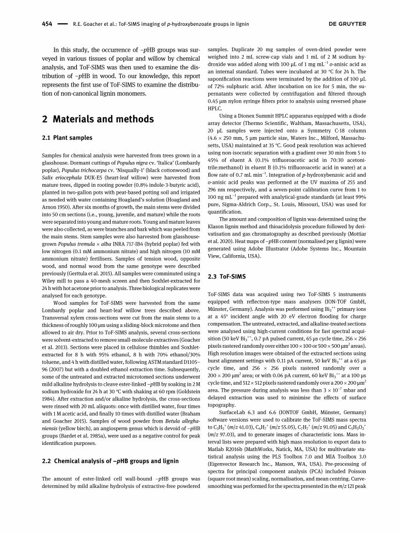
To begin, the distribution of –pHB was surveyedthroughout the vegetative tissues of poplar and willow.As –pHB moieties are ester-linked to the lignin backbone,mild alkaline hydrolysis was used to liberate these groupsprior to analysis by HPLC. This survey revealed substantialvariability between tissues, but also among the differenttaxa more broadly (Figure 2a–c). In agreement with pre-vious reports, Lombardy poplar had the highest –pHBlevels, while heart-leaf willow had the lowest (Pearl et al.1957; Venverloo 1971).
In all three genotypes examined, the stems containedthe greatest amounts of –pHB with levels ranging from 5to 29mg –pHB/g lignin, or in other words, 0.5–2.9% of thelignin (Figure 2a–c). By examining a developmentalgradient, it became clear that the juvenile stems had themost –pHB (i.e. not the youngest stems, nor the oldest).This may help explain the discord in the literature wheresome reported higher levels in younger stems (Chen et al.2017), while others found more in mature stems (Ven-verloo 1969; Wang et al. 2020) and at least one groupreported no differences whatsoever (Nakano et al. 1961).In the present study, more –pHB was observed in matureroots compared to young roots, particularly in Lombardypoplar where –pHB represented 2.2% of the total lignin inthemature roots. Bark containedmuch less –pHB (at most0.2% of the lignin), with the leaves having only tracelevels. As–pHBwasmost prevalent in the stems, the effectof perturbations in stem development were considerednext.
Nitrogen fertilisation of trees has been shown to lead tochanges in development and resource allocation, generallyresulting in fast-growing stems with wider vessels andreduced total lignin content (Li et al. 2012). In the hybridpoplar samples analysed in the present study, –pHBgroups were more abundant in the xylem of trees fertilisedwith more nitrogen, in agreement with previous observa-tions (Figure 2d; Pitre et al. 2007). As nitrogen fertilisationis known to reduce lignin content in glasshouse-grown
poplar (Novaes et al. 2009), it should be emphasised thatthis represents a decrease in –pHB per g of lignin, notmerely per g of biomass.
Reaction wood, formed as a gravitropic response inleaning stems, is another example of wood with “natural”perturbations in xylem morphology and cell wall compo-sition. In poplar, tension wood has fibres with a thickG-layer comprising more cellulose and less lignin, as wellas vessels that are narrower and fewer in number (Jourezet al. 2001). In the hybrid poplar samples analysed in thepresent study, tension wood had 2.3 timesmore –pHB thanopposite wood, and 3.5 times more than normal wood(Figure 2d). This observation corroborates previous NMRstudies on the lignin of reaction wood (Foston et al. 2011;Hedenstrom et al. 2009).
As lignin levels are lower following nitrogen fertilisa-tion and in tension wood, it is conceivable that the meta-bolic shift away from lignin leads to greater production ofp-hydroxybenzoate, the key precursor of –pHB groups.Although the biosynthesis of p-hydroxybenzoate has notyet been fully elucidated in plants, radiotracer studies haveshown a link to lignin biosynthesis (Terashima et al. 1975).
a Lombardy poplar Black cottonwood
Heart-leaf willow
b
c
mg –pHBg lignin
30
0
5
15
10
20
25
mg –pHBg lignin
0
6
12
9
3
mg –pHBg lignin
0
6
2
d Hybrid poplar
Low Nitrogen High Nitrogen
TensionWood
OppositeWood
NormalWood
mg –pHBg lignin
0
10
20
15
5
4
Figure 2: Levels of cell wall-bound p-hydroxybenzoate groups(mg –pHB/g lignin) in vegetative tissues of (a) Lombardy poplar,(b) black cottonwood, and (c) heart-leaf willow, as well as in(d) stems of hybrid poplar grownwith low and high levels of nitrogenfertilisation, and in tension wood, opposite wood and normal wood.Tissue types depicted in a–c include stems (in 50 cm increments),branches, roots (young andmature), leaves (young andmature), andbark. Gradient scales show the lignin-normalised amount of –pHBwith yellow representing more –pHB and red representing less. Allmeasurements were performed in duplicate with extractive-freewhole cell wall material using three biological replicates.
R.E. Goacher et al.: ToF-SIMS imaging of p-hydroxybenzoate groups in lignin 455
Alternatively, variations in –pHB could be due to thepresence/absence of –pHB in different cell types, as bothreaction wood and nitrogen fertilisation result in alteredproportions of normal fibre and vessel cell walls in wood.
3.2 ToF-SIMS detection of –pHB groups
In order to better understand the occurrence of –pHB inwood, ToF-SIMS imaging was employed to examine thedistribution of these groups in the juvenile stems of poplarand willow. Principal component analysis (PCA) wasinitially used to identify characteristic secondary ions(Supplementary Material; Supplementary Figure S1). Thiswork uncovered a putative signature for –pHB at m/z121.03. It was also observed that solvent extraction wasimportant in the sample preparation, as has been reportedpreviously (Goacher et al. 2011; Goacher et al. 2013).
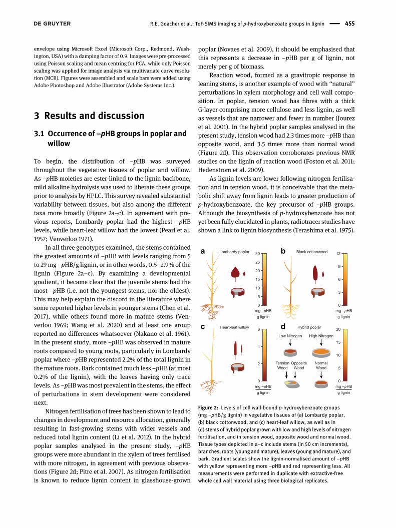
Examination of high mass resolution scans showedseveral peaks in the vicinity ofm/z 121, namelym/z 121.03,121.07 and 121.10. As a quality control measure to testwhether these were spurious peaks caused by sampleroughness or topography, the ToF-SIMS extractor bias wasaltered from 0 to +50 V to reject any stray ions originatingfrom cell sidewalls. None of the peaks at m/z 121 wereinfluenced by the extractor voltage and were therefore alldeemed representative of real chemistry and not instru-mental artefacts.
Several sample treatments were devised to examinethe nature of these three ions. First, solvent extractionresulted in decreased intensity of m/z 121.10 in Lombardypoplar (Figure 3a), consistent with the PCA analysis. Thereduction of m/z 121.10 was also observed with solventextraction of heart-leaf willow samples (data not shown).Therefore, the peak at m/z 121.10 likely corresponds to asmall-molecule aliphatic extractive, and its mass isconsistent with a peak assignment of C9H13
+.The aromatic ring structure of –pHB is identical to
H-lignin units (Figure 1c). As such, it was important to alsoconsider peaks arising from H-lignin in this study. Previ-ously reported ToF-SIMS analysis of a synthetic 8-O-4'H-type polymer resulted in the assignment of three char-acteristic ions for H-lignin: m/z 107.05 for C7H7O
+, m/z121.03 for C7H5O2
+, and m/z 121.07 for C8H9O+ (Saito et al.
2006). Of these, –pHB would most likely fragment as m/z121.03 due to cleavage of the C–Obond (Figure 1c). The ionsatm/z 107.05 and 121.07 are not expected for –pHB as theywould require a rearrangement of –pHB and its associatedlignin monomer unit to form.
As–pHBgroups are ester-linked (but H-lignin units arenot), mild alkaline hydrolysis should lead to a reduction in
any –pHB-related peaks. Indeed, treatment with sodiumhydroxide was observed to reduce the intensity of the peakat m/z 121.03 for samples of Lombardy poplar (Figure 3a),confirming this ion as characteristic of –pHB. The residualsignal observed at m/z 121.03 after treatment with sodiumhydroxide could be due to remaining, uncleaved –pHBgroups or could arise from H-lignin. Notably, the intensityof the peak at 121.07was not altered after solvent extractionor sodium hydroxide treatment. Such insensitivity to sol-vents and alkaline hydrolysis would be expected for peakscorresponding to H-lignin units.
Chemical analysis showed that juvenile stems ofLombardy poplar had seven times as much –pHB asheart-leaf willow but a similar amount of H-lignin(Supplementary Table S1), thus a comparison of thepeak envelopes for these samples could also be infor-mative. The intensity of the peak at m/z 121.03 was onlyabout four times greater in Lombardy poplar than inheart-leaf willow (Figure 3b). And, while m/z 121.03 had
Solvent-extracted & NaOH-treated
NaOH-treated
Solvent-extracted
Untreated
Lombardy poplar
Heart-leaf willow
Yellow birch
a
b
Lombardy poplar:
Solvent-extracted:
121.10
121.03
121.07
121.03
121.07
0
500
1000
1500
2000
120.9 121.0 121.1 121.2 121.3
Nor
mal
ised
inte
nsity
m/z
0
500
1000
1500
2000
120.9 121.0 121.1 121.2 121.3
Nor
mal
ised
inte
nsity
m/z
Figure 3: ToF-SIMS positive-ion mass spectra of the m/z 121 peakenvelope for (a) various treatments of Lombardy poplar wood, andfor (b) solvent-extracted samples of three different species where allspectra were normalised to total ion intensity.
456 R.E. Goacher et al.: ToF-SIMS imaging of p-hydroxybenzoate groups in lignin
greater relative intensity than m/z 121.07 in both poplarand willow, the opposite was true for yellow birch,which lacks –pHB but does have H-lignin units. More-over, the contribution of H-lignin to m/z 121.03 can beestimated since yellow birch lacks –pHB but does haveH-lignin units. Following this approach, the –pHBcomponent of the m/z 121.03 peak was estimated to be7.5 times greater in poplar than willow, closer in linewith the chemical analysis. These observations providefurther support for the assignment of m/z 121.03 asoriginating from –pHB (with some contribution fromH-lignin), m/z 121.07 as originating entirely fromH-lignin units, and m/z 121.10 as corresponding to analiphatic extractive compound.
3.3 Distribution of –pHB groups in wood
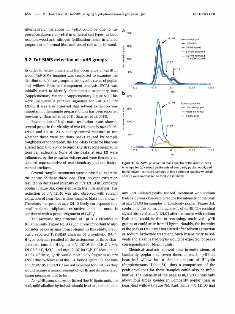
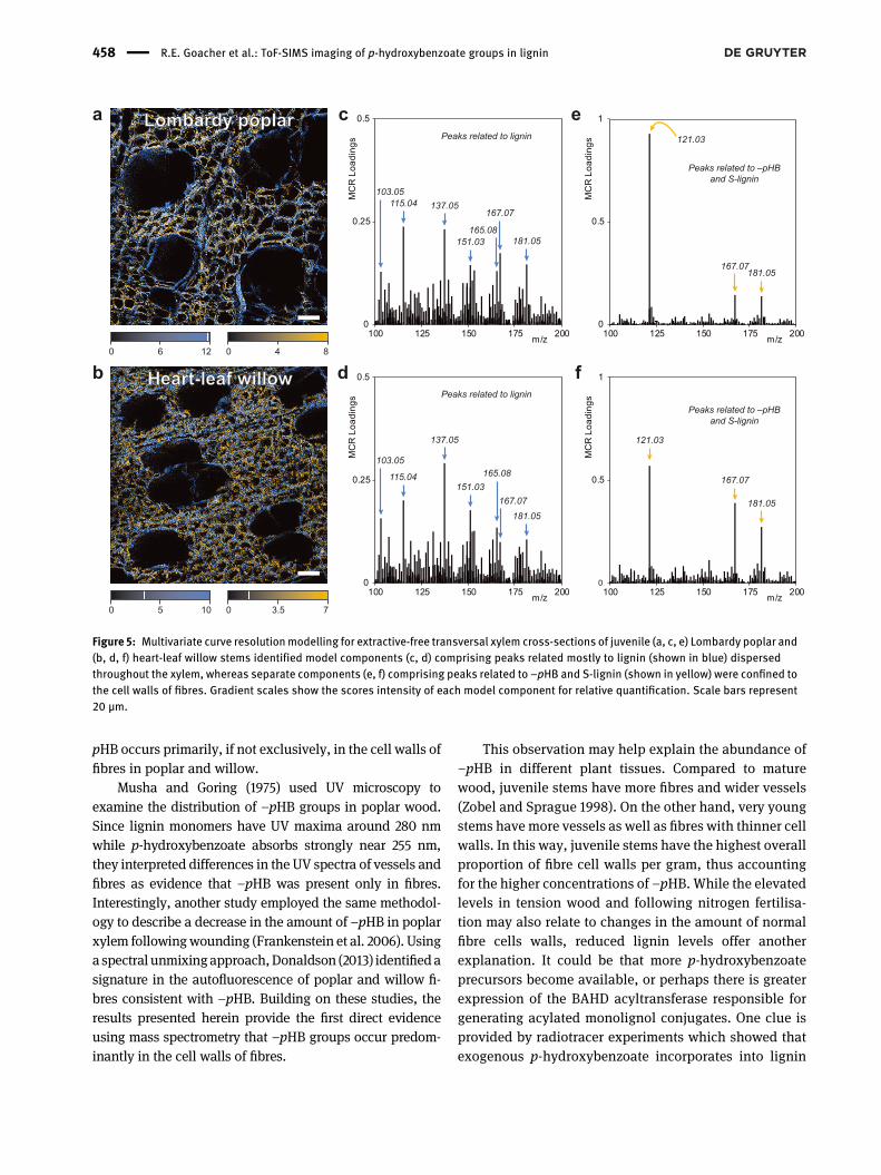
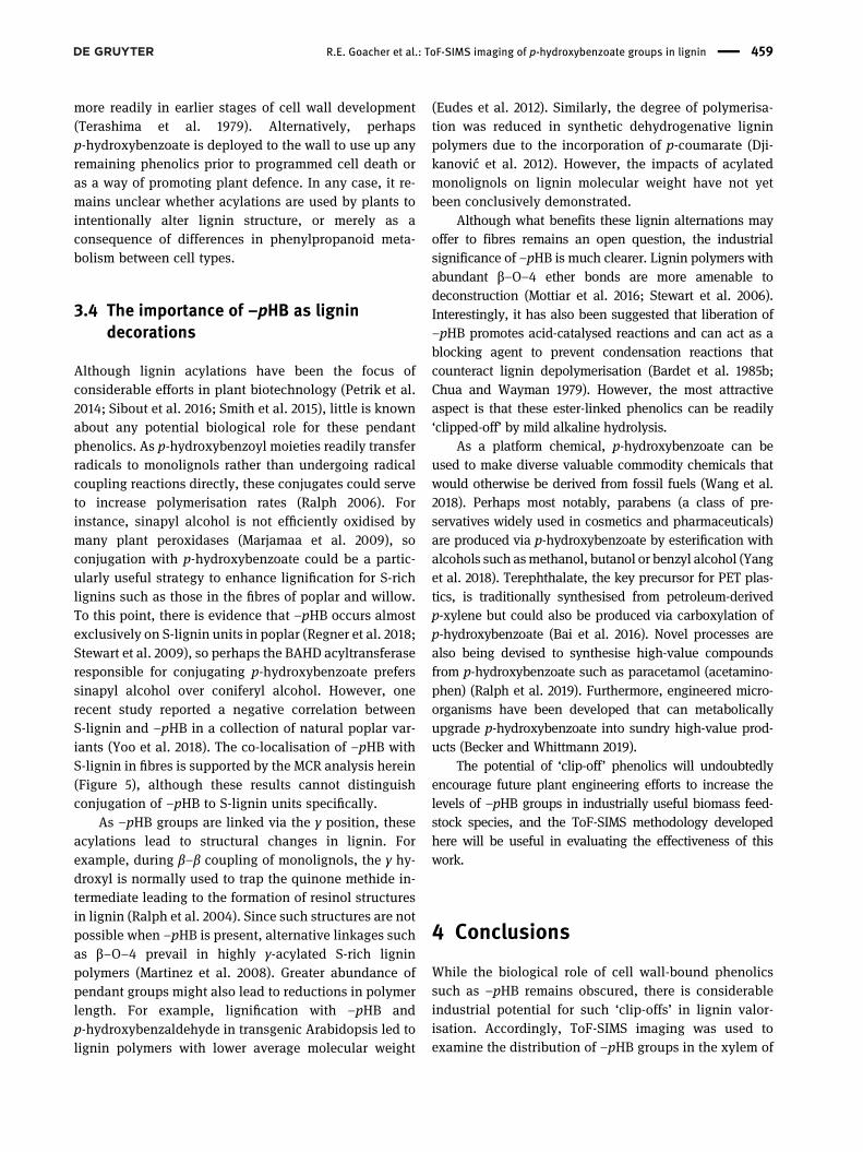
Chemical images of the m/z 121.03 peak originatingfrom –pHB were prepared using false-colour overlayswhere the –pHB signal intensity is plotted in magentaand the intensity of two aromatic ions at m/z 77.04(phenyl cation) and m/z 91.05 (tropylium cation) repre-senting the overall signal from lignin are summed andplotted in green (Figure 4). The scales represent relative,not absolute, concentrations. The ToF-SIMS chemicalimages for Lombardy poplar indicate that –pHB isenriched in the cell walls of fibres and is largely absent inareas surrounding vessel elements, as well as in someregions adjacent to ray parenchyma (Figure 4a).Although this contrast is more evident in poplar wherethere is a higher concentration of –pHB, the same patterncan also be observed in willow (Figure 4b). Additionalfalse-colour ion images are provided (SupplementaryFigure S2) to support the interpretation that –pHB islargely absent from vessels. However, it should beemphasised that some portion of the m/z 121.03 intensityin these ion images may originate from H-lignindepending on its relative abundance.
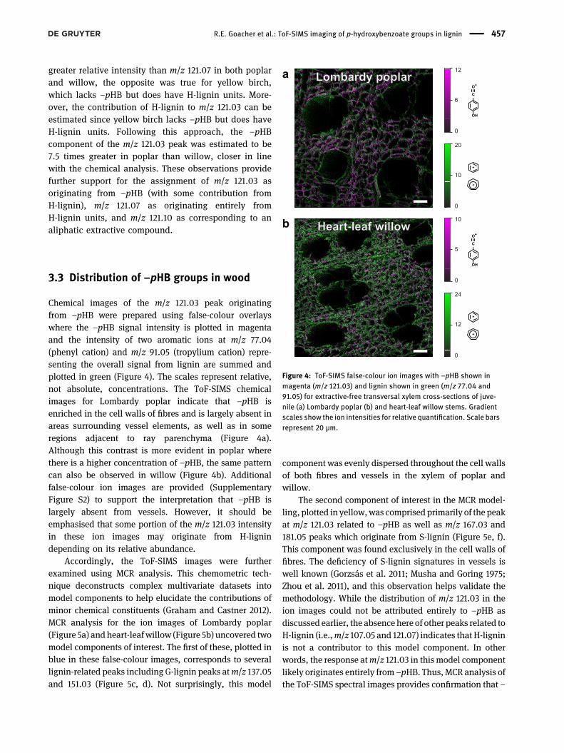
Accordingly, the ToF-SIMS images were furtherexamined using MCR analysis. This chemometric tech-nique deconstructs complex multivariate datasets intomodel components to help elucidate the contributions ofminor chemical constituents (Graham and Castner 2012).MCR analysis for the ion images of Lombardy poplar(Figure 5a) and heart-leaf willow (Figure 5b) uncovered twomodel components of interest. The first of these, plotted inblue in these false-colour images, corresponds to severallignin-related peaks including G-lignin peaks atm/z 137.05and 151.03 (Figure 5c, d). Not surprisingly, this model
component was evenly dispersed throughout the cell wallsof both fibres and vessels in the xylem of poplar andwillow.
The second component of interest in the MCR model-ling, plotted in yellow,was comprised primarily of the peakat m/z 121.03 related to –pHB as well as m/z 167.03 and181.05 peaks which originate from S-lignin (Figure 5e, f).This component was found exclusively in the cell walls offibres. The deficiency of S-lignin signatures in vessels iswell known (Gorzsás et al. 2011; Musha and Goring 1975;Zhou et al. 2011), and this observation helps validate themethodology. While the distribution of m/z 121.03 in theion images could not be attributed entirely to –pHB asdiscussed earlier, the absence here of other peaks related toH-lignin (i.e.,m/z 107.05 and 121.07) indicates that H-ligninis not a contributor to this model component. In otherwords, the response atm/z 121.03 in this model componentlikely originates entirely from –pHB. Thus, MCR analysis ofthe ToF-SIMS spectral images provides confirmation that –
a
b
OH
C
O
20
0
10
12
6
0
OH
C
O
24
0
12
10
5
0
Lombardy poplarLombardy poplar
Heart-leaf willowHeart-leaf willow
Figure 4: ToF-SIMS false-colour ion images with –pHB shown inmagenta (m/z 121.03) and lignin shown in green (m/z 77.04 and91.05) for extractive-free transversal xylem cross-sections of juve-nile (a) Lombardy poplar (b) and heart-leaf willow stems. Gradientscales show the ion intensities for relative quantification. Scale barsrepresent 20 µm.
R.E. Goacher et al.: ToF-SIMS imaging of p-hydroxybenzoate groups in lignin 457
pHB occurs primarily, if not exclusively, in the cell walls offibres in poplar and willow.
Musha and Goring (1975) used UV microscopy toexamine the distribution of –pHB groups in poplar wood.Since lignin monomers have UV maxima around 280 nmwhile p-hydroxybenzoate absorbs strongly near 255 nm,they interpreted differences in the UV spectra of vessels andfibres as evidence that –pHB was present only in fibres.Interestingly, another study employed the same methodol-ogy to describe a decrease in the amount of –pHB in poplarxylem followingwounding (Frankenstein et al. 2006). Usinga spectral unmixingapproach,Donaldson (2013) identifiedasignature in the autofluorescence of poplar and willow fi-bres consistent with –pHB. Building on these studies, theresults presented herein provide the first direct evidenceusing mass spectrometry that –pHB groups occur predom-inantly in the cell walls of fibres.
This observation may help explain the abundance of–pHB in different plant tissues. Compared to maturewood, juvenile stems have more fibres and wider vessels(Zobel and Sprague 1998). On the other hand, very youngstems have more vessels as well as fibres with thinner cellwalls. In this way, juvenile stems have the highest overallproportion of fibre cell walls per gram, thus accountingfor the higher concentrations of –pHB. While the elevatedlevels in tension wood and following nitrogen fertilisa-tion may also relate to changes in the amount of normalfibre cells walls, reduced lignin levels offer anotherexplanation. It could be that more p-hydroxybenzoateprecursors become available, or perhaps there is greaterexpression of the BAHD acyltransferase responsible forgenerating acylated monolignol conjugates. One clue isprovided by radiotracer experiments which showed thatexogenous p-hydroxybenzoate incorporates into lignin
a
b120 6 840
c
0
0.25
0.5
100 125 150 175 200
MC
R L
oadi
ngs
m/z
Peaks related to lignin
115.04 137.05
181.05151.03
103.05
dM
CR
Loa
ding
s
0
0.25
0.5
100 125 150 175 200m/z
137.05
115.04151.03
165.08
181.05
103.05
Peaks related to lignin
e
MC
R L
oadi
ngs
0
0.5
1
100 125 150 175 200m/z
121.03
167.07181.05
Peaks related to –pHBand S-lignin
f
181.05
121.03
167.07
Peaks related to –pHBand S-lignin
MC
R L
oadi
ngs
0
0.5
1
100 125 150 175 200m/z
167.07
167.07
165.08
100 5 73.50
Lombardy poplarLombardy poplar
Heart-leaf willowHeart-leaf willow
Figure 5: Multivariate curve resolutionmodelling for extractive-free transversal xylem cross-sections of juvenile (a, c, e) Lombardy poplar and(b, d, f) heart-leaf willow stems identified model components (c, d) comprising peaks related mostly to lignin (shown in blue) dispersedthroughout the xylem, whereas separate components (e, f) comprising peaks related to –pHB and S-lignin (shown in yellow) were confined tothe cell walls of fibres. Gradient scales show the scores intensity of each model component for relative quantification. Scale bars represent20 µm.
458 R.E. Goacher et al.: ToF-SIMS imaging of p-hydroxybenzoate groups in lignin
more readily in earlier stages of cell wall development(Terashima et al. 1979). Alternatively, perhapsp-hydroxybenzoate is deployed to the wall to use up anyremaining phenolics prior to programmed cell death oras a way of promoting plant defence. In any case, it re-mains unclear whether acylations are used by plants tointentionally alter lignin structure, or merely as aconsequence of differences in phenylpropanoid meta-bolism between cell types.
3.4 The importance of –pHB as lignindecorations
Although lignin acylations have been the focus ofconsiderable efforts in plant biotechnology (Petrik et al.2014; Sibout et al. 2016; Smith et al. 2015), little is knownabout any potential biological role for these pendantphenolics. As p-hydroxybenzoyl moieties readily transferradicals to monolignols rather than undergoing radicalcoupling reactions directly, these conjugates could serveto increase polymerisation rates (Ralph 2006). Forinstance, sinapyl alcohol is not efficiently oxidised bymany plant peroxidases (Marjamaa et al. 2009), soconjugation with p-hydroxybenzoate could be a partic-ularly useful strategy to enhance lignification for S-richlignins such as those in the fibres of poplar and willow.To this point, there is evidence that –pHB occurs almostexclusively on S-lignin units in poplar (Regner et al. 2018;Stewart et al. 2009), so perhaps the BAHD acyltransferaseresponsible for conjugating p-hydroxybenzoate preferssinapyl alcohol over coniferyl alcohol. However, onerecent study reported a negative correlation betweenS-lignin and –pHB in a collection of natural poplar var-iants (Yoo et al. 2018). The co-localisation of –pHB withS-lignin in fibres is supported by the MCR analysis herein(Figure 5), although these results cannot distinguishconjugation of –pHB to S-lignin units specifically.
As –pHB groups are linked via the γ position, theseacylations lead to structural changes in lignin. Forexample, during β–β coupling of monolignols, the γ hy-droxyl is normally used to trap the quinone methide in-termediate leading to the formation of resinol structuresin lignin (Ralph et al. 2004). Since such structures are notpossible when –pHB is present, alternative linkages suchas β–O–4 prevail in highly γ-acylated S-rich ligninpolymers (Martinez et al. 2008). Greater abundance ofpendant groups might also lead to reductions in polymerlength. For example, lignification with –pHB andp-hydroxybenzaldehyde in transgenic Arabidopsis led tolignin polymers with lower average molecular weight
(Eudes et al. 2012). Similarly, the degree of polymerisa-tion was reduced in synthetic dehydrogenative ligninpolymers due to the incorporation of p-coumarate (Dji-kanović et al. 2012). However, the impacts of acylatedmonolignols on lignin molecular weight have not yetbeen conclusively demonstrated.
Although what benefits these lignin alternations mayoffer to fibres remains an open question, the industrialsignificance of –pHB is much clearer. Lignin polymers withabundant β–O–4 ether bonds are more amenable todeconstruction (Mottiar et al. 2016; Stewart et al. 2006).Interestingly, it has also been suggested that liberation of–pHB promotes acid-catalysed reactions and can act as ablocking agent to prevent condensation reactions thatcounteract lignin depolymerisation (Bardet et al. 1985b;Chua and Wayman 1979). However, the most attractiveaspect is that these ester-linked phenolics can be readily‘clipped-off’ by mild alkaline hydrolysis.
As a platform chemical, p-hydroxybenzoate can beused to make diverse valuable commodity chemicals thatwould otherwise be derived from fossil fuels (Wang et al.2018). Perhaps most notably, parabens (a class of pre-servatives widely used in cosmetics and pharmaceuticals)are produced via p-hydroxybenzoate by esterification withalcohols such asmethanol, butanol or benzyl alcohol (Yanget al. 2018). Terephthalate, the key precursor for PET plas-tics, is traditionally synthesised from petroleum-derivedp-xylene but could also be produced via carboxylation ofp-hydroxybenzoate (Bai et al. 2016). Novel processes arealso being devised to synthesise high-value compoundsfrom p-hydroxybenzoate such as paracetamol (acetamino-phen) (Ralph et al. 2019). Furthermore, engineered micro-organisms have been developed that can metabolicallyupgrade p-hydroxybenzoate into sundry high-value prod-ucts (Becker and Whittmann 2019).
The potential of ‘clip-off’ phenolics will undoubtedlyencourage future plant engineering efforts to increase thelevels of –pHB groups in industrially useful biomass feed-stock species, and the ToF-SIMS methodology developedhere will be useful in evaluating the effectiveness of thiswork.
4 Conclusions
While the biological role of cell wall-bound phenolicssuch as –pHB remains obscured, there is considerableindustrial potential for such ‘clip-offs’ in lignin valor-isation. Accordingly, ToF-SIMS imaging was used toexamine the distribution of –pHB groups in the xylem of
R.E. Goacher et al.: ToF-SIMS imaging of p-hydroxybenzoate groups in lignin 459
poplar and willow stems. The results presented herecorroborate historical UV-microscopy studies and showthat –pHB is found predominantly, if not exclusively, inthe cell walls of fibres. This observation helps explain theoccurrence of –pHB in different plant tissues andperhaps also in reaction wood and following nitrogenfertilisation. An improved understanding of –pHB couldhelp elucidate the biological role of lignin decorationsand may inform future efforts in plant biotechnology andlignin engineering.
Abbreviations
G lignin guaiacyl lignin unitH lignin p-hydroxyphenyl lignin unitMCR multivariate curve resolutionPCA principal component analysis–pHB ester-linked p-hydroxybenzoate groupS lignin syringyl lignin unitToF-SIMS time-of-flight secondary-ion mass spectrometry
Acknowledgements: Thanks to L. Tran (University of Vic-toria) and A. Groover (University of California Davis) forproviding the nitrogen-fertilised and reaction wood hybridpoplar samples.Author contribution: All the authors have acceptedresponsibility for the entire content of this submittedmanuscript and approved submission.Research funding:We gratefully acknowledge funding fromthe Natural Sciences and Engineering Research Council ofCanada to Y.M. and S.D.M., from the Great Lakes BioenergyResearch Center, U.S. Department of Energy, Office ofScience, Office of Biological and Environmental Researchunder Award Numbers DE-SC0018409 and DE-FC02-07ER64494, to S.D.M., and from the Niagara UniversityAcademic Center for Integrated Sciences to R.E.G.Conflict of interest statement: The authors declare noconflicts of interest regarding this article.
References
ASTM standard D1105–96. (2007). Standard test method forpreparation of extractive-free wood. ASTM International, WestConshohocken, PA, USA.
Bai, Z., Phuan, W.C., Ding, J., Heng, T.H., Luo, J., and Zhu, Y. (2016).Production of terephthalic acid from lignin-based phenolic acidsby a cascade fixed-bed process. ACS Catal. 6: 6141−6145.
Bardet, M., Foray, M.-F., and Robert, D. (1985a). Use of the DEPT pulsesequence to facilitate the 13C NMR structural analysis of lignins.Makromol. Chem. 186: 1495−1504.
Bardet, M., Robert, D.R., and Lundquist, K. (1985b). On the reactionsand degradation of the lignin during steam hydrolysis of aspenwood. Svensk Papperstidning 6: R61–R67.
Becker, J. and Wittmann, C. (2019). A field of dreams: ligninvalorization into chemicals, materials, fuels, and health-careproducts. Biotechnol. Adv. 37: 107360.
Beckham, G.T., Johnson, C.W., Karp, E.M., Salvachúa, D., and Vardon,D.R. (2016). Opportunities and challenges in biological ligninvalorization. Curr. Opin. Biotechnol. 42: 40–53.
Boerjan, W., Ralph, J., and Baucher, M. (2003). Lignin biosynthesis.Annu. Rev. Plant Biol. 54: 519–546.
Braham, E.J. and Goacher, R.E. (2015). Identifying and minimizingbuffer interferences in ToF-SIMS analyses of lignocellulose. Surf.Interface Anal. 47: 120–126.
Chen, F., Tobimatsu, Y., Havkin-Frenkel, D., Dixon, R.A., and Ralph, J.(2012). A polymer of caffeyl alcohol in plant seeds. Proc. Natl.Acad. Sci. U.S.A. 109: 1772–1777.
Chen, T.-Y., Wang, B., Wu, Y.-Y., Wen, J.-L., Liu, C.-F., Yuan, T.-Q., andSun, R.C. (2017). Structural variations of lignin macromoleculefromdifferent growth years of triploid of Populus tomentosaCarr.Int. J. Biol. Macromol. 101: 747–757.
Chua, M.G.S. and Wayman, M. (1979). Characterization ofautohydrolysis aspen (P. tremuloides) lignins. Part 1.Composition and molecular weight distribution of extractedautohydrolysis lignin. Can. J. Chem. 57: 1141–1149.
Del Río, J.C., Rencoret, J., Gutierrez, A., Kim, H., and Ralph, J. (2017).Hydroxystilbenes are monomers in palm fruit endocarp lignins.Plant Physiol. 174: 2072–2082.
Djikanović, D., Simonović, J., Savić, A., Ristić, I., Bajuk-Bogdanović,D., Kalauzi, A., Cakić, S., Budinski-Simendić, J., Jeremić, M., andRadotić, K. (2012). Structural differences between lignin modelpolymers synthesized from variousmonomers. J. Polym. Environ.20: 607–617.
Donaldson, L. (2013). Softwood and hardwood lignin fluorescencespectra of wood cell walls in different mounting media. IAWA J.34: 3–19.
Eudes, A., George, A., Mukerjee, P., Kim, J.S., Pollet, B., Benke, P.I.,Yang, F., Mitra, P., Sun, L., Cetinkol, O.P., et al. (2012).Biosynthesis and incorporation of side-chain-truncated ligninmonomers to reduce lignin polymerization and enhancesaccharification. Plant Biotechnol. J. 10: 609–620.
Foston, M., Hubbell, C.A., Samuel, R., Jung, S., Fan, H., Ding, S.-Y.,Zeng, Y., Jawdy, S., Davis, M., Sykes, R., et al. (2011). Chemical,ultrastructural and supramolecular analysis of tension wood inPopulus tremula x alba as a model substrate for reducedrecalcitrance. Energy Environ. Sci. 4: 4962–4971.
Frankenstein, C., Schmitt, U., and Koch, G. (2006). Topochemicalstudies on modified lignin distribution in the xylem ofpoplar (Populus spp.) after wounding. Ann. Bot. 97: 195–204.
Gerttula, S., Zinkgraf, M., Muday, G.K., Lewis, D.R., Ibatullin, F.M.,Brumer, H., Hart, F., Mansfield, S.D., Filkov, V., and Groover, A.(2015). Transcriptional and hormonal regulation of gravitropismof woody stems in Populus. Plant Cell 27: 2800–2813.
Glasser, W.G. (2019). About making lignin great again some lessonsfrom the past. Front. Chem. 7: 565.
Goacher, R.E., Jeremic, D., and Master, E.M. (2011). Expanding thelibrary of secondary ions that distinguish lignin andpolysaccharides in time-of-flight secondary ion massspectrometry analysis of wood. Anal. Chem. 83: 804–812.
460 R.E. Goacher et al.: ToF-SIMS imaging of p-hydroxybenzoate groups in lignin
Goacher, R.E., Edwards, E.A., Yakunin, A.F., Mims, C.A., and Master,E.R. (2012). Application of time-of-flight-secondary ion massspectrometry for the detection of enzyme activity on solid woodsubstrates. Anal. Chem. 84: 4443–4451.
Goacher, R.E., Tsai, A.Y.-L., and Master, E.R. (2013). Towards practicaltime-of-flight secondary ion mass spectrometry lignocellulolyticenzyme assays. Biotechnol. Biofuels 6: 132.
Goacher, R.E., Selig, M.J., and Master, E.R. (2014). Advancinglignocellulose bioconversion through direct assessment ofenzyme action on insoluble substrates. Curr. Opin. Biotechnol.27: 123–133.
Goldstein, I.S. (1984). Degradation of wood by chemicals. In: Rowell,R. (Ed.). The chemistry of solid wood. American Chemical Society,Washington, DC, USA, pp. 575–586.
Gorzsás, A., Stenlund, H., Persson, P., Trygg, J., and Sundberg, B.(2011). Cell-specific chemotyping and multivariate imaging bycombined FT-IR microspectroscopy and orthogonal projectionsto latent structures (OPLS) analysis reveals the chemicallandscape of secondary xylem. Plant J. 66: 903–914.
Grabber, J.H., Ralph, J., Lapierre, C., and Barriere, Y. (2004). Geneticandmolecular basis of grass cell-wall degradability. I. Lignin-cellwall matrix interactions. C. R. Biol. 327: 455–465.
Graham, D.J. and Castner, D.G. (2012). Multivariate analysis ofToF-SIMS data from multicomponent systems: the why, when,and how. Biointerphases 7: 49.
Hatfield, R.D., Marita, J.M., Frost, K., Grabber, J., Ralph, J., Lu, F., andKim, H. (2009). Grass lignin acylation: p-coumaroyl transferaseactivity and cell wall characteristics of C3 and C4 grasses. Planta229: 1253–1267.
Hedenström, M., Wiklund-Lindström, S., Oman, T., Lu, F., Gerber, L.,Schatz, P., Sundberg, B., and Ralph, J. (2009). Identification oflignin and polysaccharide modifications in Populus wood bychemometric analysis of 2D NMR spectra from dissolved cellwalls. Mol. Plant 2: 933–942.
Hoagland, D.R. and Arnon, D.I. (1950). The water-culture method forgrowingplantswithout soil. Calif. Agric. Exp. Stn. Circ. 347: 1–32.
Jourez, B., Riboux, A., and Leclercq, A. (2001). Anatomicalcharacteristics of tension wood and opposite wood in younginclined stems of poplar (Populus euramericana cv ʻGhoyʼ). IAWAJ. 22: 133–157.
Kai, D., Tan, M.J., Chee, P.L., Chua, Y.K., Yap, Y.L., and Loh, X.J. (2016).Towards lignin-based functional materials in a sustainableworld. Green Chem. 18: 1175–1200.
Karlen, S.D., Free, H.C.A., Padmakshan, D., Smith, B.G., Ralph, J., andHarris, P.J. (2018). Commelinid monocotyledon lignins areacylated by p-coumarate. Plant Physiol. 177: 513–521.
Kim, H., Ralph, J., Lu, F., Ralph, S.A., Boudet, A.-M., MacKay, J.J.,Sederoff, R.R., Ito, T., Kawai, S., Ohashi, H., et al. (2003). NMRanalysis of lignins in CAD-deficient plants. Part 1. Incorporationof hydroxycinnamaldehydes and hydroxybenzaldehydes intolignins. Org. Biomol. Chem. 1: 268–281.
Li, H., Li, M., Luo, J., Cao, X., Qu, L., Gai, Y., Jiang, X., Liu, T., Bai, H.,Janz, D., et al. (2012). N-fertilization has different effects on thegrowth, carbon and nitrogen physiology, and wood propertiesof slow-and fast-growing Populus species. J. Exp. Bot. 63:6173–6185.
Lu, F., Ralph, J., Morreel, K., Messens, E., and Boerjan, W. (2004).Preparation and relevance of a cross-coupling product betweensinapyl alcohol and sinapyl p-hydroxybenzoate. Org. Biomol.Chem. 2: 2888–2890.
Lu, F., Karlen, S.D., Regner, M., Kim, H., Ralph, S.A., Sun, R.-C.,Kuroda, K.-I., Augustin, M.A., Mawson, R., Sabarez, H., et al.(2015). Naturally p-hydroxybenzoylated lignins in palms.Bioenergy Res. 8: 934–952.
Marjamaa, K., Kukkola, E.M., and Fagerstedt, K.V. (2009). The roleof xylem class III peroxidases in lignification. J. Exp. Bot. 60:367–376.
Martínez, Á.T., Rencoret, J., Marques, G., Gutierrez, A., Ibarra, D.,Jimenez-Barbero, J., and del Río, J.C. (2008). Monolignolacylation and lignin structure in some nonwoody plants: a 2DNMR study. Phytochemistry 69: 2831–2843.
Morreel, K., Ralph, J., Kim, H., Lu, F., Goeminne, G., Ralph, S.,Messens, E., and Boerjan, W. (2004). Profiling of oligolignolsreveals monolignol coupling conditions in lignifying poplarxylem. Plant Physiol. 136: 3537–3549.
Mottiar, Y., Vanholme, R., Boerjan, W., Ralph, J., and Mansfield, S.D.(2016). Designer lignins: harnessing the plasticity oflignification. Curr. Opin. Biotechnol. 37: 190–200.
Mottiar, Y., Gierlinger, N., Jeremic, D., Master, E.R., and Mansfield,S.D. (2020). Atypical lignification in eastern leatherwood (Dircapalustris). New Phytol. 226: 704–713.
Musha, Y. and Goring, D.A.I. (1975). Distribution of syringyl andguaiacyl moieties in hardwoods as indicated by ultravioletmicroscopy. Wood Sci. Technol. 9: 45–58.
Nakamura, Y. and Higuchi, T. (1978). Ester linkage of p-coumaric acidin bamboo lignin. III. Dehydrogenative polymerization ofconiferyl p-hydroxybenzoate and coniferyl p-coumarate. Cellul.Chem. Technol. 12: 209–221.
Nakano, J., Ishizu, A., and Migita, N. (1961). Studies on lignin XXXII.Ester groups of lignin. TAPPI 44: 30–32.
Novo-Uzal, E., Pomar, F., Gómez Ros, L.V., Espiñeira, J.M., andRos Barceló, A. (2012). Evolutionary history of lignins. Adv. Bot.Res. 61: 311–350.
Novaes, E., Osorio, L., Drost, D.R., Miles, B.L., Boaventura-Novaes,C.R., Benedict, C., Dervinis, C., Yu, Q., Sykes, R., Davis, M., et al.(2009). Quantitative genetic analysis of biomass and woodchemistry of Populus under different nitrogen levels. New Phytol.182: 878–890.
Pearl, I.A., Beyer, D.L., Johnson, B., and Wilkinson, S. (1957). Alkalinehydrolysis of representative hardwoods. TAPPI 40: 374–378.
Petrik, D.L., Karlen, S.D., Cass, C.L., Padmakshan, D., Lu, F., Liu, S.,Le Bris, P., Antelme, S., Santoro, N.,Wilkerson, C.G., et al. (2014).p-Coumaroyl-CoA:monolignol transferase (PMT) acts specificallyin the lignin biosynthetic pathway in Brachypodium distachyon.Plant J. 77: 713–726.
Pitre, F.E., Pollet, B., Lafarguette, F., Cooke, J.E.K., Mackay, J.J., andLapierre, C. (2007). Effects of increased nitrogen supply on thelignificationof poplarwood. J. Agric. FoodChem. 55: 10306–10314.
Ralph, J. (2006). What makes a good monolignol substitute? In:Hayashi, T. (Ed.). The science and lore of the plant cell wallbiosynthesis, structure and function. Brown Walker Press, BocaRaton, FL, USA, pp. 285–293.
Ralph, J., Hatfield, R.D., Quideau, S., Helm, R.F., Grabber, J.H., andJung, H.-J.G. (1994). Pathway of p-coumaric acid incorporationinto maize lignin as revealed by NMR. J. Am. Chem. Soc. 116:9448–9456.
Ralph, J., Lundquist, K., Brunow, G., Lu, F., Kim, H., Schatz, P.F.,Marita, J.M., Hatfield, R.D., Ralph, S.A., Christensen, J.H., et al.(2004). Lignins: natural polymers from oxidative coupling of4-hydroxyphenylpropanoids. Phytochem. Rev. 3: 29–60.
R.E. Goacher et al.: ToF-SIMS imaging of p-hydroxybenzoate groups in lignin 461
Ralph, J., Karlen, S., and Mobley, J. (2019). Synthesis of paracetamol(acetaminophen) from biomass-derived p-hydroxybenzamide.United States Patent US10286504B2.
Regner, M., Bartuce, A., Padmakshan, D., Ralph, J., and Karlen, S.D.(2018). Reductive cleavage method for quantitation ofmonolignols and low-abundance monolignol conjugates.ChemSusChem 11: 1600–1605.
Rencoret, J., Marques, G., Serrano, O., Kaal, J., Martínez, A. T., del Río,J.C., and Gutiérrez, A. (2020). Deciphering the unique structureand acylation pattern of Posidonia oceanica lignin. ACS Sustain.Chem. Eng. 8: 12521–12533.
Rinaldi, R., Jastrzebski, R., Clough, M.T., Ralph, J., Kennema, M.,Bruijnincx, P.C., and Weckhuysen, B.M. (2016). Paving the wayfor lignin valorisation: recent advances in bioengineering,biorefining and catalysis. Angew. Chem. Int. Ed. 55: 8164–8215.
Saito, K., Kato, T., Takamori, H., Kishimoto, T., Yamamoto, A., andFukushima, K. (2006). A new analysis of the depolymerizedfragments of lignin polymer in the plant cell walls usingToF-SIMS. Appl. Surf. Sci. 252: 6734–6737.
Schutyser, W., Renders, T., Van den Bosch, S., Koelewijn, S.F.,Beckham, G.T., and Sels, B.F. (2018). Chemicals from lignin: aninterplay of lignocellulose fractionation, depolymerisation, andupgrading. Chem. Soc. Rev. 47: 852–908.
Shimada, M., Fukuzuka, T., and Higuchi, T. (1971). Ester linkages ofp-coumaric acid in bamboo and grass lignins. TAPPI 54: 72–78.
Sibout, R., Le Bris, P., Legee, F., Cezard, L., Renault, H., and Lapierre,C. (2016). Structural redesigning Arabidopsis lignins into alkali-soluble lignins through the expression of p-coumaroyl-CoA:monolignol transferase PMT. Plant Physiol. 170: 1358–1366.
Smith, D.C.C. (1955). p-hydroxybenzoate groups in the lignin of aspen(Populus tremula). J. Chem. Soc. 3: 2347–2351.
Smith, R.A., Gonzales-Vigil, E., Karlen, S.D., Park, J.-Y., Lu, F.,Wilkerson, C.G., Samuels, L., Ralph, J., and Mansfield, S.D.(2015). Engineering monolignol p-coumarate conjugates intopoplar and Arabidopsis lignins. Plant Physiol. 169: 2992–3001.
Stewart, J.J., Kadla, J.F., andMansfield, S.D. (2006). The influence oflignin chemistry and ultrastructure on the pulping efficiency ofclonal aspen (Populus tremuloidesMichx.). Holzforschung 60:111–122.
Stewart, J.J., Akiyama, T., Chapple, C., Ralph, J., and Mansfield, S.D.(2009). The effects on lignin structure of overexpression of ferulate5-hydroxylase in hybrid poplar. Plant Physiol. 150: 621–635.
Terashima, N., Mori, I., and Kanda, T. (1975). Biosynthesis ofp-hydroxybenzoic acid in poplar lignin. Phytochemistry 14:1991–1992.
Terashima, N., Okada, M., and Tomimura, Y. (1979). Heterogeneity information of lignin. I. Heterogeneous incorporation ofp-hydroxybenzoic acid into poplar lignins. Mokuzai Gakkaishi25: 422–426.
Venverloo, C.J. (1969). The lignin of Populus nigra L. cv. ‘Italica’: acomparative study of the lignified structure in tissue cultures andthe tissues of the tree. Acta Bot. Neerl. 18: 241–314.
Venverloo, C.J. (1971). The lignin of Populus nigra L. cv. ‘Italica’ andsome other Salicaceae. Holzforschung 25: 18–24.
Wang, H.-M., Ma, C.-Y., Li, H.-Y., Chen, T.-Y., Wen, J.-L., Cao, X.-F.,Wang, X.-L., Yuan, T.-Q., and Sun, R.-C. (2020). Structuralvariations of lignin macromolecules from early growth stages ofpoplar cell walls. ACS Sustain. Chem. Eng. 8: 1813–1822.
Wang, S., Bilal, M., Hu, H., Wang, W., and Zhang, X. (2018).4-Hydroxybenzoic acid a versatile platform intermediate forvalue-added compounds. Appl. Microbiol. Biotechnol. 102:3561–3571.
Withers, S., Lu, F., Kim, H., Zhu, Y., Ralph, J., and Wilkerson, C.G.(2012). Identification of grass-specific enzyme that acylatesmonolignols with p-coumarate. J. Biol. Chem. 287: 8347–8355.
Yang, H., Zhang, F., and Wu, H. (2018). Review on life cycle ofparabens: synthesis, degradation, characterization and safetyanalysis. Curr. Org. Chem. 22: 769–779.
Yoo, C.G., Dumitrache, A., Muchero, W., Natzke, J., Akinosho, H., Li,M., Sykes, R.W., Brown, S.D., Davison, B., Tuskan, G.A., et al.(2018). Significance of lignin S/G ratio in biomass recalcitranceof Populus trichocarpa variants for bioethanol production. ACSSustain. Chem. Eng. 6: 2162–2168.
Zobel, B.J. and Sprague, J.R. (1998). Juvenile wood in forest trees.Springer-Verlag, Berlin Heidelberg.
Zhou, C., Li, Q., Chiang, V.L., Lucia, L.A., and Griffis, D.P. (2011).Chemical and spatial differentiation of syringyl and guaiacyllignins in poplar wood via time-of-flight secondary ion massspectrometry. Anal. Chem. 83: 7020–7026.
Supplementary material: Supplementary material to this article canbe found online at (https://doi.org/10.1515/hf-2020-0130).
462 R.E. Goacher et al.: ToF-SIMS imaging of p-hydroxybenzoate groups in lignin