EL ROL DEL EJERCICIO Y LA PGC1 ALFA EN LA INFLAMACION Y LAS ENCT.pdf
15
The role of exercise and PGC1α in inflammation and chronic disease Christoph Handschin 1 and Bruce M. Spiegelman 2 1 Institute of Physiology and Zurich Center for Integrative Human Physiology (ZIHP), University of Zurich, CH-8057 Zurich, Switzerland 2 Dana-Farber Cancer Institute and Department of Cell Biology, Harvard Medical School, Boston, MA 02115, USA Abstract Inadequate physical activity is linked to many chronic diseases. However, the mechanisms that tie muscle activity to health are unclear. The peroxisome proliferator-activated receptor γ co-activator 1α (PGC-1α) controls several exercise-related aspects of muscle function. We propose here mechanisms by which this protein controls muscle plasticity, suppresses a broad inflammatory response and mediates the beneficial effects of exercise. The reduction in physical activity, resulting from shifts in the nature of work and the replacement of muscle with machine in the developed world, has driven a dramatic increase in the incidence of many chronic diseases. In addition to the more obvious consequences associated with reduced activity, such as obesity, cardiovascular diseases, hypertension and type 2 diabetes, lack of sufficient exercise has also been linked to certain important types of cancer, pulmonary diseases, immune dysfunction, musculoskeletal diseases and several types of neurodegenerative disorders 1 (Fig. 1). In fact, a sedentary lifestyle is a major risk factor for many chronic pathologies and it has been shown unequivocally that inactivity increases the morbidity and mortality of these diseases 2,3 . Exercise capacity is a strong predictor of overall mortality, regardless of health and race 4 . Unfortunately, more than 50% of US adults do not exercise enough to achieve health benefits and 25% of adults shun any form of physical activity in their leisure time (Source: Center for Disease Control and Prevention, www.cdc.gov) 5 ; especially alarming is the rising trend of physical inactivity among young people 6 . Devastating effects of insufficient physical activity are likewise observed in the elderly 7 . Decreased muscle function in this population is not only directly linked to sarcopenia and the prevalence of a number of chronic diseases, but contributes enormously to the overall quality of life by diminishing strength, the ability to perform daily chores and social interactions, mobility, cognitive performance and life expectancy 7 . Even in the early elderly years, changes in physical activity have drastic consequences for health and lifespan. For example, sedentary behaviour in a 70-year-old man reduces the probability of survival to age 90 from 54% to 44% 8 . In contrast, increasing physical activity is an effective preventative measure for many chronic disorders. Furthermore, exercise is an excellent therapeutic intervention for pathologies such as obesity, type 2 diabetes, neurodegeneration, osteoporosis and sarcopenia 1 ; in terms of efficacy, exercise can rival the effects of drugs that are prescribed for many of these diseases, e.g. type 2 diabetes 9 . Correspondence should be addressed to C.H. ([email protected]) or B.M.S. ([email protected]). Competing Interests statement. The authors declare no competing financial interests. NIH Public Access Author Manuscript Nature. Author manuscript; available in PMC 2008 November 25. Published in final edited form as: Nature. 2008 July 24; 454(7203): 463–469. doi:10.1038/nature07206. NIH-PA Author Manuscript NIH-PA Author Manuscript NIH-PA Author Manuscript
-
Upload
ignacio-duque -
Category
Documents
-
view
221 -
download
0
Transcript of EL ROL DEL EJERCICIO Y LA PGC1 ALFA EN LA INFLAMACION Y LAS ENCT.pdf
The role of exercise and PGC1α in inflammation and chronicdisease
Christoph Handschin1 and Bruce M. Spiegelman2
1Institute of Physiology and Zurich Center for Integrative Human Physiology (ZIHP), University of Zurich,CH-8057 Zurich, Switzerland 2Dana-Farber Cancer Institute and Department of Cell Biology, HarvardMedical School, Boston, MA 02115, USA
AbstractInadequate physical activity is linked to many chronic diseases. However, the mechanisms that tiemuscle activity to health are unclear. The peroxisome proliferator-activated receptor γ co-activator1α (PGC-1α) controls several exercise-related aspects of muscle function. We propose heremechanisms by which this protein controls muscle plasticity, suppresses a broad inflammatoryresponse and mediates the beneficial effects of exercise.
The reduction in physical activity, resulting from shifts in the nature of work and thereplacement of muscle with machine in the developed world, has driven a dramatic increasein the incidence of many chronic diseases. In addition to the more obvious consequencesassociated with reduced activity, such as obesity, cardiovascular diseases, hypertension andtype 2 diabetes, lack of sufficient exercise has also been linked to certain important types ofcancer, pulmonary diseases, immune dysfunction, musculoskeletal diseases and several typesof neurodegenerative disorders1 (Fig. 1). In fact, a sedentary lifestyle is a major risk factor formany chronic pathologies and it has been shown unequivocally that inactivity increases themorbidity and mortality of these diseases2,3. Exercise capacity is a strong predictor of overallmortality, regardless of health and race4. Unfortunately, more than 50% of US adults do notexercise enough to achieve health benefits and 25% of adults shun any form of physical activityin their leisure time (Source: Center for Disease Control and Prevention, www.cdc.gov)5;especially alarming is the rising trend of physical inactivity among young people6. Devastatingeffects of insufficient physical activity are likewise observed in the elderly7. Decreased musclefunction in this population is not only directly linked to sarcopenia and the prevalence of anumber of chronic diseases, but contributes enormously to the overall quality of life bydiminishing strength, the ability to perform daily chores and social interactions, mobility,cognitive performance and life expectancy7. Even in the early elderly years, changes inphysical activity have drastic consequences for health and lifespan. For example, sedentarybehaviour in a 70-year-old man reduces the probability of survival to age 90 from 54% to 44%8. In contrast, increasing physical activity is an effective preventative measure for many chronicdisorders. Furthermore, exercise is an excellent therapeutic intervention for pathologies suchas obesity, type 2 diabetes, neurodegeneration, osteoporosis and sarcopenia1; in terms ofefficacy, exercise can rival the effects of drugs that are prescribed for many of these diseases,e.g. type 2 diabetes9.
Correspondence should be addressed to C.H. ([email protected]) or B.M.S. ([email protected]).Competing Interests statement. The authors declare no competing financial interests.
NIH Public AccessAuthor ManuscriptNature. Author manuscript; available in PMC 2008 November 25.
Published in final edited form as:Nature. 2008 July 24; 454(7203): 463–469. doi:10.1038/nature07206.
NIH
-PA Author Manuscript
NIH
-PA Author Manuscript
NIH
-PA Author Manuscript
Inactivity, inflammation and chronic diseaseMany chronic diseases have been found to be associated with a sterile, persistent, low-gradeinflammation (Fig. 2). For example, the development of insulin resistance and type 2 diabetestissue is closely correlated with immune cell infiltration and inflammation in white adiposetissue10. In cardiovascular diseases, activated immune cells and inflammation play a majorrole, particularly in the etiology of atherosclerosis11,12. Importantly, tumor initiation,promotion, and progression is stimulated by systemic elevation of pro-inflammatorycytokines13.
A number of neurodegenerative diseases are linked to a local inflammatory response in thebrain (neuroinflammation). For example, neuroinflammation influences activation of glia cellsand subsequent release of pro-inflammatory cytokines such as tumor necrosis factor α(TNFα); these are thought to promote the death of dopaminergic neurons in the substantia nigraand thereby contribute to the pathology of Parkinson’s disease14,15. Similarly,interleukin-1β (IL-1β), TNF-related apoptosis-inducing ligand (TRAIL) and other cytokineshave been postulated to be involved in the etiology of Alzheimer’s disease16, as has amyloid-β, itself exhibiting pro-inflammatory effects17. It is important to note that in addition to theneuroinflammation found in many neurodegenerative disorders, systemic inflammation furtherexacerbates these diseases and promotes the progression of neurodegeneration18.
Physical activity, inflammation and immunity are tightly linked in an interesting and complexway19. Regular, moderate exercise reduces systemic inflammation20. The mediators of thisbeneficial effect of exercise are unclear; however, several candidate mechanisms have beenidentified. First, exercise increases the release of epinephrine, cortisol, growth hormone,prolactin and other factors that have immunomodulatory effects21. Furthermore, exerciseresults in decreased expression of Toll-like receptor on monocytes suggested to be involvedin mediating whole body inflammation22. In contrast to the reduction of chronic inflammationby regular, moderate exercise, prolonged, high intensity training results in increased systemicinflammation and elevated risk of infection20. In fact, subsequent to this type of exercise,athletes exhibit a transient exercise-induced immunodepression23.
The recent discovery of “myokines”, cytokines produced and secreted from skeletal muscle,analogous to “adipokines” made from fat tissue, shed light on this bivalent association betweenexercise and inflammation19. The first myokine to be described was interleukin-6 (IL-6);similar factors synthesized and secreted upon contraction of muscle fibers include IL-8 andIL-1524. In addition to these muscle-derived cytokines, increased IL-1 receptor antagonist(IL-1ra), IL-10 and TNFα are found in the circulation after exercise24. However, systemicelevation of TNFα is restricted to physical activity of extremely high intensity and thereforecould be responsible for the elevated inflammatory state upon prolonged, intense exercise.
Once released transiently into the blood stream, myokines mediate some of the systemic andbeneficial effects of exercise in non-muscle tissue, e.g. modulation of hepatic glucoseproduction through IL-6. Some of these cytokines are clearly pro- (e.g. IL-1, TNFα) or anti-inflammatory (e.g. IL-10, IL-1ra). Paradoxically, both pro- and anti-inflammatory effects havebeen attributed to others25. For example, chronically elevated serum IL-6 levels have apredictive value for obesity and type 2 diabetes. In addition, chronically elevated levels ofsystemic IL-6, IL-8, IL-10, IL-1 and TNFα have been linked to the development of manydiseases associated with inflammation including cancer, and other age-associated diseases,such as sarcopenia, neurodegeneration and depression10,11,13,26–28. Finally, chronicelevation of IL-6 and TNFα results in skeletal muscle atrophy and inhibition of muscleregeneration, respectively29,30. Thus, the transient fluctuations of myokines followingphysical activity might contribute to the beneficial effects of exercise on organs other than
Handschin and Spiegelman Page 2
Nature. Author manuscript; available in PMC 2008 November 25.
NIH
-PA Author Manuscript
NIH
-PA Author Manuscript
NIH
-PA Author Manuscript
muscle in a hormone-like fashion, whereas chronic elevation of many of these same moleculesis almost certainly pro-inflammatory and detrimental. Obviously, then, the increase of IL-6and other cytokines secreted from muscle in exercise and their subsequent return to basal levelsmust be tightly regulated.
Effects of endurance and strength trainingDistinct exercise regimens are useful for the prevention and treatment of different pathologies.For example, endurance training improves cardiovascular parameters31, strength trainingreduces sarcopenia32, and the combination of both training regiments was recently reportedto be the most beneficial paradigm for type 2 diabetic patients33. For other diseases, the optimaltraining form remains to be defined. It is clear that sedentary behaviour increases the risk fordeveloping certain types of cancer1,34. Likewise, the type of exercise that confers the greatestprotection against neurodegenerative diseases is unknown. Interestingly, moderate exercise(e.g. walking) is sufficient to reduce the risk of developing dementia, as shown in a prospectivestudy with persons 65 years of age or older35.
Mechanistically, resistance training and endurance exercise activate distinct signallingpathways and result in specific adaptations of skeletal muscle. A significant proportion of thecapacity for adaptations of skeletal muscle is determined by the relative number and cross-sectional area of different muscle fiber types within a particular muscle bed36. Endurance isimproved with increased numbers of type I and type IIa fibers and endurance training increasesthe number of these fibers. Type I and type IIa fibers are red in appearance and are characterizedby a high number of mitochondria, elevated myoglobin and vascularization, and express aspecific set of myofibrillar proteins. As a result, they display resistance to fatigue and slowcontractions with low peak force generation37. The main source of ATP is the oxidativephosphorylation of glucose and fatty acids. In contrast, the white type IIb fibers (type IIx fibersin humans) have a relatively low number of mitochondria and mainly use anaerobicphosphocreatine and glucose metabolism to generate ATP. These fibers fatigue rapidly, butare able to generate fast contractions with a high peak force37. When stimulated by strengthtraining, type IIb muscle fibers can undergo substantial hypertrophy38.
Regulation and role of the PGC-1 coactivators in skeletal muscle physiologyContraction of skeletal muscle is initiated by motor neuron-induced calcium signalling. Theadaptation of muscle fibers to endurance vs. strength training is mediated by different firingpatterns of their respective motor neurons39. Type I and IIa-specific gene expression patternsare achieved by frequent bursts of sarcoplasmic calcium with low amplitude, as seen inendurance training. Strength training results in intermittent rises in sarcoplasmic calcium withhigh amplitude; this promotes transcription of the genes that mediate a type IIb-specificresponse and fiber hypertrophy39. Elevated sarcoplasmic calcium, in turn, activates the proteinphosphatase calcineurin A (CnA) and the calcium/calmodulin-dependent protein kinases(CaMK), which then alter the phosphorylation state of multiple transcription factors andcoactivators40.
This heightened calcium signalling activates several important transcription factors: cAMPresponsive element binding protein (CREB), the myocyte enhancer factors 2 (MEF2C andMEF2D) and the nuclear factor of activated T cells (NFAT). This results in altered expressionof exercise-regulated muscle genes, particularly the powerful transcriptional coactivatorPGC-1α41. Accordingly, PGC-1α expression is rapidly induced by these proteins following asingle bout of endurance exercise in vivo42. When physical activity is stopped, PGC-1α mRNAand protein levels quickly revert to the pre-exercised quantity42. In acute bouts of exercise, itis likely that increased expression of PGC-1α is primarily a mechanism for modulating
Handschin and Spiegelman Page 3
Nature. Author manuscript; available in PMC 2008 November 25.
NIH
-PA Author Manuscript
NIH
-PA Author Manuscript
NIH
-PA Author Manuscript
metabolic fluxes in skeletal muscle as a response to decreased ATP and altered fueldemands43. The multi-faceted interaction of PGC-1α with the AMP-activated protein kinase(AMPK) is likely to play a major role in this process44. In contrast, PGC-1α is found at anelevated level in chronically exercised skeletal muscle, even between individual bouts ofexercise, when compared to untrained muscle45. This reflects short term vs. long termadaptation of skeletal muscle to endurance exercise.
Thus, changes in muscle plasticity induced by chronic exercise, for example fiber-typeswitching towards the more oxidative and high endurance type IIa and type I fibers, correlatewith an increased basal expression of PGC-1α45. Furthermore, higher levels of PGC-1α arefound in oxidative fibers compared to glycolytic fibers, even in a rested state46. Transgenicelevation of PGC-1α in the skeletal muscle of animals up to the levels seen in type I fibersresults in a stable and robust fiber-type switch towards both type IIa and type I oxidativefibers46. Individual muscle fibers from these mice are more fatigue resistant, compared tofibers from wild type animals, and transgenic animals perform better in endurance exerciseindicating that chronic elevation of PGC-1α mediates many, if not all of the phenotypic changesseen in endurance-trained muscle46,47. The fiber switch promoted by PGC-1α is characterizedby increased mitochondrial density and function, increased oxidative metabolism, elevatedexpression of myofibrillar proteins characteristic of type I and type IIa muscle fibers and aswitch in substrate fuel usage46,48. Conversely, animals with PGC-1α specifically ablatedfrom skeletal muscle exhibit a higher number of glycolytic muscle fibers and have a reducedendurance exercise capacity49. Taken together, it is clear that PGC-1α is a key mediator ofmany of the known beneficial effects of physical activity on skeletal muscle physiology50,51.
Protective effects of PGC-1α in muscle biology: Suppression of chronicinflammation and muscle catabolism
One of the most important effects of exercise in human health is to prevent muscle catabolismand muscle wasting. Limb immobilization, prolonged hospitalization and various musculardystrophies are conditions where developing an exercised muscle phenotype by the patientwould improve the disease and the overall quality of life - but these patients often cannot traineffectively. Several studies indicate that PGC-1α prevents protein catabolism and musclewasting in a number of different contexts. Denervation-induced muscle atrophy, Duchennemuscular dystrophy and muscle damage via treatment with statin drugs are all greatlyameliorated when PGC-1α levels are maintained or elevated52–54.
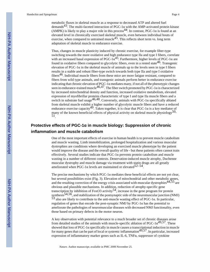
The precise mechanisms by which PGC-1α mediates these beneficial effects are not yet clear,but several possibilities exist (Fig. 3). Elevation of mitochondrial and other metabolic genes,and the resulting correction of the energy crisis associated with muscular dystrophies44,55 areobvious and plausible mechanisms. In addition, reduction of atrophy-specific genetranscription by inhibition of FoxO3 activity54, increase in the gene program for proteinsynthesis54,56, and stabilization of the postsynaptic side of the neuromuscular junction (NMJ)53 also are likely to contribute to the anti-muscle wasting effect of PGC-1α. In particular,regulation of genes that encode the post-synaptic NMJ by PGC-1α has the potential toameliorate the pathologies of neuromuscular diseases with decreased NMJ functionality, eventhose based on primary defects in the motor neuron.
A key observation with potential relevance to a much broader set of chronic diseases arosefrom detailed studies of the animals with muscle-specific ablation of PGC-1α49,57. Theseshowed that loss of PGC-1α specifically in muscle causes a transcriptional induction in musclefor many genes that can be part of local or systemic inflammation49,57. In particular, increasedexpression of inflammatory marker genes such as IL-6, TNFα, suppressor of cytokine
Handschin and Spiegelman Page 4
Nature. Author manuscript; available in PMC 2008 November 25.
NIH
-PA Author Manuscript
NIH
-PA Author Manuscript
NIH
-PA Author Manuscript
signalling 1 (SOCS1), SOCS3 and CD68 was observed in skeletal muscle of muscle-specificPGC-1α knockout animals in vivo49,57. Mice heterozygous for PGC-1α showed a smaller butsignificantly elevation expression of many of these same pro-inflammatory genes57. In bothcases, chronic elevation of circulating IL-6 was observed57. Primary muscle cells with agenetic ablation of PGC-1α exhibit higher levels of TNF-α and IL-6 mRNA than wild typemyotubes57 and elevated IL-6 protein was observed in the culture medium of the PGC-1αknockout cells compared to control cells57. Conversely, adenoviral expression of PGC-1α inC2C12 myotubes in culture reduced the expression of TNFα and IL-6 mRNA57. These datastrongly suggest that at least part of the circulating pro-inflammatory cytokines in vivo withablation of PGC-1α is originating from the muscle cells themselves. Of course, amplificationof this program may well involve subsequent recruitment to muscle of immune cells that are“specialists” at amplifying a pro-inflammatory response.
Importantly, mice with a heterozygous mutation in PGC-1α in muscle have a reduction inmRNA expression of this coactivator that is quantitatively comparable to the transcriptionaldysregulation of 36% observed in muscle of type 2 diabetic humans compared to healthyvolunteers58,59. Furthermore, the drop in PGC-1α expression in muscle-specific heterozygousanimals57 and skeletal muscle of type 2 diabetic patients59, respectively, correspondsquantitatively to the decreased expression of PGC-1α in an inactive vs. an active muscle inmice54. Although the establishment of causality between these reduced expression ofPGC-1α and the expression of pro-inflammatory genes is not possible in humans, these patientsdo also exhibit increased transcription of pro-inflammatory genes such as IL-6 and TNFα inskeletal muscle, as well as elevated IL-6 serum concentration57. Thus, the reduction ofPGC-1α mRNA in skeletal muscle of type 2 diabetics is likely to be closely linked to thechronic, low-grade inflammation present in these patients. Finally, the lower PGC-1α levelsobserved in the muscle of pre-diabetic individuals likely contributes to increases in systemicIL-6, a strong predictor for the development of type 2 diabetes60. Indeed, skeletal musclePGC-1α levels correlate inversely with expression of IL-6 and TNFα in individuals with normalglucose tolerance and in type 2 diabetic patients57. In contrast, body mass index, fastingglucose and fasting insulin levels exhibit no significant correlation to these inflammatorymarkers in this population57. Taken together, these finding also strongly suggest a causalrelationship between the increases in PGC-1α expression observed in human skeletal musclefollowing physical activity and the reduction of cytokine release from skeletal muscle knownto occur with moderate exercise. Conversely, the effects of loss of even one allele of thePGC-1α gene in mice, which stimulates the expression of a broad program of cytokineexpression and release, strongly suggests that something very similar is occurring in humanswho engage in chronically sedentary behavior. Accordingly, PGC-1α muscle-specificknockout animals exhibit decreased exercise capacity and a fiber-type switch towardsglycolytic muscle fibers49.
The molecular mechanisms that link PGC-1α and inflammatory gene expression in muscle areunknown, but they may reflect the role of PGC-1α in the control of reactive oxygen species(ROS). It has previously been shown that PGC-1α has a powerful suppressive effect on ROSproduction, in parallel to its effects in elevating mitochondrial respiration. This occurs throughthe PGC-1α-mediated expression of genes involved in ROS detoxification, as well as theexpression of uncoupling proteins that can attenuate ROS production61,62. In fact, increasedoxidative stress and inflammation are well-known to go hand in hand in many skeletal muscle-associated diseases63. Specifically, ROS have been shown to induce pro-inflammatorycytokine production in skeletal muscle64. Thus, the decreased expression of the anti-ROSgenes in muscle-specific PGC-1α knockouts57 are very likely to make a substantialcontribution to the increases seen in cytokine expression. Obviously though, there may beadditional, more directs effects of PGC-1α on the expression of genes with either pro- or anti-inflammatory action.
Handschin and Spiegelman Page 5
Nature. Author manuscript; available in PMC 2008 November 25.
NIH
-PA Author Manuscript
NIH
-PA Author Manuscript
NIH
-PA Author Manuscript
Analysis of muscle-specific PGC-1α knockout animals revealed that dysregulation ofPGC-1α in skeletal muscle does not cause insulin resistance in this tissue per se, but precipitatesabnormal whole body glucose and insulin homeostasis due to reduced insulin levels andabnormal pancreatic islet morphology. This unexpected, distal signalling apparently resultsfrom a noxious cross-talk between skeletal muscle and pancreatic β-cells in these animals57.Elevation of systemic inflammation is one likely mechanism by which skeletal muscle withdysregulated PGC-1α expression modulates β-cell function: elevation of IL-6 in the blood ofPGC-1α muscle-specific knockout animals correlates with the ability of IL-6 to suppressglucose-stimulated insulin secretion in isolated islets57. These data indicate unambiguouslythat the levels of skeletal muscle PGC-1α can powerfully influence the function of pancreaticislets; inescapably, these same data also suggest that muscle PGC-1α levels likely affect thestructure and functions of other tissues and organs too.
Systemic effects of exercise and PGC-1αWe suggest here that the decrease in PGC-1α gene expression in skeletal muscle due tosedentary behavior can set off a low level but chronic pro-inflammatory response that impactsmany other tissues negatively. As noted above, many if not most chronic diseases of aging,including heart disease, cancer and neurodegeneration are associated with chronicinflammation. In many cases that association has been shown to be causal in definedexperimental systems. The suppression of chronic inflammation in muscle via exercise-mediated induction of PGC-1α gene expression would be expected to lower the frequency and/or severity of these very same disorders. In terms of clinical data, exercise has manyneurological benefits, most notably improvement of learning and memory, protection againstneurodegeneration and amelioration of depression as well as other mood disorders17. Cancerof the colon, breast, prostate, endometrium, pancreas and skin all exhibit an increased incidencein inactive individuals, compared to those who exercise1,65. Thus, multi-organ health andplasticity as a result of exercise might be strongly influenced by altered systemic inflammationcontrolled by skeletal muscle PGC-1α activity.
It is important to note that it is very unlikely that any of the chronic diseases being discussedhere are caused by reduced PGC-1α alone. Rather, these are multi-factorial diseases requiringmultiple hits to yield full-blown disease. The multi-hit nature of human cancer now serves asuseful conceptual model for most chronic diseases. These multiple insults may originate in thegenetic heritage of an individual patient, may be acquired due to spontaneous somaticmutations, and may also be brought about by environmental or lifestyle factors. Thus, variablessuch as sedentary behavior and reduced PGC-1α levels in muscle may be viewed, on apopulation basis, as shifting the likelihood of disease to the left on a standard plot of incidencevs. age, compared to individuals with average exercise/physical activity (Fig 4). For any givenindividual, it is difficult or impossible to know what impact sedentary behaviour had on theirrisk for cancer or brain disease, for example. But that it does have an impact on the totalpopulation is a certainty. Conversely, populations with above average physical activity andthus increased PGC-1α levels experience a reduced incidence of disease, relative to those withaverage activity and PGC-1α.
Obesity: A most dangerous co-conspiratorSedentary behavior often coexists with and is a contributing factor in the development ofobesity1,66,67. Conversely, obese individuals are less likely to exercise66,67. Furthermore,inactivity and obesity are independent risk factors for many of the same chronic diseases. Infact, inactivity worsens the prevalence of chronic diseases in individuals regardless of theirBMI. Thus, being an independent risk factor, inadequate physical activity exacerbates thedetrimental effects of obesity1. In keeping with the mechanisms discussed above, we predict
Handschin and Spiegelman Page 6
Nature. Author manuscript; available in PMC 2008 November 25.
NIH
-PA Author Manuscript
NIH
-PA Author Manuscript
NIH
-PA Author Manuscript
a negative interaction between the lack of exercise and obesity in the specific molecularprograms discussed here. Obesity has been strongly associated with the expression of a pro-inflammatory program of gene expression including TNFα, IL-1 and IL-610. More precisely,it has been known for more than a decade that adipose tissue, in the context of obesity, beginsto secrete elevated levels of these “adipokines”68,69. This is true in both rodents andhumans70,71. Moreover, a functional role for TNFα and other adipokines has been shown inthe insulin-resistance of obese mice10. If we assume that there is a quantitative threshold ofcytokines required both chronically and systemically to bring about pathology in other (non-adipose, non-muscle) tissues, then inactivity combined with obesity is much more likely toreach such a threshold (Fig 4). Furthermore, if the age of onset of a particular disease, or theextent of disease is dose-responsive with respect to the levels of systemic pro-inflammatorymolecules, obesity with sedentary behavior would be expected to bring about earlier and/ormore severe disease72. Lastly, we do not know the full array of specific cytokines necessaryto contribute to the onset of any particular disease, but obesity and sedentary behavior mayinteract in ways that are not just quantitative. Indeed, they may bring together a particularcombination of adipokines/myokines that act synergistically in the causation of disease. Howbig are the synergistic effects of obesity and sedentary behavior in humans? Like othermodifiable factors such as smoking, diabetes and hypertension, obesity is predicted to reducelife expectancy between 1 to 5 years. In contrast, physical activity is estimated to add up to 5years. Importantly, a composite lifestyle of healthy behaviours has been proposed to potentiallyadd 10 years to the average life expectancy8,73.
Conclusion and perspectives: testing the HypothesisIt is obvious from the hypotheses presented here that many aspects of the physiological andpathophysiological effects of modulating PGC-1α in skeletal muscle and other organs remainenigmatic. Even less is known about the functions and therapeutic potential of the other twomembers of this gene family, PGC-1β and PGC-1-related coactivator (PRC). Similar toPGC-1α muscle-specific transgenic animals, ectopic expression of PGC-1β increasesendurance exercise capacity; however, different mechanisms seem to control the exercise-likephenotype in these two animal models74. Alterations in the amount and/or activity ofPGC-1α protein in muscle are likely to have future applications in the prevention and treatmentof a number of diseases. Amelioration of disuse-induced muscle atrophy54, DMD53 and statin-mediated muscle wasting52 through PGC-1α has already been described in animal models.The potential role of skeletal muscle PGC-1α as a modulator of non-muscular diseases, asdiscussed here, is not known but these ideas are readily testable in experimental animal models.It is hypothesized here that mice lacking one or both copies of the PGC-1α gene specificallyin skeletal muscle will be more susceptible to cancer, heart and brain disease. For example,mutant mice can be treated with chemical carcinogens that cause cancer of the breast, colonand other tissues and the rate of tumor formation and progression can be determinedquantitatively. Likewise, the same mutant strains of mice can be given challenges that induceheart failure, or certain kinds of neurodegeneration that model Parkinson’s disease orAlzheimer’s disease, and the rates of disease incidence and progression can be carefullymonitored.
Determination of the proper exercise regimens to protect from diseases linked to muscle-basedinflammation will be important. But chemical modulation of the PGC-1α pathway in skeletalmuscle is also of obvious significance. That PGC-1α gene expression can be modulated bydrugs and drug-like compounds has been demonstrated56,75,76. In addition, a number oftranscription factors that complex with PGC-1α in controlling skeletal muscle genetranscription are already known50,51 and may represent therapeutic targets. Co-activation ofthe estrogen-related receptor α (ERRα, official nomenclature NR3B1) specifies PGC-1α toinduce the same mitochondrial oxidative phosphorylation genes that are dysregulated in muscle
Handschin and Spiegelman Page 7
Nature. Author manuscript; available in PMC 2008 November 25.
NIH
-PA Author Manuscript
NIH
-PA Author Manuscript
NIH
-PA Author Manuscript
of type 2 diabetic patients56. Pharmacological disruption of the interaction between these twoproteins provokes a metabolic phenotype in cultured muscle cells resembling that of type 2diabetic muscle in vivo56. These findings provide a lead as to how selectivity in targetingPGC-1α could be achieved77. If successful, therapeutic modulation of PGC-1α has a hugeclinical potential for muscle wasting, sarcopenia, type 2 diabetes, muscular dystrophies andother very serious non-muscular chronic diseases.
AcknowledgementsWe thank Eric Smith for assistance with graphics as well as our colleagues and the members of our labs for commentson the manuscript, in particular Susan Loffredo, Jennifer Estall, Zoltan Arany, Goran Hansson, Serge Summermatter,Marco Toigo and Urs A. Meyer. C.H. is supported by the University Research Priority Program “Integrative HumanPhysiology” of the University of Zurich, an SNSF-Professorship of the Swiss National Science Foundation and theMuscular Dystrophy Association USA. B.M.S. is supported by several grants from the US National Institutes of Health.
References1. Booth FW, Chakravarthy MV, Gordon SE, Spangenburg EE. Waging war on physical inactivity: using
modern molecular ammunition against an ancient enemy. J Appl Physiol 2002;93(1):3–30. [PubMed:12070181]
2. Erikssen G, et al. Changes in physical fitness and changes in mortality. Lancet 1998;352(9130):759–762. [PubMed: 9737279]
3. Hu FB, et al. Adiposity as compared with physical activity in predicting mortality among women. NEngl J Med 2004;351(26):2694–2703. [PubMed: 15616204]
4. Kokkinos P, et al. Exercise capacity and mortality in black and white men. Circulation 2008;117(5):614–622. [PubMed: 18212278]
5. Booth FW, Lees SJ. Fundamental questions about genes, inactivity, and chronic diseases. PhysiolGenomics 2007;28(2):146–157. [PubMed: 17032813]
6. McCracken M, Jiles R, Blanck HM. Health behaviors of the young adult U.S. population: behavioralrisk factor surveillance system, 2003. Preventing chronic disease 2007;4(2):A25. [PubMed: 17362616]
7. Hollmann W, Struder HK, Tagarakis CV, King G. Physical activity and the elderly. Eur J CardiovascPrev Rehabil 2007;14(6):730–739. [PubMed: 18043292]
8. Yates LB, et al. Exceptional longevity in men: modifiable factors associated with survival and functionto age 90 years. Arch Intern Med 2008;168(3):284–290. [PubMed: 18268169]
9. Knowler WC, et al. Reduction in the incidence of type 2 diabetes with lifestyle intervention ormetformin. N Engl J Med 2002;346(6):393–403. [PubMed: 11832527]
10. Hotamisligil GS. Inflammation and metabolic disorders. Nature 2006;444(7121):860–867. [PubMed:17167474]
11. Haffner SM. The metabolic syndrome: inflammation, diabetes mellitus, and cardiovascular disease.Am J Cardiol 2006;97(2A):3A–11A. [PubMed: 16675316]
12. Matter CM, Handschin C. RANTES (regulated on activation, normal T cell expressed and secreted),inflammation, obesity, and the metabolic syndrome. Circulation 2007;115(8):946–948. [PubMed:17325252]
13. Lin WW, Karin M. A cytokine-mediated link between innate immunity, inflammation, and cancer.J Clin Invest 2007;117(5):1175–1183. [PubMed: 17476347]
14. Tansey MG, et al. Neuroinflammation in Parkinson's disease: is there sufficient evidence formechanism-based interventional therapy? Front Biosci 2008;13:709–717. [PubMed: 17981581]
15. Whitton PS. Inflammation as a causative factor in the aetiology of Parkinson's disease. Br J Pharmacol2007;150(8):963–976. [PubMed: 17339843]
16. Zipp F, Aktas O. The brain as a target of inflammation: common pathways link inflammatory andneurodegenerative diseases. Trends Neurosci 2006;29(9):518–527. [PubMed: 16879881]
17. Cotman CW, Berchtold NC, Christie LA. Exercise builds brain health: key roles of growth factorcascades and inflammation. Trends Neurosci 2007;30(9):464–472. [PubMed: 17765329]
Handschin and Spiegelman Page 8
Nature. Author manuscript; available in PMC 2008 November 25.
NIH
-PA Author Manuscript
NIH
-PA Author Manuscript
NIH
-PA Author Manuscript
18. Perry VH, Cunningham C, Holmes C. Systemic infections and inflammation affect chronicneurodegeneration. Nat Rev Immunol 2007;7(2):161–167. [PubMed: 17220915]
19. Febbraio MA. Exercise and inflammation. J Appl Physiol 2007;103(1):376–377. [PubMed:17446409]
20. Gleeson M. Immune function in sport and exercise. J Appl Physiol 2007;103(2):693–699. [PubMed:17303714]
21. Nieman DC. Current perspective on exercise immunology. Curr Sports Med Rep 2003;2(5):239–242.[PubMed: 12959703]
22. Gleeson M, McFarlin B, Flynn M. Exercise and Toll-like receptors. Exerc Immunol Rev 2006;12:34–53. [PubMed: 17201071]
23. Gleeson M, Nieman DC, Pedersen BK. Exercise, nutrition and immune function. J Sports Sci 2004;22(1):115–125. [PubMed: 14971437]
24. Pedersen BK, Akerstrom TC, Nielsen AR, Fischer CP. Role of myokines in exercise and metabolism.J Appl Physiol 2007;103(3):1093–1098. [PubMed: 17347387]
25. Kristiansen OP, Mandrup-Poulsen T. Interleukin-6 and diabetes: the good, the bad, or the indifferent?Diabetes 2005;54(Suppl 2):S114–124. [PubMed: 16306329]
26. Sarkar D, Fisher PB. Molecular mechanisms of aging-associated inflammation. Cancer Lett 2006;236(1):13–23. [PubMed: 15978720]
27. Bremmer MA, et al. Inflammatory markers in late-life depression: Results from a population-basedstudy. J Affect Disord. epub ahead of print (2007)
28. Roubenoff R. Physical activity, inflammation, and muscle loss. Nutr Rev 2007;65(12 Pt 2):S208–212. [PubMed: 18240550]
29. Haddad F, Zaldivar F, Cooper DM, Adams GR. IL-6-induced skeletal muscle atrophy. J Appl Physiol2005;98(3):911–917. [PubMed: 15542570]
30. Coletti D, et al. Tumor necrosis factor-alpha gene transfer induces cachexia and inhibits muscleregeneration. Genesis 2005;43(3):120–128. [PubMed: 16158413]
31. Manson JE, et al. A prospective study of walking as compared with vigorous exercise in the preventionof coronary heart disease in women. N Engl J Med 1999;341(9):650–658. [PubMed: 10460816]
32. Thomas DR. Loss of skeletal muscle mass in aging: examining the relationship of starvation,sarcopenia and cachexia. Clinical nutrition (Edinburgh, Scotland) 2007;26(4):389–399.
33. Sigal RJ, et al. Effects of aerobic training, resistance training, or both on glycemic control in type 2diabetes: a randomized trial. Annals of internal medicine 2007;147(6):357–369. [PubMed:17876019]
34. Zhou JR, Blackburn GL, Walker WA. Symposium introduction: metabolic syndrome and the onsetof cancer. Am J Clin Nutr 2007;86(3):s817–819. [PubMed: 18265474]
35. Larson EB, et al. Exercise is associated with reduced risk for incident dementia among persons 65years of age and older. Annals of internal medicine 2006;144(2):73–81. [PubMed: 16418406]
36. Pette D. Historical Perspectives: plasticity of mammalian skeletal muscle. J Appl Physiol 2001;90(3):1119–1124. [PubMed: 11181628]
37. Flück M, Hoppeler H. Molecular basis of skeletal muscle plasticity--from gene to form and function.Rev Physiol Biochem Pharmacol 2003;146:159–216. [PubMed: 12605307]
38. Glass DJ. Skeletal muscle hypertrophy and atrophy signaling pathways. Int J Biochem Cell Biol2005;37(10):1974–1984. [PubMed: 16087388]
39. Chin ER, et al. A calcineurin-dependent transcriptional pathway controls skeletal muscle fiber type.Genes Dev 1998;12(16):2499–2509. [PubMed: 9716403]
40. Berchtold MW, Brinkmeier H, Muntener M. Calcium ion in skeletal muscle: its crucial role for musclefunction, plasticity, and disease. Physiol Rev 2000;80(3):1215–1265. [PubMed: 10893434]
41. Puigserver P, et al. A cold-inducible coactivator of nuclear receptors linked to adaptive thermogenesis.Cell 1998;92(6):829–839. [PubMed: 9529258]
42. Pilegaard H, Saltin B, Neufer PD. Exercise induces transient transcriptional activation of thePGC-1alpha gene in human skeletal muscle. J Physiol 2003;546(Pt 3):851–858. [PubMed: 12563009]
43. Hood DA, Irrcher I, Ljubicic V, Joseph AM. Coordination of metabolic plasticity in skeletal muscle.The Journal of experimental biology 2006;209(Pt 12):2265–2275. [PubMed: 16731803]
Handschin and Spiegelman Page 9
Nature. Author manuscript; available in PMC 2008 November 25.
NIH
-PA Author Manuscript
NIH
-PA Author Manuscript
NIH
-PA Author Manuscript

44. Jager S, Handschin C, St-Pierre J, Spiegelman BM. AMP-activated protein kinase (AMPK) actionin skeletal muscle via direct phosphorylation of PGC-1alpha. Proc Natl Acad Sci U S A 2007;104(29):12017–12022. [PubMed: 17609368]
45. Russell AP, et al. Endurance training in humans leads to fiber type-specific increases in levels ofperoxisome proliferator-activated receptor-gamma coactivator-1 and peroxisome proliferator-activated receptor-alpha in skeletal muscle. Diabetes 2003;52(12):2874–2881. [PubMed: 14633846]
46. Lin J, et al. Transcriptional co-activator PGC-1 alpha drives the formation of slow-twitch musclefibres. Nature 2002;418(6899):797–801. [PubMed: 12181572]
47. Calvo JA, et al. Muscle-specific expression of PPAR{gamma} coactivator-1{alpha} improvesexercise performance and increases peak oxygen uptake. J Appl Physiol 2008;104(5):1304–1312.[PubMed: 18239076]
48. Wende AR, et al. A Role for the Transcriptional Coactivator PGC-1{alpha} in Muscle Refueling. JBiol Chem 2007;282(50):36642–36651. [PubMed: 17932032]
49. Handschin C, et al. Skeletal muscle fiber-type switching, exercise intolerance, and myopathy inPGC-1alpha muscle-specific knock-out animals. J Biol Chem 2007;282(41):30014–30021.[PubMed: 17702743]
50. Handschin C, Spiegelman BM. PGC-1 coactivators, energy homeostasis, and metabolism. EndocrRev 2006;27:728–735. [PubMed: 17018837]
51. Lin J, Handschin C, Spiegelman BM. Metabolic control through the PGC-1 family of transcriptioncoactivators. Cell Metab 2005;1(6):361–370. [PubMed: 16054085]
52. Hanai JI, et al. The muscle-specific ubiquitin ligase atrogin-1/MAFbx mediates statin-induced muscletoxicity. J Clin Invest 2007;117(12):3940–3951. [PubMed: 17992259]
53. Handschin C, et al. PGC-1alpha regulates the neuromuscular junction program and amelioratesDuchenne muscular dystrophy. Genes Dev 2007;21(7):770–783. [PubMed: 17403779]
54. Sandri M, et al. PGC-1{alpha} protects skeletal muscle from atrophy by suppressing FoxO3 actionand atrophy-specific gene transcription. Proc Natl Acad Sci U S A 2006;103(44):16260–16265.[PubMed: 17053067]
55. Wu Z, et al. Mechanisms controlling mitochondrial biogenesis and respiration through thethermogenic coactivator PGC-1. Cell 1999;98(1):115–124. [PubMed: 10412986]
56. Mootha VK, et al. Erralpha and Gabpa/b specify PGC-1alpha-dependent oxidative phosphorylationgene expression that is altered in diabetic muscle. Proc Natl Acad Sci U S A 2004;101(17):6570–6575. [PubMed: 15100410]
57. Handschin C, et al. Abnormal glucose homeostasis in skeletal muscle-specific PGC-1alpha knockoutmice reveals skeletal muscle-pancreatic beta cell crosstalk. J Clin Invest 2007;117(11):3463–3474.[PubMed: 17932564]
58. Mootha VK, et al. PGC-1alpha-responsive genes involved in oxidative phosphorylation arecoordinately downregulated in human diabetes. Nat Genet 2003;34(3):267–273. [PubMed:12808457]
59. Patti ME, et al. Coordinated reduction of genes of oxidative metabolism in humans with insulinresistance and diabetes: Potential role of PGC1 and NRF1. Proc Natl Acad Sci U S A 2003;100(14):8466–8471. [PubMed: 12832613]
60. Alexandraki K, et al. Inflammatory process in type 2 diabetes: The role of cytokines. Ann N Y AcadSci 2006;1084:89–117. [PubMed: 17151295]
61. St-Pierre J, et al. Suppression of Reactive Oxygen Species and Neurodegeneration by the PGC-1Transcriptional Coactivators. Cell 2006;127(2):397–408. [PubMed: 17055439]
62. Valle I, et al. PGC-1alpha regulates the mitochondrial antioxidant defense system in vascularendothelial cells. Cardiovascular research 2005;66(3):562–573. [PubMed: 15914121]
63. Moylan JS, Reid MB. Oxidative stress, chronic disease, and muscle wasting. Muscle Nerve 2007;35(4):411–429. [PubMed: 17266144]
64. Ji LL. Modulation of skeletal muscle antioxidant defense by exercise: Role of redox signaling. Freeradical biology & medicine 2008;44(2):142–152. [PubMed: 18191750]
65. Brown WJ, Burton NW, Rowan PJ. Updating the evidence on physical activity and health in women.Am J Prev Med 2007;33(5):404–411. [PubMed: 17950406]
Handschin and Spiegelman Page 10
Nature. Author manuscript; available in PMC 2008 November 25.
NIH
-PA Author Manuscript
NIH
-PA Author Manuscript
NIH
-PA Author Manuscript
66. Perusse L, Bouchard C. Genotype-environment interaction in human obesity. Nutr Rev 1999;57(5 Pt2):S31–37. [PubMed: 10391023]discussion S37–38
67. Rippe JM, Hess S. The role of physical activity in the prevention and management of obesity. J AmDiet Assoc 1998;98(10 Suppl 2):S31–38. [PubMed: 9787734]
68. Hotamisligil GS, Spiegelman BM. Tumor necrosis factor alpha: a key component of the obesity-diabetes link. Diabetes 1994;43(11):1271–1278. [PubMed: 7926300]
69. Hotamisligil GS, Shargill NS, Spiegelman BM. Adipose expression of tumor necrosis factor-alpha:direct role in obesity-linked insulin resistance. Science 1993;259(5091):87–91. [PubMed: 7678183]
70. Hofmann C, et al. Altered gene expression for tumor necrosis factor-alpha and its receptors duringdrug and dietary modulation of insulin resistance. Endocrinology 1994;134(1):264–270. [PubMed:8275942]
71. Hotamisligil GS, et al. Increased adipose tissue expression of tumor necrosis factor-alpha in humanobesity and insulin resistance. J Clin Invest 1995;95(5):2409–2415. [PubMed: 7738205]
72. Hamilton MT, Hamilton DG, Zderic TW. Role of low energy expenditure and sitting in obesity,metabolic syndrome, type 2 diabetes, and cardiovascular disease. Diabetes 2007;56(11):2655–2667.[PubMed: 17827399]
73. Fraser GE, Shavlik DJ. Ten years of life: Is it a matter of choice? Arch Intern Med 2001;161(13):1645–1652. [PubMed: 11434797]
74. Arany Z, et al. The transcriptional coactivator PGC-1beta drives the formation of oxidative type IIXfibers in skeletal muscle. Cell Metab 2007;5(1):35–46. [PubMed: 17189205]
75. Wagner BK, et al. Large-scale chemical dissection of mitochondrial function. Nat Biotechnol 2008;26(3):343–351. [PubMed: 18297058]
76. Arany Z, et al. Gene expression-based screening identifies microtubule inhibitors as inducers ofPGC-1alpha and oxidative phosphorylation. Proc Natl Acad Sci U S A 2008;105(12):4721–4726.[PubMed: 18347329]
77. Handschin C, Mootha VK. Estrogen-related receptor alpha (ERRalpha): A novel target in type 2diabetes. Drug Discov Today Ther Strateg 2005;2(2):151–156.
Handschin and Spiegelman Page 11
Nature. Author manuscript; available in PMC 2008 November 25.
NIH
-PA Author Manuscript
NIH
-PA Author Manuscript
NIH
-PA Author Manuscript
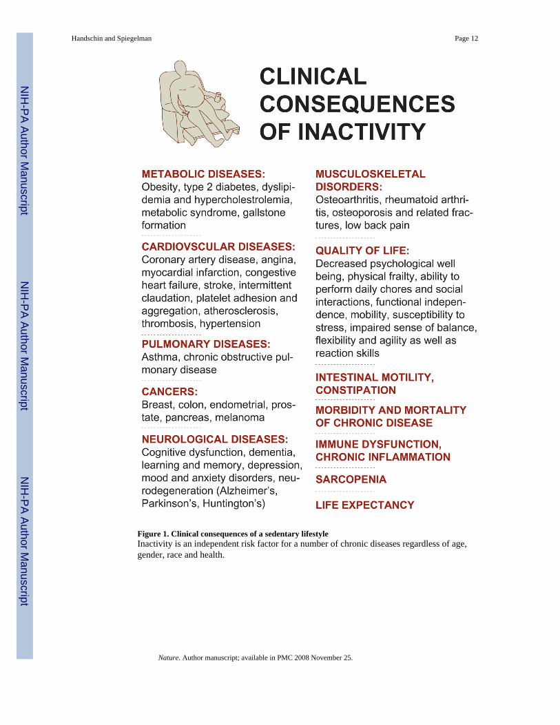
Figure 1. Clinical consequences of a sedentary lifestyleInactivity is an independent risk factor for a number of chronic diseases regardless of age,gender, race and health.
Handschin and Spiegelman Page 12
Nature. Author manuscript; available in PMC 2008 November 25.
NIH
-PA Author Manuscript
NIH
-PA Author Manuscript
NIH
-PA Author Manuscript
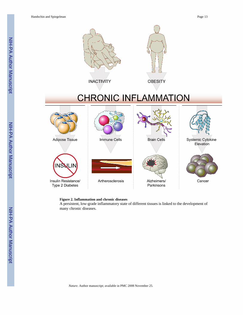
Figure 2. Inflammation and chronic diseasesA persistent, low-grade inflammatory state of different tissues is linked to the development ofmany chronic diseases.
Handschin and Spiegelman Page 13
Nature. Author manuscript; available in PMC 2008 November 25.
NIH
-PA Author Manuscript
NIH
-PA Author Manuscript
NIH
-PA Author Manuscript

Figure 3. Protective effect of PGC-1α on skeletal muscleThe relative level of PGC-1α in skeletal muscle is determined by physical activity. PGC-1α,in turn, controls muscle fiber adaptation to exercise and confers a number of beneficial changes.As a result, a reduction of systemic inflammation is observed in exercised individuals, possiblymediated through elevation of PGC-1α. In contrast, inactivity, and thus low skeletal musclePGC-1α, results in a chronic inflammatory state and thereby causes serious pathologicalconsequences. This inactivity-driven systemic inflammation is further exacerbated by obesity.
Handschin and Spiegelman Page 14
Nature. Author manuscript; available in PMC 2008 November 25.
NIH
-PA Author Manuscript
NIH
-PA Author Manuscript
NIH
-PA Author Manuscript

Figure 4. Inactivity and obesity are independent risk factors in the etiology of chronic diseasesA theoretical depiction of how sedentary lifestyle and obesity lower the threshold for age ofonset and disease incidence. Together, inactivity and obesity worsen the relative risk fordeveloping chronic diseases.
Handschin and Spiegelman Page 15
Nature. Author manuscript; available in PMC 2008 November 25.
NIH
-PA Author Manuscript
NIH
-PA Author Manuscript
NIH
-PA Author Manuscript

![REPRESENTACIÓN DE FUNCIONESºBach...Ejercicio nº 18.- Dada la función: f (x) = cosx − senx, x ∈ [0, 2π] Halla los puntos de corte con los ejes y los máximos y mínimos. Utilizando](https://static.fdocument.org/doc/165x107/5fef7342b8bf3f063904443e/representacin-de-funciones-bach-ejercicio-n-18-dada-la-funcin-f-x.jpg)